Incorporating evolutionary processes into population viability models

61J. Mamm. Ova Res. Vol. 28, 61–67, 2011
—Original—
Effect of KIT Ligand on the Viability of Porcine Primordial Follicles In Vitro
Manjula P. S. Magamage*, Mohammad Moniruzzaman and Takashi Miyano
Laboratory of Reproductive Biology and Biotechnology, Graduate School of Agricultural Science, Kobe University, Kobe 657-8501, Japan
Abstract: In mammals, the mechanisms regulating thein i t ia t ion of fo l l ic le development remain poor lyunderstood. We previously reported that testosteroneinduced the activation of porcine primordial follicles after7 days of culture. Herein, the effect of KIT ligand (KL) onthe viability, activation and development of porcineprimordial follicles was examined. Ovarian stripscontaining primordial follicles were cultured for 7 days ina medium supplemented with KL (10, 100, 250 or 500ng/ml). The percentage of degenerated follicles wassignificantly decreased (39 2%) in the 100 ng/ml KL-treated group compared to the control (56 1%),whereas a lmos t a l l o f the p r imord ia l fo l l i c lesdegenerated at higher concentrations of KL. Nopr imordia l fo l l ic les developed in cul ture at anyconcentration of KL. When ovarian strips were culturedin medium supplemented with testosterone (10–6 M) +KL (100 ng/ml), primordial follicles developed to earlysecondary fol l ic les (5 3%), however the totalpercentages of developing follicles in the testosteronegroup (21 5%) and the testosterone + KL group (28 5%) were not significantly different. These resultssuggest that KL does not promote primordial follicleactivation, although it does promote follicle viability inculture.Key words: In vitro culture, KIT ligand, Pig, Primordialfollicle, Testosterone
Introduction
In mammals, the mechanisms regulating the initiationof follicle development remain poorly understood [1–3].Many kinds of molecules that regulate primordial follicle
activation have been reported. KIT ligand (KL) [4–6],basic fibroblast growth factor [7], leukemia inhibitoryfactor [8], insulin [9, 10] and bone morphogeneticprotein 4 [11] have been shown to promote theprimordial to primary follicle transition in rodents. TheKIT receptor ty ros ine k inase and KL are we l l -documented molecules that are involved in theactivation of primordial follicles in rodents [1, 12]. KL isproduced by granulosa cells [13], and KIT is present inoocytes [14, 15]. In mice bearing spontaneousmutations of Steel, the gene encoding KL [16], folliculardevelopment is impaired [4]. Similarly, in mice bearing amutation of the gene encoding KIT [17] or treated withan antibody against KIT [5], folliculogenesis is impaired.Although the role of KL is controversial in the follicleactivation process in the mouse [18, 19], pig [15] andrabbit [13], KL might not be required for fol l ic leactivation. KL is predominantly a survival factor forprimordial germ cells, protecting them from apoptosis[20], and it stimulates granulosa cell proliferation in thepresence of oocytes [21].
Recently we reported that testosterone positivelyregulates the activation of primordial follicles derivedfrom prepubertal pigs [22]. Testosterone at 10–6 Minduced the pr imordia l fo l l ic le t ransi t ion to theintermediate and primary stages after 7 days of culture.Furthermore, testosterone-treated primordial folliclesdeveloped to the antral stage after 2 months ofxenografting.
The objective of the present study was to determinethe effect of KL on the activation and development ofcultured porcine primordial follicles. We histologicallyexamined the effect of KL on the activat ion anddevelopment of porcine primordial follicles and then theeffect of KL in combination with testosterone in culturedovarian tissues.
Received: November 8, 2010Accepted: December 20, 2010*To whom correspondence should be addressed.e-mail: [email protected]
62 J. Mamm. Ova Res. Vol. 28, 2011
Materials and Methods
Collection of ovarian cortical strips containing primordialfollicles
Ovaries were collected from 6-month-old crossbredgilts at a slaughterhouse in Kobe, Japan. Since pigsreach puberty at 6–7 months, ovaries without corpusluteum were collected from prepubertal gilts. Theova r ies we re washed once i n 0 .2% (w /v )cetyltrimethylammonium bromide (Wako Pure ChemicalIndustries, Osaka, Japan) and Dulbecco’s phosphatebuffered saline (PBS) supplemented with 0.1% (w/v)polyvinyl alcohol (PVA; Sigma, MO, USA) 3 times.Cortical strips, from randomly selected ovaries, with athickness of 0.5 mm (approximately) were dissectedwith surgical blades (No.11; Feather Safety Razor,Osaka, Japan). The strips were examined under adissection microscope and then under an invertedmicroscope, and st r ips contain ing pr imary andsecondary follicles were avoided. Primordial follicleswere identified as those having oocytes with diameterswithin a range of 30–35 m and oocytes containing alarge spherical nucleus surrounded by small l ipiddroplets [23]. Ovarian strips (approximately 2 mm × 1mm × 0.5 mm) that contained primordial follicles wereselected and cut into two pieces (each approximately 1mm × 1 mm × 0.5 mm). One part was fixed immediatelyfor histological examination to assess the foll iclenumber and morphology, and the other part waswashed 3 times and immersed in HEPES-bufferedTCM-199 (pH 7.4; Nissui Pharmaceutical, Tokyo,Japan) before culture. The HEPES-buffered TCM-199contained 25 mM HEPES (Sigma), 10 mM NaHCO3,0.1% PVA, and 0.08 mg/ml kanamycin sulphate (Sigma)in TCM 199.
In vitro cultureIn each experiment, cortical strips (1 mm × 1 mm ×
0.5 mm) were cultured on a cellulose acetate floatingmembrane filter (pore size 0.45 m and diameter 25mm; Advantec, Toyo Roshi, Tokyo, Japan) in an organculture dish (# 3037; BD Falcon, NJ, USA) for 7 daysunder a humidified atmosphere of 5% CO2 and 95% airat 38.5oC. The basic culture medium was alphaminimum essential medium (-MEM; Invitrogen, NY,USA) supplemented with 0.08 mg/ml kanamycinsu lpha te , 2 .2 mg /m l NaHCO 3 , 50 M 2-mercaptoethanol (Nacalai Tesque, Kyoto, Japan), 0.1mg/ml sodium pyruvate (Sigma), 1% (v/v) Insulin-Transferrin-Selenium (ITS; Invitrogen) and 5% (v/v)bovine plasma (Nippon Biotest Laboratories, Tokyo,
Japan) after heat inactivation. During the cultureperiod, half of the volume of the total medium waschanged every 2 days. In the experimental groups, thebasic culture medium was supplemented with 0, 10,100, 250 or 500 ng/ml (w/v) of KL (recombinant mousestem cell factor # 455-MC; R&D Systems, MN, USA). Inthe combination experiment, the basic culture mediumwas supplemented with 100 ng/ml KL with or without10–6 M testosterone (Nacalai Tesque). Testosteronewas dissolved in absolute ethanol, and 1 l of thesolution was added to each 1 ml of culture medium toobtain the final concentration just before use. As thevehicle, 1 l of ethanol was added to the control groups.
Assessment of follicular developmentOvarian cortical strips before and after culture were
fixed in 3% (w/v) paraformaldehyde in PBS, thendehydrated, embedded in methacrylate resin (JB-4;Polysciences, IL, USA), serially sectioned into 5-msections, and stained with hematoxylin and eosin. Thenumbers of follicles at different stages were recorded.The follicles were counted in every section in which anoocyte nucleus was seen. Double counting in adjacentsections was avoided. The follicles were classified intofou r ca tego r ies acco rd ing to the number andmorphology of granulosa cell layers: primordial follicleswi th a s ingle layer of f la t tened granulosa cel lssurrounding the oocyte, intermediate follicles with asingle layer containing a mixture of flat and cuboidalgranulosa cells, primary follicles with a single layer ofcuboidal granulosa cells and secondary follicles withmore than one layer of cuboidal granulosa cells or thebeginning of a second layer of granulosa cel ls.Degenerated follicles were classified by the followingstaining properties of the oocyte and granulosa cells:follicles having oocytes with pale cytoplasm, darkpyknotic nucleus, shrunken or extensive cytoplasmicvacuolations, and/or a disintegrated granulosa celllayer. The diameters of the oocytes were measured bytaking the average of two perpendicular measurementsto the nearest 1 m with an ocular micrometer (Nikon,Tokyo, Japan) attached to an inverted microscope.
Data and statistical analysisAll the data were analyzed by the Graph Pad InStat
(Version 3.0) statistical analysis program (GraphPadSoftware, CA, USA). The quantitative results arepresented as the mean ± S.E.M. The data wereanalyzed using ANOVA followed by Tukey-Kramermultiple comparisons among multiple groups whenappropriate. For statistical comparisons between two

63Magamage, et al.
groups, the t test was used. All percentile data weret ransformed to arc-s in va lues before analys is .Differences with P < 0.05 were considered statisticallysignificant.
Results
Effect of KL on primordial follicle activationHistological examination confirmed that dissected
porcine ovarian strips contained only primordial folliclesbefore culture (Fig. 1a). Each oocyte contained a largenucleus and cytoplasmic lipid droplets. The percentageof degenerated follicles was 14 ± 3% before culture(Table 1). After 7 days of culture, the strips had
changed slightly due to the appearance of degeneratedsomatic cells (Figs. 1b-f) and an increase in thepercentage of degenerated follicles (Table 1). Therewas no significant difference in the percentages ofdegenerated follicles between the control and the 10 ng/ml KL-treated groups (Figs. 1b, 1c and Table 1),whereas the percentage was significantly decreased by100 ng/ml KL (39 ± 2%, Fig. 1d). However, the meannumbers of follicles were decreased by 250 and 500 ng/ml KL (Table 1). At these concentrations of KL, most ofthe follicles were degenerated (Figs. 1e and 1f). Nodeveloping follicles were observed in any strip treatedwith any concentration of KL.
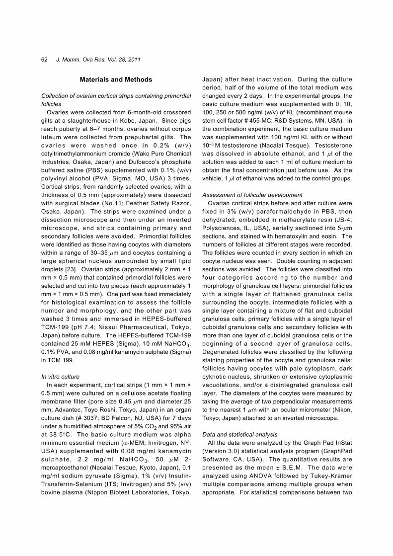
Fig. 1. Histological sections of porcine ovarian strips before (a) and after culture withKL at the concentration of 0 (b: control), 10 (c), 100 (d), 250 (e) or 500 ng/ml (f) for 7 days. The percentages of healthy intact primordial follicles werehigher in the 100 ng/ml KL-treated group (d) than in the other cultured groups.There were no developing follicles in any cultured group. Scale bar represents40 m.

64 J. Mamm. Ova Res. Vol. 28, 2011
Effect of KL in combination with testosterone onprimordial follicle development
To examine the effect of KL on primordial follicleactivat ion and development in combination withtestosterone, prepubertal porcine ovarian strips werecultured with KL (100 ng/ml) and testosterone (10–6 M)for 7 days (Table 2). Ovarian strips before culturecontained only primordial follicles (Fig. 2a). Afterculture, there were no developing follicles in the KL-treated group, however the percentage of primordialfollicles (62 ± 2%) was significantly higher than in thevehicle-treated control (46 ± 1%; Table 2, Figs. 2b and2c). On the other hand, intermediate and primaryfollicles were observed in the ovarian strips treated withtestosterone, irrespective of KL treatment (Figs. 2d and2e). Furthermore, in the testosterone-alone andtes tos terone + KL groups, the percentages o fdegenerated follicles were reduced compared to thecontro l (Table 2) , and the percentages of tota l
developing follicles including intermediate, primary andsecondary follicles, were increased to 21 ± 5% and 28 ±5%, respectively.
Discussion
The recruitment of primordial follicles into the growthphase may involve both stimulatory and inhibitorymechanisms. Although the exact mechanisms thatregulate the activation of primordial follicles remainunelucidated, the study of rodents has shown that KITand KL are involved in this process [6, 20]. The role ofKIT has been demonstrated by inhibiting experimentsusing its specific antibody, ACK2. Yoshida et al. [5]injected mice with ACK2 at various times during the first2 weeks after birth and reported that the neutralizationof KIT caused disturbances in initial follicle recruitment.KIT has been reported to express a stimulatory effect onprimordial follicle activation in different animal species
Table 1. Distribution of different types of follicles in porcine ovarian strips after culture with KIT ligand (KL)
Treatment* Day of No. of tissues No. of No. (%) of follicles**
culture examined follicles/tissue Primordial Intermediate Primary Secondary Degenerated
None 0 8 36 ± 8a 31 ± 7 0 ± 0 0 ± 0 0 ± 0 5 ± 2 (86 ± 3)a (14 ± 3)a
Control 0 ng/ml KL 7 8 33 ± 5a 14 ± 1 0 ± 0 0 ± 0 0 ± 0 18 ± 3
(44 ± 1)b (56 ± 1)b
10 ng/ml KL 7 8 36 ± 3a 15 ± 4 0 ± 0 0 ± 0 0 ± 0 20 ± 4(42 ± 3)b (58 ± 3)b
100 ng/ml KL 7 8 32 ± 3a 20 ± 2 0 ± 0 0 ± 0 0 ± 0 12 ± 1(61 ± 2)c (39 ± 2)c
250 ng/ml KL 7 8 21 ± 3a 2 ± 1 0 ± 0 0 ± 0 0 ± 0 19 ± 2(10 ± 3)d (90 ± 3)d
500 ng/ml KL 7 8 10 ± 3b 0 ± 0 0 ± 0 0 ± 0 0 ± 0 10 ± 3(100 ± 0)d
*Ovarian strips (approximately 1 mm × 1 mm × 0.5 mm) containing primordial follicles were cultured in medium containing 0, 10100, 250 or 500 ng/ml KL for 7 days. **Each value represents the mean ± S.E.M. a–d Values with different superscripts within thesame colum differ significantly (P < 0.05).
Table 2. Oocyte growth and follicular development in porcine ovarian strips after culture with KIT ligand (KL) and testosterone
Day of No. of No. of No. (%) of oocytes on No. (%) of follicles**Treatment* tissues follicles/ the basis of diameterculture examined tissue ≤35 m 36–55 m 56 m ≤ Primordial Intermediate Primary Secondary Degenerated
None 0 4 35 ± 5a 31 ± 4 0± 0 0 ± 0 31 ± 4 0± 0 0 ± 0 0 ± 0 4 ± 1(88 ± 2)a (88 ± 2)a (12 ± 2)a
Vehicle (Control) 7 4 36 ± 3a 15 ± 2 2 ± 1 0 ± 0 17 ± 1 0± 0 0 ± 0 0 ± 0 19 ± 2(41 ± 2)b (5 ± 2)a (46 ± 1)b (54 ± 1)b
KL 7 4 35 ± 7a 16 ± 3 6 ± 3 0 ± 0 22 ± 4 0± 0 0 ± 0 0 ± 0 14 ± 3(46 ± 7)b (16 ± 2)b (62 ± 2)c (38 ± 2)c
Testosterone 7 4 41 ± 5a 18 ± 3 13 ± 2 0± 0 22 ± 4 6 ± 2 3 ± 1 0 ± 0 10 ± 1(44 ± 4)b (32 ± 3)c (55 ± 5)bc (14± 4)a (7± 2)a (24 ± 4)d
Testosterone + KL 7 4 36 ± 6a 12 ± 4 13 ± 3 2 ± 1 17 ± 5 3 ± 1 5 ± 1 2 ± 1 8 ± 2 (35 ± 9)b (37 ± 4)c (5 ± 3) (49 ± 9)b (9± 2)a (14 ± 2)a (5 ± 3) (23 ± 4)d
*Ovarian strips (approximately 1 mm × 1 mm × 0.5 mm) containing primordial follicles were cultured with 100 ng/ml KL in com-bination with 10–6 M testosterone. **Each value represents the mean ± S.E.M. a–d Values with different superscripts within thesame colum differ significantly (P < 0.05).

65Magamage, et al.
including rats [6], mice [24, 25] and sheep [26] in culturecondit ions. In the present study, 100 ng/ml KLincreased the number of viable porcine primordialfollicles while reducing the number of degeneratedfollicles after 7 days of culture. This result suggests thatKL at this concentration maintains the follicle viabilityduring culture, and it is in agreement with previousresults for the pig [15], rabbit [13], human [27, 28] andmouse [18]. Although much higher concentrations of KLinduced degeneration of primordial follicles, KL did notsuppor t p r imor d i a l f o l l i c l e ac t i va t i on a t anyconcentration in the present study. Carlsson et al. [28]
showed that KL (10 and 100 ng/ml) did not affect thefollicular activation in cultured human ovarian tissues,although the number of viable follicles increased. In ourprevious study, the treatment of primordial oocytes fromprepubertal pigs with KL (50 and 100 ng/ml) for 3 daysin vitro followed by transplantation to severe combinedimmunodeficient (SCID) mice for 2 months did notinduce oocyte growth or follicular development [15].Hutt et al. [13], reported that KL (50 and 150 ng/ml) hadno effect on rabbits’ primordial follicle development.
KL at 100 ng/ml yielded the highest percentages ofviable primordial follicles in the 7-day culture performed
Fig. 2. Histological sections of porcine ovarian strips before and after culture with KLand testosterone. The ovarian strips before culture contained healthy primordialfollicles (a). 10–6 M testosterone treatment (d) yielded intermediate follicles(arrowhead) or early primary follicles (double arrowheads). The ovarian stripstreated with 10–6 M testosterone + 100 ng/ml KL (e) yielded both primary(arrows) and early secondary follicles (double arrows). In the ovarian stripstreated with KL 100 ng/ml (c) and in the control treated with vehicle (b), theprimordial follicles had not initiated development. Scale bar represents 40 m.

66 J. Mamm. Ova Res. Vol. 28, 2011
in the present study. We used commercially availablemouse recombinant KL instead of porcine KL. It isreasonable to think that high concentrations of mouseKL are required to activate porcine primordial follicles.However, KL at concentrations of 250 and 500 ng/mlinduced follicle degeneration in our culture conditionsfor unknown reasons.
Porcine primordial follicles cultured with testosteronedeve loped to the an t ra l s tage 2 months a f te rxenografting [22]. Testosterone-containing culturemedium in the present study yielded intermediate andprimary follicles, a result that is in agreement with ourprevious report. In the medium containing bothtestosterone and KL, some follicles developed to theearly secondary stage, although the total percentage ofdeveloping follicles was not significantly different fromthat produced by culture in testosterone alone. Thisresult suggests that there is no specific effect of KL onprimordial foll icle activation. On the other hand,testosterone alone decreased the degeneration ofprimordial follicles indicating two possible roles fortestosterone: primordial foll icle activation and/orpromotion of follicle viability. Testosterone may directlyaffect the primordial follicle activation via the androgenreceptor in oocytes [22], and it may also induceexpression of KL, thereby maintaining follicle viability.Further studies are needed to explore this idea thattestosterone induces KL action.
In summary, in the culture conditions in the presentstudy, 100 ng/ml KL decreased the degeneration ofporcine primordial follicles in prepubertal pigs during a7-day culture. Some primordial follicles developed tothe early secondary stage in the medium containingboth KL and testosterone, although KL did not inducethe activation of porcine primordial follicles.
Acknowledgements
We are grateful to the staff of Kobe Meat InspectionOffice for supplying the pig ovaries. This study wassupported in part by a Grant-in-Aid for Scientif icResearch from the Japan Society for the Promotion ofScience to MM and TM.
References
1) Skinner, M.K. (2005): Regulation of primordial follicleassembly and development. Hum. Reprod. Update, 11,461–471.
2) Picton, H.M., Harris, S.E., Muruvi, W. and Chambers, E.L.(2008): The in vitro growth and maturation of follicles.
Reproduction, 136, 703–715.3) Adhikari, D. and Liu, K. (2009): Molecular mechanisms
underlying the activation of mammalian primordialfollicles. Endocr. Rev., 30, 438–464.
4) Huang, E.J., Manova, K., Packer, A.I., Sanchez, S.,Bachvarova, R.F. and Besmer, P. (1993): The murine steelpanda mutation affects kit ligand expression and growth ofearly ovarian follicles. Dev. Biol., 157, 100–109.
5) Yoshida, H., Takakura, N., Kataoka, H., Kunisada, T.,Okamura, H. and Nishikawa, S.I. (1997): Stepwiserequirement of c-kit tyrosine kinase in mouse ovarianfollicle development. Dev. Biol., 184, 122–137.
6) Parrott, J.A. and Skinner, M.K. (1999): Kit-ligand/stem cellfactor induces primordial follicle development and initiatesfolliculogenesis. Endocrinology, 140, 4262–4271.
7) Nilsson, E., Parrott, J.A. and Skinner, M.K. (2001): Basicfibroblast growth factor induces primordial follicledevelopment and initiates folliculogenesis. Mol. Cell.Endocrinol., 175, 123–130.
8) Nilsson, E.E., Kezele, P. and Skinner, M.K. (2002):Leukemia inhibitory factor (LIF) promotes the primordialto primary follicle transition in rat ovaries. Mol. Cell.Endocrinol., 188, 65–73.
9) Yu, N. and Roy, S.K. (1999): Development of primordialand prenatal follicles from undifferentiated somatic cellsand oocytes in the hamster prenatal ovary in vitro: Effect ofinsulin. Biol. Reprod., 61, 1558–1567.
10) Kezele, P.R., Nilsson, E.E. and Skinner, M.K. (2002):Insulin but not insulin-like growth factor-1 promotes theprimordial to primary follicle transition. Mol. Cell.Endocrinol., 192, 37–43.
11) Nilsson, E.E. and Skinner, M.K. (2003): Bonemorphogenetic protein-4 acts as an ovarian follicle survivalfactor and promotes primordial follicle development. Biol.Reprod., 69, 1265–1272.
12) Reddy, P., Shen, L., Ren, C., Boman, K., Lundin, E.,Ottander, U., Lindgren, P., Liu, Y.X., Sun, Q.Y. and Liu,K. (2005): Activation of Akt (PKB) and suppression ofFKHRL1 in mouse and rat oocytes by stem cell factorduring follicular activation and development. Dev. Biol.,281, 160–170.
13) Hutt, K.J., McLaughlin, E.A. and Holland, M.K. (2006):K I T / K I T l i g a n d i n m a m m a l i a n o o g e n e s i s a n dfolliculogenesis: roles in rabbit and murine ovarian follicleactivation and oocyte growth. Biol. Reprod., 75, 421–433.
14) Manova, K., Huang, E.J., Angeles, M., De Leon, V.,Sanchez, S., Pronovost, S.M., Besme, P. and Bachvarova,R.F. (1993): The expression pattern of the c-kit ligand ingonads of mice supports a role for the c-kit receptor inoocyte growth and in proliferation of spermatogonia. Dev.Biol., 157, 85–99.
15) Moniruzzaman, M. and Miyano, T. (2007): KIT-KIT ligandin the growth of porcine oocytes in primordial follicles. J.Reprod. Dev., 53, 1273–1281.
16) Besmer, P. (1991): The kit ligand encoded at the murineSteel locus: a pleiotropic growth and differentiation factor.Curr. Opin. Cell Biol., 3, 939–946.

67Magamage, et al.
17) Chabot, B., Stephenson, D.A., Chapman, V.M., Besmer, P.and Bernstein, A. (1988): The proto-oncogene c-kitencoding a transmembrane tyrosine kinase receptor mapsto the mouse W locus. Nature, 335, 88–89.
18) John, G.B., Shidler, M.J., Besmer, P. and Castrillon, D.H.(2009): Kit signaling via PI3K promotes ovarian folliclematuration but is dispensable for primordial follicleactivation. Dev. Biol., 331, 292–299.
19) Moniruzzaman, M., Sakamaki, K., Akazawa, Y. andMiyano, T. (2007): Oocyte growth and fol l iculardevelopment in KIT-deficient Fas-knockout mice.Reproduction, 133, 117–125.
20) Driancourt, M.A., Reynaud, K., Cortvrindt, R. and Smitz,J. (2000): Roles of KIT and KIT LIGAND in ovarianfunction. Rev. Reprod., 5, 143–152.
21) Otsuka, F. and Shimasaki, S. (2002): A negative feedbacksystem between oocyte bone morphogenetic protein 15 andgranulosa cell kit ligand: its role in regulating granulosacell mitosis. Proc. Natl. Acad. Sci. USA, 99, 8060–8065.
22) Magamage, M.P.S., Zengyo, M., Moniruzzaman, M. andMiyano, T. (2011): Testosterone induces activation ofporcine primordial follicles in vitro. Reprod. Med. Biol. (inpress)
23) Moniruzzaman, M., Lee, J., Zengyo, M. and Miyano, T.
(2010): Knockdown of FOXO3 induces primordial oocyteactivation in pigs. Reproduction, 139, 349–357.
24) Thomas, F.H., Ismail, R.S., Jiang, J.Y. and Vanderhyden,B.C. (2008): Kit ligand 2 promotes murine oocyte growthin vitro. Biol. Reprod., 78, 167–175.
25) Gougeon, A., Delangle, A., Arouche, N., Stridsberg, M.,Gotteland, J.P. and Loumaye, E. (2010): Kit ligand and thesomatostatin receptor antagonist, BIM-23627, stimulate invitro resting follicle growth in the neonatal mouse ovary.Endocrinology, 151, 1299–1309.
26) Muruvi, W., Picton, H.M., Rodway, R.G. and Joyce, I.M.(2005): In vitro growth of oocytes from primordial folliclesisolated from frozen-thawed lamb ovaries. Theriogenology,64, 1357–1370.
27) Abir, R., Fisch, B., Jin, S., Barnett, M., Kessler-Icekson, G.and Ao, A. (2004): Expression of stem cell factor and itsreceptor in human fetal and adult ovaries. Fertil. Steril., 82,1235–1243.
28) Carlsson, I.B., Laitinen, M.P., Scott, J.E., Louhio, H.,Velentzis, L., Tuuri, T., Aoltonen, J., Ritvos, O., Winston,R.N. and Hovatta, O. (2006): Kit ligand and c-kit areexpressed during early human ovarian foll icle aredevelopment and interactions is required for the survival offollicles in long term culture. Reproduction, 131, 641–649.
Copyright © 2022 FDOKUMEN