Towards traceable size determination of extracellular vesicles
Upload
independentCategory
view
0download
0
Regulation of Extracellular Signal-Regulated Kinase byCannabinoids in Hippocampus
Pascal Derkinderen,1* Emmanuel Valjent,1,2* Madeleine Toutant,1 Jean-Christophe Corvol,1 Herve Enslen,1
Catherine Ledent,3 James Trzaskos,4 Jocelyne Caboche,2 and Jean-Antoine Girault1
1Institut National de la Sante et de la Recherche Medicale/Université Pierre et Marie Curie U536, Institut du Fer a Moulin, Paris, France 75005, 2Laboratoirede Neurobiologie des Processus Adaptatifs, Centre National de la Recherche Scientifique and Universite Pierre et Marie Curie, UPMC Unite Mixte deRecherche 7102, Paris, France 75005, 3Institut de Recherche Interdisciplinaire en Biologie Humaine et Nucleaire, Universite Libre de Bruxelles, B1070,Brussels, Belgium, and 4Bristol-Myers Squibb Company, Wilmington, Delaware 19880-0400
Endocannabinoids form a novel class of intercellular messengers, the functions of which include retrograde signaling in the brain andmediation or modulation of several types of synaptic plasticity. Yet, the signaling mechanisms and long-term effects of the stimulation ofCB1 cannabinoid receptors (CB1-R) are poorly understood. We show that anandamide, 2-arachidonoyl-glycerol, and �9-tetrahydrocannabinol (THC) activated extracellular signal-regulated kinase (ERK) in hippocampal slices. In living mice, THC activatedERK in hippocampal neurons and induced its accumulation in the nuclei of pyramidal cells in CA1 and CA3. Both effects were attributableto stimulation of CB1-R and activation of MAP kinase/ERK kinase (MEK). In hippocampal slices, the stimulation of ERK was independentof phosphatidyl-inositol-3-kinase but was regulated by cAMP. The endocannabinoid-induced stimulation of ERK was lost in Fyn knock-out mice, in slices and in vivo, although it was insensitive to inhibitors of Src-family tyrosine kinases in vitro, suggesting a noncatalyticrole of Fyn. Finally, the effects of cannabinoids on ERK activation were dependent on the activity of glutamate NMDA receptors in vivo,but not in hippocampal slices, indicating the existence of several pathways linking CB1-R to the ERK cascade. In vivo THC induced theexpression of immediate-early genes products (c-Fos protein, Zif268, and BDNF mRNAs), and this induction was prevented by aninhibitor of MEK. The strong potential of cannabinoids for inducing long-term alterations in hippocampal neurons through the activa-tion of the ERK pathway may be important for the physiological control of synaptic plasticity and for the general effects of THC in thecontext of drug abuse.
Key words: hippocampus; cannabinoids; 2-AG; anandamide; CB1-R; THC; LPA; ERK; phosphorylation; Fyn; immediate-early genes;c-Fos; Zif268; BDNF; slices; rat; mouse
IntroductionThe endocannabinoid system, which has recently emerged as amajor player in the control of synaptic plasticity, is the pharma-cological target of cannabis, the most widely used illicit drug ofabuse. Cannabinoid receptors of the CB1 subtype (CB1-Rs) arehighly expressed in the brain (Matsuda et al., 1990). They are thetargets for endogenous ligands (endocannabinoids), includinganandamide and 2-arachidonoyl glycerol (2-AG) (for review, seeDi Marzo et al., 1998). In hippocampus, CB1-Rs are abundantand enriched in nerve terminals, especially those of a subpopula-tion of inhibitory interneurons (Tsou et al., 1998, 1999; Katona etal., 1999, 2000). The production of 2-AG is increased after stim-ulation of Schaeffer collaterals (Stella et al., 1997). Massive acti-vation of CB1-Rs decreases long-term potentiation (LTP) and
depression (LTD) (Misner and Sullivan, 1999) and impairsshort-term memory tasks associated with the firing of hippocam-pal neurons (Hampson and Deadwyler, 2000). Endocannabi-noids play a major role in the modulation of synaptic transmis-sion: they are released after depolarization of postsynapticneurons and act backward on presynaptic CB1-Rs to suppressinhibitory neurotransmitter release (Ohno-Shosaku et al., 2001;Wilson and Nicoll, 2001), a phenomenon called depolarization-induced suppression of inhibition. Thus, in physiological condi-tions the endocannabinoid system facilitates the induction ofLTP (Carlson et al., 2002).
Despite the recent progress in understanding the actions ofendocannabinoids on synaptic transmission, the signal transduc-tion pathways regulated by Gi/o-coupled CB1-Rs in hippocam-pus are poorly characterized. Most of the data were obtained innon-neuronal cell lines, in which stimulation of CB1-Rs activatesthe extracellular-regulated kinase (ERK) subtype of mitogen-activated protein (MAP) kinases (Bouaboula et al., 1995a; Wart-mann et al., 1995), leading to the expression of the immediate-earlygene (IEG) Zif268 (also known as egr-1, NGFI-A, or Krox-24)(Bouaboula et al., 1995a). CB1-Rs are also coupled to the activationof protein kinase B/Akt (PKB) through the phosphatidylinositol 3(PI3)-kinase pathway (Gomez del Pulgar et al., 2000, 2002). Little isknown about the signaling pathways regulated by cannabinoids in
Received Aug. 29, 2002; revised Dec. 18, 2002; accepted Dec. 24, 2002.P.D. was supported by a Poste d’accueil Institut National de la Sante et de la Recherche Medicale. This work was
supported in part by grants from Mission Interministerielle de Lutte contre les Drogues et la Toxicomanie, HumanFrontier Science Programme Organization, Fondation pour la Recherche Medicale, Fondation Schlumberger pourl’Enseignement et la Recherche, and Action Concertee Incitative (Biologie du Developpement et Physiologie Inte-grative). Prof. Seth Grant (University of Edinburgh) is gratefully acknowledged for providing some of the Fyn knock-out mice used in this study.
*P.D. and E.V. contributed equally to this work.Correspondence should be addressed to Dr. Jean-Antoine Girault, Institut National de la Sante et de la Recherche
Medicale U536, Institut du Fer a Moulin, 17 rue du Fer a Moulin, 75005 Paris, France. E-mail: [email protected] © 2003 Society for Neuroscience 0270-6474/03/232371-12$15.00/0
The Journal of Neuroscience, March 15, 2003 • 23(6):2371–2382 • 2371

the adult nervous system. Intraperitoneal injection of cannabinoidagonists increases the expression of IEGs, products including Zif268,c-Fos, and c-Jun, in rat forebrain (Mailleux et al., 1994; Glass andDragunow, 1995), but the mechanism of these responses is notknown. We have shown previously that cannabinoids augment ty-rosine phosphorylation of the neuronal splice isoform of focal adhe-sion kinase (FAK) (Derkinderen et al., 1996, 2001b; Burgaya et al.,1997) and its association with the Src family tyrosine kinase Fyn(Derkinderen et al., 2001b). Cannabinoids also activate p38-MAPK,but not c-Jun N-terminal kinase, in hippocampal slices (Derkin-deren et al., 2001a).
The aim of the present study was to determine whether can-nabinoids could regulate ERK in hippocampus, to examine thesignaling pathways involved, and to determine the role of thispathway in IEG expression. We report that stimulation of CB1-Rsactivates the ERK cascade both in hippocampal slices and in vivowhere it controls the expression of IEGs.
Materials and MethodsReagents. Anandamide, cremophor El, �9-tetrahydrocannabinol (THC),diethyl pyrocarbonate, lysophosphatidic acid (LPA), protein A-Sepharose, and tetrodotoxin were purchased from Sigma (Saint QuentinFallavier, France). 2-Arachidonoyl glycerol and WIN55212–2 were fromResearch Biochemicals (Saint Quentin Fallavier, France). CP55940 wasfrom Pfizer. 4-Amino-5-(4-chlorophenyl)-7-(t-butyl)pyrazolo[3,4-D]-pyrimidine (PP2) was from Calbiochem (Meudon, France). LY290004was from Biomol (Le Perray en Yvelines, France). PD98059 was fromNew England Biolabs (Ozyme, Orsay France). SR141716A was fromSanofi (Montpellier, France). U0126 and SL327 were kindly provided byDr. James Trzaskos (DuPont Merck). Artificial CSF (ACSF) contained(in mM): 125 NaCl, 2.4 KCl, 0.83 MgCl2, 1.1 CaCl2, 0.5 KH2PO4, Na2SO4,27 NaHCO3, 10 glucose, 11 HEPES, pH 7.4. Ca 2�-free ACSF had thesame composition except for MgCl2, which was 1.93 mM. The followingcommercially available phosphospecific antibodies were used for West-ern blotting: monoclonal anti-MAP kinase, activated (clone MAPK-YT,Sigma; 1:10,000), anti-diphospho-ERK (Promega; 1:5000), or anti-diphospho-ERK (New England Biolabs; diluted 1:1000), anti-phospho-Ser218-Ser222 MEK1/2 (New England Biolabs; diluted 1:1000). Mousemonoclonal antibodies (Zymed; 1:1000) or rabbit affinity-purified IgG(Upstate Biotechnology, Lake Placid, NY; 1 �g/10 ml) was used to detectthe total amounts of ERK 1 and 2 proteins in homogenates. Monoclonalantibodies to phosphotyrosine (4G10) were purchased from UpstateBiotechnology (diluted 1:4000). For immunocytochemistry, polyclonalanti-phospho-Thr/Tyr-ERK antibodies (New England Biolabs; diluted1:200) and polyclonal anti-c-Fos antibodies (Santa Cruz Biotechnology,Santa Cruz, CA; diluted 1:500) were used.
Rat and mice hippocampal slices. Rat hippocampal slices (300 �mthick) were prepared from young male Sprague Dawley rats (150 –200gm) with a McIlwain tissue chopper, as described previously (Siciliano etal., 1994). Briefly, slices were dissected in ice-cold, Ca 2�-free ACSF andplaced for 10 min in polypropylene tubes (three slices per tube) contain-ing 1 ml of Ca 2�-free ACSF at 35°C, equilibrated at pH 7.4 in O2/CO2
(95:5, v/v). They were then incubated at 35°C in 900 �l ACSF containing1.1 mM Ca 2� and 1 �M TTX, for 45 min before pharmacological treat-ment. Treatment of control slices was performed by the addition of ve-hicle. TTX was added to prevent indirect effects attributable to neuronalfiring and had no effects on tyrosine phosphorylation by itself (data notshown). At the end of the experiment, ACSF was aspirated, and sliceswere immediately frozen on dry ice and kept at �80°C. Hippocampalslices from wild-type, CB1-R knock-out (Ledent et al., 1999), and Fynknock-out mice (Grant et al., 1992) were prepared as rat slices except forthe use of a Vibratome instead of a McIllwain chopper.
Western blot analysis. Slices were lysed by sonication in 1% SDS (v/v)containing 1 mM sodium orthovanadate at 100°C. Equal amounts oflysate slices (60 �g) were separated by SDS-PAGE (8 or 10%) beforeelectrophoretic transfer onto nitrocellulose membrane (Hybond Pure,Amersham, Orsay, France). Membranes were blocked for 1 hr at room
temperature in Tris-buffered saline (TBS) (100 mM NaCl, 10 mM Tris, pH7.5) with 0.1% Tween 20 or 5% nonfat dry milk for the detection ofphosphorylated and nonphosphorylated proteins, respectively. Mem-branes were incubated overnight at 4°C with the primary antibodies.Bound antibodies were detected with horseradish peroxidase-conjugatedanti-rabbit or anti-mouse antibodies (Amersham; diluted 1:4000) andvisualized by enhanced chemiluminescent detection (ECL, Amersham).When necessary, membranes were stripped in buffer [containing 100 mM
glycine, pH 2.5, 200 mM NaCl, 0.1% Tween 20 (v/v) and 0.1% (v/v)�-mercaptoethanol] for 45 min at room temperature, followed by exten-sive washing in TBS before reblocking and reprobing. The relevant im-munoreactive bands were quantified with laser-scanning densitometryusing Scion Image software. To allow comparison between different au-toradiographic films, the density of the bands was expressed as a percent-age of the average of control (untreated) slices. The value of activediphospho-ERK2 (P-ERK2) was normalized to the amount of total ERK2in the same sample and expressed as a percentage of controls. Statisticalanalysis was done by ANOVA followed by t test using Prism 3.02software.
In vivo experiments. Male CD-1 mice (Charles River) weighing 20 –22gm were used. They were housed in cages in groups of five in atemperature-controlled room (21 � 1°C) 1 week before the experimentswere started. The mice were given access to food and water ad libitum andwere maintained on a 12 hr light/dark cycle. Animal care was conductedin accordance with standard ethical guidelines (Guide for the Care andUse of Laboratory Animals, National Academy Press, 1996) and approvedby the local ethics committee. THC was purchased as a 10 mg/ml solutionin ethanol, which was diluted 1:100 in distilled water containing 5%ethanol and 5% cremophor El. A volume of 0.1 ml per 10 gm of bodyweight of this mixture containing 0.1 mg/ml THC was injected intraperi-toneally. The CB1-R antagonist SR141716A (3 mg/kg) was dissolved in asolution of 10% ethanol, 10% cremophor El, and 80% distilled water,and injected intraperitoneally in a final volume of 0.2 ml per 10 gm ofbody weight 15 min before THC injection. SL327 (100 mg/kg in DMSO)or DMSO was injected intraperitoneally 1 hr before THC injection. Atspecified times after receiving THC (1 mg/kg, i.p.) brains were fixed byintracardiac perfusion of 4% paraformaldehyde (PFA) in 0.1 M
Na2HPO4/NaH2PO4 buffer, pH 7.5, as described previously (Valjent etal., 2000). Brains were removed and postfixed in the same fixative solu-tion overnight, sectioned (30 �m) on a Vibratome (Leitz), and then keptin a solution containing 30% ethylene glycol, 30% glycerol, 0.1 M phos-phate buffer, 0.1% diethyl pyrocarbonate at �20°C until processed for insitu hybridization or immunohistochemistry.
Immunohistochemistry. Detection of active ERKs or c-Fos proteins wasperformed as described previously (Atkins et al., 1998; Valjent et al.,2000). Briefly, free-floating sections were rinsed in TBS (0.25 M Tris, 0.5M NaCl, pH 7.5), incubated for 5 min in TBS containing 3% H2O2 and10% methanol, and rinsed three times 10 min each in TBS (0.1 mM NaFwas included in all buffers and incubation solutions). After 15 min incu-bation in 0.2% Triton X-100 in TBS, sections were rinsed three times inTBS and incubated overnight with the primary antibody (see below) at4°C. After three rinses in TBS, sections were incubated for 2 hr at roomtemperature with the secondary biotinylated antibody (anti-IgG) using adilution twice that of the first antibody in TBS. After three rinses in TBS,sections were incubated overnight in avidin– biotin–peroxidase complex(ABC) solution (Vector Laboratories; final dilution 1:50). Sections werethen washed two times in TBS and two times in TB (Tris 0.25 M, pH 7,5),10 min each, placed in a solution of TB containing 0.1% 3–3� diamino-benzidine (50 mg/100 ml), and developed by adding H2O2 (0.02%). Afterprocessing, tissue sections were mounted onto gelatin-coated slides anddehydrated through alcohol to xylene for light microscopic examination.
In situ hybridization. The antisense (complementary to cellularmRNA) probes were 35S-radiolabeled riboprobes. For Zif268 and BDNFriboprobes, murine cDNA subclones were used. Zif268 insert corre-sponding to 1.6 kbp was linearized after HindIII digestion and tran-scribed with T7 RNA polymerase. The BDNF (327 bp) riboprobe wastranscribed with T7 RNA polymerase after linearization with SmaI.Transcription reactions contained 1 �M [� 35S]-UTP (1000 Ci/mmol;Isotopchim), 250 �M ATP, CTP, GTP, and unlabeled UTP (10.5 �M) and
2372 • J. Neurosci., March 15, 2003 • 23(6):2371–2382 Derkinderen et al. • Regulation of ERK by Cannabinoids in Hippocampus
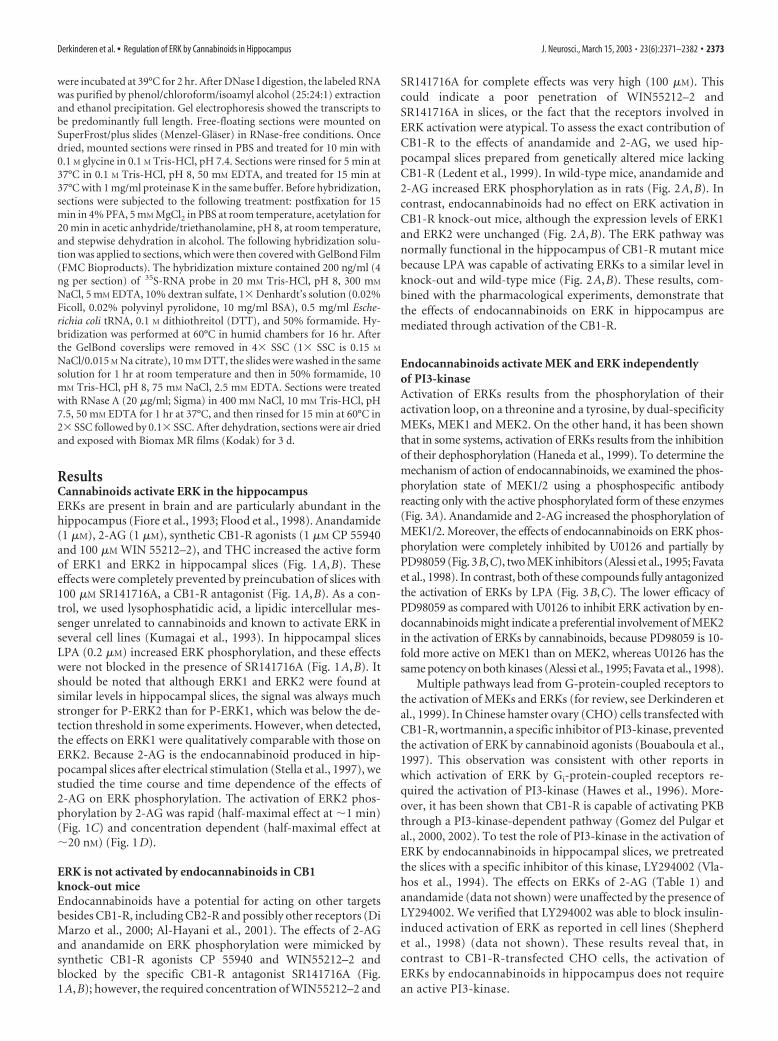
were incubated at 39°C for 2 hr. After DNase I digestion, the labeled RNAwas purified by phenol/chloroform/isoamyl alcohol (25:24:1) extractionand ethanol precipitation. Gel electrophoresis showed the transcripts tobe predominantly full length. Free-floating sections were mounted onSuperFrost/plus slides (Menzel-Glaser) in RNase-free conditions. Oncedried, mounted sections were rinsed in PBS and treated for 10 min with0.1 M glycine in 0.1 M Tris-HCl, pH 7.4. Sections were rinsed for 5 min at37°C in 0.1 M Tris-HCl, pH 8, 50 mM EDTA, and treated for 15 min at37°C with 1 mg/ml proteinase K in the same buffer. Before hybridization,sections were subjected to the following treatment: postfixation for 15min in 4% PFA, 5 mM MgCl2 in PBS at room temperature, acetylation for20 min in acetic anhydride/triethanolamine, pH 8, at room temperature,and stepwise dehydration in alcohol. The following hybridization solu-tion was applied to sections, which were then covered with GelBond Film(FMC Bioproducts). The hybridization mixture contained 200 ng/ml (4ng per section) of 35S-RNA probe in 20 mM Tris-HCl, pH 8, 300 mM
NaCl, 5 mM EDTA, 10% dextran sulfate, 1� Denhardt’s solution (0.02%Ficoll, 0.02% polyvinyl pyrolidone, 10 mg/ml BSA), 0.5 mg/ml Esche-richia coli tRNA, 0.1 M dithiothreitol (DTT), and 50% formamide. Hy-bridization was performed at 60°C in humid chambers for 16 hr. Afterthe GelBond coverslips were removed in 4� SSC (1� SSC is 0.15 M
NaCl/0.015 M Na citrate), 10 mM DTT, the slides were washed in the samesolution for 1 hr at room temperature and then in 50% formamide, 10mM Tris-HCl, pH 8, 75 mM NaCl, 2.5 mM EDTA. Sections were treatedwith RNase A (20 �g/ml; Sigma) in 400 mM NaCl, 10 mM Tris-HCl, pH7.5, 50 mM EDTA for 1 hr at 37°C, and then rinsed for 15 min at 60°C in2� SSC followed by 0.1� SSC. After dehydration, sections were air driedand exposed with Biomax MR films (Kodak) for 3 d.
ResultsCannabinoids activate ERK in the hippocampusERKs are present in brain and are particularly abundant in thehippocampus (Fiore et al., 1993; Flood et al., 1998). Anandamide(1 �M), 2-AG (1 �M), synthetic CB1-R agonists (1 �M CP 55940and 100 �M WIN 55212–2), and THC increased the active formof ERK1 and ERK2 in hippocampal slices (Fig. 1A,B). Theseeffects were completely prevented by preincubation of slices with100 �M SR141716A, a CB1-R antagonist (Fig. 1A,B). As a con-trol, we used lysophosphatidic acid, a lipidic intercellular mes-senger unrelated to cannabinoids and known to activate ERK inseveral cell lines (Kumagai et al., 1993). In hippocampal slicesLPA (0.2 �M) increased ERK phosphorylation, and these effectswere not blocked in the presence of SR141716A (Fig. 1A,B). Itshould be noted that although ERK1 and ERK2 were found atsimilar levels in hippocampal slices, the signal was always muchstronger for P-ERK2 than for P-ERK1, which was below the de-tection threshold in some experiments. However, when detected,the effects on ERK1 were qualitatively comparable with those onERK2. Because 2-AG is the endocannabinoid produced in hip-pocampal slices after electrical stimulation (Stella et al., 1997), westudied the time course and time dependence of the effects of2-AG on ERK phosphorylation. The activation of ERK2 phos-phorylation by 2-AG was rapid (half-maximal effect at �1 min)(Fig. 1C) and concentration dependent (half-maximal effect at�20 nM) (Fig. 1D).
ERK is not activated by endocannabinoids in CB1knock-out miceEndocannabinoids have a potential for acting on other targetsbesides CB1-R, including CB2-R and possibly other receptors (DiMarzo et al., 2000; Al-Hayani et al., 2001). The effects of 2-AGand anandamide on ERK phosphorylation were mimicked bysynthetic CB1-R agonists CP 55940 and WIN55212–2 andblocked by the specific CB1-R antagonist SR141716A (Fig.1A,B); however, the required concentration of WIN55212–2 and
SR141716A for complete effects was very high (100 �M). Thiscould indicate a poor penetration of WIN55212–2 andSR141716A in slices, or the fact that the receptors involved inERK activation were atypical. To assess the exact contribution ofCB1-R to the effects of anandamide and 2-AG, we used hip-pocampal slices prepared from genetically altered mice lackingCB1-R (Ledent et al., 1999). In wild-type mice, anandamide and2-AG increased ERK phosphorylation as in rats (Fig. 2A,B). Incontrast, endocannabinoids had no effect on ERK activation inCB1-R knock-out mice, although the expression levels of ERK1and ERK2 were unchanged (Fig. 2A,B). The ERK pathway wasnormally functional in the hippocampus of CB1-R mutant micebecause LPA was capable of activating ERKs to a similar level inknock-out and wild-type mice (Fig. 2A,B). These results, com-bined with the pharmacological experiments, demonstrate thatthe effects of endocannabinoids on ERK in hippocampus aremediated through activation of the CB1-R.
Endocannabinoids activate MEK and ERK independentlyof PI3-kinaseActivation of ERKs results from the phosphorylation of theiractivation loop, on a threonine and a tyrosine, by dual-specificityMEKs, MEK1 and MEK2. On the other hand, it has been shownthat in some systems, activation of ERKs results from the inhibitionof their dephosphorylation (Haneda et al., 1999). To determine themechanism of action of endocannabinoids, we examined the phos-phorylation state of MEK1/2 using a phosphospecific antibodyreacting only with the active phosphorylated form of these enzymes(Fig. 3A). Anandamide and 2-AG increased the phosphorylation ofMEK1/2. Moreover, the effects of endocannabinoids on ERK phos-phorylation were completely inhibited by U0126 and partially byPD98059 (Fig. 3B,C), two MEK inhibitors (Alessi et al., 1995; Favataet al., 1998). In contrast, both of these compounds fully antagonizedthe activation of ERKs by LPA (Fig. 3B,C). The lower efficacy ofPD98059 as compared with U0126 to inhibit ERK activation by en-docannabinoids might indicate a preferential involvement of MEK2in the activation of ERKs by cannabinoids, because PD98059 is 10-fold more active on MEK1 than on MEK2, whereas U0126 has thesame potency on both kinases (Alessi et al., 1995; Favata et al., 1998).
Multiple pathways lead from G-protein-coupled receptors tothe activation of MEKs and ERKs (for review, see Derkinderen etal., 1999). In Chinese hamster ovary (CHO) cells transfected withCB1-R, wortmannin, a specific inhibitor of PI3-kinase, preventedthe activation of ERK by cannabinoid agonists (Bouaboula et al.,1997). This observation was consistent with other reports inwhich activation of ERK by Gi-protein-coupled receptors re-quired the activation of PI3-kinase (Hawes et al., 1996). More-over, it has been shown that CB1-R is capable of activating PKBthrough a PI3-kinase-dependent pathway (Gomez del Pulgar etal., 2000, 2002). To test the role of PI3-kinase in the activation ofERK by endocannabinoids in hippocampal slices, we pretreatedthe slices with a specific inhibitor of this kinase, LY294002 (Vla-hos et al., 1994). The effects on ERKs of 2-AG (Table 1) andanandamide (data not shown) were unaffected by the presence ofLY294002. We verified that LY294002 was able to block insulin-induced activation of ERK as reported in cell lines (Shepherdet al., 1998) (data not shown). These results reveal that, incontrast to CB1-R-transfected CHO cells, the activation ofERKs by endocannabinoids in hippocampus does not requirean active PI3-kinase.
Derkinderen et al. • Regulation of ERK by Cannabinoids in Hippocampus J. Neurosci., March 15, 2003 • 23(6):2371–2382 • 2373

Role of cAMP in the control of ERK activation bycannabinoids in hippocampal slicesStimulation of CB1-R inhibits adenylyl cyclase, thus decreasingcAMP levels (Howlett, 1995). Depending on the cell type andexperimental conditions, cAMP can either stimulate or inhibitthe ERK pathway, through various signaling pathways (Stork andSchmitt, 2002). In various non-neuronal cells, cAMP preventsthe activation of ERK by mitogens, whereas in neuronal cells suchas PC12, an increase in the intracellular concentration of cAMPactivates the ERK pathway. Because we had demonstrated previ-ously that cAMP was involved in the effects of endocannabinoidson protein tyrosine phosphorylation in hippocampal slices (Der-kinderen et al., 1996, 2001b), we examined its role in ERK regu-lation. We treated hippocampal slices with 8-Bromo (Br)-cAMP,a cell-permeant analog of cAMP, or forskolin, a stimulator ofadenylyl cyclase, for different periods of time. Treatment of sliceswith these compounds for �30 min induced an increase in ERKphosphorylation, in agreement with previous reports (Impey et
al., 1998, 1999). In contrast, when slices were treated for periodsranging from 30 to 45 min, ERK phosphorylation dramaticallydecreased as compared with controls (Fig. 4A and unpublishedresults). A 45 min pretreatment with 8-Br-cAMP completely pre-vented the activation of ERK by endocannabinoids (Fig. 4B,C),whereas LPA was still capable of activating ERKs in these condi-tions (data not shown). Because a major and prolonged effect ofcAMP is to activate cAMP-dependent protein kinase (PKA), thisresult prompted us to test whether inhibition of PKA could par-ticipate in ERK activation. We used two unrelated PKA inhibi-tors: Rp-cAMPS, which prevents cAMP binding to the regulatorysubunit of the kinase, and H-89, an inhibitor of the catalyticsubunit of PKA. Remarkably, both compounds stimulated ERKactivity in hippocampal slices (Fig. 4B,C). Taken together, theseresults underline the complex effects of cAMP on ERK phosphor-ylation in hippocampus, revealing a time-dependent combina-tion of stimulatory and inhibitory effects. They also suggest that adecrease in cAMP levels and, consequently, in PKA activity, may
Figure 1. Cannabinoid agonists stimulate ERK phosphorylation in rat hippocampal slices. A, Rat hippocampal slices were incubated at 35°C, as described in Materials and Methods, for 50 minbefore the addition of vehicle (Control ), 1 �M anandamide, 1 �M 2-AG, 1 �M CP 55940, 100 �M WIN 55212–2, 0.2 �M LPA, or 0.1 �M �9-THC for 5 min, in the absence or in the presence of 100 �M
SR 141716A applied 30 min before. Slices were homogenized in SDS; 60 �g of protein per sample were subjected to immunoblot analysis using antibodies specific for the dually phosphorylated(active) forms of ERK1 and ERK2 (Blot P-ERK ). After stripping, the membranes were reprobed with anti-ERK (Blot ERK ) antibodies. B, For quantification the optical densities of P-ERK2-immunoreactive bands were measured, normalized to the optical densities of total ERK2 in the same samples, and expressed as percentages of controls. Data correspond to means � SEM. Statisticalanalysis was done with ANOVA (F(13,24) 22.8; p � 0.0001) followed by t test (treated vs control: ***p � 0.001, **p � 0.01; treated in the presence of SR141716A vs in its absence: °° p � 0.01,° p � 0.05). C, D, Quantification of the effects of 2-AG on ERK2 active form: time course (drug concentration 1 �M) ( C); concentration–response curve (treatment for 5 min) ( D). Immunoreactivitywas quantified by scanning densitometry using NIH image 1.62 software. Values are means � SEM of four to eight independent experiments and are expressed as percentages of the maximalincrease above unstimulated control values.
2374 • J. Neurosci., March 15, 2003 • 23(6):2371–2382 Derkinderen et al. • Regulation of ERK by Cannabinoids in Hippocampus

participate in the effects of CB1-R on the ERK pathway inhippocampus.
Role of Fyn in the effects of endocannabinoidsWe have shown previously that stimulation of CB1-Rs in hip-pocampus activates a protein tyrosine phosphorylation pathway,involving neuronal isoforms of FAK and an associated protein,p130-Cas (Derkinderen et al., 1996, 2001b). In cell lines, afterautophosphorylation on Tyr-397, FAK recruits Src-family ty-rosine kinases, including Src and Fyn, which in turn phosphory-late multiple residues in the kinase domain and C-terminal re-gion of FAK, as well as in p130-Cas (Girault et al., 1999; Schaller,2001). Our recent results showed that, in hippocampal slices,endocannabinoids increase the association of Fyn, but not Src,with FAK (Derkinderen et al., 2001b). In several cell types, re-cruitment of Src-family kinases by FAK has been shown to resultin the activation of ERK through various signaling mechanisms(Girault et al., 1999; Schaller, 2001). Moreover, other pathwaysinvolving Src-family kinases can activate ERKs downstream ofG-protein-coupled receptors, independently of FAK (Luttrell etal., 1996). To determine the role of Src-family kinases in theeffects of endocannabinoids, we used PP2, a compound that in-hibits potently the catalytic activity of this group of kinases(Hanke et al., 1996). As reported previously (Derkinderen et al.,1996), endocannabinoids increased tyrosine phosphorylation ofa number of proteins in hippocampal slices (Fig. 5A). Pretreat-ment of slices with PP2 dramatically decreased the basal tyrosinephosphorylation of most proteins and abolished the effects ofendocannabinoids (Fig. 5A), demonstrating that they are almostcompletely accounted for by Src-family kinases. However, de-spite the massive inhibition of protein tyrosine phosphorylationby PP2, the activation of ERK by endocannabinoids was stillpresent (Fig. 5A).
Because the endocannabinoids specifically increase the asso-ciation of Fyn with FAK (Derkinderen et al., 2001b), we exam-ined whether the effects of endocannabinoids on ERK were al-tered in Fyn knock-out mice. In these mice the effects of 2-AG onprotein tyrosine phosphorylation were reduced dramatically(Fig. 5B), confirming the critical role of Fyn in tyrosine phos-phorylation in hippocampus (Grant et al., 1995) and its specificinvolvement in the action of cannabinoids (Derkinderen et al.,2001b). Moreover, endocannabinoids were no longer capable ofstimulating ERK (Fig. 5B), showing that Fyn is required for thiseffect. It is important to note that the levels of ERK (Fig. 5B) andCB1-R (Derkinderen et al., 2001b) were not altered in these mice.Altogether these results strongly suggest that Fyn in hippocam-pus is necessary for the activation of ERK by CB1-Rs, but that thisrequirement is independent from acute Fyn catalytic activity.
THC, a cannabinoid agonist, activates ERKs in thehippocampus in vivoOur results demonstrated that endocannabinoids activate theERK pathway in hippocampal slices. To determine whether thiseffect could be also induced by cannabinoids in vivo, we used
Figure 2. The regulation of ERKs by endocannabinoids is absent in hippocampal slices fromCB1-R knock-out mice. A, Hippocampal slices from wild-type (CB1-R �/�) and CB1-R knock-out (CB1-R �/�) mice were incubated at 35°C, as described in Materials and Methods, for 50min before the addition of vehicle (Control ), 1 �M anandamide, 1 �M 2-AG, or 0.2 �M LPA for 5min. ERK phosphorylation was assayed as described in the legend to Figure 1. B, Quantificationof the results for P-ERK2 as described in the legend to Figure 1. Data correspond to means �SEM. Statistical analysis was done with ANOVA (anandamide: F(3,20) 40.2, p � 0.0001; 2-AGand LPA: F(5,24) 8.2, p � 0.0001) followed by t test (treated vs control: ***p � 0.001, **p �0.01, *p � 0.05; treated in knock-out vs wild type: ° p � 0.05).
Figure 3. Role of MEK in the effects of endocannabinoids in rat hippocampal slices. A, Rathippocampal slices were incubated as described in Materials and Methods, for 50 min before theaddition of vehicle (Control ), 1 �M anandamide, or 1 �M 2-AG for 5 min. Homogenates (60 �gof protein per sample) were analyzed by immunoblotting with antibodies specific for the phos-phorylated forms of MEK1/2. Quantification of P-MEK immunoreactivity (mean � SEM): con-trols 100 � 8, anandamide 857 � 29, 2-AG 514 � 57 (F(2,5) 226, p � 0.001; t test treatedvs control: p � 0.0001). B, Slices were treated with the same compounds as in A, or with 0.2 �M
LPA, in the absence or in the presence of 50 �M PD98059 or 30 �M U0126, two MEK inhibitors.Homogenates were analyzed for active dually phosphorylated ERK by immunoblotting. C,Quantification of the results for P-ERK2 as described in the legend to Figure 1. Data correspondto mean�SEM. Statistical analysis was done with ANOVA (F(11,45 7.4; p �0.0001) followedby t test (treated vs control: ***p � 0.001; treated in the presence of MEK inhibitor vs in itsabsence: ° p � 0.05).
Derkinderen et al. • Regulation of ERK by Cannabinoids in Hippocampus J. Neurosci., March 15, 2003 • 23(6):2371–2382 • 2375

THC, the cannabinoid agonist widely abused by humans, whichhas potent effects when administrated at the periphery. As dem-onstrated above, THC was a powerful stimulator of ERKs in hip-pocampal slices (Fig. 1). We analyzed ERK activation in hip-pocampus by immunocytochemistry with a phosphorylationstate-specific antibody after injection of �9-THC (1 mg/kg, i.p.).A strong P-ERK-like immunoreactivity was detected in the pyra-midal cell layers of CA1 (Fig. 6A) and CA3 (Fig. 6B) 10 min afterTHC injection (number of positive cells per section, mean �SEM: vehicle 30 � 4 vs THC 125 � 7; n 4 mice; t test; p � 0.01).This effect was transient because the number of P-ERK-positivecells decreased rapidly: 57 � 13 at 20 min, 31 � 8 at 30 min, and18 � 4 at 60 min (n 4 mice per time point; no significantdifference with controls). No significant effect was observed indentate gyrus (data not shown). Analysis at a higher magnifica-tion indicated that ERK was activated in the soma and dendritesof many neurons, although only some principal neurons werelabeled in response to THC (Fig. 6A,B). In CA1, P-ERK-immunoreactivity was detected in pyramidal cells, whereas inCA3, both pyramidal and nonpyramidal cells were labeled with adense plexiform dendritic network extending into the stratumradiatum. The activation of ERK in response to THC was pre-vented in CB1-R knock-out mice (Fig. 6C) as well as by pretreat-ment with SR141716A (3 mg/kg, i.p.) (Fig. 6D), demonstratingthat THC activates ERK by acting on CB1-R. Because a dramaticalteration in the response to stimulation of CB1-R was observedin hippocampal slices of Fyn�/� mice, we examined whether theresponse to THC was also hampered in vivo in these animals. Inthese mutant mice, no P-ERK immunoreactivity was detectedafter injection of THC (Fig. 6E), revealing that Fyn is necessaryfor the activation of ERK.
In hippocampus, most CB1-Rs are located on GABA nerveterminals (Tsou et al., 1998, 1999; Katona et al., 1999, 2000), andtheir stimulation is capable of enhancing the activation of pyra-midal cells by glutamatergic inputs through a decrease in GABAinhibitory tone (Ohno-Shosaku et al., 2001; Wilson and Nicoll,2001). Therefore, we tested the contribution of glutamate neuro-transmission in the effects of THC on the ERK pathway. In micepretreated with MK801 (0.1 mg/kg, i.p.), a noncompetitive an-tagonist of NMDA receptors that crosses the blood– brain bar-rier, THC did not increase P-ERK immunoreactivity in hip-pocampal neurons (Fig. 6F). This result suggests that the effectsof CB1-R on ERK activation in vivo either are mediated by anincrease in NMDA receptor stimulation or require the concom-itant activation of these receptors. To determine whether the ac-tivation of the ERK pathway by cannabinoids in hippocampuswas exclusively a consequence of an increased sensitivity to exci-tatory inputs, we examined the contribution of glutamate in the
effects of endocannabinoids in hippocampal slices. In this prep-aration, MK801 had a small stimulatory effect on ERK phosphor-ylation by itself but did not prevent ERK activation by either2-AG (Table 1) or THC (data not shown). In contrast, the stim-ulatory effect of glutamate on ERK phosphorylation was pre-vented in the presence of MK801 (data not shown). These resultsindicate that ERK activation by cannabinoids may be achieved byseveral mechanisms, one of which depends on NMDA receptorsand appears predominant in vivo.
Role of ERK in the regulation of gene expression by THCin vivoThe strong accumulation of P-ERK in nuclei (Fig. 7A) suggestedits possible role in gene regulation via phosphorylation of tran-scription factors. To examine the consequences of ERK stimula-tion by THC in vivo we used SL327, a drug that crosses the blood–brain barrier and prevents the activation of ERK by inhibitingMEK (Atkins et al., 1998; Valjent et al., 2000). This drug, closelyrelated to U0126, which inhibited completely ERK phosphoryla-tion in hippocampal slices (Fig. 3), had a dramatic effect onP-ERK immunoreactivity in hippocampal neurons and pre-
Figure 4. Role of cAMP in the effects of endocannabinoids on ERK phosphorylation. A, Rathippocampal slices were incubated with forskolin (50 �M) for the indicated period of time.Homogenates were analyzed for active dually phosphorylated ERK by immunoblotting. B, Rathippocampal slices were incubated as described in Materials and Methods, in the presence or inthe absence of 4 mM 8-Br-cAMP for 45 min before the addition of vehicle (Control ), 1 �M
anandamide, or 2-AG for 5 min. In other experiments, slices were incubated in the presence ofthe PKA inhibitors H-89 (100 �M) or Rp-cAMPS (1 mM) for 20 min. Homogenates were analyzedfor active dually phosphorylated ERK by immunoblotting as described in the legend to Figure 1.C, Quantification of the results for P-ERK2 as described in the legend to Figure 1. Data correspondto mean � SEM. Statistical analysis was done with ANOVA (8-Br-cAMP: F(5,18) 14.5; H89 andRpcAMPs: F(2,7) 16.1; p � 0.01) followed by t test (treated vs control: ***p � 0.001,**p � 0.01, *p � 0.05; treated in the presence of 8-Br-cAMP vs in its absence: °° p � 0.01,° p � 0.05).
Table 1. Activation of ERK2 by 2-AG is independent of PI3-kinase and NMDAreceptors in rat hippocampal slices
Control 2-AG p n
Vehicle 100 � 28 800 � 211 �0.01 6LY294002 52 � 8 626 � 146 �0.001 3p NS NSVehicle 100 � 17 900 � 140 �0.01 4MK801 200 � 18 1100 � 197 �0.01 4p �0.05 NS
Hippocampal slices were treated for 5 min with 1 �M 2-AG, in the absence (Vehicle) or presence of 50 �M LY294002(added 50 min before 2-AG), a PI3-kinase inhibitor, or, in a different series of experiments, in the absence (Vehicle)or presence of 20 �M MK801 (added 25 min before 2-AG). The homogenates were analyzed for the active duallyphosphorylated form of ERK2 by immunoblotting and quantified as indicated in the legend to Figure 1. Statisticalanalysis was performed by ANOVA followed by t test.
2376 • J. Neurosci., March 15, 2003 • 23(6):2371–2382 Derkinderen et al. • Regulation of ERK by Cannabinoids in Hippocampus

vented its enhancement after THC treatment (Fig. 7B). ERKphosphorylates and activates transcription factors of the ternarycomplex factor family, including Elk-1, that bind to the serumresponse element (SRE) (Whitmarsh and Davis, 1996; Valjent etal., 2001a). Moreover, ERK regulates genes that contain thecAMP/Ca 2� response element (CRE) in their promoter, possiblyby phosphorylating and activating MAPK-activated protein ki-nase 2, which phosphorylates cAMP response element-bindingprotein (CREB) (Impey et al., 1999). Therefore, we examined therole of ERK in the induction of IEGs by THC, focusing our atten-tion on c-Fos and Zif268, which contain both SRE and CRE intheir promoter, and the expression of which is known to increasein response to cannabinoids in neurons (Mailleux et al., 1994;Glass and Dragunow, 1995). We analyzed c-Fos protein by im-munohistochemistry, with a specific antibody that did not recog-nize Fos-related antigens (Fig. 7C), and we analyzed Zif268mRNA by in situ hybridization (Fig. 7D). THC increased thenumber and immunoreactivity of c-Fos-expressing neurons inCA1 (Fig. 7C) and in CA3 (data not shown). Zif268 mRNA levelswere increased in CA1 and CA3 but not in the dentate gyrus (Fig.7D,F). Both effects were prevented by injection of SL327 beforeTHC (Fig. 7C,D,F).
We also examined the effects of THC on the expression ofBDNF, a growth factor that behaves as an IEG, transcribed in
response to bursts of neuronal activity through its CREB-bindingsites (Shieh et al., 1998; Tao et al., 1998), and plays an importantrole in hippocampal synaptic plasticity (Kang and Schuman,1995; Hartmann et al., 2001; Patterson et al., 2001; Ying et al.,2002). We found a significant upregulation of BDNF mRNA lev-els in CA1 and CA3 1 hr after injection of THC (Fig. 7E,F). Thisincrease in BDNF mRNA was prevented by pretreatment withSL327 (Fig. 7E,F). Thus, acute injection of THC can induceBDNF mRNA transcription through the ERK signaling pathway.
DiscussionRegulation of ERKs by cannabinoids in hippocampusThe present study shows that cannabinoids activate ERKs in hip-pocampus and in slices incubated in vitro, as well as in living mice.In slices, 2-AG, an endocannabinoid produced in hippocampusin response to stimulation of Schaeffer collaterals (Stella et al.,1997), increased ERK phosphorylation rapidly and at low con-centrations. This effect was likewise achieved with other canna-binoids, including synthetic agonists and THC. Injection of THCin mice was also able to induce ERK activation in the hippocam-pus. The effects of THC on ERK activation were mediated byCB1-Rs because they were prevented by pretreatment withSR141716A, a specific CB1-R antagonist, and they were not ob-served in CB1-R knock-out mice. In the hippocampus, the neu-
Figure 5. Role of Src-family kinases in the effects of endocannabinoids in rat hippocampal slices. A, Rat hippocampal slices were incubated as described in Materials and Methods, in the presenceor absence of 5 �M PP2 for 45 min before the addition of vehicle (Control ), 1 �M anandamide, or 2-AG for 5 min. Homogenates were analyzed by immunoblotting with antibodies specific foranti-phospho-tyrosine (Blot P-Tyr), active dually phosphorylated ERK (Blot P-ERK ), or total ERK (Blot ERK ). The optical densities of P-ERK2 were as follows: in the absence of PP2: control 100 � 43,anandamide 767 � 231, and 2-AG 413 � 92; in the presence of PP2: control 89 � 28, anandamide 1358 � 280, and 2-AG 861 � 191 (F(5,14) 7.1, p � 0.001; followed by t test, p � 0.05 forendocannabinoid-treated vs control in both groups). B, Hippocampal slices from wild-type (Fyn �/�) and Fyn knock-out (Fyn �/�) mice were incubated for 50 min before the addition of eithervehicle (Control ) or 1 �M 2-AG for 5 min. The optical densities of P-ERK2 were as follows: in Fyn �/� slices: control 100 � 8, 2-AG 1198 � 351; in Fyn �/� slices: control 115 � 23, 2-AG 108 �63 (F(3,8) 9.3, p � 0.01; followed by t test, p � 0.05, 2-AG vs control in wild-type slices, and p � 0.05, 2-AG in knock-out vs control slices).
Derkinderen et al. • Regulation of ERK by Cannabinoids in Hippocampus J. Neurosci., March 15, 2003 • 23(6):2371–2382 • 2377

rons that expressed the strongest P-ERK immunoreactivity afterin vivo administration of THC were pyramidal cells in CA1 andCA3. In contrast, CB1-R-immunoreactive neurons are located inall subfields in hippocampus and are mostly interneurons, with alarge predominance of cholecystokinin-positive cells (Pettit et al.,1998; Katona et al., 1999; Tsou et al., 1999). In fact, CB1-Rs arehighly concentrated in nerve terminals, and endocannabinoidshave recently been shown to mediate retrograde signaling at hip-pocampal synapses (Ohno-Shosaku et al., 2001; Wilson andNicoll, 2001). Although we cannot formally rule out a contribu-
tion of CB1-Rs in principal cells, which express low levels ofCB1-R mRNA (Marsicano and Lutz, 1999), THC is likely to acton pyramidal cells indirectly by modulating inputs at the presyn-aptic level. Because CB1-Rs are mostly located on GABA termi-nals, they could modulate indirectly the sensitivity to excitatoryinputs by decreasing the inhibitory tone on pyramidal neurons(Ohno-Shosaku et al., 2001; Wilson and Nicoll, 2001). Stimula-tion of NMDA glutamate receptors is capable of activating theERK pathway in vitro and in vivo (Valjent et al., 2001a). Interest-ingly, however, the contribution of NMDA glutamate receptors
Figure 6. THC activates ERK in hippocampus in vivo by stimulating CB1 receptors. Mice were injected with vehicle (Veh) or THC (1 mg/kg, i.p.) 10 min before they were killed. Active ERK1 and ERK2were detected by peroxidase immunocytochemistry in the hippocampus, using antibodies against the doubly phosphorylated protein. Results in CA1 ( A) and CA3 ( B) regions are shown. The rightpanel corresponds to a higher magnification of the THC-treated sections. In THC-treated mice, immunoreactive cells are mostly present in the pyramidal cell layer of CA1 and CA3. A strong labelingis visible in the cytoplasm (including the dendrites) and the nucleus. C, THC failed to activate ERK in CA1 of CB1 �/� mice, whereas it was active in CB1 �/� matched controls. D, In the presenceof the CB1-R antagonist, SR141716A (3 mg/kg) injected alone (Veh � SR141716A), or 15 min before THC injection (THC � SR141716A), no activation of ERK was observed in CA1. E, THC failed toactivate ERK in CA1 of Fyn �/� mice, whereas its effects were present in Fyn �/� matched controls. F, Stimulation of ERK phosphorylation by THC was abolished by the NMDA receptor antagonistMK801 (0.1 mg/kg), injected 15 min before THC. The results obtained in CB1 �/�, SR141716A-treated, Fyn �/�, and MK801-treated mice in CA3 (data not shown) were similar to thoseillustrated here in CA1.
2378 • J. Neurosci., March 15, 2003 • 23(6):2371–2382 Derkinderen et al. • Regulation of ERK by Cannabinoids in Hippocampus

in the effects of THC appeared different in vitro and in vivo. Inliving mice, MK801, an NMDA receptor antagonist, preventedthe effects of THC on the ERK pathway, suggesting that THCeffects on ERK in vivo either were mediated by an increased stim-ulation or sensitivity of NMDA receptors or required the con-comitant activity of these receptors.
Signaling pathways involved in ERK regulation by CB1-RsAlthough the effects of in vivo CB1-R stimulation were sensitiveto a NMDA receptor inhibitor, a strong stimulation of ERK phos-phorylation was observed in hippocampal slices in the presenceof TTX, which prevents neuronal firing. This effect was also re-sistant to MK801, arguing in favor of a direct stimulation of theERK pathway by CB1-Rs in this preparation. It should be pointedout that the precise cellular localization of the activation of ERK
by CB1-Rs in hippocampal slices is notknown. Nevertheless, the mechanism bywhich stimulation of CB1-Rs led to ERKactivation in hippocampal slices appearsto be different from what has been re-ported in transfected cells. Studies inCHO cells supported a role for �� ratherthan for �i subunits in CB1-R–ERK cou-pling (Bouaboula et al., 1995a,b). Activa-tion of ERKs in cell lines by Gi-coupledreceptors can be mediated through re-cruitment of PI3-kinase by �� sub-units, independently of the inhibition ofadenylyl cyclase (Hawes et al., 1996;Lopez-Ilasaca et al., 1997). However, inhippocampal slices, the cannabinoid-induced activation of ERKs was insensi-tive to the PI3-kinase inhibitor LY294002,strongly arguing against a role of the ��subunits/PI3-kinase pathway in this ef-fect. Other pathways linking G-protein-coupled receptors to MAP kinases involvetyrosine kinases of the Src family (Luttrellet al., 1997). For example, PP2, a potentinhibitor of these kinases, blocked the ef-fects of various extracellular messengerson the activation of ERK in CHO cells(Igishi and Gutkind, 1998). We haveshown previously that stimulation ofCB1-Rs increases the tyrosine phosphor-ylation of the neuronal isoform of FAK inhippocampus and its association with Fyn(Derkinderen et al., 1996, 2001b). Thus,FAK provides a possible link for the acti-vation of ERK through recruitment ofSrc-family kinases or PI3-kinase (Giraultet al., 1999; Schaller, 2001). However, theactivation of ERK by CB1-Rs in hip-pocampal slices cannot be accounted forby these known pathways linking FAK toERK, because it was resistant to inhibitorsof PI3-kinase and Src-family kinases. Itwas particularly striking that PP2 did notprevent ERK activation in hippocampalslices, despite its dramatic effect on pro-tein tyrosine phosphorylation. Yet, theSrc-family kinase Fyn appeared to be crit-ical for the activation of ERK by CB1-Rs
because this activation was absent in Fyn mutant mice, both invitro and in vivo. Although we verified that CB1-R and ERK levelswere unaltered in Fyn�/� mice, we cannot rule out the possibil-ity that the absence of Fyn throughout development impairedindirectly the signaling pathways at other levels. An alternativeexplanation of our findings is that stimulation of CB1-Rs leads tothe recruitment of Fyn and to the activation of ERK by a mecha-nism independent of Fyn catalytic activity. In support of thishypothesis, the closely related tyrosine kinase Src is known toexert some of its biological effects independently of its tyrosinekinase activity (Kaplan et al., 1995; Schwartzberg et al., 1997).
The role of cAMP in ERK regulation in hippocampusSeveral additional pathways have been described that linkG-protein-coupled receptors to ERK activation and could ac-
Figure 7. ERK-dependent induction of immediate-early genes by THC in hippocampus in vivo. A, Mice were injected with 1mg/kg THC 10 min before they were killed, as in Figure 6 A, High magnification of a peroxidase-labeled section shows the nuclearstaining for dually phosphorylated ERK. B, The effects of THC (middle panel ) were prevented in the CA1 region of mice injected withSL327 (100 mg/kg) 60 min before THC (right panel ). Results shown are representative of four to six animals for each group. C, c-Fosimmunoreactivity (peroxidase reaction) was examined in CA1 of mice injected with either vehicle (Veh) or 1 mg/kg THC 60 minbefore they were killed. SL327 (100 mg/kg) was injected 60 min before THC. D, Zif268 mRNA expression was analyzed by in situhybridization in mouse hippocampus 1 hr after injection of vehicle, THC (1 mg/kg, i.p.), or THC and SL327 (100 mg/kg, 60 minbefore THC). Note the increased hybridization signals in the CA1 and CA3 regions. E, BDNF mRNA expression was analyzed by in situhybridization in mouse hippocampus 1 hr after injection of vehicle, THC (1 mg/kg, i.p.), or THC and SL327 (100 mg/kg, 60 minbefore THC). F, Signals for mRNA hybridization were quantified using an image analyzer for six animals for each treatment.Statistical analyses used one-way ANOVA followed by a post hoc comparison with Newman–Keuls test; *p � 0.001 when com-paring THC-treated mice with control mice; ˆ p � 0.001 when comparing SL � THC with THC alone (n 6 mice per group).
Derkinderen et al. • Regulation of ERK by Cannabinoids in Hippocampus J. Neurosci., March 15, 2003 • 23(6):2371–2382 • 2379

count for the effects of endocannabinoids (Marinissen and Gut-kind, 2001). It is well established that CB1-Rs inhibit adenylylcyclase, including in hippocampus (Bidaut-Russell et al., 1990),and our observations reveal that ERK activation by cannabinoidswas closely associated with cAMP regulation. The effects of en-docannabinoids on ERK were prevented by pretreatment of sliceswith cAMP analogs, suggesting that their mechanism of actionmay involve an inhibition of cAMP production. Similar effects ofcAMP on the activation of ERK induced by anandamide havebeen reported in a human breast cancer cell line (Melck et al.,1999). The role of cAMP in the effects of CB1-Rs in hippocampuswas further supported by the fact that two different inhibitors ofPKA, acting on the regulatory or the catalytic subunit of PKA,also activated ERK in hippocampal slices. These effects of cAMPare reminiscent of those reported previously for FAK (Derkin-deren et al., 1996, 2001b). Interestingly, a negative effect of cAMPon both FAK- and ERK-dependent responses has also been ob-served in a completely different context, when adherent NIH3T3cells were placed in suspension (Howe and Juliano, 2000). Theseeffects of cAMP might appear paradoxical in light of the knownstimulation of ERK by cAMP in hippocampal slices (Impey et al.,1998, 1999). Indeed, we confirmed in our experimental condi-tions that 8-Br-cAMP as well as forskolin were also capable ofactivating ERK, in agreement with these previous reports. Thesestimulatory effects lasted for a short period of time and werefollowed by a decrease in ERK phosphorylation and its insensi-tivity to endocannabinoids. The effects of cAMP on ERK in hip-pocampus are complex, similar to what has been reported inother cell types (Stork and Schmitt, 2002). cAMP appears to exerttwo opposing effects on the ERK pathway in hippocampus, astimulatory effect that may be partly independent of PKA and, asindicated by our findings, an inhibitory effect mediated by PKA.Thus, in hippocampal slices, stimulation of CB1-Rs could be acritical modulator of cAMP signaling, by opposing some of itseffects and enhancing some others. Further work will have todetermine whether these apparently opposing effects take place atdifferent cellular or subcellular locations.
Functional implications of the regulation of ERK andimmediate-early genes by cannabinoids in hippocampusOur findings have two types of implications concerning the pos-sible physiological function of the endocannabinoid system inhippocampus and the effects of THC in the context of drug abuse.Recent work from several laboratories has unveiled the importantrole of the endocannabinoid system in hippocampus. Althoughmassive stimulation of CB1-Rs inhibits several forms of synapticplasticity in hippocampus, in vivo and in vitro, including LTP andLTD in CA1 neurons (Collins et al., 1995; Terranova et al., 1995;Misner and Sullivan, 1999), it appears that focal stimulation ofthese receptors has more subtle effects. By decreasing locally theGABAergic inhibition of hippocampal neurons (Ohno-Shosakuet al., 2001; Wilson and Nicoll, 2001), endocannabinoids facili-tate the induction of LTP (Carlson et al., 2002). In addition tothese direct effects on synaptic transmission, our results showthat stimulation of CB1-Rs has potent effects on signaling path-ways that are known to be critical for long-term synaptic plastic-ity. Stimulation of CB1-Rs activates ERK, a protein kinase knownto be critical in several forms of synaptic plasticity and in learningand memory (English and Sweatt, 1997; Atkins et al., 1998). TheERK pathway appears essential for some forms of LTP in hip-pocampus (Winder et al., 1999) and for the regulation of geneexpression in the context of synaptic plasticity (Impey et al.,1999). Remarkably, the activation of ERK by THC was absent in
the hippocampus of mice lacking the tyrosine kinase Fyn, whichdisplay altered synaptic plasticity (Grant et al., 1992).
CB1-R-mediated stimulation of ERK was responsible for theinduction of Zif268, a transcription factor essential for transitionfrom short- to long-term synaptic plasticity and for the expres-sion of long-term memories (Jones et al., 2001). Moreover, wedemonstrate that THC also induced the expression of BDNF, aneurotrophic factor that increases synaptic efficiency (Kang andSchuman, 1995; Patterson et al., 2001; Ying et al., 2002). Thus,stimulation of CB1-Rs is by itself capable of activating signalingcascades known to be important for synaptic plasticity. Localizedincreases in endocannabinoid production could facilitate localsynaptic efficiency not only by inhibiting presynaptic release ofGABA but also by activating signaling pathways important forlong-term modifications.
In contrast to this possible physiological role of the local acti-vation of CB1-Rs, their general pharmacological stimulation inthe whole tissue clearly has a negative effect on plasticity (Collinset al., 1995; Terranova et al., 1995; Misner and Sullivan, 1999;Bohme et al., 2000). The effects of THC on ERK phosphorylationin mice, reported here, were observed at doses (1 mg/kg) that arelow for experimental studies in animals and may correspond tothe amounts of THC absorbed by human subjects during heavyintoxication (Barnett et al., 1985). This dose of THC has minimalaversive effects in mice, and, given after a priming injection in thehome cage, is capable of inducing conditioned place preference(Valjent and Maldonado, 2000). It should be pointed out thatgeneral administration of THC also stimulates the ERK pathwayin other brain regions, including the dorsal striatum and nucleusaccumbens (Valjent et al., 2001b). In these brain regions, how-ever, the activation of ERK was entirely dependent on activationof dopamine D1 receptors, whereas in hippocampus the effects ofTHC were still observed in the presence of SCH23390, a potentblocker of D1 receptors (our unpublished observations). Thus,the present study demonstrates that THC can activate ERK inbrain by several pathways, and in regions beyond those usuallythought to be the major targets of drugs of abuse. Therefore it willbe important to determine whether effects of THC on ERK inhippocampus and their consequences on gene expression mayparticipate in the cognitive and memory impairment reported inheavy cannabis users (Pope et al., 2001; Solowij et al., 2002).
ReferencesAlessi DR, Cuenda A, Cohen P, Dudley DT, Saltiel AR (1995) PD 098059 is
a specific inhibitor of the activation of mitogen-activated protein kinasekinase in vitro and in vivo. J Biol Chem 270:27489 –27494.
Al-Hayani A, Wease KN, Ross RA, Pertwee RG, Davies SN (2001) The en-dogenous cannabinoid anandamide activates vanilloid receptors in therat hippocampal slice. Neuropharmacology 41:1000 –1005.
Atkins CM, Selcher JC, Petraitis JJ, Trzaskos JM, Sweatt JD (1998) TheMAPK cascade is required for mammalian associative learning. Nat Neu-rosci 1:602– 609.
Barnett G, Licko V, Thompson T (1985) Behavioral pharmacokinetics ofmarijuana. Psychopharmacology 85:51–56.
Bidaut-Russell M, Devane WA, Howlett AC (1990) Cannabinoid receptorsand modulation of cyclic AMP accumulation in the rat brain. J Neuro-chem 55:21–26.
Bohme GA, Laville M, Ledent C, Parmentier M, Imperato A (2000) En-hanced long-term potentiation in mice lacking cannabinoid CB1 recep-tors. Neuroscience 95:5–7.
Bouaboula M, Bourrie B, Rinaldi-Carmona M, Shire D, Le Fur G, Casellas P(1995a) Stimulation of cannabinoid receptor CB1 induces krox-24 ex-pression in human astrocytoma cells. J Biol Chem 270:13973–13980.
2380 • J. Neurosci., March 15, 2003 • 23(6):2371–2382 Derkinderen et al. • Regulation of ERK by Cannabinoids in Hippocampus

Bouaboula M, Poinot-Chazel C, Bourrie B, Canat X, Calandra B, Rinaldi-Carmona M, Le Fur G, Casellas P (1995b) Activation of mitogen-activated protein kinases by stimulation of the central cannabinoid recep-tor CB1. Biochem J 312:637– 641.
Bouaboula M, Perrachon S, Milligan L, Canat X, Rinaldi-Carmona M, PortierM, Barth F, Calandra B, Pecceu F, Lupker J, Maffrand JP, Le Fur G,Casellas P (1997) A selective inverse agonist for central cannabinoid re-ceptor inhibits mitogen-activated protein kinase activation stimulated byinsulin or insulin-like growth factor 1: evidence for a new model of recep-tor/ligand interactions. J Biol Chem 272:22330 –22339.
Burgaya F, Toutant M, Studler JM, Costa A, Le Bert M, Gelman M, Girault JA(1997) Alternatively spliced focal adhesion kinase in rat brain with in-creased autophosphorylation activity. J Biol Chem 272:28720 –28725.
Carlson G, Wang Y, Alger BE (2002) Endocannabinoids facilitate the induc-tion of LTP in the hippocampus. Nat Neurosci 5:723–724.
Collins DR, Pertwee RG, Davies SN (1995) Prevention by the cannabinoidantagonist, SR141716A, of cannabinoid-mediated blockade of long-termpotentiation in the rat hippocampal slice. Br J Pharmacol 115:869 – 870.
Derkinderen P, Toutant M, Burgaya F, Le Bert M, Siciliano JC, De FranciscisV, Gelman M, Girault JA (1996) Regulation of a neuronal form of focaladhesion kinase by anandamide. Science 273:1719 –1722.
Derkinderen P, Enslen H, Girault JA (1999) The ERK/MAP-kinase cascadein the nervous system. NeuroReport 10:R24 –34.
Derkinderen P, Ledent C, Parmentier M, Girault JA (2001a) Cannabinoidsactivate p38 mitogen-activated protein kinases through CB1 receptors inhippocampus. J Neurochem 77:957–960.
Derkinderen P, Toutant M, Kadare G, Ledent C, Parmentier M, Girault JA(2001b) Dual role of Fyn in the regulation of FAK�6,7 by cannabinoidsin hippocampus. J Biol Chem 276:38289 –38296.
Di Marzo V, Melck D, Bisogno T, De Petrocellis L (1998) Endocannabi-noids: endogenous cannabinoid receptor ligands with neuromodulatoryaction. Trends Neurosci 21:521–528.
Di Marzo V, Breivogel CS, Tao Q, Bridgen DT, Razdan RK, Zimmer AM,Zimmer A, Martin BR (2000) Levels, metabolism, and pharmacologicalactivity of anandamide in CB(1) cannabinoid receptor knockout mice:evidence for non-CB(1), non-CB(2) receptor-mediated actions of anan-damide in mouse brain. J Neurochem 75:2434 –2444.
English JD, Sweatt JD (1997) A requirement for the mitogen-activated pro-tein kinase cascade in hippocampal long term potentiation. J Biol Chem272:19103–19106.
Favata MF, Horiuchi KY, Manos EJ, Daulerio AJ, Stradley DA, Feeser WS,Van Dyk DE, Pitts WJ, Earl RA, Hobbs F, Copeland RA, Magolda RL,Scherle PA, Trzaskos JM (1998) Identification of a novel inhibitor ofmitogen-activated protein kinase kinase. J Biol Chem 273:18623–18632.
Fiore RS, Bayer VE, Pelech SL, Posada J, Cooper JA, Baraban JM (1993) p42mitogen-activated protein kinase in brain: prominent localization in neu-ronal cell bodies and dendrites. Neuroscience 55:463– 472.
Flood DG, Finn JP, Walton KM, Dionne CA, Contreras PC, Miller MS, BhatRV (1998) Immunolocalization of the mitogen-activated protein ki-nases p42MAPK and JNK1, and their regulatory kinases MEK1 andMEK4, in adult rat central nervous system. J Comp Neurol 398:373–392.
Girault JA, Costa A, Derkinderen P, Studler JM, Toutant M (1999) FAK andPYK2/CAK in the nervous system, a link between neuronal activity, plas-ticity and survival? Trends Neurosci 22:257–263.
Glass M, Dragunow M (1995) Induction of the Krox 24 transcription factorin striosomes by a cannabinoid agonist. NeuroReport 6:241–244.
Gomez del Pulgar T, Velasco G, Guzman M (2000) The CB1 cannabinoidreceptor is coupled to the activation of protein kinase B/Akt. Biochem J347:369 –373.
Gomez del Pulgar T, De Ceballos ML, Guzman M, Velasco G (2002) Can-nabinoids protect astrocytes from ceramide-induced apoptosis throughthe phosphatidylinositol 3-kinase/protein kinase B pathway. J Biol Chem277:36527–36533.
Grant SGN, O’Dell TJ, Karl KA, Stein PL, Soriano P, Kandel ER (1992)Impaired long-term potentiation, spatial learning, and hippocampal de-velopment in fyn mutant mice. Science 258:1903–1910.
Grant SGN, Karl KA, Kiebler MA, Kandel ER (1995) Focal adhesion kinasein the brain: novel subcellular localization and specific regulation by Fyntyrosine kinase in mutant mice. Genes Dev 9:1909 –1921.
Hampson RE, Deadwyler SA (2000) Cannabinoids reveal the necessity ofhippocampal neural encoding for short-term memory in rats. J Neurosci20:8932– 8942.
Haneda M, Sugimoto T, Kikkawa R (1999) Mitogen-activated protein ki-nase phosphatase: a negative regulator of the mitogen-activated proteinkinase cascade. Eur J Pharmacol 365:1–7.
Hanke JH, Gardner JP, Dow RL, Changelian PS, Brissette WH, Weringer EJ,Pollok K, Connelly PA (1996) Discovery of a novel, potent, and Srcfamily-selective tyrosine kinase inhibitor: study of Lck- and FynT-dependent T cell activation. J Biol Chem 271:695–701.
Hartmann M, Heumann R, Lessmann V (2001) Synaptic secretion of BDNFafter high-frequency stimulation of glutamatergic synapses. EMBO J20:5887–5897.
Hawes BE, Luttrell LM, Van Biesen T, Lefkowitz RJ (1996) Phosphatidylinosi-tol 3-kinase is an early intermediate in the G�gamma-mediated mitogen-activated protein kinase signaling pathway. J Biol Chem 271:12133–12136.
Howe AK, Juliano RL (2000) Regulation of anchorage-dependent signaltransduction by protein kinase A and p21-activated kinase. Nat Cell Biol2:593– 600.
Howlett AC (1995) Pharmacology of cannabinoid receptors. Annu RevPharmacol Toxicol 35:607– 634.
Igishi T, Gutkind JS (1998) Tyrosine kinases of the Src family participate insignaling to MAP kinase from both Gq, and Gi-coupled receptors. Bio-chem Biophys Res Commun 244:5–10.
Impey S, Obrietan K, Wong ST, Poser S, Yano S, Wayman G, Deloulme JC,Chan G, Storm DR (1998) Cross talk between ERK and PKA is requiredfor Ca 2� stimulation of CREB-dependent transcription and ERK nucleartranslocation. Neuron 21:869 – 883.
Impey S, Obrietan K, Storm DR (1999) Making new connections: role ofERK/MAP kinase signaling in neuronal plasticity. Neuron 23:11–14.
Jones MW, Errington ML, French PJ, Fine A, Bliss TV, Garel S, Charnay P,Bozon B, Laroche S, Davis S (2001) A requirement for the immediateearly gene Zif268 in the expression of late LTP and long-term memories.Nat Neurosci 4:289 –296.
Kang H, Schuman EM (1995) Long-lasting neurotrophin-induced en-hancement of synaptic transmission in the adult hippocampus. Science267:1658 –1662.
Kaplan KB, Swedlow JR, Morgan DO, Varmus HE (1995) c-Src enhancesthe spreading of src�/� fibroblasts on fibronectin by a kinase-independent mechanism. Genes Dev 9:1505–1517.
Katona I, Sperlagh B, Sik A, Kafalvi A, Vizi ES, Mackie K, Freund TF (1999)Presynaptically located CB1 cannabinoid receptors regulate GABA releasefrom axon terminals of specific hippocampal interneurons. J Neurosci19:4544 – 4558.
Katona I, Sperlagh B, Magloczky Z, Santha E, Kofalvi A, Czirjak S, Mackie K, ViziES, Freund TF (2000) GABAergic interneurons are the targets of cannabi-noid actions in the human hippocampus. Neuroscience 100:797–804.
Kumagai N, Morii N, Fujisawa K, Yoshimasa T, Nakao K, Narumiya S (1993)Lysophosphatidic acid induces tyrosine phosphorylation and activationof MAP-kinase and focal adhesion kinase in cultured Swiss 3T3 cells.FEBS Lett 329:273–276.
Ledent C, Valverde O, Cossu C, Petitet F, Aubert LF, Beslot F, Bohme GA,Imperato A, Pedrazzini T, Roques BP, Vassart G, Fratta W, Parmentier M(1999) Unresponsiveness to cannabinoids and reduced addictive effectsof opiates in CB1 receptor knockout mice. Science 283:401– 404.
Lopez-Ilasaca M, Crespo P, Pellici PG, Gutkind JS, Wetzker R (1997) Link-age of G protein-coupled receptors to the MAPK signaling pathwaythrough PI 3-kinase gamma. Science 275:394 –397.
Luttrell LM, Hawes BE, Van Biesen T, Luttrell DK, Lansing TJ, Lefkowitz RJ(1996) Role of c-Src tyrosine kinase in G protein-coupled receptor- andG�gamma subunit-mediated activation of mitogen-activated protein ki-nases. J Biol Chem 271:19443–19450.
Luttrell LM, Van Biesen T, Hawes BE, Koch WJ, Krueger KM, Touhara K,Lefkowitz RJ (1997) G-protein-coupled receptors and their regulation:activation of the MAP kinase signaling pathway by G-protein-coupledreceptors. Adv Second Messenger Phosphoprotein Res 31:263–277.
Mailleux P, Verslype M, Preud’homme X, Vanderhaeghen J-J (1994) Acti-vation of multiple transcription factor genes by tetrahydrocannabinol inrat forebrain. NeuroReport 5:1265–1268.
Marinissen MJ, Gutkind JS (2001) G-protein-coupled receptors and signal-ing networks: emerging paradigms. Trends Pharmacol Sci 22:368 –376.
Marsicano G, Lutz B (1999) Expression of the cannabinoid receptor CB1 indistinct neuronal subpopulations in the adult mouse forebrain. EurJ Neurosci 11:4213– 4225.
Matsuda LA, Lolait SJ, Brownstein MJ, Young AC, Bonner TI (1990) Struc-
Derkinderen et al. • Regulation of ERK by Cannabinoids in Hippocampus J. Neurosci., March 15, 2003 • 23(6):2371–2382 • 2381

ture of a cannabinoid receptor and functional expression of the clonedcDNA. Nature 346:561–564.
Melck D, Rueda D, Galve-Roperh I, De Petrocellis L, Guzman M, Di Marzo V(1999) Involvement of the cAMP/protein kinase A pathway and ofmitogen-activated protein kinase in the anti-proliferative effects of anan-damide in human breast cancer cells. FEBS Lett 463:235–240.
Misner DL, Sullivan JM (1999) Mechanism of cannabinoid effects on long-term potentiation and depression in hippocampal CA1 neurons. J Neu-rosci 19:6795– 6805.
Ohno-Shosaku T, Maejima T, Kano M (2001) Endogenous cannabinoidsmediate retrograde signals from depolarized postsynaptic neurons to pre-synaptic terminals. Neuron 29:729 –738.
Patterson SL, Pittenger C, Morozov A, Martin KC, Scanlin H, Drake C, Kan-del ER (2001) Some forms of cAMP-mediated long-lasting potentiationare associated with release of BDNF and nuclear translocation ofphospho-MAP kinase. Neuron 32:123–140.
Pettit DA, Harrison MP, Olson JM, Spencer RF, Cabral GA (1998) Immu-nohistochemical localization of the neural cannabinoid receptor in ratbrain. J Neurosci Res 51:391– 402.
Pope Jr HG, Gruber AJ, Hudson JI, Huestis MA, Yurgelun-Todd D (2001)Neuropsychological performance in long-term cannabis users. Arch GenPsychiatry 58:909 –915.
Schaller MD (2001) Biochemical signals and biological responses elicited bythe focal adhesion kinase. Biochim Biophys Acta 1540:1–21.
Schwartzberg PL, Xing LP, Hoffmann O, Lowell CA, Garrett L, Boyce BF,Varmus HE (1997) Rescue of osteoclast function by transgenic expres-sion of kinase-deficient Src in src�/� mutant mice. Genes Dev11:2835–2844.
Shepherd PR, Withers DJ, Siddle K (1998) Phosphoinositide 3-kinase: thekey switch mechanism in insulin signaling. Biochem J 333:471– 490.
Shieh PB, Hu SC, Bobb K, Timmusk T, Ghosh A (1998) Identification of asignaling pathway involved in calcium regulation of BDNF expression.Neuron 20:727–740.
Siciliano JC, Gelman M, Girault JA (1994) Depolarization and neurotrans-mitters increase neuronal protein tyrosine phosphorylation. J Neuro-chem 62:950 –959.
Solowij N, Stephens RS, Roffman RA, Babor T, Kadden R, Miller M, Chris-tiansen K, McRee B, Vendetti J (2002) Cognitive functioning of long-term heavy cannabis users seeking treatment. JAMA 287:1123–1131.
Stella N, Schweitzer P, Piomelli D (1997) A second endogenous cannabi-noid that modulates long-term potentiation. Nature 388:773–778.
Stork PJ, Schmitt JM (2002) Crosstalk between cAMP and MAP kinase sig-naling in the regulation of cell proliferation. Trends Cell Biol 12:258 –266.
Tao X, Finkbeiner S, Arnold DB, Shaywitz AJ, Greenberg ME (1998) Ca 2�
influx regulates BDNF transcription by a CREB family transcriptionfactor-dependent mechanism. Neuron 20:709 –726.
Terranova JP, Michaud JC, Le Fur G, Soubrie P (1995) Inhibition of long-termpotentiation in rat hippocampal slices by anandamide and WIN55212–2:reversal by SR141716 A, a selective antagonist of CB1 cannabinoid receptors.Naunyn Schmiedebergs Arch Pharmacol 352:576–579.
Tsou K, Nogueron MI, Muthian S, Sanudo-Pena MC, Hillard CJ, DeutschDG, Walker JM (1998) Fatty acid amide hydrolase is located preferen-tially in large neurons in the rat central nervous system as revealed byimmunohistochemistry. Neurosci Lett 254:137–140.
Tsou K, Mackie K, Sanudo-Pena MC, Walker JM (1999) Cannabinoid CB1receptors are localized primarily on cholecystokinin-containing GABAer-gic interneurons in the rat hippocampal formation. Neuroscience93:969 –975.
Valjent E, Maldonado R (2000) A behavioural model to reveal place prefer-ence to delta 9- tetrahydrocannabinol in mice. Psychopharmacology(Berl) 147:436 – 438.
Valjent E, Corvol JC, Pages C, Besson MJ, Maldonado R, Caboche J (2000)Involvement of the extracellular signal-regulated kinase cascade forcocaine-rewarding properties. J Neurosci 20:8701– 8709.
Valjent E, Caboche J, Vanhoutte P (2001a) Mitogen-activated protein ki-nase/extracellular signal-regulated kinase induced gene regulation inbrain: a molecular substrate for learning and memory? Mol Neurobiol23:83–99.
Valjent E, Pages C, Rogard M, Besson MJ, Maldonado R, Caboche J (2001b)Delta 9-tetrahydrocannabinol-induced MAPK/ERK and Elk-1 activationin vivo depends on dopaminergic transmission. Eur J Neurosci14:342–352.
Vlahos CJ, Matter WF, Hui KY, Brown RF (1994) A specific inhibitor of phos-phatidylinositol 3-kinase,2-(4-morpholinyl)-8-phenyl-4H-1-benzopyran-4-one (LY294002). J Biol Chem 269:5241–5248.
Wartmann M, Campbell D, Subramanian A, Burstein SH, Davis RJ (1995)The MAP kinase signal transduction pathway is activated by the endoge-nous cannabinoid anandamide. FEBS Lett 359:133–136.
Whitmarsh AJ, Davis RJ (1996) Transcription factor AP-1 regulation bymitogen-activated protein kinase signal transduction pathways. J MolMed 74:589 – 607.
Wilson RI, Nicoll RA (2001) Endogenous cannabinoids mediate retrogradesignaling at hippocampal synapses. Nature 410:588 –592.
Winder DG, Martin KC, Muzzio IA, Rohrer D, Chruscinski A, Kobilka B,Kandel ER (1999) ERK plays a regulatory role in induction of LTP bytheta frequency stimulation and its modulation by beta-adrenergic recep-tors. Neuron 24:715–726.
Ying SW, Futter M, Rosenblum K, Webber MJ, Hunt SP, Bliss TV, Bramham CR(2002) Brain-derived neurotrophic factor induces long-term potentiation inintact adult hippocampus: requirement for ERK activation coupled to CREBand upregulation of Arc synthesis. J Neurosci 22:1532–1540.
2382 • J. Neurosci., March 15, 2003 • 23(6):2371–2382 Derkinderen et al. • Regulation of ERK by Cannabinoids in Hippocampus
Copyright © 2022 FDOKUMEN