Friendship stability in adolescence is associated with ventral ...

Neurobiology of Learning and Memory 86 (2006) 72–81
www.elsevier.com/locate/ynlme
EVects of ventral and dorsal CA1 subregional lesions on trace fear conditioning
Jason L. Rogers 1, Michael R. Hunsaker, Raymond P. Kesner ¤
Department of Psychology, University of Utah, 380 South 1530 East, Room 502, Salt Lake City, UT 84112-0251, USA
Received 25 October 2005; revised 3 January 2006; accepted 12 January 2006Available online 28 February 2006
Abstract
Recent lines of research have focused on dissociating function between the dorsal and ventral hippocampus along space and anxietydimensions. In the dorsal hippocampus, the CA1 subregion has been implicated in the acquisition of contextual fear as well as in the traceinterval in trace fear conditioning. The present study was designed to test the relative contributions of dorsal (dCA1) and ventral CA1(vCA1) in trace fear conditioning. Long–Evans rats received ibotenate lesions of the ventral CA1 (nD 7), dorsal CA1 (nD 9), or vehiclecontrol lesions (nD 8) prior to trace fear conditioning acquisition. Results suggest dCA1 and vCA1 groups show no signiWcant deWcitsduring acquisition when compared to control groups. dCA1 and vCA1 both show deWcits in the retention of contextual fear when tested24 h post-acquisition (P < .05 and P < .01, respectively), and vCA1 was impaired relative to dCA1 (P < .05). This is suggestive of a gradedinvolvement in contextual retention between the dorsal and ventral aspects of CA1. dCA1 showed no deWcit for retention of conditionedfear to the tone or the trace when tested 48 h post-acquisition, whereas vCA1 did show a signiWcant deWcit for the trace interval and aslight, non-signiWcant reduction in freezing to the tone, when compared to the control group (p < .05). Overall the data are suggestive of agraded involvement in retention of fear conditioning between the dorsal and ventral aspects of CA1, but it is likely that vCA1 may becritically involved in retention of trace fear conditioning.© 2006 Elsevier Inc. All rights reserved.
Keywords: Trace fear conditioning; Ventral hippocampus; Dorsal hippocampus; CA1
1. Introduction
In addition to its role in associating conditioned fearwith contextual stimuli (Kim & Fanselow, 1992; Maren,Aharonov, & Fanselow, 1997), the hippocampus appears tobe necessary to form associations between a conditionedstimulus (CS) and unconditional stimulus (US) when atemporal gap or ‘trace’ separates the CS from the US(“trace” conditioning). In both eye-blink conditioning inrabbits (Kim, Rison, & Fanselow, 1995; McEchron & Dis-terhoft, 1999; Moyer, Deyo, & Disterhoft, 1990) and fearconditioning in rats (McEchron, Bouwmeester, Tseng,
* Corresponding author. Fax: +1 801 581 5841.E-mail address: [email protected] (R.P. Kesner).
1 Present address: Department of Neurosciences, Medical University ofSouth Carolina, 173 Ashley Ave, BSD 403, Charleston, NC 29425, USA.
1074-7427/$ - see front matter © 2006 Elsevier Inc. All rights reserved.doi:10.1016/j.nlm.2006.01.002
Weiss, & Disterhoft, 1998; Quinn, Oommen, Morrison, &Fanselow, 2002; Tseng, Guan, Disterhoft, & Weiss, 2004;Weiss, Bouwmeester, Power, & Disterhoft, 1999), lesions ofthe hippocampus retard or attenuate trace conditioning;however, both Weiss et al. (1999) and Moyer et al. (1990)reported that when the CS and US coincide (delay condi-tioning) animals with lesions of the hippocampus performas well as controls. These data suggest the hippocampusplays a role in the acquisition of trace fear conditioning butnot classical conditioning of fear. In all of the above stud-ies, except the Quinn et al. (2002), lesions were made to thehippocampus prior to conditioning, suggesting that the hip-pocampus is necessary for the acquisition of trace condi-tioning. Plasticity in the hippocampus plays an importantrole given that in mice and rats the acquisition of trace fearor trace eye-lid conditioning is impaired with NMDAreceptor antagonist (APV) injections into the dorsal

J.L. Rogers et al. / Neurobiology of Learning and Memory 86 (2006) 72–81 73
hippocampus prior to learning (Misane et al., 2005; Quinn,Loya, Quang, & Fanselow, 2005; Sakamoto et al., 2005). Inhumans, Knight, Cheng, Smith, Stein, and Helmstetter(2004) and Buchel, Dolan, Armony, and Friston (1999)found an increase in hippocampal activity during trace, butnot delay conditioning using functional magnetic resonanceimaging (fMRI); supporting the Wndings in both rabbitsand rats. Furthermore, post-conditioning lesions of the hip-pocampus appear to cause a temporally graded retrogradeamnesia (Anagnostaras, Maren, & Fanselow, 1999; Take-hara, Kawahara, & Kirino, 2003), suggesting that the hip-pocampus may play a role in the consolidation and/orretrieval of previously conditioned contextual fear (forreview see Maren & Holt, 2000). It appears that the hippo-campus is necessary for the acquisition, consolidation, andretrieval of temporal information in trace fear condition-ing; an idea supported by both computational and cogni-tive models (Kesner & Rogers, 2004; Kesner, 1998; Rolls,1996).
The hippocampus primarily receives cortical informa-tion from the entorhinal cortex (EC) (Siegal & Tassoni,1971; Swanson & Cowan, 1977). SteVenach, Witter, Moser,and Moser (2005) found that Wber-sparing lesions of thedorsolateral band of the EC, which projects mainly to thedorsal hippocampus, retarded rats’ retention as well asinhibited new learning on the water maze while showing nodiscernible eVect on anxiety-related behaviors, measured asactivity on the elevated plus-maze. In contrast, Wber-spar-ing lesions of the ventromedial band of the EC, which pro-jects mainly to the ventral hippocampus, reduced anxiety-related behaviors on the elevated plus-maze, but had nodiscernible eVect on water maze training. Some investiga-tors suggest that the contribution of the EC could vary as afunction of the targets within the hippocampus. For exam-ple, axons terminating in the dorsal hippocampus, deWnedas the 50% of hippocampal volume starting at the septalpole (homologous to the posterior hippocampus in prima-tes and humans), are thought to mediate behaviors relatedto spatial learning and memory; whereas axons terminatingin the ventral hippocampus, 50% of hippocampal volumestarting at the temporal pole (homologous to the anteriorhippocampus), are thought to mediate behaviors related toanxiety, fear, and/or stress (Bannerman et al., 1999, 2004;Moser & Moser, 1998). Furthermore, the ventral hippo-campus appears to have more connections to other limbicstructures implicated in anxiety, fear, and/or stress relatedbehaviors: namely the prefrontal cortex (Barbas & Blatt,1995; Goldman-Rakic, Selemon, & Schwartz, 1984), bednucleus of the stria terminalis, and the amygdala (Henke,1990) which has reciprocal connections with the ventralhippocampus (Pitkanen, Pikkarainen, Nurminen, & Ylinen,2000). Bast and colleagues (2001) reported that inactivationof the ventral hippocampus attenuated freezing to discreteand contextual cues during delay conditioning, oVeringbehavioral support to the anatomical data; however,Kjelstrup et al. (2002) have reported that ventral hippocam-pal lesions fail to impair contextual conditioning. These
authors also reported that ventral hippocampal lesionsshow reduced anxiety-related behaviors on the elevatedplus-maze, a Wnding supported by Trivedi and Coover(2004). Recent Wndings from Burman, Starr, and Gewirtz(2006) found that lesions of the dorsal hippocampus atten-uated acquisition of trace conditioning, whereas, largerlesions, encompassing both dorsal and ventral hippocam-pus, impaired retention of trace conditioning. Despite theseapparent contradictions, the anatomical data suggest apotential dissociation for hippocampal function along thedorsal–ventral axis with the dorsal hippocampus implicatedin spatial learning and memory and the ventral hippocam-pus implicated in more anxiety-related behaviors.
In their review of the roles of the diVerent subregions ofthe hippocampus in learning and memory, Kesner, Lee, andGilbert (2004) suggest that the CA1 subregion mediatestemporal information processing by means of temporally“chunking” information and temporal pattern separation.In support of this view, Gilbert, Kesner, and Lee (2001)found that lesions of the CA1 subregion disrupted memoryfor temporal order using an eight-arm radial maze. Huerta,Sun, Wilson, and Tonegawa (2000) reported that mice lack-ing NMDA receptors in the CA1 subregion (dorsal plusventral) failed to learn a 30-s trace fear conditioning para-digm, but were unimpaired during delay conditioning. Fur-thermore, McEchron, Tseng, and Disterhoft (2003) foundthat a signiWcant portion of CA1 neurons Wred maximallyfollowing CS oVset (trace interval) during a 10- and 20-strace conditioning paradigm in rabbits. These authors alsoreported that neuronal Wring was largest when the fearresponses were greatest, suggesting a relationship betweenCA1 activity and behavior. These data suggest that theCA1 subregion of the hippocampus is critically involved ininformation processing involving CS–US associations if atemporal component (trace) is interposed.
Given that the CA1 subregion of the hippocampusseems to be important for temporal processing, the presentstudy sought to investigate potential diVerences betweenthe respective roles of the dorsal and ventral components ofCA1 in trace fear conditioning. Based on the Wndingsabove, it was hypothesized that lesions of dorsal CA1would produce a deWcit in contextual conditioning; how-ever, the inconsistent Wndings with the ventral hippocam-pus made it unclear whether or not ventral CA1 lesionswould attenuate contextual conditioning. In addition, itwas unclear whether either lesion would selectively impairacquisition, retention, or would impair both. Furthermore,it was hypothesized that ventral CA1 lesions would attenu-ate tone and trace conditioning, but the extent to whichventral CA1 lesions would impair freezing to the tone wasunknown. Finally, if the ventral hippocampus is involved inanxiety-related behavior, an overall increase in activity maybe found, whereas if the dorsal hippocampus processes spa-tial learning and memory a deWcit in context and/or trace,but not the tone, would be seen. At present, no study hasexamined the role of the ventral hippocampal subregions(i.e., CA1, CA3) in any behavioral task. Understanding the
74 J.L. Rogers et al. / Neurobiology of Learning and Memory 86 (2006) 72–81
role of hippocampal subregions along the dorsal–ventralaxis is critical in order to create a holistic theory/model ofhippocampal function. In the present paradigm, a 10-s tem-poral gap, or trace interval, separated the tone stimulus(CS) from the foot-shock (US). In addition, rats were givena 2-min pre-exposure period, critical for encoding contex-tual information (Fanselow, 1990). This paradigm providesa measure of CA1 function in the dorsal and ventral com-ponents of the hippocampus as it (a) allows for the investi-gation of the encoding and retrieval of fear conditioningduring the trace interval separate from the shock, tone, orcontext and (b) separates encoding and retention of fearconditioning for the context from the shock, tone, or trace.
2. Materials and methods
2.1. Animals
Twenty-four Long–Evans rats (Simonsen Laboratories, Inc., Gilroy,CA), approximately 4 months of age and weighing 300–400 g at the startof experimentation served as subjects. The rats were housed individually inplastic tubs located in a colony with a 12-h light–dark cycle. All testingwas conducted during the light portion the light–dark cycle. All rats werefree fed and had ad libitum access to water. All experiments were con-ducted according to the NIH Guide for the Care and Use of LaboratoryAnimals and the University of Utah Institutional Animal Care and UseCommittee.
2.2. Surgery
Rats were randomly assigned to a surgery group. Rats were anesthe-tized and maintained with sodium pentobarbitol (60 mg/kg i.p.) and givenatropine sulfate (0.2 mg/kg i.m.) as a prophylactic. Rats that received alesion of the ventral CA1 subregion were given 0.75 mL diazepam (2 mg/mL i.p.) 10 min prior to surgery to prevent any seizure activity that mayresult from the excitotoxic lesion. Each rat was placed in a stereotaxicapparatus (David Kopf Instruments, Tunjana, CA) with its head level. Thescalp was incised and retracted to expose bregma and lambda which wereadjusted into the same horizontal plane by moving the incisor bar dorso-ventrally. Dorsal CA1 lesions (n D 9) were made using ibotenic acid (Bio-search Technologies, Inc., San Rafael, CA, 8 mg/mL), infused using amicro infusion pump (Cole-Parmer; Vernon Hills, IL) and a 10-�L Hamil-ton syringe (Hamilton; Reno, NV) at a rate of 6 �L/hr and a volume of0.10 or 0.15 �L depending on the site, bilaterally into three sites. After infu-sion, the cannula remained in each site for 1 min to allow diVusion of theinjected excitotoxin. The coordinates for dorsal CA1 lesions based on thePaxinos and Watson atlas (1997) were 3.6 mm posterior to bregma, 1.0 mmlateral to the midline, 2.4 mm ventral to dura (0.1 �L volume ibotenic acidinjected); 3.6 mm posterior to bregma, 2.0 mm lateral to the midline,2.1 mm ventral to dura (0.1 �L); 3.6 mm posterior to bregma, 3.0 mm lat-eral to the midline, 2.3 mm ventral to dura (0.15 �L). Ventral CA1 lesions(n D 7) were also made using ibotenic acid, infused following the same pro-cedures as dorsal CA1 lesions, into three sites within the ventral hippo-campus located 5.3 mm posterior to bregma, 3.0 mm lateral to the midline,2.8 mm ventral to dura (0.1 �L); 5.3 mm posterior to bregma, 5.2 mm lat-eral to the midline, 4.0 mm ventral to dura (0.1 �L); 5.3 mm posterior tobregma, 5.8 mm lateral to the midline, 6.2 mm ventral to dura (0.15 �L).Vehicle control lesions (vCA1 vehicle nD 4 and dCA1 vehicle n D 4) weremade using the same coordinates and procedures as the dCA1 group;however, physiological saline was infused. Following surgery, the incisionwas sutured, 1.5 mL saline was injected into each hip subcutaneously toexpel the anesthetic, and the rats were allowed to recover on a heating padbefore returning to their home cage. In addition, rats received acetamino-phen (Children’s Tylenol; 200 mg/100 mL water) as an analgesic andmashed food for three days following surgery. The behavior of all animals
was monitored for epileptiform activity for 7 days post-surgery. No epilep-tic behavior was observed.
2.3. Histology
After all behavioral testing was completed, rats were euthanized with alethal dose of sodium pentobarbital (70 mg/mL i.p.) and perfused intracar-dially with 0.9% phosphate buVered saline (pH 6.0) for 2 min followed by10% buVered formalin (pH 7.0) for another 5 min. The brains were thenextracted and stored in 30% sucrose formalin for 72 h before being frozenand sliced into 40 �m sections with a freezing-stage microtome. Controland dorsal CA1 lesions were cut along the coronal plane, whereas ventralCA1 lesions were cut along the transverse plane. In both cases, every thirdsection from the tissue block containing the hippocampus was mountedon microscopic slides and stained with cresyl violet for microscopic veriW-cation of the lesions. The percent damage for each animal was averagedfrom the sections and then summed across animals. Sections were photo-graphed and imported into Image J (National Institute of Health,Bethesda MD) for quantitative lesion analysis.
2.4. Experimental apparatus
Two observation chambers were used during the three consecutivedays of testing. The Wrst chamber was used for conditioning and the con-textual retention test. This chamber (28 £ 21£ 22 cm; Coulbourn Instru-ments, Allentown, PA) consisted of two clear Plexiglas walls (rear wall andfront door) and two aluminum sidewalls. The chamber Xoor contained 18steel rods connected to a precision-regulated shocker (Coulbourn Instru-ments, Allentown, PA) delivering an electric foot-shock stimulus. Aspeaker was inserted into one of the aluminum sidewalls of the condition-ing chamber to deliver the tone. A computer program (Graphic State,Coulbourn Instruments, Allentown, PA) controlled the presentation of allstimuli. The chamber was located in an isolated room lit with Xuorescentand halogen lamps. Numerous visual cues such as toys and posters werelocated around the conditioning chamber to provide contextual cues. Avideo camera recorded the animal’s behavior. The chamber was cleanedusing a weakened cleaning solution (HDQ cleaner). A second observationchamber tested the retention of the tone stimulus and trace interval in theabsence of any contextual cues or shock. This chamber (32 £ 32 £ 32 cm)was constructed from clear Plexiglas on all sides of the chamber. A speakerwas attached to a hole (2.5 cm diameter) made on one of the walls of thechamber to deliver auditory stimuli. The chamber was located in a diVer-ent room surrounded by completely diVerent visual cues. The chamber wascleaned between sessions to remove stress odors using water.
2.5. Procedure
2.5.1. Day 1—acquisitionRats were placed in the fear conditioning chamber for 2 min without a
tone stimulus as a baseline. After the 2 min baseline period, rats received15 trials of tone-trace-shock pairings. A tone (32 s, 2 kHz, 85-dB), pre-sented through a speaker, initiated each trial. A trace period (10-s) sepa-rated the tone and shock. An electric foot-shock (2-s, 0.5 mA) wasdelivered through the shock-Xoor after the trace period. A 72-s intertrialinterval (ITI) separated each successive trial. After the 15th and Wnal tone-trace-shock pairing, the rats remained in the chamber for an additional2 min without tone or shock stimuli. A freezing response (e.g., absence ofmovement minus respiratory movement) was measured by a blindobserver, who scored freezing behavior every 8-s for the baseline, tone, andITI and every 5-s for the trace; resulting in 15 total baseline observations,as well as 4 tone observations, 2 trace observations, and 9 ITI observationsfor each trial.
2.5.2. Day 2—contextual retention testEach rat was tested for retrieval of contextual conditioning 24 h after
acquisition. The rat was placed in the same chamber used during theacquisition period for 8 min in the absence of the tone stimulus. Freezing
J.L. Rogers et al. / Neurobiology of Learning and Memory 86 (2006) 72–81 75
behavior was measured every 8 s by a blind observer. Due to extinction,only the Wrst 6 min of testing was analyzed.
2.5.3. Day 3—tone and trace retention testEach rat was placed in the clear Plexiglas chamber 48 h after acquisi-
tion. The rat received a 2-min pre-exposure period followed by 15 tone-trace combinations (the same tone presented in Day 1 acquisition). Thetone was presented for 32 s, followed by a 12-s trace period. The 12 s tracereXected the 10-s trace period during acquisition plus 2 s for the durationof the shock which was absent in the test. Freezing behavior was measuredevery 8 s by a blind observer during the 2-min baseline and the 15 tone-trace periods. Due to extinction, only the Wrst 9 tone-trace periods wereanalyzed.
2.6. Data analysis
Freezing scores were transformed to percent scores of total observa-tions and blocked into groups of three trials for data analysis. Repeatedmeasures analysis of variance (ANOVA) was employed for testing groupdiVerences during acquisition and retention of the context, tone stimulus,and trace interval. Furthermore, a post hoc comparison (Student–New-man–Keuls (SNK)) was made when necessary. All eVects were consideredstatistically signiWcant if P < .05. Acquisition data are presented in Wveblocks of trials with three trials per block, shown as means § SEMs.
3. Results
3.1. Histology
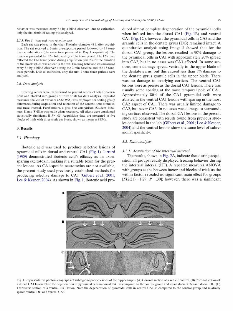
Ibotenic acid was used to produce selective lesions ofpyramidal cells in dorsal and ventral CA1 (Fig. 1). Jarrard(1989) demonstrated ibotenic acid’s eYcacy as an axon-sparing excitotoxin, making it a suitable toxin for the pres-ent lesions. As CA1-speciWc neurotoxins are not available,the present study used previously established methods forproducing selective damage to CA1 (Gilbert et al., 2001;Lee & Kesner, 2004). As shown in Fig. 1, ibotenic acid pro-
duced almost complete degeneration of the pyramidal cellswhen infused into the dorsal CA1 (Fig. 1B) and ventralCA1 (Fig. 1C); however, the pyramidal cells in CA3 and thegranule cells in the dentate gyrus (DG) remained intact. Aquantitative analysis using Image J showed that for thedorsal CA1 group, the lesions resulted in 90% damage tothe pyramidal cells in CA1 with approximately 20% spreadinto CA2, but in no cases was CA3 aVected. In some sec-tions, some damage spread ventrally to the upper blade ofthe dentate gyrus, but this caused less than 5% damage tothe dentate gyrus granule cells in the upper blade. Therewas no damage to overlying cortices. The ventral CA1lesions were as precise as the dorsal CA1 lesions. There wasusually some sparing at the most temporal pole of CA1.Approximately 80% of the CA1 pyramidal cells wereablated in the ventral CA1 lesions with sparing in the mostCA2 aspect of CA1. There was usually limited damage toCA2, but never CA3. In no cases was damage to surround-ing cortices observed. The dorsal CA1 lesions in the presentstudy are consistent with results found from previous stud-ies conducted in the lab (Gilbert et al., 2001; Lee & Kesner,2004) and the ventral lesions show the same level of subre-gional speciWcity.
3.2. Data analysis
3.2.1. Acquisition of the intertrial intervalThe results, shown in Fig. 2A, indicate that during acqui-
sition all groups readily displayed freezing behavior duringthe intertrial interval (ITI). A repeated measures ANOVAwith groups as the between factor and blocks of trials as thewithin factor revealed no signiWcant main eVect for groups[F (2,21)D 1.29; PD .30]; however, there was a signiWcant
Fig. 1. Representative photomicrographs of subregion-speciWc lesions of the hippocampus. (A) Coronal section of a vehicle control. (B) Coronal section ofa dorsal CA1 lesion. Note the degeneration of pyramidal cells in dorsal CA1 as compared to the control group and intact dorsal CA3 and dorsal DG. (C)Transverse section of a ventral CA1 lesion. Note the degeneration of pyramidal cells in ventral CA1 as compared to the control group and relativelyspared ventral DG and ventral CA3.

76 J.L. Rogers et al. / Neurobiology of Learning and Memory 86 (2006) 72–81
Fig. 2. Percent freezing during acquisition. (A) Context following the pre-acquisition baseline period (PRE) on Day 1. Data are presented as blocksof trials where each block represents three trials. Note that there are nosigniWcant diVerences among the groups. (B) Trace interval following thepre-acquisition baseline period (PRE) on Day 1. Data are presented asblocks of trials where each block represents three trials. Note that thereare no signiWcant diVerences among the groups. (C) Tone stimulus follow-ing the pre-acquisition baseline period (PRE) on Day 1. Data are pre-sented as blocks of trials where each block represents three trials. Notethat there are no signiWcant diVerences among the groups.
main eVect for blocks of trials [F (4,84)D3.06; PD .02], sug-gesting that all rats learned the task. There were no signiW-cant interactions between the groups and trials during ITIacquisition [F (8,84)D0.47; PD .87].
3.2.2. Acquisition of the trace intervalThe results, shown in Fig. 2B, indicate that during acqui-
sition all groups readily displayed freezing behavior duringthe trace interval. A repeated measures ANOVA withgroups as the between factor and blocks of trials as thewithin factor revealed no signiWcant main eVect for groups[F (2,21)D 3.11; PD0.06]. Since the P value approached sig-niWcance and the trace acquisition graph suggested a possi-ble dissociation between lesion groups that may have beenmissed by the ANOVA, a SNK post hoc was performed.Neither dCA1 nor vCA1 were signiWcantly diVerent fromcontrols (P > .05). There was a signiWcant main eVect forblocks of trials [F (4,84)D9.31; P < .0001], suggesting thatall rats learned the task. There were no signiWcant interac-tions between the groups and trials during acquisition ofthe trace interval [F (8,84)D 1.18; PD .32].
3.2.3. Acquisition of the tone stimulusThe results, shown in Fig. 2C, indicate that during acqui-
sition all groups readily displayed freezing behavior duringthe tone stimulus. A repeated measures ANOVA withgroups as the between factor and blocks of trials as thewithin factor revealed no signiWcant main eVect for groups[F (2,21)D 1.38; PD .27]; there was a signiWcant main eVectfor blocks of trials [F(4,84)D 11.59; P < .0001], suggestingthat all rats learned the task. There were no signiWcantinteractions between the groups and trials during acquisi-tion of the tone stimulus [F (8,84)D 1.75; PD .10].
3.2.4. Contextual retentionAlthough the animals were tested on context retention
for 8 min, only 6 min was analyzed and divided into threeblocks of 2 min trials, as the control group began to extin-guish to the context after 6 min. The results, shown inFig. 3A, indicate that during retention of the context theventral CA1 group froze signiWcantly less than either thedorsal CA1 group or control group, whereas the dorsalCA1 group froze signiWcantly less than the control group.A repeated measures ANOVA conducted on the contexttest administered 24 h post-training with groups as thebetween factor and blocks of trials as the within factorrevealed a signiWcant main eVect for groups[F (2,21)D 13.56; P < .0006]. Post hoc analysis (SNK) indi-cated that the ventral CA1 lesion group froze signiWcantlyless than either the control (P < .01) or dorsal CA1 groups(P < .05), whereas the dorsal CA1 group froze signiWcantlyless than the control group (P < .05). This is suggestive of agraded involvement in contextual retention between thedorsal and ventral aspects of CA1. There was no main eVectfor blocks of trials [F (2,42)D2.08; PD .14], nor was therean interaction between groups and blocks of trials[F (4,42)D 0.60; PD .66].

J.L. Rogers et al. / Neurobiology of Learning and Memory 86 (2006) 72–81 77
Fig. 3. Percent freezing during retention tests given post-conditioning. (A)Contextual retention tested 24 h post-conditioning. Data are presented asa function of blocks of trials where each block represent a 2-min timeperiod. Note that the vCA1 group froze signiWcantly less than either con-trol or dCA1 groups, whereas the dCA1 group froze signiWcantly less thanthe control group. (B) Trace retention test given following pre-retentionbaseline period (PRE) 48 h post-conditioning. Data are presented asblocks of trials where each block represents three trials. Note that thevCA1 group froze signiWcantly less than either dCA1 group or controlgroup, whereas the dCA1 group was not signiWcantly diVerent from thecontrol group. (C) Tone retention test given following pre-retention base-line period (PRE) 48 h post-conditioning. Data are presented as blocks oftrials where each block represents three trials. Note that there are no sig-niWcant diVerences between groups.
3.2.5. Trace interval retentionThe results, shown in Fig. 3B, indicate that during reten-
tion the ventral CA1 group froze signiWcantly less than thecontrols, whereas the dorsal CA1 group showed no impair-ment during the trace test administered 48 h post-training.A repeated measures ANOVA with groups as the betweenfactor and blocks of trials as the within factor revealed asigniWcant main eVect for groups during retention of thetrace interval [F (2,21)D 7.07; PD .005]. Post hoc analysis(Student–Newman–Keuls) indicated that the ventral CA1lesion group froze signiWcantly less than the control group(P < .01) as well as signiWcantly less compared to the dCA1group (P < .05). Dorsal CA1 lesion animals did not diVerfrom controls (P > .05). The vCA1 appears to be criticallyinvolved in subserving the trace component of fear condi-tioning, whereas dCA1 seems to play only a minor role.There was no main eVect for blocks of trials during reten-tion of the trace period [F (2,42)D0.81; PD .45], nor wasthere an interaction between the groups and blocks of trials[F (4, 42)D0.62; PD .58].
3.2.6. Tone stimulus retentionThe results, shown in Fig. 3C, indicate that during reten-
tion all groups readily displayed freezing behavior duringthe tone test administered 48 h post-training. A repeatedmeasures ANOVA with groups as the between factor andblocks of trials as the within factor revealed no signiWcantmain eVect for groups during retention of the tone stimulus[F (2,21)D 2.96; PD .08]. Since the P value approached sig-niWcance, an SNK post hoc analysis was performed on thedata. No signiWcant diVerences between groups wasrevealed (P > .05). Furthermore, there was no main eVectfor blocks of trials during retention of the tone period [F (2,42)D0.56; PD .57], nor was there an interaction betweenthe groups and blocks of trials [F (4,42)D0.70; PD .59].
4. Discussion
The present study sought to investigate potential dissoci-ations between the roles of the dorsal and ventral compo-nents of CA1 during trace fear conditioning. It washypothesized that lesions of the dorsal CA1 (dCA1) wouldproduce a deWcit in contextual conditioning; however, itwas unclear whether or not ventral CA1 (vCA1) lesionswould attenuate contextual conditioning. In addition, itwas unclear whether either lesion would impair acquisition,retention, or both. Furthermore, it was hypothesized thatventral CA1 lesions would attenuate trace conditioning,but the extent to which ventral CA1 lesions would impairfreezing to the tone was unknown. The present paradigmemployed a 10-s trace interval to separate the tone stimulus(CS) from the foot-shock (US). In addition, rats were givena 2-min pre-exposure period, critical for encoding contex-tual information (Fanselow, 1990).
Surprisingly, acquisition of conditioned fear was unin-terrupted by lesions of the dorsal or ventral CA1. Animalsreadily froze to the context during the intertrial interval
78 J.L. Rogers et al. / Neurobiology of Learning and Memory 86 (2006) 72–81
(ITI) (Fig. 2A), trace interval (Fig. 2B), or tone stimulus(Fig. 2C). Although it appears that there was some retarda-tion of freezing during acquisition for the vCA1 lesiongroup, this eVect was not statistically signiWcant and wasdeemed unreliable. The data also indicate that both dCA1and vCA1 lesions signiWcantly reduced freezing during thecontext test administered 24 h post-conditioning (Fig. 3A);however, the vCA1 lesion group froze signiWcantly less thaneither control or dCA1 lesion groups. Freezing behavior ofthe vCA1 lesion group was all but abolished when returnedto the conditioning chamber for the context test. The pat-tern of the data is suggestive of a gradient in function alongthe dorso-ventral axis of the hippocampus. The dCA1showed a deWcit when compared to controls, but vCA1showed a deWcit when compared to dCA1 and a larger deW-cit when compared to controls. This means that dCA1 is infact involved in contextual fear recollection, but vCA1appears to be more important.
Similarly, the vCA1 lesion group froze signiWcantly lessthan either control or dCA1 lesion group during the tracetest administered 48 h post-conditioning (Fig. 3B). ThedCA1 group showed a mild attenuation of freezing duringthe trace test; however, this was not reliable. The dCA1group was not signiWcantly diVerent than controls, but thedata indicate a marginal eVect. Finally, no signiWcant groupdiVerences were observed during the tone test also adminis-tered 48 h post-conditioning (Fig. 3C). The lesion groupsshowed attenuation of freezing, but this eVect was not reli-able. Thus, the Wndings indicate no diVerences among thegroups during acquisition of trace fear conditioning, orretention of the tone (CS). The vCA1 lesion group dis-played signiWcantly reduced freezing during the context testas well as during the trace test, whereas the dCA1 lesionedgroup displayed signiWcantly reduced freezing only duringthe context test and a small, insigniWcant attenuation offreezing to the trace interval. One caveat is that all animalsreceived the context test before the tone-trace test.Although there were no overlapping stimuli, future studiesmay be needed to determine if any order eVects exist.
It should be noted that in the present experiment theventral CA1 lesion animals did not display hyperactivity. Aclose look at the acquisition data reveal the ventral CA1animals were able to acquire the trace fear conditioningtask as readily as the dCA1 and control groups. The vCA1group was able to display freezing behavior during theacquisition phase. Also, the tone test data suggest the ven-tral CA1 animals, although slightly impaired, were able tofreeze to the discrete CS 48 h post-acquisition; thus it isargued that hyperexcitability or hyperactivity were not fac-tors in the deWcits displayed by the ventral CA1 lesion ani-mals. Also, during the 2-min baseline period prior toacquisition as well as prior to the trace and tone test thevCA1 animals did not show any excessive running, rearing,jumping, or other ambulatory behaviors compared to anyother group.
The reduced freezing during the context test for thedCA1 group is consistent with previous Wndings with dor-
sal hippocampal lesions (Kim & Fanselow, 1992; Marenet al., 1997), as well as dCA1 lesions (Lee & Kesner, 2004),during delay conditioning. The reduced freezing during thecontext test for the vCA1 group is consistent with previousWndings from Bast, Zhang, and Feldon (2001), who showedthat temporary inactivation of the ventral hippocampus viatetrodotoxin (TTX) or the GABAA agonist muscimolattenuated freezing when animals were returned to the con-ditioned fear chamber. Rudy and Matus-Amat (2005)found that muscimol inactivation of the ventral hippocam-pus prior to conditioning and anisomycin infusions into theventral hippocampus prior to a retention test after 48 hpost-acquisition signiWcantly reduced rats’ freezing. Thus,the present Wndings are clearly consistent with these Wnd-ings, suggesting that the hippocampus may be necessary forcontextual associations during conditioned fear. The pres-ent data indicate that for conditioned contextual fear dCA1is involved in retention but vCA1 appears to be even moreimportant in the retention of conditioned fear.
Given that mice lacking NMDA receptors in the CA1subregion (dorsal plus ventral) failed to learn a 30-s tracefear conditioning paradigm (Huerta et al., 2000) and CA1neurons encoded trace duration during a 10- and 20-s traceconditioning paradigm in rabbits (McEchron et al., 2003),one might expect in the present study that acquisitionwould be retarded by the CA1 lesion groups. Although thelesions were present during acquisition, lesions of the CA1subregion, either dorsal or ventral, seemed to have littleeVect on acquisition but did have an eVect on the retentiontests conducted 24 and 48 h post-conditioning. These datawould suggest that the CA1 subregion of the hippocampusis not necessary for the acquisition of contextual or tempo-ral information. One potential explanation might be thatother subregions (i.e., DG or CA3, or some combination) ofthe hippocampus may play an important role in the acqui-sition of trace fear conditioning. For example, a recentstudy by Fendt, Koch, and Fanselow (2005) found thatNMDA lesions of the dorsal hippocampus signiWcantlyimpaired acquisition of trace, but not classical, fear condi-tioning when measured using fear potentiated startle.Quinn et al. (2005) found that infusions of the NMDAantagonist APV into the dorsal hippocampus prior to traceconditioning, but not the test, signiWcantly reduced freezingduring the context test. Furthermore, infusions of APV intothe dorsal hippocampus either prior to trace conditioningor prior to the test signiWcantly reduced freezing during thetrace interval during the tone test. Both of these studieswere aimed at the dorsal hippocampus, whereas the presentstudy speciWcally investigated the CA1 subregion in boththe dorsal and ventral components of the hippocampus. Itis conceivable, therefore, that the entire hippocampus, orperhaps the CA3 subregion in conjunction with CA1, isnecessary for the acquisition of trace fear conditioning (seeKesner, 2005).
Another hypothesis behind the present study was thatlesions of the CA1 would impair trace conditioning. Thepresent Wndings oVer some support for this hypothesis;
J.L. Rogers et al. / Neurobiology of Learning and Memory 86 (2006) 72–81 79
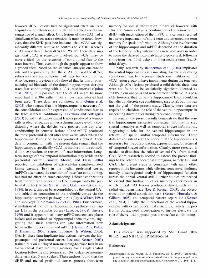
however dCA1 lesions had no signiWcant eVect on traceacquisition or retention, although the graphed results aresuggestive of a small eVect. Only lesions of the vCA1 had asigniWcant eVect on trace retention. It must be noted, how-ever, that the SNK post hoc indicated that vCA1 was sig-niWcantly diVerent relative to controls to P > .01, whereasvCA1 was diVerent from dCA1 to P > .05. These data sug-gest that dCA1 is somehow involved, but vCA1 may bemore critical for the retention of conditioned fear to thetrace interval. Thus, even though the graphs appear to showa graded eVect, based on the statistical analysis one cannotrule out the possibility that the vCA1, but not the dCA1,subserves the trace component of trace fear conditioning.Also, because a previous study showed that lesions or phar-macological blockade of the dorsal hippocampus disruptstrace fear conditioning with a 30-s trace interval (Quinnet al., 2005), it is possible that the dCA1 might be moreimportant if a 30-s rather than a 10-s trace interval hadbeen used. These data are consistent with Quinn et al.(2002) who suggest that the hippocampus is necessary forthe consolidation and/or expression of conditioned fear tothe trace interval. Additionally, Takehara and colleagues(2003) found that hippocampal lesions produced a tempo-rally graded retrograde amnesia during trace eye-blink con-ditioning where the deWcits were greatest one day afterconditioning. In contrast, lesions of the mPFC producedthe most profound deWcit after four weeks, after which thehippocampal lesions no longer produced a deWcit. Thesedata in conjunction with the present data suggest that thehippocampus, speciWcally vCA1, is involved in the consoli-dation, expression, or retrieval of trace information. Long-term storage of this temporal information may reside in theprefrontal cortex. Runyan, Moore, and Dash (2004)reported that inhibition of extracellular signal-regulatedkinase cascade (Erk) in the medial prefrontal cortex(mPFC) attenuated the retention of trace fear conditioning,but had no eVect on trace encoding. EVerent connectionsfrom the ventral hippocampus CA1 synapse onto the pre-frontal cortex (Barbas & Blatt, 1995; Goldman-Rakic et al.,1984). In part, this can be accomplished by the ventral CA1and subiculum connection to the prelimbic cortex via thehippocampo-temporal pathway in rats (Jay & Witter, 1991)and monkeys (Goldman-Rakic et al., 1984). Furthermore,stimulation of the ventral hippocampus pathway can trig-ger LTP in the prelimbic cortex (Laroche, Jay, & Thierry,1990) and it appears that many mPFC neurons are phaselocked and entrained to hippocampal theta rhythm, sug-gesting that these neurons can gate information Xowbetween the hippocampus and mPFC (Hyman, Zilli, Paley,& Hasselmo, 2005; Siapis, Lubenov, & Wilson, 2005).Clearly, these data implicate interactions between the hip-pocampus and prefrontal cortex. Lee and Kesner (2003)trained rats on a delayed non-matching-to-place task in an8-arm radial maze requiring memory for a single spatiallocation following short-term (i.e., 10-s) delays or interme-diate-term (i.e., 5-min) delays. These authors found that thedHIP and medial prefrontal cortex process short-term
memory for spatial information in parallel; however, with10-s and 5-min delays a combination of a lesion of thedHIP with inactivation of the mPFC or vice versa resultedin a severe impairment of short-term and intermediate-termmemory for spatial information. Although the involvementof the hippocampus and mPFC depended on the durationof the temporal delay, interactions were necessary in orderto solve the delayed non-matching-to-place task followingshort-term (i.e., 10-s) delays or intermediate-term (i.e., 5-min) delays.
Finally, research by Bannerman et al. (2004) implicatesthe ventral hippocampus in associating discrete cues duringconditioned fear. In the present study, one might expect thevCA1 lesion group to have impairments during the tone test.Although vCA1 lesions produced a mild deWcit, these datawere not found to be statistically signiWcant (deWned asP < .05 in our analysis) and were deemed unreliable. It is pos-sible, however, that full ventral hippocampal lesions could, infact, disrupt discrete cue conditioning (i.e., tone), but this wasnot the goal of the present study. Clearly, more data arerequired to elucidate the role of the ventral hippocampus inassociating discrete cues during trace conditioning.
In general, the present results demonstrate that the ven-tral hippocampus processes contextual information (i.e.,spatial memory) as well as temporal (trace) information,suggesting a role for the ventral hippocampus in theretrieval of spatial and/or temporal information. Thesedata are consistent with the notion that the hippocampus isnecessary for the consolidation, expression, and/or retrievalof temporal (trace) information. Clearly, more research isneeded to dissociate the roles of ventral CA1 from dorsalCA1. More research is needed to extend the present Wnd-ings to the other hippocampal subregions, namely DG andCA3. The present study is consistent with many otherreports in the literature, while oVering some novel Wndings:namely a subregional analysis of hippocampal functionacross the dorsal–ventral axis. Further studies are neededto extend this Wnding to other memory experiments inwhich dorsal CA1 lesions produce a deWcit, such as theradial eight-arm maze (Lee & Kesner, 2003), the object-trace-odor paired-association task (Kesner, Hunsaker, &Gilbert, 2005), and temporal pattern separation (Kesneret al., 2004). Finally, the interactions of the ventral hippo-campus with extrahippocampal structures, namely the pre-frontal cortex, need investigation to further elucidate therole of the ventral hippocampus in trace fear conditioning.
Acknowledgments
This research was supported by NSF Grant IBN-0135273 and NIH Grant R1MH065314.
References
Anagnostaras, S. G., Maren, S., & Fanselow, M. S. (1999). Temporallygraded retrograde amnesia of contextual fear after hippocampal dam-age in rats: within-subjects examination. Neuroscience, 19, 1106–1114.

80 J.L. Rogers et al. / Neurobiology of Learning and Memory 86 (2006) 72–81
Bannerman, D. M., Yee, B. K., Good, M. A., Heupel, M. J., Iversen, S. D.,& Rawlins, J. N. P. (1999). Double dissociation of function within thehippocampus: A comparison of dorsal, ventral, and complete hippo-campal cytotoxic lesions. Behavioral Neuroscience, 113, 1170–1188.
Bannerman, D. M., Rawlins, J. N. P., McHugh, S. B., Deacon, R. M. J.,Yee, B. K., Bast, T., et al. (2004). Regional dissociations within the hip-pocampus—memory and anxiety. Neuroscience and BiobehavioralReviews, 28, 273–283.
Barbas, H., & Blatt, G. J. (1995). Topographically speciWc hippocampalprojections target functionally distinct prefrontal areas in the rhesusmonkey. Hippocampus, 5, 511–533.
Bast, T., Zhang, W., & Feldon, J. (2001). The ventral hippocampus andfear conditioning in rats. Experimental Brain Research, 139, 39–52.
Buchel, C., Dolan, R. J., Armony, J. L., & Friston, K. J. (1999). Amygdala-hippocampal involvement in human aversive trace conditioningrevealed through event-related functional magnetic resonance imaging.Neuroscience, 19, 10869–10876.
Burman, M. A., Starr, M. J., & Gewirtz, J. C. (2006). Dissociable eVects ofhippocampus lesions on expression of fear and trace fear conditioningmemories in rats. Hippocampus, 16, 103–113.
Fanselow, M. S. (1990). Factors governing one trial contextual condition-ing. Animal Learning and Behavior, 18, 264–270.
Fendt, M., Koch, M., & Fanselow, M. S. (2005). Lesions of the dorsal hip-pocampus block trace fear conditioned potentiated startle. BehavioralNeuroscience, 119, 834–838.
Gilbert, P. E., Kesner, R. P., & Lee, I. (2001). Dissociating hippocampalsubregions: A double dissociation between dentate gyrus and CA1.Hippocampus, 11, 626–636.
Goldman-Rakic, P. S., Selemon, L. D., & Schwartz, M. L. (1984). Dualpathways connecting the dorsolateral prefrontal cortex with the hippo-campal formation and parahippocampal gyrus in the rhesus monkey.Neuroscience, 12, 719–742.
Henke, P. G. (1990). Hippocampal pathway to the amygdala and stressulcer development. Brain Research Bulletin, 25, 691–695.
Huerta, P. T., Sun, L. D., Wilson, M. A., & Tonegawa, S. (2000). Formationof temporal memory requires NMDA receptors within CA1 pyramidalneurons. Neuron, 25, 473–480.
Hyman, J. M., Zilli, E. A., Paley, A. M., & Hasselmo, M. E. (2005). Medialprefrontal cortex cells show dynamic modulation with the hippocam-pal theta rhythm dependent on behavior. Hippocampus, 15, 739–749.
Jarrard, L. E. (1989). On the use of ibotenic acid to lesion selectively diVer-ent components of the hippocampal formation. Neuroscience Methods,29, 251–259.
Jay, T. M., & Witter, M. P. (1991). Distribution of hippocampal CA1 andsubicular eVerents in the prefrontal cortex of the rat studied by meansof anterograde transport of Phaseolus vulgaris-leucoagglutinin. Journalof Comparative Neurology, 313, 574–586.
Kesner, R. P., & Rogers, J. L. (2004). An analysis of independence andinteractions of brain substrates that subserve multiple attributes, mem-ory systems, and underlying processes. Neurobiology of Learning andMemory, 82, 199–215.
Kesner, R. P., Lee, I., & Gilbert, P. E. (2004). A behavioral assessment ofhippocampal function based on a subregional analysis. Reviews in Neu-roscience, 15, 333–351.
Kesner, R. P., Hunsaker, M. R., & Gilbert, P. E. (2005). The role of CA1 inthe acquisition of an object-trace-odor paired associate task. Behav-ioral Neuroscience, 119, 781–786.
Kesner, R. P. (1998). Neural mediation of memory for time: Role of thehippocampus and medial prefrontal cortex. Psychonomic BulletinReview, 5, 585–596.
Kesner, R. P. (2005). Temporal processing of information: The role of themedial prefrontal cortex and hippocampus: Theoretical comment onGilmartin and McEchron. Behavioral Neuroscience, 119, 1705–1709.
Kim, J. J., & Fanselow, M. S. (1992). Modality speciWc retrograde amnesiaof fear following hippocampal lesions. Science, 256, 675–677.
Kim, J. J., Rison, R. A., & Fanselow, M. S. (1995). EVects of amygdala, hip-pocampus, and periaqueductal gray lesions on short- and long-termcontextual fear. Behavioral Neuroscience, 107, 1093–1098.
Kjelstrup, K. G., Tuvnes, F. A., SteVenach, H., Murison, R., Moser, E. I., &Moser, M. (2002). Reduced fear expression after lesions of the ventralhippocampus. Proceedings of the National Academy of Sciences UnitedStates of America, 99, 10825–10830.
Knight, D. C., Cheng, D. T., Smith, C. N., Stein, E. A., & Helmstetter, F. J.(2004). Neural substrates mediating human delay and trace fear condi-tioning. Neuroscience, 24, 218–228.
Laroche, S., Jay, T. M., & Thierry, A. M. (1990). Long-term potentiation inthe prefrontal cortex following stimulation of the hippocampal CA1/subicular region. Neuroscience Letters, 114, 184–190.
Lee, I., & Kesner, R. P. (2003). Time-dependent relationship between thedorsal hippocampus and the prefrontal cortex in spatial memory. Jour-nal of Neuroscience, 23, 1517–1523.
Lee, I., & Kesner, R. P. (2004). DiVerential contributions of dorsal hippo-campal subregions to memory acquisition and retrieval in contextualfear-conditioning. Hippocampus, 14, 1–10.
Maren, S., & Holt, W. (2000). The hippocampus and contextual memoryretrieval in Pavlovian conditioning. Behavioral Brain Research, 110,97–108.
Maren, S., Aharonov, G., & Fanselow, M. S. (1997). Neurotoxic lesions ofthe dorsal hippocampus and Pavlovian fear conditioning in rats.Behavioral Brain Research, 88, 261–274.
McEchron, M. D., Tseng, W., & Disterhoft, J. F. (2003). Single neurons inCA1 hippocampus encode trace interval duration during trace heart rate(fear) conditioning in rabbit. Journal of Neuroscience, 23, 1535–1547.
McEchron, M. D., & Disterhoft, J. F. (1999). Hippocampal encoding ofnon-spatial trace conditioning. Hippocampus, 9, 385–396.
McEchron, M. D., Bouwmeester, H., Tseng, W., Weiss, C., & Disterhoft, J.F. (1998). Hippocampectomy disrupts auditory trace fear conditioningand contextual fear conditioning in the rat. Hippocampus, 8, 638–646.
Misane, I., Tovote, P., Meyer, M., Spiess, J., Ogren, S. O., & Stiedl, O.(2005). Time-dependent involvement of the dorsal hippocampus intrace fear conditioning in mice. Hippocampus, 15, 418–426.
Moyer, J. R., Jr., Deyo, R. A., & Disterhoft, J. F. (1990). Hippocampec-tomy disrupts trace eye-blink conditioning in rabbits. Behavioral Neu-roscience, 104, 243–252.
Moser, M. B., & Moser, E. I. (1998). Functional diVerentiation in the hip-pocampus. Hippocampus, 8, 608–619.
Paxinos, G., & Watson, C. (1997). The rat brain in stereotaxic coordinates.NY: New York: Academy of Sciences.
Pitkanen, A., Pikkarainen, M., Nurminen, N., & Ylinen, A. (2000). Recip-rocal connections between the amygdala and the hippocampal forma-tion, perirhinal cortex, and postrhinal cortex in rat. A review. Annals ofthe New York Academy of Sciences, 911, 369–391.
Quinn, J. J., Oommen, S. S., Morrison, G. E., & Fanselow, M. S. (2002).Post-training excitotoxic lesions of the dorsal hippocampus attenuateforward trace, backward trace, and delay fear conditioning in a tempo-rally speciWc manner. Hippocampus, 12, 495–504.
Quinn, J. J., Loya, F., Quang, D. M., & Fanselow, M. S. (2005). Dorsal hip-pocampus NMDA receptors diVerentially mediate trace and contex-tual fear conditioning. Hippocampus, 15, 665–674.
Rolls, E. T. (1996). A theory of hippocampal function in memory. Hippo-campus, 6, 601–620.
Rudy, J. W., & Matus-Amat, P. (2005). The ventral hippocampus supportsa memory representation of context and contextual fear conditioning:Implications for a unitary function of the hippocampus. BehavioralNeuroscience, 119, 154–163.
Runyan, J. D., Moore, A. N., & Dash, P. K. (2004). A role for prefrontalcortex in memory storage for trace fear conditioning. Neuroscience, 24,1288–1295.
Sakamoto, T., Takatsuki, K., Kawahara, S., Kirino, Y., Niki, H., & Mish-ina, M. (2005). Role of the hippocampal NMDA receptors in trace eye-blink conditioning. Brain Research, 1039, 130–136.
Siapis, A. G., Lubenov, E. V., & Wilson, M. A. (2005). Prefrontal phaselocking to hippocampal theta oscillations. Neuron, 46, 141–151.
Siegal, A., & Tassoni, J. P. (1971). DiVerential eVerent projections from theventral and dorsal hippocampus of the cat. Brain Behavior and Evolu-tion, 4, 185–200.

J.L. Rogers et al. / Neurobiology of Learning and Memory 86 (2006) 72–81 81
SteVenach, H.-A., Witter, B., Moser, M.-B., & Moser, E. I. (2005). Spatialmemory in the rat requires the dorsolateral band of the entorhinal cor-tex. Neuron, 45, 301–313.
Swanson, L. W., & Cowan, W. M. (1977). An autoradiographic study ofthe organization of the eVerent connections of the hippocampal forma-tion in the rat. Journal of Comparative Neurology, 172, 49–84.
Takehara, K., Kawahara, S., & Kirino, Y. (2003). Time-dependent reorga-nization of the brain components underlying memory retention intrace eyeblink conditioning. Neuroscience, 23, 9897–9905.
Trivedi, M. A., & Coover, G. D. (2004). Lesions of the ventral hippocam-pus, but not the dorsal hippocampus, impair conditioned fear expres-sion and inhibitory avoidance on the elevated T-maze. Neurobiology ofLearning and Memory, 81, 172–184.
Tseng, W., Guan, R., Disterhoft, J. F., & Weiss, C. (2004). Trace eyeblink con-ditioning is hippocampally dependent in mice. Hippocampus, 14, 58–65.
Weiss, C., Bouwmeester, H., Power, J. M., & Disterhoft, J. F. (1999). Hip-pocampal lesions prevent trace eyeblink conditioning in the freelymoving rat. Behavioral Brain Research, 99, 123–132.
Copyright © 2022 FDOKUMEN