Open Repair of Ventral Incisional Hernias
23
Open Repair of Ventral Incisional Hernias Dan H. Shell IV, MD a , Jorge de la Torre, MD a, * , Patricio Andrades, MD a,b , Luis O. Vasconez, MD a a Division of Plastic Surgery, University of Alabama at Birmingham, 510 20th Street S, Birmingham, AL 35294-3411, USA b Division of Transplant Immunology, University of Alabama at Birmingham, 510 20th Street S, Birmingham, AL 35294-3411, USA Incisional hernia is a common and often debilitating complication after laparotomy. Despite significant advances in many areas of surgery, correc- tion of incisional hernias continues to be problematic, with recurrence rates ranging from 5% to 63% depending on the type of repair used [1–8]. Recur- rence rates are likely underestimated because of a lack of long-term follow- up and objective criteria in the literature to determine true recurrence. More than 2 million laparotomies are performed annually in the United States, with a reported 2% to 11% incidence of incisional hernia [1,5,9–11]. It is the most common complication after laparotomy by a 2:1 ratio over bowel obstruction and is the most common indication for reoperation by a 3:1 ratio over adhesive small bowel obstruction [12]. Approximately 100,000 hernia repairs are performed annually in the United States [13]. The associated morbidity secondary to incarceration, strangulation, and bowel obstruction is significant. In a retrospective review of 206 patients who underwent incisional hernia repair, Read and Yoder [9,10] found that strangulation or incarceration was the indication for repair in 17% of patients. The gradual enlargement of the hernia over time results in a rel- ative loss of abdominal domain, with adverse effects on postural mainte- nance, respiration, micturition, defecation, and biomechanical properties, which have a profound impact on patients’ overall physical capacity and quality of life. As patients are forced to alter their lifestyle, their ability to work becomes impaired, which has negative economic consequences. Pro- gressive enlargement of the hernia also results in a cosmetic deformity, which is detrimental to patients’ self-esteem. * Corresponding author. E-mail address: [email protected] (J. de la Torre). 0039-6109/08/$ - see front matter Ó 2008 Elsevier Inc. All rights reserved. doi:10.1016/j.suc.2007.10.008 surgical.theclinics.com Surg Clin N Am 88 (2008) 61–83
-
Upload
independent -
Category
Documents
-
view
1 -
download
0
Transcript of Open Repair of Ventral Incisional Hernias
Open Repair of Ventral IncisionalHernias
Dan H. Shell IV, MDa, Jorge de la Torre, MDa,*,Patricio Andrades, MDa,b, Luis O. Vasconez, MDa
aDivision of Plastic Surgery, University of Alabama at Birmingham,
510 20th Street S, Birmingham, AL 35294-3411, USAbDivision of Transplant Immunology, University of Alabama at Birmingham,
510 20th Street S, Birmingham, AL 35294-3411, USA
Incisional hernia is a common and often debilitating complication afterlaparotomy. Despite significant advances in many areas of surgery, correc-tion of incisional hernias continues to be problematic, with recurrence ratesranging from 5% to 63% depending on the type of repair used [1–8]. Recur-rence rates are likely underestimated because of a lack of long-term follow-up and objective criteria in the literature to determine true recurrence.
More than 2 million laparotomies are performed annually in the UnitedStates, with a reported 2% to 11% incidence of incisional hernia [1,5,9–11].It is the most common complication after laparotomy by a 2:1 ratio overbowel obstruction and is the most common indication for reoperation bya 3:1 ratio over adhesive small bowel obstruction [12]. Approximately100,000 hernia repairs are performed annually in the United States [13].The associated morbidity secondary to incarceration, strangulation, andbowel obstruction is significant. In a retrospective review of 206 patientswho underwent incisional hernia repair, Read and Yoder [9,10] foundthat strangulation or incarceration was the indication for repair in 17%of patients. The gradual enlargement of the hernia over time results in a rel-ative loss of abdominal domain, with adverse effects on postural mainte-nance, respiration, micturition, defecation, and biomechanical properties,which have a profound impact on patients’ overall physical capacity andquality of life. As patients are forced to alter their lifestyle, their ability towork becomes impaired, which has negative economic consequences. Pro-gressive enlargement of the hernia also results in a cosmetic deformity,which is detrimental to patients’ self-esteem.
Surg Clin N Am 88 (2008) 61–83
* Corresponding author.
E-mail address: [email protected] (J. de la Torre).
0039-6109/08/$ - see front matter � 2008 Elsevier Inc. All rights reserved.
doi:10.1016/j.suc.2007.10.008 surgical.theclinics.com
62 SHELL et al
Incisional hernias are the only abdominal hernias that are iatrogenic [10].Controversy exists regarding the ideal treatment of incisional hernias. No-where in surgery does the phrase ‘‘if there are multiple ways of fixing a prob-lem then there is not one good way’’ hold true more so than with incisionalhernia repair. The approach to incisional hernia repair is often based on tra-dition rather than evidence. Several important contributions to the literaturein recent years have helped our understanding of the causes of incisionalhernia formation and the important physiologic and functional propertiesof the abdominal wall. An appreciation for the dynamic function of the ab-dominal wall has led to technical refinements and the recognition of impor-tant principles that are necessary for successful repair.
Etiology
Many patient-related risk factors have been implicated in the develop-ment of incisional hernias, including obesity, smoking, aneurismal disease,chronic obstructive pulmonary disease, male gender, malnourishment,corticosteroid dependency, renal failure, malignancy, and prostatism[1–8,10,11,14–23]. Many of these risk factors may contribute to the develop-ment of an incisional hernia, but no single factor is so regularly associatedthat it may be declared as serving a truly etiologic role [22].
In a study by Condon and colleagues [21] of complications associatedwith closure of 1000 midline laparotomies, no single factor was associatedwith incisional hernia on univariate analysis. On multivariate analysis,only the combination of reopening and reclosing previous incisions coupledwith wound infection influenced the development of incisional hernia. Post-operative wound infection has been found in additional studies to be the sin-gle most significant prognostic factor in the development of incisional hernia[1,7,10,14,19,21]. Bucknall and colleagues [1] reported a 23% incidence ofincisional hernia formation in patients who developed a wound infection.
Obesity often has been cited as a risk factor, with an incisional hernia rateof 15% to 20% [24–26]. In a prospective, randomized evaluation that com-pared fascial closure techniques, Brolin [24] found a reduction in incisionalhernia occurrence from 18% to 10% with the use of double-stranded #1PDS placed in continuous fashion compared with #1 Ethibond placed in in-terrupted figure-of-eight fashion. Aneurysmal disease also has been found inmultiple studies to be an independent risk factor in the development of inci-sional hernias [22,27–29]. A recent multicenter, prospective study by Rafettoand colleagues [22] found a 28.2% incidence of incisional hernia formationin patients undergoing surgery for aortic aneurysm repair. After correctingfor other risk factors, this figure represents a ninefold increase in the inci-dence of incisional hernia formation compared with surgery for aortic occlu-sive disease. It has been suggested that a defect in collagen metabolism witha decreased ratio of type I to type III procollagen may play a role; however,
63OPEN REPAIR OF VENTRAL INCISIONAL HERNIAS
further studies are needed before a causal relationship can be established[22,30].
Incisional hernias differ from other abdominal wall hernias in their iatro-genic origin. Surgeon-related technical errors are responsible for most inci-sional hernias. Closure under tension results in fascial strangulation andhernia formation. Studies have shown that 50% of hernia recurrences aredetected in the first postoperative year, 75% are detected at 2 years, and90% are detected at 3 years, with continued failure rates of 2% per yearthereafter [2,5,6,11]. These findings implicate technical factors in earlywound failure and patient-related factors in late wound failure. Playforthand colleagues [31] applied radiopaque staples to the margins of incised fas-cia. Serial radiographs were taken at time intervals up to 1 year. In patientswho developed incisional hernias at 1 year, there was separation of the sta-ples at 1 week postoperatively. This finding supports faulty surgical tech-nique as the primary cause of early wound failure. Poole [32] concludedin a comprehensive review that local technical factors were of greater signif-icance than patient-related conditions in the development of incisional her-nias. Given these findings, it is incumbent on surgeons to identify and useappropriate techniques and materials to minimize the incidence of incisionalhernias.
Controversy exists regarding the optimal closure material and techniqueused to avoid incisional hernias. Carlson and colleagues [21] compared theincisional hernia rate of midline, transverse, and paramedian incisions. Mid-line incisions had the highest hernia rated10.5% compared with 7.5% withtransverse incisions and 2.5% with paramedian incisions. A meta-analysis ofrandomized, controlled trials that compared suture material and techniquefound that abdominal fascial closure with nonabsorbable monofilament su-ture in a continuous fashion had a significantly lower rate of incisional her-nia [33]. In work that has been reinforced by others, Jenkins [34–36] foundthat a suture length-to-incision ratio of 4:1 was optimal for fascial closure.To use this length of suture, bites should encompass 1 cm of tissue at 1-cmintervals with attention to simply approximate the fascia. They also foundnonabsorbable suture in continuous fashion to be the material and tech-nique of choice.
Presentation and natural history
Patients typically present with a bulge in a portion of the healed surgicalincision. Complaints of dull abdominal discomfort and associated nauseaare common and are related to stretching of the bowel mesentery as it pro-trudes through the defect [10,11]. Bowel obstruction may result from incar-ceration in the hernia sac but is more often caused by twisting of the bowelaround adhesions at the lateral margins of the hernia defect [10,11]. The nat-ural history of incisional hernias is gradual enlargement. The linea albaserves as the midline anchor for the aponeurotic insertions of the rectus
64 SHELL et al
sheath and oblique musculature [37]. Disruption results in gradual enlarge-ment of the hernia defect because of unopposed lateral contraction of theoblique musculature. As the hernia defect widens, task-dependent functionsof the abdominal wall musculature are interfered with and significant phys-iologic derangements occur [11,38,39].
The abdominal wall has important functions in respiration. As the herniadefect widens, the diaphragm loses synergy with the abdominal wall, as evi-denced by paradoxic abdominal respiratory motion [37]. Puckree and col-leagues [39] demonstrated that the internal oblique and transversusabdominus muscles receive neural impulses from central expiratory neurons.Misuri and colleagues [40] demonstrated by ultrasound assessment that thetransversus abdominus muscle is a major contributor to the generation ofexpiratory forces. Trunk motion abnormalities are common in patientswith incisional hernias. Myrinkas and colleagues [41] measured stretch re-flexes of the rectus abdominus muscles and found that a crossed monosyn-aptic communication exists between the right and left rectus muscles, whichcontrols trunk flexion and extension. Trunk rotation results from joint con-traction of one external oblique and the contralateral internal oblique. Blon-deel and colleagues [42] demonstrated in isokinetic dynamometric studiesthat displacement of the oblique fibers insertions results in statistically sig-nificant reductions in trunk rotation.
The abdominal wall plays an important role in posture maintenance andsupport of the lumbar spine [43–45]. Patients with large incisional hernias of-ten have significant lumbar lordosis and disabling back pain. Children withprune belly syndrome are functionally impaired by the associated scoliosis[45]. Ramirez and colleagues [46] demonstrated complete relief of backpain after repair of large incisional hernias by restoration of midline myofas-cial continuity. In a study by Toranto [43], resolution of back pain wasobserved in 24 of 25 patients after wide rectus plication. This resolution ispostulated to result from a restoration of the counterbalancing effect ofthe abdominal wall muscles with the back musculature. The lateral pull ofthe internal oblique-transversus abdominus musculature on the lumbodorsalfascia is responsible for a reduction in intervertebral joint stress [46].
Expulsive functions are compromised and may become problematic asthe hernia enlarges. Contraction of the abdominal wall musculature andgeneration of intra-abdominal pressure are important in functions such ascoughing, micturition, and defecation.
Dermatologic changes may occur as the hernia enlarges. As the overlyingskin is stretched, the subcutaneous tissue atrophies and the skin at the apexbecomes ischemic, which renders it susceptible to ulceration and infection.
Repair principles
The presence of an incisional hernia is an indication for repair; the herniawill only enlarge in size and lead to progressive physiologic derangements.
65OPEN REPAIR OF VENTRAL INCISIONAL HERNIAS
The actual size of the hernia is defined by the size of the parietal defect to berepaired, which is often significantly larger than the palpable clinical defect.This includes all secondary hernias and zones of weakened fascia [47]. Mul-tiple repair techniques have been used in the past; however, there is lack ofa general consensus regarding the optimal technique. Several importantprinciples have been defined to aid in the surgical approach to this difficultproblem [48–51]. The goals of hernia repair should be as follows:
1. Prevention of visceral eventration2. Incorporation of the remaining abdominal wall in the repair3. Provision of dynamic muscular support4. Restoration of abdominal wall continuity in a tension-free manner
The high recurrence rates with primary suture repair have led to an in-creased use of prosthetic mesh to provide for a ‘‘tension-free’’ repair. Thisapproach has resulted in a decline in recurrence rates; however, mesh-relatedcomplications, such as infection, extrusion, and fistula formation, are signif-icant problems. Recent emphasis on the importance of restoration of mid-line myofascial continuity and dynamic abdominal wall support has led tothe application of numerous techniques of autologous reconstruction.
Primary suture repair
Until the 1990s, simple suture repair of incisional hernias was the goldstandard. Multiple retrospective studies in the literature have demonstratedhigh recurrence rates (25%–63%) of primary suture repair of even small(! 5 cm) fascial defects [3,4,7,9,11]. Various techniques have been applied;however, the continued presence of tension at the site of repair has led tohigh recurrence rates (Table 1). Additional hernias and areas of fascialweakening may not be appreciated by the limited exposure of primary su-ture repair and may result in future recurrences. In a study of recurrent her-nias by Girotto and colleagues [55], 50% of patients were noted to havemore than one hernia at the time of exploration.
The high recurrence rates of primary suture repair were supported ina large, prospective, randomized trial by Luijendijk and colleagues [3]. Ina study that compared mesh and primary suture repair for incisional herniassmaller than 6 cm in greatest dimension, they found a 46% recurrence ratein the primary suture repair group compared with 23% in the mesh repairgroup [3]. A long-term follow-up of the study by Burger and colleagues[4] revealed a 10-year cumulative rate of recurrence of 63% for the suturerepair group compared with 32% for the mesh repair group, which ledthe authors to conclude that ‘‘primary suture repair of incisional herniasshould be completely abandoned.’’ An expert panel on incisional hernior-rhaphy concluded that primary suture repair should be used only for small(! 5 cm) hernias and if the repair is oriented horizontally with nonresorb-able, monofilament suture with a suture-to-wound length ratio of 4:1 [56].
Table 1
Results of primary suture repair techniques
Author/Year N Recurrence (%) Follow-up (mo)
Langer, et al, 1985 [6] 154 31 48–120
George, et al, 1986 [2] 81 46 14
Van der Linden, et al, 1988 [52] 151 49 39
Read, et al, 1989 [9] 206 24.8
Gecim, et al, 1996 [15] 109 45 7–92
Luijendijk, et al, 2000 [3] 97 46 36
Burger, et al, 2004 [4] 97 63 120
Sauerland, et al, 2005 [53] 305 18 60
Al-Salamah, et al, 2006 [54] 72 20.8 37.5
Adapted from Cassar K, Munro A. Surgical treatment of incisional hernia. Br J Surg
2002;89:534–45; with permission of Blackwell Science Ltd.
66 SHELL et al
Mesh repair
High recurrence rates associated with primary suture repair have led toan increased application of prosthetic mesh for the repair of incisional her-nias. The use of synthetic mesh in incisional hernia repairs increased from34.2% in 1987 to 65.5% in 1999 [57]. The American Hernia Society has de-clared that the use of mesh currently represents the standard of care in inci-sional hernia repair [58]. Placement of mesh allows for a tension-freerestoration of the structural integrity of the abdominal wall. Advantagesto the use of mesh include availability, absence of donor site morbidity,and strength of the repair [59]. The ideal prosthetic material should be non-toxic, nonimmunogenic, and nonreactive [59,60]. The ultimate goal when us-ing mesh is for it to become incorporated into the surrounding tissues.
Tensile strength is another important property of the synthetic material.Tensile strength of the abdominal wall may be calculated as the product oftension strength according to LaPlace’s formula (DP ¼ 2T/r) and the area ofcross-section of the abdomen [59]. In an average-sized human, the maximumrequired tensile strength to maintain abdominal closure is 16 N/cm [59]. Ingeneral, prosthetic materials have a tensile strength more than 32 N/cm [61].Rarely is there a true failure of the mesh material. Recurrences seen aftermesh repair typically occur laterally at the mesh-tissue interface. The phys-ical properties of this interface are important in determining the ultimatestrength and durability of the repair.
The two most commonly used permanent prosthetic materials are poly-propylene and expanded polytetrafluoroethylene (ePTFE). Polypropylenewas first introduced in the 1950s by Usher [62]. The large pore size of thepolypropylene mesh allows for macrophage and neutrophil infiltration,which provides greater resistance to infection. Its porosity also allows forbetter fibrovascular ingrowth and a reduced incidence of seroma formation[59]. ePTFE (Goretex; W.L. Gore and Associates, Flagstaff, Arizona) hasa microporous structure that minimizes cellular infiltration and tissue incor-poration. Studies have shown ePTFE prosthesis to be stronger than marlex
67OPEN REPAIR OF VENTRAL INCISIONAL HERNIAS
and equivalent to polypropylene in terms of suture retention strength [63].As a result of its flexible, soft, and conforming qualities and minimal tissueingrowth, it can be placed directly on bowel [59]. The disadvantages ofePTFE are related to its microporous structure. The material is virtually im-penetrable, which prevents host tissue incorporation and leads to seromaformation. Once infected, ePTFE requires explantation. The microporesrange from 3 to 41 mm in size, which are large enough for bacteria (1 mm)to infiltrate but too small for macrophages (O 50 mm) [59].
In an effort to reduce mesh-related complications and more closely dupli-cate abdominal wall physiology, research has focused on the development ofcomposite materials that combine nonabsorbable and absorbable materials.Well-designed, comparative studies with long-term follow-up are stillneeded. Knowledge of the structural anatomy and an appreciation of thephysiology of the abdominal wall are necessary for successful abdominalwall reconstruction. Recurrence after mesh repair is rarely caused by intrin-sic failure of the prosthetic material. Failure to identify healthy fascia andtechnical error in securing the mesh to the fascia commonly lead to recur-rence at the mesh-fascia interface (Table 2).
Several methods of securing the mesh to the fascia have been described,with the most common being mesh onlay, mesh inlay, retrorectus placement,and intraperitoneal underlay. The onlay technique (Fig. 1A) is popularamong surgeons because it avoids direct contact with the bowel and impartsless tension on the repair. In a survey of more than 1000 surgeons, Milliken[11] reported that 50% of surgeons use this repair without closing the fascialdefect. The disadvantages are that it requires wide tissue undermining,which may predispose to wound-related complications, and that the pres-sure required to disrupt the mesh from the anterior abdominal wall is lessthan other repairs. Chevrel and Rath [76,77] reported their results of 389patients and found a recurrence rate of 18.4% (n ¼ 153) without the useof mesh compared with 5.5% (n ¼ 133) with the use of polypropylene onlaymesh and 0.97% (n ¼ 103) with the use of fibrin glue in addition to themesh. Their technique consisted of relaxing incisions in the anterior rectussheath with primary approximation of the linea alba and medial turnoverof the anterior rectus sheath followed by mesh placement.
The inlay technique involves excision of the hernia sac and identificationof healthy fascial margins (Fig. 1B). This technique provides for a tensionlessrepair at the time of surgery and avoids the wide undermining of the onlayrepair. Without the overlying support of the anterior abdominal wall, activ-ities that increase intra-abdominal pressure impart significant tension to themesh-fascial interface, which is the weakest point of the repair [76]. High re-currence rates of 10% to 20% have resulted in use of other techniques tooptimize strength of the mesh-fascia interface [3,11]. Retrorectus placementof mesh, popularized by Rives and Stoppa, has been used with increasingfrequency [11,78,79]. In this technique, the hernia sac is preserved andused as a buffer between the mesh and underlying viscera. The mesh is
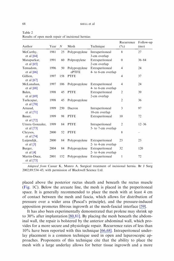
Table 2
Results of open mesh repair of incisional hernias
Author Year N Mesh Technique
Recurrence
(%)
Follow-up
(mo)
McCarthy,
et al [64]
1981 25 Polypropylene Intraperitoneal
3-cm overlap
8 27
Matapurkar,
et al [65]
1991 60 Polpropylene Extraperitoneal
2-cm overlap
0 36–84
Temudom,
et al [66]
1996 50 Polypropylene
ePTFE
Extraperitoneal
4- to 6-cm overlap
4 24
Gillion,
et al [67]
1997 158 PTFE 4 37
McLanahan,
et al [68]
1997 106 Polypropylene Extraperitoneal
4- to 6-cm overlap
4 24
Balen,
et al [69]
1998 45 PTFE Extraperitoneal
2-cm overlap
2 39
Turkcapar,
et al [70]
1998 45 Polypropylene 2 36
Arnaud,
et al [71]
1999 250 Dacron Intraperitoneal
10-cm overlap
3 97
Bauer,
et al [72]
1999 98 PTFE Extraperitoneal 10 72
Utrera Gonzalez,
et al [73]
1999 84 PTFE Intraperitoneal
5- to 7-cm overlap
2 12–36
Chrysos,
et al [74]
2000 52 PTFE 8 –
Luijendijk,
et al [3]
2000 84 Polypropylene Extraperitoneal
2- to 4-cm overlap
23 23
Burger,
et al [4]
2004 84 Polypropylene Extraperitoneal
2- to 4-cm overlap
32 120
Martin-Duce,
et al [75]
2001 152 Polypropylene Extraperitoneal 1 72
Adapted from Cassar K, Munro A. Surgical treatment of incisional hernia. Br J Surg
2002;89:534–45; with permission of Blackwell Science Ltd.
68 SHELL et al
placed above the posterior rectus sheath and beneath the rectus muscle(Fig. 1C). Below the arcuate line, the mesh is placed in the preperitonealspace. It is generally recommended to place the mesh with at least 4 cmof contact between the mesh and fascia, which allows for distribution ofpressure over a wider area (Pascal’s principle), and the pressure-inducedapposition promotes fibrous ingrowth at the mesh-fascial interface [59].
It has also been experimentally demonstrated that prolene may shrink upto 30% after implantation [80,81]. By placing the mesh beneath the abdom-inal wall, the repair is bolstered by the anterior abdominal wall, which pro-vides for a more secure and physiologic repair. Recurrence rates of less than10% have been reported with this technique [66,68]. Intraperitoneal under-lay placement is a common technique used in open and laparoscopic ap-proaches. Proponents of this technique cite that the ability to place themesh with a large underlay allows for better tissue ingrowth and a more
Fig. 1. Mesh placement techniques. (A) Onlay technique. (B) Inlay technique. (C) Retrorectus
underlay technique.
69OPEN REPAIR OF VENTRAL INCISIONAL HERNIAS
secure mesh-fascial interface [11]. Fixation techniques vary from approx-imation at the fascial margins to full-thickness lateral fixation [82]. Recur-rence rates of less than 5% have been reported with this technique [82].
Advances in laparoscopic surgery have led to an increased application ofthis technology to the treatment of incisional hernias. This technique in-volves intraperitoneal mesh placement, which is secured with either a tackingdevice or transabdominal sutures or both. Advocates of this technique citelower recurrence rates of 2% to 4%, shorter hospital stay, decreased infec-tion rate, and reduced wound complication rates as advantages. Severalcomparative studies have concluded that it is a superior technique [83–87].Restoration of dynamic abdominal wall function by midline myofascial ap-proximation and cosmetic improvement of the abdomen by excision of ex-cess tissue and scar are important objectives of hernia repair that are notaccomplished by the laparoscopic approach [58].
Although the application of mesh has resulted in a significant improve-ment in recurrence rates, the use of mesh is associated with specific compli-cations that may range from being relatively minor to life threatening.Infection is one of the most feared complications after mesh placement.The average rate of early and late mesh infections is approximately 7%[8,59,72,88–90] and depends on the type of mesh used. The most common
70 SHELL et al
organisms are Staphyloccocus aureus and Staphylococcus epidermidis [59].Reports exist in the literature of mesh salvage in the face of infection; how-ever, in most cases mesh removal is required [91]. Law [92] examined theeffects of infection on the mesh-fascial interface and found significantweakening, which predisposes to higher recurrence rates. Robertson andcolleagues [93–106] demonstrated that isolation of the incision away fromthe hernia repair through an abdominoplasty approach is associated withlower complication and recurrence rates. It was particularly helpful in obesepatients and patients with multiple or recurrent hernias.
Seroma is a common complication after hernia repair and comprises up to16% of the overall complications [8,88,107]. Reduction of the hernia leavesa potential space for fluid accumulation. Combined with inflammation, dis-ruption of lymphatics, and continued irritation caused by the foreign bodyreaction from the prosthetic material, this complication results in fluid accu-mulation [59]. Seromas often resolve with time; however, continued pros-thetic irritation may result in persistent seroma requiring surgical drainage.
Inadequate soft tissue coverage may result in mesh extrusion. Less pliablematerials, such as marlex, are associated with a higher extrusion rate. Whenextrusion is noted, most authors agree that mesh removal is indicated. En-teric fistula formation is a potentially devastating complication that occurswhen the prosthetic material erodes into the underlying bowel. Leber andcolleagues [8] demonstrated that excision of the hernia sac, lack of omentalinterposition, and the presence of a fascial gap were factors associated witha higher incidence of fistula formation.
Bioprosthetics
Justified concern regarding mesh-related complications has led to thesearch for more biocompatible prosthetic material. Advances in tissue engi-neering technology have led to the development of biomaterials derivedfromhumanand animal tissues. Thesematerials differ in that they heal by a re-generative process rather than by scar tissue formation. The collagen-basedextracellular matrix is preserved, which allows for maintenance of mechanicalintegrity while providing a scaffold for host tissue regeneration. These mate-rials have demonstrated resistance to infection, tolerance of cutaneous expo-sure, and mechanical stability when used in incisional hernia repair.Disadvantages are the high cost and lack of long-term follow-up studies.
Components separation technique
A significant contribution to the repair of incisional hernias was the de-scription by Ramirez and colleagues [46] of the components separation tech-nique (Fig. 2). The evolution of the components separation technique isbased on early descriptions by Vasconez and colleagues [108] of transverse
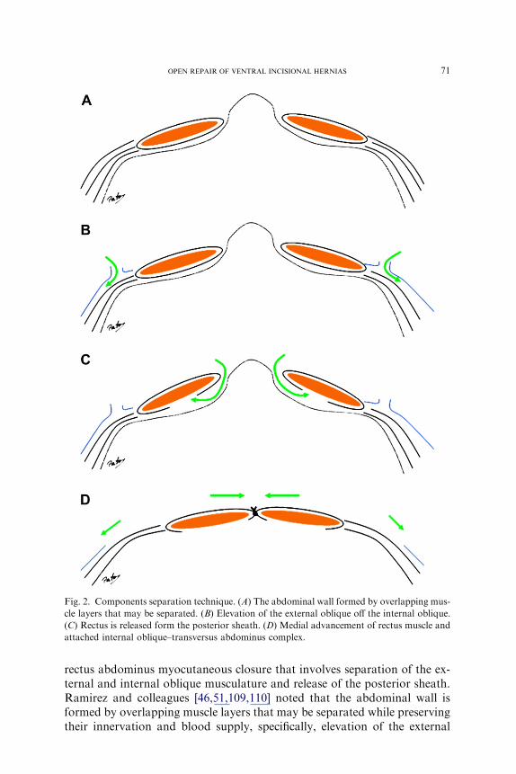
Fig. 2. Components separation technique. (A) The abdominal wall formed by overlapping mus-
cle layers that may be separated. (B) Elevation of the external oblique off the internal oblique.
(C) Rectus is released form the posterior sheath. (D) Medial advancement of rectus muscle and
attached internal oblique–transversus abdominus complex.
71OPEN REPAIR OF VENTRAL INCISIONAL HERNIAS
rectus abdominus myocutaneous closure that involves separation of the ex-ternal and internal oblique musculature and release of the posterior sheath.Ramirez and colleagues [46,51,109,110] noted that the abdominal wall isformed by overlapping muscle layers that may be separated while preservingtheir innervation and blood supply, specifically, elevation of the external
72 SHELL et al
oblique off the internal oblique while maintaining the neurovascular supplyto the rectus abdominus, which travels in a segmental fashion between theinternal oblique and transversus abdominus. The rectus then can be releasedfrom the posterior sheath. Once this procedure is accomplished, medial ad-vancement of a compound flap of rectus muscle and attached internal obli-que-transversus abdominus complex can be used to cover large midlineabdominal defects. Unilateral advancement of 5 cm in the epigastric region,10 cm at the umbilicus, and 3 cm in the suprapubic region has been de-scribed. Fabian and colleagues [111,112] described a modification that in-volved division of the internal oblique of the anterior rectus sheath, whichallowed for unilateral advancement of 8 to 10 cm in the epigastric area, 10to 15 cm in the mid abdomen, and 6 to 8 cm in the suprapubic region. Alower hernia recurrence rate, avoidance of prosthetic material, restorationof dynamic abdominal wall function, and improvement in back and posturalabnormalities have been cited in the literature (Table 3). Wound-relatedcomplications have been problematic with this technique and are relatedto the wide undermining required. Recent work has demonstrated a reduc-tion in wound-related complications with preservation of periumbilical per-forators [121].
In a recent review, Ramirez [110] attributed the success of the procedureto five principles:
1. Translation of the muscular layer of the abdominal wall to enlarge thetissue surface area.
2. Separation of muscle layers that allows for maximal individual expan-sion of each muscle unit.
3. Disconnection of the muscle unit from its fascial sheath envelope, whichrestricts horizontal motion and thereby facilitates expansion.
4. Abdominal wall musculature in approximately 70% of its surface is cov-ering hollow viscus, which is more easily compressed than solidstructures.
5. Bilateral mobilization works more efficiently than unilateral advance-ment by equilibrating forces of the abdominal wall and centralizingthe midline.
Flap reconstruction
Local and distant flaps have been used to reconstruct hernia defects inwhich there is significant absolute loss of domain and in lateral defectsthat are not amenable to advancement techniques. Fasciocutaneous flapsmay be used to reconstruct partial-thickness defects of the skin and subcu-taneous tissues and full-thickness defects when used in combination withmesh. The thoracoepigastric flap is useful for defects of the upper third ofthe abdominal wall. The iliolumbar bipedicled flap based on the superficialcircumflex iliac and lumbar perforators may be used for middle third
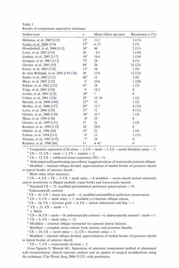
Table 3
Results of components separation technique
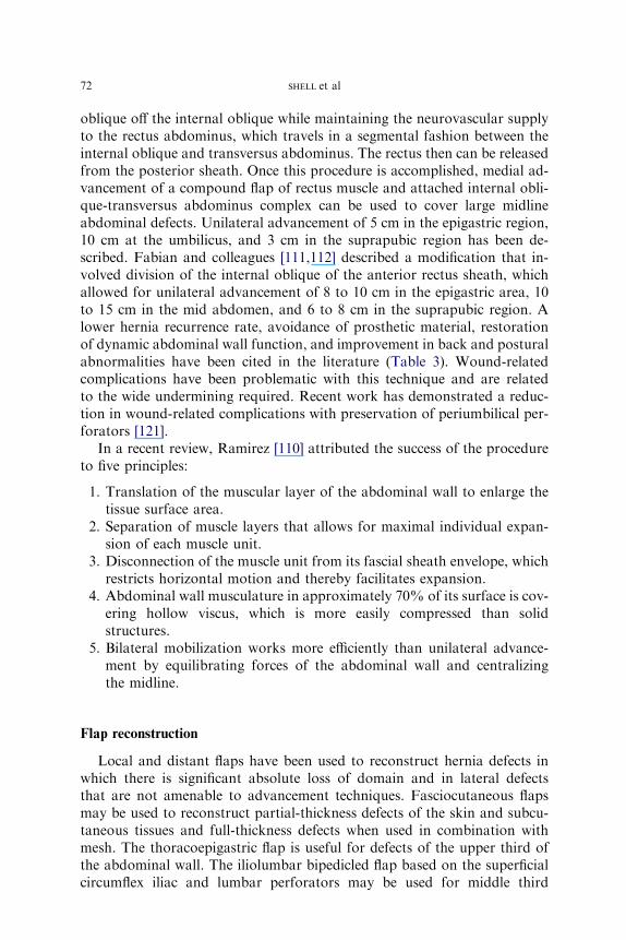
Author/year n Mean follow-up (mo) Recurrence n (%)
Hultman, et al, 2005 [113] 13a 11.5 2 (15)
Vargo, et al, 2004 [114] 27b 6–27 2 (7)
Howdieshell, et al, 2004 [115] 18c 48 2 (11)
Lowe, et al, 2003 [116] 30 9.5 3 (10)
Lindsey, et al, 2003 [117] 10d 18.6 1 (10)
Jernigan, et al, 2003 [112] 73e 24 4 (5)
Girotto, et al, 2003 [55] 96f 26 21 (22)
Ewart, et al, 2003 [118] 11g 10 1 (9)
de vries Reilingh, et al, 2003 [119,120] 38 15.6 12 (32)
Saulis, et al, 2002 [121] 66h 12 3 (8)
Maas, et al, 2002 [122] 5i 10.6 1 (20)
Sukkar, et al, 2001 [123] 47j 24 1 (2)
Voigt, et al, 2001 [124] 9 14.2 0
Levine, et al, 2001 [125] 10k ? 0
Cohen, et al, 2001 [126] 29l 12–36 1 (3)
Shestak, et al, 2000 [109] 22m 52 1 (5)
Mathes, et al, 2000 [127] 26n 13.7 4 (15)
Lowe, et al, 2000 [128] 37o 12 4 (11)
Girotto, et al, 2000 [129] 20p 25.7 1 (5)
Maas, et al, 1999 [130] 4q 18 0
Girotto, et al, 1999 [131] 37 21 2 (5)
Kuzbari, et al, 1998 [132] 10r 28.8 0
Dibello, et al, 1996 [50] 35s 22 3 (9)
Fabian, et al, 1994 [111] 9t 11 1 (11)
Thomas, et al, 1993 [133] 7u 18 0
Ramirez, et al, 1990 [46] 11 4–42 0
a Component separation (CS) alone ¼ 3, CS þ mesh ¼ 5, CS þ mesh/Alloderm onlay ¼ 5.b CS ¼ 23, CS þ mesh ¼ 2, CS þ surgisis ¼ 2.c CS ¼ 12, CS þ subfascial tissue expansion (TE) ¼ 6.d Abdominal wall partitioning (accordion), staggered release of tranversalis/external oblique.e Modified ¼ internal oblique divided, approximation of medial border of posterior sheath
to lateral border of anterior sheath.f Mesh onlay when necessary.g CS ¼ 4, CS þ TE ¼ 3, CS þ mesh onlay ¼ 4; modified ¼ rectus sheath incised anteriorly
and/or posteriorly or flipped medially (open book) and transversalis incised.h Standard CS ¼ 25, modified (periumbilical perforator preservation) ¼ 41.i Endoscopically assisted.j CS ¼ 41, CS þ fascia lata graft ¼ 6, modified periumbilical perforator preservation.k CS ¼ 9, CS þ mesh onlay ¼ 1, modified (�) internal oblique release.l CS ¼ 24, CS þ Goretex graft ¼ 4, CS þ lateral abdominal wall flap ¼ 1.m CS ¼ 21, CS þ mesh ¼ 1.n � Mesh.o CS¼ 20, CSþmesh¼ 10, endoscopically assisted¼ 6, endoscopically assistedþmesh¼ 1.p CS ¼ 8, CS þ mesh onlay ¼ 12.q Modified ¼ external oblique transected via separate lateral incision.r Modified ¼ complete rectus release from anterior and posterior sheaths.s CS ¼ 20, CS þ vicryl onlay ¼ 12, CS þ Goretex onlay ¼ 3.t Modified ¼ internal oblique divided, approximation of medial border of posterior sheath
to lateral border of anterior sheath.u CS ¼ 7, CS þ transversalis division ¼ 2.
From Nguyen V, Shestak KC. Separation of anatomic components method of abdominal
wall reconstruction: clinical outcome analysis and an update of surgical modifications using
the technique. Clin Plastic Surg 2006;33:255; with permission.
74 SHELL et al
defects. Lower third defects may be covered with a groin flap, which mayreach to the umbilicus [49,127,134–137]. Superficial inferior epigastric arteryand deep inferior epigastric artery flaps are useful for lower abdominal andgroin defects [134,138–140].
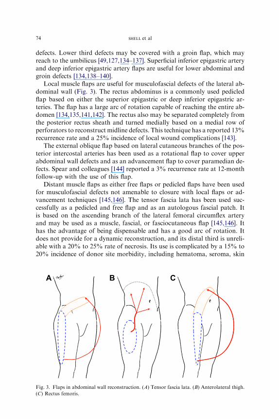
Local muscle flaps are useful for musculofascial defects of the lateral ab-dominal wall (Fig. 3). The rectus abdominus is a commonly used pedicledflap based on either the superior epigastric or deep inferior epigastric ar-teries. The flap has a large arc of rotation capable of reaching the entire ab-domen [134,135,141,142]. The rectus also may be separated completely fromthe posterior rectus sheath and turned medially based on a medial row ofperforators to reconstruct midline defects. This technique has a reported 13%recurrence rate and a 25% incidence of local wound complications [143].
The external oblique flap based on lateral cutaneous branches of the pos-terior intercostal arteries has been used as a rotational flap to cover upperabdominal wall defects and as an advancement flap to cover paramedian de-fects. Spear and colleagues [144] reported a 3% recurrence rate at 12-monthfollow-up with the use of this flap.
Distant muscle flaps as either free flaps or pedicled flaps have been usedfor musculofascial defects not amenable to closure with local flaps or ad-vancement techniques [145,146]. The tensor fascia lata has been used suc-cessfully as a pedicled and free flap and as an autologous fascial patch. Itis based on the ascending branch of the lateral femoral circumflex arteryand may be used as a muscle, fascial, or fasciocutaneous flap [145,146]. Ithas the advantage of being dispensable and has a good arc of rotation. Itdoes not provide for a dynamic reconstruction, and its distal third is unreli-able with a 20% to 25% rate of necrosis. Its use is complicated by a 15% to20% incidence of donor site morbidity, including hematoma, seroma, skin
Fig. 3. Flaps in abdominal wall reconstruction. (A) Tensor fascia lata. (B) Anterolateral thigh.
(C) Rectus femoris.
75OPEN REPAIR OF VENTRAL INCISIONAL HERNIAS
graft loss, and lateral knee instability [145,146]. Recurrence rates are alsosignificant and range from 9% to 42% in the literature [145,146].
The anterolateral thigh flap has been used in the reconstruction of lowerabdominal wall defects as either a free or pedicled flap based on septocuta-neous perforating branches of the transverse and deep branches of the lat-eral femoral circumflex artery [147]. With the adjunctive use of mesh, thistechnique has demonstrated low recurrence rates in small series. It alsomay be used in combination with the tensor fascia lata to provide a compos-ite graft up to 35 � 20 cm in dimension. In small series, the use of this tech-nique has demonstrated no recurrences or flap loss in follow-up up to 24months [134,147,148].
The rectus femoris muscle has been used successfully as either a free flapwith preservation of the motor nerve or as a pedicled flap in reconstructionof the lower two thirds of the abdomen. It is a dispensable muscle with a con-sistent anatomy. Reports have indicated a weakness in terminal knee exten-sion after muscle harvest, which can be minimized by approximating thevastus medialis and lateralis. It is based on the lateral femoral circumflexartery and has a large arc of superior and contralateral rotation. It can bedesigned as a musculofascial or musculofasciocutaneous flap based on thelocation and extent of the defect [149–151]. Electromyographic studieshave documented motor function of the transferred muscle.
The latissimus dorsi muscle has been used as a musculocutaneous flap fordefects of the upper third of the abdomen. It is based on the thoracodorsalpedicle and can be designed as either a pedicled or a free flap. As a pedicledflap, its arc of rotation is limited to coverage of upper abdominal wall de-fects. The area of coverage can be increased by including the preglutealand lumbodorsal fascia. In 1979, Bostwick [152] reported the use of the lat-issimus as a free flap for abdominal wall reconstruction. In 1998, Ninkovic[153] reported the use of a free, innervated latissimus flap in conjunctionwith prolene mesh for abdominal wall reconstruction. No flap failureswere reported, and electromyographic testing demonstrated reinnervationof the muscle.
The gracilis muscle has been used to reconstruct lower third abdominalwall defects. It is a thin, narrow, dispensable muscle based on the ascendingbranch of the medial circumflex femoral artery. It can be designed as eithera muscular or musculofasciocutaneous flap and is limited to small defectsbecause of its size and the poor reliability of its distal skin [154]. The vastuslateralis can be used as a muscular flap for reconstruction of lower third ab-dominal wall defects. It does not have a fascial component and its use is pri-marily reserved for salvage situations [155].
Tissue expansion
Tissue expansion has been used to provide well-vascularized, autologous,innervated tissue for abdominal wall reconstruction. Its use has been
76 SHELL et al
demonstrated in the reconstruction of congenital defects and large hernias[156–161]. Expanders may be placed in either the subcutaneous or intermus-cular plane. Placement in the avascular plane between the external and in-ternal oblique muscles allows superficial expansion of the external obliqueand deep expansion of the internal oblique-tranversus abdominus musculo-fascial layer while preserving innervation and blood supply. Hobar and col-leagues [157,158] demonstrated an approximate doubling of the layers of theanterior abdominal wall with normal function and clinically demonstratedinnervated composite reconstruction of defects exceeding 50% of the ab-dominal surface.
Summary
Despite advances in many fields of surgery, incisional hernias still remaina significant problem. There is a lack of general consensus among surgeonsregarding optimal treatment. A surgeon’s approach is often based on tradi-tion rather than clinical evidence. The surgeon’s treatment plan should becomprehensive, with attention focused not merely on restoration of struc-tural continuity. An understanding of the structural and functional anatomyof the abdominal wall and an appreciation of the importance of restoringdynamic function are necessary for the successful reconstruction of the ab-dominal wall.
References
[1] Bucknall TE, Cox PJ, Ellis H. Burst abdomen and incisional hernia: a prospective study of
1129 major laparotomies. Br Med J (Clin Res Ed) 1982;284(6364):519–20.
[2] GeorgeCD, EllisH. The results of incisional hernia repair: a twelve year review.AnnRColl
Surg Engl 1986 Jul;68(4):185–7.
[3] Luijendijk RW, HopWC, Van den Tol MP, et al. A comparison of suture repair with mesh
repair for incisional hernia. N Engl J Med 2000;343(6):392–8.
[4] Burger JW, Luijendijk RW, Hop WC, et al. Long term follow up of a randomized con-
trolled trial of suture versus mesh repair of incisonal hernia. Ann Surg 2004;240(4):
578–83 [discussion: 583–5].
[5] Mudge M, Hughes LE. Incisional hernia: a ten year prospective study of incidence and at-
titudes. Br J Surg 1985;72:70–1.
[6] Langer S, Christiansen J. Long-term results after incisional hernia repair. Acta Chir Scand
1985;151:217–9.
[7] Anthony T, Bergen PC, Kim LT. Factors affecting recurrence following incisional hernior-
rhaphy. World J Surg 2000;24(1):95–101.
[8] Leber GE, Garb JL, Alexander AJ, et al. Long term complications associated with pros-
thetic repair of incisional hernias. Arch Surg 1998;133(4):378–82.
[9] ReadRC,YoderG.Recent trends inmanagement of incisional herniation.Arch Surg 1989;
124:485–8.
[10] Santora TA, Rosalyn JJ. Incisional hernia. Surg Clin North Am 1993;73:557–70.
[11] Millikan KW. Incisional hernia repair. Surg Clin North Am 2003;83:1223–34.
77OPEN REPAIR OF VENTRAL INCISIONAL HERNIAS
[12] Duepree HJ, Senagore AJ, Delaney CP, et al. Does means of access affect the incidence of
small bowel obstruction and ventral hernia after bowel resection? Laparoscopy versus lap-
arotomy. J Am Coll Surg 2003;197(2):177–81.
[13] Rutkow IM. Epidemiologic, economic and sociologic aspects of hernia surgery in the
United States in the 1990s. Surg Clin North Am 1998;78:941–51.
[14] Hesselink VJ, Luijendijk RW, De Wilt JH, et al. An evaluation of risk factors in incisional
hernia recurrence. Surg Gynecol Obstet 1993;176(3):228–34.
[15] Gecim IE,Kocak S, Ersoz S, et al. Recurrence after incisional hernia repair: results and risk
factors. Surg Today 1996;26(8):607–9.
[16] Sorenson LT, Hemmingsen UB, Kirkeby LT, et al. Smoking is a risk factor for incisional
hernia. Arch Surg 2005;140(2):119–23.
[17] Sugerman HJ, Kellum JM Jr, Reines HD, et al. Greater risk of incisional hernia with mor-
bidly obese than steroid-dependent patients and low recurrence with prefascial polypropyl-
ene mesh. Am J Surg 1996;171(1):80–4.
[18] Koller R,Miholic J, Jakl RJ. Repair of incisional hernias with polytetrafluoroethylene. Eur
J Surg 1997;163(4):261–6.
[19] Lamont PM, Ellis H. Incisional hernia in re-opened abdomens: an overlooked risk factor.
Br J Surg 1988;75(4):374–6.
[20] Makela JT,Kiviniemi H, Juvonen T, et al. Factors influencing wound dehiscence aftermid-
line laparotomy. Am J Surg 1995;170(4):387–90.
[21] Carlson MA, Ludwig KA, Condon RE. Ventral hernia and other complications of 1,000
midline laparotomies. South Med J 1995;88(4):450–3.
[22] Raffetto JD, Cheung Y, Fisher JB, et al. Incision and abdominal wall hernias in patients
with aneurysm or occlusive aortic disease. J Vasc Surg 2003;37:1150–4.
[23] Condon RE. Ventral abdominal hernia. In: Baker RJ, Fischer JE, editors. Mastery of sur-
gery. 4th edition. Philadelphia: Lippincott Williams & Wilkins; 2001.
[24] Brolin RE. Prospective, randomized evaluation of midline fascial closure in gastric bypass
operations. Am J Surg 1996;172(4):328–31.
[25] ThompsonWR,Amaral JF, CaldwellMD, et al. Complications and weight loss in 150 con-
secutive gastric exclusion patients: critical review. Am J Surg 1983;146(5):602–12.
[26] Yale CE. Gastric surgery for morbid obesity: complications and long-term weight control.
Arch Surg 1989;124(8):941–6.
[27] Adye B, Luna G. Incidence of abdominal wall hernia in aortic surgery. Am J Surg 1998;
175(5):400–2.
[28] Stevick CA, Long JB, Jamasbi B, et al. Ventral hernia following abdominal aortic recon-
struction. Am Surg 1988;54(5):287–9.
[29] Johnson B, Sharp R, Thursby P. Incisional hernias: incidence following abdominal aortic
aneurysm repair. J Cardiovasc Surg (Torino) 1995;36(5):487–90.
[30] Si Z, Bhardwaj R, Rosch R, et al. Impaired balance of type I and type III procollagen
mRNA in cultured fibroblasts of patients with incisional hernia. Surgery 2002;131(3):
324–31.
[31] PlayforthMJ, Sauven PD, EvansM, et al. The prediction of incisional hernia by radio-opa-
que markers. Ann R Coll Surg Engl 1986;68(2):82–4.
[32] Poole GV Jr. Mechanical factors in abdominal wound closure: the prevention of fascial de-
hiscence. Surgery 1985;97(6):631–40.
[33] Hodgson NC, Malthaner RA, Østbye T. The search for an ideal method of fascial closure:
a meta-analysis. Ann Surg 2000;231(3):436–42.
[34] Jenkins TP. The burst abdominal wound: a mechanical approach. Br J Surg 1976;63(11):
873–6.
[35] Trimbos JB, van Rooij J. Amount of suture material needed for continuous or interrupted
wound closure: an experimental study. Eur J Surg 1993;159(3):141–3.
[36] Cassar K, Munro A. Surgical treatment of incisional hernia. Br J Surg 2002;89:534–45.
[37] Abrahamsom J, Eldar S. Abdominal incision. Lancet 1989;1(8642):847.
78 SHELL et al
[38] GreviousMA, CohenM, Shah SR, et al. Structural and functional anatomy of the abdom-
inal wall. Clin Plast Surg 2006;33:169–79.
[39] Puckree T, Cerny F, Bishop B. Abdominal motor unit activity during respiratory and non-
respiratory tasks. J Appl Physiol 1998;84(5):1707–15.
[40] Misuri G, Colagrande S, Gorini M. In vivo ultrasound assessment of respiratory function
of abdominal muscles in normal subjects. Eur Respir J 1997;10(12):2861–7.
[41] Myrinkas SE, Beith ID, Harrison PJ. Stretch reflexes in the rectus abdominus muscle in
man. Exp Physiol 2000;85(4):445–50.
[42] Blondeel N, Vanderstraeten GG,Monstrey SJ. The donor site morbidity of free DIEP flaps
and free TRAM flaps in breast reconstruction. Br J Plast Surg 1997;50(5):322–30.
[43] Toranto IR. Resolution of back pain with wide abdominal rectus plication abdomino-
plasty. Plast Reconstr Surg 1990;85(4):545–55.
[44] Gracovetsky S, FarfanH, Helleur C. The abdominal mechanism. Spine 1985;10(4):317–24.
[45] Lam KS, Mehdian H. The importance of an intact abdominal musculature mechanism in
maintaining spinal sagittal balance: case illustration in prune-belly syndrome. Spine 1999;
24(7):712–22.
[46] Ramirez OM, Ruas E, Dellon L. ‘‘Components separation’’ method for closure of abdom-
inal-wall defects: an anatomic and clinical study. Plast Reconstr Surg 1990;86(3):521–6.
[47] WantzGE. Incisional hernia: the problemand the cure. JAmColl Surg 1999;188(4):433–47.
[48] Core GB, Grotting JC. Reoperative surgery of the abdominal wall. In: Grotting JC, editor.
Aesthetic and reconstructive plastic surgery. St Louis (MO): Quality Medical Publishing,
Incorporated; 1995. p. 1327–75.
[49] Rohrich RJ, Lowe JB, Baty JD, et al. An algorithm for abdominal wall reconstruction.
Plast Reconstr Surg 2000;105(1):202–16.
[50] DiBello JN Jr, Moore JH Jr. Sliding myofascial flap of rectus abdominus muscles for the
closure of recurrent ventral hernias. Plast Reconstr Surg 1996;98(3):464–9.
[51] NguyenV, ShestakKC. Separation of anatomic componentsmethod of abdominal wall re-
construction: clinical outcome analysis and an update of surgical modifications using the
technique. Clin Plast Surg 2006;33:247–57.
[52] Van der Linden FT, vanVroonhoven TJ. Long term results after surgical correction of inci-
sional hernia. Neth J Surg 1988;40:127–43.
[53] Sauerland S, Schmedt CG, Lein S, et al. Primary incisional herniarepair with or without
polypropylene mesh: a report on 384 patients with 5 year follow up. Langenbecks Arch
Surg 2005;390(5):408–12.
[54] Al-Salamah SM, Hussain MI, Khalid K, et al. Suture vs mesh repair for incisional hernia.
Saudi Med J 2006;27(5):652–6.
[55] Girotto JA, Chiaramonte M, Menon NG, et al. Recalcitrant abdominal wall hernias: long
term results of autologous tissue repair. Plast Reconstr Surg 2003;112(1):106–14.
[56] KorenkovM, Paul A, Sauerland S, et al. Classification and surgical treatment: results of an
experts’ meeting. Langenbecks Arch Surg 2001;386:65–73.
[57] Flum DR, Horvath K, Koepsell T. Have outcomes with incisional hernia repair improved
with time? A population-based analysis. Ann Surg 2003;237(1):129–35.
[58] Voeller GR, Ramshaw B, Park AE, et al. Incisional hernia. J Am Coll Surg 1999;189(6):
635–7.
[59] Grevious MA, Cohen M, Jean-Pierre F, et al. The use of prosthetics in abdominal wall re-
construction. Clin Plast Surg 2006;33:181–97.
[60] Klosterhalfen B, Rosch R, Junge K. Long term inertness of meshes. In: Schumpelick NL,
editor. Meshes: benefits and risks. Berlin: Springer; 2004. p. 170–8.
[61] Cobb WS, Kercher KW, Heniford BT. The argument for lightweight polypropylene mesh
in hernia repair. Surg Innov 2005;12(1):T1–7.
[62] Usher FC, Ochsner J, Tuttle Jr LL. Use of Marlex mesh in the repair of incisional hernias.
Am Surg 1958;24:969–74.
79OPEN REPAIR OF VENTRAL INCISIONAL HERNIAS
[63] Stelzner F. Function of the abdominal wall and development and therapy of hernias.
Langenbecks Arch Chir 1994;379(2):109–19.
[64] McCarthy JD, Twiest MW. Intraperitoneal polypropylene mesh support of incisional her-
niorrhaphy. Am J Surg 1981;142:707–11.
[65] Matapurkar BG, Gupta AK, Agarwal AK. A new technique of ‘‘marlex peritoneal sand-
wich’’ in the repair of large incisional hernias. World J Surg 1991;15:768–70.
[66] Temudom T, Siadati M, Sarr MG. Repair of complex giant or recurrent ventral hernias by
using tension-free intraparietal prosthetic mesh (Stoppa technique): lessons learned from
our initial experience (fifty patients). Surgery 1996;120:738–43.
[67] Gillion JF, Begin GF, Marecos C, et al. Expanded polytetrafluoroethylene patches used in
the intraperitoneal or extraperitoneal position for repair on incisional hernias of the antero-
lateral abdominal wall. Am J Surg 1997;174:16–9.
[68] McLanahanD,King LT,WeemsC, et al. Retrorectus prosthetic mesh repair of midline ab-
dominal hernia. Am J Surg 1997;173:445–9.
[69] Balen EM, Diez-Caballero A, Hernandez-Lizoain JL, et al. Repair of ventral hernias using
expanded polytetrafluoroethylene patch. Br J Surg 1998;85:1415–8.
[70] Turkcapar AG, Yerdel MA, Aydinuraz K, et al. Repair of midline incisional hernias using
polypropylene grafts. Surg Today 1998;28:59–63.
[71] Arnaud JP, Tuech JJ, Pessaux P, et al. Surgical treatment of postoperative incisional hernia
by intraperitoneal insertion ofDacronmesh and an aponeurotic graft: a report of 250 cases.
Arch Surg 1999;134:1260–2.
[72] Bauer JJ, Kreel I, Gelernt IM. Twelve year experience with expanded polytetrafluoroethy-
lene in the repair of abdominal defects. Mt Sinai Med J 1999;66(1):20–5.
[73] Utrera Gonzalez A, de la Portilla de Juan F, Carranza Albarran G. Large incisional hernia
repair using intraperitoneal placement of expanded polytetrafluoroethylene. Am J Surg
1999;177:291–3.
[74] Chrysos E, Athanasakis E, Saridaki Z, et al. Surgical repair of incisional ventral hernias:
tension free technique using prosthetic materials (expanded polytetrafluoroethylene Gore-
tex dual mesh). Am Surg 2000;66:679–82.
[75] Martin-Duce A, Noguerales F, Villeta R, et al. Modifications to Rives technique for mid-
line incisional hernia repair. Hernia 2001;5:70–2.
[76] Chevrel JP, Rath AM. The use of fibrin glues in the surgical treatment of incisional hernias.
Hernia 1997;1:9–14.
[77] Larson GM. Plastic mesh repair of incisional hernias. Am J Surg 1978;135:559–63.
[78] Stoppe RE. Treatment of complicated groin and incisional hernias. World J Surg 1989;13:
545–54.
[79] Rives J, Pire JC, Flement JP, et al. Major incisional hernia. In: Chevrel JP, editor. Surgery
of the abdominal wall. New York: Springer-Verlag; 1987. p. 116–44.
[80] KlingeU,Klosterhalfen B, Conze J, et al.Modifiedmesh for hernia repair that is adapted to
the physiology of the abdominal wall. Eur J Surg 1998;164(12):951–60.
[81] KlingeU,Conze B,KlosterhalfenB, et al. Changes in abdominal wall mechanics aftermesh
implantation: experimental changes in mesh stability. Langenbecks Arch Chir 1996;381(6):
323–32.
[82] MillikanKW, BaptistaM,Amin B, et al. Intraperitoneal underlay ventral hernia repair uti-
lizing bilayer ePTFE and polypropylene mesh. Am Surg 2003;69:258–63.
[83] Carbajo MA, Martin del Olmo JC, Blanco JI, et al. Laparoscopic treatment versus open
surgery in the solution of major incisional and abdominal wall hernias with mesh. Surg
Endosc 1999;13:250–2.
[84] Ramshaw BJ, Schwab J, Mason EM, et al. Comparison of laparoscopic and open ventral
herniorrhaphy. Am Surg 1999;65:827–31.
[85] Park A, Burch DW, Lovrics P. Laparoscopic and open incisional hernioplasty. Surg
Endosc 1997;11:32–5.
80 SHELL et al
[86] HenifordBT, ParkA, RamshawBJ, et al. Laparoscopic ventral and incisional hernia repair
in 407 patients. J Am Coll Surg 2000;190:645–50.
[87] Heniford BT, Park A, Ramshaw BJ, et al. Laparoscopic repair of ventral hernias: nine
years’ experience with 850 consecutive cases. Ann Surg 2003;238:391–400.
[88] CobbW IV, Harris JB, Lokey JS, et al. Incisional herniorrhaphy with intraperitoneal com-
posite mesh: a report of 95 cases. Am Surg 2003;69(9):784–7.
[89] CarlsonG, et al. Abdominal wall reconstruction. In: Achauer BM, Eriksson E, Guturon B,
editors. Plastic surgery: indications, operations, and outcomes. St Louis (MO): Mosby;
2004. p. 563–74.
[90] Jones JW, Jurkovich GJ. Polypropylene mesh closure of infected abdominal wounds. Am
Surg 1989;55:73–6.
[91] Kelly ME, Behrman SW. The safety and efficacy of prosthetic hernia repair in clean-con-
taminated and contaminated wounds. Am Surg 2002;68(6):524–8 [discussion: 528–9].
[92] LawNW. A comparison of polypropylene mesh and expanded polytetrafluoroethylene for
repair of contaminated abdominal wall defects: an experimental study. Surgery 1991;
109(5):652–5.
[93] Robertson JD, de la Torre JI, Gardner PM, et al. Abdominoplasty repair for abdominal
wall hernias. Ann Plast Surg 2003;51:10–6.
[94] Badylak SF,Kokini K, Tullius B, et al.Morphologic study of small intestinal submucosa as
a body wall repair device. J Surg Res 2002;103:190–202.
[95] Buinewicz B,RosenB.Acellular cadaveric dermis (Alloderm): a newalternative for abdom-
inal hernia repair. Ann Plast Surg 2004;52:188–94.
[96] Butler CE, Langstein HN, Kornowitz SJ. Pelvic, abdominal, and chest wall reconstruction
with Alloderm in patients at increased risk for mesh-related complications. Plast Reconstr
Surg 2005;116:1263–75.
[97] Espinosa-de-los-Monteros A, de la Torre JI, Marrero I, et al. Utilization of human ca-
daveric acellular dermis for abdominal hernia reconstruction. Ann Plast Surg 2007;58:
264–7.
[98] Adedeji OA, Bailey CA, Varma JS. Porcine dermal collagen graft in abdominal wall recon-
struction. Br J Plast Surg 2002;55:85–6.
[99] Shell DH 4th, Croce MA, Cagiannos C, et al. Comparison of small-intestinal submucosa
and expanded polytetrafluoroethylene as a vascular conduit in the presence of gram-posi-
tive contamination. Ann Surg 2005;241(6):995–1001 [discussion: 1001–4].
[100] Jernigan TW, Croce MA, Cagiannos C. Small intestinal submucosa for vascular recon-
struction in the presence of gastrointestinal contamination. Ann Surg 2004;239(5):733–8
[discussion: 738–40].
[101] Butler CE. The role of bioprosthetics in abdominal wall reconstruction. Clin Plast Surg
2006;33:199–211.
[102] Ueno T, Pickett LC, de la Fuente SG, et al. Clinical application of porcine small intestinal
submucosa in the management of infected or potentially contaminated abdominal defects.
J Gastrointest Surg 2004;8:109–12.
[103] Helton WS, Fisichella PM, Berger R, et al. Short term outcomes with small intestinal sub-
mucosa for ventral abdominal hernia. Arch Surg 2005;140:549–62.
[104] Choe JM, Kothandapani R, James L, et al. Autologous, cadaveric, and synthetic ma-
terials used in sling surgery: comparative biomechanical analysis. Urology 2001;58:
482–6.
[105] Silverman RP, Li EN, Holton LH III, et al. Ventral hernia repair using allogenic acellular
dermal matrix in a swine model. Hernia 2004;8:336–42.
[106] Butler CE, Prieto VG. Reduction of adhesions with composite Alloderm/polypropylene
mesh implants for abdominal wall reconstruction. Plast Reconstr Surg 2004;114:464–73.
[107] Balique JG, Bouillot JL, Flament JB, et al. Intraperitoneal treatment of incisional and um-
bilical hernias using an innovative composite mesh: four year results of a prospective mul-
ticenter clinical trial. Hernia 2005;9:68–74.
81OPEN REPAIR OF VENTRAL INCISIONAL HERNIAS
[108] Vasconez LO, Lejour M, Gamboa-Babadilla M. Atlas of breast reconstruction. Philadel-
phia: JB Lippincott; 1991.
[109] Shestak KC, Edington HJ, Johnson RR. The separation of anatomic components tech-
nique for the reconstruction of massive midline abdominal wall defects: anatomy, surgical
technique, applications, and limitations revisited. Plast Reconstr Surg 2000;105(2):731–8.
[110] Ramirez OM. Inception and evolution of the components separation technique: personal
recollections. Clin Plast Surg 2006;33:241–6.
[111] FabianTC,CroceMA, PritchardFE. Planned ventral hernia: stagedmanagement for acute
abdominal wall defects. Ann Surg 1994;219(6):643–50 [discussion: 651].
[112] Jernigan TW, Fabian TC, CroceMA. Stagedmanagement of giant abdominal wall defects:
acute and long term results. Ann Surg 2003;238(3):349–55.
[113] Hultman CS, Pratt B, Cairns BA. Multi-disciplinary approach to abdominal wall recon-
struction after decompressive laparotomy for abdominal compartment syndrome. Ann
Plast Surg 2005;54(3):269–75.
[114] Vargo D. Component separation in the management of the difficult abdominal wall. Am J
Surg 2004;188(6):633–7.
[115] Howdieshell TR, Proctor CD, Sternberg E. Temporary abdominal closure followed by de-
finitive abdominal wall reconstruction of the open abdomen. Am J Surg 2004;188(3):301–6.
[116] Lowe JB III, Lowe JB, Baty JD.Risks associatedwith components separation for closure of
complex abdominal wall defects. Plast Reconstr Surg 2003;111(3):1276–83 [discussion:
1286–8].
[117] Lindsey JT. Abdominal wall partitioning (the accordion effect) for reconstruction of major
defects: a retrospective review of 10 patients. Plast Reconstr Surg 2003;112(2):477–85.
[118] Ewart CJ, Lankford AB, GamboaMG. Successful closure of abdominal wall hernias using
the components separation technique. Ann Plast Surg 2003;50(3):269–73 [discussion:
273–4].
[119] de Vries Reileigh TS. Components separation technique for the repair of large abdominal
wall hernias. J Am Coll Surg 2003;196(1):32–7.
[120] Losanoff JE, Richman BW, Sauter ER. Component separationmethod for abdominal wall
reconstruction. J Am Coll Surg 2003;196(5):825–6.
[121] Saulis AS, Dumanian GA. Periumbilical rectus abdominis perforator preservation signifi-
cantly reduces superficial wound complications in separation of parts hernia repair. Plast
Reconstr Surg 2002;109(7):2275–80 [discussion: 2281–2].
[122] Maas SM, van Engel M, LeeksmaNG. Amodification of the components separation tech-
nique for the repair of complicated ventral hernias. J Am Coll Surg 2002;194(3):388–90.
[123] Sukkar SM, Dumanian GA, Szczerba SM. Challenging abdominal wall defects. Am J Surg
2001;181(2):115–21.
[124] Voigt M, Andree C, Galla TJ. Reconstruction of abdominal wall midline defects: the ab-
dominal wall components separation. Zentralbl Chir 2001;126(12):1000–4.
[125] Levine JP, Karp NS. Restoration of abdominal wall integrity as a salvage procedure in dif-
ficult recurrent abdominal wall hernias using amethod of widemyofascial release. Plast Re-
constr Surg 2001;107(3):706–17.
[126] CohenM,Morales R Jr, Fildes J. Staged reconstruction after gunshot wounds to the abdo-
men. Plast Reconstr Surg 2001;108(1):83–92.
[127] Mathes SJ, Steinwald PM, Foster RD, et al. Complex abdominal wall reconstruction:
a comparison of flap and mesh closure. Ann Surg 2000;232(4):586–96.
[128] Lowe JB, Garza IR, Bowman JL. Endoscopically assisted components separation tech-
nique for closure of abdominal wall defects. Plast Reconstr Surg 2000;105(2):720–9.
[129] Girotto JA, Malaisrie SC, Bulkely G. Recurrent ventral herniation in Ehlers-Danlos syn-
drome. Plast Reconstr Surg 2000;106(7):1520–6.
[130] Maas SM, van Engel M, LeeksmaNG. Amodification of the components separation tech-
nique for closure of abdominal wall defects in the presence of an enterostomy. J Am Coll
Surg 1999;189(1):138–40.
82 SHELL et al
[131] Girotto JA, KoMJ, Redett R. Closure of chronic abdominal wall defects: a long term eval-
uation of the components separation method. Ann Plast Surg 1999;42(4):385–94.
[132] Kuzbari R, Worseg AP, Tairych G. Sliding door technique for the repair of midline inci-
sional hernias. Plast Reconstr Surg 1998;101(5):1235–42.
[133] Thomas WO III, Parry SW, Rodning CB. Ventral/incisional abdominal herniorrhaphy by
fascial partition/release. Plast Reconstr Surg 1993;91(6):1080–6.
[134] Lowe JB. Updated algorithm for abdominal wall reconstruction. Clin Plast Surg 2006;33:
225–40.
[135] Mathes SJ, Nahai F. Clinical application of muscle and musculocutaneous flaps. St Louis
(MO): Mosby; 1982.
[136] Mathes SJ, Nahai F. Reconstructive surgery: principles, anatomy, and technique. New
York: Churchill Livingstone; 1997.
[137] Ohtsuka H, Ochi K, Seike H. Reconstruction of a large lateral abdominal wall defect with
an iliolumbar bipedicled flap. Br J Plast Surg 1984;37(3):327–9.
[138] Gottlieb ME, Chandrasekhar B, Terz JJ, et al. Clinical applications of the extended deep
inferior epigastric flap. Plast Reconstr Surg 1986;78(6):782–92.
[139] Stern HS, Nahai F. The versatile superficial inferior epigastric artery free flap. Br J Plast
Surg 1992;45(4):270–4.
[140] Classen D. The extended deep inferior epigastric flap: a case series. Ann Plast Surg 1999;
42(2):137–41.
[141] Parkash S, Palepu J. Rectus abdominus myocutaneous flap: clinical experience with ipsilat-
eral and contralateral flaps. Br J Surg 1983;70(2):68–70.
[142] Mathes SJ, Bostwick J III. A rectus abdominus myocutaneous flap to reconstruct abdom-
inal wall defects. Br J Plast Surg 1977;30(4):282–3.
[143] DeFranzo AJ, KingmanGJ, Sterchi JM, et al. Rectus turnover flaps for the reconstruction
of large midline abdominal wall defects. Ann Plast Surg 1996;37(1):18–23.
[144] Spear SL, Walker RK. The external oblique flap for reconstruction of the rectus sheath.
Plast Reconstr Surg 1992;90(4):608–13.
[145] Disa JJ, Goldberg NH, Carlton JM, et al. Restoring abdominal wall integrity in contami-
nated tissue deficient wounds using autologous fascia grafts. Plast Reconstr Surg 1988;
101(4):979–86.
[146] Williams JK, Carlson GW, deChalain T, et al. Role of tensor fascia lata in abdominal wall
reconstruction. Plast Reconstr Surg 1998;101(3):802–5.
[147] KimataY,UchiyamaK, SekidoM, et al. Anterolateral thigh flap for abdominal wall recon-
struction. Plast Reconstr Surg 1999;103(4):1191–7.
[148] Sasaki K, Nozaki M, Nakazawa H, et al. Reconstruction of a large abdominal wall defect
using combined free tensor fascia latae musculocutaneous flap and anterolateral thigh flap.
Plast Reconstr Surg 1998;102(6):2244–52.
[149] McCraw JB, Dibbell DG, Carraway JH. Clinical definition of independent myocutaneous
vascular territories. Plast Recostr Surg 1977;60(3):341–52.
[150] Koshima I, Nanba Y, Tutsui T, et al. Dynamic reconstruction of large abdominal defects
using free rectus femoris. Ann Plast Surg 2003;50(4):420–4.
[151] Caulfield WH, Curtsinger L, Powell G. Donor leg morbidity after pedicled rectus femoris
muscle flap transfer for abdominal and pelvic reconstruction. Ann Plast Surg 1994;32(4):
377–982.
[152] Bostwick J 3rd, Vasconez LO, Nahai F, et al. Sixty latissimus dorsi flaps. Plast Reconstr
Surg 1979;63(1):31–41.
[153] Ninkovic M, Kronberger P, Harpf C. Free innervated latissimus dorsi muscle for recon-
struction of full-thickness abdominal wall defects. Plast Reconstr Surg 1998;101(4):971–8.
[154] Venugopalan S. Repair of midline abdominal incisional hernia by gracilis muscle transfer.
Br J Plast Surg 1980;33(1):43–5.
[155] DowdenRV,McCraw JB. The vastus lateralismuscle flap: technique and applications.Ann
Plast Surg 1980;4(5):396–404.
83OPEN REPAIR OF VENTRAL INCISIONAL HERNIAS
[156] JacobsenWM, Petty PM, Bite U, et al. Massive abdominal wall hernia reconstruction with
expanded external/internal oblique and transversalis fascia. Plast Reconstr Surg 1997;
100(2):326–35.
[157] Hobar PC, Rohrich RJ, Byrd HS. Abdominal-wall reconstruction with expanded muscu-
lofascial tissue in a posttraumatic defect. Plast Reconstr Surg 1994;94(2):379–83.
[158] Byrd HS, Hobar PC. Abdominal wall expansion in congenital defects. Plast Reconstr Surg
1989;84(2):347–52.
[159] Carlson GW, Elwood E, Losken A, et al. The role of tissue expansion in abdominal wall
reconstruction. Ann Plast Surg 2000;44(2):147–53.
[160] Espinosa-de-los-Monteros A, de la Torre JI, Ahumada LA, et al. Reconstruction of the ab-
dominal wall for incisional hernia repair. Am J Surg 2006;191:173–7.
[161] Ger R, Duboys E. The prevention and repair of large abdominal wall defects by muscle
transposition: a preliminary communication. Plast Reconstr Surg 1983;72(2):170–5.