Incisional ventral hernias: Review of the literature and recommendations regarding the grading and...
15
Incisional ventral hernias: Review of the literature and recommendations regarding the grading and technique of repair The Ventral Hernia Working Group: Karl Breuing, MD, a Charles E. Butler, MD, FACS, b Stephen Ferzoco, MD, FACS, a Michael Franz, MD, c Charles S. Hultman, MD, MBA, FACS, d Joshua F. Kilbridge, e Michael Rosen, MD, f Ronald P. Silverman, MD, FACS, g and Daniel Vargo, MD, FACS, h Boston, MA, Houston, TX, Ann Arbor, MI, Chapel Hill, NC, San Francisco, CA, Cleveland, OH, Baltimore, MD, and Salt Lake City, UT Despite advances in surgical technique and prosthetic technologies, the risks for recurrence and infection are high following the repair of incisional ventral hernias. High-quality data suggest that all ventral hernia repairs should be reinforced with prosthetic repair materials. The current standard for reinforced hernia repair is synthetic mesh, which can reduce the risk for recurrence in many patients. However, permanent synthetic mesh can pose a serious clinical problem in the setting of infection. Assessing patients’ risk for wound infection and other surgical-site occurrences, therefore, is an outstanding need. To our knowledge, there currently exists no consensus in the literature regarding the accurate assessment of risk of surgical-site occurrences in association with or the appropriate techniques for the repair of incisional ventral hernias. This article proposes a novel hernia grading system based on risk factor characteristics of the patient and the wound. Using this system, surgeons may better assess each patient’s risk for surgical-site occurrences and thereby select the appropriate surgical technique, repair material, and overall clinical approach for the patient. A generalized approach and technical considerations for the repair of incisional ventral hernias are outlined, including the appropriate use of component separation and the growing role of biologic repair materials. (Surgery 2010;148:544-58.) From Brigham and Women’s/Faulkner Hospital, Harvard Medical School, a Boston, MA; University of Texas, M. D. Anderson Cancer Center, b Houston, TX; University of Michigan Health System, c Ann Arbor, MI; University of North Carolina at Chapel Hill, d Chapel Hill, NC; Kilbridge Associates, e San Francisco, CA; University Hospital, Case Medical Center, f Cleveland, OH; University of Maryland School of Medicine, g Baltimore, MD; and University of Utah Health Science Center, h Salt Lake City, UT THE REPAIR OF INCISIONAL VENTRAL HERNIAS is a com- mon surgical procedure; in the United States, it is estimated that 250,000 ventral hernia repairs are performed each year. 1 The indications for repair are well established. However, controversies exist with regard to technique of repair, whether the repair should be reinforced, and, if so, what type of material should be used. One reason for these controversies is the lack of consensus as to when specific techniques and materials should be applied. In addition, a controversy has developed as to what the most important endpoint is in the repair of a ventral hernia: surgical-site occurrence (SSO) or hernia recurrence. The American Medical Association published a system for the development of evidence-based guidelines that provides for best-practice measures to be employed in patient care. 2 Over the last 15 years, this system has been used in various areas of medicine to arrive at best-care recommenda- tions. To date, no guidelines have been established to address ventral hernia repair. A Ventral Hernia Working Group (VHWG) has been established to evaluate new technologies and techniques as they apply to ventral hernia repair. Supported by funding for the VHWG provided by LifeCell Cor- poration, Branchburg, NJ. Editorial support was provided by Medisys Health Communications, High Bridge, NJ. Writing assistance provided by Joshua Kilbridge of Kilbridge Associates, San Francisco, CA. Accepted for publication January 14, 2010. Reprint requests: Michael Franz, MD, University of Michigan Health System, 2922H Taubman Health Care Center, 1500 E. Medical Center Drive, Ann Arbor, MI 48109-5331. E-mail: [email protected]. 0039-6060/$ - see front matter Ó 2010 Mosby, Inc. All rights reserved. doi:10.1016/j.surg.2010.01.008 544 SURGERY
Transcript of Incisional ventral hernias: Review of the literature and recommendations regarding the grading and...
SupportporationMedisysassistancSan Fra
Accepte
ReprintHealthMedicalmfranz@
0039-60
� 2010
doi:10.1
544 S
Incisional ventral hernias: Review ofthe literature and recommendationsregarding the grading and techniqueof repairThe Ventral Hernia Working Group: Karl Breuing, MD,a Charles E. Butler, MD, FACS,b StephenFerzoco, MD, FACS,a Michael Franz, MD,c Charles S. Hultman, MD, MBA, FACS,d
Joshua F. Kilbridge,e Michael Rosen, MD,f Ronald P. Silverman, MD, FACS,g andDaniel Vargo, MD, FACS,h Boston, MA, Houston, TX, Ann Arbor, MI, Chapel Hill, NC, San Francisco, CA,Cleveland, OH, Baltimore, MD, and Salt Lake City, UT
Despite advances in surgical technique and prosthetic technologies, the risks for recurrence and infectionare high following the repair of incisional ventral hernias. High-quality data suggest that all ventralhernia repairs should be reinforced with prosthetic repair materials. The current standard for reinforcedhernia repair is synthetic mesh, which can reduce the risk for recurrence in many patients. However,permanent synthetic mesh can pose a serious clinical problem in the setting of infection. Assessingpatients’ risk for wound infection and other surgical-site occurrences, therefore, is an outstanding need.To our knowledge, there currently exists no consensus in the literature regarding the accurate assessmentof risk of surgical-site occurrences in association with or the appropriate techniques for the repair ofincisional ventral hernias. This article proposes a novel hernia grading system based on risk factorcharacteristics of the patient and the wound. Using this system, surgeons may better assess each patient’srisk for surgical-site occurrences and thereby select the appropriate surgical technique, repair material,and overall clinical approach for the patient. A generalized approach and technical considerations forthe repair of incisional ventral hernias are outlined, including the appropriate use of componentseparation and the growing role of biologic repair materials. (Surgery 2010;148:544-58.)
From Brigham and Women’s/Faulkner Hospital, Harvard Medical School,a Boston, MA; University of Texas,M. D. Anderson Cancer Center,b Houston, TX; University of Michigan Health System,c Ann Arbor, MI;University of North Carolina at Chapel Hill,d Chapel Hill, NC; Kilbridge Associates,e San Francisco, CA;University Hospital, Case Medical Center,f Cleveland, OH; University of Maryland School of Medicine,g
Baltimore, MD; and University of Utah Health Science Center,h Salt Lake City, UT
THE REPAIR OF INCISIONAL VENTRAL HERNIAS is a com-mon surgical procedure; in the United States, itis estimated that 250,000 ventral hernia repairsare performed each year.1 The indications forrepair are well established. However, controversiesexist with regard to technique of repair, whether
ed by funding for the VHWG provided by LifeCell Cor-, Branchburg, NJ. Editorial support was provided byHealth Communications, High Bridge, NJ. Writing
e provided by Joshua Kilbridge of Kilbridge Associates,ncisco, CA.
d for publication January 14, 2010.
requests: Michael Franz, MD, University of MichiganSystem, 2922H Taubman Health Care Center, 1500 E.
Center Drive, Ann Arbor, MI 48109-5331. E-mail:umich.edu.
60/$ - see front matter
Mosby, Inc. All rights reserved.
016/j.surg.2010.01.008
URGERY
the repair should be reinforced, and, if so, whattype of material should be used. One reason forthese controversies is the lack of consensus as towhen specific techniques and materials should beapplied. In addition, a controversy has developedas to what the most important endpoint is in therepair of a ventral hernia: surgical-site occurrence(SSO) or hernia recurrence.
The American Medical Association published asystem for the development of evidence-basedguidelines that provides for best-practice measuresto be employed in patient care.2 Over the last 15years, this system has been used in various areasof medicine to arrive at best-care recommenda-tions. To date, no guidelines have been establishedto address ventral hernia repair.
A Ventral Hernia Working Group (VHWG) hasbeen established to evaluate new technologies andtechniques as they apply to ventral hernia repair.
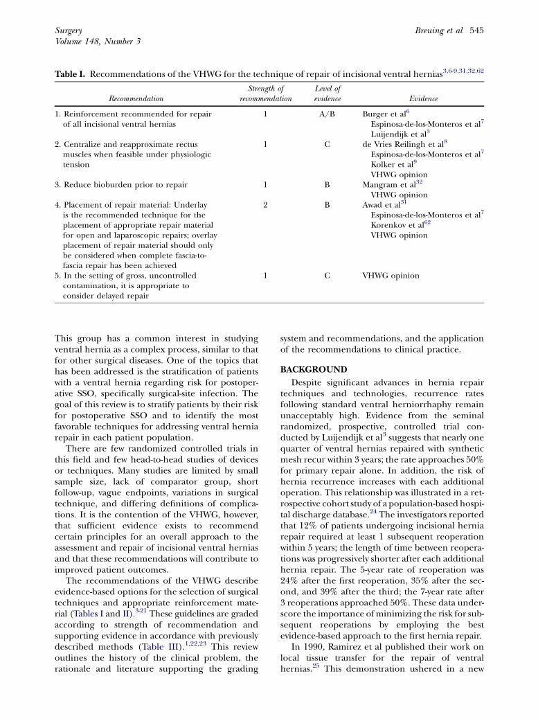
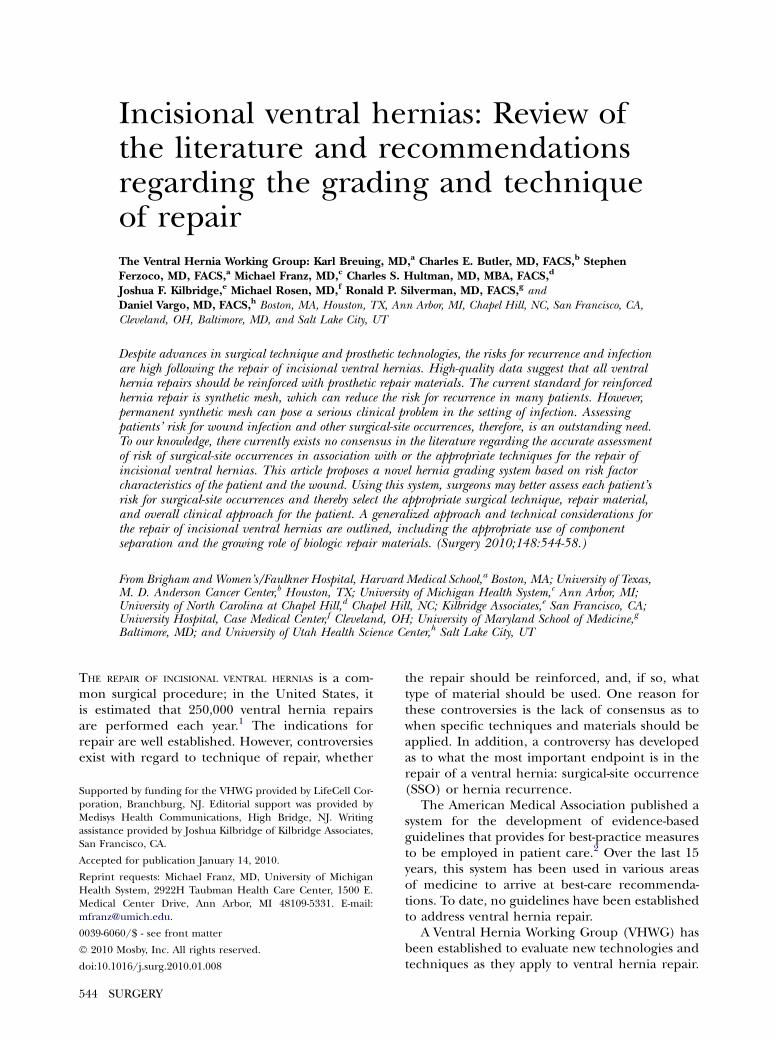
Table I. Recommendations of the VHWG for the technique of repair of incisional ventral hernias3,6-9,31,32,62
RecommendationStrength of
recommendationLevel ofevidence Evidence
1. Reinforcement recommended for repairof all incisional ventral hernias
1 A/B Burger et al6
Espinosa-de-los-Monteros et al7
Luijendijk et al3
2. Centralize and reapproximate rectusmuscles when feasible under physiologictension
1 C de Vries Reilingh et al8
Espinosa-de-los-Monteros et al7
Kolker et al9
VHWG opinion3. Reduce bioburden prior to repair 1 B Mangram et al32
VHWG opinion4. Placement of repair material: Underlay
is the recommended technique for theplacement of appropriate repair materialfor open and laparoscopic repairs; overlayplacement of repair material should onlybe considered when complete fascia-to-fascia repair has been achieved
2 B Awad et al31
Espinosa-de-los-Monteros et al7
Korenkov et al62
VHWG opinion
5. In the setting of gross, uncontrolledcontamination, it is appropriate toconsider delayed repair
1 C VHWG opinion
SurgeryVolume 148, Number 3
Breuing et al 545
This group has a common interest in studyingventral hernia as a complex process, similar to thatfor other surgical diseases. One of the topics thathas been addressed is the stratification of patientswith a ventral hernia regarding risk for postoper-ative SSO, specifically surgical-site infection. Thegoal of this review is to stratify patients by their riskfor postoperative SSO and to identify the mostfavorable techniques for addressing ventral herniarepair in each patient population.
There are few randomized controlled trials inthis field and few head-to-head studies of devicesor techniques. Many studies are limited by smallsample size, lack of comparator group, shortfollow-up, vague endpoints, variations in surgicaltechnique, and differing definitions of complica-tions. It is the contention of the VHWG, however,that sufficient evidence exists to recommendcertain principles for an overall approach to theassessment and repair of incisional ventral herniasand that these recommendations will contribute toimproved patient outcomes.
The recommendations of the VHWG describeevidence-based options for the selection of surgicaltechniques and appropriate reinforcement mate-rial (Tables I and II).3-21 These guidelines are gradedaccording to strength of recommendation andsupporting evidence in accordance with previouslydescribed methods (Table III).1,22,23 This reviewoutlines the history of the clinical problem, therationale and literature supporting the grading
system and recommendations, and the applicationof the recommendations to clinical practice.
BACKGROUND
Despite significant advances in hernia repairtechniques and technologies, recurrence ratesfollowing standard ventral herniorrhaphy remainunacceptably high. Evidence from the seminalrandomized, prospective, controlled trial con-ducted by Luijendijk et al3 suggests that nearly onequarter of ventral hernias repaired with syntheticmesh recur within 3 years; the rate approaches 50%for primary repair alone. In addition, the risk ofhernia recurrence increases with each additionaloperation. This relationship was illustrated in a ret-rospective cohort study of a population-based hospi-tal discharge database.24 The investigators reportedthat 12% of patients undergoing incisional herniarepair required at least 1 subsequent reoperationwithin 5 years; the length of time between reopera-tions was progressively shorter after each additionalhernia repair. The 5-year rate of reoperation was24% after the first reoperation, 35% after the sec-ond, and 39% after the third; the 7-year rate after3 reoperations approached 50%. These data under-score the importance of minimizing the risk for sub-sequent reoperations by employing the bestevidence-based approach to the first hernia repair.
In 1990, Ramirez et al published their work onlocal tissue transfer for the repair of ventralhernias.25 This demonstration ushered in a new
Table II. Recommendations of the VHWG for choice of repair material for incisional ventral hernias, bygrade4,5,11-21
RecommendationStrength of
recommendationLevel ofevidence Evidence
Grade 1 Choice of repair material by surgeon preferenceand patient factors
1 C VHWG opinion
Grade 2 Increased risk for surgical site occurrencesuggests additive risk of permanent syntheticrepair material, and potential advantage forappropriate biologic reinforcement
1 B Dunne et al12
Finan et al13
Pessaux et al14
Petersen et al20
VHWG opinionGrade 3 Permanent synthetic repair material generally
not recommended; potential advantage tobiologic repair material
1 B Diaz et al5
Houck et al11
Jones et al18
Kim et al4
Grade 4 Permanent synthetic repair material notrecommended; biologic repair materialshould be considered
1 A Diaz et al5
Jones et al18
Kim et al4
Paton et al16
Patton et al15
Sczczerba et al19
van’t Riet et al21
Voyles et al17
SurgerySeptember 2010
546 Breuing et al
era in hernia repair, where incisions to releasefascia allowed for a tension-free closure of themidline. In an effort to improve recurrence rates,synthetic mesh was employed to reinforce herniarepairs.6 However, there were significant complica-tions associated with use of synthetic mesh, includ-ing infection of the prosthesis and the formationof enterocutaneous fistulae.17,26-28 In the late1990s, biologic repair materials were introducedas a possible ventral hernia solution. Althoughmultiple products are available for use, no consen-sus exists as to the indicated patient population,how they should be implanted, and their overallrisk of complication and recurrence.
THE VHWG PROCESS
In September 2008, the VHWG met for a 2-daysummit with the goal of developing an initialstatement regarding the repair of incisional ventralhernias. The group consisted of 8 surgeons (4general and 4 plastic), all of whom have extensiveexperience in abdominal wall reconstruction. Thepurpose of the summit was 2-fold: (1) to propose agrading system to guide surgeons in the assessmentof patients with incisional ventral hernias withregard to risk for SSO, especially infection; and(2) to propose evidence-based recommendationsregarding the approach to advanced surgical tech-niques for the repair of incisional ventral hernia.All aspects related to hernia repair were evaluatedand broken down to their core components. Aliterature search was then undertaken to identify
known best practices in each core area determinedto be important to a successful ventral herniarepair. These articles were graded based on levelof evidence and used to develop the recommen-dations, grading system, and treatment algorithm.
RESULTS OF LITERATURE REVIEW
Initial discussions identified SSO and recur-rence as the 2 main issues in ventral hernia repair.For SSO, patient factors, wound factors, andchoice of implant were deemed to be most impor-tant. For recurrence, surgical technique wasthought to be most important, although patientand wound factors should also be considered. Asearch of the literature identified various factorsrelated to the status of the patient and wound thatshould be addressed when evaluating the overallcomplication risk in a patient with ventral hernia(discussed in the following paragraphs).
Infection and other SSOs. Common SSO fol-lowing ventral hernia repair include infection,seroma, wound dehiscence, and the formation ofenterocutaneous fistulae. Each of these complica-tions conveys morbidity and the risk for additionalsequelae. Each also relates to the management ofthe wound and to risks associated with the use ofrepair materials. A wound dehiscence, for exam-ple, may lead to exposure of the repair material; ifthe material is a permanent synthetic mesh, then itwill likely require removal because of continuedrisk for infection.3 Infection is a common and sig-nificant postoperative occurrence that increases
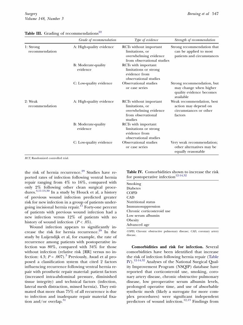
Table III. Grading of recommendations22
Grade of recommendation Type of evidence Strength of recommendation
1: Strongrecommendation
A: High-quality evidence RCTs without importantlimitations, oroverwhelming evidencefrom observational studies
Strong recommendation thatcan be applied to mostpatients and circumstances
B: Moderate-qualityevidence
RCTs with importantlimitations or strongevidence fromobservational studies
C: Low-quality evidence Observational studiesor case series
Strong recommendation, butmay change when higherquality evidence becomesavailable
2: Weakrecommendation
A: High-quality evidence RCTs without importantlimitations, oroverwhelming evidencefrom observationalstudies
Weak recommendation, bestaction may depend oncircumstances or otherfactors
B: Moderate-qualityevidence
RCTs with importantlimitations or strongevidence fromobservational studies
C: Low-quality evidence Observational studiesor case series
Very weak recommendation;other alternatives may beequally reasonable
RCT, Randomized controlled trial.
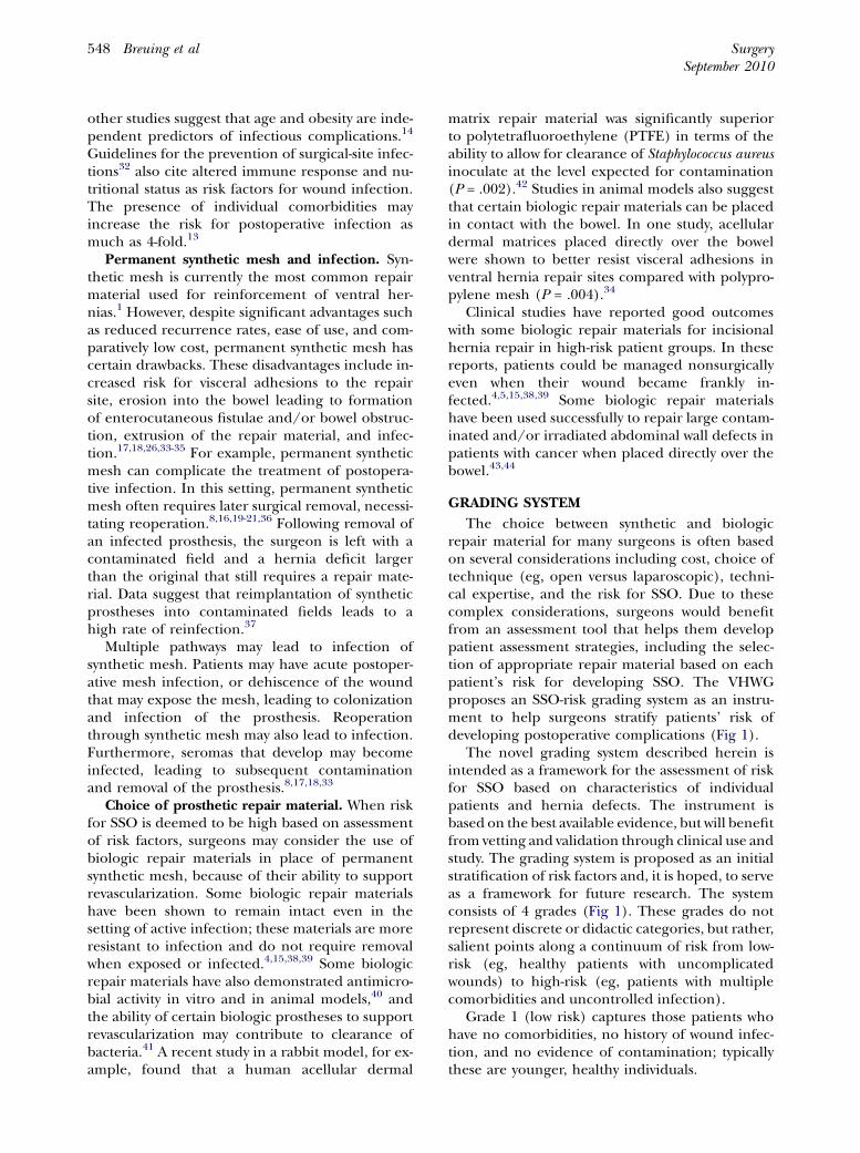
Table IV. Comorbidities shown to increase the riskfor postoperative infection12-14,32
SmokingDiabetesCOPDCADNutritional statusImmunosuppressionChronic corticosteroid useLow serum albuminObesityAdvanced age
COPD, Chronic obstructive pulmonary disease; CAD, coronary arterydisease.
SurgeryVolume 148, Number 3
Breuing et al 547
the risk of hernia recurrence.29 Studies have re-ported rates of infection following ventral herniarepair ranging from 4% to 16%, compared withonly 2% following other clean surgical proce-dures.3,11-13,30 In a study by Houck et al, a historyof previous wound infection predicted greaterrisk for new infection in a group of patients under-going incisional hernia repair.11 Forty-one percentof patients with previous wound infection had anew infection versus 12% of patients with nohistory of wound infection (P < .05).
Wound infection appears to significantly in-crease the risk for hernia recurrence.29 In thestudy by Luijendijk et al, for example, the rate ofrecurrence among patients with postoperative in-fection was 80%, compared with 34% for thosewithout infection (relative risk [RR] versus no in-fection: 4.3; P = .007).3 Previously, Awad et al pro-posed a classification system that cited 2 factorsinfluencing recurrence following ventral hernia re-pair with prosthetic repair material: patient factors(increased intra-abdominal pressure, diminishedtissue integrity) and technical factors (infection,lateral mesh distraction, missed hernia). They esti-mated that more than 75% of all recurrence is dueto infection and inadequate repair material fixa-tion and/or overlap.31
Comorbidities and risk for infection. Severalcomorbidities have been identified that increasethe risk of infection following hernia repair (TableIV).12-14,32 Analyses of the National Surgical Qual-ity Improvement Program (NSQIP) database havereported that corticosteroid use, smoking, coro-nary artery disease, chronic obstructive pulmonarydisease, low preoperative serum albumin levels,prolonged operative time, and use of absorbablesynthetic mesh (likely a surrogate for more com-plex procedures) were significant independentpredictors of wound infection.12,13 Findings from
SurgerySeptember 2010
548 Breuing et al
other studies suggest that age and obesity are inde-pendent predictors of infectious complications.14
Guidelines for the prevention of surgical-site infec-tions32 also cite altered immune response and nu-tritional status as risk factors for wound infection.The presence of individual comorbidities mayincrease the risk for postoperative infection asmuch as 4-fold.13
Permanent synthetic mesh and infection. Syn-thetic mesh is currently the most common repairmaterial used for reinforcement of ventral her-nias.1 However, despite significant advantages suchas reduced recurrence rates, ease of use, and com-paratively low cost, permanent synthetic mesh hascertain drawbacks. These disadvantages include in-creased risk for visceral adhesions to the repairsite, erosion into the bowel leading to formationof enterocutaneous fistulae and/or bowel obstruc-tion, extrusion of the repair material, and infec-tion.17,18,26,33-35 For example, permanent syntheticmesh can complicate the treatment of postopera-tive infection. In this setting, permanent syntheticmesh often requires later surgical removal, necessi-tating reoperation.8,16,19-21,36 Following removal ofan infected prosthesis, the surgeon is left with acontaminated field and a hernia deficit largerthan the original that still requires a repair mate-rial. Data suggest that reimplantation of syntheticprostheses into contaminated fields leads to ahigh rate of reinfection.37
Multiple pathways may lead to infection ofsynthetic mesh. Patients may have acute postoper-ative mesh infection, or dehiscence of the woundthat may expose the mesh, leading to colonizationand infection of the prosthesis. Reoperationthrough synthetic mesh may also lead to infection.Furthermore, seromas that develop may becomeinfected, leading to subsequent contaminationand removal of the prosthesis.8,17,18,33
Choice of prosthetic repair material. When riskfor SSO is deemed to be high based on assessmentof risk factors, surgeons may consider the use ofbiologic repair materials in place of permanentsynthetic mesh, because of their ability to supportrevascularization. Some biologic repair materialshave been shown to remain intact even in thesetting of active infection; these materials are moreresistant to infection and do not require removalwhen exposed or infected.4,15,38,39 Some biologicrepair materials have also demonstrated antimicro-bial activity in vitro and in animal models,40 andthe ability of certain biologic prostheses to supportrevascularization may contribute to clearance ofbacteria.41 A recent study in a rabbit model, for ex-ample, found that a human acellular dermal
matrix repair material was significantly superiorto polytetrafluoroethylene (PTFE) in terms of theability to allow for clearance of Staphylococcus aureusinoculate at the level expected for contamination(P = .002).42 Studies in animal models also suggestthat certain biologic repair materials can be placedin contact with the bowel. In one study, acellulardermal matrices placed directly over the bowelwere shown to better resist visceral adhesions inventral hernia repair sites compared with polypro-pylene mesh (P = .004).34
Clinical studies have reported good outcomeswith some biologic repair materials for incisionalhernia repair in high-risk patient groups. In thesereports, patients could be managed nonsurgicallyeven when their wound became frankly in-fected.4,5,15,38,39 Some biologic repair materialshave been used successfully to repair large contam-inated and/or irradiated abdominal wall defects inpatients with cancer when placed directly over thebowel.43,44
GRADING SYSTEM
The choice between synthetic and biologicrepair material for many surgeons is often basedon several considerations including cost, choice oftechnique (eg, open versus laparoscopic), techni-cal expertise, and the risk for SSO. Due to thesecomplex considerations, surgeons would benefitfrom an assessment tool that helps them developpatient assessment strategies, including the selec-tion of appropriate repair material based on eachpatient’s risk for developing SSO. The VHWGproposes an SSO-risk grading system as an instru-ment to help surgeons stratify patients’ risk ofdeveloping postoperative complications (Fig 1).
The novel grading system described herein isintended as a framework for the assessment of riskfor SSO based on characteristics of individualpatients and hernia defects. The instrument isbased on the best available evidence, but will benefitfrom vetting and validation through clinical use andstudy. The grading system is proposed as an initialstratification of risk factors and, it is hoped, to serveas a framework for future research. The systemconsists of 4 grades (Fig 1). These grades do notrepresent discrete or didactic categories, but rather,salient points along a continuum of risk from low-risk (eg, healthy patients with uncomplicatedwounds) to high-risk (eg, patients with multiplecomorbidities and uncontrolled infection).
Grade 1 (low risk) captures those patients whohave no comorbidities, no history of wound infec-tion, and no evidence of contamination; typicallythese are younger, healthy individuals.

• Previous woundinfection
• Stoma present
• Violation of thegastrointestinaltract
Grade 3PotentiallyContaminated
Grade 2Co-Morbid
• Smoker
• Obese
• Diabetic
• Immunosuppressed
• COPD
Grade 1Low Risk
• Low risk of complications
• No history ofwound infection
Grade 4Infected
• Infected mesh
• Septic dehiscence
Fig 1. Hernia grading system: assessment of risk for surgical site occurrences. Wound infection defined as being con-tained within the skin or subcutaneous tissue (superficial), or involving the muscle and/or fascia (deep).13
SurgeryVolume 148, Number 3
Breuing et al 549
Grade 2 (comorbid) includes patients who havecomorbidities that increase the risk for surgical-siteinfection (Table IV), but who do not have evidenceof wound contamination or active infection. Therelative contribution of different comorbidities is amatter for consideration and debate. To our knowl-edge, no data currently exist that dictate whichcomorbidities carry the most weight, or whichcombination of comorbidities increases risk.Similarly, there are only minimal data to delineatethe tipping point for a characteristic to be consid-ered a comorbidity (eg, how recent a history ofinfection, how much smoking, what degree ofmalnutrition, how much corticosteroid use).Certain thresholds have been described. Thresh-olds at which the risk for infection increases includeblood glucose $110 mg/dL (hemoglobin A1c >7.0)and age $75 years.45,46 Further research is requiredto better understand the contribution of comorbid-ities to risk. Until such data become available,surgeons must rely on their clinical judgment.
Grade 3 (potentially contaminated) is a higher-risk category based on evidence of contaminationof the wound. Factors that suggest contaminationinclude the presence of a nearby stoma, violationof the gastrointestinal tract, or history of woundinfection. Grade 4 (infected) patients are at high-est risk for SSO. Characteristics in grade 4 includeactive infection, especially infected synthetic mesh,and septic dehiscence. Each of these grades rep-resents a wide swath of risk and patient types.Assessment of risk, therefore, will continue to relyto some degree on individual surgeon judgmentand experience. The inclusion criteria for eachgrade will be further refined as new data regardingcomorbidities and outcomes become available.
Each grade relates to the aforementioned riskfactors for SSO but does not consider the size orcomplexity of the defect or the proposed approachto repair. For example, relatively small hernias withinfected mesh would still be considered grade 4
because of the presence of active infection. Con-versely, relatively large hernias in a healthy individ-ual may be considered grade 1 if there are nocomorbidities or signs of contamination, such asviolation of the bowel or history of wound infection.
There are characteristics of the patient, defect,and surgical site that may influence the risk forrecurrence as well as SSO. For example, a greaternumber of previous repairs increases the risk ofhernia recurrence.24 For the current statement,however, the VHWG concluded that there are stillinsufficient data in the literature to reliably gradethe risk of recurrence according to the proposedgrading scale. It was also agreed that inclusion ofhernia recurrence risk in the grading scale wouldmake it too complex for its intended purpose,which is to serve as a simple and memorable guideassessing a patient’s risk of SSO.
VHWG APPROACH TO THE TECHNIQUE FORTHE REPAIR OF INCISIONAL VENTRALHERNIAS
The application of advanced surgical techniquesand materials may reduce the risks of recurrenceand SSO such as infection. With the goal of mini-mizing recurrence and complications, the VHWGoffers evidence-based recommendations regardingtechnical approaches to the repair of incisionalventral hernias (Table I). Although these recom-mendations pertain mainly to open repairs, laparo-scopic approaches will be discussed briefly.
The recommendations are not intended to beprescriptive or definitive but to serve as principlesto guide the selection of surgical techniques. TheVHWG noted significant variation in technicaldetails between surgeons, both within the paneland in the community, and concluded that anyextensive discussion of technique is beyond thescope of this article. Therefore, the details of thetechniques cited in this statement are not fullydescribed herein.
Table V. Principles for the repair of incisionalventral hernia
Optimize patient conditionNutritional statusBlood sugar levelsSmoking cessation
Prepare woundReduce bioburdenTake down adhesions, fistulae
Reapproximate midline to the extent possible usingcomponent separation when appropriate
Use appropriate reinforcement materialConsider biologic repair material in patients atincreased risk for surgical-site occurrences
SurgerySeptember 2010
550 Breuing et al
The overall principles agreed on by the VHWG(Table V) are optimizationof the patient,preparationof the wound, centralization and reapproximation ofthe rectus muscles along the midline to the extentpossible, and the use of appropriate prosthetic repairmaterial to reinforce the closure. Surgical principlesare described in relation to each of the 4 grades ofrisk in the grading system described above and willfocus primarily on open repair.
Patient optimization. Patient optimization in-cludes encouraging smoking cessation ($4 weekspreoperatively), maintaining blood glucose levels(<110 mg/dL), improving oxygenation in patientswith chronic hypoxia (using bronchodilators, in-haled corticosteroids, and/or prostaglandin inhib-itors), and setting patient expectations.12,39,47
Additional factors include weight loss, optimiza-tion of nutritional status, and management/con-trol of any infection, if possible. Relevant sites ofdistal infection include an ileal conduit or thebladder in a patient who requires chronic catheter-ization to drain urine.
Wound preparation. There are 2 stages of woundpreparation. The first occurs prior to surgery; thisstage may include percutaneous drainage of anyabscesses or management of skin irritation from anenterocutaneous fistula. The second stage occurs inthe operating room; sharp debridement of all devi-talized or infected tissue to reduce the bioburden ofthe wound is critical, and contaminated woundsshould be cleaned by pulse lavage.32 If the biobur-den can be successfully managed, then immediatereconstruction can be performed. If not, then astaged approach with multiple wound debride-ments prior to reconstruction may be needed. All fis-tulae should be definitively managed with excisionand reanastomosis or externalization, and infectedsynthetic prostheses should be removed.
Reapproximation of the rectus muscles. It is therecommendation of the VHWG to centralize and
reapproximate the rectus muscles along the mid-line for ventral hernia repairs to the extent possi-ble. This step attempts to restore the functional,innervated abdominal wall and create a true dy-namic repair without undue tension. The phrase‘‘without undue tension’’ refers to the attempt torestore normal physiologic tension. The abdomi-nal wall is a load-bearing structure and reactsdynamically to internal and external forces (hence‘‘dynamic repair’’). Too little tension in a herniarepair results in wound edge separation and poorcollagen organization in the incision; too muchtension leads to ischemia and wound dehiscence.Physiologic tension attempts to achieve a balancebetween these opposing outcomes.48
Techniques for the repair of ventral herniascommonly used by the VHWG and communitysurgeons include retrorectus (ie, Rives-Stoppa pro-cedure) and component separation. Retrorectusrepair has been widely employed in Europe and isconsidered by some surgeons to be the standardfor repair of ventral hernias. The technique allowsfor placement of repair material behind the defectwithout contacting the viscera. The technique ofretrorectus repair is described in detail by otherauthors.49,50 Consideration should be given to theuse of biologic or synthetic repair materials withlower risk for adhesions in case the posteriorsheath is absent or breaks down. Retrorectus repairalone, however, does not reduce large defects orcentralize the midline.
For larger defects, formal component separa-tion, as first described by Ramirez et al25 and mod-ified by numerous authors,8,9,51-57 is the preferredapproach for reapproximating the midline withminimal or no tension. Component separation cre-ates a dynamic repair by using incisions that createfascial release to bring the rectus muscles togetherat the midline, thereby recreating an innervated,functional abdominal wall. Elements of each tech-nique may be used in conjunction. The VHWG rec-ommends the use of component separation orother appropriate techniques to reapproximatethe midline for all ventral hernias, except forvery small defects or cases where reapproximationis not feasible.
Case series suggest that open component sepa-ration has utility in challenging cases, and canreduce recurrence53,58,59; however, patients willstill benefit from prosthetic repair material, partic-ularly in complex defects (eg, degraded fascia,tight closure, multiple comorbidities, contamina-tion).7,8,9,58 A recent retrospective review, for ex-ample, compared component separation withoutreinforcement to component separation plus
SurgeryVolume 148, Number 3
Breuing et al 551
biologic repair material overlay.7 This study re-ported a significantly lower recurrence rate whencomponent separation was reinforced with bio-logic repair material (0%, component separationplus overlay versus 13%, component separationalone; P = .006). One randomized, prospective trialcompared component separation to primary re-pair with expanded PTFE (ePTFE).8 An interimanalysis reported hernia recurrence in 10 of 19 pa-tients in the component separation group (meantime to recurrence, 7 months) and 4 of 18 in theePTFE group (mean time to recurrence, 22months). Seven patients in the ePTFE group hadan infection of the mesh that required removalof the prosthesis, followed by reconstruction usingcomponent separation. It should be noted, how-ever, that no published data have been founddirectly comparing component separation to pri-mary repair alone (or any other repair technique),nor are there any prospective data evaluating theaddition of prosthetic repair material to compo-nent separation.
SELECTION AND USE OF PROSTHETICREPAIR MATERIAL
Level 1A data from the study by Luijendijk et alindicate that all clean, grade 1 ventral herniarepairs should be reinforced with some type ofrepair material.3,6 Even in the small hernias in rel-atively healthy patients included in this study (fas-cial defect length or width #6 cm), the use ofprosthetic repair material halved the rate of recur-rence, both over short-term (23% vs 46%; P =.005)3 and longer-term (32% vs 63%; P < .001) fol-low-up.6 Based on these data, the VHWG recom-mends the use of prosthetic repair material toreinforce the repair of all incisional ventral her-nias, regardless of whether or not the midline fas-cia can be reapproximated.
The diversity of synthetic and biologic repairmaterials available for the reinforcement of herniarepair complicates the selection of an appropriateprosthesis. At least 80 different prosthetic mate-rials are available for hernia repair,60 and the char-acteristics and types of prostheses vary considerablyeven within the classes of synthetic and biologicmaterials. The choice of material may be basedon a variety of considerations, including character-istics of the patient and defect, surgeon familiaritywith material, and cost. The risk for SSO and sub-sequent infection may determine the selection of asynthetic versus a biologic repair material. Basedon the grading system described above, theVHWG recommends that biologic repair materialswith specific characteristics (see below) are
preferred over synthetic mesh for use in infectedfields and should be strongly considered whencontamination is suspected (Table II). TheVHWG also notes that the increased risk for SSOassociated with comorbidities within grade 2 maysuggest potential advantages to some biologic re-pair materials, depending on choice of technique(eg, open versus laparoscopic) and the balanceof benefits and risks. It should be emphasizedthat this suggestion is based on the presumptionthat certain patients with comorbidities (ie, grade2) will, in fact, develop SSOs such as wound infec-tion, and that biologic repair materials may facili-tate management of infection withoutnecessitating removal. To date, we have found nopublished controlled clinical studies comparing bi-ologic and synthetic repair materials in this patientpopulation.
Although the VHWG does not make any recom-mendation regarding choice of specific prostheticrepair materials, certain features of synthetic andbiologic repair materials should be considered dur-ing the selection process. The VHWG calls attentionto specific characteristics such as adequate strength,ease of handling during procedures, ability to resistadhesions when placed in contact with the bowel,and reduced risk of infection through support fortissue incorporation and revascularization.
Synthetic repair materials. Synthetic meshes aremost often categorized as macroporous, micropo-rous, or composite.61,62 Macroporous meshesinclude monofilament and double-filament polypro-pylene, among many others. These materials havelarge pore sizes that allow for in-growth of scar tissue.When placed in contact with abdominal viscera, mac-roporous meshes are associated with the formationof bowel adhesions and obstructions and enterocuta-neous fistulae.63,64 Therefore, these materialsshould be avoided or used in combination with vas-cularized tissue (eg, greater omentum, hernia sac)or antiadhesive barriers when contact with the bowelis likely. Microporous meshes, such as ePTFE, have asmaller pore size that does not allow for tissue in-growth, but may lead to encapsulation and the persis-tence of bacteria. Therefore, microporous mesh hasa lower affinity for adhesions, but may be more sus-ceptible to infection.
A wide variety of composite materials is nowavailable that combine different qualities, such ashaving macroporous mesh on one side to promotetissue in-growth and microporous mesh on theother to reduce risk for adhesions to the mesh (eg,polypropylene/ePTFE). Synthetic meshes withantiadhesive coatings have also been developed.Such coatings include nonabsorbable (eg, titanium,
SurgerySeptember 2010
552 Breuing et al
polyurethane) and absorbable coatings (eg, omega-3 fatty acid, collagen hydrogel, oxygenated regen-erated cellulose). Preclinical evidence suggestsreduced risk of adhesions to composite and coatedsynthetic meshes compared with traditionalsynthetic meshes.65-69 The relative benefits of thesedifferent prostheses with regard to adhesion forma-tion and risk for infection vary according todifferent study models, methodologies, and out-comes.63,67,70-73 Furthermore, prospective data arelacking regarding the clinical benefits of theseprostheses for ventral hernia repair, and no compar-ative clinical data are currently available.
Finally, a new category of lightweight mesh iscurrently being used in both open and laparo-scopic hernia repairs. There are data to suggestbetter functional outcomes than those achievedwith traditional synthetic mesh, although definitivestudies are lacking.74
Biologic repair materials. Biologic repair mate-rials are an equally diverse and expanding class.Certain specific characteristics are thought to con-tribute to the successful use of particular biologicrepair materials in the setting of contamination orlow-grade infection, whereas others are contra-indicated. These properties include intact extra-cellular matrix and the ability to support tissueregeneration through revascularization and cellrepopulation in a clinically relevant timeframe. Ithas been hypothesized that resistance to infectionfor some biologic repair materials may be relatedto the in-growth of cells and vasculature.75 Numer-ous animal studies have shown that altering the ex-tracellular matrix through suboptimal processingand/or crosslinking may have a negative impacton host response to the repair material.76,77 Theneovascularization demonstrated in studies ofsome biologic repair materials may allow thesematerials to better resist infection when placed ina potentially contaminated field.42,75
The ability of some biologic repair materials tosupport regeneration is based on studies in animalmodels that describe the immunologic response ofthe host to the prosthesis. Positive recognition (ie,recognition of the prosthesis as ‘‘self’’) leads toregeneration and integration of the repair materialinto native tissue. Negative recognition (ie, recog-nition of the prosthesis as foreign) may lead toresorption or encapsulation.76,78 Resorption andencapsulation have been demonstrated with severalbiologic repair materials in a nonhuman primatemodel of abdominal wall repair.76 The investigatorssuggested that the lack of integration and tissue re-generation with these materials may account forpoor initial wound healing. Integration of 1 non--
cross-linked, intact biologic repair material into na-tive tissue was demonstrated in the same nonhumanprimate model. These results are similar to those re-ported in clinical studies.44,79 In one study of ab-dominal repair following harvest of transverserectus abdominus musculocutaneous flaps forbreast reconstruction, biopsies of the biologic re-pair material showed similar cell density, vascula-ture, and collagen orientation to those of normalabdominal fascial tissue.79 A second study foundthat explanted biologic repair material from an irra-diated, contaminated abdominal wall repair site 14months after implantation demonstrated remodel-ing of the biologic repair material, including revas-cularization and cellular repopulation.44
It should be emphasized that no comparativetrials have been performed to date evaluatingdifferent biologic repair materials in incisionalhernia repair, and differentiation between pro-ducts is based on early findings with a limitednumber of the available prostheses. Data describ-ing the qualities of biologic repair materials areonly available for certain prostheses. Similar ani-mal and clinical studies are awaited for the major-ity of products in this class.
TECHNIQUE OF PLACEMENT
There are technical aspects of the use of bio-logic repair material that must be considered inorder to achieve successful outcomes. Studies havedocumented high rates of seroma, diastasis, bulg-ing, and recurrence with biologic repair mate-rials80,81; critical techniques of placement weredescribed that may influence the risk of these com-plications.43 In one study, recurrence was reducedwhen component separation was combined withbiologic repair material; conversely, bridging withbiologic repair material without reducing the sizeof the defect was associated with a recurrencerate of 80%.81 The tensile qualities of repair mate-rials differ and may impact technique. The VHWGnotes that most biologic repair materials should beimplanted under appropriate tension to help pre-vent the development of laxity. (This use of tensionfor repair material implantation should be distin-guished from the avoidance of undue tension---orphysiologic tension---that describes the fascial clo-sure.) Surgeons should be aware that the use of abiologic repair material necessitates technicalfamiliarity with its appropriate placement.
Overlay, underlay, or interpositional placementof prosthetic repair material. In open incisionalhernia repair, prosthetic repair material may beplaced to reinforce a primary repair or to bridge aremaining defect if reapproximation of the
SurgeryVolume 148, Number 3
Breuing et al 553
midline is not possible. The repair material may besutured superficial to the primary repair or fascialedges (overlay), deep to the primary repair orfascial edges (underlay), or to the edge of thedefect with minimal overlap (interpositional). Theoverlay technique is easier to perform, does notrequire devascularization of the rectus, and pre-vents contact between the repair material and theunderlying viscera. Overlay placement also allowsfor reinforcement of the lateral releasing incisionsafter component separation, if desired. Overlayplacement, therefore, may be preferred for typesof synthetic mesh that are associated with forma-tion of bowel adhesions to minimize the risk thatthe mesh may erode into the abdominal compart-ment and become exposed to the viscera.
There are also theoretical advantages to theplacement of repair material as an underlay. Whenthe material is placed deep to the abdominalmusculature, increases in intra-abdominal pressurepress the repair material into the defect andagainst the native tissue, rather than away fromthe defect. Intra-abdominal forces may also bemore evenly distributed across the repair materialwhen placed as an underlay.82 Furthermore, cuta-neous exposure does not result in exposure ofthe repair material, because the prosthesis remainsbelow the musculofascial layer.
Bridging of defects, which refers to the use ofprosthetic repair material to span tissue gaps whenreapproximation of the fascial edges is not possi-ble, has been associated with high rates of recur-rence and complications. Bridging may notgenerally be recommended except in cases wherecomponent separation is not feasible or is insuffi-cient to bring the fascial edges together (seediscussion of algorithm, below).83
The VHWG notes that underlay may be pre-ferred because of the theoretical advantages of thistechnique. However, there are no reliable datasupporting the use of one technique over an-other.83 Patient factors and surgeon preferenceshould also be considered. Regardless of place-ment, repair material should overlap with intactfascia by at least 3--5 cm.34,84-89
TECHNICAL OPTIONS BY GRADE
The overriding recommendation of the VHWGregarding the repair of incisional ventral hernia isto reinforce the primary fascial closure with aprosthetic repair material.6 The selection of typeof repair material between biologic and syntheticwith regard to hernia grade should be based onrisk for SSO (Table II). For patients at low riskfor SSO (grade 1), the choice of reinforcement
should be based on surgeon preference and pa-tient factors. Grade 2 encompasses patients withcomorbidities, such as smoking, diabetes, or mal-nutrition (Table IV). Data from analyses of theNSQIP database and other studies suggest that pa-tients in grade 2 have a wound infection rate that is4-fold greater than what is predicted based solelyon wound classification.12,13 Current published ev-idence does not delineate the relative contributionof each comorbidity to increased risk. Ongoingand future clinical studies may provide a morethorough evidence-based estimate of which andhow many comorbidities contribute most signifi-cantly to increased risk of SSO. In the absence ofmore definitive data, the VHWG notes that the in-creased risk associated with these comorbiditiessuggests a potential advantage for the use of appro-priate biologic repair material for reinforcementof open repairs.
Grade 3 includes patients with contamination ofthe wound or suspicion of contamination, includ-ing a previous wound infection. Based on theincreased risk for infection associated with contam-inated wounds, the VHWG notes that permanentsynthetic mesh is generally not recommended forpatients considered to be grade 3. Appropriatebiologic repair material is a good option for rein-forcement in these patients, because it does notnecessitate removal even in the setting of activeinfection.
Grade 4 patients have frankly infected wounds,most notably those associated with an existinginfected synthetic mesh. Studies suggest that thereplacement of infected synthetic mesh with newpermanent synthetic mesh leads to a high rate ofreoperation and additional mesh infection andreplacement.8 The use of permanent syntheticmesh in patients considered to be grade 4, there-fore, is not recommended by the VHWG. In accor-dance with the surgical principles outlined aboveand in Table V, infected wounds should be thor-oughly prepared by meticulously reducing the bio-burden prior to placement of repair material anddefinitive closure. No repair material should beused in the setting of gross, uncontrolled contam-ination, and surgeons may consider a delayedrepair in such situations.
LAPAROSCOPIC REPAIR OF INCISIONALVENTRAL HERNIA
This statement focuses primarily on the openrepair of incisional ventral hernia. However, thegrowing popularity of laparoscopic techniques de-serves discussion with relation to the gradingsystem and recommendations of the VHWG.
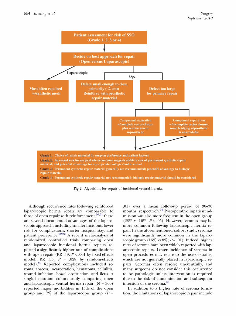
Decide on best approach for repair(Open versus Laparascopic)
Patient assessment for risk of SSO (Grade 1, 2, 3 or 4)
Grade 1: Choice of repair material by surgeon preference and patient factors
Grade 2: Increased risk for surgical site occurrence suggests additive risk of permanent synthetic repair material, and potential advantage for appropriate biologic reinforcement
Grade 3: Permanent synthetic repair material generally not recommended; potential advantage to biologic repair material
Grade 4: Permanent synthetic repair material not recommended; biologic repair material should be considered
Most often repaired w/synthetic mesh
Defect small enough to closeprimarily (≤2 cm):
Reinforce with prostheticrepair material
Laparascopic Open
Defect too large for primary repair
Component separation w/incomplete rectus closure, some bridging w/prosthetic
is unavoidable
Component separation w/complete rectus closure
plus reinforcement w/prosthetic
Fig 2. Algorithm for repair of incisional ventral hernia.
SurgerySeptember 2010
554 Breuing et al
Although recurrence rates following reinforcedlaparoscopic hernia repair are comparable tothose of open repair with reinforcement,90,91 thereare several documented advantages of the laparo-scopic approach, including smaller incisions, lowerrisk for complications, shorter hospital stay, andpatient preference.90-92 A recent meta-analysis ofrandomized controlled trials comparing openand laparoscopic incisional hernia repairs re-ported a significantly higher rate of complicationswith open repair (RR .49, P < .001 by fixed-effectsmodel; RR .53, P = .028 by random-effectsmodel).89 Reported complications included se-roma, abscess, incarceration, hematoma, cellulitis,wound infection, bowel obstruction, and ileus. Asingle-institution cohort study comparing openand laparoscopic ventral hernia repair (N = 360)reported major morbidities in 15% of the opengroup and 7% of the laparoscopic group (P =
.01) over a mean follow-up period of 30--36months, respectively.91 Postoperative inpatient ad-mission was also more frequent in the open group(28% vs 16%; P < .05). However, seromas may bemore common following laparoscopic hernia re-pair. In the aforementioned cohort study, seromaswere significantly more common in the laparo-scopic group (16% vs 8%; P = .01). Indeed, higherrates of seroma have been widely reported with lap-aroscopic repairs. Lower incidence of seroma inopen procedures may relate to the use of drains,which are not generally placed in laparoscopic re-pairs. Seromas often resolve uneventfully, andmany surgeons do not consider this occurrenceto be pathologic unless intervention is requireddue to the risk of contamination and subsequentinfection of the seroma.82
In addition to a higher rate of seroma forma-tion, the limitations of laparoscopic repair include
SurgeryVolume 148, Number 3
Breuing et al 555
the inability to restore functional abdominal wallanatomy. Other difficulties include the inability tomanage skin redundancy and the hernia sac.Current approaches to laparoscopic repair do notroutinely employ extensive mobilization of tissue,meaning that the repair material is almost alwaysbridging some aspect of the defect. Laparoscopi-cally inserted repair material is placed intraperito-neally as an underlay below the fascial defect.82
These repairs do not recreate an innervatedabdominal wall under physiologic tension.
Recently, several investigators have describedminimally invasive techniques of component sep-aration.54 Experience with these techniques hasbeen reported in studies of cadavers,55 a porcinemodel,56 select patients with infected repair mate-rial,57 and small comparative groups.54 Preliminaryresults suggest that minimally invasive techniquesare feasible, and may be associated with fewercomplications.
TREATMENT ALGORITHM
The first step in the treatment of ventral herniais patient assessment, starting with risk factors andsize of the defect. Smaller defects (#2 cm) may besuitable for primary repair; larger defects wherethe fascia does not meet without undue tensionshould be reduced as much as possible. Eachpatient’s risk for SSO should be assessed usingthe grading system.
A proposed algorithm for the treatment of inci-sional ventral hernias is illustrated in Fig 2. Followingassessment for risk of SSO, patients are categorizedby size of defect. Very small defects may be closed pri-marily along with reinforcing prosthetic repair mate-rial, potentially using a retrorectus repair. Mostdefects too large for primary repair can be closedwith component separation and reinforced withprosthetic repair material. For the rare cases inwhich component separation is not feasible or is in-sufficient to completely reduce the defect, surgeonsmay consider bridging the defect with prosthetic re-pair material. (The repair material should underliethe rectus muscles by at least 5 cm.) Examples of pa-tients for whom component separation may not befeasible include those with intensive radiation treat-ment of the abdominal wall or extensive scarring ofthe rectus muscles. Surgeons should exercise theirjudgment when considering the feasibility of compo-nent separation. When using component separationand/or other techniques to reapproximate the rec-tus muscles, the authors find that bridging of defectswith biologic repair material is rarely necessary.
The nature of a laparoscopic ventral herniarepair as currently performed leads to a bridged
repair. For surgeons who practice laparoscopicrepairs, patients in grade 1, many in grade 2, andsome in grade 3 may be suitable for this approach,depending on individual risk for infection andother considerations. Hernias in grade 4 should berepaired with open procedures. The same princi-ples of selecting prosthetic repair material applyregardless of technique (open versus laparo-scopic): most patients in grade 1, some in grade2, and a few in grade 3 may be suitable for repairwith permanent synthetic mesh; all patients con-sidered at increased risk for SSO (including somein grade 2, most in grade 3, and all in grade 4)should be considered for repair with appropriatebiologic repair material.
OTHER CONSIDERATIONS IN SELECTION OFREPAIR MATERIAL AND TECHNIQUE
One key consideration in the selection of pros-thetic repair material deserves mention. Currently,there is wide variation in the cost of availableprostheses. For some institutions and practices,cost may limit or eliminate the use of moreexpensive devices. A thorough discussion of costconsiderations is not the intended purpose of thisarticle. However, future analyses of the cost-benefitrelationship accounting for the expense of mate-rials, surgical procedures, and potential complica-tions would be greatly beneficial to practitionersand administrators alike.
Many of the advanced techniques described inthis consensus statement require extensive hospitalresources and a high level of training. Surgeons insettings with less extensive resources may giveconsideration to the referral of resource-intensivepatients to tertiary care centers that have appro-priate surgical resources.
SUMMARY
Incisional ventral hernias are common andchallenging for surgeons. The lack of high-qualityevidence leaves surgeons without clear guidanceregarding the selection of technique or material.The ultimate goal of this effort was to produce asimple, generally accepted grading system andsurgical technique recommendations for the re-pair of incisional ventral hernias. The first step inthis effort was the creation of an initial literaturereview and set of recommendations. This state-ment represents the current state-of-the-art tech-nique and materials as described by thoughtleaders in the field and supported by the bestavailable evidence. It is hoped that the gradingsystem and recommendations will serve to assistsurgeons and stimulate discussion and research.
SurgerySeptember 2010
556 Breuing et al
As new data become available, the VHWG willrevisit this statement to reflect the evolving under-standing of ventral hernias. Future updates will beprovided as data emerge and novel techniques andmaterials are developed.
REFERENCES
1. Millennium Research Group. US markets for soft tissue re-pair 2009. Toronto, ON: Millennium Research Group, Inc;2008.
2. American Medical Association. Attributes to guide the de-velopment and evaluation of practice parameters/guide-lines. Chicago: American Medical Association; 1996.
3. Luijendijk RW, Hop WC, van den Tol MP, de Lange DC,Braaksma MM, IJzermans JN, et al. A comparison of suturerepair with mesh repair for incisional hernia. N Engl J Med2000;343:392-8.
4. Kim H, Bruen K, Vargo D. Acellular dermal matrix in themanagement of high-risk abdominal wall defects. AmJ Surg 2006;192:705-9.
5. Diaz JJ Jr, Guy J, Berkes MB, Guillamondegui O, Miller RS.Acellular dermal allograft for ventral hernia repair in thecompromised surgical field. Am Surg 2006;72:1181-7.
6. Burger JW, Luijendijk RW, Hop WC, Halm JA, VerdaasdonkEG, Jeekel J. Long-term follow-up of a randomized con-trolled trial of suture versus mesh repair of incisional her-nia. Ann Surg 2004;240:578-83.
7. Espinosa-de-los-Monteros A, de la Torre JI, Marrero I, An-drades P, Davis MR, Vasconez LO. Utilization of human cadav-eric acellular dermis for abdominal hernia reconstruction.Ann Plast Surg 2007;58:264-7.
8. de Vries Reilingh TS, van Goor H, Charbon JA, Rosman C,Hesselink EJ, van der Wilt GJ, et al. Repair of giant midlineabdominal wall hernias: ‘‘components separation tech-nique’’ versus prosthetic repair: interim analysis of a ran-domized controlled trial. World J Surg 2007;31:756-63.
9. Kolker AR, Brown DJ, Redstone JS, Scarpinato VM, WallackMK. Multilayer reconstruction of abdominal wall defectswith acellular dermal allograft (AlloDerm) and componentseparation. Ann Plast Surg 2005;55:36-41.
10. Fabian TC, Croce MA, Pritchard FE, Minard G, HickersonWL, Howell RL, et al. Planned ventral hernia. Staged man-agement for acute abdominal wall defects. Ann Surg 1994;219:643-50.
11. Houck JP, Rypins EB, Sarfeh IJ, Juler GL, Shimoda KJ. Repairof incisional hernia. Surg Gynecol Obstet 1989;169:397-9.
12. Dunne JR, Malone DL, Tracy JK, Napolitano LM. Abdomi-nal wall hernias: risk factors for infection and resource uti-lization. J Surg Res 2003;111:78-84.
13. Finan KR, Vick CC, Kiefe CI, Neumayer L, Hawn MT. Pre-dictors of wound infection in ventral hernia repair. AmJ Surg 2005;190:676-81.
14. Pessaux P, Lermite E, Blezel E, et al. Predictive risk score forinfection after inguinal hernia repair. Am J Surg 2006;192:165-71.
15. Patton JH Jr, Berry S, Kralovich KA. Use of human acellulardermal matrix in complex and contaminated abdominalwall reconstructions. Am J Surg 2007;193:360-3.
16. PatonBL,Novitsky YW, ZereyM, SingRF, KercherKW,HenifordBT. Management of infections of polytetrafluoroethylene-basedmesh. Surg Infect (Larchmt) 2007;8:337-41.
17. Voyles CR, Richardson JD, Bland KI, Tobin GR, Flint LM,Polk HC Jr. Emergency abdominal wall reconstruction
with polypropylene mesh: short-term benefits versus long-term complications. Ann Surg 1981;194:219-23.
18. Jones JW, Jurkovich GJ. Polypropylene mesh closure of in-fected abdominal wounds. Am Surg 1989;55:73-6.
19. Szczerba SR, Dumanian GA. Definitive surgical treatment ofinfected or exposed ventral hernia mesh. Ann Surg 2003;237:437-41.
20. Petersen S, Henke G, Freitag M, Faulhaber A, Ludwig K. Deepprosthesis infection in incisional hernia repair: predictive fac-tors and clinical outcome. Eur J Surg 2001;167:453-7.
21. van ’t Riet M, de Vos van Steenwijk PJ, Bonjer HJ, SteyerbergEW, Jeekel J. Mesh repair for postoperative wound dehis-cence in the presence of infection: is absorbable mesh saferthan non-absorbable mesh? Hernia 2007;11:409-13.
22. Guyatt G, Gutterman D, BaumannMH, Addrizzo-Harris D, Hy-lek EM, Phillips B,et al. Grading strengthof recommendationsand quality of evidence in clinical guidelines: report from anamerican college of chest physicians task force. Chest 2006;129:174-81.
23. Shekelle PG, Woolf SH, Eccles M, Grimshaw J. Clinicalguidelines: developing guidelines. BMJ 1999;318:593-6.
24. Flum DR, Horvath K, Koepsell T. Have outcomes of inci-sional hernia repair improved with time? A population-based analysis. Ann Surg 2003;237:129-35.
25. Ramirez OM, Ruas E, Dellon AL. ‘‘Components separation’’method for closure of abdominal-wall defects: an anatomicand clinical study. Plast Reconstr Surg 1990;86:519-26.
26. Leber GE, Garb JL, Alexander AI, Reed WP. Long-termcomplications associated with prosthetic repair of incisionalhernias. Arch Surg 1998;133:378-82.
27. Cobb WS, Harris JB, Lokey JS, McGill ES, Klove KL. Inci-sional herniorrhaphy with intraperitoneal compositemesh: a report of 95 cases. Am Surg 2003;69:784-7.
28. Karakousis CP, Volpe C, Tanski J, Colby ED, Winston J, Dris-coll DL. Use of a mesh for musculoaponeurotic defects ofthe abdominal wall in cancer surgery and the risk of bowelfistulas. J Am Coll Surg 1995;181:11-6.
29. Iqbal CW, Pham TH, Joseph A, Mai J, Thompson GB, SarrMG. Long-term outcome of 254 complex incisional herniarepairs using the modified Rives-Stoppa technique. WorldJ Surg 2007;31:2398-404.
30. White TJ, Santos MC, Thompson JS. Factors affectingwound complications in repair of ventral hernias. AmSurg 1998;64:276-80.
31. Awad ZT, Puri V, LeBlanc K, et al. Mechanisms of ventralhernia recurrence after mesh repair and a new proposedclassification. J Am Coll Surg 2005;201:132-40.
32. Mangram AJ, Horan TC, Pearson ML, Silver LC, Jarvis WR.Guideline for prevention of surgical site infection, 1999.Hospital Infection Control Practices Advisory Committee.Infect Control Hosp Epidemiol 1999;20:250-78.
33. Stone HH, Fabian TC, Turkleson ML, Jurkiewicz MJ. Man-agement of acute full-thickness losses of the abdominalwall. Ann Surg 1981;193:612-8.
34. Butler CE, Prieto VG. Reduction of adhesions with compos-ite AlloDerm/polypropylene mesh implants for abdominalwall reconstruction. Plast Reconstr Surg 2004;114:464-73.
35. Bauer JJ, Harris MT, Kreel I, Gelernt IM. Twelve-year expe-rience with expanded polytetrafluoroethylene in the repairof abdominal wall defects. Mt Sinai J Med 1999;66:20-5.
36. Martin-Duce A, Noguerales F, Villeta R, et al. Modificationsto Rives technique for midline incisional hernia repair. Her-nia 2001;5:70-2.
37. Clagett GP, Bowers BL, Lopez-Viego MA, Rossi MB, Valen-tine RJ, Myers SI, et al. Creation of a neo-aortoiliac system

SurgeryVolume 148, Number 3
Breuing et al 557
from lower extremity deep and superficial veins. Ann Surg1993;218:239-48.
38. Helton WS, Fisichella PM, Berger R, Horgan S, Espat NJ,Abcarian H. Short-term outcomes with small intestinal sub-mucosa for ventral abdominal hernia. Arch Surg 2005;140:549-60.
39. Maurice SM, Skeete DA. Use of human acellular dermal ma-trix for abdominal wall reconstructions. Am J Surg 2009;197:35-42.
40. Sarikaya A, Record R, Wu CC, Tullius B, Badylak S, LadischM. Antimicrobial activity associated with extracellular matri-ces. Tissue Eng 2002;8:63-71.
41. Badylak SF, Coffey AC, Lantz GC, Tacker WA, Geddes LA.Comparison of the resistance to infection of intestinal sub-mucosa arterial autografts versus polytetrafluoroethylene ar-terial prostheses in a dog model. J Vasc Surg 1994;19:465-72.
42. Milburn ML, Holton LH, Chung TL, Li EN, Bochicchio GV,Goldberg NH, et al. Acellular dermal matrix compared withsynthetic implant material for repair of ventral hernia in thesetting of peri-operative Staphylococcus aureus implantcontamination: a rabbit model. Surg Infect (Larchmt)2008;9:433-42.
43. Butler CE, Langstein HN, Kronowitz SJ. Pelvic, abdominal,and chest wall reconstruction with AlloDerm in patients atincreased risk for mesh-related complications. Plast Re-constr Surg 2005;116:1263-75.
44. Nemeth NL, Butler CE. Complex torso reconstruction withhuman acellular dermal matrix: long-term clinical follow-up. Plast Reconstr Surg 2009;123:192-6.
45. Malone DL, Genuit T, Tracy JK, Gannon C, Napolitano LM.Surgical site infections: reanalysis of risk factors. J Surg Res2002;103:89-95.
46. van den Berghe G, Wouters P, Weekers F, Verwaest C, Bruy-ninckx F, Schetz M, et al. Intensive insulin therapy in thecritically ill patients. N Engl J Med 2001;345:1359-67.
47. Lindstrom D, Sadr Azodi O, Wladis A, Tonnesen H, LinderS, Nasell H, et al. Effects of a perioperative smoking cessa-tion intervention on postoperative complications: a ran-domized trial. Ann Surg 2008;248:739-45.
48. Franz MG. The biology of hernias and the abdominal wall.Hernia 2006;10:462-71.
49. Bauer JJ, Harris MT, Gorfine SR, Kreel I. Rives-Stoppa pro-cedure for repair of large incisional hernias: experiencewith 57 patients. Hernia 2002;6:120-3.
50. Heartsill L, Richards ML, Arfai N, et al. Open Rives-Stoppaventral hernia repair made simple and successful but notfor everyone. Hernia 2005;9:162-6.
51. Vargo D. Component separation in the management of thedifficult abdominal wall. Am J Surg 2004;188:633-7.
52. Ennis LS, Young JS, Gampper TJ, Drake DB. The ‘‘open-book’’ variation of component separation for repair of mas-sive midline abdominal wall hernia. Am Surg 2003;69:733-42.
53. de Vries Reilingh TS, van Goor H, Rosman C, BemelmansMH, de Jong D, van Nieuwenhoven EJ, et al. ‘‘Componentsseparation technique’’ for the repair of large abdominalwall hernias. J Am Coll Surg 2003;196:32-7.
54. Lowe JB, Garza JR, Bowman JL, Rohrich RJ, Strodel WE. En-doscopically assisted ‘‘components separation’’ for closure ofabdominal wall defects. Plast Reconstr Surg 2000;105:720-9.
55. Milburn ML, Shah PK, Friedman EB, et al. Laparoscopicallyassisted components separation technique for ventral inci-sional hernia repair. Hernia 2007;11:157-61.
56. Rosen MJ, Williams C, Jin J, et al. Laparoscopic versus open-component separation: a comparative analysis in a porcinemodel. Am J Surg 2007;194:385-9.
57. Rosen MJ, Jin J, McGee MF, Williams C, Marks J, Ponsky JL.Laparoscopic component separation in the single-stagetreatment of infected abdominal wall prosthetic removal.Hernia 2007;11:435-40.
58. DiBello JN Jr, Moore JH Jr. Sliding myofascial flap of therectus abdominus muscles for the closure of recurrent ven-tral hernias. Plast Reconstr Surg 1996;98:464-9.
59. Levine JP, Karp NS. Restoration of abdominal wall integrityas a salvage procedure in difficult recurrent abdominal wallhernias using a method of wide myofascial release. Plast Re-constr Surg 2001;107:707-16.
60. Kingsnorth A, LeBlanc K. Hernias: inguinal and incisional.Lancet 2003;362:1561-71.
61. den Hartog D, Dur AH, Tuinebreijer WE, Kreis RW. Opensurgical procedures for incisional hernias. Cochrane Data-base Syst Rev 2008:CD006438.
62. Korenkov M, Paul A, Sauerland S, et al. Classification andsurgical treatment of incisional hernia. Results of an ex-perts’ meeting. Langenbecks Arch Surg 2001;386:65-73.
63. Harrell AG, Novitsky YW, Peindl RD, Cobb WS, Austin CE,Cristiano JA, et al. Prospective evaluation of adhesion for-mation and shrinkage of intra-abdominal prosthetics in arabbit model. Am Surg 2006;72:808-13.
64. Novitsky YW, Harrell AG, Cristiano JA, Paton BL, NortonHJ, Peindl RD, et al. Comparative evaluation of adhesionformation, strength of ingrowth, and textile properties ofprosthetic meshes after long-term intra-abdominal implan-tation in a rabbit. J Surg Res 2007;140:6-11.
65. Bellon JM, Rodriguez M, Garcia-Honduvilla N, Gomez-GilV, Pascual G, Bujan J. Postimplant behavior of lightweightpolypropylene meshes in an experimental model of abdom-inal hernia. J Invest Surg 2008;21:280-7.
66. Emans PJ, Schreinemacher MH, Gijbels MJ, Beets GL,Greve JW, Koole LH, et al. Polypropylene meshes to pre-vent abdominal herniation. Can stable coatings preventadhesions in the long term? Ann Biomed Eng 2009;37:410-8.
67. van ’t Riet M, de Vos van Steenwijk PJ, Bonthuis F, MarquetRL, Steyerberg EW, Jeekel J, et al. Prevention of adhesion toprosthetic mesh: comparison of different barriers using anincisional hernia model. Ann Surg 2003;237:123-8.
68. Schug-Pass C, Sommerer F, Tannapfel A, Lippert H,Kockerling F. The use of composite meshes in laparo-scopic repair of abdominal wall hernias: are there differ-ences in biocompatibility?: experimental results obtainedin a laparoscopic porcine model. Surg Endosc 2009;23:487-95.
69. Pierce RA, Perrone JM, Nimeri A, Sexton JA, Walcutt J, Fri-sella MM, et al. 120-day comparative analysis of adhesiongrade and quantity, mesh contraction, and tissue responseto a novel omega-3 fatty acid bioabsorbable barrier macro-porous mesh after intraperitoneal placement. Surg Innov2009;16:46-54.
70. Harrell AG, Novitsky YW, Kercher KW, Foster M, Burns JM,Kuwada TS, et al. In vitro infectability of prosthetic mesh bymethicillin-resistant Staphylococcus aureus. Hernia 2006;10:120-4.
71. Schreinemacher MH, Emans PJ, Gijbels MJ, Greve JW, BeetsGL, Bouvy ND. Degradation of mesh coatings and intraper-itoneal adhesion formation in an experimental model. BrJ Surg 2009;96:305-13.
72. de Vries Reilingh TS, van Goor H, Koppe MJ, Bodegom ME,Hendriks T, Bleichrodt RP. Interposition of polyglactinmesh does not prevent adhesion formation between visceraand polypropylene mesh. J Surg Res 2007;140:27-30.
SurgerySeptember 2010
558 Breuing et al
73. Burger JW, Halm JA, Wijsmuller AR, ten Raa S, Jeekel J.Evaluation of new prosthetic meshes for ventral herniarepair. Surg Endosc 2006;20:1320-5.
74. Cobb WS, Kercher KW, Heniford BT. The argumentfor lightweight polypropylene mesh in hernia repair. SurgInnov 2005;12:63-9.
75. Holton LH 3rd, Kim D, Silverman RP, Rodriguez ED, Singh N,Goldberg NH. Human acellular dermal matrix for repair of ab-dominal wall defects: review of clinical experience and exper-imental data. J Long Term Eff Med Implants 2005;15:547-58.
76. Sandor M, Xu H, Connor J, Lombardi J, Harper JR, Silver-man RP, et al. Host response to implanted porcine-derivedbiologic materials in a primate model of abdominal wallrepair. Tissue Eng Part A 2008;14:2021-31.
77. Jarman-Smith ML, Bodamyali T, Stevens C, Howell JA, Hor-rocks M, Chaudhuri JB. Porcine collagen crosslinking, deg-radation and its capability for fibroblast adhesion andproliferation. J Mater Sci Mater Med 2004;15:925-32.
78. Xu H, Wan H, Sandor M, Qi S, Ervin F, Harper JR, et al.Host response to human acellular dermal matrix transplan-tation in a primate model of abdominal wall repair. TissueEng Part A 2008;14:2009-19.
79. Glasberg SB, D’Amico RA. Use of regenerative human acel-lular tissue (AlloDerm) to reconstruct the abdominal wallfollowing pedicle TRAM flap breast reconstruction surgery.Plast Reconstr Surg 2006;118:8-15.
80. Gupta A, Zahriya K, Mullens PL, Salmassi S, Keshishian A.Ventral herniorrhaphy: experience with two different bio-synthetic mesh materials, Surgisis and Alloderm. Hernia2006;10:419-25.
81. Jin J, Rosen MJ, Blatnik J, McGee MF, Williams CP, Marks J,et al. Use of acellular dermal matrix for complicated ventral
hernia repair: does technique affect outcomes? J Am CollSurg 2007;205:654-60.
82. Turner PL, Park AE. Laparoscopic repair of ventral incisionalhernias: pros and cons. Surg Clin North Am 2008;88:85-100.
83. Kingsnorth A. The management of incisional hernia. AnnR Coll Surg Engl 2006;88:252-60.
84. Wassenaar EB, Raymakers JT, Rakic S. Impact of the meshfixation technique on operation time in laparoscopic repairof ventral hernias. Hernia 2008;12:23-5.
85. Tsimoyiannis EC, Tsimogiannis KE, Pappas-Gogos G, NikasK, Karfis E, Sioziou H. Seroma and recurrence in laparo-scopic ventral hernioplasty. JSLS 2008;12:51-7.
86. Klinge U, Conze J, Krones CJ, Schumpelick V. Incisionalhernia: open techniques. World J Surg 2005;29:1066-72.
87. LeBlanc KA. Laparoscopic incisional and ventral hernia re-pair: complications-how to avoid and handle. Hernia 2004;8:323-31.
88. Schumpelick V, Klinge U, Junge K, Stumpf M. Incisional ab-dominal hernia: the open mesh repair. Langenbecks ArchSurg 2004;389:1-5.
89. Tagaya N, Mikami H, Aoki H, Kubota K. Long-term compli-cations of laparoscopic ventral and incisional hernia repair.Surg Laparosc Endosc Percutan Tech 2004;14:5-8.
90. Sajid MS, Bokhari SA, Mallick AS, Cheek E, Baig MK. Lap-aroscopic versus open repair of incisional/ventral hernia:a meta-analysis. Am J Surg 2009;197:64-72.
91. Bingener J, Buck L, Richards M, Michalek J, Schwesinger W,Sirinek K. Long-term outcomes in laparoscopic vs open ven-tral hernia repair. Arch Surg 2007;142:562-7.
92. Goodney PP, Birkmeyer CM, Birkmeyer JD. Short-term out-comes of laparoscopic and open ventral hernia repair: ameta-analysis. Arch Surg 2002;137:1161-5.