Comparison of ventral organ development across Pycnogonida ...
26
RESEARCH ARTICLE Open Access Comparison of ventral organ development across Pycnogonida (Arthropoda, Chelicerata) provides evidence for a plesiomorphic mode of late neurogenesis in sea spiders and myriapods Georg Brenneis 1,2* , Gerhard Scholtz 2 and Barbara S. Beltz 1 Abstract Background: Comparative studies of neuroanatomy and neurodevelopment provide valuable information for phylogenetic inference. Beyond that, they reveal transformations of neuroanatomical structures during animal evolution and modifications in the developmental processes that have shaped these structures. In the extremely diverse Arthropoda, such comparative studies contribute with ever-increasing structural resolution and taxon coverage to our understanding of nervous system evolution. However, at the neurodevelopmental level, in-depth data remain still largely confined to comparably few laboratory model organisms. Therefore, we studied postembryonic neurogenesis in six species of the bizarre Pycnogonida (sea spiders), which – as the likely sister group of all remaining chelicerates – promise to illuminate neurodevelopmental changes in the chelicerate lineage. Results: We performed in vivo cell proliferation experiments with the thymidine analogs 5-bromo-2′-deoxyuridine and 5-ethynl-2′-deoxyuridine coupled to fluorescent histochemical staining and immunolabeling, in order to compare ventral nerve cord anatomy and to localize and characterize centers of postembryonic neurogenesis. We report interspecific differences in the architecture of the subesophageal ganglion (SEG) and show the presence of segmental “ventral organs” (VOs) that act as centers of neural cell production during gangliogenesis. These VOs are either incorporated into the ganglionic soma cortex or found on the external ganglion surface. Despite this difference, several shared features support homology of the two VO types, including (1) a specific arrangement of the cells around a small central cavity, (2) the presence of asymmetrically dividing neural stem cell-like precursors, (3) the migration of newborn cells along corresponding pathways into the cortex, and (4) the same VO origin and formation earlier in development. Conclusions: Evaluation of our findings relative to current hypotheses on pycnogonid phylogeny resolves a bipartite SEG and internal VOs as plesiomorphic conditions in pycnogonids. Although chelicerate taxa other than Pycnogonida lack comparable VOs, they are a characteristic feature of myriapod gangliogenesis. Accordingly, we propose internal VOs with neurogenic function to be part of the ground pattern of Arthropoda. Further, our findings illustrate the importance of dense sampling in old arthropod lineages – even if as gross-anatomically uniform as Pycnogonida – in order to reliably differentiate plesiomorphic from apomorphic neurodevelopmental characteristics prior to outgroup comparison. Keywords: Nervous system, Ventral nerve cord, Cell proliferation, Evolution, 5-bromo-2′ -deoxyuridine, 5-ethynyl-2′ - deoxyuridine, Callipallenidae, Ammotheidae, Pycnogonidae, Phoxichilidiidae, * Correspondence: [email protected] 1 Wellesley College, Neuroscience Program, 106 Central Street, Wellesley, MA 02481, USA 2 Humboldt-Universität zu Berlin, Institut für Biologie, Vergleichende Zoologie, Philippstraße 13, Haus 2, 10115 Berlin, Germany © The Author(s). 2018 Open Access This article is distributed under the terms of the Creative Commons Attribution 4.0 International License (http://creativecommons.org/licenses/by/4.0/), which permits unrestricted use, distribution, and reproduction in any medium, provided you give appropriate credit to the original author(s) and the source, provide a link to the Creative Commons license, and indicate if changes were made. The Creative Commons Public Domain Dedication waiver (http://creativecommons.org/publicdomain/zero/1.0/) applies to the data made available in this article, unless otherwise stated. Brenneis et al. BMC Evolutionary Biology https://doi.org/10.1186/s12862-018-1150-0
-
Upload
khangminh22 -
Category
Documents
-
view
2 -
download
0
Transcript of Comparison of ventral organ development across Pycnogonida ...

RESEARCH ARTICLE Open Access
Comparison of ventral organ developmentacross Pycnogonida (Arthropoda,Chelicerata) provides evidence for aplesiomorphic mode of late neurogenesisin sea spiders and myriapodsGeorg Brenneis1,2* , Gerhard Scholtz2 and Barbara S. Beltz1
Abstract
Background: Comparative studies of neuroanatomy and neurodevelopment provide valuable information forphylogenetic inference. Beyond that, they reveal transformations of neuroanatomical structures during animalevolution and modifications in the developmental processes that have shaped these structures. In the extremelydiverse Arthropoda, such comparative studies contribute with ever-increasing structural resolution and taxon coverageto our understanding of nervous system evolution. However, at the neurodevelopmental level, in-depth data remainstill largely confined to comparably few laboratory model organisms. Therefore, we studied postembryonic neurogenesisin six species of the bizarre Pycnogonida (sea spiders), which – as the likely sister group of all remaining chelicerates –promise to illuminate neurodevelopmental changes in the chelicerate lineage.
Results: We performed in vivo cell proliferation experiments with the thymidine analogs 5-bromo-2′-deoxyuridine and5-ethynl-2′-deoxyuridine coupled to fluorescent histochemical staining and immunolabeling, in order to compare ventralnerve cord anatomy and to localize and characterize centers of postembryonic neurogenesis. We report interspecificdifferences in the architecture of the subesophageal ganglion (SEG) and show the presence of segmental “ventral organs”(VOs) that act as centers of neural cell production during gangliogenesis. These VOs are either incorporated into theganglionic soma cortex or found on the external ganglion surface. Despite this difference, several shared features supporthomology of the two VO types, including (1) a specific arrangement of the cells around a small central cavity, (2) thepresence of asymmetrically dividing neural stem cell-like precursors, (3) the migration of newborn cells along correspondingpathways into the cortex, and (4) the same VO origin and formation earlier in development.
Conclusions: Evaluation of our findings relative to current hypotheses on pycnogonid phylogeny resolves a bipartite SEGand internal VOs as plesiomorphic conditions in pycnogonids. Although chelicerate taxa other than Pycnogonida lackcomparable VOs, they are a characteristic feature of myriapod gangliogenesis. Accordingly, we propose internal VOswith neurogenic function to be part of the ground pattern of Arthropoda. Further, our findings illustrate the importanceof dense sampling in old arthropod lineages – even if as gross-anatomically uniform as Pycnogonida – in order to reliablydifferentiate plesiomorphic from apomorphic neurodevelopmental characteristics prior to outgroup comparison.
Keywords: Nervous system, Ventral nerve cord, Cell proliferation, Evolution, 5-bromo-2′-deoxyuridine, 5-ethynyl-2′-deoxyuridine, Callipallenidae, Ammotheidae, Pycnogonidae, Phoxichilidiidae,
* Correspondence: [email protected] College, Neuroscience Program, 106 Central Street, Wellesley, MA02481, USA2Humboldt-Universität zu Berlin, Institut für Biologie, Vergleichende Zoologie,Philippstraße 13, Haus 2, 10115 Berlin, Germany
© The Author(s). 2018 Open Access This article is distributed under the terms of the Creative Commons Attribution 4.0International License (http://creativecommons.org/licenses/by/4.0/), which permits unrestricted use, distribution, andreproduction in any medium, provided you give appropriate credit to the original author(s) and the source, provide a link tothe Creative Commons license, and indicate if changes were made. The Creative Commons Public Domain Dedication waiver(http://creativecommons.org/publicdomain/zero/1.0/) applies to the data made available in this article, unless otherwise stated.
Brenneis et al. BMC Evolutionary Biology (2018) 18:47 https://doi.org/10.1186/s12862-018-1150-0

BackgroundThe astounding species diversity of arthropods and themany different forms of their segmented bodies havefascinated zoologists for many centuries. Resolving thephylogenetic relationships within this taxon and under-standing the evolutionary transformations leading to itsextreme morphological diversification has proved to bean enormous challenge, with some issues persisting tothis day [1, 2]. With the advent of molecular phyloge-netics and its rapid advances in the last two decades,some traditional morphology-based arthropod relation-ships have been seriously questioned. As a result ofdiscrepancies between morphological and molecularanalyses, (re)investigation of morphological charactercomplexes has been intensified in order to criticallyevaluate the evidence for debated nodes in the arthropodtree. For instance, scrutiny of different features pertain-ing to nervous system development and adult neuro-anatomy (e.g., [3–6]) helped to clarify the relationshipsof the major mandibulate groups Myriapoda, Hexapodaand crustaceans: in agreement with molecular evidence(e.g., [7–9]), the neural characters consolidated supportfor the taxon Tetraconata (paraphyletic crustaceans +Hexapoda), thereby overhauling the traditionally advo-cated clade of myriapods and hexapods (see [1] forreview). Over the last decades, the assessment and inter-pretation of neuroanatomical and neurodevelopmentalcharacter complexes in the context of arthropod phyl-ogeny and evolution – more recently even in fossilrepresentatives – has morphed into a prospering field,occasionally referred to as “neurophylogeny” or “neuralcladistics” [10–17].In this context, numerous studies on initial embryonic
neurogenesis in chelicerate and mandibulate arthropodsand their close relatives Onychophora have provided uswith many details on the cell types and dynamics in-volved, as well as the gene network governing the neuro-genic processes (see [18–20] for reviews). Yet, recentinvestigations of neurogenic processes in more advanceddevelopmental stages – including free-living postem-bryonic instars in groups with anamorphic development– are still restricted to few taxa outside of Hexapoda.Especially the paucity of data in representatives of thechelicerate and myriapod lineages hampers well-foundedcomparison of late neurogenesis across arthropods.As a first step to address this issue, our recent study
on the pycnogonid Meridionale sp. (formerly known asPseudopallene sp.; see [21]; Fig. 1e) followed the neuro-genic processes in the ventral nerve cord (VNC) duringthe postembryonic developmental phase [22]. Pycnogo-nida (sea spiders) is an old lineage of marine arthropodsdating back at least to the Ordovician [23], if not even tothe Cambrian [24, 25]. Notably, it is currently consideredthe sister group to all other extant chelicerates [7, 26–28].
Owing to this phylogenetic position, Pycnogonida hasbeen recognized as one of the crucial taxa to investigate atthe morphological and developmental levels when seekingto reliably reconstruct plesiomorphic versus apomorphicfeatures in the chelicerate lineage (e.g., [29]) and poten-tially even in arthropods in general. During the postem-bryonic development of Meridionale sp., the so-called“ventral organs” (VOs) are the locations of neurogenic cellproliferation in the VNC. In advanced postembryonicinstars, these VOs represent bilaterally paired and segmen-tally arranged cell clusters that sit on the ventral surface ofthe ganglia and are connected to the ganglionic somacortex via slender fibrous strands that penetrate throughthe neural sheath. Additional ganglion cells are produced inthe ventral clusters and appear to move along the strandsinto the soma cortex – the strands accordingly acting ascell migration streams [22]. Importantly, however, despitePycnogonida being a taxon with a comparably uniformgross architecture of the central nervous system (CNS)[30–32], classical developmental studies on representativesof different pycnogonid genera give deviating descriptionsregarding the presence, exact position, and potential func-tion(s) of the VOs [33–39]. These reported discrepanciesnecessitate further ingroup investigations beyond ourrecent single-species-approach, in order to confidentlyevaluate these neurodevelopmental characters of the taxonPycnogonida in an arthropod-wide framework. Only suchcomparative studies guided by hypotheses on sea spiderphylogeny enable reliable identification of plesiomorphicfeatures of crown group Pycnogonida and meaningful com-parison to the other arthropod lineages.Hence, we set out to expand our understanding of the
pycnogonid VOs and their function during postembryo-nic development in five pycnogonid species in additionto Meridionale sp., all of which belonging to differentgenera (Fig. 1). Two of these species are thought to beclosely related to Meridionale sp. (Fig. 1a, b) with whichthey are traditionally placed in the Callipallenidae thatmay, however, represent a paraphyletic assemblage (Fig.1g) (e.g., [40, 41]). The remaining three species are rep-resentatives of different taxa that are well-separated inthe pycnogonid tree (Fig. 1c, d, f, g). In an attempt tomaximize direct comparability of the semaphoronts ex-amined, we focused on late postlarval and juvenile in-stars that are part of the postembryonic developmentalpathways of all six species, in spite of distinct differencesin earlier developmental periods (see [42] for review).Animals were exposed to in vivo cell proliferationmarkers followed by immunolabeling procedures to testwhether or not (1) the presence and location of the VOscan be confirmed in the additional five species, (2) thecharacterization of postembryonic VOs as niches ofneurogenic cell proliferation can be validated beyond thegenus Meridionale, (3) any indications for specific neural
Brenneis et al. BMC Evolutionary Biology (2018) 18:47 Page 2 of 26

precursor cell types can be found, and (4) cell migrationpatterns of newborn cells can be reconstructed. Resultsfrom these experiments are evaluated in the context ofcurrent hypotheses on sea spider phylogeny and plesio-morphic character states for crown group Pycnogonidaare deduced and discussed within an arthropod-wideperspective.
MethodsTerminologyAll species names of Pycnogonida have been updated ac-cording to the latest suggestions by Bamber and colleagues[43]. The designation of postembryonic stages as specificpostlarval or juvenile instars follows the general definitionsrecently suggested by Brenneis and colleagues [42].
Specimen collectionPhoxichilidium femoratum (Rathke, 1799), Tanystylumorbiculare Wilson, 1878, and Callipallene brevirostris(Johnston, 1837)Specimens of Phoxichilidium femoratum (Phoxichilidiidae)were collected in June and July 2015 close to Prescott Parkin Portsmouth, NH, USA. During the summer months,adults, juveniles and the last postlarval instar of this speciescling to colonies of the hydrozoan Tubularia larynx [44].Specimens of Tanystylum orbiculare (Ammotheidae) andCallipallene brevirostris (Callipallenidae) were collected inJuly 2015 and 2016 near Vineyard Haven and Menemshaon Martha’s Vineyard, MA, USA. At this time of year, thesespecies are found in high abundances on colonies of thehydrozoan genus Pennaria as previously reported by Mor-gan [33]. Patches of the respective hydrozoan colonies were
Fig. 1 Investigated pycnogonid species and two hypotheses on their phylogenetic relationships. a Callipallene brevirostris (Johnston, 1837). Adult malebearing egg packages, dorsal view, 70% ethanol preservation. b Stylopallene cheilorhynchus Clark, 1963. Dorsal view of live female. c Tanystylum orbiculareWilson, 1878. Adult female, dorsal view, 70% ethanol preservation. d Pycnogonum litorale Strøm, 1762. Male clinging to female during copula, dorsal view.e Meridionale sp. (member of “variabilis”-complex sensu Arango and Brenneis [45]). Antero-ventral view of live female. f Phoxichilidium femoratum (Rathke,1799). Adult male, dorsal view, 70% ethanol preservation. g Distribution of the studied species shown against the backbone of two competing hypothesesof pycnogonid relationships [40, 41]. Note that the position of the genus Phoxichilidium remains questionable in the left cladogram, owing to its rathersurprising separation from the genus Anoplodactylus. Both genera are generally accepted to constitute the morphologically and developmentally stronglysupported taxon Phoxichilidiidae
Brenneis et al. BMC Evolutionary Biology (2018) 18:47 Page 3 of 26

manually removed from floating docks and maintained forthe duration of the experiments in the Animal Care Facilityat Wellesley College. Small pieces of the colonies werescreened under a stereomicroscope and pycnogonidstogether with some hydrozoans were manually transferredinto 50 mL Falcon tubes with cut-out “windows” coveredby a fine mesh to be left freely floating in a recirculatingartificial sea water system.
Stylopallene cheilorhynchus Clark, 1963, and Meridionale sp.Specimens of both callipallenid species were collected inNovember 2015 in coastal waters near Eaglehawk Neck,Tasmania, Australia. During SCUBA diving, animalswere collected by hand from their bryozoan prey Orthos-cuticella sp. For the duration of the experiments, pycno-gonids were kept with patches of bryozoan colonies insmall mesh cages submerged in aerated tanks filled withlocal natural sea water. All Meridionale specimens inves-tigated are part of the “variabilis”-complex, thetaxonomy of which being still unsatisfactorily resolved atthe species level [45]. Only specimens of the morpho-type with blackened chela and proboscis tips were used.
Pycnogonum litorale Strøm, 1762Postlarval instars and juveniles of P. litorale (Pycnogoni-dae) were lab-reared in a recirculating artificial sea watersystem in Berlin, the setup of which being adapted fromprevious successful studies [46–49].
In vivo cell proliferation experimentsPycnogonids were transferred into 6-well plates filledwith filtered natural sea water containing either the thy-midine analog 5-bromo-2′-deoxyuridine (BrdU, 5 mg/mL;Sigma-Aldrich, #B5002) or 5-ethynyl-2′-deoxyuridine(EdU, 100 μM, provided in Click-iT® EdU Alexa Fluor®488 or 594® Imaging Kits; Invitrogen – Molecular Probes®#C10337 or #C10339, respectively). The plates were slowlyrotated on a horizontal shaker for the duration of thenucleoside exposure. Exposure times differed betweenexperiments (see results for specific time periods). Indouble-nucleoside labeling experiments, BrdU was usedas first marker, followed by EdU after a variable interven-ing time period in sea water tanks.
Specimen fixation and pre-staining processingAfter nucleoside exposure, animals were fixed for 1 to 2nights at 4 °C in 4% PFA/SW (16% formaldehyde indouble-distilled H2O (methanol-free, Electron Micros-copy Sciences, #15710) diluted 1:4 in filtered natural seawater) and subsequently either directly processed oralternatively stored at − 20 °C in cryoprotectant buffer(0.05 M sodium phosphate buffer, 300 g/L sucrose, 30%(v/v) ethylene glycol, 10 g/L polyvinylpyrrolidone) untilfurther use. In the majority of cases, the complete CNS
was dissected in phosphate-buffered saline (PBS; 75 mMNa2HPO4, 20 mM KH2PO4, 100 mM NaCl, pH 7.4). Insome specimens of Meridionale sp. and P. litorale, thewalking legs, ovigers and the strongly sclerotized por-tions of the cheliphores (if present) were removed, theremaining animal briefly coated in 0.01% Poly-L-Lysineand embedded in 5% agar (dissolved in double-distilledH2O) for sectioning. Sagittal sections of 50 μm thicknesswere produced with a Leica VT 1000S vibratome.Dissected CNSs and sections were thereafter ex-
posed to 10 μg/mL Proteinase K (Ambion, #AM2546)in PBS for 10–15 min at 37 °C, followed by severalquick rinses in PBS under gentle shaking at roomtemperature (RT). In the case of subsequent BrdUlabeling, an additional incubation step in 2 N HCl for20 min at RT was performed.
Immunohistochemistry and fluorescent histochemistryPrior to antibody exposure, samples were permeabilizedin PBS + 0.3% Triton-X (PBTx) for at least 2 hours withseveral changes of the buffer. The primary and second-ary antibodies were diluted in PBTx, with incubationtimes lasting 12–48 h at 4 °C. In developing and adultpycnogonid ganglia, immunolabeling of the structuralprotein acetylated α-tubulin can be used to visualize theneurites and part of the cortical cytoskeleton of theneuronal somata, thus giving an excellent overview ofthe ganglionic architecture (e.g., [22, 50]). A primarymonoclonal mouse antibody (anti-ac-α-tub IgG 2bIsotype, clone 6-11 B-1, Sigma T6793, dilution 1:200)was used in conjunction with a Cy3- or Alexa Fluor®647-coupled secondary goat antibody (anti-mouse IgG(H + L), Jackson/Dianova, #115-165-146 (Cy3) & #115-605-166 (A647), dilution 1:200). For detection of BrdU-positive cells, a monoclonal rat anti-BrdU primary anti-body was applied (Accurate Chemical & Scientific Cor-poration, clone BU1/75 (ICR1), # OBT0030G, dilution1:50) followed by a Alexa Fluor®488-coupled goat anti-rat secondary antibody (IgG(H + L), Jackson/Dianova,#112-545-167, dilution 1:200). Detection of cells in Mphase was performed with a polyclonal rabbit anti-phosphorylated histone H3 antibody (Upstate Biotech-nology, Lake Placid, NY, Cat# 07-492, dilution 1:200) incombination with a Cy3-coupled goat anti-rabbitsecondary antibody (IgG(H + L), Jackson/Dianova, #111-165-144, dilution 1:200). All antibody incubations werefollowed by rinsing in PBTx for at least 4 h at RT withgentle agitation on a horizontal shaker. As control fornon-specific binding of the secondary antibodies, pri-mary antibodies were omitted; this resulted in completeloss of signal.EdU-positive cells were detected with Click-iT® EdU
Alexa Fluor® 488 or 594® Imaging Kits (Invitrogen –Molecular Probes®, #C10337 or #C10339, respectively)
Brenneis et al. BMC Evolutionary Biology (2018) 18:47 Page 4 of 26

according to the guidelines of the manufacturer’sprotocol but with an extended Click-iT® reaction timeof 2–3 h.F-actin labeling for visualization of the cortical cyto-
skeleton in Ph. femoratum was performed with AlexaFluor®488 phalloidin (Invitrogen – Molecular Probes®#A12379, 1:50 in PBTx) overnight at 4 °C.Dissected CNSs of 70% ethanol-preserved adult speci-
mens of the species Nymphon gracile Leach, 1814, Parapal-lene australiensis (Hoek, 1881), Endeis spinosa Montagu,1808, and Cilunculus japonicus (Turpaeva, 1990) werelabeled overnight at 4 °C with the lipophilic marker FM 1-43FX (Invitrogen Molecular Probes®, #F35355, 5 μg/mL indouble-distilled H2O).Nuclear counterstaining was performed with Hoechst
(H33342, Invitrogen Molecular Probes®, #H1399, 1 μg/mLin PBS) after all other labeling procedures. Incubationlasted at least 1 h (sections) and was occasionally extendedovernight at 4 °C (complete CNSs). After final rinsing inPBS, samples were cleared in Vectashield® MountingMedium (Vector Laboratories, Inc.) and mounted onmicroscopic slides with tiny plasticine pieces attached tothe corners of the cover slips to prevent compression ofthe samples.
Data documentation, analysis and presentationImages of adult specimens preserved in 70% ethanol arebased on z-stacks that were manually taken with a LeicaM165C FC stereomicroscope equipped with a LeicaDFC310 FX camera. Images were thereafter aligned inAdobe Photoshop CS3. Fluorescent overview images ofCNSs were taken with a Zeiss Lumar V12, aligned z-stacks being automatically created with Zeiss AxioVisionsoftware (Version 4.7.10). Each z-stack was merged to asingle image with extended depth of field using HeliconFocus software (Heliconsoft, Version 6.7.1).
Confocal laser scanning microscopy of fluorescent label-ing was performed with a Leica DMI 6000 CS microscopecoupled to a Leica TCS SP5 II scan unit. Based on the exci-tation spectra of the applied fluorochromes, a combinationof UV laser (405 nm→Hoechst), argon laser (488 nm→Alexa Fluor® 488) and helium-neon laser (543 nm→Cy™3;594 nm→Alexa Fluor® 594; 633 nm→Alexa Fluor® 647)was selected for imaging.The 3D reconstruction software Imaris (Bitplane AG,
Version 7.0.0) was used for subsequent analyses. Softwaretools were applied as previously described (e.g., [19]). Glo-bal contrast and brightness values of some of the imageswere adjusted using Adobe Photoshop CS3. All figureswere compiled with Adobe Illustrator CS3. Supplementarymovies were generated in Imaris (Animation module) andsubsequently transformed into MP4-format using the free-ware FormatFactory (version 2.96, www.pcfreetime.com).
ResultsGross architecture of the VNC and its development inpostlarval and juvenile instarsThe adult VNC of each of the six species investigated com-prises a chain of ganglia that has been formed by the neuro-meres of the palpal, the ovigeral and the four walking legsegments (Fig. 2). In the three callipallenid representatives(Meridionale sp., S. cheilorhynchus, C. brevirostris), thepalpal and ovigeral neuromeres fuse during developmentinto a bipartite subesophageal ganglion (SEG; Fig. 2b, c). Incontrast to this, the SEG of the other three species (Ph.femoratum, T. orbiculare, P. litorale) is tripartite, encom-passing not only of the palpal and ovigeral neuromeres butalso the neuromere of walking leg segment 1 (Figs. 2a, cand 3b). The fusion of the three neuromeres begins in theearly postlarval phase and is already well-advanced in thelate postlarval and juvenile instars that are the focus of thepresent study. In both types of SEG, the neuronal somataare arranged in one contiguous cortex that surrounds the
Table 1 Overview of species, postembryonic stages and number of specimens analyzed in the course of this study
Species Family # scanned & analyzed specimens
penultimate postlarval instar last postlarval instar 1st juvenile instar later juvenile instar
Phoxichilidium femoratum(Rathke, 1799)
Phoxichilidiidae 1 31 12 –
Tanystylum orbiculareWilson, 1878
Ammotheidae 4 5 4 1
Meridionale sp.(“variabilis” complex sensuArango & Brenneis, 2013 [45])
Callipallenidae – – – 6
Callipallene brevirostris(Johnston, 1837)
Callipallenidae – 6 – –
Stylopallene cheilorhynchusClark, 1963
Callipallenidae – – 5 1
Pycnogonum litoraleStrøm, 1762
Pycnogonidae 8 5 1 –
Brenneis et al. BMC Evolutionary Biology (2018) 18:47 Page 5 of 26

ganglionic neuropil. Within the neuropil delineation of thecontributing segmental units remains possible, especially inthe case of walking leg neuromere 1 in the tripartite SEG ofPh. femoratum, T. orbiculare and P. litorale (Figs. 2a, cand 3b).In the last postlarval and the first juvenile instars, the
developing ganglia of the walking leg segments are well-defined morphological units (e.g., Fig. 3b). However, aspostembryonic development of all species – with the ex-ception of C. brevirostris – is anamorphic, the size ofthe growing ganglia decreases along an anterior-posterior (a-p) developmental gradient, which is espe-cially evident in the last postlarval instar (e.g., Fig. 3b).In this instar, the a-p gradient is also externallyrecognizable by the incompletely differentiated limbbuds of walking leg pair 4 (Fig. 3a).In addition to the walking leg ganglia, one or two small
posterior ganglion anlagen are transiently formed duringthe postlarval phase but subsequently fuse with the lastwalking leg ganglion in juveniles (e.g., Figs. 2a and 3b;Additional file 1; see [22] for more details).Among the numerous longitudinal and transverse tracts
in the walking leg ganglia of pycnogonids [50], one ventrallongitudinal tract is particularly prominent in all studied
species (e.g., Figs. 2c and 4a; Additional file 2). It encirclesthe ganglionic neuropil on its ventral surface and is posi-tioned in line with the interganglionic connectives. Thistract could be used as an important landmark for the cellproliferation studies.
Confirmation of cell proliferation in the external VOs ofMeridionale sp.In Meridionale sp., only advanced juvenile instars wereinvestigated (originally described as postembryonic stage6, although this stage may encompass a number of sub-sequent instars [51]). Our previous study on this speciesdescribed the development of the VOs and resolved theirfunction via documentation of mitoses and cell countsin the ganglionic soma cortex [22]. Here, cell prolifera-tion experiments were performed to independently val-idate previous results, study the direction of cellmovement and enable direct comparison to the otherpycnogonid species.In corroboration of the earlier results, double-nucleoside
labeling with BrdU and EdU (4 h BrdU exposure, 92 h seawater, 4 h EdU exposure, 20 h sea water, sacrifice = 5 d sur-vival time) reveals the expected cell proliferation in theexternal VO-clusters and along the streams that penetrate
Fig. 2 Gross anatomy of the adult VNC of four studied species. Immunolabeling of acetylated tubulin (white) with nuclear counterstain (blue),maximum projections of vibratome sections (a, b) or wholemount VNCs (c). Asterisks mark fused vestiges of the transient posterior ganglion anlagen.a P. litorale, sagittal section of first juvenile instar, anterior to the left. Note the inclusion of walking leg neuromere 1 in the subesophageal ganglion.b Meridionale sp., sagittal section of adult male, anterior to the left. Note the distinct anatomical separation of the subesophageal ganglion and theone of walking leg segment 1. c Adult VNC of C. brevirostris (left) and T. orbiculare (right), ventral view. Note anatomical separation of the subesophagealganglion and walking leg ganglion 1 in C. brevirostris, but their fusion in the antero-posteriorly compressed VNC of T. orbiculare. Arrowheads highlight theventral longitudinal tracts that are particularly conspicuous in the left VNC. Abbreviations: br – brain, eso – esophagus, mg – midgut, ot – ocular tubercle,ovn – ovigeral neuromere, pan – palpal neuromere, pha – pharynx, seg – subesophageal ganglion, wlg – walking leg ganglion, wln – walkingleg neuromere
Brenneis et al. BMC Evolutionary Biology (2018) 18:47 Page 6 of 26
through the neural sheath into the ganglionic soma cortex(Fig. 4; Additional file 2). Beyond that, the labeling profilesof nuclei within the VOs provide strong evidence that cellmigration proceeds from the clusters into the somacortex: Labeling for BrdU – the marker that was pre-sented first – is found in the external VO-cluster, butin addition to this, almost a third of all BrdU-positive(BrdU+) cells are located in the streams and evendeeper within the soma cortex (258 out of 857 BrdU+ cells [= 30.1%] in streams and cortices of six speci-mens) (Fig. 4b–d; Additional file 2). In contrast, thegreat majority of cells labeling for EdU – the markerpresented second – remain confined to the externalVO-clusters (352 out of 383 EdU+ cells [= 91.9%] inthe clusters of six specimens) and only a low numberof them is encountered in the proximal parts of thestreams (Fig. 4e; Additional file 2). This pattern withits more restricted distribution of the EdU+ cells con-firms the external VO-cluster as the predominantproliferation center, acting as a neurogenic niche fornew neural precursor cells that subsequently migrateinto the interior of the ganglion.
Centers of cell proliferation in the postlarval and juvenileVNC of other pycnogonid speciesIn addition to juveniles of Meridionale sp., we con-ducted in vivo cell proliferation experiments withpostlarval and/or juvenile instars of all other speciesexcept P. litorale.For Ph. femoratum, T. orbiculare and C. brevirostris, the
last postlarval instars were available. Additionally, juvenileinstars could be studied in Ph. femoratum and T. orbiculare(Table 1). Owing to the comparably fast postembryonicdevelopment of the three species (see [33, 44]), we exposedspecimens to EdU for only 4 h prior to sacrifice. In all threespecies, this relatively short time window proved sufficientto detect significant numbers of EdU+ cells in the CNS. Al-most all of the EdU+ cells are concentrated in paired, seg-mentally arrayed regions in the VNC ganglia as well as inthe brain (Figs. 5a, c, 6a, c and 7a; Additional files 1, 3). Fur-thermore, few single EdU+ cells are found in deeper layersof the cortex (Additional file 1) or are scattered more per-ipherally, often directly underlying the neural sheath be-tween two adjacent ganglia or close to the segmental nerveroots (Figs. 5c and 6a, c). The peripheral location and the
Fig. 3 Last postlarval and first juvenile instars of Ph. femoratum and anatomy of their VNC. a Stereomicroscope images of the last postlarval instar(left) and the first juvenile instar (right), ventral view, anterior to the top, 70% ethanol preservation. Note incomplete differentiation of walking legpair 4 in the last postlarval instar. b Maximum projections of wholemount VNCs of the last postlarval and first juvenile instars in ventral view,anterior to the top. Labeling of acetylated tubulin (white), phalloidin-F-actin (glow) and nuclear counterstain (blue). The decreasing size of thewalking leg ganglia along an a-p developmental gradient is especially evident in the postlarval instar. Asterisks mark transient posterior ganglionanlagen. Abbreviations: seg – subesophageal ganglion, wl – walking leg, wlg – walking leg ganglion, wln – walking leg neuromere
Brenneis et al. BMC Evolutionary Biology (2018) 18:47 Page 7 of 26

frequently flattened nuclei suggest a glial nature of most ofthese scattered EdU+ cells.In order to further characterize the paired ganglionic
regions with the concentrated cell proliferation, EdU detec-tion was coupled with tubulin immunolabeling. The EdU+cells are located ventrally in the soma cortex, show moreintense tubulin labeling than the surrounding somata andare arranged in conspicuous formations of spherical toellipsoid shape. Within these formations, centrally directedprocesses of the intrinsic cells converge around a tiny cavity(Figs. 5b, b’, d, d’, 6b, b’, d, d’ and 7b, c; Additional file 1).This specific cell arrangement identifies these structures asthe internal VOs that have been previously reported insome pycnogonids [33, 36, 38] and is strikingly similar tothe arrangement in the external VOs of Meridionale sp.([19, 22], see above). In further correspondence to theexternal VOs, the internal VOs undergo characteristicchanges during development. The older a developing gan-glion is, the more compact the internal VO that houses theEdU+ cells tends to be (Figs. 5b, d, 6b, d and 7b). Early
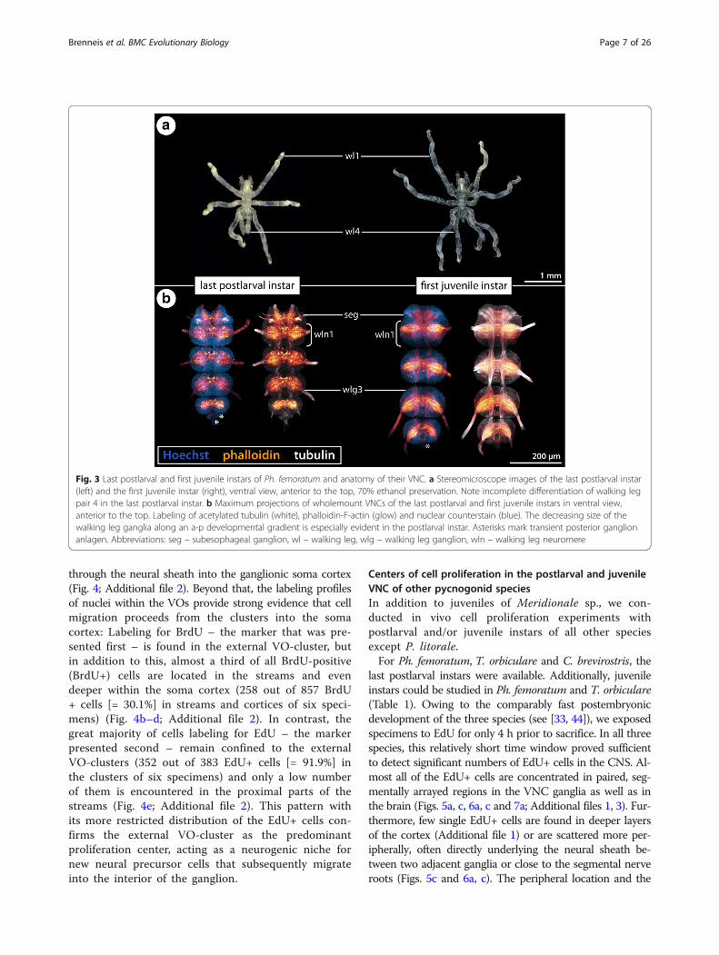
stages of the internal VOs encompass many cells that fea-ture a large nucleus with euchromatic DNA (judging fromstaining intensity), being distinctly larger than the nuclei ofadjacent VO cells and the nuclei of cells in the surroundingcortex (e.g., Additional file 1). In contrast, the nuclei inmore advanced (i.e., older) internal VOs are smaller andmore densely packed than the ones in the surrounding cor-tical regions (Figs. 5b, d, 6b, d and 7b, c; Additional file 4).In accordance with the age-related changes in nuclearmorphology, the number of EdU+ cells changes over time.In the ganglia of postlarval instars, higher numbers ofEdU+ nuclei are found than in the ganglia of the olderjuvenile instars (Figs. 5a, c, 6a, c and 7a). This suggestsa decrease in cell production with ongoing postembryo-nic development. Similarly, the intra-individual devel-opmental gradient is reflected in higher numbers ofEdU+ cells in the more posterior walking leg ganglia withtheir more voluminous VOs. Interestingly, while thisgradient along the VNC appears rather linear in thepostlarval instars of Ph. femoratum and T. orbiculare
Fig. 4 BrdU-EdU double-labeling in the VNC of a juvenile instar of Meridionale sp. Labeling of acetylated tubulin (white), BrdU (green) and EdU(red) with Hoechst nuclear counterstain (blue) after 5-day experiment (4 h BrdU exposure, 92 h sea water, 4 h EdU exposure, 20 h sea water,sacrifice). Sagittal vibratome section, anterior to the left. Arrows highlight the anterior and posterior migratory streams that penetrate through theneural sheath into the soma cortex. Arrowheads mark the ventral longitudinal tract. Stars indicate the small central cavity around which tubulin-rich cell processes converge. a Walking leg ganglia 3 and 4. The black rectangle indicates the region of interest shown in (b–e). The ellipse highlightsthe external VO-cluster of walking leg ganglion 3, featuring numerous BrdU-positive cells. b–e Detail of the external VO-cluster and the migratorystreams penetrating into the cortex of walking leg ganglion 4. The images depict different marker combinations: b all four markers, c tubulin, BrdUand EdU, d BrdU and EdU, e EdU. Note the presence of exclusively BrdU-positive cells in deeper cortex layers (stippled areas). EdU-positive cellsco-label for BrdU and are restricted to the external VO-cluster and the proximal part of the streams. Abbreviations: lm – longitudinal muscle bundles,mg – midgut, wlg – walking leg ganglion
Brenneis et al. BMC Evolutionary Biology (2018) 18:47 Page 8 of 26

Fig. 5 Cell proliferation in the VNC of postlarval and juvenile Ph. femoratum instars. Labeling of acetylated tubulin (white) and EdU (red) with nuclearcounterstain (blue) after 4 h EdU exposure. Maximum projections (a, c) and optical sections (b, b’, d, d’) of wholemount CNSs, anterior to the top.Stippled ovals highlight the internal VOs of one body half. Stars indicate the central cavity around which tubulin-rich cell processes converge. Whitearrows indicate broad tubulin-rich cell bands (b’) and more condensed fibrous strands (d’) extending dorsally from the VOs towards the neuropil.Asterisks mark transient posterior ganglion anlagen. a: CNS of the last postlarval instar, ventral view, EdU labeling shown separately on the right. Notethe gradual antero-posterior increase of cell proliferation. b & b’: Wlg3–4, horizontal and sagittal sections. Black arrows mark larger nuclei in the VOs ofwlg4. Note also wider cavities of the VOs in wlg4. c: CNS of the first juvenile instar, ventral view, EdU labeling shown separately on the right. Arrowheads marksingle EdU-positive cells in the periphery of the ganglia. d & d’: Wlg3–4, horizontal and sagittal sections. Only few large VO nuclei remain (black arrow), VOnuclei being generally smaller than the neuronal nuclei in the soma cortex. Arrowheads mark the ventral longitudinal tract in wlg3. Abbreviations:br – brain, ovn – ovigeral neuromere, pan – palpal neuromere, seg – subesophageal ganglion, wlg – walking leg ganglion, wln – walking leg neuromere
Brenneis et al. BMC Evolutionary Biology (2018) 18:47 Page 9 of 26
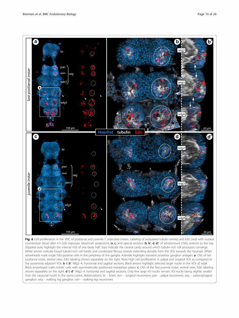
Fig. 6 Cell proliferation in the VNC of postlarval and juvenile T. orbiculare instars. Labeling of acetylated tubulin (white) and EdU (red) with nuclearcounterstain (blue) after 4 h EdU exposure. Maximum projections (a, c) and optical sections (b, b’, d, d’) of wholemount CNSs, anterior to the top.Stippled ovals highlight the internal VOs of one body half. Stars indicate the central cavity around which tubulin-rich cell processes converge.White arrows indicate broad tubulin-rich cell bands and condensed fibrous strands extending dorsally from the VOs towards the neuropil. Whitearrowheads mark single EdU-positive cells in the periphery of the ganglia. Asterisks highlight transient posterior ganglion anlagen. a: CNS of lastpostlarval instar, ventral view, EdU labeling shown separately on the right. Note high cell proliferation in palpal and ovigeral VOs as compared tothe posteriorly adjacent VOs. b & b’: Wlg2–4, horizontal and sagittal sections. Black arrows highlight selected larger nuclei in the VOs of wlg4.Black arrowheads mark mitotic cells with asymmetrically positioned metaphase plates. c: CNS of the first juvenile instar, ventral view, EdU labelingshown separately on the right. d & d’: Wlg2–4, horizontal and sagittal sections. Only few large VO nuclei remain, VO nuclei being slightly smallerthan the neuronal nuclei in the soma cortex. Abbreviations: br – brain, ovn – ovigeral neuromere, pan – palpal neuromere, seg – subesophagealganglion, wlg – walking leg ganglion, wln – walking leg neuromere
Brenneis et al. BMC Evolutionary Biology (2018) 18:47 Page 10 of 26
Fig. 7 (See legend on next page.)
Brenneis et al. BMC Evolutionary Biology (2018) 18:47 Page 11 of 26

(Figs. 5a and 6a), a sharp increase in EdU+ cells isdetectable between walking leg ganglia 3 and 4 of C.brevirostris (Fig. 7a, b). Further, the internal VOs ofthe palpal and ovigeral neuromeres in Ph. femoratumand T. orbiculare break the pattern, since they con-tinue to exhibit comparably high cell proliferation injuveniles, even as the numbers of EdU+ cells in theposteriorly adjacent walking leg neuromeres decrease(Figs. 5c and 6c).In contrast to the external VO-clusters of Meridionale
sp., the internal VOs are not anatomically distinct fromthe surrounding soma cortex and any directed movementof cells between VOs and the cortex is more challengingto reconstruct in fixed samples (see below for T. orbicu-lare). However, it is interesting to note that anterior andposterior cell bands extend dorsally from each VOthrough the soma cortex towards the ganglionic neuropil,similar to the cell streams found in Meridionale sp.[19, 22]. While these strongly tubulin-positive cell bandsare still rather difficult to delimit from the surroundingcells in earlier stages, they condense to more compactstrands with accompanying small nuclei in later stages(Figs. 5b’, 6b’ and 7b; Additional files 1, 4). These strandsare positioned roughly at the same level as the prominentventral longitudinal tract that encircles the neuropil.In S. cheilorhynchus, only first juvenile instars and
a single specimen of the second juvenile instar wereavailable for in vivo cell proliferation studies (12 hBrdU, 12 h sea water, sacrifice) (Table 1). The gen-eral findings for this callipallenid species are identi-cal to the other three species (Fig. 7d–f ’) includingwell-defined fibrous strands emanating from thecompact internal VOs into the surrounding corticalareas (Fig. 7e’–f ’). In line with the juveniles of theother species, the developmental gradient along thea-p axis is not as pronounced in the first and secondjuvenile instars of S. cheilorhynchus.In P. litorale, no in vivo cell proliferation markers
could be used owing to the lack of live postembryonicinstars at the time of the experiments. However, tubulinimmunolabeling coupled with nuclear counterstaining
demonstrates similar internal VOs in postlarval andjuvenile instars of this species (Fig. 8). In particular inthe first juvenile instar, the VOs with their centrally con-verging cell processes and the dorsally emanating cellbands are easily discernible in the soma cortex of theventral ganglia (Fig. 8c–f ).
Asymmetric divisions in internal VOs and first insightsinto the duration of the S/G2/M phaseRecent investigations on the neurogenic processes inMeridionale sp. [19, 22] have supported classical histo-logical works on pycnogonid development that claimeda neural stem cell (NSC)-like precursor type to reside inthe segmental VOs of pycnogonids [33, 36, 39]. Afterconfirming the presence of the internal VOs, we furtherscrutinized them in three of the species (Ph. femoratum,C. brevirostris, T. orbiculare) to elucidate whether spe-cific cell types can be morphologically distinguished andto gain more insights into their cell cycle and cell div-ision characteristics.In Ph. femoratum (for which the highest number of
specimens was available, see Table 1), we performedphalloidin labeling of F-actin to visualize the corticalcytoskeleton in combination with immunolabeling of themitosis marker phosphorylated histone H3 (PH3) on thelast postlarval and first juvenile instars. In both instars,this staining revealed mitoses in the internal VOs(Fig. 9a–b’), predominantly in the central area housingthe larger cells with euchromatic nuclei. Starting inmetaphase, mitoses of these cells were observed to bedistinctly asymmetric, the metaphase plate being shiftedfar towards one pole of the cell (Fig. 9a–b’). Also in thesubsequent mitotic phases this asymmetry remainsdiscernible (Fig. 9b). This observation provides firstmorphological evidence for the presence of an asymmet-rically dividing NSC-like precursor type in the VOs ofPh. femoratum. In other postlarval and juvenile speci-mens, we combined EdU labeling (4 h exposure, sacri-fice) with subsequent immunolabeling against PH3. TheEdU+ cells within the VOs were found to be significantlymore numerous than the PH3+ cells, especially in the
(See figure on previous page.)Fig. 7 Cell proliferation in the VNC of C. brevirostris and S. cheilorhynchus instars. Labeling of acetylated tubulin (white) with nuclear counterstain (blue).C. brevirostris: 4 h EdU (red, a–c). S. cheilorhynchus: 12 h BrdU + 12 h sea water (red, d–f’). Maximum projections (a, d) and optical sections (b, c, e–f’) ofwholemount VNCs. Stippled ovals highlight internal VOs of one body half. Stars indicate cavities around which tubulin-rich cell processes converge.Black arrows highlight larger nuclei in the VO of wlg4. White arrows indicate cell bands and condensed fibrous strands extending dorsally from theVOs towards the neuropil. Asterisks mark transient posterior ganglion anlagen. a: VNC of last postlarval instar, ventral view, EdU signal shown separately onthe right. Note conspicuously higher number of EdU+ cells in wlg4. b: Wlg2–4, sagittal section. Black arrowheads mark potential pycnotic bodiesindicative of cell death occurring in addition to cell proliferation. White arrowheads mark the ventral longitudinal tract c: Wlg4, cross section. d: VNC offirst juvenile instar, ventral view, BrdU labeling shown separately on the right. White arrowheads mark single BrdU-positive cells in the periphery of theganglia. e & e’: Wlg3–4 of first juvenile instar, horizontal and sagittal sections. Black arrowhead indicates a PH3-labeled (yellow) mitotic cell. Whitearrowheads mark ventral longitudinal tracts. f & f’: Wlg3–4 of second juvenile instar, horizontal and sagittal sections. White arrowheadsmark ventral longitudinal tracts. Abbreviations: ovn – ovigeral neuromere, pan – palpal neuromere, seg – subesophageal ganglion, wlg – walking legganglion, wln – walking leg neuromere
Brenneis et al. BMC Evolutionary Biology (2018) 18:47 Page 12 of 26
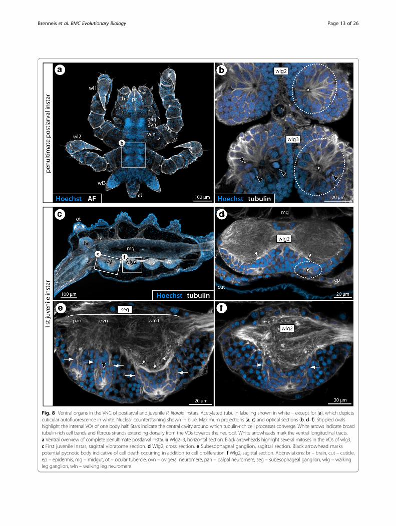
Fig. 8 Ventral organs in the VNC of postlarval and juvenile P. litorale instars. Acetylated tubulin labeling shown in white – except for (a), which depictscuticular autofluorescence in white. Nuclear counterstaining shown in blue. Maximum projections (a, c) and optical sections (b, d–f). Stippled ovalshighlight the internal VOs of one body half. Stars indicate the central cavity around which tubulin-rich cell processes converge. White arrows indicate broadtubulin-rich cell bands and fibrous strands extending dorsally from the VOs towards the neuropil. White arrowheads mark the ventral longitudinal tracts.a Ventral overview of complete penultimate postlarval instar. bWlg2–3, horizontal section. Black arrowheads highlight several mitoses in the VOs of wlg3.c First juvenile instar, sagittal vibratome section. d Wlg2, cross section. e Subesophageal ganglion, sagittal section. Black arrowhead markspotential pycnotic body indicative of cell death occurring in addition to cell proliferation. f Wlg2, sagittal section. Abbreviations: br – brain, cut – cuticle,ep – epidermis, mg – midgut, ot – ocular tubercle, ovn – ovigeral neuromere, pan – palpal neuromere, seg – subesophageal ganglion, wlg – walkingleg ganglion, wln – walking leg neuromere
Brenneis et al. BMC Evolutionary Biology (2018) 18:47 Page 13 of 26

Fig. 9 Asymmetric divisions, cell cycle aspects and BrdU clearing time of VO cells in Ph. femoratum. a, c: Walking leg ganglion 4 of last postlarvalinstar. b, b’, d: Walking leg ganglion 4 of first juvenile instar. Stippled ovals highlight the internal VOs of one body half. Stars indicate the centralcavity around which tubulin-rich cell processes converge. a, b: Horizontal optical sections showing phalloidin-F-actin staining (red) in combinationwith PH3 labeling (yellow) and nuclear counterstain (blue). Black arrows point to mitotic cells with asymmetrically positioned metaphase plates.Black arrowhead marks later stage of an asymmetric division. b’: Higher magnification of a PH3-labeled metaphase plate with distinct shift towardsone pole of the cell (the same metaphase is highlighted in b). c, d: Horizontal optical sections showing EdU staining (4 h exposure, red) incombination with acetylated tubulin (white) and PH3 (yellow) labeling and nuclear counterstain (blue). The PH3-positive mitotic cells show no co-labelingfor EdU. The white arrow indicates the anterior fibrous strand extending dorsally from the VO. e: VO of last postlarval instar, extended optical section(5 μm) through one hemisphere of walking leg ganglion 3 after double-nucleoside labeling (6 h BrdU, 12 h sea water, 3 h EdU, sacrifice).Detection of BrdU (green) and EdU (red) combined with acetylated tubulin (white) and nuclear counterstain (blue). Black arrows mark selected BrdU+/EdU+ nuclei. White arrowheads highlight some BrdU+/EdU− nuclei. Black arrowheads indicate BrdU−/EdU+ nuclei
Brenneis et al. BMC Evolutionary Biology (2018) 18:47 Page 14 of 26
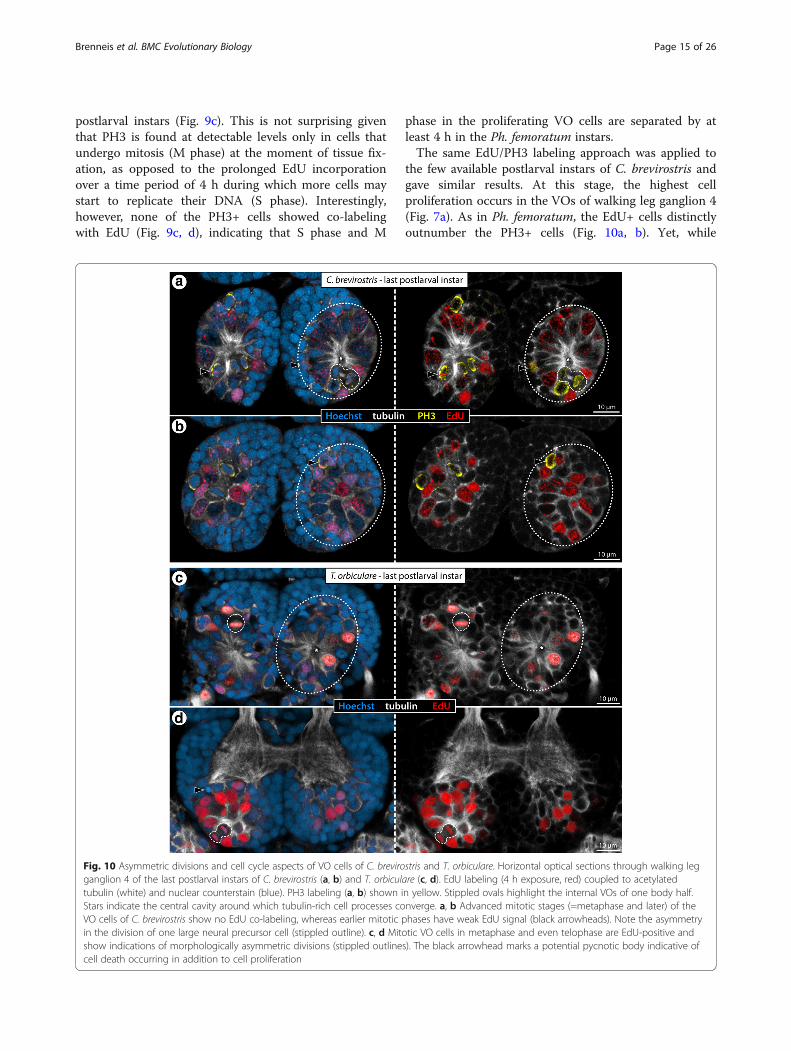
postlarval instars (Fig. 9c). This is not surprising giventhat PH3 is found at detectable levels only in cells thatundergo mitosis (M phase) at the moment of tissue fix-ation, as opposed to the prolonged EdU incorporationover a time period of 4 h during which more cells maystart to replicate their DNA (S phase). Interestingly,however, none of the PH3+ cells showed co-labelingwith EdU (Fig. 9c, d), indicating that S phase and M
phase in the proliferating VO cells are separated by atleast 4 h in the Ph. femoratum instars.The same EdU/PH3 labeling approach was applied to
the few available postlarval instars of C. brevirostris andgave similar results. At this stage, the highest cellproliferation occurs in the VOs of walking leg ganglion 4(Fig. 7a). As in Ph. femoratum, the EdU+ cells distinctlyoutnumber the PH3+ cells (Fig. 10a, b). Yet, while
Fig. 10 Asymmetric divisions and cell cycle aspects of VO cells of C. brevirostris and T. orbiculare. Horizontal optical sections through walking legganglion 4 of the last postlarval instars of C. brevirostris (a, b) and T. orbiculare (c, d). EdU labeling (4 h exposure, red) coupled to acetylatedtubulin (white) and nuclear counterstain (blue). PH3 labeling (a, b) shown in yellow. Stippled ovals highlight the internal VOs of one body half.Stars indicate the central cavity around which tubulin-rich cell processes converge. a, b Advanced mitotic stages (=metaphase and later) of theVO cells of C. brevirostris show no EdU co-labeling, whereas earlier mitotic phases have weak EdU signal (black arrowheads). Note the asymmetryin the division of one large neural precursor cell (stippled outline). c, d Mitotic VO cells in metaphase and even telophase are EdU-positive andshow indications of morphologically asymmetric divisions (stippled outlines). The black arrowhead marks a potential pycnotic body indicative ofcell death occurring in addition to cell proliferation
Brenneis et al. BMC Evolutionary Biology (2018) 18:47 Page 15 of 26

advanced mitotic stages were found to be EdU-negative,some PH3+ cells in prophase showed weak EdU co-labeling (Fig. 10a, b). This observation speaks for ashorter G2 phase between the S and M phases in thelarge proliferating VO cells and may indicate a generallyfaster cycling compared to Ph. femoratum. Close inspec-tion of later mitotic stages of these large cells also re-vealed a slight asymmetry in the divisions (Fig. 10a),which is in agreement with previous claims of NSC-likeprecursors in the VOs of C. brevirostris [33].In T. orbiculare, the VOs of the last postlarval instar
were studied after EdU exposure (4 h exposure, sacri-fice). Not all of the EdU+ cells in the VOs show notablesize differences compared to the somata in the sur-rounding cortex (Fig. 10c, d). Nonetheless, several of theobserved mitotic stages in larger cells show the sameindications of morphological asymmetry with shiftedmetaphase plates (Figs. 6b and 10c) and persisting slightdifferences in later mitotic stages (Fig. 10d). Notably,very strong EdU labeling was encountered in cells in allmitotic phases, including the telophase (Fig. 10c, d). Atthe same time, however, some mitotic cells were devoidof EdU labeling (Fig. 6b). Accordingly, the cycling timesof the proliferating VO cells in T. orbiculare seem to bemore heterogeneous, comprising faster (S phase to Mphase < 4 h) and slower (S phase to M phase > 4 h) cyc-ling cells. With the available data it cannot be deter-mined whether these cell cycle differences relate todifferent neural precursor types in the VOs.
Double-nucleoside labeling using BrdU and EdU –estimation of BrdU clearing time and further evidencefor NSC-like precursor types in pycnogonid VOsDuring the postembryonic development of Meridionalesp., the direction of cell movement from external VO-clusters via the streams into the soma cortex has beenpreviously reconstructed [22] and independently validatedhere with double-nucleoside cell proliferation experiments(Fig. 4; Additional file 2). Aiming to investigate whethercell migration occurs in species with internal VOs, weconducted similar experiments with postembryonic instarsof Ph. femoratum and T. orbiculare. Experiments withpostlarval instars and juveniles of T. orbiculare weresuccessful and lasted 3 days in total (see next section). InPh. femoratum, by contrast, survival of postembryonicinstars in good condition could only be accomplished withshort time windows of less than 24 h. This duration wastoo short to extract cell migration patterns with anyconfidence (Additional file 4). However, although theexperiments failed in this respect, the relatively shorttime intervals between the two proliferation markerpulses provided insights into the marker clearing timein pycnogonid instars falling in the studied size range:The shortest applied sea water interval between BrdU
and EdU exposures was 12 h. After this experiment,the VOs of Ph. femoratum showed a mixed pattern ofBrdU+/EdU- and BrdU+/EdU+ cells, but importantlyalso some BrdU-/EdU+ cells (Fig. 9e; Additional file4). The presence of EdU-only labeled cells indicatesthat BrdU was no longer available at detectable levelsafter the intervening 12 h in sea water, resulting inthe exclusive incorporation of EdU in cells passingthrough S phase during the EdU exposure period. Ac-cordingly, 12 h can be deduced as a conservative esti-mate for the clearing time of BrdU from the animals.This may still be an overestimation since shorterinter-pulse intervals have not been tested.Regardless of the uncertainty pertaining to shorter
inter-pulse intervals, this finding has important impli-cations for experiments using longer inter-pulse inter-vals with the similar-sized postembryonic instars ofMeridionale sp. The intervening sea water interval inour double-labeling experiments on Meridionale sp.juveniles lasted almost 4 days (92 h), that is, almost 8-times the 12 h clearing time established in Ph. femora-tum. Therefore, it seems highly unlikely that any ofthe double-labeled cells observed in Meridionale sp.(Fig. 4b–e; Additional file 2) incorporated BrdUduring the EdU exposure period. As a consequence,double labeling with both proliferation markers sug-gests that cells have repeatedly passed through S phaseduring the experiment. Such double-labeled cells arealmost exclusively found in the external VO-clustersof Meridionale sp. and only occasionally in the prox-imal portions of the migratory streams (Fig. 4b–e;Additional file 2). Further, although BrdU-only labeledcells were readily identifiable in the external VO-clusters and deep within the soma cortex, EdU label-ing is detected in the overwhelming majority of casesin combination with BrdU labeling (381 out of 383EdU+ cells [= 99.5%] with BrdU co-labeling in sixspecimens). This pattern strongly suggests that repeat-edly dividing neural precursor cells reside within theexternal VO-clusters, their progeny migrating throughthe streams into the cortex. Morphological delimita-tion of different cell types is not possible in the exter-nal VOs of juvenile instars of Meridionale sp. [22].However, since the external clusters are derivatives ofthe earlier VO-stages that house large asymmetricallydividing NSC-like precursors [19], it is feasible thatthe double-labeled cells are of the same type, havingdecreased in size with ongoing cycling during postem-bryonic development.
Double-nucleoside labeling in T. orbiculare: cell migrationfrom the VOs into the soma cortexIn T. orbiculare, 3-day-long double-nucleoside labelingexperiments were conducted (4 h BrdU exposure, 64 h
Brenneis et al. BMC Evolutionary Biology (2018) 18:47 Page 16 of 26

sea water, 4 h EdU exposure, sacrifice) with the penulti-mate and last postlarval instars as well as with the firstjuvenile instar. Investigation of this developmental seriesenabled us to evaluate labeling patterns intra-individually along the a-p developmental gradient butalso between specific segmental ganglia across differentinstars.The overall patterns across instars and along the a-p
gradient show that the highest cell proliferation correlates
well with the major growth periods of the different ventralganglia, being followed by a notable decrease of prolifera-tion rates in later stages (Fig. 11). Without exception, thenumber of cells labeling for the first marker BrdU signifi-cantly exceeds the second marker EdU (Figs. 11 and 12;Additional file 3). Cells that have undergone S phase duringthe 4 h EdU pulse are predominantly restricted to the ven-tral soma cortex, being located in the internal VOs (Figs. 6and 12). During the major growth period of the ganglia, the
Fig. 11 Double-nucleoside labeling experiments with postlarval and juvenile instars of T. orbiculare. BrdU (green) and EdU (red) labeling (4 h Brdu,64 h sea water, 4 h EdU, sacrifice) with nuclear counterstain (blue). All images show maximum projections in ventral view, apart from those positionedto the right of stippled vertical lines, which are lateral views of the hemiganglia of one body half. For a clearer depiction of the proliferation markerpatterns, the nuclear counterstain and BrdU signal have been successively removed from left to right. Stippled ovals highlight the position of theinternal VOs in one body half. Curved stippled lines highlight the characteristic sickle-like arrangement of the BrdU-positive cells. Black arrows pointat exclusively EdU-labeled nuclei in advanced VO-stages. White arrowheads indicate exclusively EdU-labeled, flattened nuclei of peripheral glial cells.White arrows mark selected EdU-positive nuclei of cells just outside the VOs, potentially indicative of further divisions after cells have started to migrate.a–a”: Wlg 2–4 of the penultimate postlarval instar. b–b”: Wlg 2–4 of the last postlarval instar. c–c”: Wlg 2–4 of the first juvenile instar. Abbreviations:wlg – walking leg ganglion
Brenneis et al. BMC Evolutionary Biology (2018) 18:47 Page 17 of 26

VOs house EdU+ cells of different cell sizes (Fig. 12a–c),which might point to the presence of different neuralprecursor cell types/generations during the peak ofcell proliferation activity. Irrespective of size differ-ences, however, almost all EdU+ cells show also BrdUco-labeling (Figs. 11 and 12; Additional file 3). How-ever, in more advanced VO-stages, cells labeled exclu-sively with EdU are more frequently detectable(Fig. 11c–c”). The most obvious EdU-only cells arelocated in the periphery of the ganglia of juvenile
instars, featuring flattened nuclei that suggest a glialnature (Fig. 11c–c”). Even though few in number, thepresence of these BrdU-/EdU+ cells confirms that un-bound BrdU was not available during the final EdUpulse, as predicted by the clearing time estimateobtained in Ph. femoratum. Consequently, the double-labeling of cells in the VOs of T. orbiculare indicatestheir repeated passage through S phase during the ex-periments, in accordance with the results obtained forthe external VO-clusters of Meridionale sp.
Fig. 12 Distribution of BrdU- and EdU-positive cells in walking leg ganglion 3 of T. orbiculare. BrdU (green) and EdU (red) labeling (4 h Brdu, 64 hsea water, 4 h EdU, sacrifice) with acetylated tubulin (white) and nuclear counterstain (blue). All images show horizontal optical sections throughwalking leg ganglion 3 of the last postlarval instar, proceeding from apical to basal. For clearer depiction of the proliferation marker patterns,tubulin labeling plus nuclear counterstain and the BrdU signal have been successively removed from left to right. The wide stippled outlines tracethe extension of the ganglionic soma cortex. Stippled ovals highlight the position of the internal VOs in one body half (a–c). Black arrows (d)point at BrdU and EdU co-labeled nuclei in the cell band emanating dorsally from the VO and running posterior to the neuropil, suggestive offurther divisions during cell migration. White arrowheads (d) mark neurite bundles of the ventral longitudinal tract which becomes recognizablein these stages. White arrows (b–d) point at potential pycnotic bodies indicative of cell death occurring in addition to cell proliferation in the VOsand the migratory cell bands. Note the heterogeneous nucleus sizes in the internal VOs (a–c) as well as similar nucleus size and morphology ofBrdU-positive cells and surrounding somata in areas outside the VOs and deeper in the cortex (c–e)
Brenneis et al. BMC Evolutionary Biology (2018) 18:47 Page 18 of 26

In contrast to the EdU labeling, BrdU+ cells are spreadfar beyond the VOs into the growing soma cortex of thedeveloping ganglia. They do not only surround the VOsbut also reach further dorsally towards the formingganglionic neuropil. Especially in advanced stages ofganglion development, the BrdU+ cells extend anteriorlyand posteriorly from the neuropil, leading in lateral viewto a sickle-like arrangement embedded in the cortex(Fig. 11a, a’, b, b’, c, c’; Additional file 3). This is stronglysuggestive of active cell migration. Also in ventral view,the overall pattern of BrdU+ cells changes over time.During the early phases of rapid ganglion growth, theyare spread out in broad, almost concentric rings aroundthe VOs (Fig. 11a, a’, b, b’) as would be expected fromthe migration of newborn cells in all directions from themain centers of cell proliferation. In later stages, how-ever, the BrdU+ cells are arranged in more elongatedband-like patterns with an oblique a-p axis (Fig. 11c, c’).The VOs are still in the center of these cell bands butmigration of newborn cells has become restricted lat-erally and is instead concentrated along two pathwaysthat encircle the central neuropil anteriorly and pos-teriorly, in close proximity to the ventral longitudinaltract (Additional file 3). Incidentally, already in earlierstages, cells that have recently undergone DNA syn-thesis (i.e., are EdU+) and have started to migratedorsally are encountered in close vicinity to the sametract (Fig. 12d).Importantly, the considerable number of BrdU+ cells
labeled during the rapid ganglion growth may not be ex-clusively attributable to cell proliferation in the VOs butmay in part be explained by additional cell divisions dur-ing migration. Although this aspect cannot be conclu-sively resolved with the methods applied here, thealmost complete lack of EdU labeling in deeper layers(e.g., Fig. 12c, d) suggests that such divisions of newborncells leaving the VOs would have to occur in early stagesof migration. The majority of BrdU+ cells located deeperin the cortex are therefore more likely to be differentiat-ing/differentiated postmitotic neural cells, which notionreceives further support from their similar nuclearmorphology compared to the surrounding BrdU-negative neuronal somata (Fig. 12d, e).
DiscussionGross architecture of the VNC: independent inclusion ofwalking leg neuromere 1 into the SEG of differentpycnogonid lineagesIn accordance with the conserved basic body organizationof the various sea spider taxa (excepting the few extra-legged species), the pycnogonid CNS shows a remarkablyuniform architecture [31, 32, 52]. In the majority of species,the VNC features the exact same set of separate adult
ganglia, although the relative distance between them candiffer significantly (Fig. 13). The latter phenomenon corre-lates well with the general habitus of a given species (slen-der, tube-like trunk vs. compact, disk-shaped trunk).Our study highlights one of the few taxon-specific archi-
tectural differences of the pycnogonid VNC: the three calli-pallenid representatives possess an anatomically separatewalking leg ganglion 1 posterior to a bipartite SEG thatcomprises the palpal and ovigeral neuromeres. By contrast,the remaining three species have a tripartite adult SEG thatincludes the neuromere of walking leg segment 1. Import-antly, however, outside of Phoxichilidiidae, Pycnogonidaeand the genus Tanystylum, all pycnogonid taxa hithertostudied have a bipartite SEG as found in the three callipalle-nids (Fig. 13a–f, also [30, 52]). This taxon list includes otherammotheid genera (e.g., Fig. 13d) as well as groups whichhave been indicated to branch off close to the base of thepycnogonid tree in the most exhaustive phylogenetic ana-lyses [40, 41, 53] (Fig. 13g), such as Austrodecidae (Fig. 13f)and Colossendeidae (e.g., [31, 54]). Further, it also encom-passes Ascorhynchidae (e.g., [31, 55]) which has been pro-posed as the earliest (paraphyletic) offshoot in one 18SrRNA-based molecular analysis [56] (but see, e.g., [53] forAscorhynchidae well-nested in the pycnogonid tree).Hence, when mapping the SEG architecture on currentlydiscussed topologies of pycnogonid interrelationships, a bi-partite composition is resolved as the most parsimoniousplesiomorphic state (Fig. 13g). The tripartite SEG of someextant taxa, on the other hand, is indicated to go back tothree independent events in pycnogonid crown group evo-lution, being the result of the fusion of the palpal, ovigeraland walking leg 1 neuromeres during postembryonic gang-liogenesis. Notably, this interpretation holds true regardlessof the morphologically questionable splitting of the Phoxi-chilidiidae, which has been recovered due to the rather sur-prising placement of the genus Phoxichilidium in one ofthe recent analyses [40] (Fig. 13g).
Variations of a common theme – external versus internalpycnogonid VOsIn the developing ventral ganglia of all pycnogonid speciesstudied here, we could confirm the existence of segmentalVOs, which appear as conspicuous paired clusters of cellswith centrally directed processes converging around a smallcavity. However, in contrast to Meridionale sp., where theVOs represent separate clusters that lie external to the gan-glia, the ones in the five other species are incorporated intothe ganglionic soma cortex (Fig. 14a), in which they appearas intensely tubulin-labeled cell formations with centrallydirected processes. The latter finding confirms classicalhistological studies focusing on the VNC development inpycnogonid taxa other than Nymphonidae [33, 36–39].The deviating position of the external and internal VOs
raises the questions whether they serve the same function
Brenneis et al. BMC Evolutionary Biology (2018) 18:47 Page 19 of 26

and if they are homologous structures across pycnogonids.With regard to the functional role, our cell proliferationexperiments and mitosis marker labeling unambiguouslycharacterize internal VOs as centers of significant cell pro-liferation during VNC development, in correspondence tothe external VOs of Meridionale sp. [19, 22]. Further, re-gardless of VO position, our double-nucleoside labelingapproach illustrates migration of significant numbers ofnewborn cells from the VOs into the growing soma cortexof the respective ganglion. Accordingly, this indicates thatexternal and internal VOs act as postembryonic
neurogenic niches in the ventral ganglia – and refutes pre-vious speculations on the predominantly neurosecretorynature of the internal VOs [37, 38].With regard to the question of homology, additional
characteristics can be considered: beyond the func-tional correspondence of both VO types as niches ofpostembryonic neurogenesis, the presence of a morpho-logically conspicuous large cell type – especially in earl-ier stages – has been noted in all previous descriptions(e.g., [33, 36, 39]). In the present study, we have shownasymmetric divisions of these cells in three species with
Fig. 13 Gross architecture of the VNC of different sea spider families evaluated against the backbone of pycnogonid phylogeny. a–d Fluorescentstereomicroscope images of partially dissected nervous systems after labeling with the lipophilic marker FM 1-43FX. e–f Fluorescent stereomicroscopeimages of nervous systems with nuclear labeling. Note that all species feature an anatomically separate walking leg ganglion 1, even though it isalways close to the subesophageal ganglion. a Nymphon gracile, adult CNS, dorsal view. b Parapallene australiensis, adult VNC, dorsal view. c Endeisspinosa, adult VNC, dorsal view. d Cilunculus japonicus, adult CNS, dorsal view. e Propallene sp., adult VNC, dorsal view. f Pantopipetta armoricana, CNSof late juvenile, ventral view. g The bipartite subesophageal ganglion is resolved as plesiomorphic condition of Pycnogonida after mappingon current hypotheses of pycnogonid relationships and parsimony-based reconstruction. Abbreviations: br – brain, seg – subesophagealganglion, wlg – walking leg ganglion
Brenneis et al. BMC Evolutionary Biology (2018) 18:47 Page 20 of 26

internal VOs (Ph. femoratum, T. orbiculare, C. breviros-tris), in line with previous findings in the external VOsof Meridionale sp. [19]. The occurrence of such largeand asymmetrically dividing cells has been recognizedalso in earlier VO stages of S. cheilorhynchus [19]. Fur-ther, our double-nucleoside labeling experiments with T.orbiculare have provided evidence for repeated divisionsof cells residing in the internal VOs. In combinationwith the observed asymmetry of some divisions, thesefindings support the presence of a NSC-like precursortype as a shared feature of external and internal VOs(Fig. 14a). Beyond that, the developmental series of T.orbiculare instars investigated with double-nucleoside la-beling has revealed more specific similarities to the cellmigration patterns of Meridionale sp. ([19, 22], presentstudy): starting with a rather homogeneous movement indorsal direction and proceeding via two broad, antero-and posterodorsally oriented cell bands, the newborncells become increasingly restricted to two migrationstreams with ongoing ganglion development. These cellmigration streams extend in close vicinity to the sameventral longitudinal tract in both species (Fig. 14a), andcorresponding fibrous components extending along thispathway have been detected in all other species with in-ternal VOs. Finally, the origin and earlier developmental
stages of both VO types are virtually identical. Both arederivatives of segmentally paired, pseudo-stratifiedregions of the ventral neuroectoderm from which thefirst ganglion cells immigrate (e.g., [19, 57]). With on-going development, these regions begin to invaginate(Fig. 15a–c) and eventually detach from the apical ecto-derm, thus forming the conspicuous paired VOs at theventral side of the ganglion anlage, the VOs’ central cav-ities being lined by the formerly apical poles of the neu-roectodermal cells [19, 22, 33, 35, 39]. Considering all ofthese correspondences, we find strong support for thehomology of both types of VO in the various pycnogo-nid taxa, in spite of the positional difference. This dif-ference is the result of an incorporation process intothe soma cortex during postembryonic gangliogenesisof some species, which does not take place in others.In an evolutionary framework, it remains then to be
clarified which of the two VO types represents the ple-siomorphic condition of the taxon Pycnogonida. Acrossthe groups studied so far, external VOs have been exclu-sively discovered in Nymphonidae (e.g., [35, 36, 58]) andthe callipallenid genera Meridionale ([22], present study)and Pseudopallene [58]. In all other taxa investigated,including the callipallenid genera Stylopallene and Calli-pallene ([33, 38, 39], present study), the VOs are
Fig. 14 Internal versus external VOs and reconstruction of the plesiomorphic VO type in crown group Pycnogonida. a Schematic representation ofexternal and internal VOs from postlarval to juvenile instars, showing an idealized sagittal section. Lightly-colored, larger cells in the VOs represent theNSC-like precursors. Black cells detach from the VOs and start migration. Yellow cells are neural precursors that divide again while they migrate intothe soma cortex (as shown in [22]). b Mapping of the VO types on two competing hypotheses on the phylogenetic relationships of extant pycnogonidtaxa. Although several lineages remain unstudied (labeled with question marks), the distribution of the two VO types across the taxa investigatedsuggests internal VOs to be the plesiomorphic condition of Pycnogonida. Abbreviations: GN – glomerulus-like neuropil, VLT – ventral longitudinal tract,VO – ventral organ
Brenneis et al. BMC Evolutionary Biology (2018) 18:47 Page 21 of 26
incorporated into the soma cortex (Fig. 14b). Irrespect-ive of which phylogenetic hypothesis is favored [40, 41,53, 56], a parsimony-based assessment of the distribu-tion of VO types suggests the external VOs to be anapomorphic condition that has emerged within thecallipallenid-nymphonid lineage (Fig. 14b). To date, theexact relationships of the nymphonid and callipallenid taxaare still unsatisfactorily resolved. However, with further pro-gress in this field, the presence of external VOs may provephylogenetically informative, as a potential apomorphiccharacter state of a monophylum comprised of a subset ofcallipallenid species and nymphonids.
Interspecific differences and deviations from the a-p gradientof VO cell proliferation correlate to specific events ofpostembryonic developmentCompared to the rather linear a-p gradient of ventralganglion development in Ph. femoratum and T. orbiculare,the last postlarval instar of C. brevirostris shows an abruptincrease in cell proliferation between the VOs of walkingleg ganglia 3 and 4 (Fig. 7a). This observation coincideswith a different mode of postembryonic development. Thegenus Tanystylum shows a pronounced anamorphic devel-opment with a minute protonymphon larva as hatchingstage and strictly sequential addition of the walking legsegments [33, 59]. This developmental type is widely con-sidered to be plesiomorphic for Pycnogonida (e.g., [42]).Likewise, all members of the Phoxichilidiidae follow an
anamorphic – albeit accelerated – mode with a slight butperceivable a-p gradient in walking leg segment formation[44, 60]. By contrast, the two genera Callipallene and Neo-pallene feature the most pronounced embryonization ofdevelopment known in Pycnogonida [33, 38, 39, 51, 54].One hallmark of their development is the synchronizeddifferentiation of walking leg segments 1–3 early duringembryogenesis, whereas walking leg segment 4 is formeddistinctly later [42]. As a consequence, the first three walk-ing leg segments and their ganglia are well-differentiatedin the postlarval instar, but walking leg segment 4 is stillin the period of major ganglion growth with VOs thathouse numerous large NSC-like precursors (Fig. 7b,c). Accordingly, the observed differences in overallcell proliferation patterns between postlarval instarsof the different species examined reflect deviations inthe timing of walking leg segment formation duringtheir development.Another case of proliferation patterns deviating from a
monotonous a-p developmental gradient is encounteredin the anterior palpal and ovigeral VOs that are affiliatedwith the SEG. While cell proliferation of the posteriorlyfollowing VOs in walking leg segments 1 and 2 de-creases between the last postlarval and the first juvenileinstar, the number of EdU+ cells in the palpal and ovig-eral VOs was found to remain distinctly higher in Ph.femoratum and T. orbiculare (Figs. 5A, c and 6a, c). Again,these observations correlate with developmental events in
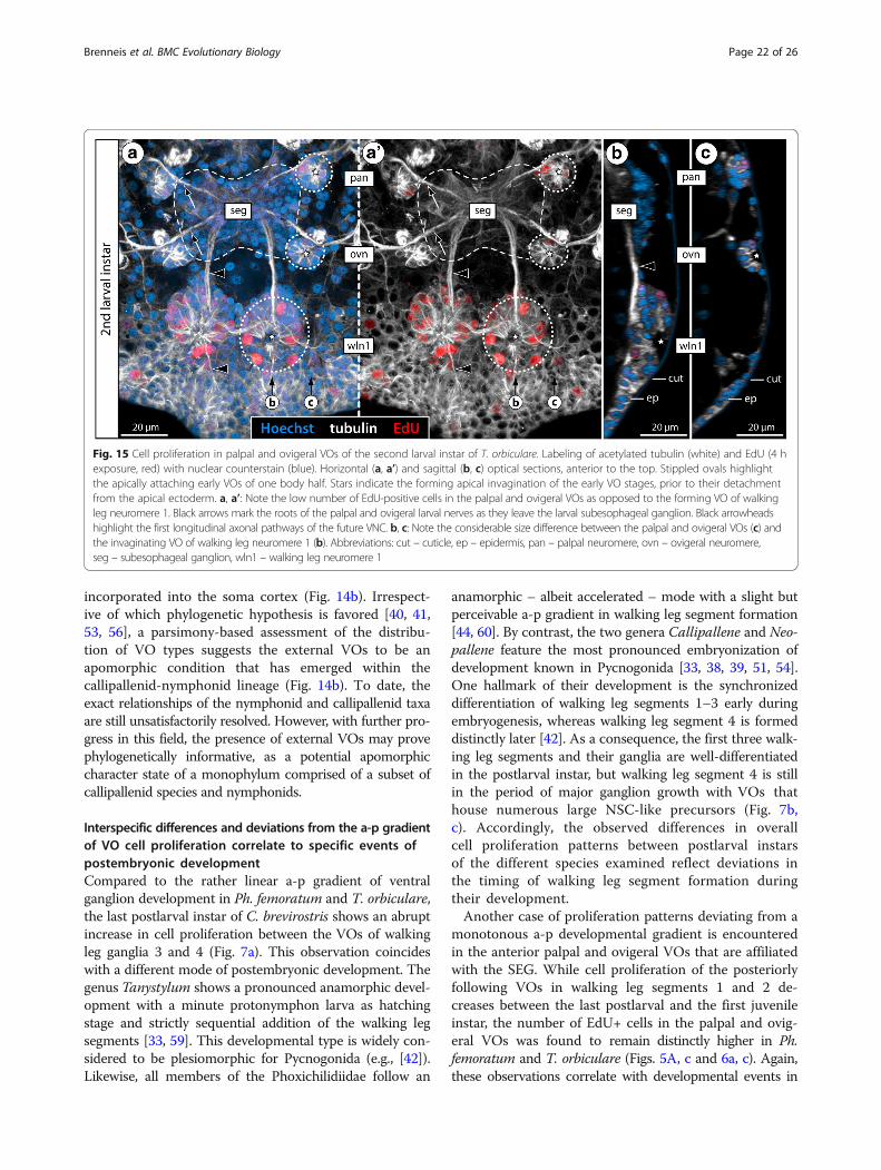
Fig. 15 Cell proliferation in palpal and ovigeral VOs of the second larval instar of T. orbiculare. Labeling of acetylated tubulin (white) and EdU (4 hexposure, red) with nuclear counterstain (blue). Horizontal (a, a’) and sagittal (b, c) optical sections, anterior to the top. Stippled ovals highlightthe apically attaching early VOs of one body half. Stars indicate the forming apical invagination of the early VO stages, prior to their detachmentfrom the apical ectoderm. a, a’: Note the low number of EdU-positive cells in the palpal and ovigeral VOs as opposed to the forming VO of walkingleg neuromere 1. Black arrows mark the roots of the palpal and ovigeral larval nerves as they leave the larval subesophageal ganglion. Black arrowheadshighlight the first longitudinal axonal pathways of the future VNC. b, c: Note the considerable size difference between the palpal and ovigeral VOs (c) andthe invaginating VO of walking leg neuromere 1 (b). Abbreviations: cut – cuticle, ep – epidermis, pan – palpal neuromere, ovn – ovigeral neuromere,seg – subesophageal ganglion, wln1 – walking leg neuromere 1
Brenneis et al. BMC Evolutionary Biology (2018) 18:47 Page 22 of 26
this particular region: all pycnogonid taxa with a protonym-phon larva as hatching stage possess small three-segmentedpalpal and ovigeral larval limb pairs at the beginning of thepostlarval phase [57, 58, 61–63]. However, one of the mostpeculiar aspects of pycnogonid development is the meta-morphosis of these limbs into the adult palps and ovigers(if present) which starts at the end of the postlarval phase.In the case of the oviger, this metamorphosis even includesthe almost complete atrophy of the larval limb prior to a denovo outgrowth of the appendage during the first instars ofthe juvenile phase (see [59] for Tanystylum, [51] for review).Accordingly, major neuroarchitectural changes are to beexpected in the SEG during this period, owing to the loss ofneural elements of the palpal and ovigeral appendagenerves of the larva (Fig. 15a, a’) as well as the integration ofnew afferent and efferent neurons into the existing circuitsas the adult limb pairs differentiate. Embedded in this de-velopmental framework, the higher cell proliferation ratesin the corresponding VOs appear a logical consequence,and may serve as additional evidence for the VOs' indi-cated neurogenic role.
Beyond sea spiders: Internal VOs as a plesiomorphicfeature of arthropod gangliogenesisOur new data on the internal VOs in some pycnogonidtaxa and the reconstruction of this VO type as the plesio-morphic condition of Pycnogonida has implications in abroader arthropod-wide framework.Similarities and differences between neurogenic pro-
cesses in the major arthropod groups and their closestrelatives have been discussed in depth in numerous con-tributions of the last decades (e.g., [18–20]), often withfocus on the initial embryonic phase and to the inclusionof the involved gene networks. In this context, thediscovery of detailed similarities between the neuralprecursor types and neural cell lineages underlying earlyembryonic neurogenesis in hexapods and (some) crusta-ceans (see [3, 6, 18, 64]) has contributed compellingneurodevelopmental arguments in favor of the taxonTetraconata that is strongly supported in molecularanalyses (e.g., [7–9]). In similar fashion, correspondencesbetween the early neurogenic processes in the non-pycnogonid chelicerates (represented by horseshoe crabsand spiders) and myriapods have been briefly discussedas supporting a sister group relationship of both lineages[65–67] but are now more generally considered as theplesiomorphic condition of Arthropoda (e.g., [19, 20]).When putting the focus on later phases of neurogenesis,
VO-like structures as centers of neural cell production havenot been reported in any of the recently investigated cheli-cerate taxa other than Pycnogonida [19, 68, 69], nor arethey known from any tetraconate taxon. Intriguingly, how-ever, VO-like cell formations are invariably formed during
advanced neurogenesis of the myriapod taxa [70–74], insome studies having even been designated as “ventralorgans” [71, 72, 74]. As is the case in pycnogonids, thesemyriapod VOs develop via an invagination of the centralarea of the neuroectoderm of each hemisegment. Moreover,in accordance with the here deduced pycnogonid groundpattern, myriapod VOs are subsequently incorporated intothe ventral soma cortex of the ganglia, where they havebeen reported to produce additional ganglion cells [70–73].In light of increasingly better support for myriapods assister group of Tetraconata within the taxon Mandibulata(e.g., [7, 27, 75]), we therefore propose internal VOs as cen-ters of late neurogenesis to be a symplesiomorphic featureof gangliogenesis in pycnogonids and myriapods. In otherwords, we suggest such internalized VOs to be part of theground pattern of Arthropoda.Also in the likely arthropod sister group Onychophora,
conspicuous segmental thickenings of the ventral ecto-derm are formed during advanced development. As forMyriapoda, histological studies have termed these struc-tures “ventral organs” (e.g., [76–78]) and in part suggestedtheir involvement in late embryonic neurogenesis (e.g.,[78]). However, several recent investigations have refutedthis role (e.g., [79, 18]), as the onychophoran VOs neitherinvaginate, nor retain a connection to the underlyingCNS. Instead, they gradually atrophy to tiny sclerotizedepidermal structures that act as muscle attachment sitesfor limb muscles in the adult animals [79, 80]. Owing tothese differences, homologization of these onychophoranstructures with the pycnogonid and myriapod VOs seemsnot tenable. A notable exception to this may be found inthe protocerebral neuroectoderm of onychophorans. Here,the central area invaginates during development to formthe hypocerebral organ – an external cell cluster that isattached to the ventral side of brain. To this day, thefunction of the hypocerebral organ remains largelyunresolved, and apart from a (neuro)secretory nature (e.g.,[37, 81, 82]), a role in late and perhaps even adult neuro-genic processes has been considered [22]. Clearly, furtherstudies on this organ are needed for a better understand-ing of its constituting cell types and their function.Similarly, it will be the task of future studies to further
dissect the cellular processes and their underlyinggenetic networks in the internal VOs of pycnogonidsand myriapods in more detail. For instance, the presenceof specialized NSC-like precursors comparable to thosein pycnogonids needs to be critically reinvestigated in myr-iapod taxa, as the currently available data are in this respectstill contradictory and inconclusive (e.g., [65, 74, 83]).Beyond that, the potential persistence of internal VOs inthe ganglia of adult pycnogonids and myriapods – as shownfor the external VOs of Meridionale sp. [22] – and therelated issue of life-long neurogenic processes in these twoarthropod groups remain to be clarified.
Brenneis et al. BMC Evolutionary Biology (2018) 18:47 Page 23 of 26

ConclusionsOur comparative study on six different pycnogonid speciesprovides new insights into the evolution of neurodevelop-mental processes and adult neuroanatomy in one of theoldest extant arthropod lineages. By evaluating our findingsin light of current hypotheses on pycnogonid phylogeny,we reveal a bipartite SEG as the plesiomorphic condition ofsea spiders and find evidence for the convergent fusion ofwalking leg neuromere 1 to this SEG in three different pyc-nogonid lineages. These fusion events are clearly independ-ent of the apomorphic postoral CNS condensation in theprosoma of euchelicerates (e.g., [12, 84]), as well as of thedifferent SEG formation/expansion events discussed formandibulate taxa (e.g., [85]). Further, we reconstruct thepresence of internal VOs instead of external VOs as plesio-morphic character state of sea spider gangliogenesis.Although chelicerate taxa other than Pycnogonida lackcomparable VOs, they are a characteristic feature of myria-pod gangliogenesis and, accordingly, we propose internalVOs with neurogenic function to be part of the groundpattern of Arthropoda. Finally, our study demonstrates theimportance of an expanded taxon sampling in neurodeve-lopmental and neuroanatomical investigations, even whendealing with arthropod lineages that show such a conservedbasic body organization as Pycnogonida. This enables thedocumentation of ingroup variation and more reliabledifferentiation of plesiomorphic from apomorphic characterstates prior to outgroup comparison.
Additional files
Additional file 1: Movie of walking leg ganglia 3 and 4 in a young lastpostlarval instar of Ph. femoratum. Labeling of acetylated tubulin (white)and EdU (4 h exposure, red) with nuclear counterstain (blue). Differentcombinations of the three signals are shown during the movie, in orderto show specific aspects in more detail. The movie starts in ventral view,anterior is to the top. The object turns 90° around the a-p axis towardsthe left to demonstrate that the VOs containing the proliferating cells areembedded in the ventral soma cortex (for better view, one body half isclipped away after the turn). Note the single EdU+ nucleus deep in thecortex of walking leg ganglion 3. After a 90° rotation back into ventral view,a clipping plane is briefly added to show only the ventralmost portion ofthe soma cortices, which house the strongly tubulin-labeled VOs with theircentral cavities and the proliferating cells. Note also the presence of EdU+cells in the posterior ganglion anlagen, which will begin to merge withwalking leg ganglion 4 during subsequent development towards the first ju-venile instar. (MP4 17,615 kb) (MP4 17615 kb)
Additional file 2: Movie of walking leg ganglion 3 in a juvenile ofMeridionale sp. Labeling of acetylated tubulin (white), BrdU (green) andEdU (red) (4 h BrdU exposure, 92 h sea water, 4 h EdU exposure, 20 hsea water) with nuclear counterstain (blue). Different combinations ofthe four signals are shown during the movie to highlight specific aspectsin more detail. The movie starts in ventral view, anterior is to the top. Theobject turns almost 90° around the a-p axis towards the left to demonstratethat the VOs containing the majority of the proliferating cells (as indicatedby the BrdU+ and EdU+ nuclei) are located externally on the ventral surfaceof the ganglion. Only BrdU+ cells are found along the migratory streamsthat extend into the soma cortex. EdU+ nuclei are located exclusively in theexternal VOs, showing BrdU co-labeling. Note also the very prominent andcurved ventral longitudinal tract that is visible whenever the tubulin signal is
shown, e.g., during the rotation back into ventral view at the end of themovie. (MP4 16,957 kb) (MP4 16957 kb)
Additional file 3: Movie of walking leg ganglia 2–4 in the last postlarvalinstar of T. orbiculare. Labeling of acetylated tubulin (white), BrdU (green)and EdU (red) (4 h BrdU exposure, 64 h sea water, 4 h EdU exposure)with nuclear counterstain (blue). Different combinations of the foursignals are shown during the movie to highlight specific aspects in moredetail. The movie starts in ventral view, anterior is to the top. The objectturns 90° around the a-p axis towards the right to demonstrate that theVOs containing the proliferating cells (as indicated by the EdU+ nuclei)are embedded in the ventral soma cortex (for better view, one body halfis clipped away after the turn). Note the presence of a higher number oflarge EdU+ nuclei in the more posterior ganglia and the occurrence ofdistinctly smaller nuclei in between and around the proliferating VO cells.After the rotation, the BrdU signal is added to demonstrate the predominantdouble labeling with the EdU+ cells (yellow-orange signal due to overlay ofgreen and red) as well as the wider distribution of BrdU-only cells in the somacortices. Note the sickle-like arrangement of the BrdU+ cells as they extendand migrate around the growing neuropil in the more anterior walking legganglia. Note also the curved ventral longitudinal tract that is at this stagewell-differentiated in walking leg ganglion 2, being visible once the tubulinsignal is added in lateral view and during the final rotation back into ventralview. (MP4 19,277 kb) (MP4 19277 kb)
Additional file 4: Movie of walking leg ganglia 2 and 3 in a premoltinglast postlarval instar of Ph. femoratum. Labeling of acetylated tubulin(white), BrdU (green) and EdU (red) (6 h BrdU exposure, 12 h sea water,3 h EdU exposure) with nuclear counterstain (blue). Different combinationsof the four signals are shown during the movie, in order to highlightspecific aspects more clearly. The movie starts in ventral view, anterior is tothe top. Note the more intense nuclear staining of many smaller VO cells.The object turns 90° around the a-p axis towards the right to demonstratethat the VOs containing the proliferating cells (as indicated by the BrdU+and EdU+ nuclei) are embedded in the ventral soma cortex (for better view,one body half is clipped away after the turn). Note a single dorsal BrdU+ cellthat lies close to the segmental nerve root in walking leg ganglion 2. Notealso the curved ventral longitudinal tract, which is visible dorsal to the VOsonce the tubulin signal is added in lateral view and during the final rotationback into ventral view. The final ventral aspect focuses on walkingleg ganglion 3, a clipping plane having been added to remove structuresthat lie dorsal to the VOs. Switching between the BrdU and EdUchannels demonstrates the mixed pattern of BrdU+/EdU+, BrdU+/EdU−and BrdU−/EdU+ nuclei. (MP4 20,632 kb) (MP4 20632 kb)
AbbreviationsAF: autofluorescence; a-p: anterior-posterior; br: brain; BrdU: 5-bromo-2′-deoxyuridine; ch: cheliphore; CNS: central nervous system; cut: cuticle;EdU: 5-ethynyl-2′-deoxyuridine; ep: epidermis; eso: esophagus; GN: glomerulus-like neuropil; lm: longitudinal muscles; mg: midgut; ot: ocular tubercle;ovn: ovigeral neuromere; pan: palpal neuromere; PH3: phosphorylated histoneH3; pha: pharynx; pr: proboscis; SEG/seg: subesophageal ganglion; vlt: ventrallongitudinal tract; VNC: ventral nerve cord; VO: ventral organ; wl: walking leg;wlg: walking leg ganglion; wln: walking leg neuromere
AcknowledgementsWe would like to thank Eric Lovely for sharing the location for the collectionof Phoxichilidium femoratum. Karen Gowlett-Holmes, Mick Baron and Paulineand Ben Nuttens of the Eaglehawk Neck Dive Centre significantly contributedto the successful collection of Meridionale sp. and Stylopallene cheilorhynchus.David Staples, Prashant P. Sharma as well as Katsumi Miyazaki and Günter Passare thanked for providing specimens of Parapallene australiensis, Pantopipettaarmoricana and Cilunculus japonicus, respectively. Jeanne L. Benton gaveinvaluable advice regarding the BrdU and EdU labeling procedures. Theassistance of Julia S. Lord and Sharon Gobes with the vibratome sectioningis gratefully acknowledged. Pat Carey and Valerie LePage of the Animal CareFacility at Wellesley College contributed to the well-being of the live animalsduring the experiments. We thank two anonymous reviewers for their helpfulcomments. Collection of Australian sea spiders was made possible by a permitof the Tasmanian Department of Primary Industries, Parks, Water and Environment(permit no. 15111).
Brenneis et al. BMC Evolutionary Biology (2018) 18:47 Page 24 of 26

FundingGB is funded by the Deutsche Forschungsgemeinschaft (BR 5039/1-1, BR 5039/2-1). Data generation on P. litorale was funded by a grant of the DeutscheForchungsgemeinschaft awarded to GS (Scho 442/9-1); during part of this timeGB was supported by the Studienstiftung des deutschen Volkes. BSB receivedfunding from the National Science Foundation (NSF-IOS-1656103).
Availability of data and materialsRaw data generated in this study are in the care of the first author (GB).
Authors’ contributionsGB conceived and designed the experiments, performed the experimentalwork, data acquisition, data analysis and interpretation, and wrote the firstdraft of the manuscript. GS and BSB provided lab space and contributed todata interpretation and manuscript writing. All authors read and approvedthe final manuscript.
Ethics approvalThe animals studied are non-regulated invertebrates.
Consent for publicationNot applicable.
Competing interestsThe authors declare that they have no competing interests.
Publisher’s NoteSpringer Nature remains neutral with regard to jurisdictional claims inpublished maps and institutional affiliations.
Received: 25 October 2017 Accepted: 6 March 2018
References1. Giribet G, Edgecombe GD. The Arthropoda: a phylogenetic framework.
In: Minelli A, Boxshall G, Fusco G, editors. Arthropod biology andevolution molecules, development, morphology. Berlin, Heidelberg:Springer; 2013. p. 17–40.
2. Edgecombe GD, Legg DA. Frontiers in Palaeontology. Origins and earlyevolution of arthropods. Palaeontology. 2014;57:457–68.
3. Dohle W. Are the insects terrestrial crustaceans? A discussion of somenew facts and arguments and the proposal of the proper name‘Tetraconata’ for the monophyletic unit Crustacea + Hexapoda. Ann SocEntomol Fr. 2001;37:85–103.
4. Richter S. The Tetraconata concept: hexapod-crustacean relationships andthe phylogeny of Crustacea. Org Divers Evol. 2002;2:217–37.
5. Müller CHG, Rosenberg J, Richter S, Meyer-Rochow VB. The compound eyeof Scutigera coleoptrata (Linnaeus, 1758) (Chilopoda: Notostigmophora): anultrastructural reinvestigation that adds support to the Mandibulataconcept. Zoomorphology. 2003;122:191–209.
6. Ungerer P, Scholtz G. Filling the gap between identified neuroblastsand neurons in crustaceans adds new support for Tetraconata. Proc RSoc B. 2008;275:369–76.
7. Regier JC, Shultz JW, Zwick A, Hussey A, Ball B, Wetzer R, Martin JW,Cunningham CW. Arthropod relationships revealed by phylogenomicanalysis of nuclear protein-coding sequences. Nature. 2010;463:1079–83.
8. von Reumont BM, Jenner RA, Wills MA, Dell’Ampio E, Pass G, Ebersberger I,Meyer B, Koenemann S, Iliffe TM, Stamatakis A, et al. Pancrustaceanphylogeny in the light of new phylogenomic data: support for Remipediaas the possible sister group of Hexapoda. Mol Biol Evol. 2012;29:1031–45.
9. Schwentner M, Combosch D, Nelson J, Giribet G. A phylogenomic solutionto the origin of insects by resolving crustacean-hexapod relationships. CurrBiol. 2017;27:1818–24.
10. Harzsch S. Neurophylogeny: architecture of the nervous system and a freshview on arthropod phylogeny. Integr Comp Biol. 2006;46:162–94.
11. Strausfeld NJ, Andrew DR. A new view of insect-crustacean relationships I.Inferences from neural cladistics and comparative neuroanatomy. ArthropodStruct Dev. 2011;40:276–88.
12. Strausfeld NJ. Arthropod brains. Evolution, functional elegance, and historicalsignificance. Cambridge (Massachusetts). London: Belknap Press of HarvardUniversity Press; 2012.
13. Loesel R, Wolf H, Kenning M, Harzsch S, Sombke A. Architectural principlesand evolution of the arthropod central nervous system. In: Minelli A,Boxshall G, Fusco G, editors. Arthropod biology and evolution molecules,development, morphology. Berlin, Heidelberg: Springer; 2013. p. 299–342.
14. Strausfeld NJ, Ma X, Edgecombe GD. Fossils and the evolution of thearthropod brain. Curr Biol. 2016;26:R989–R1000.
15. Sombke A, Stemme T. Serotonergic neurons in the ventral nerve cord ofChilopoda - a mandibulate pattern of individually identifiable neurons.Zoological Lett. 2017;3:9.
16. Martin C, Gross V, Hering L, Tepper B, Jahn H, Oliveira IS, Stevenson PA, MayerG. The nervous and visual systems of onychophorans and tardigrades: learningabout arthropod evolution from their closest relatives. J Comp Physiol A. 2017;203:565–90.
17. Wolff G, Thoen H, Marshall J, Sayre M, Strausfeld NJ. An insect-like mushroombody in a crustacean brain. elife. 2017;6:e29889.
18. Whitington PM, Mayer G. The origins of the arthropod nervous system:insights from the Onychophora. Arthropod Struct Dev. 2011;40:193–209.
19. Brenneis G, Stollewerk A, Scholtz G. Embryonic neurogenesis inPseudopallene sp. (Arthropoda, Pycnogonida) includes two subsequentphases with similarities to different arthropod groups. EvoDevo. 2013;4:32.
20. Stollewerk A. A flexible genetic toolkit for arthropod neurogenesis. PhilosTrans R Soc Lond. 2016;371:20150044.
21. Staples D. A revision of the callipallenid genus Pseudopallene Wilson, 1878(Pycnogonida, Callipallenidae). Zootaxa. 2014;3765:339–59.
22. Brenneis G, Scholtz G. The ‘ventral organs’ of Pycnogonida (Arthropoda)are neurogenic niches of late embryonic and post-embryonic nervoussystem development. PLoS One. 2014;9(4):e95435. https://doi.org/10.1371/journal.pone.0095435.
23. Rudkin DM, Cuggy MB, Young GA, Thompson DP. An Ordovician pycnogonid(sea spider) with serially subdivided ‘head’ region. J Paleontol. 2013;87:395–405.
24. Waloszek D, Dunlop JA. A larval sea spider (Arthropoda: Pycnogonida) fromthe upper Cambrian “Orsten” of Sweden, and the phylogenetic position ofpycnogonids. Palaeontology. 2002;45:421–46.
25. Dunlop JA. Geological history and phylogeny of Chelicerata. ArthropodStruct Dev. 2010;39:124–42.
26. Dunlop JA, Arango CP. Pycnogonid affinities: a review. J Zool Syst EvolRes. 2005;43:8–21.
27. Rehm P, Meusemann K, Borner J, Misof B, Burmester T. Phylogeneticposition of Myriapoda revealed by 454 transcriptome sequencing. MolPhylogenet Evol. 2014;77:25–33.
28. Sharma PP, Kaluziak ST, Pérez-Porro AR, González VL, Hormiga G, WheelerWC, Giribet G. Phylogenomic interrogation of Arachnida reveals systemicconflicts in phylogenetic signal. Mol Biol Evol. 2014;31:2963–84.
29. Schwager EE, Schönauer A, Leite DJ, Sharma PP, McGregor AP. Chelicerata. In:Wanninger A, editor. Evolutionary developmental biology of invertebratesvolume 3 Ecdysozoa I: non-Tetraconata. Wien: Springer; 2015. p. 99–139.
30. Helfer H, Schlottke E. Pantopoda. Bronns Klassen und Ordnungen des Tierreichs,Bd. 5, Abt. IV, Buch 2. Leipzig: Akademische Verlagsgesellschaft m.b.H; 1935.
31. Henry LM. The nervous system of the Pycnogonida. Microentomology.1953;18:16–36.
32. Brenneis G. Pycnogonida (Pantopoda). In: Schmidt-Rhaesa A, Harzsch S,Purschke G, editors. Structure and evolution of invertebrate nervoussystems. Oxford: Oxford University Press; 2016. p. 419–27.
33. Morgan TH. A contribution to the embryology and phylogeny of thepycnogonids, Studies from the biological Laboratory of the Johns HopkinsUniversity Baltimore, vol. 5; 1891. p. 1–76.
34. Meisenheimer J. Beiträge zur Entwicklungsgeschichte der Pantopoden. I. DieEntwicklung von Ammothea echinata Hodge bis zur Ausbildung derLarvenform. Z Wiss Zool. 1902;72:191–248.
35. Dogiel V. Studien über die Entwicklungsgeschichte der Pantopoden.Nervensystem und Drüsen der Pantopodenlarven. Z Wiss Zool. 1911;99:109–46.
36. Dogiel V. Embryologische Studien an Pantopoden. Z Wiss Zool. 1913;107:575–741.
37. Sanchez S. Cellules neurosécrétrices et organes infracérébraux de Peripatopsismoseleyi Wood (Onychophores) et neurosécrétion chez Nymphon gracile Leach(Pycnogonides). Arch Zool Exp Gén Notes Revue. 1958;96:57–62.
38. Sanchez S. Le développement des Pycnogonides et leurs affinités avec lesArachnides. Arch Zool Exp Gén. 1959;98:1–102.
39. Winter G. Beiträge zur Morphologie und Embryologie des vorderenKörperabschnitts (Cephalosoma) der Pantopoda Gerstaecker, 1863. I. Entstehungund Struktur des Zentralnervensystems. Z Zool Syst Evol. 1980;18:27–61.
Brenneis et al. BMC Evolutionary Biology (2018) 18:47 Page 25 of 26

40. Arango CP, Wheeler WC. Phylogeny of the sea spiders (Arthropoda,Pycnogonida) based on direct optimization of six loci and morphology.Cladistics. 2007;23:1–39.
41. Arabi J, Cruaud C, Couloux A, Hassanin A. Studying sources of incongruencein arthropod molecular phylogenies: sea spiders (Pycnogonida) as a casestudy. C R Biol. 2010;333:438–53.
42. Brenneis G, Bogomolova EV, Arango CP, Krapp F. From egg to “no-body”: anoverview and revision of developmental pathways in the ancient arthropodlineage Pycnogonida. Front Zool. 2017;14:6.
43. Bamber RN, El Nagar A, Arango CP. Pycnobase: World PycnogonidaDatabase. Available online at http://www.marinespecies.org/pycnobase.Accessed 13 March 2018.
44. Lovely EC. The life history of Phoxichilidium tubulariae (Pycnogonida:Phoxichilidiidae). Northeast Nat. 2005;12:77–92.
45. Arango CP, Brenneis G. New species of Australian Pseudopallene (Pycnogonida:Callipallenidae) based on live colouration, morphology and DNA. Zootaxa.2013;3616:401–36.
46. Behrens W. Larvenentwicklung und metamorphose von Pycnogonum litorale(Chelicerata, Pantopoda). Zoomorphology. 1984;104:266–79.
47. Tomaschko KH, Wilhelm E, Bückmann D. Growth and reproduction ofPycnogonum litorale (Pycnogonida) under laboratory conditions. Mar Biol.1997;129:595–600.
48. Vilpoux K, Waloszek D. Larval development and morphogenesis of the seaspider Pycnogonum litorale (Ström, 1762) and the tagmosis of the body ofPantopoda. Arthropod Struct Dev. 2003;32:349–83.
49. Ungerer P, Scholtz G. Cleavage and gastrulation in Pycnogonum litorale(Arthropoda, Pycnogonida): morphological support for the Ecdysozoa?Zoomorphology. 2009;128:263–74.
50. Brenneis G, Scholtz G. Serotonin-immunoreactivity in the ventral nerve cordof Pycnogonida – support for individually identifiable neurons as ancestralfeature of the arthropod nervous system. BMC Evol Biol. 2015;15:136.
51. Brenneis G, Arango CP, Scholtz G. Morphogenesis of Pseudopallene sp.(Pycnogonida, Callipallenidae) II: postembryonic development. Dev GenesEvol. 2011;221:329–50.
52. Loman JCC. Beiträge zur Anatomie und Biologie der Pantopoden. TijdschrNed Dierkundige Vereeniging Ser 2. 1917;16:53–102.
53. Sabroux R, Corbari L, Krapp F, Bonillo C, Le Prieur S, Hassanin A.Biodiversity and phylogeny of Ammotheidae (Arthropoda: Pycnogonida).Eur J Taxon. 2017;286:1–33.
54. Hoek PPC. Report on the Pycnogonida, dredged by H.M.S. challengerduring the years 1873-76. Challenger report. Zoology. 1881;3:1–167.
55. Dohrn A. Die Pantopoden des Golfes von Neapel und der angrenzendenMeeres-Abschnitte. Leipzig: Verlag von Wilhelm Engelmann; 1881.
56. Nakamura K, Kano Y, Suzuki N, Namatame T, Kosaku A. 18S rRNA phylogenyof sea spiders with emphasis on the position of Rhynchothoracidae. MarBiol. 2007;153:213–23.
57. Alexeeva N, Bogomolova EV, Tamberg Y, Shunatova N. Oligomeric larvae ofthe pycnogonids revisited. J Morphol. 2017;9:1284–304.
58. Bogomolova EV, Malakhov VV. Larvae of sea spiders (Arthropoda,Pycnogonida) from the White Sea. Entomol Rev. 2003;83:222–36.
59. Gillespie JM, Bain BA. Postembryonic development of Tanystylum bealensis(Pycnogonida, Ammotheidae) from Barkley sound, British Columbia, Canada.J Morphol. 2006;267:308–17.
60. Maxmen A. Pycnogonid development and the evolution of the arthropodbody plan. Cambridge: PhD thesis, Harvard University; 2006.
61. Burris ZP. Larval morphologies and potential developmental modes of eightsea spider species (Arthropoda: Pycnogonida) from the southern Oregoncoast. J Mar Biol Assoc U K. 2011;91:845–55.
62. Cano Sánchez E, López-González PJ. New data concerning postembryonicdevelopment in Antarctic Ammothea species (Pycnogonida: Ammotheidae).Polar Biol. 2013;36:1175–93.
63. Hübner J, Wagner P, Lehmann T, Melzer RR. Testing species delimitation withlarval morphology: scanning electron microscopy analysis of protonymphonlarvae of two closely related sea spiders, Pallenopsis patagonica (Hoek) andPallenopsis yepayekae Weis. Invertebr Syst. 2017;31:363–74.
64. Ungerer P, Eriksson BJ, Stollewerk A. Neurogenesis in the water flea Daphniamagna (Crustacea, Branchiopoda) suggests different mechanisms ofneuroblast formation in insects and crustaceans. Dev Biol. 2011;357:42–52.
65. Dove H, Stollewerk A. Comparative analysis of neurogenesis in themyriapod Glomeris marginata (Diplopoda) suggests more similarities tochelicerates than to insects. Development. 2003;130:2161–71.
66. Kadner D, Stollewerk A. Neurogenesis in the chilopod Lithobius forficatussuggests more similarities to chelicerates than to insects. Dev Genes Evol.2004;214:367–79.
67. Mayer G, Whitington PM. Velvet worm development links myriapods withchelicerates. Proc R Soc B. 2009a;276(1673):3571–9.
68. Stollewerk A, Weller M, Tautz D. Neurogenesis in the spider Cupiennius salei.Development. 2001;128:2673–88.
69. Mittmann B. Early neurogenesis in the horseshoe crab Limulus polyphemusand its implication for arthropod relationships. Biol Bull. 2002;203:221–2.
70. Heymons R. Die Entwicklungsgeschichte der Scolopender. Zoologica.1901;33:1–244.
71. Tiegs OW. The embryology and affinities of the Symphyla, based on a studyof Hanseniella agilis. Q J Microsc Sci. 1940;82:1–225.
72. Tiegs OW. The development and affinities of the Pauropoda, based on astudy of Pauropus silvaticus. Part I. Q J Microsc Sci. 1947;88:165–267.
73. Dohle W. Die Embryonalentwicklung von Glomeris marginata (Villers) imVergleich zur Entwicklung anderer Diplopoden. Zool Jahrb Anat. 1964;81:241–310.
74. Knoll HJ. Untersuchungen zur Entwicklungsgeschichte von Scutigeracoleoptrata L. (Chilopoda). Zool Jahrb Anat. 1974;92:47–132.
75. Lozano-Fernandez J, Carton R, Tanner AR, Puttnick MN, Blaxter M,Vinther J, Olesen J, Giribet G, Edgecombe GD, Pisani D. A molecularpalaeobiological exploration of arthropod terrestrialization. Philos TransR Soc B. 2016;371:20150133.
76. Kennel J. Entwicklungsgeschichte von Peripatus edwardsii Blanch. undPeripatus torquatus n.sp, Arbeiten aus dem zoologisch-zootomischen InstitutWürzburg. 1886;8:1–93.
77. Sedgwick A. A monograph of the development of Peripatus capensis. In:Studies from the morphological Laboratory in the University of Cambridge.Cambridge: Cambridge University Press; 1888. p. 1–146.
78. Pflugfelder O. Entwicklung von Paraperipatus amboinensis n. spec. ZoolJahrb Anat. 1948;69:443–92.
79. Mayer G, Whitington PM. Neural development in Onychophora (velvetworms) suggests a step-wise evolution of segmentation in the nervoussystem of Panarthropoda. Dev Biol. 2009;335:263–75.
80. de Sena Oliveira I, Tait NN, Strübing I, Mayer G. The role of ventral andpreventral organs as attachment sites for segmental limb muscles inOnychophora. Front Zool. 2013;10:73.
81. Eriksson BJ, Tait NN, Norman JM, Budd GE. An ultrastructural investigation ofthe hypocerebral organ of the adult Euperipatoides kanangrensis(Onychophora, Peripatopsidae). Arthropod Struct Dev. 2005;34:407–18.
82. Eriksson BJ, Samadi L, Schmid A. The expression pattern of the genesengrailed, pax6, otd and six3 with special respect to head and eyedevelopment in Euperipatoides kanangrensis Reid 1996 (Onychophora:Peripatopsidae). Dev Genes Evol. 2013;223:237–46.
83. Whitington PM, Meier T, King P. Segmentation, neurogenesis and formationof early axonal pathways in the centipede, Ethmostigmus rubripes (Brandt).Rouxs Arch Dev Biol. 1991;199:349–63.
84. Scholtz G. Perspective—heads and brains in arthropods: 40 years after the‘Endless dispute’. In: Schmidt-Rhaesa A, Harzsch S, Purschke G, editors.Structure and evolution of invertebrate nervous systems. Oxford: OxfordUniversity Press; 2016.
85. Stegner MEJ, Brenneis G, Richter S. The ventral nerve cord in Cephalocarida(Crustacea): new insights into the ground pattern of Tetraconata. J Morphol.2014;275:269–94.
Brenneis et al. BMC Evolutionary Biology (2018) 18:47 Page 26 of 26