Resting-state functional connectivity of ventral parietal regions associated with attention...
9
ORIGINAL RESEARCH ARTICLE published: 22 February 2013 doi: 10.3389/fnhum.2013.00038 Resting-state functional connectivity of ventral parietal regions associated with attention reorienting and episodic recollection S. M. Daselaar 1,2 *, W. Huijbers 3 , K. Eklund 1 , M. Moscovitch 4 and R. Cabeza 1 1 Department of Psychology and Neuroscience, Duke University, Durham, NC, USA 2 Donders Institute for Brain, Cognition and Behaviour, Radboud University Nijmegen, Nijmegen, Netherlands 3 Martinos Center for Biomedical Imaging, Harvard Medical School, Brigham and Women’s Hospital, Boston, MA, USA 4 Department of Psychology, University of Toronto, ON, Canada Edited by: Leun J. Otten, University College London, UK Reviewed by: Stefano F. Cappa, Vita-Salute San Raffaele University, Italy Simon Hanslmayr, University of Konstanz, Germany Michael B. Miller, University of California, Santa Barbara, USA *Correspondence: S. M. Daselaar, Department of Psychology and Neuroscience, Duke University, Durham, NC, USA. Donders Institute for Brain, Cognition and Behaviour, Radboud University Nijmegen, Montessorilaan 3, 6525 HR Nijmegen, PO Box 9104, 6500 HE Nijmegen, Netherlands. e-mail: [email protected] In functional neuroimaging studies, ventral parietal cortex (VPC) is recruited by very different cognitive tasks. Explaining the contributions of VPC to these tasks has become a topic of intense study and lively debate. Perception studies frequently find VPC activations during tasks involving attention-reorienting, and memory studies frequently find them during tasks involving episodic recollection. According to the Attention to Memory (AtoM) model, both phenomena can be explained by the same VPC function: bottom-up attention. Yet, a recent functional MRI (fMRI) meta-analysis suggested that attention-reorienting activations are more frequent in anterior VPC, whereas recollection activations are more frequent in posterior VPC. Also, there is evidence that anterior and posterior VPC regions have different functional connectivity patterns. To investigate these issues, we conducted a resting-state functional connectivity analysis using as seeds the center-of-mass of attention-reorienting and recollection activations in the meta-analysis, which were located in the supramarginal gyrus (SMG, around the temporo-parietal junction—TPJ) and in the angular gyrus (AG), respectively. The SMG seed showed stronger connectivity with ventrolateral prefrontal cortex (VLPFC) and occipito-temporal cortex, whereas the AG seed showed stronger connectivity with the hippocampus and default network regions. To investigate whether these connectivity differences were graded or sharp, VLPFC and hippocampal connectivity was measured in VPC regions traversing through the SMG and AG seeds. The results showed a graded pattern: VLPFC connectivity gradually decreases from SMG to AG, whereas hippocampal connectivity gradually increases from SMG to AG. Importantly, both gradients showed an abrupt break when extended beyond VPC borders. This finding suggests that functional differences between SMG and AG are more subtle than previously thought. These connectivity differences can be explained by differences in the input and output to anterior and posterior VPC regions, without the need of postulating markedly different functions. These results are as consistent with integrative accounts of VPC function, such as the AtoM model, as they are with models that ascribe completely different functions to VPC regions. Keywords: bottom-up attention, episodic memory, functional connectivity, resting state fMRI, ventral parietal cortex INTRODUCTION Located ventrally to the intraparietal sulcus, the ventral parietal cortex (VPC) is comprised of the supramarginal gyrus [SMG (BA40)] and angular gyrus [AG (BA39)]. In line with traditional views linking the parietal cortex to attention (Mesulam, 1981), there is abundant functional MRI (fMRI) evidence of the involve- ment of VPC in attentional reorienting to sensory stimuli outside the immediate focus of attention (Corbetta and Shulman, 2002; Corbetta et al., 2008). At the same time, there are abundant fMRI data linking VPC to vivid episodic recollection (Ciaramelli et al., 2008; Vilberg and Rugg, 2008). For example, VPC shows greater activity for items remembered with than without contex- tual details and for items with high than low confidence (Henson et al., 1999; Eldridge et al., 2000; Wheeler and Buckner, 2004; Yonelinas et al., 2005; Daselaar et al., 2006). According to the Attention to Memory (AtoM) model, the involvement of VPC in attention-reorienting during perception tasks and in recollec- tion during memory tasks reflect the same underlying cognitive function: bottom-up attention (Cabeza et al., 2008). This model defines bottom-up attention as attention captured by informa- tion entering working memory. Since the latter includes not only incoming sensory information but also information retrieved from long-term memory, this model assumes that bottom-up attention can be captured not only by salient external stimuli but also by vivid memories. Thus, the AtoM model provides a parsi- monious account for VPC activations in perception and memory Frontiers in Human Neuroscience www.frontiersin.org February 2013 | Volume 7 | Article 38 | 1 HUMAN NEUROSCIENCE
Transcript of Resting-state functional connectivity of ventral parietal regions associated with attention...

ORIGINAL RESEARCH ARTICLEpublished: 22 February 2013
doi: 10.3389/fnhum.2013.00038
Resting-state functional connectivity of ventral parietalregions associated with attention reorienting andepisodic recollectionS. M. Daselaar 1,2*, W. Huijbers 3, K. Eklund1, M. Moscovitch 4 and R. Cabeza1
1 Department of Psychology and Neuroscience, Duke University, Durham, NC, USA2 Donders Institute for Brain, Cognition and Behaviour, Radboud University Nijmegen, Nijmegen, Netherlands3 Martinos Center for Biomedical Imaging, Harvard Medical School, Brigham and Women’s Hospital, Boston, MA, USA4 Department of Psychology, University of Toronto, ON, Canada
Edited by:
Leun J. Otten, University CollegeLondon, UK
Reviewed by:
Stefano F. Cappa, Vita-Salute SanRaffaele University, ItalySimon Hanslmayr, University ofKonstanz, GermanyMichael B. Miller, University ofCalifornia, Santa Barbara, USA
*Correspondence:
S. M. Daselaar, Department ofPsychology and Neuroscience,Duke University, Durham, NC, USA.
Donders Institute for Brain,Cognition and Behaviour, RadboudUniversity Nijmegen,Montessorilaan 3, 6525 HRNijmegen, PO Box 9104, 6500 HENijmegen, Netherlands.e-mail: [email protected]
In functional neuroimaging studies, ventral parietal cortex (VPC) is recruited by verydifferent cognitive tasks. Explaining the contributions of VPC to these tasks has become atopic of intense study and lively debate. Perception studies frequently find VPC activationsduring tasks involving attention-reorienting, and memory studies frequently find themduring tasks involving episodic recollection. According to the Attention to Memory (AtoM)model, both phenomena can be explained by the same VPC function: bottom-up attention.Yet, a recent functional MRI (fMRI) meta-analysis suggested that attention-reorientingactivations are more frequent in anterior VPC, whereas recollection activations are morefrequent in posterior VPC. Also, there is evidence that anterior and posterior VPC regionshave different functional connectivity patterns. To investigate these issues, we conducteda resting-state functional connectivity analysis using as seeds the center-of-mass ofattention-reorienting and recollection activations in the meta-analysis, which were locatedin the supramarginal gyrus (SMG, around the temporo-parietal junction—TPJ) and inthe angular gyrus (AG), respectively. The SMG seed showed stronger connectivity withventrolateral prefrontal cortex (VLPFC) and occipito-temporal cortex, whereas the AGseed showed stronger connectivity with the hippocampus and default network regions.To investigate whether these connectivity differences were graded or sharp, VLPFC andhippocampal connectivity was measured in VPC regions traversing through the SMG andAG seeds. The results showed a graded pattern: VLPFC connectivity gradually decreasesfrom SMG to AG, whereas hippocampal connectivity gradually increases from SMG to AG.Importantly, both gradients showed an abrupt break when extended beyond VPC borders.This finding suggests that functional differences between SMG and AG are more subtlethan previously thought. These connectivity differences can be explained by differences inthe input and output to anterior and posterior VPC regions, without the need of postulatingmarkedly different functions. These results are as consistent with integrative accounts ofVPC function, such as the AtoM model, as they are with models that ascribe completelydifferent functions to VPC regions.
Keywords: bottom-up attention, episodic memory, functional connectivity, resting state fMRI, ventral parietal
cortex
INTRODUCTIONLocated ventrally to the intraparietal sulcus, the ventral parietalcortex (VPC) is comprised of the supramarginal gyrus [SMG(BA40)] and angular gyrus [AG (BA39)]. In line with traditionalviews linking the parietal cortex to attention (Mesulam, 1981),there is abundant functional MRI (fMRI) evidence of the involve-ment of VPC in attentional reorienting to sensory stimuli outsidethe immediate focus of attention (Corbetta and Shulman, 2002;Corbetta et al., 2008). At the same time, there are abundantfMRI data linking VPC to vivid episodic recollection (Ciaramelliet al., 2008; Vilberg and Rugg, 2008). For example, VPC showsgreater activity for items remembered with than without contex-tual details and for items with high than low confidence (Henson
et al., 1999; Eldridge et al., 2000; Wheeler and Buckner, 2004;Yonelinas et al., 2005; Daselaar et al., 2006). According to theAttention to Memory (AtoM) model, the involvement of VPCin attention-reorienting during perception tasks and in recollec-tion during memory tasks reflect the same underlying cognitivefunction: bottom-up attention (Cabeza et al., 2008). This modeldefines bottom-up attention as attention captured by informa-tion entering working memory. Since the latter includes not onlyincoming sensory information but also information retrievedfrom long-term memory, this model assumes that bottom-upattention can be captured not only by salient external stimuli butalso by vivid memories. Thus, the AtoM model provides a parsi-monious account for VPC activations in perception and memory
Frontiers in Human Neuroscience www.frontiersin.org February 2013 | Volume 7 | Article 38 | 1
HUMAN NEUROSCIENCE
Daselaar et al. Parietal connectivity, attention, and memory
domains, as well as in other cognitive domains (Cabeza et al.,2012).
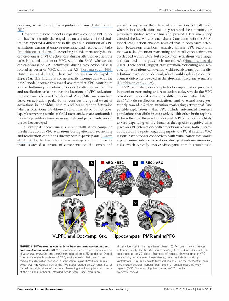
However, the AtoM model’s integrative account of VPC func-tion has been recently challenged by a meta-analysis of fMRI stud-ies that reported a difference in the spatial distribution of VPCactivations during attention-reorienting and recollection tasks(Hutchinson et al., 2009). According to this meta-analysis, thecenter-of-mass of VPC activations during attention-reorientingtasks is located in anterior VPC, within the SMG, whereas thecenter-of-mass of VPC activations during recollection tasks islocated in posterior VPC, within the AG (Corbetta et al., 2008;Hutchinson et al., 2009). These two locations are displayed inFigure 1A. This finding is not necessarily incompatible with theAtoM model because this model assumes that VPC contributessimilar bottom-up attention processes to attention-reorientingand recollection tasks, not that the locations of VPC activationsin these two tasks must be identical. Also, fMRI meta-analysesbased on activation peaks do not consider the spatial extent ofactivations in individual studies and hence cannot determinewhether activations for different conditions do or do not over-lap. Moreover, the results of fMRI meta-analyses are confoundedby many possible differences in methods and participants amongthe studies surveyed.
To investigate these issues, a recent fMRI study comparedthe distribution of VPC activations during attention-reorientingand recollection conditions directly within-participants (Cabezaet al., 2011). In the attention-reorienting condition, partic-ipants searched a stream of consonants on the screen and
pressed a key when they detected a vowel (an oddball task),whereas in a recollection task, they searched their memory forpreviously studied word-chains and pressed a key when theydetected the last word of each chain. Consistent with the AtoMmodel, conjunction analyses revealed that in both tasks detec-tion (bottom-up attention) activated similar VPC regions inthe two tasks. Attention-reorienting and recollection activationsoverlapped within SMG, but recollection activations were largerand extended more posteriorly toward AG (Hutchinson et al.,2009). These results suggest that attention-reorienting and rec-ollection activations can overlap within-participants but the dis-tributions may not be identical, which could explain the center-of-mass difference detected in the aforementioned meta-analysis(Hutchinson et al., 2009).
If VPC contributes similarly to bottom-up attention processesin attention-reorienting and recollection tasks, why do the VPCactivations they elicit show some differences in spatial distribu-tion? Why do recollection activations tend to extend more pos-teriorly toward AG than attention-reorienting activations? Onepossible explanation is that VPC includes intermixed neuronalpopulations that differ in connectivity with other brain regions.If this is the case, the exact locations of fMRI activations are likelyto vary depending on the demands that specific cognitive tasksplace on VPC interactions with other brain regions, both in termsof inputs and outputs. Regarding inputs to VPC, if anterior VPCregions have stronger connectivity with visual cortex that wouldexplain more anterior activations during attention-reorientingtasks, which typically involve visuospatial stimuli (Hutchinson
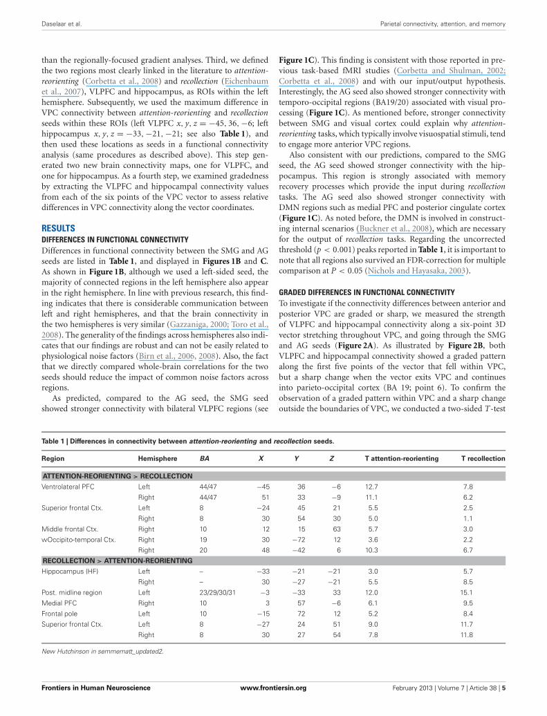
FIGURE 1 | Differences in connectivity between attention-reorienting
and recollection seeds. (A) VPC coordinates derived from meta-analysesof attention-reorienting and recollection plotted on a 3D rendering. Dottedlines indicate the boundaries of VPC, and the solid black line in themiddle the distinction between supramarginal gyrus (SMG) and angulargyrus (AG). (B) Comparison of the two seeds plotted on 3D renderings ofthe left and right sides of the brain, illustrating the hemispheric symmetryof the findings. Although left-sided seeds were used, results are
virtually identical in the right hemisphere. (C) Regions showing greaterVPC connectivity for the attention-reorienting (red) and recollection (blue)seeds plotted on 2D slices. Examples of regions showing greater VPCconnectivity for the attention-reorienting seed include left and rightventrolateral PFC, and occipito-temporal regions. For the recollection seed,they include bilateral hippocampus, and the “default mode network”regions (PCC, Posterior cingulate cortex; mPFC, medialprefrontal cortex).
Frontiers in Human Neuroscience www.frontiersin.org February 2013 | Volume 7 | Article 38 | 2

Daselaar et al. Parietal connectivity, attention, and memory
et al., 2009). Conversely, if posterior VPC regions have strongerconnectivity with medial temporal lobe (MTL) regions, thatwould explain more posterior activations during recollectiontasks, which depend on input from MTL memory regions, suchas the hippocampus. Regarding outputs from VPC, if anteriorVPC regions have stronger connectivity with premotor frontalregions (Kelly et al., 2010), that would explain more anterioractivations during attention-reorienting tasks, which typicallyrequire speeded responses. On the other hand, if posterior VPCregions have stronger connectivity with the default mode net-work (DMN) regions associated with the construction of internalscenarios (Buckner et al., 2008), that could explain more poste-rior activations during recollection tasks, which usually requirethe creation of such internal scenarios before a response can bemade. Preliminary evidence that these hypothetical differencesin connectivity do exist has been provided by recent resting-state functional connectivity studies (Vincent et al., 2008; Nelsonet al., 2010; Uddin et al., 2010; Yeo et al., 2011). For example,these studies found that more anterior VPC regions have strongerconnectivity with ventrolateral PFC (VLPFC), whereas more pos-terior VPC regions have stronger connectivity with MTL andDMN regions.
However, there are two problems in using this functionalconnectivity evidence as evidence in favor of our input-outputhypothesis. First, given that the anatomical borders within VPCare still relatively ill-defined (Uddin et al., 2010), it is unclear towhat extent the anterior and posterior VPC regions investigatedin previous resting state connectivity studies (Vincent et al., 2008;Nelson et al., 2010; Uddin et al., 2010; Yeo et al., 2011) match withthe locations associated with attention-reorienting and recollec-tion tasks in the fMRI meta-analysis (Hutchinson et al., 2009).For example, in one study the anterior VPC regions were inanterior AG rather than in SMG (Uddin et al., 2010). Thus, inorder to link attention-reorienting and recollection differencesto connectivity differences, it is critical to investigate connectiv-ity differences study using as seeds the actual center-of-mass ofattention-reorienting and recollection activations. Second, ouroutput-input hypothesis would be more consistent with anterior-posterior differences in VPC connectivity if these differences aregraded than if they are sharp. Sharp differences would fit bet-ter with the idea of very different cognitive functions in SMGvs. AG (Nelson et al., 2010). To investigate if anterior-posteriordifferences in VPC connectivity are graded one needs to sam-ple connectivity in a continuous manner between anterior andposterior VPC regions.
The goals of present study were to address these two prob-lems. First, to link connectivity differences to the distributionof attention-reorienting and recollection activations, we con-ducted a resting-state functional connectivity using as seeds thecenter-of-mass of attention-reorienting and recollection activa-tions in the aforementioned fMRI meta-analysis (Hutchinsonet al., 2009). Since there is a tendency for recollection studies toshow left-sided activations, we used VPC seeds in the left hemi-sphere only, but explored both left- and right-hemisphere con-nectivity. Despite the close proximity of the attention-reorientingand recollection seeds (see Figure 1A), we expected differences infunctional connectivity patterns. In particular, we predicted that
the SMG seed would show stronger connectivity with VLPFC,which is a region previously associated with attention-reorienting(Corbetta and Shulman, 2002), whereas the AG seed wouldshow stronger connectivity with MTL, and in particular withthe hippocampus, which is a region previously associated withrecollection (Eichenbaum et al., 2007).
Second, we investigated whether functional connectivity dif-ferences between the anterior SMG seed and the posterior AGseed are sharp or graded. For this analysis, we focused onVPC connectivity with the two regions most strongly associatedwith attention-reorienting and recollection, namely VLPFC andthe hippocampus, respectively. Instead of a categorical contrastbetween two VPC locations, as in previous studies, we sampledconnectivity continuously between the anterior SMG seed andthe posterior AG seed. If one assumes that peak activations indifferent regions of VPC reflect differences in cognitive function,and that the latter goes hand-in-hand with differences in con-nectivity, then marked differences in function can be assumed toentail sharp differences in connectivity. Alternatively, these dif-ferent regions may have a common function, with differencesin peak activation reflecting the different inputs and outputs onwhich that function is applied. If that is the case, then differencesin connectivity between the peaks are likely to be graded. In linewith our hypothesis, we predicted that differences in VLPFC andhippocampal connectivity would be graded rather than sharp,with VLPFC connectivity decreasing gradually from anterior toposterior regions and hippocampal connectivity showing theopposite trend.
METHODSPARTICIPANTSResting-state scans were acquired from 24 participants (14 female,mean age 23) recruited from the University of Amsterdam com-munity. All participants were in good health and right-handed.Their native language was Dutch and they were paid 25 euro forparticipation. Participants gave their written informed consentand the study met all criteria for approval of the ethical boardof the Amsterdam Medical Center.
SCANNINGfMRI images were collected on a Phillips Intera 3.0T using a6-channel standard SENSE head coil and a T2∗ sensitive gradi-ent echo sequence (96 × 96 matrix, TR 2000 ms, TE 30 ms, FA80◦, 34 slices, 2.3 × 2.3 mm voxel size, 3-mm thick transverseslices). Additionally, a high-resolution T1-weighted structuralscan (256 × 256 matrix, TR 12 ms, TE 5 ms, FOV 24 cm, 68slices, 1 mm slice thickness) was collected. Two 8-min rest blockswere administered to each participant. Each block consisted ofa black screen with a white fixation cross-hair in the center.Participants were instructed to keep focused on the cross-hairduring scanning.
IMAGE PREPROCESSINGStatistical Parametric Mapping (SPM5; http://www.fil.ion.ucl.ac.uk/spm) software was used to preprocess and analyze theMR data. The images were slice-time and motion-corrected,and then normalized. First, individual normalization parameters
Frontiers in Human Neuroscience www.frontiersin.org February 2013 | Volume 7 | Article 38 | 3
Daselaar et al. Parietal connectivity, attention, and memory
were obtained by normalizing the segmentedstructural scan ofeach subject using the Montreal Neurological Institute (MNI)T1 template image. These normalization parameters were thenapplied to the functional images. Next, the normalized functionalimages were resliced to a resolution of 3 × 3 × 3 mm and spa-tially smoothed using an 8-mm isotropic Gaussian kernel. Next,treating the volumes as a time series, the data were temporallysmoothed using linear detrending, and a temporal filter (0.01 Hz< f < 0.08 Hz) was applied to remove low-frequency drifts andphysiological high-frequency noise.
VPC SEEDSFor the identification of the seeds, we used the center-of-massof attention-reorienting activations in an fMRI meta-analysis(Hutchinson et al., 2009), which also reported the center-of-massof recollection activations previously estimated by other reviews(Ciaramelli et al., 2008; Vilberg and Rugg, 2008). In the presentstudy, we used the MNI coordinate system. Since the metaanalysisalso included studies reporting their results in Talairach space, wefirst converted these coordinates to MNI space (Brett et al., 2001).Then we calculated the center-of-mass using the median of theresulting x, y, and z coordinates for both attention-reorienting andrecollection studies. For the attention-reorienting studies (N = 23)the center of mass was x, y, z = −48,−45, 34, for recollectionstudies (N = 20), it was x, y, z = −43,−58, 36, (Vilberg andRugg, 2008). Figure 1A shows a rendering of VPC including theboundaries between SMG and AG using the CARET softwarepackage (http://brainvis.wustl.edu/wiki/index.php/Caret:About).As can be seen, the attention-reorienting seed (red dot) waslocated more anteriorly, falling within SMG (BA 40) in a regioncommonly referred to as the temporo-parietal junction (TPJ)(Corbetta et al., 2008). In contrast, the recollection seed (bluedot) was located more posteriorly falling within the boundariesof AG (BA 39).
FUNCTIONAL CONNECTIVITY ANALYSESConnectivity differences between SMG vs. AG seedsFor analysis of the connectivity differences between the SMGand AG seeds we used the REST toolbox (V1.3; www.restfmri.net) integrated with SPM5. A seed correlation analysis was per-formed in a voxel-wise way with the six head motion parametersas covariates. Individual r-maps were normalized to Z-mapsby using Fisher’s Z transformation. For the seed analysis, twospherical regions of interest (ROIs; radius = 6 mm) were cen-tered at the MNI coordinates derived from the two meta-analysesof attention-reorienting and recollection. Next, the Z-maps wereaveraged across runs for each participant. Subsequently, differ-ence maps were created by subtracting the two resulting Z-maps.To assess relative differences in connectivity between the twoseeds, we used a random effects analysis on these difference mapswith a two-sided one-sample t-test (P < 0.001, minimum clustersize = 25).
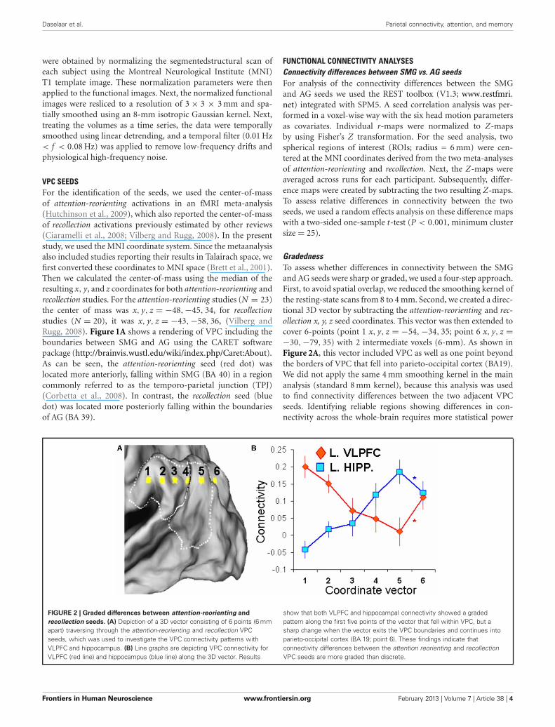
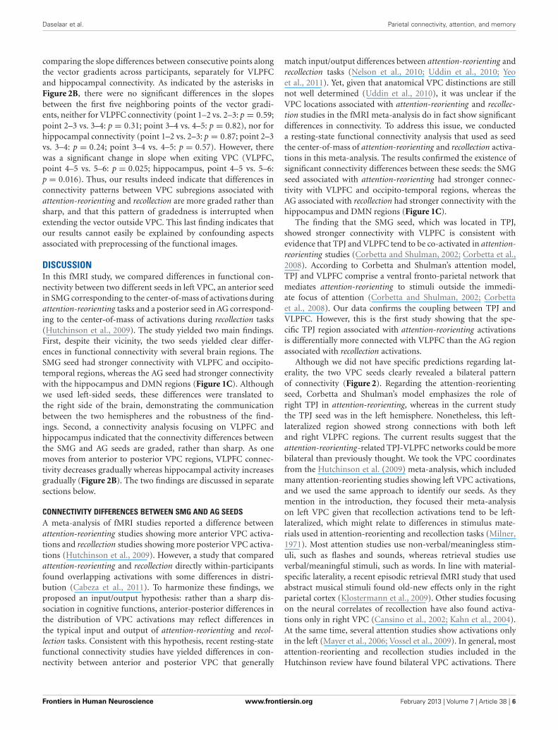
GradednessTo assess whether differences in connectivity between the SMGand AG seeds were sharp or graded, we used a four-step approach.First, to avoid spatial overlap, we reduced the smoothing kernel ofthe resting-state scans from 8 to 4 mm. Second, we created a direc-tional 3D vector by subtracting the attention-reorienting and rec-ollection x, y, z seed coordinates. This vector was then extended tocover 6-points (point 1 x, y, z = −54, −34, 35; point 6 x, y, z =−30, −79, 35) with 2 intermediate voxels (6-mm). As shown inFigure 2A, this vector included VPC as well as one point beyondthe borders of VPC that fell into parieto-occipital cortex (BA19).We did not apply the same 4 mm smoothing kernel in the mainanalysis (standard 8 mm kernel), because this analysis was usedto find connectivity differences between the two adjacent VPCseeds. Identifying reliable regions showing differences in con-nectivity across the whole-brain requires more statistical power
FIGURE 2 | Graded differences between attention-reorienting and
recollection seeds. (A) Depiction of a 3D vector consisting of 6 points (6 mmapart) traversing through the attention-reorienting and recollection VPCseeds, which was used to investigate the VPC connectivity patterns withVLPFC and hippocampus. (B) Line graphs are depicting VPC connectivity forVLPFC (red line) and hippocampus (blue line) along the 3D vector. Results
show that both VLPFC and hippocampal connectivity showed a gradedpattern along the first five points of the vector that fell within VPC, but asharp change when the vector exits the VPC boundaries and continues intoparieto-occipital cortex (BA 19; point 6). These findings indicate thatconnectivity differences between the attention reorienting and recollectionVPC seeds are more graded than discrete.
Frontiers in Human Neuroscience www.frontiersin.org February 2013 | Volume 7 | Article 38 | 4
Daselaar et al. Parietal connectivity, attention, and memory
than the regionally-focused gradient analyses. Third, we definedthe two regions most clearly linked in the literature to attention-reorienting (Corbetta et al., 2008) and recollection (Eichenbaumet al., 2007), VLPFC and hippocampus, as ROIs within the lefthemisphere. Subsequently, we used the maximum difference inVPC connectivity between attention-reorienting and recollectionseeds within these ROIs (left VLPFC x, y, z = −45, 36, −6; lefthippocampus x, y, z = −33,−21, −21; see also Table 1), andthen used these locations as seeds in a functional connectivityanalysis (same procedures as described above). This step gen-erated two new brain connectivity maps, one for VLPFC, andone for hippocampus. As a fourth step, we examined gradednessby extracting the VLPFC and hippocampal connectivity valuesfrom each of the six points of the VPC vector to assess relativedifferences in VPC connectivity along the vector coordinates.
RESULTSDIFFERENCES IN FUNCTIONAL CONNECTIVITYDifferences in functional connectivity between the SMG and AGseeds are listed in Table 1, and displayed in Figures 1B and C.As shown in Figure 1B, although we used a left-sided seed, themajority of connected regions in the left hemisphere also appearin the right hemisphere. In line with previous research, this find-ing indicates that there is considerable communication betweenleft and right hemispheres, and that the brain connectivity inthe two hemispheres is very similar (Gazzaniga, 2000; Toro et al.,2008). The generality of the findings across hemispheres also indi-cates that our findings are robust and can not be easily related tophysiological noise factors (Birn et al., 2006, 2008). Also, the factthat we directly compared whole-brain correlations for the twoseeds should reduce the impact of common noise factors acrossregions.
As predicted, compared to the AG seed, the SMG seedshowed stronger connectivity with bilateral VLPFC regions (see
Figure 1C). This finding is consistent with those reported in pre-vious task-based fMRI studies (Corbetta and Shulman, 2002;Corbetta et al., 2008) and with our input/output hypothesis.Interestingly, the AG seed also showed stronger connectivity withtemporo-occipital regions (BA19/20) associated with visual pro-cessing (Figure 1C). As mentioned before, stronger connectivitybetween SMG and visual cortex could explain why attention-reorienting tasks, which typically involve visuospatial stimuli, tendto engage more anterior VPC regions.
Also consistent with our predictions, compared to the SMGseed, the AG seed showed stronger connectivity with the hip-pocampus. This region is strongly associated with memoryrecovery processes which provide the input during recollectiontasks. The AG seed also showed stronger connectivity withDMN regions such as medial PFC and posterior cingulate cortex(Figure 1C). As noted before, the DMN is involved in construct-ing internal scenarios (Buckner et al., 2008), which are necessaryfor the output of recollection tasks. Regarding the uncorrectedthreshold (p < 0.001) peaks reported in Table 1, it is important tonote that all regions also survived an FDR-correction for multiplecomparison at P < 0.05 (Nichols and Hayasaka, 2003).
GRADED DIFFERENCES IN FUNCTIONAL CONNECTIVITYTo investigate if the connectivity differences between anterior andposterior VPC are graded or sharp, we measured the strengthof VLPFC and hippocampal connectivity along a six-point 3Dvector stretching throughout VPC, and going through the SMGand AG seeds (Figure 2A). As illustrated by Figure 2B, bothVLPFC and hippocampal connectivity showed a graded patternalong the first five points of the vector that fell within VPC,but a sharp change when the vector exits VPC and continuesinto parieto-occipital cortex (BA 19; point 6). To confirm theobservation of a graded pattern within VPC and a sharp changeoutside the boundaries of VPC, we conducted a two-sided T-test
Table 1 | Differences in connectivity between attention-reorienting and recollection seeds.
Region Hemisphere BA X Y Z T attention-reorienting T recollection
ATTENTION-REORIENTING > RECOLLECTION
Ventrolateral PFC Left 44/47 −45 36 −6 12.7 7.8
Right 44/47 51 33 −9 11.1 6.2
Superior frontal Ctx. Left 8 −24 45 21 5.5 2.5
Right 8 30 54 30 5.0 1.1
Middle frontal Ctx. Right 10 12 15 63 5.7 3.0
wOccipito-temporal Ctx. Right 19 30 −72 12 3.6 2.2
Right 20 48 −42 6 10.3 6.7
RECOLLECTION > ATTENTION-REORIENTING
Hippocampus (HF) Left – −33 −21 −21 3.0 5.7
Right – 30 −27 −21 5.5 8.5
Post. midline region Left 23/29/30/31 −3 −33 33 12.0 15.1
Medial PFC Right 10 3 57 −6 6.1 9.5
Frontal pole Left 10 −15 72 12 5.2 8.4
Superior frontal Ctx. Left 8 −27 24 51 9.0 11.7
Right 8 30 27 54 7.8 11.8
New Hutchinson in semmematt_updated2.
Frontiers in Human Neuroscience www.frontiersin.org February 2013 | Volume 7 | Article 38 | 5
Daselaar et al. Parietal connectivity, attention, and memory
comparing the slope differences between consecutive points alongthe vector gradients across participants, separately for VLPFCand hippocampal connectivity. As indicated by the asterisks inFigure 2B, there were no significant differences in the slopesbetween the first five neighboring points of the vector gradi-ents, neither for VLPFC connectivity (point 1–2 vs. 2–3: p = 0.59;point 2–3 vs. 3–4: p = 0.31; point 3–4 vs. 4–5: p = 0.82), nor forhippocampal connectivity (point 1–2 vs. 2–3: p = 0.87; point 2–3vs. 3–4: p = 0.24; point 3–4 vs. 4–5: p = 0.57). However, therewas a significant change in slope when exiting VPC (VLPFC,point 4–5 vs. 5–6: p = 0.025; hippocampus, point 4–5 vs. 5–6:p = 0.016). Thus, our results indeed indicate that differences inconnectivity patterns between VPC subregions associated withattention-reorienting and recollection are more graded rather thansharp, and that this pattern of gradedness is interrupted whenextending the vector outside VPC. This last finding indicates thatour results cannot easily be explained by confounding aspectsassociated with preprocessing of the functional images.
DISCUSSIONIn this fMRI study, we compared differences in functional con-nectivity between two different seeds in left VPC, an anterior seedin SMG corresponding to the center-of-mass of activations duringattention-reorienting tasks and a posterior seed in AG correspond-ing to the center-of-mass of activations during recollection tasks(Hutchinson et al., 2009). The study yielded two main findings.First, despite their vicinity, the two seeds yielded clear differ-ences in functional connectivity with several brain regions. TheSMG seed had stronger connectivity with VLPFC and occipito-temporal regions, whereas the AG seed had stronger connectivitywith the hippocampus and DMN regions (Figure 1C). Althoughwe used left-sided seeds, these differences were translated tothe right side of the brain, demonstrating the communicationbetween the two hemispheres and the robustness of the find-ings. Second, a connectivity analysis focusing on VLPFC andhippocampus indicated that the connectivity differences betweenthe SMG and AG seeds are graded, rather than sharp. As onemoves from anterior to posterior VPC regions, VLPFC connec-tivity decreases gradually whereas hippocampal activity increasesgradually (Figure 2B). The two findings are discussed in separatesections below.
CONNECTIVITY DIFFERENCES BETWEEN SMG AND AG SEEDSA meta-analysis of fMRI studies reported a difference betweenattention-reorienting studies showing more anterior VPC activa-tions and recollection studies showing more posterior VPC activa-tions (Hutchinson et al., 2009). However, a study that comparedattention-reorienting and recollection directly within-participantsfound overlapping activations with some differences in distri-bution (Cabeza et al., 2011). To harmonize these findings, weproposed an input/output hypothesis: rather than a sharp dis-sociation in cognitive functions, anterior-posterior differences inthe distribution of VPC activations may reflect differences inthe typical input and output of attention-reorienting and recol-lection tasks. Consistent with this hypothesis, recent resting-statefunctional connectivity studies have yielded differences in con-nectivity between anterior and posterior VPC that generally
match input/output differences between attention-reorienting andrecollection tasks (Nelson et al., 2010; Uddin et al., 2010; Yeoet al., 2011). Yet, given that anatomical VPC distinctions are stillnot well determined (Uddin et al., 2010), it was unclear if theVPC locations associated with attention-reorienting and recollec-tion studies in the fMRI meta-analysis do in fact show significantdifferences in connectivity. To address this issue, we conducteda resting-state functional connectivity analysis that used as seedthe center-of-mass of attention-reorienting and recollection activa-tions in this meta-analysis. The results confirmed the existence ofsignificant connectivity differences between these seeds: the SMGseed associated with attention-reorienting had stronger connec-tivity with VLPFC and occipito-temporal regions, whereas theAG associated with recollection had stronger connectivity with thehippocampus and DMN regions (Figure 1C).
The finding that the SMG seed, which was located in TPJ,showed stronger connectivity with VLPFC is consistent withevidence that TPJ and VLPFC tend to be co-activated in attention-reorienting studies (Corbetta and Shulman, 2002; Corbetta et al.,2008). According to Corbetta and Shulman’s attention model,TPJ and VLPFC comprise a ventral fronto-parietal network thatmediates attention-reorienting to stimuli outside the immedi-ate focus of attention (Corbetta and Shulman, 2002; Corbettaet al., 2008). Our data confirms the coupling between TPJ andVLPFC. However, this is the first study showing that the spe-cific TPJ region associated with attention-reorienting activationsis differentially more connected with VLPFC than the AG regionassociated with recollection activations.
Although we did not have specific predictions regarding lat-erality, the two VPC seeds clearly revealed a bilateral patternof connectivity (Figure 2). Regarding the attention-reorientingseed, Corbetta and Shulman’s model emphasizes the role ofright TPJ in attention-reorienting, whereas in the current studythe TPJ seed was in the left hemisphere. Nonetheless, this left-lateralized region showed strong connections with both leftand right VLPFC regions. The current results suggest that theattention-reorienting-related TPJ-VLPFC networks could be morebilateral than previously thought. We took the VPC coordinatesfrom the Hutchinson et al. (2009) meta-analysis, which includedmany attention-reorienting studies showing left VPC activations,and we used the same approach to identify our seeds. As theymention in the introduction, they focused their meta-analysison left VPC given that recollection activations tend to be left-lateralized, which might relate to differences in stimulus mate-rials used in attention-reorienting and recollection tasks (Milner,1971). Most attention studies use non-verbal/meaningless stim-uli, such as flashes and sounds, whereas retrieval studies useverbal/meaningful stimuli, such as words. In line with material-specific laterality, a recent episodic retrieval fMRI study that usedabstract musical stimuli found old-new effects only in the rightparietal cortex (Klostermann et al., 2009). Other studies focusingon the neural correlates of recollection have also found activa-tions only in right VPC (Cansino et al., 2002; Kahn et al., 2004).At the same time, several attention studies show activations onlyin the left (Mayer et al., 2006; Vossel et al., 2009). In general, mostattention-reorienting and recollection studies included in theHutchinson review have found bilateral VPC activations. There
Frontiers in Human Neuroscience www.frontiersin.org February 2013 | Volume 7 | Article 38 | 6
Daselaar et al. Parietal connectivity, attention, and memory
is one study (Guerin and Miller, 2009) that found a retrievalsuccess effect in parietal cortex that showed a tendency for left-lateralized activity compared to attention, regardless of stimu-lus type (words/faces). However, this study investigated old/neweffects, and not specifically recollection. They also did not reportan effect in VPC, but in dorsal parietal cortex. Thus, in generaland in line with our connectivity data, findings indicate that thelateralization difference in VPC activity between recollection andattention-reorienting studies is more relative than absolute.
In terms of our input/output hypothesis, the strong connec-tivity of the SMG seed with VLPFC could explain why VPCactivations tend to be more anterior for attention-reorientingthan recollection. Most attention-reorienting tasks require speededresponses, and VLPFC is directly linked to premotor regions(Kelly et al., 2010) important for such output. In contrast,recollection tasks are less dependent on rapid responses. Theinput/output hypothesis fits also well with the finding that theSMG seed was more strongly connected with visual occipito-temporal regions (BA 19/20) than the AG seed. Most attention-reorienting tasks involve visuospatial stimuli and hence emphasizeoccipito-temporal input to VPC.
Turning to the AG seed associated with recollection, thestronger connectivity of this seed with the hippocampal for-mation is consistent with the role of the well-known role ofhippocampus in recollection. The AG seed also showed strongerconnections with DMN regions, including the posterior cingu-late cortex and medial prefrontal cortex. The DMN tends tobe more activated during rest than during active task condi-tions. According to one account, the processes mediated by theDMN represent an internal mode of cognition (Buckner et al.,2008; Huijbers et al., 2011), mediating internally oriented pro-cesses involving internal reflections, which follow a memorycue or are generated spontaneously. Interpreted in terms of ourinput/output hypothesis, these functional connectivity resultscould explain the association between AG and recollection tasks.During recollection tasks, the input of VPC are long-term memo-ries recovered by MTL and the output involves the constructionof an internal scenario mediated by DMN. Thus, because of itsinput and output characteristics, recollection tasks are more likelyto engage posterior VPC regions that have stronger connectivitywith MTL and DMN regions.
In terms of the relation between our functional connectiv-ity findings and underlying neural processes, it should be notedthat we cannot measure direct neural communication with fMRI,but only slow oscillations in brain activity. However, there issubstantial evidence indicating that these slow oscillations repre-sent fluctuations in the power of synchronized neuronal activity(Anderson, 2008; Nir et al., 2008; de Pasquale et al., 2010),and that they show correlations with structural brain connec-tivity measures (Honey et al., 2007; van den Heuvel et al., 2009;Uddin et al., 2010). Moreover, fMRI connectivity measures havebeen reliably linked to fluctuations in multi-unit neural activ-ity. For instance, a recent brain connectivity study that combinedintracortical neurophysiological recording techniques with fMRI(Shmuel and Leopold, 2008) found clear correlations in brainconnectivity across visual cortex regions between fMRI mea-sures and multi-unit recordings (spiking rate and power changes).Thus, even though fMRI cannot provide a measure of direct
neural communication, it certainly yields insights into the neu-ral coupling between different brain areas, and on a larger spatialscale than can be achieved with electrophysiological techniques.
GRADED CONNECTIVITY PATTERNSAlthough the differences in functional connectivity we foundbetween the SMG and AG seeds were generally consistent withour input/output hypothesis, it is important to determine if thesedifferences are graded or sharp. As noted before, graded differ-ences would fit well with the input/output hypothesis, whereassharp differences would be more consistent with different cog-nitive operations in SMG and AG (Hutchinson et al., 2009).To investigate whether connectivity differences were grade orsharp, we sampled connectivity in a continuous manner betweenthe SMG and AG seeds. As illustrated by Figure 2B, the resultsusing critical VLPFC and hippocampal seeds clearly supporteda graded pattern: as one moves from anterior to posterior VPC,VLPFC connectivity decreases gradually whereas hippocampalconnectivity increases gradually.
The graded connectivity patterns we observed (Figure 2B) aremore likely to be associated with a common function medi-ated by adjacent regions of VPC, as we predicted, than withmarkedly different functions subserved by these regions whichwould entail sharp differences in connectivity. Thus, the gradedconnectivity patterns fit well with the idea that SMG and AG servea common function mediated by intermixed neuronal popula-tions, whose proportions differ from anterior to posterior VPCregions. Anterior VPC regions have stronger connections withVLPFC, which could explain a more anterior distribution of VPCactivations during attention-reorienting tasks, whereas posteriorVPC regions have stronger connections with the hippocampus,which could partly explain a more posterior distribution of VPCactivations during recollection tasks.
The finding of graded connectivity patterns within VPC seemsat odds with cyto- and receptor-architectonic evidence indicat-ing that this region is anatomically heterogeneous (Brodmann,1911; Caspers et al., 2012). However, there is abundant evidencethat the distribution of different cell types within specializedregions of the brain, and their respective connections, are gen-erally more graded than discrete. Graded patterns of cell behaviorand intermixed cell populations have been found in the visual cor-tex (Barone et al., 2000), auditory cortex (Castro and Kandler,2010), MTL (Xiang and Brown, 1998), and frontal lobes (Barbas,1986). Regarding VPC, using structural connectivity measures,Caspers et al. (2011) found that the middle VPC (approximatelycorresponding to TPJ) shared connection patterns of both ros-tral (posterior supramarginal) and caudal (angular) VPC areas,again suggesting that the functional/anatomical boundaries arenot that clear, and rather show a rostro-caudal gradient in termsof structural connectivity. Thus, despite abundant evidence formodularity in the brain, available evidence suggests that thecytoarchitectonic boundaries between anatomical subregions, atleast, within modules are more graded than sharp. Nevertheless,despite the fact that we found clear breaks of the gradients out-side of VPC, some caveat is required regarding the effect of spatialsmoothing on the reported gradients. Even though we used aminimal smoothing kernel of 4 mm with a 6-mm gradient gap,there could be some residual blurring effects.
Frontiers in Human Neuroscience www.frontiersin.org February 2013 | Volume 7 | Article 38 | 7

Daselaar et al. Parietal connectivity, attention, and memory
The finding that connectivity differences between anterior andposterior VPC regions are more graded rather than sharp isimportant because it supports the development of over-archingmodels of VPC function, such as output buffer and evidenceaccumulator models (Wagner et al., 2005). The AtoM model isanother exemplar of an overarching model, because it assumesthat the area of VPC that encompasses both AG and SMG plays aan overarching role in bottom-up attention and that both ante-rior (SMG) and posterior (AG) VPC regions contribute to it.This model can parsimoniously account for VPC activations dur-ing both attention-reorienting and recollection tasks because bothtasks depend on bottom-up attention. Importantly, this modelis not incompatible with evidence that the spatial distributionof VPC activations is not identical for attention-reorienting andrecollection tasks, because the specific locations of activationsdepend on several factors, including input/output differences.The current results fit well with this input/out hypothesis.
In addition to addressing a specific issue regarding the func-tion of the VPC, this study also speaks to a general prob-lem in interpreting functional neuroimaging data. Differencesin regions of peak activation, and in functional connectivitywith those regions, typically are interpreted as indicative of dif-ferences in function mediated by those regions (fractionationhypothesis). As we have argued, this need not be the case.These differences could just as easily reflect differences in apply-ing a common (overarching) function to different domains,in our case perception and memory, about which informa-tion is conveyed via different pathways (Cabeza et al., 2012).Although we acknowledge that finding a graded response isnot solid evidence for the “overarching function hypothesis”against the “fractionation hypothesis,” we hope that it is suffi-ciently compelling to encourage others to entertain both typesof interpretations of functional neuroimaging data in otherfields.
REFERENCESAnderson, J. S. (2008). Origin of syn-
chronized low-frequency blood oxy-gen level-dependent fluctuations inthe primary visual cortex. Am. J.Neuroradiol. 29, 1722–1729.
Barbas, H. (1986). Pattern in the lam-inar origin of corticocortical con-nections. J. Comp. Neurol. 252,415–422.
Barone, P., Batardiere, A., Knoblauch,K., and Kennedy, H. (2000).Laminar distribution of neuronsin extrastriate areas projecting tovisual areas V1 and V4 correlateswith the hierarchical rank andindicates the operation of a distancerule. J. Neurosci. 20, 3263–3281.
Birn, R. M., Diamond, J. B., Smith,M. A., and Bandettini, P. A.(2006). Separating respiratory-variation-related fluctuations fromneuronal-activity-related fluctu-ations in fMRI. Neuroimage 31,1536–1548.
Birn, R. M., Murphy, K., andBandettini, P. A. (2008). Theeffect of respiration variations onindependent component analysisresults of resting state functionalconnectivity. Hum. Brain Mapp.29, 740–750.
Brett, M., Christoff, K., Cusack, R.,and Lancaster, J. (2001). Using thetalairach atlas with the MNI tem-plate. Neuroimage 13, s85.
Brodmann, K. (1911). VergleichendeLokalisationslehre der Grosshirnrindein ihren Prinzipien dargestellt aufGrund des Zellenbaues. Leipzig:Barth.
Buckner, R. L., Andrews-Hanna, J. R.,and Schacter, D. L. (2008). Thebrain’s default network: anatomy,function, and relevance to disease.Ann. N.Y. Acad. Sci. 1124, 1–38.
Cabeza, R., Ciaramelli, E., andMoscovitch, M. (2012). Cognitivecontributions of the ventral parietalcortex: an integrative theoreticalaccount. Trends Cogn. Sci. 16,338–352.
Cabeza, R., Ciaramelli, E., Olson,I. R., and Moscovitch, M.(2008). The parietal cortex andepisodic memory: an attentionalaccount. Nat. Rev. Neurosci. 9,613–625.
Cabeza, R., Mazuz, Y. S., Stokes,J., Kragel, J. E., Woldorff, M.G., Ciaramelli, E., et al. (2011).Overlapping parietal activity inmemory and perception: evidencefor the attention to memory model.J. Cogn. Neurosci. 23, 3209–3217.
Cansino, S., Maquet, P., Dolan, R.J., and Rugg, M. D. (2002). Brainactivity underlying encoding andretrieval of source memory. Cereb.Cortex 12, 1048–1056.
Caspers, S., Eickhoff, S. B., Rick, T.,von Kapri, A., Kuhlen, T., Huang,R., et al. (2011). Probabilistic fibretract analysis of cytoarchitectoni-cally defined human inferior pari-etal lobule areas reveals similari-ties to macaques. Neuroimage 58,362–380.
Caspers, S., Schleicher, A., Bacha-Trams, M., Palomero-Gallagher, N.,Amunts, K., and Zilles, K. (2012).Organization of the human infe-rior parietal lobule based on recep-tor architectonics. Cereb. Cortex 23,615–628.
Castro, J. B., and Kandler, K. (2010).Changing tune in auditory cortex.Nat. Neurosci. 13, 271–273.
Ciaramelli, E., Grady, C. L., andMoscovitch, M. (2008). Top-downand bottom-up attention to mem-ory: a hypothesis (AtoM) on the role
of the posterior parietal cortex inmemory retrieval. Neuropsychologia46, 1828–1851.
Corbetta, M., Patel, G., and Shulman,G. L. (2008). The reorienting sys-tem of the human brain: from envi-ronment to theory of mind. Neuron58, 306–324.
Corbetta, M., and Shulman, G. L.(2002). Control of goal-directedand stimulus-driven attention inthe brain. Nat. Rev. Neurosci. 3,201–215.
Daselaar, S. M., Fleck, M. S., andCabeza, R. (2006). Triple disso-ciation in the medial temporallobes: recollection, familiarity,and novelty. J. Neurophysiol. 96,1902–1911.
de Pasquale, F., Della Penna, S.,Snyder, A. Z., Lewis, C., Mantini,D., Marzetti, L., et al. (2010).Temporal dynamics of spontaneousMEG activity in brain networks.Proc. Natl. Acad. Sci. U.S.A. 107,6040–6045.
Eichenbaum, H., Yonelinas, A. P.,and Ranganath, C. (2007). Themedial temporal lobe and recogni-tion memory. Annu. Rev. Neurosci.30, 123–152.
Eldridge, L. L., Knowlton, B. J.,Furmanski, C. S., Bookheimer,S. Y., and Engel, S. A. (2000).Remembering episodes: a selec-tive role for the hippocampusduring retrieval. Nat. Neurosci. 3,1149–1152.
Gazzaniga, M. S. (2000). Cerebralspecialization and interhemi-spheric communication. Brain 123,1293–1326.
Guerin, S. A., and Miller, M. B.(2009). Lateralization of the parietalold/new effect: an event-relatedfMRI study comparing recognition
memory for words and faces.Neuroimage 44, 232–242.
Henson, R. A., Rugg, M. D., Shallice,T., Josephs, O., and Dolan, R. J.(1999). Recollection and familiarityin recognition memory: an event-related functional magnetic reso-nance imaging study. J. Neurosci. 19,3962–3972.
Honey, C. J., Kotter, R., Breakspear,M., and Sporns, O. (2007). Networkstructure of cerebral cortex shapesfunctional connectivity on multipletime scales. Proc. Natl. Acad. Sci.U.S.A. 104, 10240–10245.
Huijbers, W., Pennartz, C. M., Rubin,D. C., and Daselaar, S. M. (2011).Imagery and retrieval of auditoryand visual information: neural cor-relates of successful and unsuccess-ful performance. Neuropsychologia49, 1730–1740.
Hutchinson, J. B., Uncapher, M. R.,and Wagner, A. D. (2009). Posteriorparietal cortex and episodicretrieval: convergent and divergenteffects of attention and memory.Learn. Mem. 16, 343–356.
Kahn, I., Davachi, L., and Wagner, A. D.(2004). Functional-neuroanatomiccorrelates of recollection:implications for models of recog-nition memory. J. Neurosci. 24,4172–4180.
Kelly, C., Uddin, L. Q., Shehzad, Z.,Margulies, D. S., Castellanos, F.X., Milham, M. P., et al. (2010).Broca’s region: linking humanbrain functional connectivity dataand non-human primate tracinganatomy studies. Eur. J. Neurosci.32, 383–398.
Klostermann, E. C., Loui, P., andShimamura, A. P. (2009). Activationof right parietal cortex duringmemory retrieval of nonlinguistic
Frontiers in Human Neuroscience www.frontiersin.org February 2013 | Volume 7 | Article 38 | 8

Daselaar et al. Parietal connectivity, attention, and memory
auditory stimuli. Cogn. Affect.Behav. Neurosci. 9, 242–248.
Mayer, A. R., Harrington, D., Adair, J.C., and Lee, R. (2006). The neuralnetworks underlying endoge-nous auditory covert orientingand reorienting. Neuroimage 30,938–949.
Mesulam, M. M. (1981). A corticalnetwork for directed attention andunilateral neglect. Ann. Neurol. 10,309–325.
Milner, B. (1971). Interhemispheric dif-ferences in the localization of psy-chological processes in man. Br.Med. Bull. 27, 272–277.
Nelson, S. M., Cohen, A. L., Power,J. D., Wig, G. S., Miezin, F. M.,Wheeler, M. E., et al. (2010). Aparcellation scheme for human leftlateral parietal cortex. Neuron 67,156–170.
Nichols, T., and Hayasaka, S. (2003).Controlling the familywise errorrate in functional neuroimaging: acomparative review. Stat. MethodsMed. Res. 12, 419–446.
Nir, Y., Mukamel, R., Dinstein, I.,Privman, E., Harel, M., Fisch, L.,et al. (2008). Interhemisphericcorrelations of slow spontaneousneuronal fluctuations revealedin human sensory cortex. Nat.Neurosci. 11, 1100–1108.
Shmuel, A., and Leopold, D. A. (2008).Neuronal correlates of spontaneousfluctuations in fMRI signals inmonkey visual cortex: implica-tions for functional connectivityat rest. Hum. Brain Mapp. 29,751–761.
Toro, R., Fox, P. T., and Paus, T. (2008).Functional coactivation map of thehuman brain. Cereb. Cortex 18,2553–2559.
Uddin, L. Q., Supekar, K., Amin,H., Rykhlevskaia, E., Nguyen,D. A., Greicius, M. D., et al.(2010). Dissociable connectiv-ity within human angular gyrusand intraparietal sulcus: evidencefrom functional and structuralconnectivity. Cereb. Cortex 20,2636–2646.
van den Heuvel, M. P., Mandl, R. C.W., Kahn, R. S., and Pol, H. E. H.(2009). Functionally linked resting-state networks reflect the underlyingstructural connectivity architectureof the human brain. Hum. BrainMapp. 30, 3127–3141.
Vilberg, K. L., and Rugg, M. D. (2008).Memory retrieval and the pari-etal cortex: a review of evidencefrom a dual-process perspective.Neuropsychologia 46, 1787–1799.
Vincent, J. L., Kahn, I., Snyder, A.Z., Raichle, M. E., and Buckner,
R. L. (2008). Evidence for a fron-toparietal control system revealedby intrinsic functional connectivity.J. Neurophysiol. 100, 3328–3342.
Vossel, S., Weidner, R., Thiel, C. M., andFink, G. R. (2009). What is “odd” inPosner’s location-cueing paradigm?Neural responses to unexpectedlocation and feature changes com-pared. J. Cogn. Neurosci. 21, 30–41.
Wagner, A. D., Shannon, B. J., Kahn, I.,and Buckner, R. L. (2005). Parietallobe contributions to episodicmemory retrieval. Trends Cogn. Sci.9, 445–453.
Wheeler, M. E., and Buckner, R. L.(2004). Functional-anatomic corre-lates of remembering and knowing.Neuroimage 21, 1337–1349.
Yeo, B. T., Krienen, F. M., Sepulcre,J., Sabuncu, M. R., Lashkari, D.,Hollinshead, M., et al. (2011). Theorganization of the human cerebralcortex estimated by intrinsic func-tional connectivity. J. Neurophysiol.106, 1125–1165.
Yonelinas, A. P., Otten, L. J., Shaw,K. N., and Rugg, M. D. (2005).Separating the brain regionsinvolved in recollection and famil-iarity in recognition memory.J. Neurosci. 25, 3002–3008.
Xiang, J. Z., and Brown, M. W. (1998).Differential neuronal encoding of
novelty, familiarity and recencyin regions of the anterior tempo-ral lobe. Neuropharmacology 37,657–676.
Conflict of Interest Statement: Theauthors declare that the researchwas conducted in the absence of anycommercial or financial relationshipsthat could be construed as a potentialconflict of interest.
Received: 10 February 2012; accepted:02 February 2013; published online: 22February 2013.Citation: Daselaar SM, Huijbers W,Eklund K, Moscovitch M and CabezaR (2013) Resting-state functional con-nectivity of ventral parietal regionsassociated with attention reorientingand episodic recollection. Front. Hum.Neurosci. 7:38. doi: 10.3389/fnhum.2013.00038Copyright © 2013 Daselaar, Huijbers,Eklund, Moscovitch and Cabeza. This isan open-access article distributed underthe terms of the Creative CommonsAttribution License, which permits use,distribution and reproduction in otherforums, provided the original authorsand source are credited and subject to anycopyright notices concerning any third-party graphics etc.
Frontiers in Human Neuroscience www.frontiersin.org February 2013 | Volume 7 | Article 38 | 9