RNA silencing of Mcl-1 enhances ABT-737-mediated apoptosis in melanoma: role for a...
11
RNA Silencing of Mcl-1 Enhances ABT-737-Mediated Apoptosis in Melanoma: Role for a Caspase-8-Dependent Pathway Angela M. Keuling 1 , Kathleen E. A. Felton 2 , Arabesque A. M. Parker 1 , Majid Akbari 3 , Susan E. Andrew 1 , Victor A. Tron 2 * 1 Department of Medical Genetics, University of Alberta, Edmonton, Alberta, Canada, 2 Department of Pathology and Molecular Medicine, Queen’s University, Kingston, Ontario, Canada, 3 Department of Pathology, Vancouver Coastal Health, Lions Gate Hospital Site, Vancouver, British Columbia, Canada Abstract Background: Malignant melanoma is resistant to almost all conventional forms of chemotherapy. Recent evidence suggests that anti-apoptotic proteins of the Bcl-2 family are overexpressed in melanoma and may contribute to melanoma’s striking resistance to apoptosis. ABT-737, a small-molecule inhibitor of Bcl-2, Bcl-xl and Bcl-w, has demonstrated efficacy in several forms of leukemia, lymphoma as well as solid tumors. However, overexpression of Mcl-1, a frequent observance in melanoma, is known to confer ABT-737 resistance. Methodology/Principal Findings: Here we report that knockdown of Mcl-1 greatly reduces cell viability in combination with ABT-737 in six different melanoma cell lines. We demonstrate that the cytotoxic effect of this combination treatment is due to apoptotic cell death involving not only caspase-9 activation but also activation of caspase-8, caspase-10 and Bid, which are normally associated with the extrinsic pathway of apoptosis. Caspase-8 (and caspase-10) activation is abrogated by inhibition of caspase-9 but not by inhibitors of the death receptor pathways. Furthermore, while caspase-8/-10 activity is required for the full induction of cell death with treatment, the death receptor pathways are not. Finally, we demonstrate that basal levels of caspase-8 and Bid correlate with treatment sensitivity. Conclusions/Significance: Our findings suggest that the combination of ABT-737 and Mcl-1 knockdown represents a promising, new treatment strategy for malignant melanoma. We also report a death receptor-independent role for extrinsic pathway proteins in treatment response and suggest that caspase-8 and Bid may represent potential markers of treatment sensitivity. Citation: Keuling AM, Felton KEA, Parker AAM, Akbari M, Andrew SE, et al. (2009) RNA Silencing of Mcl-1 Enhances ABT-737-Mediated Apoptosis in Melanoma: Role for a Caspase-8-Dependent Pathway. PLoS ONE 4(8): e6651. doi:10.1371/journal.pone.0006651 Editor: Mikhail V. Blagosklonny, Roswell Park Cancer Institute, United States of America Received May 22, 2009; Accepted July 17, 2009; Published August 17, 2009 Copyright: ß 2009 Keuling et al. This is an open-access article distributed under the terms of the Creative Commons Attribution License, which permits unrestricted use, distribution, and reproduction in any medium, provided the original author and source are credited. Funding: This work is supported by the Canadian Institutes for Health Research. AMK is funded by a Natural Sciences and Engineering Research Council of Canada scholarship, an Alberta Heritage Foundation for Medical Research (AHFMR) studentship and an Honorary Isaak Walton Killam Memorial Scholarship (University of Alberta). SEA is an AHFMR Senior Scholar. The funders had no role in study design, data collection and analysis, decision to publish, or preparation of the manuscript. Competing Interests: V. Tron is owner/director of Gentron Pharma * E-mail: [email protected] Introduction Over the past 40 years, the incidence of melanoma has increased more rapidly than any other type of cancer [1]. If melanoma is diagnosed early, it can be cured by surgical removal of the tumor [2]. However, metastatic melanoma is usually incurable, with a 5-year survival rate less than 10% and a median survival time of 7.5 months after diagnosis [3]. Currently, dacarbazine (DTIC) is the standard treatment for advanced cases of melanoma; however, complete remission is achieved in only 5% of patients [4]. In the past few years, new treatments have been developed but, as yet, none have significantly prolonged survival time [4,5,6]. Recent studies have suggested that the Bcl-2 family of apoptotic proteins plays a critical role in chemoresistance in melanoma [7]. The Bcl-2 family consists of both pro- and anti-apoptotic proteins. Pro-apoptotic Bcl-2 proteins are further divided into multidomain and BH3-only proteins. The multidomain pro- apoptotic proteins Bak and Bax oligomerize in the mitochondrial membrane to allow release of cytochrome c and other apoptotic effectors into the cytoplasm [8]. Bak and Bax activity are facilitated by BH3-only proteins (e.g. Bim, Bid, Bad, Noxa, and Puma) and inhibited by anti-apoptotic Bcl-2 proteins (Bcl-2, Bcl- xL, Mcl-1, Bcl-w and A1) [9,10,11,12]. A number of studies have reported overexpression of Bcl-2, Mcl-1 and Bcl-xL in melanoma compared to normal tissue or benign nevi, although there is some controversy as to the role of Bcl-2 expression in chemoresistance [7,13,14,15]. Therapeutic strategies to reduce levels of these proteins enhance the effects of conventional chemotherapeutics in pre-clinical melanoma models [reviewed in 5]. ABT-737 is a potent small-molecule inhibitor of Bcl-xL, Bcl-2 and Bcl-w (K i #1 nM), which has demonstrated single-agent activity in a number of hematopoietic cancers and solid tumors PLoS ONE | www.plosone.org 1 August 2009 | Volume 4 | Issue 8 | e6651
Transcript of RNA silencing of Mcl-1 enhances ABT-737-mediated apoptosis in melanoma: role for a...

RNA Silencing of Mcl-1 Enhances ABT-737-MediatedApoptosis in Melanoma: Role for a Caspase-8-DependentPathwayAngela M. Keuling1, Kathleen E. A. Felton2, Arabesque A. M. Parker1, Majid Akbari3, Susan E. Andrew1,
Victor A. Tron2*
1 Department of Medical Genetics, University of Alberta, Edmonton, Alberta, Canada, 2 Department of Pathology and Molecular Medicine, Queen’s University, Kingston,
Ontario, Canada, 3 Department of Pathology, Vancouver Coastal Health, Lions Gate Hospital Site, Vancouver, British Columbia, Canada
Abstract
Background: Malignant melanoma is resistant to almost all conventional forms of chemotherapy. Recent evidence suggeststhat anti-apoptotic proteins of the Bcl-2 family are overexpressed in melanoma and may contribute to melanoma’s strikingresistance to apoptosis. ABT-737, a small-molecule inhibitor of Bcl-2, Bcl-xl and Bcl-w, has demonstrated efficacy in severalforms of leukemia, lymphoma as well as solid tumors. However, overexpression of Mcl-1, a frequent observance inmelanoma, is known to confer ABT-737 resistance.
Methodology/Principal Findings: Here we report that knockdown of Mcl-1 greatly reduces cell viability in combinationwith ABT-737 in six different melanoma cell lines. We demonstrate that the cytotoxic effect of this combination treatment isdue to apoptotic cell death involving not only caspase-9 activation but also activation of caspase-8, caspase-10 and Bid,which are normally associated with the extrinsic pathway of apoptosis. Caspase-8 (and caspase-10) activation is abrogatedby inhibition of caspase-9 but not by inhibitors of the death receptor pathways. Furthermore, while caspase-8/-10 activity isrequired for the full induction of cell death with treatment, the death receptor pathways are not. Finally, we demonstratethat basal levels of caspase-8 and Bid correlate with treatment sensitivity.
Conclusions/Significance: Our findings suggest that the combination of ABT-737 and Mcl-1 knockdown represents apromising, new treatment strategy for malignant melanoma. We also report a death receptor-independent role for extrinsicpathway proteins in treatment response and suggest that caspase-8 and Bid may represent potential markers of treatmentsensitivity.
Citation: Keuling AM, Felton KEA, Parker AAM, Akbari M, Andrew SE, et al. (2009) RNA Silencing of Mcl-1 Enhances ABT-737-Mediated Apoptosis in Melanoma:Role for a Caspase-8-Dependent Pathway. PLoS ONE 4(8): e6651. doi:10.1371/journal.pone.0006651
Editor: Mikhail V. Blagosklonny, Roswell Park Cancer Institute, United States of America
Received May 22, 2009; Accepted July 17, 2009; Published August 17, 2009
Copyright: � 2009 Keuling et al. This is an open-access article distributed under the terms of the Creative Commons Attribution License, which permitsunrestricted use, distribution, and reproduction in any medium, provided the original author and source are credited.
Funding: This work is supported by the Canadian Institutes for Health Research. AMK is funded by a Natural Sciences and Engineering Research Council ofCanada scholarship, an Alberta Heritage Foundation for Medical Research (AHFMR) studentship and an Honorary Isaak Walton Killam Memorial Scholarship(University of Alberta). SEA is an AHFMR Senior Scholar. The funders had no role in study design, data collection and analysis, decision to publish, or preparation ofthe manuscript.
Competing Interests: V. Tron is owner/director of Gentron Pharma
* E-mail: [email protected]
Introduction
Over the past 40 years, the incidence of melanoma has
increased more rapidly than any other type of cancer [1]. If
melanoma is diagnosed early, it can be cured by surgical removal
of the tumor [2]. However, metastatic melanoma is usually
incurable, with a 5-year survival rate less than 10% and a median
survival time of 7.5 months after diagnosis [3]. Currently,
dacarbazine (DTIC) is the standard treatment for advanced cases
of melanoma; however, complete remission is achieved in only 5%
of patients [4]. In the past few years, new treatments have been
developed but, as yet, none have significantly prolonged survival
time [4,5,6]. Recent studies have suggested that the Bcl-2 family of
apoptotic proteins plays a critical role in chemoresistance in
melanoma [7].
The Bcl-2 family consists of both pro- and anti-apoptotic
proteins. Pro-apoptotic Bcl-2 proteins are further divided into
multidomain and BH3-only proteins. The multidomain pro-
apoptotic proteins Bak and Bax oligomerize in the mitochondrial
membrane to allow release of cytochrome c and other apoptotic
effectors into the cytoplasm [8]. Bak and Bax activity are
facilitated by BH3-only proteins (e.g. Bim, Bid, Bad, Noxa, and
Puma) and inhibited by anti-apoptotic Bcl-2 proteins (Bcl-2, Bcl-
xL, Mcl-1, Bcl-w and A1) [9,10,11,12].
A number of studies have reported overexpression of Bcl-2,
Mcl-1 and Bcl-xL in melanoma compared to normal tissue or
benign nevi, although there is some controversy as to the role of
Bcl-2 expression in chemoresistance [7,13,14,15]. Therapeutic
strategies to reduce levels of these proteins enhance the effects of
conventional chemotherapeutics in pre-clinical melanoma models
[reviewed in 5].
ABT-737 is a potent small-molecule inhibitor of Bcl-xL, Bcl-2
and Bcl-w (Ki#1 nM), which has demonstrated single-agent
activity in a number of hematopoietic cancers and solid tumors
PLoS ONE | www.plosone.org 1 August 2009 | Volume 4 | Issue 8 | e6651

in pre-clinical trials [16,17,18,19]. However, several studies have
shown that high levels of Mcl-1 confer ABT-737 resistance
[16,20,21]. Concordantly, down-regulation of Mcl-1 by genetic
and chemical strategies restores treatment sensitivity. The
combination of Mcl-1 down-regulation and ABT-737 appears to
be an efficient means of inducing apoptosis in multiple tumor
types.
A recent study demonstrated that ABT-737 induces cell death
in melanoma cell lines when combined with proteasome inhibitor
MG-132 [22] The authors also perform an experiment indicating
that ABT-737-dependent cell death can be enhanced by
knockdown of Mcl-1. Here we confirm this observation and
further provide the first in-depth characterization of the combined
effect of Mcl-1 small interfering RNA (siRNA) and ABT-737 in
malignant melanoma. We examined the effects of both single
agents and the combination treatment on the induction of cell
death in six melanoma cell lines. While neither single agent
induces a significant amount of death in all cell lines, the
combination treatment is consistently effective in reducing overall
viability and inducing apoptosis in melanoma cell lines. Further-
more, we observed that the combination treatment was accom-
panied by death receptor-independent activation of caspase-8,
caspase-10, and Bid. Finally, we demonstrate correlations between
steady-state levels of cleaved caspase-8 and Bid and sensitivity to
the combination treatment suggesting their potential as markers
for measuring treatment efficacy. Overall, our studies demonstrate
that the combination treatment of Mcl-1 knockdown and ABT-
737 is a promising new treatment strategy for melanoma that calls
for further investigation in vivo.
Results
Overexpression of Mcl-1 in MelanomaIn a previous study from our group, we reported increased
expression of Mcl-1 in malignant melanoma compared to benign
nevi in an immunohistochemical comparison of 5 nevi and 15
melanoma samples (10 primary and 5 metastases) [7]. In the current
study, we expanded these results utilizing a tissue microarray. By
comparing 25 benign nevi and 65 melanoma samples (41 primary
and 24 metastases), we observed a statistically significant difference
in Mcl-1 score between benign nevi and primary melanoma
(P,0.0001) and between primary melanoma and metastatic disease
(P = 0.04) (Figure 1A). Furthermore, there was a trend towards
increased Mcl-1 levels with increased tumor depth (Figure 1B).
Knockdown of Mcl-1 using Dicer-substrate siRNATo examine the effects of Mcl-1 knockdown and ABT-737 on
melanoma in vitro, we used six melanoma cell lines: Lox IMVI,
Malme-3M, MeWo, SK-MEL-2, SK-MEL-5 and SK-MEL-28.
All cell lines, with the exception of MeWo, are part of the National
Cancer Institute’s NCI-60 panel of cancer cell lines. We observed
detectable Mcl-1 expression in all cell lines, with three of the six
displaying increased expression compared to normal human
melanocytes (Figure 2A). To reduce Mcl-1 protein levels, we used
Dicer-substrate siRNA (DsiRNA). These 27-mer RNA duplexes
display an up to 100-fold increase in potency compared to
conventional siRNA [23]. Indeed, we observed noticeably reduced
levels of Mcl-1 with as little as 0.01 nM Mcl-1 DsiRNA. In all cell
lines, maximum knockdown is achieved at a dose of 10 nM
DsiRNA (Figure 2B and data not shown). When optimizing
knockdown, we also examined levels of cleaved PARP as an
indication of apoptosis induction. Increased PARP cleavage was
not observed in all cell lines; however, in Lox IMVI, we observed
maximum PARP cleavage at the 10 nM dose (Figure 2B). We
therefore utilized 10 nM as a standard dose in subsequent assays.
In all cell lines, maximum knockdown is achieved at 24 hours and
is maintained for up to 5 days (Figure 2C and data not shown). We
observed maximum PARP cleavage at 24 hours in Lox IMVI and
thus 24 hours is used as our standard time point. We next
determined whether Mcl-1 knockdown alone could reduce
viability of melanoma cells. Compared to Scrambled DsiRNA,
Mcl-1 DsiRNA significantly decreases viability in two cell lines,
Lox IMVI and SK-MEL-2 (Figure 2D).
Combination of Mcl-1 knockdown and ABT-737decreases viability in multiple melanoma cell lines
As knockdown of Mcl-1 alone was not effective in the induction of
cell death in all cell lines, we tested the effects of ABT-737, which
inhibits Bcl-2, Bcl-xl and Bcl-w [17]. Bcl-2 and Bcl-xL are both
known to be upregulated in melanoma [24]. ABT-737, alone or in
combination with Scrambled DsiRNA, has only minor effects on cell
viability in the six cell lines (Figure 3 and Supplementary Figure S1).
Figure 1. Results of tissue microarray demonstrating overex-pression of Mcl-1 in melanoma. (A) Increased Mcl-1 staining inprimary melanoma compared to benign nevi (p,0.0001) and inmelanoma metastases compared to primary melanoma (p = 0.04). Datarepresent mean6SEM. P values are based on two-tailed, unpaired ttests. (B) Mcl-1 expression increases with tumor depth. Data representmean6SEM.doi:10.1371/journal.pone.0006651.g001
Mcl-1 and ABT-737 in Melanoma
PLoS ONE | www.plosone.org 2 August 2009 | Volume 4 | Issue 8 | e6651

SK-MEL-5 displays the strongest response; however, even at the
highest dose of ABT-737, viability remains over 50%. Furthermore,
Malme-3M and MeWo show little to no response. We therefore
examined the combined effect of Mcl-1 knockdown and ABT-737.
In contrast to the single treatments, the combination of Mcl-1
DsiRNA and ABT-737 decreases viability in all cell lines examined
(Figure 3). To assess the synergistic interaction between Mcl-1
knockdown and ABT-737, we used two-way ANOVA calculations
as previously described [25]. In all cell lines there was a statistically
significant interaction between the two treatments, with cell lines
varying in the extent of the synergism. SK-MEL-5 displays the
weakest effect of the interaction (i.e. interaction accounts for 1.97%
of total variation, P = 0.001) whereas Malme-3M shows the greatest
synergistic effect (interaction accounts for 24.62% of variation,
P,0.0001) The Malme-3M cell line shows only minor effects of the
single treatments but displays a strong viability decrease with the
combination treatment, illustrating the strength of the synergistic
effect of the combination therapy.
Mcl-1 knockdown and ABT-737 induce apoptotic celldeath
We then confirmed that the observed decreases in viability were
due to corresponding increases in apoptosis (Figure 4A). To
quantify apoptosis, we measured DNA fragmentation using the
TUNEL assay. In SK-MEL-2, which responds to both Mcl-1
DsiRNA and ABT-737 alone and in combination, there are
statistically significant increases in apoptosis induction with all
three treatments (Figure 4A). The marked increase in apoptosis
with the combination treatment (Mcl-1 DsiRNA and ABT-737)
confirms the synergistic effect on viability observed in Figure 3. In
Malme-3M, which shows almost no change in viability with the
single agents, there is also little change in apoptosis induction
(Figure 4A). However, there is a significant increase in apoptosis
with the combination treatment. Increases in apoptosis as assessed
by TUNEL correspond to increased PARP cleavage by western
immunoblot, demonstrating caspase-dependent death (Figure 4B).
Combination treatment results in caspase-9-dependentcleavage of caspase-8, caspase-10 and Bid
A key event in the intrinsic pathway of apoptosis is cleavage of
caspase-9 and formation of the apoptosome. A number of studies
have shown that caspase-9 is cleaved following ABT-737
treatment [16,26,27]. We confirmed these results and further
demonstrate that caspase-9 cleavage increases dramatically with
the addition of Mcl-1 DsiRNA (Figure 5A).
Some studies have also observed increased caspase-8 cleavage
with ABT-737 (single) treatment; however, the role of the extrinsic
pathway in treatment response remains to be elucidated
[16,26,27]. We observed caspase-8 cleavage following both Mcl-
1 knockdown and ABT-737 treatment in all cell lines that respond
to the single treatments (Figure 5A and data not shown).
Moreover, caspase-8 cleavage dramatically increases with the
combination treatment. A major target of active caspase-8 is the
pro-apoptotic Bcl-2 protein Bid, which is cleaved to produce
active, truncated Bid (tBid). Increased levels of cleaved caspase-8
are accompanied by increased levels of tBid (Figure 5A).
Furthermore, the Mcl-1 DsiRNA and ABT-737 combination
treatment significantly decreases levels of procaspase-10
(Figure 5A). Decreased procaspase-10 has previously been shown
to correspond to caspase-10 cleavage and activation [28,29,30].
Caspase-8 and caspase-10 are initiator caspases of the death
receptor pathway; however, there is some evidence that they can
also be cleaved downstream of the mitochondria subsequent to
Figure 2. Knockdown of Mcl-1 with Dicer substrate siRNA(DsiRNA). (A) Immunoblot showing endogenous Mcl-1 levels in sixmelanoma cell lines compared to normal human epidermal melano-cytes (HEM). Cells were harvested for protein at approximately equalconfluency one day after splitting. Values represent quantification ofbands using the amido black stain to correct for loading andnormalization to the HEM band. (B) Western blot analysis demonstrat-ing dose-dependent knockdown by Mcl-1 DsiRNA. Lox IMVI cells weretreated with Lipofectamine-only, 100 nM Scrambled DsiRNA orincreasing doses of Mcl-1 DsiRNA for 24 hours. Immunoblotting wasperformed using antibodies directed against Mcl-1, cleaved PARP(apoptosis marker) and c–tubulin (loading control). (C) Western blotanalysis showing time course of Mcl-1 knockdown. Lox IMVI cells weretreated with Lipofectamine-only for 24 hours or with 10 nM Mcl-1DsiRNA for the indicated time points. (D) Cell viability following Mcl-1DsiRNA treatment. Cells were transfected with 10 nM Mcl-1 orScrambled DsiRNA for 24 hours then assessed for viability by MTTassay. Viability is expressed as a percentage of the Scrambledtreatment. Data represent mean6SEM (n = 4). Significant differencesbetween the Mcl-1 and Scrambled treatments were determined usingtwo-tailed, unpaired t tests. **P#0.005, ***P#0.0005.doi:10.1371/journal.pone.0006651.g002
Mcl-1 and ABT-737 in Melanoma
PLoS ONE | www.plosone.org 3 August 2009 | Volume 4 | Issue 8 | e6651

Figure 3. Synergistic effect of Mcl-1 knockdown and ABT-737 on cell viability in melanoma cell lines. Cells were transfected with either10 nM Mcl-1 or Scrambled DsiRNA and treated with 0, 0.1, 1, 10, or 50 mM of ABT-737. Each dose was increased by two orders of magnitude beforethe logarithm was taken to allow us to graph all doses as positive values. Viability was assessed 24 hours post-transfection via MTT assay. Each curvewas normalized back to the 0 mM ABT-737 treatment: the Scrambled DsiRNA curve was normalized to cells treated with only Scrambled DsiRNA(10 nM) and the Mcl-1 DsiRNA curve was normalized to cells treated with only Mcl-1 DsiRNA (10 nM). Data represent mean6SEM (n = 4).doi:10.1371/journal.pone.0006651.g003
Mcl-1 and ABT-737 in Melanoma
PLoS ONE | www.plosone.org 4 August 2009 | Volume 4 | Issue 8 | e6651

caspase-9 activation [31]. We therefore assessed whether the
caspase-8/-10 cleavage observed following combination treatment
is dependent on caspase-9 activity. We demonstrate that inhibition
of caspase-9 decreases cleavage of caspase-8 in a dose-dependent
fashion (Figure 5B). At the 50 mM dose of the caspase-9 inhibitor,
caspase-8 cleavage is almost completely abolished. The caspase-9
inhibitor also completely restores full-length caspase-10 (Figure 5B).
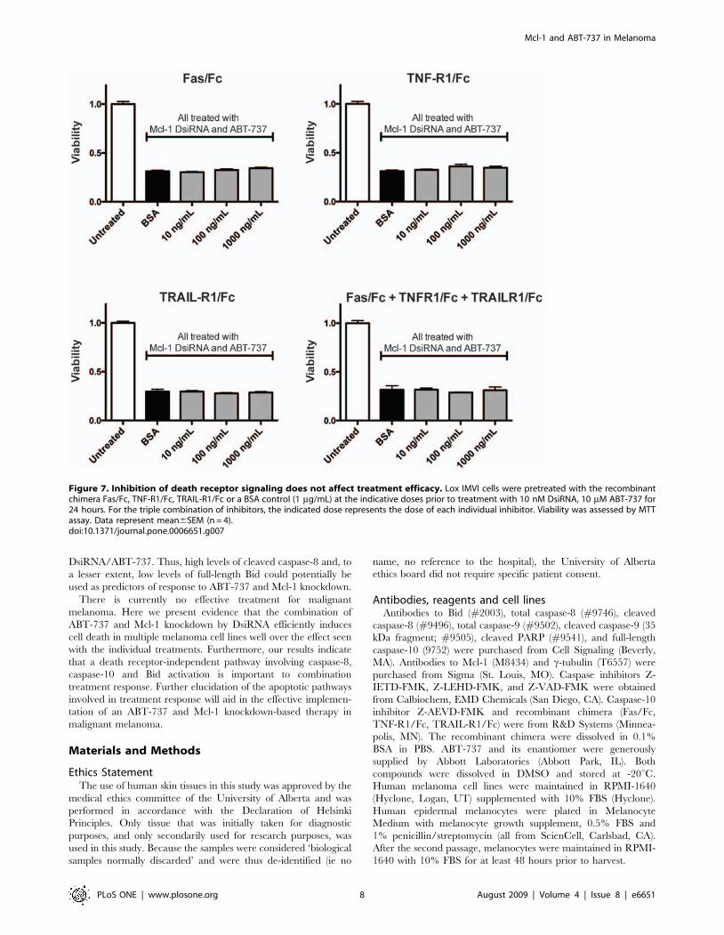
To assess the contribution of death receptor signaling to
caspase-8/-10 activation, we used death receptor recombinant
chimera (Fas/Fc, TNF-R1/Fc, and TRAIL-R1/Fc). These soluble
receptors bind all corresponding ligand (both soluble and
membrane-bound), thus preventing ligand from interacting with
endogenous receptors and preventing death receptor-mediated
apoptosis [32,33]. We show that neither the individual recombi-
nant chimera nor the combination of all three had any effect on
caspase-8/-10 activation in response to Mcl-1 DsiRNA/ABT-737
treatment (Figure 5C).
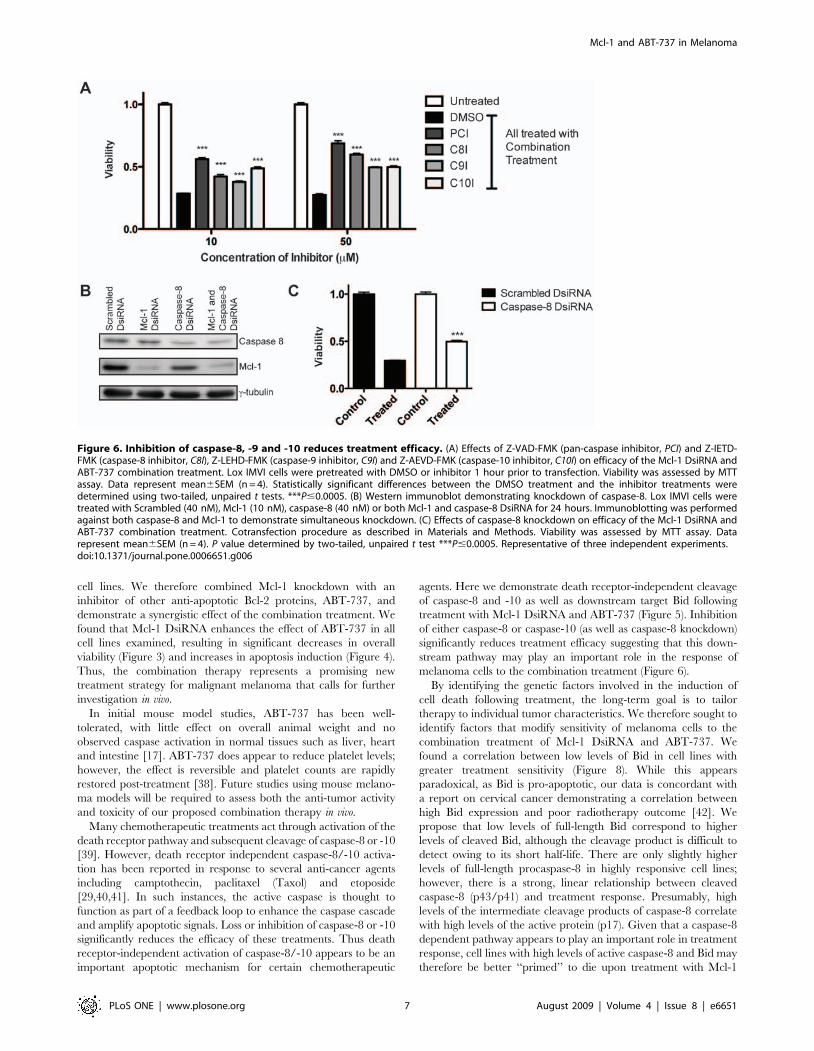
Response to combination treatment requires caspaseactivity but not death receptor signaling
To elucidate the pathways required for response to the
combination treatment (Mcl-1 DsiRNA and ABT-737) we
examined the effects of inhibitors of caspase-8 (Z-IETD-FMK), -
9 (Z-LEHD-FMK) and -10 (Z-AEVD-FMK). The pan-caspase
inhibitor Z-VAD-FMK is also shown for reference. All inhibitors
significantly increase viability (and thus decrease treatment
response) in cells treated with Mcl-1 DsiRNA and ABT-737
(Figure 6A).
To further confirm that caspase-8 plays a role in treatment
response we also knocked down caspase-8 expression. Using
DsiRNA, we were able to knockdown caspase-8 protein levels by
,50% without interfering with Mcl-1 knockdown (Figure 6B).
Pre-treatment with caspase-8 DsiRNA significantly increases
viability in cells treated with the Mcl-1 DsiRNA/ABT-737
combination (P,0.0001; Figure 6C). Knockdown of caspase-8
was less effective in reducing treatment efficacy compared to the
caspase-8 inhibitor, which may be due to incomplete knockdown.
However, while the effect was not as strong as the peptide
inhibitor, caspase-8 knockdown confirms that down-regulation of
caspase-8 activity decreases efficacy of the combination treatment.
To assess the requirement for death receptor signaling, we
examined the effects of the recombinant chimera Fas/Fc, TNF-
R1/Fc and TRAIL-R1/Fc on treatment response. Neither the
individual chimera, nor the combination of all three, had any
effect on viability of cells treated with the Mcl-1 DsiRNA and
ABT-737 combination treatment (Figure 7).
Levels of cleaved caspase-8 correlate with combinationtreatment response
Recent studies have demonstrated that expression levels of both
pro- and anti-apoptotic Bcl-2 proteins affect ABT-737 sensitivity
[34,35]. However, to the authors’ knowledge, no modifiers of the
response to ABT-737 in combination with Mcl-1 knockdown have
been identified. Given the dramatic caspase-8 and Bid cleavage
observed following treatment with Mcl-1 DsiRNA and ABT-737
in melanoma, we investigated whether expression of either protein
correlates with combination treatment response.
As we were unable to calculate accurate EC50 values for all the
cell lines (owing to the fact that some of the survival curves level off
before reaching 50%) we calculated the surviving fraction of cells
treated with 10 mM ABT-737, combined with our standard dose
of 10 nM Mcl-1 DsiRNA. At this dose of the combination
treatment, there is an almost two-fold difference in viability
between the most responsive cell line (SK-MEL-2) and the least
responsive (MeWo) (P,0.0001; Supplementary Figure S2). When
the surviving fraction is plotted against basal expression levels of
Bid and caspase-8, several trends are apparent. There is a
statistically significant correlation between levels of Bid and
survival, with the most responsive cell lines displaying lower levels
of full-length Bid (Figure 8A). Cell lines with a strong response to
treatment also have slightly higher levels of full-length procaspase-
8 (Figure 8B). While we could not consistently detect tBid in
Figure 4. Mcl-1 knockdown and ABT-737 induce apoptotic celldeath. (A) Assessment of apoptosis via TUNEL assay. SK-MEL-2 andMalme-3M cells were treated with No Treatment, RNA Control (10 nMScrambled DsiRNA), Drug Control (10 mM Enantiomer of ABT-737),10 nM Mcl-1 DsiRNA, 10 mM ABT-737, 10 nM Scrambled DsiRNA and10 mM ABT-737, or 10 nM Mcl-1 DsiRNA and 10 mM ABT-737. Cells wereharvested for TUNEL 24 hours post-transfection. Induction of apoptosisis expressed as a fold increase over No Treatment. Data representmean6SEM (n = 3). Statistical significance was determined using two-tailed, unpaired t tests. *P#0.05 **P#0.005, ***P#0.0005. (B) Assess-ment of apoptosis via PARP cleavage. SK-MEL-2 and Malme-3M cellswere treated with 10 nM Scrambled DsiRNA, 10 nM Mcl-1 DsiRNA and/or 10 mM ABT-737 in the combinations indicated for 24 hours.doi:10.1371/journal.pone.0006651.g004
Mcl-1 and ABT-737 in Melanoma
PLoS ONE | www.plosone.org 5 August 2009 | Volume 4 | Issue 8 | e6651

untreated cells, we were able to detect and quantify the
intermediate cleavage products of caspase-8 (p41/p43 isoforms).
We demonstrated a strong, direct correlation between steady-state
levels of cleaved caspase-8 and the surviving fraction (Figure 8C).
As cleaved caspase-8 levels increase, there is an increase in
treatment response. In contrast, there is no correlation between
caspase-10 expression and treatment response (data not shown).
Cell line differences are not due to known CASP8polymorphisms
In a recent report by Li et al., two putatively functional
polymorphisms of CASP8 were shown to contribute to melanoma
susceptibility [36]. We therefore sequenced both the D302H
(rs1045485:G.C) and 2652 6N ins/del (rs3834129: 2/
CTTACT) polymorphisms in the six melanoma cell lines used
in this study. None of the cell lines has the G to C transition to
produce the D302H amino acid change. With regard to the 2652
6N ins/del polymorphism: Malme-3M, MeWo, SK-MEL-2 and
SK-MEL-5 are homozygous for the insertion allele, SK-MEL-28 is
homozygous for the deletion and Lox IMVI is a heterozygote. The
2652 6N deletion is thought to reduce expression of CASP8 by
eliminating an SP1 promoter binding site [37]. However, given
that we observe highest levels of full-length caspase-8 in Lox IMVI
(a heterozygote) and lowest levels in MeWo (an insertion
homozygote), genotype at this locus likely does not play a major
role in determining caspase-8 expression levels in the cell lines in
this study.
Discussion
Malignant melanoma is highly resistant to chemotherapeutic
treatments largely due to an intrinsic resistance of the neoplastic
melanocytes to undergo apoptosis. Our group was one of the first
to describe increased expression of the anti-apoptotic protein Mcl-
1 in melanoma [7]. In the present study, we have expanded these
results using a tissue microarray. Mcl-1 expression increases with
tumor depth and stage (Figure 1A,B) suggesting that Mcl-1 is a
critical obstacle to apoptosis induction in melanoma.
However, we report that reduction of Mcl-1 expression alone is
not enough to significantly reduce survival in multiple melanoma
Figure 5. The combination of Mcl-1 knockdown and ABT-737 induces death receptor-independent cleavage of caspase-9, caspase-8, caspase-10 and Bid. (A) Western analysis showing caspase-9, -8, -10 and Bid levels with treatment. SK-MEL-2 and Malme-3M were treated with10 nM Mcl-1 DsiRNA and 10 mM ABT-737, alone and in combination for 24 hours. (B) Western blots showing reduced caspase-8/-10 activation withinhibition of caspase-9. Lox IMVI cells were pretreated with 50 mM DMSO or the indicated doses of Z-LEHD-FMK (caspase-9 inhibitor, C9I) one hourprior to transfection. Cells were then treated with the combination of 10 nM Mcl-1 DsiRNA and 10 mM ABT-737. Control cells were pretreated withDMSO and transfected with Scrambled DsiRNA. (C) Western analysis showing that caspase-8/-10 activation is not affected by death receptorinhibitors. Lox IMVI cells were pre-treated with 1 mg/mL Fas/Fc, TNF-R1/Fc, TRAIL-R1/Fc or the combination of all three (all at 1 mg/mL) prior totransfection. Cells were then treated with 10 nM Mcl-1 DsiRNA and 10 mM ABT-737 for 24 hours.doi:10.1371/journal.pone.0006651.g005
Mcl-1 and ABT-737 in Melanoma
PLoS ONE | www.plosone.org 6 August 2009 | Volume 4 | Issue 8 | e6651
cell lines. We therefore combined Mcl-1 knockdown with an
inhibitor of other anti-apoptotic Bcl-2 proteins, ABT-737, and
demonstrate a synergistic effect of the combination treatment. We
found that Mcl-1 DsiRNA enhances the effect of ABT-737 in all
cell lines examined, resulting in significant decreases in overall
viability (Figure 3) and increases in apoptosis induction (Figure 4).
Thus, the combination therapy represents a promising new
treatment strategy for malignant melanoma that calls for further
investigation in vivo.
In initial mouse model studies, ABT-737 has been well-
tolerated, with little effect on overall animal weight and no
observed caspase activation in normal tissues such as liver, heart
and intestine [17]. ABT-737 does appear to reduce platelet levels;
however, the effect is reversible and platelet counts are rapidly
restored post-treatment [38]. Future studies using mouse melano-
ma models will be required to assess both the anti-tumor activity
and toxicity of our proposed combination therapy in vivo.
Many chemotherapeutic treatments act through activation of the
death receptor pathway and subsequent cleavage of caspase-8 or -10
[39]. However, death receptor independent caspase-8/-10 activa-
tion has been reported in response to several anti-cancer agents
including camptothecin, paclitaxel (Taxol) and etoposide
[29,40,41]. In such instances, the active caspase is thought to
function as part of a feedback loop to enhance the caspase cascade
and amplify apoptotic signals. Loss or inhibition of caspase-8 or -10
significantly reduces the efficacy of these treatments. Thus death
receptor-independent activation of caspase-8/-10 appears to be an
important apoptotic mechanism for certain chemotherapeutic
agents. Here we demonstrate death receptor-independent cleavage
of caspase-8 and -10 as well as downstream target Bid following
treatment with Mcl-1 DsiRNA and ABT-737 (Figure 5). Inhibition
of either caspase-8 or caspase-10 (as well as caspase-8 knockdown)
significantly reduces treatment efficacy suggesting that this down-
stream pathway may play an important role in the response of
melanoma cells to the combination treatment (Figure 6).
By identifying the genetic factors involved in the induction of
cell death following treatment, the long-term goal is to tailor
therapy to individual tumor characteristics. We therefore sought to
identify factors that modify sensitivity of melanoma cells to the
combination treatment of Mcl-1 DsiRNA and ABT-737. We
found a correlation between low levels of Bid in cell lines with
greater treatment sensitivity (Figure 8). While this appears
paradoxical, as Bid is pro-apoptotic, our data is concordant with
a report on cervical cancer demonstrating a correlation between
high Bid expression and poor radiotherapy outcome [42]. We
propose that low levels of full-length Bid correspond to higher
levels of cleaved Bid, although the cleavage product is difficult to
detect owing to its short half-life. There are only slightly higher
levels of full-length procaspase-8 in highly responsive cell lines;
however, there is a strong, linear relationship between cleaved
caspase-8 (p43/p41) and treatment response. Presumably, high
levels of the intermediate cleavage products of caspase-8 correlate
with high levels of the active protein (p17). Given that a caspase-8
dependent pathway appears to play an important role in treatment
response, cell lines with high levels of active caspase-8 and Bid may
therefore be better ‘‘primed’’ to die upon treatment with Mcl-1
Figure 6. Inhibition of caspase-8, -9 and -10 reduces treatment efficacy. (A) Effects of Z-VAD-FMK (pan-caspase inhibitor, PCI) and Z-IETD-FMK (caspase-8 inhibitor, C8I), Z-LEHD-FMK (caspase-9 inhibitor, C9I) and Z-AEVD-FMK (caspase-10 inhibitor, C10I) on efficacy of the Mcl-1 DsiRNA andABT-737 combination treatment. Lox IMVI cells were pretreated with DMSO or inhibitor 1 hour prior to transfection. Viability was assessed by MTTassay. Data represent mean6SEM (n = 4). Statistically significant differences between the DMSO treatment and the inhibitor treatments weredetermined using two-tailed, unpaired t tests. ***P#0.0005. (B) Western immunoblot demonstrating knockdown of caspase-8. Lox IMVI cells weretreated with Scrambled (40 nM), Mcl-1 (10 nM), caspase-8 (40 nM) or both Mcl-1 and caspase-8 DsiRNA for 24 hours. Immunoblotting was performedagainst both caspase-8 and Mcl-1 to demonstrate simultaneous knockdown. (C) Effects of caspase-8 knockdown on efficacy of the Mcl-1 DsiRNA andABT-737 combination treatment. Cotransfection procedure as described in Materials and Methods. Viability was assessed by MTT assay. Datarepresent mean6SEM (n = 4). P value determined by two-tailed, unpaired t test ***P#0.0005. Representative of three independent experiments.doi:10.1371/journal.pone.0006651.g006
Mcl-1 and ABT-737 in Melanoma
PLoS ONE | www.plosone.org 7 August 2009 | Volume 4 | Issue 8 | e6651
DsiRNA/ABT-737. Thus, high levels of cleaved caspase-8 and, to
a lesser extent, low levels of full-length Bid could potentially be
used as predictors of response to ABT-737 and Mcl-1 knockdown.
There is currently no effective treatment for malignant
melanoma. Here we present evidence that the combination of
ABT-737 and Mcl-1 knockdown by DsiRNA efficiently induces
cell death in multiple melanoma cell lines well over the effect seen
with the individual treatments. Furthermore, our results indicate
that a death receptor-independent pathway involving caspase-8,
caspase-10 and Bid activation is important to combination
treatment response. Further elucidation of the apoptotic pathways
involved in treatment response will aid in the effective implemen-
tation of an ABT-737 and Mcl-1 knockdown-based therapy in
malignant melanoma.
Materials and Methods
Ethics StatementThe use of human skin tissues in this study was approved by the
medical ethics committee of the University of Alberta and was
performed in accordance with the Declaration of Helsinki
Principles. Only tissue that was initially taken for diagnostic
purposes, and only secondarily used for research purposes, was
used in this study. Because the samples were considered ‘biological
samples normally discarded’ and were thus de-identified (ie no
name, no reference to the hospital), the University of Alberta
ethics board did not require specific patient consent.
Antibodies, reagents and cell linesAntibodies to Bid (#2003), total caspase-8 (#9746), cleaved
caspase-8 (#9496), total caspase-9 (#9502), cleaved caspase-9 (35
kDa fragment; #9505), cleaved PARP (#9541), and full-length
caspase-10 (9752) were purchased from Cell Signaling (Beverly,
MA). Antibodies to Mcl-1 (M8434) and c-tubulin (T6557) were
purchased from Sigma (St. Louis, MO). Caspase inhibitors Z-
IETD-FMK, Z-LEHD-FMK, and Z-VAD-FMK were obtained
from Calbiochem, EMD Chemicals (San Diego, CA). Caspase-10
inhibitor Z-AEVD-FMK and recombinant chimera (Fas/Fc,
TNF-R1/Fc, TRAIL-R1/Fc) were from R&D Systems (Minnea-
polis, MN). The recombinant chimera were dissolved in 0.1%
BSA in PBS. ABT-737 and its enantiomer were generously
supplied by Abbott Laboratories (Abbott Park, IL). Both
compounds were dissolved in DMSO and stored at -20uC.
Human melanoma cell lines were maintained in RPMI-1640
(Hyclone, Logan, UT) supplemented with 10% FBS (Hyclone).
Human epidermal melanocytes were plated in Melanocyte
Medium with melanocyte growth supplement, 0.5% FBS and
1% penicillin/streptomycin (all from ScienCell, Carlsbad, CA).
After the second passage, melanocytes were maintained in RPMI-
1640 with 10% FBS for at least 48 hours prior to harvest.
Figure 7. Inhibition of death receptor signaling does not affect treatment efficacy. Lox IMVI cells were pretreated with the recombinantchimera Fas/Fc, TNF-R1/Fc, TRAIL-R1/Fc or a BSA control (1 mg/mL) at the indicative doses prior to treatment with 10 nM DsiRNA, 10 mM ABT-737 for24 hours. For the triple combination of inhibitors, the indicated dose represents the dose of each individual inhibitor. Viability was assessed by MTTassay. Data represent mean6SEM (n = 4).doi:10.1371/journal.pone.0006651.g007
Mcl-1 and ABT-737 in Melanoma
PLoS ONE | www.plosone.org 8 August 2009 | Volume 4 | Issue 8 | e6651

Figure 8. Levels of Bid and cleaved caspase-8 correlate with response to the combination treatment. Right, western analyses ofendogenous levels of full-length Bid (A), full-length caspase-8 (B) and cleaved caspase-8 (C). Cells were harvested for protein at approximately equalconfluency one day after splitting. Left, relative expression levels were quantified by densitometry, normalized to the amido black loading control,then plotted against the surviving fraction at 10 nM DsiRNA, 10 mM ABT-737 (expressed as a fraction of cells treated with Scrambled DsiRNA only).Data presented are the mean (6SEM) of three independent experiments. Correlation was assessed by Pearson correlation analysis: R (correlationcoefficient) and P value are indicated on the graph. Lox, Lox IMVI; Malme, Malme-3M; SK2, SK-MEL-2; SK5, SK-MEL-5; SK28, SK-MEL-28.doi:10.1371/journal.pone.0006651.g008
Mcl-1 and ABT-737 in Melanoma
PLoS ONE | www.plosone.org 9 August 2009 | Volume 4 | Issue 8 | e6651

Tissue Microarray (TMA)Formalin-fixed, paraffin-embedded tissues from 28 human
normal nevi, 49 primary melanomas and 32 metastatic melano-
mas were obtained from the Department of Laboratory Medicine
and Pathology at the University of Alberta. The most represen-
tative tumor area was carefully selected and marked on the
hematoxylin and eosin-stained slide. 0.6 mm-thick tissue cores
were taken from each biopsy specimen. Multiple 4-mm sections
were cut with a Leica microtome (Leica Microsystems Inc,
Bannockburn, IL) and transferred to adhesive-coated slides.
Immunohistochemistry of TMAThe TMA slides were dewaxed with xylene and rehydrated with
graded alcohol washes. Antigen retrieval was performed by heating
the samples at 95uC for 30 minutes in 10 mM sodium citrate (pH 6.0).
Endogenous peroxidase activity was blocked with 3% hydrogen
peroxide for 20 minutes. After blocking with universal blocking serum
(DAKO Diagnostics, Mississauga, ON, Canada) for 30 minutes, the
slides were incubated with anti-Mcl-1 antibody (1:800) at 4uCovernight. The sections were incubated with biotin-labeled secondary
antibody and streptavidin-peroxidase for 30 minutes each (DAKO
Diagnostics). The samples were developed with 3,39-diaminobenzi-
dine substrate (Vector Laboratories, Burlington, ON, Canada) and
counterstained with hematoxylin. Negative controls omitted Mcl-1
antibody during the primary antibody incubation.
Evaluation of immunostainingMcl-1 staining in the TMA was scored by a pathologist and the
total staining score was derived from the staining intensity
multiplied by the percentage positive cells. Score for staining
intensity: 0 = negative, 1 = weak staining, 2 = moderate staining,
3 = strong staining. Score for positive percentage: 1 = ,20%
positive, 2 = 20–39% positive, 3 = 40–59% positive, 4 = 60–79%
positive, 5 = 80–100% positive.
siRNA transfectionCell lines were transfected with the indicated doses of Dicer-
substrate siRNA (DsiRNA) directed against Mcl-1
(HSC.RNAI.N021960.2.3), caspase-8 (ACC NM_001228.4_3) or
a universal negative control scrambled sequence (DS Scrambled
Neg) (Integrated DNA Technologies, Coralville, IA) using Lipo-
fectamine 2000 (Invitrogen, Carlsbad, CA) according to manufac-
turer’s instructions. Where indicated, cells were treated with the
appropriate dose of ABT-737 eight hours after DsiRNA transfec-
tion. To assess the effect of caspase-8 knockdown on survival, cells
were first transfected with 40 nM caspase-8 or Scrambled DsiRNA
in 60 mm plates, then split into 96 well plates 24 hours post-
transfection. 24 hours later, cells were transfected with Mcl-1 or
Scrambled DsiRNA and/or treated with ABT-737 as indicated.
Viability and Apoptosis assaysPercent viability was assessed colorimetrically using MTT (3-
(4,5-dimethyl-thiazol-2-yl)-2,5-diphenyltetrazolium bromide; Sig-
ma). Twenty-four hours after transfection, plates were incubated
with 0.5 mg/ml MTT for 6 hours. Following incubation, the
formazan precipitate was solubilized with 24:1 isopropanol:HCl
and the absorbance was measured at 570 nm. Percent viability
was calculated in comparison to control treatments. To measure
apoptosis: 24 hours post-transfection, adherent and suspension
cells were collected and fixed in 1% paraformaldehyde. Terminal
deoxynucleotidyl dUTP nick end labeling (TUNEL) was per-
formed using the Apo-BrdU In Situ DNA Fragmentation assay kit
(Biovision, Mountain View, CA) according to manufacturer’s
instructions. Labeled cells were analyzed by fluorescence-activated
cell sorter (FACS; BD Biosciences, San Jose, CA).
Western blot analysisAdherent and suspension cells were collected at indicated time
points. Whole cell lysis, SDS-PAGE and western immunoblotting
were performed as previously described [43]. Densitometry was
performed using Quantity One software (Bio-Rad, Mississauga,
ON, Canada). All blots were done in triplicate. In experiments
comparing treatments within the same cell line, c-tubulin is used
as the loading control; however, as c-tubulin levels can vary
between cell lines, for experiments comparing different cell lines, a
representative section of the amido black stain is shown.
PCR and sequencingDNA was extracted using the QIAquick DNA extraction
columns (Qiagen, Valencia, CA). Previously described primers
were used to amplify the 2652 6N del polymorphism [37], and
the D302H polymorphism [36]. Bands were purified using the
QIAquick Gel Extraction Kit (Qiagen) and sequenced using the
BigDye Terminator cycle sequencing kit (Applied Biosystems,
Foster City, CA). Sequencing reactions were run on 313061 or
3100-Avant Applied Biosystems genetic analyzers.
Statistical analysesAll statistical analyses were performed on GraphPad Prism
version 5 for Macintosh (GraphPad Software, San Diego, CA). To
assess the synergistic effect of the combination treatment, we used
two-way ANOVA as previously described [25]. Briefly, overall
viability by MTT assay (ie absorbance at 570 nm without
normalization) of cells treated with the combination treatment
(Mcl-1 DsiRNA 10 nM and ABT-737 10 mM) were compared
against cells treated with either single treatment or cells treated
with only a Scrambled DsiRNA control. The same data is shown
in its normalized form in Figure 3. P-values less than 0.05 were
considered statistically significant.
Supporting Information
Figure S1 Effect of ABT-737 compared to drug enantiomer on
cell viability in melanoma cell lines. Cells were treated with 0, 0.1,
1, 10, or 50 mM of ABT-737 or enantiomer. Each dose was
increased by two orders of magnitude before the logarithm was
taken to allow us to graph all doses as positive values. Viability was
assessed at 24 hours via MTT assay. Curves were normalized to
the 0 mM treatment. Data points represent mean6SEM (n = 4).
Found at: doi:10.1371/journal.pone.0006651.s001 (1.42 MB TIF)
Figure S2 Survival at 10 nM DsiRNA, 10 mM ABT-737 in 6
melanoma cell lines. Survival is expressed as a fraction of cells treated
with Scrambled DsiRNA only (10 nM). Data represent mean6SEM
(n = 4). The statistical significance of the difference between the least
and most responsive cell lines (MeWo and SK-MEL-2, respectively)
was determined by a two-tailed, unpaired t test.
Found at: doi:10.1371/journal.pone.0006651.s002 (1.11 MB TIF)
Acknowledgments
The authors thank Lindsay Schultz and Raylene Kramchynsky for
excellent technical assistance and Leah Young for helpful discussion.
Author Contributions
Conceived and designed the experiments: AMK KEAF SEA VT.
Performed the experiments: AMK AAMP MA VT. Analyzed the data:
AMK KEAF AAMP. Wrote the paper: AMK SEA VT.
Mcl-1 and ABT-737 in Melanoma
PLoS ONE | www.plosone.org 10 August 2009 | Volume 4 | Issue 8 | e6651

References
1. Chudnovsky Y, Khavari PA, Adams AE (2005) Melanoma genetics and the
development of rational therapeutics. J Clin Invest 115: 813–824.2. Gray-Schopfer V, Wellbrock C, Marais R (2007) Melanoma biology and new
targeted therapy. Nature 445: 851–857.3. Barth A, Wanek LA, Morton DL (1995) Prognostic factors in 1,521 melanoma
patients with distant metastases. J Am Coll Surg 181: 193–201.4. Stein JA, Brownell I (2008) Treatment approaches for advanced cutaneous
melanoma. J Drugs Dermatol 7: 175–179.
5. Eberle J, Kurbanov BM, Hossini AM, Trefzer U, Fecker LF (2007) Overcomingapoptosis deficiency of melanoma-hope for new therapeutic approaches. Drug
Resist Updat 10: 218–234.6. Lorigan P, Eisen T, Hauschild A (2008) Systemic therapy for metastatic
malignant melanoma—from deeply disappointing to bright future? Exp
Dermatol 17: 383–394.7. Tang L, Tron VA, Reed JC, Mah KJ, Krajewska M, et al. (1998) Expression of
apoptosis regulators in cutaneous malignant melanoma. Clin Cancer Res 4:1865–1871.
8. Mikhailov V, Mikhailova M, Degenhardt K, Venkatachalam MA, White E, et
al. (2003) Association of Bax and Bak homo-oligomers in mitochondria. Baxrequirement for Bak reorganization and cytochrome c release. J Biol Chem 278:
5367–5376.9. Chen L, Willis SN, Wei A, Smith BJ, Fletcher JI, et al. (2005) Differential
targeting of prosurvival Bcl-2 proteins by their BH3-only ligands allowscomplementary apoptotic function. Mol Cell 17: 393–403.
10. Letai A, Bassik MC, Walensky LD, Sorcinelli MD, Weiler S, et al. (2002)
Distinct BH3 domains either sensitize or activate mitochondrial apoptosis,serving as prototype cancer therapeutics. Cancer Cell 2: 183–192.
11. Willis SN, Chen L, Dewson G, Wei A, Naik E, et al. (2005) Proapoptotic Bak issequestered by Mcl-1 and Bcl-xL, but not Bcl-2, until displaced by BH3-only
proteins. Genes Dev 19: 1294–1305.
12. Yin XM, Oltvai ZN, Korsmeyer SJ (1994) BH1 and BH2 domains of Bcl-2 arerequired for inhibition of apoptosis and heterodimerization with Bax. Nature
369: 321–323.13. Leiter U, Schmid RM, Kaskel P, Peter RU, Krahn G (2000) Antiapoptotic bcl-2
and bcl-xL in advanced malignant melanoma. Arch Dermatol Res 292:225–232.
14. Wong RP, Khosravi S, Martinka M, Li G (2008) Myeloid leukemia-1 expression
in benign and malignant melanocytic lesions. Oncol Rep 19: 933–937.15. Zhuang L, Lee CS, Scolyer RA, McCarthy SW, Zhang XD, et al. (2007) Mcl-1,
Bcl-XL and Stat3 expression are associated with progression of melanomawhereas Bcl-2, AP-2 and MITF levels decrease during progression of melanoma.
Mod Pathol 20: 416–426.
16. Konopleva M, Contractor R, Tsao T, Samudio I, Ruvolo PP, et al. (2006)Mechanisms of apoptosis sensitivity and resistance to the BH3 mimetic ABT-737
in acute myeloid leukemia. Cancer Cell 10: 375–388.17. Oltersdorf T, Elmore SW, Shoemaker AR, Armstrong RC, Augeri DJ, et al.
(2005) An inhibitor of Bcl-2 family proteins induces regression of solid tumours.Nature 435: 677–681.
18. Tagscherer KE, Fassl A, Campos B, Farhadi M, Kraemer A, et al. (2008)
Apoptosis-based treatment of glioblastomas with ABT-737, a novel smallmolecule inhibitor of Bcl-2 family proteins. Oncogene.
19. Trudel S, Stewart AK, Li Z, Shu Y, Liang SB, et al. (2007) The Bcl-2 familyprotein inhibitor, ABT-737, has substantial antimyeloma activity and shows
synergistic effect with dexamethasone and melphalan. Clin Cancer Res 13:
621–629.20. Chen S, Dai Y, Harada H, Dent P, Grant S (2007) Mcl-1 down-regulation
potentiates ABT-737 lethality by cooperatively inducing Bak activation and Baxtranslocation. Cancer Res 67: 782–791.
21. van Delft MF, Wei AH, Mason KD, Vandenberg CJ, Chen L, et al. (2006) The
BH3 mimetic ABT-737 targets selective Bcl-2 proteins and efficiently inducesapoptosis via Bak/Bax if Mcl-1 is neutralized. Cancer Cell 10: 389–399.
22. Miller LA, Goldstein NB, Johannes WU, Walton CH, Fujita M, et al. (2008)BH3 Mimetic ABT-737 and a Proteasome Inhibitor Synergistically Kill
Melanomas through Noxa-Dependent Apoptosis. J Invest Dermatol.
23. Kim DH, Behlke MA, Rose SD, Chang MS, Choi S, et al. (2005) Synthetic
dsRNA Dicer substrates enhance RNAi potency and efficacy. Nat Biotechnol23: 222–226.
24. Bush JA, Li G (2003) The role of Bcl-2 family members in the progression ofcutaneous melanoma. Clin Exp Metastasis 20: 531–539.
25. Slinker BK (1998) The statistics of synergism. J Mol Cell Cardiol 30: 723–731.26. Chauhan D, Velankar M, Brahmandam M, Hideshima T, Podar K, et al. (2007)
A novel Bcl-2/Bcl-X(L)/Bcl-w inhibitor ABT-737 as therapy in multiple
myeloma. Oncogene 26: 2374–2380.27. Kline MP, Rajkumar SV, Timm MM, Kimlinger TK, Haug JL, et al. (2007)
ABT-737, an inhibitor of Bcl-2 family proteins, is a potent inducer of apoptosisin multiple myeloma cells. Leukemia 21: 1549–1560.
28. Milhas D, Cuvillier O, Therville N, Clave P, Thomsen M, et al. (2005) Caspase-
10 triggers Bid cleavage and caspase cascade activation in FasL-inducedapoptosis. J Biol Chem 280: 19836–19842.
29. Filomenko R, Prevotat L, Rebe C, Cortier M, Jeannin JF, et al. (2006) Caspase-10 involvement in cytotoxic drug-induced apoptosis of tumor cells. Oncogene
25: 7635–7645.
30. Sutheesophon K, Nishimura N, Kobayashi Y, Furukawa Y, Kawano M, et al.(2005) Involvement of the tumor necrosis factor (TNF)/TNF receptor system in
leukemic cell apoptosis induced by histone deacetylase inhibitor depsipeptide(FK228). J Cell Physiol 203: 387–397.
31. Slee EA, Harte MT, Kluck RM, Wolf BB, Casiano CA, et al. (1999) Orderingthe cytochrome c-initiated caspase cascade: hierarchical activation of caspases-2,
-3, -6, -7, -8, and -10 in a caspase-9-dependent manner. J Cell Biol 144:
281–292.32. Keogh RJ, Harris LK, Freeman A, Baker PN, Aplin JD, et al. (2007) Fetal-
derived trophoblast use the apoptotic cytokine tumor necrosis factor-alpha-related apoptosis-inducing ligand to induce smooth muscle cell death. Circ Res
100: 834–841.
33. Luce A, Courtin A, Levalois C, Altmeyer-Morel S, Romeo PH, et al. (2009)Death receptor pathways mediate targeted and non-targeted effects of ionizing
radiations in breast cancer cells. Carcinogenesis 30: 432–439.34. Deng J, Carlson N, Takeyama K, Dal Cin P, Shipp M, et al. (2007) BH3
profiling identifies three distinct classes of apoptotic blocks to predict response toABT-737 and conventional chemotherapeutic agents. Cancer Cell 12: 171–185.
35. Tahir SK, Yang X, Anderson MG, Morgan-Lappe SE, Sarthy AV, et al. (2007)
Influence of Bcl-2 family members on the cellular response of small-cell lungcancer cell lines to ABT-737. Cancer Res 67: 1176–1183.
36. Li C, Zhao H, Hu Z, Liu Z, Wang LE, et al. (2008) Genetic variants andhaplotypes of the caspase-8 and caspase-10 genes contribute to susceptibility to
cutaneous melanoma. Hum Mutat.
37. Sun T, Gao Y, Tan W, Ma S, Shi Y, et al. (2007) A six-nucleotide insertion-deletion polymorphism in the CASP8 promoter is associated with susceptibility
to multiple cancers. Nat Genet 39: 605–613.38. Zhang H, Nimmer PM, Tahir SK, Chen J, Fryer RM, et al. (2007) Bcl-2 family
proteins are essential for platelet survival. Cell Death Differ 14: 943–951.39. Debatin KM, Krammer PH (2004) Death receptors in chemotherapy and
cancer. Oncogene 23: 2950–2966.
40. de Vries JF, Wammes LJ, Jedema I, van Dreunen L, Nijmeijer BA, et al. (2007)Involvement of caspase-8 in chemotherapy-induced apoptosis of patient derived
leukemia cell lines independent of the death receptor pathway and downstreamfrom mitochondria. Apoptosis 12: 181–193.
41. von Haefen C, Wieder T, Essmann F, Schulze-Osthoff K, Dorken B, et al.
(2003) Paclitaxel-induced apoptosis in BJAB cells proceeds via a death receptor-independent, caspases-3/-8-driven mitochondrial amplification loop. Oncogene
22: 2236–2247.42. Green MM, Hutchison GJ, Valentine HR, Fitzmaurice RJ, Davidson SE, et al.
(2005) Expression of the proapoptotic protein Bid is an adverse prognostic factor
for radiotherapy outcome in carcinoma of the cervix. Br J Cancer 92: 449–458.43. Young LC, Peters AC, Maeda T, Edelmann W, Kucherlapati R, et al. (2003)
DNA mismatch repair protein Msh6 is required for optimal levels of ultraviolet-B-induced apoptosis in primary mouse fibroblasts. J Invest Dermatol 121:
876–880.
Mcl-1 and ABT-737 in Melanoma
PLoS ONE | www.plosone.org 11 August 2009 | Volume 4 | Issue 8 | e6651