Regulation of Aging and Longevity by Ion Channels ... - MDPI
32
Cells 2022, 11, 1180. https://doi.org/10.3390/cells11071180 www.mdpi.com/journal/cells Review Regulation of Aging and Longevity by Ion Channels and Transporters Kartik Venkatachalam 1,2 1 Department of Integrative Biology and Pharmacology, McGovern Medical School at the University of Texas Health Sciences Center (UTHealth-Houston), Houston, TX 77030, USA; [email protected] 2 Graduate Programs in Neuroscience and Biochemistry and Cell Biology, MD Anderson Cancer Center and UTHealth Graduate School of Biomedical Sciences, Houston, TX 77030, USA Abstract: Despite significant advances in our understanding of the mechanisms that underlie age- related physiological decline, our ability to translate these insights into actionable strategies to ex- tend human healthspan has been limited. One of the major reasons for the existence of this barrier is that with a few important exceptions, many of the proteins that mediate aging have proven to be undruggable. The argument put forth here is that the amenability of ion channels and transporters to pharmacological manipulation could be leveraged to develop novel therapeutic strategies to com- bat aging. This review delves into the established roles for ion channels and transporters in the reg- ulation of aging and longevity via their influence on membrane excitability, Ca 2+ homeostasis, mi- tochondrial and endolysosomal function, and the transduction of sensory stimuli. The goal is to provide the reader with an understanding of emergent themes, and prompt further investigation into how the activities of ion channels and transporters sculpt the trajectories of cellular and organ- ismal aging. Keywords: longevity; ion channels; lifespan; aging; calcium; ER; lysosomes 1. Introduction All biological processes lose their integrity with the passage of time. Aging is the single greatest risk factor for diseases associated with diminished survivorship such as cancer, diabetes, cardiovascular diseases, and neurodegeneration [1,2]. By extension, strategies that ameliorate age-dependent diseases also extend lifespan. Over the past three decades, major advances in gerobiology have been made using model organisms such as C. elegans and Drosophila [3–6]. These pioneering studies have established that various bi- ochemical pathways influence the rates of cellular and organismal aging [2]. Emerging insights have informed investigations into the genetics of aging, and prompted the quest to identify drugs that extend healthspan by ameliorating age-related diseases or by slow- ing the rates of biological aging. The problem, however, is that barring a few exceptions (e.g., rapamycin and metfor- min [2]) not many drugs have emerged as suitable candidates to combat aging. Although this barrier has many explanations, the most obvious is the sheer complexity of aging. Age-related loss of physiological integrity stems from a host of defects, including genomic and epigenetic instability, telomere shortening, proteostatic imbalance, chronic inflamma- tion, dysregulated nutrient sensing, mitochondrial defects, cellular senescence, and stem cell exhaustion [1,2]. Most proteins and signaling cascades involved in the regulation of these hallmarks are undruggable. With perhaps the exception of G-protein coupled receptors, ion channels and trans- porters count among the most druggable targets in the proteome. This is because domains that mediate ion transport usually interface well with small molecules. Indeed, many nat- urally occurring biological toxins target ion channels and transporters with exquisite Citation: Venkatachalam, K. Regulation of Aging and Longevity by Ion Channels and Transporters. Cells 2022, 11, 1180. https://doi.org/ 10.3390/cells11071180 Academic Editor: Christian M. Grimm Received: 1 February 2022 Accepted: 29 March 2022 Published: 31 March 2022 Publisher’s Note: MDPI stays neu- tral with regard to jurisdictional claims in published maps and institu- tional affiliations. Copyright: © 2022 by the authors. Li- censee MDPI, Basel, Switzerland. This article is an open access article distributed under the terms and con- ditions of the Creative Commons At- tribution (CC BY) license (https://cre- ativecommons.org/licenses/by/4.0/).
-
Upload
khangminh22 -
Category
Documents
-
view
3 -
download
0
Transcript of Regulation of Aging and Longevity by Ion Channels ... - MDPI

Cells 2022, 11, 1180. https://doi.org/10.3390/cells11071180 www.mdpi.com/journal/cells
Review
Regulation of Aging and Longevity by Ion Channels and
Transporters
Kartik Venkatachalam 1,2
1 Department of Integrative Biology and Pharmacology, McGovern Medical School at the University of Texas
Health Sciences Center (UTHealth-Houston), Houston, TX 77030, USA; [email protected] 2 Graduate Programs in Neuroscience and Biochemistry and Cell Biology, MD Anderson Cancer Center and
UTHealth Graduate School of Biomedical Sciences, Houston, TX 77030, USA
Abstract: Despite significant advances in our understanding of the mechanisms that underlie age-
related physiological decline, our ability to translate these insights into actionable strategies to ex-
tend human healthspan has been limited. One of the major reasons for the existence of this barrier
is that with a few important exceptions, many of the proteins that mediate aging have proven to be
undruggable. The argument put forth here is that the amenability of ion channels and transporters
to pharmacological manipulation could be leveraged to develop novel therapeutic strategies to com-
bat aging. This review delves into the established roles for ion channels and transporters in the reg-
ulation of aging and longevity via their influence on membrane excitability, Ca2+ homeostasis, mi-
tochondrial and endolysosomal function, and the transduction of sensory stimuli. The goal is to
provide the reader with an understanding of emergent themes, and prompt further investigation
into how the activities of ion channels and transporters sculpt the trajectories of cellular and organ-
ismal aging.
Keywords: longevity; ion channels; lifespan; aging; calcium; ER; lysosomes
1. Introduction
All biological processes lose their integrity with the passage of time. Aging is the
single greatest risk factor for diseases associated with diminished survivorship such as
cancer, diabetes, cardiovascular diseases, and neurodegeneration [1,2]. By extension,
strategies that ameliorate age-dependent diseases also extend lifespan. Over the past three
decades, major advances in gerobiology have been made using model organisms such as
C. elegans and Drosophila [3–6]. These pioneering studies have established that various bi-
ochemical pathways influence the rates of cellular and organismal aging [2]. Emerging
insights have informed investigations into the genetics of aging, and prompted the quest
to identify drugs that extend healthspan by ameliorating age-related diseases or by slow-
ing the rates of biological aging.
The problem, however, is that barring a few exceptions (e.g., rapamycin and metfor-
min [2]) not many drugs have emerged as suitable candidates to combat aging. Although
this barrier has many explanations, the most obvious is the sheer complexity of aging.
Age-related loss of physiological integrity stems from a host of defects, including genomic
and epigenetic instability, telomere shortening, proteostatic imbalance, chronic inflamma-
tion, dysregulated nutrient sensing, mitochondrial defects, cellular senescence, and stem
cell exhaustion [1,2]. Most proteins and signaling cascades involved in the regulation of
these hallmarks are undruggable.
With perhaps the exception of G-protein coupled receptors, ion channels and trans-
porters count among the most druggable targets in the proteome. This is because domains
that mediate ion transport usually interface well with small molecules. Indeed, many nat-
urally occurring biological toxins target ion channels and transporters with exquisite
Citation: Venkatachalam, K.
Regulation of Aging and Longevity
by Ion Channels and Transporters.
Cells 2022, 11, 1180. https://doi.org/
10.3390/cells11071180
Academic Editor:
Christian M. Grimm
Received: 1 February 2022
Accepted: 29 March 2022
Published: 31 March 2022
Publisher’s Note: MDPI stays neu-
tral with regard to jurisdictional
claims in published maps and institu-
tional affiliations.
Copyright: © 2022 by the authors. Li-
censee MDPI, Basel, Switzerland.
This article is an open access article
distributed under the terms and con-
ditions of the Creative Commons At-
tribution (CC BY) license (https://cre-
ativecommons.org/licenses/by/4.0/).

Cells 2022, 11, 1180 2 of 32
sensitivity, and these entities have been used to engineer novel pharmacological modifi-
ers. Furthermore, since the activity of ion channels and transporters can be assessed in
vitro using electrophysiology or optical imaging, it is possible to conduct high-throughput
screens to discover suitable activators and inhibitors. Although it might not be possible to
extend healthspan or longevity by targeting channels that function in just one pathway, a
cogent strategy for the amelioration of age-dependent loss of function could involve the
identification and targeting of ion channels and transporters that affect multiple pathways
related to organismal aging. With this rationale in mind, this article provides an overview
of how aging and longevity are determined by the activities of various ion channels and
transporters. The overarching goal of this review is to highlight commonalities and en-
courage further investigation into the modulation of the activities of ion channels and
transporters to influence the rates of cellular and organismal aging.
2. Regulation of Aging and Longevity by Ion Channels and Transporters Related to
Ca2+ Homeostasis and Electrical Excitability
Ca2+ is a key second messenger that is involved in the regulation of a host of different
pathways, including synaptic transmission, neuronal survival, cellular bioenergetics, and
gene expression that range over timescales spanning from milliseconds to hours and days
[7,8]. Critical to cellular Ca2+ signaling are ATP-consuming transporters that maintain cy-
tosolic Ca2+ concentration ([Ca2+]) in the nanomolar range by pumping the cation against
its steep concentration gradient (~10,000-fold across the plasma membrane and endoplas-
mic reticulum (ER) membrane) [7,8]. Movement of Ca2+ down its concentration gradient
involves the opening of Ca2+-permeable channels that serve as conduits that permit tran-
sient elevations of cytosolic [Ca2+], and the attendant activation of relevant cellular path-
ways [7,9].
One of the most exciting recent findings in gerobiology is that expression of genes
associated with the maintenance of neuronal Ca2+ homeostasis, excitability, and synaptic
function is anticorrelated with lifespan in many organisms including humans [10]. Anal-
ysis of RNA-seq and microarray datasets generated from frontal cortices isolated imme-
diately post-mortem revealed the downregulation of genes associated with neurotrans-
mission in exceptionally long-lived humans [10] (Figure 1A). Advanced age is also asso-
ciated with increased neuronal activity in C. elegans, and pharmacological attenuation of
excitability and its consequences using the Cl− channel agonist, ivermectin, or the L-type
voltage-gated Ca2+ channel (VGCC) blocker, nimodipine, extended worm lifespan [10]
(Figure 1B). Roles for excitability in the regulation of neuronal viability and organismal
lifespan are also observed in Drosophila [11–15] (Figure 1B). Deletion of the fly K+ channel
genes, ether a go-go (eag) and shaker (Sh)—expected to augment neuronal excitability—led
to severe loss of motor coordination, sleep deficits, and shorter lifespan [11,15]. Dimin-
ished Na+/K+ ATPase activity, which leads to a loss of the resting membrane potential and
severe neuronal hyperexcitability, also caused shorter lifespan in flies [11,12,16].
The relationship between neuronal excitability and lifespan is conserved in mammals
[10]. As is the case in C. elegans, ivermectin extends lifespan in a mouse model of amyo-
trophic lateral sclerosis (ALS) [17]. Plasma membrane excitability is also an important de-
terminant of oncogene-induced senescence (OIS) in non-excitable mammalian cells [18]
(Figure 1B). A genetic screen revealed that loss of SCN9A, the gene encoding a voltage-
gated Na+ channel, protected human fibroblasts from OIS [18]. Oncogenic signals or acti-
vation of the tumor suppressor, p53, led to the induction of SCN9A with NF-kB as an
intermediary [18]. Elevations in SCN9A abundance, which is expected to augment plasma
membrane excitability, led to senescence via the retinoblastoma (Rb) pathway [18].
Whether NF-kB signaling, p53, or Rb also contribute to the shorter lifespan observed in
animals with chronic neuronal depolarization (see below) remains to be addressed.

Cells 2022, 11, 1180 3 of 32
Figure 1. Regulation of cellular and organismal aging by neuronal excitability. (A) Sequencing of
RNA extracted from the frontal cortices of deceased individuals revealed that expression of genes
related to neuronal excitability and excitatory synaptic transmission was relatively lower in long-
lived humans (i.e., those who died between the ages of 85–101 years) compared to that in people
who died between the ages of 70–80 years. (B) In both C. elegans and Drosophila, neuronal excitability
is inversely correlated with longevity, and pharmacological repression of neuronal activity extends
lifespan. In non-excitable cells such as fibroblasts, increased electrical excitability is associated with
precocious onset of senescence. Images created with BioRender.com (accessed on 20 January 2022).
2.1. Regulation of Aging by Ca2+ Channels in the Plasma Membrane
Converging lines of evidence indicate a central role for plasma membrane-resident
Ca2+ channels in the regulation of longevity [19,20]. Aged mammalian nervous system is
characterized by increased abundance of L-type VGCCs [21]. Concordantly, verapamil-
mediated inhibition of L-type channels ameliorated neurodegeneration and extended
lifespan in a mouse model of ALS [22]. Similarly, N-type VGCCs (Cav2.2) contribute to
age-related neuroinflammation [23]. Pharmacological inhibition of Cav2.2 channels blunts
neuroinflammation in aged mice [23]. In endothelial cells, VGCC activity contributes to
replicative senescence, and the VGCC inhibitor, nifedipine, reduces the fraction of senes-
cent cells in the endothelium [24]. These findings in mammalian models agree with those
in aged C. elegans as pertaining to the extension of lifespan conferred by the administration
of L-type VGCC inhibitors, nimodipine, and verapamil [10,19]. In further agreement,
knockdown of the VGCC subunit, unc-36, extends C. elegans lifespan [20].
Members of the transient receptor potential (TRP) superfamily of cation channels are
also important in age-dependent changes in various cell types. Loss of the gene encoding
TRPC5 protects mouse endothelial cells from the induction of senescence markers such as
p53 and p21, and from oxidative stress-induced senescence [25]. These findings point to
TRPC5 as an actionable target in the regulation of vascular aging. Activity of members of
the Melastatin subfamily of TRP channels (TRPM) also impacts the onset of cellular senes-
cence. TRPM2 conductance is positively correlated with oxidative stress-induced neu-
ronal senescence [26]. Indeed, the mitigation of oxidative stress by glutathione is

Cells 2022, 11, 1180 4 of 32
associated with the inhibition of TRPM2, whereas depletion of glutathione is associated
with augmented TRPM2 activity [26]. In contrast, TRPM7 and TRPM8 abundances are
elevated in pancreatic cancer where they play roles in the amelioration of OIS and repli-
cative senescence [27–29]. Knockdown of the genes encoding TRPM7 and TRPM8 induces
senescence in pancreatic cancer cells [27–29].
Years of study have pointed to a critical role for NMDA receptors in Ca2+ excitotoxi-
city [30]. As such, hyperactive N-methyl D-aspartate (NMDA) receptors contribute to the
shortening of lifespan observed in models of neurodegenerative disease. For instance,
spinocerebellar ataxia type 1 (SCA1), which is caused by a CAG repeat expansion in the
SCA1 gene, is associated with the pathologically elevated activity of extrasynaptic NMDA
receptors [31]. Long-term administration of the NMDA receptor antagonist, memantine,
attenuated cerebellar neurodegeneration and extended animal lifespan in a mouse model
of SCA1 [31]. Similarly, in a Drosophila model of C9ORF72 ALS, inhibition of NMDA re-
ceptors delayed motor dysfunction and extended animal lifespan [32]. These studies
demonstrate that excessive NMDA receptor activity in models of neurodegenerative dis-
eases underlies the loss of neuronal viability and shortening of lifespan.
On the other hand, loss of cognitive function associated with advanced age may stem
from an age-dependent decrease in NMDA function. NMDA receptor densities in parts
of the brain such as the hippocampus decrease with age, and in models of Alzheimer’s
disease [33–35]. These changes underlie aspects of age-dependent cognitive decline. In
this context, NMDA receptor activity has been linked to a transcriptional antioxidant re-
sponse aimed at preventing activity-dependent loss of synaptic transmission [36]. Inter-
ventions that promote NMDA receptor function in aged animals, therefore, ameliorate the
onset of learning and memory deficits [34,35,37–39]. Together, these studies point to the
importance of optimal NMDA receptor activity for the maintenance of brain resilience
with the passage of time, such that either an increase or decrease in NMDA receptor ac-
tivity can shorten lifespan.
2.2. Regulation of Aging and Longevity by Ca2+ Channels in the Endoplasmic Reticulum (ER)
The notion that deviation from the optimal range of channel activity can become toxic
is also the case for Ca2+ channels that localize to the ER [8]. Inositol trisphosphate receptors
(IP3Rs) are ER-resident Ca2+ channels that are activated by IP3 generated by phospholipase
C isoforms (PLCβ) such as those coupled to G-protein coupled receptors (GPCRs) [40]
(Figure 2). In case of the gene encoding IP3R type 1 (IP3R1, encoded by ITPR1), loss-of-
function mutations result in spinocerebellar ataxia types 15 and 29 (SCA15 and SCA29,
respectively), Gillespie syndrome, and sporadic infantile-onset cerebellar ataxia [41–46].
While most mice carrying Itpr1 deletion die as embryos, the escapers exhibit severe ataxia
and tonic-clonic seizures that lead to death by the weaning period [47]. Furthermore, mu-
tations that lowered the abundance of IP3R1 in the cerebellar Purkinje neurons induced
ataxia, and sensitized neurons to cell death during ER stress [41,48]. On the other hand,
elevated IP3R1 activity underlies the neuropathology in SCA2 and SCA3 [49,50]. Aug-
mented IP3R1 activity also contributes to neuronal cell death in Huntington disease (HD)
and Niemann-Pick C1 disease [51,52]. Therefore, either an increase or decrease in IP3R1
activity is toxic to neurons, and can thus shorten lifespan.
Neurons are also sensitive to the dosage of the IP3R2 isoform. Suggesting that IP3R2
protects against ALS, deletion of Itpr2 in the SOD1G93A mouse model of ALS promoted
inflammation and diminished animal survival [53]. Therefore, higher ITPR2 expression in
the spinal cord of humans with ALS [53], suggests the engagement of a protective re-
sponse involving IP3R2. In the absence of the ALS-related mutations, however, IP3R2 con-
tributes to the adverse consequences of advanced age. Itpr2 knockout mice exhibit de-
creased age-related pathology resulting in longer lifespan, resistance to metabolic stress,
and diminished immunosenescence [54]. Even at the cellular level, deletion or repression
of Itpr2 prevented OIS or ROS-induced senescence [54–57]. This effect on senescence is
Cells 2022, 11, 1180 5 of 32
due to diminished interorganellar transfer of Ca2+ between the ER and mitochondria,
which is otherwise mediated very effectively by IP3R2.
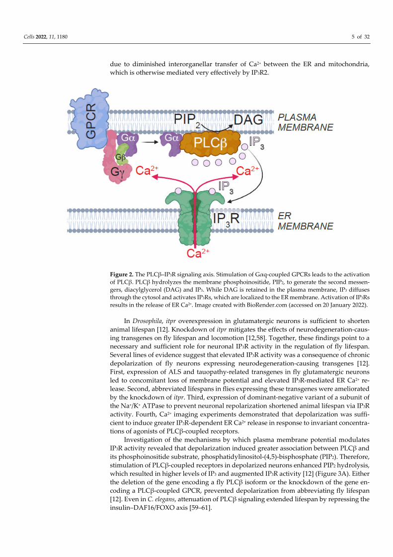
Figure 2. The PLCβ–IP3R signaling axis. Stimulation of Gαq-coupled GPCRs leads to the activation
of PLCβ. PLCβ hydrolyzes the membrane phosphoinositide, PIP2, to generate the second messen-
gers, diacylglycerol (DAG) and IP3. While DAG is retained in the plasma membrane, IP3 diffuses
through the cytosol and activates IP3Rs, which are localized to the ER membrane. Activation of IP3Rs
results in the release of ER Ca2+. Image created with BioRender.com (accessed on 20 January 2022).
In Drosophila, itpr overexpression in glutamatergic neurons is sufficient to shorten
animal lifespan [12]. Knockdown of itpr mitigates the effects of neurodegeneration-caus-
ing transgenes on fly lifespan and locomotion [12,58]. Together, these findings point to a
necessary and sufficient role for neuronal IP3R activity in the regulation of fly lifespan.
Several lines of evidence suggest that elevated IP3R activity was a consequence of chronic
depolarization of fly neurons expressing neurodegeneration-causing transgenes [12].
First, expression of ALS and tauopathy-related transgenes in fly glutamatergic neurons
led to concomitant loss of membrane potential and elevated IP3R-mediated ER Ca2+ re-
lease. Second, abbreviated lifespans in flies expressing these transgenes were ameliorated
by the knockdown of itpr. Third, expression of dominant-negative variant of a subunit of
the Na+/K+ ATPase to prevent neuronal repolarization shortened animal lifespan via IP3R
activity. Fourth, Ca2+ imaging experiments demonstrated that depolarization was suffi-
cient to induce greater IP3R-dependent ER Ca2+ release in response to invariant concentra-
tions of agonists of PLCβ-coupled receptors.
Investigation of the mechanisms by which plasma membrane potential modulates
IP3R activity revealed that depolarization induced greater association between PLCβ and
its phosphoinositide substrate, phosphatidylinositol-(4,5)-bisphosphate (PIP2). Therefore,
stimulation of PLCβ-coupled receptors in depolarized neurons enhanced PIP2 hydrolysis,
which resulted in higher levels of IP3 and augmented IP3R activity [12] (Figure 3A). Either
the deletion of the gene encoding a fly PLCβ isoform or the knockdown of the gene en-
coding a PLCβ-coupled GPCR, prevented depolarization from abbreviating fly lifespan
[12]. Even in C. elegans, attenuation of PLCβ signaling extended lifespan by repressing the
insulin–DAF16/FOXO axis [59–61].

Cells 2022, 11, 1180 6 of 32
Figure 3. Consequences and regulators of neuronal excitability. (A) Left, at resting membrane po-
tentials, PLCβ hydrolyzes the PIP2 moieties associated with the enzyme. Right, plasma membrane
depolarization results in enhanced PIP2–PLCβ clustering. Subsequent, stimulation of PLCβ-coupled
receptors in depolarized neurons results in the augmentation of PIP2 hydrolysis, IP3 production, and
IP3R-dependent ER Ca2+ release. (B) Left, REST is a master regulator of membrane excitability in
healthy neurons. Nuclear translocation of REST allows for the repression of RE1-containing gene
such as those encoding channels and pumps. Repression of these genes is necessary for the proper
maintenance of membrane potential and excitability. Right, in neurodegenerative disease, REST is
sequestered away from the nucleus resulting in derepression of target genes and the augmentation
of excitability and genes related to synaptic transmission. Images created with BioRender.com (ac-
cessed 25 January 2022).
2.3. Transcriptional Control of Excitability and Longevity
The relationship between longevity and the neuronal excitability is mediated by Re-
pressor Element-1 (RE1) Silencing Transcription Factor (REST, also called NRSF), which
represses genes carrying the RE1 motif [10,62,63]. REST appears to promote resilience to
age by decreasing the expression of genes necessary for promoting neuronal excitability
[10,64] (Figure 3B). REST-deficient mice exhibit cortical hyperexcitability, a propensity for
epileptic seizures, and enhanced mortality [10]. In C. elegans, knockdown of REST
orthologs augments neuronal excitability and shortens lifespan, whereas elevated REST
activity represses neuronal excitability and lengthens animal lifespan [10]. In worms,
REST and attendant neuronal excitability impact on longevity via the regulation of
DAF16/FOXO signaling [10,59,60].

Cells 2022, 11, 1180 7 of 32
The findings that expression of neurodegeneration-causing transgenes in Drosophila
glutamatergic neurons results in both hyperexcitability and shorter lifespan [12] is con-
sistent with the notion of REST dysregulation in models of neurodegeneration [65]. Dele-
tion of REST is sufficient to induce degeneration in mouse and C. elegans nervous systems
[66]. REST is neuroprotective in aged human neurons, iPSC-derived neurons in patients
with Alzheimer’s disease (AD), and in mouse models of AD [10,66,67]. Mechanistically,
REST function is lost in AD patients and in individuals with age-dependent cognitive im-
pairment due to increased sequestration into autophagosomes, and depletion from the
nucleus [66,67] (Figure 3B). It is also possible that REST might get trapped in protein ag-
gregates that are observed in many neurodegenerative diseases (Figure 3B), although this
possibility has not been demonstrated directly. An exception to the model that REST is
ubiquitously neuroprotective is the case in HD. In HD neurons, REST exhibits aberrantly
high nuclear localization, increased RE1 occupancy, and expected gene repression [68,69].
Whether these outcomes contribute to HD pathology, and how this phenotype relates to
that observed in AD remain incompletely understood.
3. Regulation of Aging and Longevity by Mitochondrial Ion Channels and Transporters
Mitochondria are critical for life at both cellular and organismal levels. Mitochondrial
function declines in older animals, and the underlying mechanisms are major contributors
to the adverse outcomes of advanced age [70]. This section aspires to describe the contri-
butions of mitochondrial ion channels and transporters to the regulation of aging and lon-
gevity.
3.1. Mitochondrial Ca2+ Uniporter
Uptake of Ca2+ into the mitochondrial matrix is critical for many aspects of mitochon-
drial function, including ATP production (Figure 4). Ca2+ transport across the inner mito-
chondrial membrane is mediated by the mitochondrial Ca2+ uniporter (MCU) and its reg-
ulators, mitochondrial Ca2+ uptake 1-3 (MICU1-3), mitochondrial Ca2+ uniporter regulator
1 (MCUR1), and essential MCU regulator (EMRE) [71–75]. Close coupling between the ER
and mitochondria permits the Ca2+ ions released via IP3Rs to be taken up into the mito-
chondrial matrix through the uniporter [71,76] (Figure 4). Coordinated activities of IP3Rs
and MCU, therefore, permit Ca2+ transport from the ER to mitochondria, which in turn,
stimulates mitochondrial bioenergetics (Figure 4). This mode of interorganellar Ca2+
transport is needed for C. elegans longevity [61]. In agreement, knockdown of MCU in
Drosophila neurons further abbreviates the lifespan of animals experiencing precocious
mortality due to diminished ER–mitochondria Ca2+ exchange [12]. In mice, MICU3 abun-
dance and MCU-dependent mitochondrial Ca2+ uptake decrease in the aging skeletal mus-
cle leading to sarcopenia [77]. Restoration of MICU3 abundance in mouse muscle in-
creased myogenesis and delayed sarcopenia [77].
While uniporter activity within the physiological range is necessary for animal via-
bility, transporter overactivity has been observed in many pathological conditions (Figure
4). MCU and MICU1 contribute mitochondrial Ca2+ overload that occurs in various neu-
rodegenerative and pathological conditions that shorten lifespan. Untrammeled MCU ac-
tivity contribute to neurodegeneration in SCA, and knockdown of Mcu in mouse neurons
ameliorates mitochondrial Ca2+ overload and excitotoxicity stemming from NMDA recep-
tor activation [78,79]. In mouse cardiomyocytes, deletion of Mcu protects from ischemia
reperfusion injury [80,81]. Furthermore, knockdown of the gene encoding a regulatory
partner of MCU, MCUR1, attenuates mitochondrial Ca2+ uptake and ATP production, and
drives pro-survival pathways such as AMPK activation and autophagy [75].
MCU also senses and responds to mitochondrial oxidative stress and redox balance.
Glutathione moieties can be added to MCU, which attenuates the Ca2+ transport activity
of the protein, and thereby, limits mitochondrial metabolism and reactive oxygen species
(ROS) production [82]. In Drosophila muscle, MCU is necessary for oxidative stress-in-
duced cell death, and deletion of fly MCU confers robust resistance to oxidative stress-

Cells 2022, 11, 1180 8 of 32
dependent lethality [83]. Even OIS requires MCU-dependent mitochondrial Ca2+ overload
subsequent to ER Ca2+ release via IP3Rs [56]. In view of the model that ROS production
contributes to aging [84,85], MCU could be considered as drivers of aging and senescence.
Figure 4. Interorganellar Ca2+ transfer between ER and mitochondria. ER and mitochondrial outer
membrane are physically tethered at sites called mitochondria associated membranes (MAMs).
MAMs constitute the regions of metabolite exchange between the two organelles. A fraction of IP3Rs
localize to MAMs, where they mediate the transfer of Ca2+ from the ER lumen to the perimitochon-
drial region. The mitochondrial Ca2+ uniporter resides in the inner mitochondrial membrane, and is
responsible for the transfer of Ca2+ into the mitochondrial matrix. Physiological Ca2+ elevations in
the matrix are necessary for mitochondrial bioenergetics and the production of ATP via TCA and
ETC. Ca2+ overload, however, results in pathology and eventual cell death. Image created with Bio-
Render.com (accessed 24 January 2022).
3.2. Other Mitochondrial Channels and Transporters
Opposing the effects of the uniporter are transporters that extrude matrix Ca2+. Re-
moval of matrix Ca2+ under physiological conditions is largely dependent on the mito-
chondrial Na+/Ca2+/Li+ exchanger (NCLX), with the permeability transition pore playing a
role during Ca2+ overload [86–89]. Although Ca2+/H+ exchange has also been proposed to
serve a role in the removal of mitochondrial Ca2+, molecular identity of the exchanger re-
mains controversial [89–91].
Deletion of NCLX in the adult mouse heart has led to sudden death due to heart
failure [88]. The underlying mechanism, not surprisingly, involved mitochondrial Ca2+
overload, unremitting ROS production, and cell death [88]. Conversely, increased abun-
dance of NCLX in the mouse heart protects against ischemic cell death and heart failure
[88]. In dissociated hippocampal neurons challenged with excitotoxic stimuli, knockdown
of the gene encoding NCLX further dysregulates mitochondrial Ca2+ homeostasis and pro-
motes ROS production [92]. Diminished abundance of NCLX in neurons and glia, and
attendant impairments in the removal of mitochondrial Ca2+ are sufficient to elicit neuro-
degeneration [92]. Indeed, deletion of NCLX accelerates amyloid plaque formation, tau
neuropathology, and the rate of memory decline in a mouse model of AD [93]. Mitochon-
drial [Ca2+] is also constitutively elevated due to diminished NCLX activity in multiple
mouse models of Parkinson’s disease [94–96].
Many studies have pointed to roles for K+ channels in mitochondrial function. Small
conductance Ca2+-activated K+ channels (SK2 channels) localize to the inner mitochondrial
membrane and mediate mitochondrial K+ currents [97]. Activation of mitochondrial SK2
channels protects cells against mitochondria-dependent cell death when challenged by

Cells 2022, 11, 1180 9 of 32
excitotoxic insults [97]. In C. elegans, pharmacological activation of SK channels promotes
longevity by conferring resistance to ferroptosis [98]. Big conductance Ca2+-activated K+
channels (BK channels) are also found in the inner mitochondrial membrane of cardiomy-
ocytes, where they mediate Ca2+-activated K+ currents and protect the heart from ischemic
injury [99]. In Drosophila, absence of mitochondrial BK channels led to increased ROS pro-
duction and abbreviated fly lifespan, whereas overexpression of these channels led to
long-lived animals [100].
4. Mitochondrial Uncoupling Proteins and Longevity
Uncoupling proteins (UCPs) are integral proteins of the inner mitochondrial mem-
brane that mediate the transport of protons from the intermembrane space (IMS) to the
matrix [101,102] (Figure 5). As such, mitochondrial UCPs dissipate the electrochemical
gradient across the inner mitochondrial membrane, which otherwise drives ATP synthe-
sis via ATP synthase [101] (Figure 5). Because dissipation of the protonmotive force leads
to release of the energy derived from oxidized substrates in the form of heat, UCP1-medi-
ated proton leak drives thermogenesis in brown adipose tissue [101–105]. UCPs 2-5, on
the other hand, have limited roles in thermogenesis, and are needed for the attenuation of
mitochondrial oxidative stress, regulation of cellular and organismal metabolism, and an-
timicrobial immunity [101,106–112].
Figure 5. Uncoupling protein (UCP) mediated dissipation of the mitochondrial proton (H+) gradient.
ETC involves the transfers of electrons (e−) from reducing equivalents (NADH and FADH2) to mo-
lecular oxygen (O2). e− transport is coupled with the movement of H+ from the mitochondrial matrix
to the intermembrane space (IMS). Subsequent dissipation of the electrochemical gradient through
ATP synthase drives the production of ATP. Transfer of e− to O2 also leads to the generation of
superoxide ions and ROS. By mediating the transport of H+ down their electrochemical gradient,
UCPs dissipate the protonmotive force. Loss of protonmotive force results in the production of heat
and prevents ATP production. Upon UCP-mediated H+ leak, diminished transfer of e− to O2 miti-
gates ROS production. Image created with BioRender.com (accessed 24 January 2022).
The mechanisms of UCP-dependent proton transport have been studied extensively
in the context of UCP1. These studies have revealed that UCPs are dimers of 6 transmem-
brane domain-containing proteins [102,113]. At the core of the dimer is a hydrophilic pore
[102]. UCP-mediated proton transport requires free fatty acids, which are purported to
function via one of two possible mechanisms [101,114]. The “proton buffering model” ar-
gues that fatty acids transfer protons to proton-buffering amino acids in the pore, which

Cells 2022, 11, 1180 10 of 32
then shuttle the ions across the membrane [101]. Alternatively, the “fatty acid cycling
model” contends that fatty acid anions accept protons in the intermembrane space and
transport protons across the membrane by directly traversing the pore [101].
Role of UCP Proteins in the Response to Oxidative Stress
ROS—well-established byproducts of mitochondrial metabolism—can damage
DNA, proteins, and lipids, and are therefore drivers of senescence. Because the mitochon-
drial protonmotive force is positively correlated with superoxide production [101,115],
activation of UCP2 and UCP3 mitigates cellular oxidative stress, and thereby, counters the
onset of senescence [101,102,107–110]. Conversely, loss of either UCP2 or UCP3 is associ-
ated with increased levels of mitochondrial ROS [109,110,116,117].
There are clearly some benefits associated with increased ROS in the absence of
UCPs. Elevated ROS production in macrophages isolated from Ucp2−/− mice greatly re-
stricts growth of Toxoplasma gondii, making the animals resistant to death from Toxoplasma
infection [109]. More generally, however, ROS promotes senescence and aging
[84,85,102,118–122]. Therefore, higher levels of proton leak via UCP2 or UCP3, which mit-
igate oxidative stress, are correlated with longer lifespans in mice [123,124]. Ectopic over-
expression of human UCP2 or UCP3 in fly and/or mouse neurons is sufficient to prolong
the animals’ lifespans, and confer greater resistance to ROS [120,125–127]. These beneficial
effects depend on the induction of pro-longevity genes in hypothalamic neurons of mice,
or in insulin-producing fly neurons, which are reminiscent of hypothalamic neurons [125–
127]. Given the role for the hypothalamus in the regulation of feeding and energy home-
ostasis, these findings raise the intriguing possibility that UCP activity regulates lifespan
by influencing the animals’ feeding behavior. Indeed, caloric restriction in mice—a relia-
ble pro-longevity factor—augments the expression of Ucp2 and Ucp3, promotes mitochon-
drial proton leak, and lowers ROS production [128,129]. The convergence of multiple lon-
gevity-related pathways on UCP2 and UCP3 speaks to the pro-survival roles of uncou-
pling, and supports the “uncoupling to survive” model [123,130].
Although the predominant role for UCP1 is thermogenesis in brown adipose tissue,
abundance of this protein does correlate with lower incidence of age-related disease [131].
In humans, polymorphisms in the regulatory regions of UCP1 that result in greater ex-
pression of the gene are associated with improved longevity [132]. Overexpression of
Ucp1 in mice is sufficient to extend lifespan by lowering the incidence of cancer and ath-
erosclerotic lesions, and by correcting preexisting metabolic dysfunction [131]. Con-
versely, Ucp1−/− mice exhibit greater susceptibility to obesity at advanced age when reared
on a high-fat diet (HFD) [133]. Given the adverse effects of obesity on lifespan, these find-
ings point to a role for UCP1 in mitigating the consequences of unhealthy diet on age-
related decline of healthspan.
5. Regulation of Lifespan by Ion Channels Involved in Autophagy and Lysosomal
Proteostasis
Autophagy and lysosomal protein degradation constitute major axes of proteostasis
in metazoans. By coordinating the removal of toxic macromolecules and damaged orga-
nelles, these processes counteract the stochastic accumulation of cellular debris that oth-
erwise promote aging [134–136]. Therefore, hypermethylation of autophagy and lysoso-
mal genes tends to diminish protein degradation in aged organisms [137], whereas upreg-
ulation of autophagy and lysosomal function improves proteostasis and extends healthy
lifespan [134]. Induction of autophagy is sufficient to confer resistance to oxidative stress
and insulin sensitivity in mice reared on a high-fat diet, and ameliorate the toxic conse-
quences of polyglutamine expansion in mouse and fly models [138,139]. Ubiquitous over-
expression of Atg5, a gene that encodes a protein needed for autophagosome formation,
extends mouse lifespan via the augmentation of leanness, insulin-sensitivity, and oxida-
tive stress tolerance [140]. Even the extension of lifespan brought about by diminished
insulin-signaling in Drosophila or dietary restriction in C. elegans are dependent on

Cells 2022, 11, 1180 11 of 32
autophagy and lysosomal activity [141–143]. Genetic upregulation of chaperone-mediated
autophagy (CMA), which involves LAMP-2A-dependent direct lysosomal targeting of se-
lect proteins, also enhances proteostasis, mitigates oxidative stress, and preserves organ
function in aged mammals [144,145]. Conversely, the repression of autophagy by deletion
of Atg5 or Atg7 in mice and flies promotes aging, neurodegeneration, and shorter lifespan
via dysregulation of proteostasis and mitochondrial function [146–151].
5.1. Involvement of Vesicular Ion Channels in Endolysosomal Function and Lifespan
Autophagic protein degradation is comprised of a series of vesicular trafficking
events that originate with the sequestration of cargo into autophagosomes. These double
membrane-bound organelles then fuse with endolysosomal vesicles in order to permit
hydrolytic enzymes to gain access to material destined for degradation [152]. In Drosoph-
ila, hybrid organelles formed upon the fusion of late-endosomes and autophagosomes
(amphisomes) fuse with lysosomes in a Ca2+-dependent process requiring the endolyso-
somal cation channel, TRPML [153,154]. Loss of trpml, therefore, prevents the fusion of
amphisomes with lysosomes, which leads to the accumulation of amphisomes and dimin-
ished endolysosomal degradation [153,154]. These phenotypes also characterize Muco-
lipidosis type IV (MLIV)—a lysosomal storage disease that stems from the loss of
MCOLN1, the gene encoding the human ortholog [155–158].
In flies, diminished amphisome–lysosome fusion in the absence of functional TRPML
leads to decreased production of amino acids that are generated from endolysosomal pro-
tein degradation [153,154]. Given the roles for endolysosomal amino acids in the activa-
tion of the mTORC1 kinase complex [159], loss of TRPML results in diminished mTORC1
activation—a phenotype that can be suppressed by dietary administration of a high-pro-
tein diet [153,154]. The role for TRPML1 in mTORC1 activation is also conserved in mam-
mals [160–162] (Figure 6). Interestingly, the inverse relationship between mTORC1 activ-
ity and endolysosome/autophagosome biogenesis (via TFEB, see below) ensures that loss
of TRPML results in upregulation of endolysosomal biogenesis, which may explain the
accumulation of lysosomes in MLIV [153,163–165].
Based on the aforementioned insights, one can appreciate that the regulation of aging
and lifespan by the TRPML family of endolysosomal ion channels is complex. On the one
hand, loss of TRPMLs leads to the accumulation of undigested endosomal material,
whereas their transcriptional upregulation promotes the exocytosis and clearance of toxic
macromolecules [166]. In this regard, TRPMLs are necessary for proteostasis. Indeed, the
loss of mouse TRPML1 or fly TRPML leads to proteostatic imbalance, severe loss of neu-
ronal function, and abbreviated lifespans [167,168]. It is worth noting that besides inhibit-
ing the mTORC1 complex, the FDA-approved, longevity-promoting drug, rapamycin
[169] also activates TRPML1 independently of mTORC1 [170]. Consequently, rapamycin-
induced autophagic flux is attenuated in TRPML1-deficient cells [170]. It is, therefore, pos-
sible that some of the pro-longevity effects of rapamycin stem from the activation of
TRPML1 and the attendant improvement of cellular proteostasis.
On the other hand, since decreased mTORC1 activity is associated with the extension
of lifespan in a variety of organisms [171–175], attenuation of mTORC1 activity in the ab-
sence of TRPMLs also have beneficial consequences. In agreement, abbreviation of
lifespan upon the expression of neurodegeneration-causing transgenes in fly neurons is
suppressed by the concomitant knockdown of trpml [12]. Additionally, TRPML1 contrib-
utes to the toxicity in a presenilin knockout mouse model of Alzheimer’s disease [176].
Delineating the relative roles of TRPMLs in the regulation of lifespan under physiologi-
cally normal or pathological conditions would likely require further investigation.
Two-pore channels (TPCs) are a class of endolysosomal cation channels that orches-
trate vesicular trafficking events by releasing vesicular Ca2+ and Na+ in response to
NAADP and PI(3,5)P2 [177–184]. TPC function is of relevance to autophagy, mTORC1 ac-
tivity, protein homeostasis, and cholesterol homeostasis [185–188] (Figure 6). Not surpris-
ingly, deletion of the gene encoding TPC2 in mice results in skeletal muscle atrophy due

Cells 2022, 11, 1180 12 of 32
to defective lysosomal proteolysis, accumulation of undigested autophagic vacuoles, and
heightened sensitivity to starvation [186]. Similar roles for TPCs in ensuring autophagic
and endolysosomal flux has also been reported in cardiac muscle [185]. Elevated TPC2
activity is observed in cells expressing Parkinson’s disease associated mutations in LRRK2
[189], suggesting that attenuation of TPC2 activity (as is the case of TRPML1) might be
beneficial in late-onset neurodegenerative diseases.
Figure 6. Transcriptional regulation of genes encoding endolysosomal ion channels. Within the cell
nucleus, TFEB, and related transcription factors such as TFE3 and MITF, bind to the promoters of
genes that encode autophagy and endolysosomal proteins. Figure shows dephosphorylated TFEB
bound to the promoters of MCOLN1 and TPCN2, which encode TRPML1 and TPC2, respectively.
Phosphorylated TFEB is retained in the cytosol leading to diminished expression of endolysosomal
genes. TRPML1 and TPC2 are endolysosomal cation channels that regulate nucleocytoplasmic
translocation of TFEB activity in different ways. By driving autophagic and endolysosomal flux,
TRPML1 and TPC2 are needed for mTORC1 activation, which in turn, phosphorylates TFEB leading
to retention of the transcription factor in the cytosol. On the other hand, release of endolysosomal
Ca2+ via TRPML1 and TPC2 activates calcineurin (CaN), which dephosphorylates TFEB, and
thereby, promotes the translocation of the transcription factor into the nucleus. Image created with
BioRender.com (accessed 24 January 2022).
Cl- represents the major endolysosomal counter ion. ClC-6 and ClC-7 and endolyso-
somal chloride transporters are required for normal lysosomal function [190–192]. Loss of
these transporters result in a variety of neuropathological alterations, including lysosomal
storage, progressive neuron loss, and microglial activation [193–195]. The progressive na-
ture of these phenotypes suggests a role for these proteins in the maintenance of neuronal
function and survival at advanced age.
Critical to the regulation of amino acid availability and mTORC1 activity are vesicu-
lar solute carriers belonging to the SLC family of transporters. Among the many amino
Cells 2022, 11, 1180 13 of 32
acid transporters critical for mTORC1 activation is the lysosomal amino acid transporter,
SLC38A9 [196–198]. Loss of SLC38A9 decouples mTORC1 from amino acids such as argi-
nine, whereas overexpression of SLC38A9 results in the constitutive activation of
mTORC1, even in the absence of amino acids [196–198]. Therefore, SLC38A9 is not only a
vesicular transporter needed for the efflux of amino acids from the lysosome, but is also
an amino acid sensor that synchronizes mTORC1 activity to amino acid abundance [196–
199]. Besides serving as a sensor and transporter for amino acids, SLC38A9 also harbors
cholesterol-binding motifs [200]. Association of cholesterol with SLC38A9 activates
mTORC1 independently of the transporter’s role in endolysosomal amino acid efflux
[200]. Other amino acid transporters linked to the regulation of mTORC1 include
SLC38A5, SLC7A5 (also called LAT1) and the glutamine transporter SLC1A5 (also called
ASCT2) [201–205]. Consistent with the roles for mTORC1 in aging [171–175], emerging
evidence suggests that modulation of the activity of amino acid sensors, such as the ones
described here, could be an effective strategy to influence rates of aging [206]. Indeed,
polymorphisms in genes encoding amino acid transporters are associated with age-re-
lated changes in physical performance (e.g., grip strength and walking speed) [207]. Con-
versely, there is increased abundance of amino acid transporters such as SLC7A5,
SLC1A5, and SLC38A5 in inflammation and cancer [204–206,208]. Together, these studies
point to critical roles for amino acid transporters in the regulation of aging and longevity.
5.2. Transcriptional Regulation of Endolysosomal Function
A major advancement in the field of lysosomal biology was the identification of tran-
scription factors (TFEB, TFE3, and MITF) that function as master regulators of autophagy
and endolysosomal biogenesis [163–165,209–214]. Genetic or pharmacological activation
of these transcription factors promotes the clearance of cellular debris and aggregate-
prone toxic macromolecules [166,215–217]. As such, TFEB maintains the quiescent state of
neural stems cells, and has been positively correlated with longevity in C. elegans and mice
[218–224]. TFEB and TFE3 are needed for the maintenance of whole-body metabolism in
response to changes in various environmental stimuli including diet [225]. Dietary re-
striction—an established mode of lifespan extension—promotes TFEB-dependent gene
expression in mouse hepatocytes [222].
Many genes that encode endolysosomal ion channels, including TRPML1 and TPC2,
are under the control of TFEB [211,212,226,227] (Figure 6). In turn, nuclear translocation
of TFEB, which is dependent on the phosphorylation status of the protein, is regulated by
TRPML1 and TPC2 (Figure 6). On the one hand, TRPML1- and TPC2-dependent activa-
tion of mTORC1 ensures TFEB phosphorylation and cytosolic retention [160–164] (Figure
6). These findings explain why the lack of TRPML1, results in nuclear translocation of
TFEB and unremitting endolysosomal biogenesis. On the other hand, endolysosomal Ca2+
release can also dephosphorylate and promote nuclear translocation of TFEB via the pro-
tein phosphatase, calcineurin (CaN) [228] (Figure 6). This model portends a feed-forward
cycle, whereby TRPML1 and TPC2 activation, which are under TFEB transcriptional con-
trol, promotes further TFEB-dependent endolysosomal biogenesis.
While TFEB-driven autophagy and endolysosomal biogenesis delays aging via the
clearance of toxic macromolecules, unremitting activation of TFEB can paradoxically
shorten lifespan [141]. Either the overexpression and/or constitutive nuclear localization
of TFEB and TFE3 promote the growth of various types of cancer [226,227,229–240]. Ad-
ditionally, amplification of the MITF locus and functional upregulation of MITF are potent
drivers of melanoma [213,241–246]. These findings point to the importance of maintaining
autophagy and endolysosomal function at an optimal level because deviation from this
optimum in either direction has deleterious consequences to healthy lifespan. In cancers
driven by TFEB/TFE3/MITF, expression of genes encoding endolysosomal cation chan-
nels, TRPML1, TRPML2 and TPC2, is elevated, and either the knockdown or pharmaco-
logical inhibition of these channels attenuates the growth and invasiveness of the tumors

Cells 2022, 11, 1180 14 of 32
[160,226,227,247–252]. Taken together, these studies point to the highly context-dependent
relationship between endolysosomal ion channels and healthspan.
6. Regulation of Ion Channel Activity by the Longevity Factor, Klotho
Mutations in the mouse klotho gene results in animals that develop normally, but
prematurely exhibit several features of accelerated aging including neurodegeneration,
atherosclerosis, osteoporosis, infertility, atrophy of the skin and other organs, and shorter
lifespan [253,254]. Conversely, overexpression of klotho results in a 20–30% extension of
mouse lifespan via the repression of insulin signaling and the amelioration of oxidative
stress [255,256]. Overexpressed klotho also enhances cognition by the enhancement of syn-
aptic plasticity [257]. These roles for Klotho in healthy aging are also observed in humans
[258–260].
The product of the klotho gene is a type-I single pass transmembrane protein that
localizes to the cell surface [256]. The extracellular domain of the protein exhibits homol-
ogy to glycosidases/sialidases that can cleave the b-glycosidic linkage in sugars, glycopro-
teins and glycolipids, and remove sialic acid residues from membrane proteins
[253,256,261–263]. Although expression of klotho is highest in the kidneys, the extracellular
sialidase domain of the protein product is cleaved by ADAM10 and ADAM17 transmem-
brane proteases, and released into the bloodstream where it can function as an endocrine
factor [256,261,262,264]. ADAM10/ADAM17-dependent cleavage of membrane-bound
Klotho is under the control of insulin signaling, which when taken in consideration that
soluble Klotho represses insulin signaling, points to the existence of a feedback loop to
prevent unremitting insulin signaling [255,264]. While the membrane-bound form of
Klotho serves as a coreceptor for fibroblast growth factor-23 (FGF23) and regulates phos-
phate and vitamin D metabolism, the secreted form neither binds to, nor serves as a core-
ceptor for FGF23 [256,265]. Rather, soluble Klotho serves as an enzyme capable of remov-
ing sugars and sialic acid residues from various membrane glycoproteins [253,256,261–
263]. It is via the influence on membrane glycoproteins that soluble Klotho prolongs lon-
gevity.
Regulation of Ion Channel Activity by Klotho
By removing glycan moieties from the extracellular side of the plasma membrane,
soluble Klotho promotes the surface retention and activities of TRPV5 and TRPV6 ion
channels [263,266–269]. Removal of terminal sialic acid residues allows the remaining sug-
ars to bind lectins leading to channel crosslinking on the cell surface and diminished in-
ternalization [263]. Given that TRPV5 participates in systemic Ca2+ and phosphate home-
ostasis by functioning in the kidney [270], it is not clear whether Klotho influences lon-
gevity via these channels. Nevertheless, it is possible that hyperphosphatemia and hyper-
calcemia resulting from diminished TRPV5/TRPV6 function in the absence of Klotho
could contribute to age-dependent vascular calcification and osteopenia [271]. A putative
link to longevity can be gleaned from the demonstration that Klotho promotes the surface
retention and overall activity of the Na+/K+ ATPase [272]. Given the relationship between
neuronal excitability and longevity, and demonstrated roles for the Na+/K+ ATPase in the
regulation of these parameters [10–12,16], soluble Klotho could influence lifespan by sta-
bilizing this ATPase at the cell surface.
Alternatively, Klotho could influence animal longevity by its influence on cardiac
function. Klotho counteracts cardiac arrhythmia by ensuring the cell surface expression
of KCNQ1/KCNE1 K+ channels that are involved in cardiac repolarization [273]. Loss of
soluble Klotho promotes cardiac arrhythmia owing to decreased activity of the
KCNQ1/KCNE1 channels in cardiomyocytes. Cardioprotective roles of Klotho also stem
from downregulation of TRPC6 channel activity [274,275]. TRPC6 channels have vital
roles in stress-induced pathological cardiac hypertrophy and remodeling [274]. By atten-
uating TRPC6 conductance, Klotho ameliorates cardiotoxicity [274,276].

Cells 2022, 11, 1180 15 of 32
7. Roles for Ion Channels in Regulation of Longevity by Temperature
All physiological systems are under the control of temperature. While an increase in
temperature accelerates the rates of biological reactions, a decrease in temperature has the
opposite effect [277]. The inverse relationship between rates of metabolism and longevity
is why lower body temperatures are associated with longer lifespans, whereas higher
body temperatures are more common in short-lived individuals [278–280]. In homeo-
therms, interventions that influence rates of aging and lifespan, for instance caloric re-
striction, also tend to alter body temperature in a manner that is consistent between the
aforementioned relationship between temperature and longevity [278,281]. Cold temper-
atures in homeotherms lead to the generation of heat in brown adipose tissue via UCP1
[101–105]. Given the involvement of UCP proteins in regulation of longevity (as discussed
above), cold-induced uncoupling could constitute another mechanism by which low tem-
peratures promote longevity. In agreement, overexpression of Ucp2 in the hypocretin neu-
rons of mouse hypothalamus extended the animals’ longevity by lowering their core body
temperature [125].
In poikilotherms such as C. elegans and Drosophila, a decrease in ambient temperature
promotes stress resistance and counteracts inflammatory signaling [282]. While it is
tempting to speculate that the relationship between temperature and age-related pathol-
ogy stems from the propensity of temperature to increase thermodynamic entropy
[278,283], it is likely that additional levels of complexity bear upon this relationship. In-
deed, flies cycled between hot and cold ambient temperatures live for as long as those that
are reared at steady low ambient temperature [278,279,284]. These intriguing data sug-
gested that the process of sensing a drop in ambient temperature is sufficient to impart
the benefits of low temperature [278].
7.1. Thermosensitive Channels and Lifespan
In agreement with the notion that the act of sensing ambient temperature influences
longevity, recent studies have shown that thermosensitive ion channels actuate the effects
of temperature on lifespan. A pioneering study in C. elegans challenged the passive ther-
modynamic model of aging by demonstrating that a cold-sensitive TRP channel (TRPA-
1) detects a drop in environmental temperature, and signals to the well-established mod-
ulator of longevity, DAF-16/FOXO [5,285,286] (Figure 7). trpa-1 deficient adult worms ex-
hibited significantly shorter lifespans when reared at a cool 15 °C, whereas no such differ-
ence between wild-type and trpa-1 mutants was observed in animals raised at 25 °C
[285,286]. These findings suggest that cold-induced extension of lifespan involves an ion
channel that is directly activated by lower temperature rather than a general reduction in
rates of metabolism (Figure 7).

Cells 2022, 11, 1180 16 of 32
Figure 7. Effect of low-temperature on the lifespan of C. elegans is mediated by the cold-sensitive ion
channel, TRPA-1. TRPA-1 is a cold sensitive ion channel that resides in the plasma membrane of C.
elegans neurons and intestinal cells. Activation of the channels at 15 °C, results in cytosolic Ca2+ ele-
vation and the activation of a signaling cascade that culminates in DAF-16/FOXO-mediated gene
transcription, which in turn, promotes worm longevity. Image created with BioRender.com (ac-
cessed 27 January 2022).
Mice lacking the thermosensitive TRP channel, TRPV1, exhibit youthful metabolism
and are relatively long-lived [287]. The regulation of lifespan via TRPV1 involves nuclear
localization of the CREB-regulated transcriptional coactivator, CRTC1. In animals lacking
TRPV1, CRTC1 is excluded from the nucleus, which results in diminished production of
the neuropeptide CGRP and attendant improvement of metabolic health [287]. The rela-
tionship between CGRP and systemic metabolism is ensured by the repression of insulin
release by CGRP [287]. In C. elegans, deletion of the TRPV channel genes, ocr-2 and osm-9,
led to the extension of lifespan [287,288]. Whether the activities of TRPV1, OSM-9, and
OCR-2 in these contexts involve temperature remain to be addressed.
7.2. Heat-shock Response and Lifespan
While low temperature is associated with the extension of lifespan in many species,
transient heat shocks in poikilotherms induce the expression of molecular chaperones that
enforce proteostasis and promote longevity [289–291]. Interestingly, longevity-promoting
effects of the heat-shock response in C. elegans are not cell autonomous, but rather stem
from thermosensitive neurons that express cyclic nucleotide-gated (CNG) TAX-2/TAX-4
channels [292]. Deletion of guanylyl cyclase responsible for the activation of these CNG
channels in thermosensitive neurons prevents the induction of a heat-shock response in
other tissues [292]. The underlying mechanism involves the secretion of FMRFamide neu-
ropeptide from the thermosensitive neurons, which greatly influences peripheral insulin
signaling [293]. Furthermore, induction of the C. elegans heat shock factor, HSF-1, and the
attendant effects on proteostasis can occur independently of a bona fide heat shock stim-
ulus. Cholinergic neurotransmission at the neuromuscular junction leads to a Ca2+-de-
pendent induction of the hsf-1 in postsynaptic muscle cells via a process requiring the
VGCCs and ryanodine receptors in the ER [294].

Cells 2022, 11, 1180 17 of 32
8. Regulation of Longevity by Channels Involved in Other Sensory Modalities
Besides temperature, many sensory modalities can modulate lifespan. In Drosophila,
critical insights were obtained from the demonstration that deletion of the gene encoding
a water-sensitive channel belonging to the amiloride-sensitive ENaC family of Na+ chan-
nels, pickpocket 28 (ppk28), altered the metabolic status of animals, and thereby, extended
lifespan [295–297]. Loss of ppk28 improved systemic metabolism via the augmentation of
neuronal signaling involving glucagon-related, adipokinetic hormone (AKH) [295]. Re-
markably, the apparent lack of water—mimicked by the deletion of ppk28—triggers AKH-
dependent alterations in metabolic pathways such as b-oxidation that produce molecular
water, and in the process, extends lifespan [295].
In C. elegans, activity of sensory neurons, which is partially dependent on the ENaC
channel MEC-4, is necessary for the protection of mitochondria from fragmentation in
aged animals [298]. Furthermore, the TRPV channel, OSM-9, functions in sensory neurons
to mediate the avoidance of hypertonic stress [299,300]. Deletion of osm-9 led to enhanced
proteostasis and survival of the worms when placed in hypertonic stress [300]. Deletion
of another TRPV channel gene in C. elegans, ocr-2, also extended adult lifespan [287,288].
In Drosophila, loss of a similar TRPV channel, Inactive (Iav), which has been proposed to
play roles in mechanosensation, is associated with reduced lifespan [301,302].
Relationships between lifespan and reproduction and/or food availability have been
observed across taxa. In flies, detection of female pheromones via gustatory sensory neu-
rons leads to reduction in stored nutrient reserves, greater sensitivity to starvation, and
shorter lifespan in males [303]. This deleterious effect of female pheromone sensation on
male longevity was observed only if the males were not allowed to mate, and mating re-
versed the effects of pheromone sensation on longevity. These findings indicate that the
relationship sensory and reward circuits in the brain modulates aging and lifespan [303].
Although the involvement of ion channels in this axis of longevity have not been de-
scribed, indirect evidence suggests the involvement of ENaC channels. Deletion of ppk23
and ppk25 result in diminished intensities of male courtship, likely due to altered re-
sponses to female pheromones [304,305]. It would be worth evaluating whether the ab-
sences of ppk23 and ppk25 influence the lifespan of the animals in a pheromone- and court-
ship-dependent manner. As was the case with female pheromones regulating male
lifespan in fruit flies, food-derived odors shorten fly lifespan, and even deter the longev-
ity-promoting effects of dietary restriction [306]. Therefore, deletion of the gene encoding,
Or83b, an ionotropic odorant receptor that functions as a non-selective cation channel
needed for the detection of food-derived odors, augmented stress resistance and extended
animal lifespan [306–308].
9. Closing Remarks and Future Directions
Despite the plethora of ion channels and transporters that influence aging and lon-
gevity, holistic evaluation of the studies described here also reveal commonalities. One
general principle is that mechanisms of ion transport constitute major regulatory axes in
pathways that determine the rates of cellular or organismal aging. Some channels even
function at the intersection of one or more hallmarks of aging. A better understanding of
how these channels and transporters impact age-related loss of biological integrity could
aid in the development of effective anti-aging strategies. Another theme that emerges
from these studies is that homeostatic regulation of ion channel and transporter activity
is the key to human healthspan. Any deviation of ion transport from a healthy optimum
leads to elevated rates of mortality. Consequently, the trajectory of age-dependent loss of
biological function likely varies greatly from one individual to the next. What we need,
therefore, is the development of strategies that would enable personalized approaches to
combat aging. A suite of drugs that restores ionic homeostasis in various tissues by ap-
propriately modifying channel and transporter activities could be the key to translate bi-
ological insights into actionable therapeutic strategies. Future studies into the regulation

Cells 2022, 11, 1180 18 of 32
of aging and longevity by ion channels and transporters will undoubtedly bring us closer
to realizing this vision.
Funding: Work in the Venkatachalam lab is supported by NIH grants RF1AG069076, RF1AG067414,
and R21AG072176 (all to K.V.).
Institutional Review Board Statement: Not applicable.
Informed Consent Statement: Not applicable.
Data Availability Statement: Not applicable.
Acknowledgments: The author wishes to thank Richa Gupta and Elham Rastegari for reading the
manuscript and providing valuable feedback.
Conflicts of Interest: The author declares no competing interest.
References
1. López-Otín, C.; Blasco, M.A.; Partridge, L.; Serrano, M.; Kroemer, G. The Hallmarks of Aging. Cell 2013, 153, 1194–1217.
https://doi.org/10.1016/j.cell.2013.05.039.
2. Campisi, J.; Kapahi, P.; Lithgow, G.J.; Melov, S.; Newman, J.C.; Verdin, E. From Discoveries in Ageing Research to Therapeutics
for Healthy Ageing. Nature 2019, 571, 183–192. https://doi.org/10.1038/s41586-019-1365-2.
3. Kenyon, C.; Chang, J.; Gensch, E.; Rudner, A.; Tabtiang, R. A C. elegans Mutant That Lives Twice as Long as Wild Type. Nature
1993, 366, 461–464. https://doi.org/10.1038/366461A0.
4. Friedman, D.B.; Johnson, T.E. A Mutation in the Age-1 Gene in Caenorhabditis Elegans Lengthens Life and Reduces Hermaph-
rodite Fertility. Genetics 1988, 118, 75–86. https://doi.org/10.1093/genetics/118.1.75.
5. Ogg, S.; Paradis, S.; Gottlieb, S.; Patterson, G.I.; Lee, L.; Tissenbaum, H.A.; Ruvkun, G. The Fork Head Transcription Factor
DAF-16 Transduces Insulin-like Metabolic and Longevity Signals in C. elegans. Nature 1997, 389, 994–999.
https://doi.org/10.1038/40194.
6. Tatar, M.; Kopelman, A.; Epstein, D.; Tu, M.P.; Yin, C.M.; Garofalo, R.S. A Mutant Drosophila Insulin Receptor Homolog That
Extends Life-Span and Impairs Neuroendocrine Function. Science 2001, 292, 107–110. https://doi.org/10.1126/SCIENCE.1057987.
7. Berridge, M.J. Calcium Signalling Remodelling and Disease. Biochem. Soc. Trans. 2012, 40, 297–309.
https://doi.org/10.1042/BST20110766.
8. Karagas, N.E.; Venkatachalam, K. Roles for the Endoplasmic Reticulum in Regulation of Neuronal Calcium Homeostasis. Cells
2019, 8, 1232. https://doi.org/10.3390/cells8101232.
9. Berridge, M.J. Inositol Trisphosphate and Calcium Signalling Mechanisms. Biochim. Biophys. Acta (BBA)-Mol. Cell Res. 2009, 1793,
933–940. https://doi.org/10.1016/j.bbamcr.2008.10.005.
10. Zullo, J.M.; Drake, D.; Aron, L.; O’Hern, P.; Dhamne, S.C.; Davidsohn, N.; Mao, C.-A.; Klein, W.H.; Rotenberg, A.; Bennett, D.A.;
et al. Regulation of Lifespan by Neural Excitation and REST. Nature 2019, 574, 359–364. https://doi.org/10.1038/s41586-019-1647-
8.
11. Fergestad, T.; Ganetzky, B.; Palladino, M.J. Neuropathology in Drosophila Membrane Excitability Mutants. Genetics 2006, 172,
1031. https://doi.org/10.1534/GENETICS.105.050625.
12. Wong, C.-O.; Karagas, N.E.; Jung, J.; Wang, Q.; Rousseau, M.A.; Chao, Y.; Insolera, R.; Soppina, P.; Collins, C.A.; Zhou, Y.; et al.
Regulation of Longevity by Depolarization-Induced Activation of PLC-β-IP3R Signaling in Neurons. Proc. Natl. Acad. Sci. USA
2021, 118, e2004253118. https://doi.org/10.1073/pnas.2004253118.
13. Reynolds, E.R. Shortened Lifespan and Other Age-Related Defects in Bang Sensitive Mutants of Drosophila Melanogaster. G3
Genes Genomes Genet. 2018, 8, 3953–3960. https://doi.org/10.1534/G3.118.200610.
14. Garber, G.; Smith, L.A.; Reenan, R.A.; Rogina, B. Effect of Sodium Channel Abundance on Drosophila Development, Reproduc-
tive Capacity and Aging. Fly 2012, 6, 57–67. https://doi.org/10.4161/FLY.18570.
15. Cirelli, C.; Bushey, D.; Hill, S.; Huber, R.; Kreber, R.; Ganetzky, B.; Tononi, G. Reduced Sleep in Drosophila Shaker Mutants.
Nature 2005, 434, 1087–1092. https://doi.org/10.1038/nature03486.
16. Palladino, M.J.; Bower, J.E.; Kreber, R.; Ganetzky, B. Neural Dysfunction and Neurodegeneration in Drosophila Na+/K+ ATPase
Alpha Subunit Mutants. J. Neurosci. Off. J. Soc. Neurosci. 2003, 23, 1276–1286. https://doi.org/10.1523/JNEUROSCI.23-04-
01276.2003.
17. Andries, M.; van Damme, P.; Robberecht, W.; van den Bosch, L. Ivermectin Inhibits AMPA Receptor-Mediated Excitotoxicity
in Cultured Motor Neurons and Extends the Life Span of a Transgenic Mouse Model of Amyotrophic Lateral Sclerosis. Neuro-
biol. Dis. 2007, 25, 8–16. https://doi.org/10.1016/J.NBD.2006.08.018.
18. Warnier, M.; Flaman, J.-M.; Chouabe, C.; Wiel, C.; Gras, B.; Griveau, A.; Blanc, E.; Foy, J.-P.; Mathot, P.; Saintigny, P.; et al. The
SCN9A Channel and Plasma Membrane Depolarization Promote Cellular Senescence through Rb Pathway. Aging Cell 2018, 17,
e12736. https://doi.org/10.1111/acel.12736.

Cells 2022, 11, 1180 19 of 32
19. Liu, W.; Lin, H.; Mao, Z.; Zhang, L.; Bao, K.; Jiang, B.; Xia, C.; Li, W.; Hu, Z.; Li, J. Verapamil Extends Lifespan in Caenorhabditis
Elegans by Inhibiting Calcineurin Activity and Promoting Autophagy. Aging 2020, 12, 5300–5317. https://doi.org/10.18632/AG-
ING.102951.
20. Sutphin, G.L.; Backer, G.; Sheehan, S.; Bean, S.; Corban, C.; Liu, T.; Peters, M.J.; van Meurs, J.B.J.; Murabito, J.M.; Johnson, A.D.;
et al. Caenorhabditis Elegans Orthologs of Human Genes Differentially Expressed with Age Are Enriched for Determinants of
Longevity. Aging Cell 2017, 16, 672–682. https://doi.org/10.1111/ACEL.12595.
21. Moore, S.J.; Murphy, G.G. The Role of L-Type Calcium Channels in Neuronal Excitability and Aging. Neurobiol. Learn. Mem.
2020, 173, 107230. https://doi.org/10.1016/J.NLM.2020.107230.
22. Zhang, X.; Chen, S.; Lu, K.; Wang, F.; Deng, J.; Xu, Z.; Wang, X.; Zhou, Q.; Le, W.; Zhao, Y. Verapamil Ameliorates Motor Neuron
Degeneration and Improves Lifespan in the SOD1 G93A Mouse Model of ALS by Enhancing Autophagic Flux. Aging Dis. 2019,
10, 1159–1173. https://doi.org/10.14336/AD.2019.0228.
23. Huntula, S.; Saegusa, H.; Wang, X.; Zong, S.; Tanabe, T. Involvement of N-Type Ca2+ Channel in Microglial Activation and Its
Implications to Aging-Induced Exaggerated Cytokine Response. Cell Calcium 2019, 82, 102059.
https://doi.org/10.1016/j.ceca.2019.102059.
24. Hayashi, T.; Yamaguchi, T.; Sakakibara, Y.; Taguchi, K.; Maeda, M.; Kuzuya, M.; Hattori, Y. ENOS-Dependent Antisenscence
Effect of a Calcium Channel Blocker in Human Endothelial Cells. PLoS ONE 2014, 9, e88391. https://doi.org/10.1371/jour-
nal.pone.0088391.
25. Li, Z.; Guo, G.; Wang, H.; Si, X.; Zhou, G.; Xiong, Y.; Li, S.; Dai, R.; Yang, C. TRPC5 Channel Modulates Endothelial Cells
Senescence. Eur. J. Pharmacol. 2017, 802, 27–35. https://doi.org/10.1016/j.ejphar.2017.02.037.
26. Belrose, J.C.; Xie, Y.-F.; Gierszewski, L.J.; MacDonald, J.F.; Jackson, M.F. Loss of Glutathione Homeostasis Associated with Neu-
ronal Senescence Facilitates TRPM2 Channel Activation in Cultured Hippocampal Pyramidal Neurons. Mol. Brain 2012, 5, 11.
https://doi.org/10.1186/1756-6606-5-11.
27. Yee, N.S.; Brown, R.D.; Lee, M.S.; Zhou, W.; Jensen, C.; Gerke, H.; Yee, R.K. TRPM8 Ion Channel Is Aberrantly Expressed and
Required for Preventing Replicative Senescence in Pancreatic Adenocarcinoma: Potential Role of TRPM8 as a Biomarker and
Target. Cancer Biol. Ther. 2012, 13, 592–599. https://doi.org/10.4161/cbt.20079.
28. Yee, N.S.; Zhou, W.; Lee, M. Transient Receptor Potential Channel TRPM8 Is Over-Expressed and Required for Cellular Prolif-
eration in Pancreatic Adenocarcinoma. Cancer Lett. 2010, 297, 49–55. https://doi.org/10.1016/j.canlet.2010.04.023.
29. Yee, N.S.; Zhou, W.; Lee, M.; Yee, R.K. Targeted Silencing of TRPM7 Ion Channel Induces Replicative Senescence and Produces
Enhanced Cytotoxicity with Gemcitabine in Pancreatic Adenocarcinoma. Cancer Lett. 2012, 318, 99–105.
https://doi.org/10.1016/j.canlet.2011.12.007.
30. Szydlowska, K.; Tymianski, M. Calcium, Ischemia and Excitotoxicity. Cell Calcium 2010, 47, 122–129.
https://doi.org/10.1016/J.CECA.2010.01.003.
31. Iizuka, A.; Nakamura, K.; Hirai, H. Long-Term Oral Administration of the NMDA Receptor Antagonist Memantine Extends
Life Span in Spinocerebellar Ataxia Type 1 Knock-in Mice. Neurosci. Lett. 2015, 592, 37–41.
https://doi.org/10.1016/J.NEULET.2015.02.055.
32. Xu, W.; Xu, J. C9orf72 Dipeptide Repeats Cause Selective Neurodegeneration and Cell-Autonomous Excitotoxicity in Drosoph-
ila Glutamatergic Neurons. J. Neurosci. Off. J. Soc. Neurosci. 2018, 38, 7741–7752. https://doi.org/10.1523/JNEUROSCI.0908-
18.2018.
33. Wenk, G.L.; Barnes, C.A. Regional Changes in the Hippocampal Density of AMPA and NMDA Receptors across the Lifespan
of the Rat. Brain Res. 2000, 885, 1–5. https://doi.org/10.1016/S0006-8993(00)02792-X.
34. Adams, M.M.; Shi, L.; Linville, M.C.; Forbes, M.E.; Long, A.B.; Bennett, C.; Newton, I.G.; Carter, C.S.; Sonntag, W.E.; Riddle,
D.R.; et al. Caloric Restriction and Age Affect Synaptic Proteins in Hippocampal CA3 and Spatial Learning Ability. Exp. Neurol.
2008, 211, 141–149. https://doi.org/10.1016/J.EXPNEUROL.2008.01.016.
35. Dubal, D.B.; Zhu, L.; Sanchez, P.E.; Worden, K.; Broestl, L.; Johnson, E.; Ho, K.; Yu, G.Q.; Kim, D.; Betourne, A.; et al. Life
Extension Factor Klotho Prevents Mortality and Enhances Cognition in HAPP Transgenic Mice. J. Neurosci. Off. J. Soc. Neurosci.
2015, 35, 2358–2371. https://doi.org/10.1523/JNEUROSCI.5791-12.2015.
36. Papadia, S.; Soriano, F.X.; Léveillé, F.; Martel, M.A.; Dakin, K.A.; Hansen, H.H.; Kaindl, A.; Sifringer, M.; Fowler, J.; Stefovska,
V.; et al. Synaptic NMDA Receptor Activity Boosts Intrinsic Antioxidant Defenses. Nat. Neurosci. 2008, 11, 476–487.
https://doi.org/10.1038/NN2071.
37. Majumder, S.; Caccamo, A.; Medina, D.X.; Benavides, A.D.; Javors, M.A.; Kraig, E.; Strong, R.; Richardson, A.; Oddo, S. Lifelong
Rapamycin Administration Ameliorates Age-Dependent Cognitive Deficits by Reducing IL-1β and Enhancing NMDA Signal-
ing. Aging Cell 2012, 11, 326–335. https://doi.org/10.1111/J.1474-9726.2011.00791.X.
38. Shi, L.; Adams, M.M.; Linville, M.C.; Newton, I.G.; Forbes, M.E.; Long, A.B.; Riddle, D.R.; Brunso-Bechtold, J.K. Caloric Re-
striction Eliminates the Aging-Related Decline in NMDA and AMPA Receptor Subunits in the Rat Hippocampus and Induces
Homeostasis. Exp. Neurol. 2007, 206, 70–79. https://doi.org/10.1016/J.EXPNEUROL.2007.03.026.
39. Yilmaz, N.; Vural, H.; Yilmaz, M.; Sutcu, R.; Sirmali, R.; Hicyilmaz, H.; Delibas, N. Calorie Restriction Modulates Hippocampal
NMDA Receptors in Diet-Induced Obese Rats. J. Recept. Signal Transduct. Res. 2011, 31, 214–219.
https://doi.org/10.3109/10799893.2011.569724.
40. Woll, K.A.; van Petegem, F. Calcium-Release Channels: Structure and Function of IP 3 Receptors and Ryanodine Receptors.
Physiol. Rev. 2022, 102, 209–268. https://doi.org/10.1152/physrev.00033.2020.

Cells 2022, 11, 1180 20 of 32
41. Van de Leemput, J.; Chandran, J.; Knight, M.A.; Holtzclaw, L.A.; Scholz, S.; Cookson, M.R.; Houlden, H.; Gwinn-Hardy, K.;
Fung, H.-C.; Lin, X.; et al. Deletion at ITPR1 Underlies Ataxia in Mice and Spinocerebellar Ataxia 15 in Humans. PLoS Genet.
2007, 3, e108. https://doi.org/10.1371/journal.pgen.0030108.
42. Klar, J.; Ali, Z.; Farooq, M.; Khan, K.; Wikström, J.; Iqbal, M.; Zulfiqar, S.; Faryal, S.; Baig, S.M.; Dahl, N. A Missense Variant in
ITPR1 Provides Evidence for Autosomal Recessive SCA29 with Asymptomatic Cerebellar Hypoplasia in Carriers. Eur. J. Hum.
Genet. 2017, 25, 848–853. https://doi.org/10.1038/ejhg.2017.54.
43. Sasaki, M.; Ohba, C.; Iai, M.; Hirabayashi, S.; Osaka, H.; Hiraide, T.; Saitsu, H.; Matsumoto, N. Sporadic Infantile-Onset Spino-
cerebellar Ataxia Caused by Missense Mutations of the Inositol 1,4,5-Triphosphate Receptor Type 1 Gene. J. Neurol. 2015, 262,
1278–1284. https://doi.org/10.1007/s00415-015-7705-8.
44. Huang, L.; Chardon, J.W.; Carter, M.T.; Friend, K.L.; Dudding, T.E.; Schwartzentruber, J.; Zou, R.; Schofield, P.W.; Douglas, S.;
Bulman, D.E.; et al. Missense Mutations in ITPR1 Cause Autosomal Dominant Congenital Nonprogressive Spinocerebellar
Ataxia. Orphanet J. Rare Dis. 2012, 7, 67. https://doi.org/10.1186/1750-1172-7-67.
45. Gerber, S.; Alzayady, K.J.; Burglen, L.; Brémond-Gignac, D.; Marchesin, V.; Roche, O.; Rio, M.; Funalot, B.; Calmon, R.; Durr,
A.; et al. Recessive and Dominant De Novo ITPR1 Mutations Cause Gillespie Syndrome. Am. J. Hum. Genet. 2016, 98, 971–980.
https://doi.org/10.1016/j.ajhg.2016.03.004.
46. McEntagart, M.; Williamson, K.A.; Rainger, J.K.; Wheeler, A.; Seawright, A.; De Baere, E.; Verdin, H.; Bergendahl, L.T.; Quigley,
A.; Rainger, J.; et al. A Restricted Repertoire of De Novo Mutations in ITPR1 Cause Gillespie Syndrome with Evidence for
Dominant-Negative Effect. Am. J. Hum. Genet. 2016, 98, 981–992. https://doi.org/10.1016/j.ajhg.2016.03.018.
47. Matsumoto, M.; Nakagawa, T.; Inoue, T.; Nagata, E.; Tanaka, K.; Takano, H.; Kuno, J.; Sakakibara, S.; Yamada, M.; Yoneshima,
H.; et al. Ataxia and Epileptic Seizures in Mice Lacking Type 1 Inositol 1,4,5-Trisphosphate Receptor. Nature 1996, 379, 168–171.
https://doi.org/10.1038/379168a0.
48. Higo, T.; Hamada, K.; Hisatsune, C.; Nukina, N.; Hashikawa, T.; Hattori, M.; Nakamura, T.; Mikoshiba, K. Mechanism of ER
Stress-Induced Brain Damage by IP(3) Receptor. Neuron 2010, 68, 865–878. https://doi.org/10.1016/j.neuron.2010.11.010.
49. Kasumu, A.W.; Liang, X.; Egorova, P.; Vorontsova, D.; Bezprozvanny, I. Chronic Suppression of Inositol 1,4,5-Triphosphate
Receptor-Mediated Calcium Signaling in Cerebellar Purkinje Cells Alleviates Pathological Phenotype in Spinocerebellar Ataxia
2 Mice. J. Neurosci. Off. J. Soc. Neurosci. 2012, 32, 12786–12796. https://doi.org/10.1523/JNEUROSCI.1643-12.2012.
50. Chen, X.; Tang, T.-S.; Tu, H.; Nelson, O.; Pook, M.; Hammer, R.; Nukina, N.; Bezprozvanny, I. Deranged Calcium Signaling and
Neurodegeneration in Spinocerebellar Ataxia Type 3. J. Neurosci. Off. J. Soc. Neurosci. 2008, 28, 12713–12724.
https://doi.org/10.1523/JNEUROSCI.3909-08.2008.
51. Tang, T.-S.; Tu, H.; Chan, E.Y.W.; Maximov, A.; Wang, Z.; Wellington, C.L.; Hayden, M.R.; Bezprozvanny, I. Huntingtin and
Huntingtin-Associated Protein 1 Influence Neuronal Calcium Signaling Mediated by Inositol-(1,4,5) Triphosphate Receptor
Type 1. Neuron 2003, 39, 227–239. https://doi.org/10.1016/s0896-6273(03)00366-0.
52. Tiscione, S.A.; Casas, M.; Horvath, J.D.; Lam, V.; Hino, K.; Ory, D.S.; Santana, L.F.; Simó, S.; Dixon, R.E.; Dickson, E.J. IP3R-
Driven Increases in Mitochondrial Ca2+ Promote Neuronal Death in NPC Disease. Proc. Natl. Acad. Sci. USA 2021, 118,
e2110629118. https://doi.org/10.1073/pnas.2110629118.
53. Staats, K.A.; Humblet-Baron, S.; Bento-Abreu, A.; Scheveneels, W.; Nikolaou, A.; Deckers, K.; Lemmens, R.; Goris, A.; van Gin-
derachter, J.A.; van Damme, P.; et al. Genetic Ablation of IP3 Receptor 2 Increases Cytokines and Decreases Survival of
SOD1G93A Mice. Hum. Mol. Genet. 2016, 25, 3491–3499. https://doi.org/10.1093/hmg/ddw190.
54. Ziegler, D.V.; Vindrieux, D.; Goehrig, D.; Jaber, S.; Collin, G.; Griveau, A.; Wiel, C.; Bendridi, N.; Djebali, S.; Farfariello, V.; et al.
Calcium Channel ITPR2 and Mitochondria-ER Contacts Promote Cellular Senescence and Aging. Nat. Commun. 2021, 12, 720.
https://doi.org/10.1038/s41467-021-20993-z.
55. Bartok, A.; Weaver, D.; Golenár, T.; Nichtova, Z.; Katona, M.; Bánsághi, S.; Alzayady, K.J.; Thomas, V.K.; Ando, H.; Mikoshiba,
K.; et al. IP3 Receptor Isoforms Differently Regulate ER-Mitochondrial Contacts and Local Calcium Transfer. Nat. Commun.
2019, 10, 3726. https://doi.org/10.1038/s41467-019-11646-3.
56. Wiel, C.; Lallet-Daher, H.; Gitenay, D.; Gras, B.; le Calvé, B.; Augert, A.; Ferrand, M.; Prevarskaya, N.; Simonnet, H.; Vindrieux,
D.; et al. Endoplasmic Reticulum Calcium Release through ITPR2 Channels Leads to Mitochondrial Calcium Accumulation and
Senescence. Nat. Commun. 2014, 5, 3792. https://doi.org/10.1038/ncomms4792.
57. Ma, X.; Warnier, M.; Raynard, C.; Ferrand, M.; Kirsh, O.; Defossez, P.-A.; Martin, N.; Bernard, D. The Nuclear Receptor RXRA
Controls Cellular Senescence by Regulating Calcium Signaling. Aging Cell 2018, 17, e12831. https://doi.org/10.1111/acel.12831.
58. Kim, S.H.; Zhan, L.; Hanson, K.A.; Tibbetts, R.S. High-Content RNAi Screening Identifies the Type 1 Inositol Triphosphate
Receptor as a Modifier of TDP-43 Localization and Neurotoxicity. Hum. Mol. Genet. 2012, 21, 4845–4856.
https://doi.org/10.1093/hmg/dds321.
59. Chun, L.; Gong, J.; Yuan, F.; Zhang, B.; Liu, H.; Zheng, T.; Yu, T.; Xu, X.Z.S.; Liu, J. Metabotropic GABA Signalling Modulates
Longevity in C. elegans. Nat. Commun. 2015, 6, 8828. https://doi.org/10.1038/ncomms9828.
60. Kawli, T.; Wu, C.; Tan, M.-W. Systemic and Cell Intrinsic Roles of Gqalpha Signaling in the Regulation of Innate Immunity,
Oxidative Stress, and Longevity in Caenorhabditis Elegans. Proc. Natl. Acad. Sci. USA 2010, 107, 13788–13793.
https://doi.org/10.1073/pnas.0914715107.
61. Burkewitz, K.; Feng, G.; Dutta, S.; Kelley, C.A.; Steinbaugh, M.; Cram, E.J.; Mair, W.B. Atf-6 Regulates Lifespan through ER-
Mitochondrial Calcium Homeostasis. Cell Rep. 2020, 32, 108125. https://doi.org/10.1016/j.celrep.2020.108125.
Cells 2022, 11, 1180 21 of 32
62. Schoenherr, C.J.; Anderson, D.J. The Neuron-Restrictive Silencer Factor (NRSF): A Coordinate Repressor of Multiple Neuron-
Specific Genes. Science 1995, 267, 1360–1363. https://doi.org/10.1126/science.7871435.
63. Ballas, N.; Grunseich, C.; Lu, D.D.; Speh, J.C.; Mandel, G. REST and Its Corepressors Mediate Plasticity of Neuronal Gene Chro-
matin throughout Neurogenesis. Cell 2005, 121, 645–657. https://doi.org/10.1016/J.CELL.2005.03.013.
64. Chong, J.A.; Tapia-Ramirez, J.; Kim, S.; Toledo-Aral, J.J.; Zheng, Y.; Boutros, M.C.; Altshuller, Y.M.; Frohman, M.A.; Kraner,
S.D.; Mandel, G. REST: A Mammalian Silencer Protein That Restricts Sodium Channel Gene Expression to Neurons. Cell 1995,
80, 949–957. https://doi.org/10.1016/0092-8674(95)90298-8.
65. Hwang, J.-Y.; Zukin, R.S. REST, a Master Transcriptional Regulator in Neurodegenerative Disease. Curr. Opin. Neurobiol. 2018,
48, 193–200. https://doi.org/10.1016/J.CONB.2017.12.008.
66. Lu, T.; Aron, L.; Zullo, J.; Pan, Y.; Kim, H.; Chen, Y.; Yang, T.-H.; Kim, H.-M.; Drake, D.; Liu, X.S.; et al. REST and Stress Re-
sistance in Ageing and Alzheimer’s Disease. Nature 2014, 507, 448–454. https://doi.org/10.1038/nature13163.
67. Meyer, K.; Feldman, H.M.; Lu, T.; Drake, D.; Lim, E.T.; Ling, K.-H.; Bishop, N.A.; Pan, Y.; Seo, J.; Lin, Y.-T.; et al. REST and
Neural Gene Network Dysregulation in IPSC Models of Alzheimer’s Disease. Cell Rep. 2019, 26, 1112–1127.e9.
https://doi.org/10.1016/j.celrep.2019.01.023.
68. Zuccato, C.; Tartari, M.; Crotti, A.; Goffredo, D.; Valenza, M.; Conti, L.; Cataudella, T.; Leavitt, B.R.; Hayden, M.R.; Timmusk,
T.; et al. Huntingtin Interacts with REST/NRSF to Modulate the Transcription of NRSE-Controlled Neuronal Genes. Nat. Genet.
2003, 35, 76–83. https://doi.org/10.1038/ng1219.
69. Zuccato, C.; Belyaev, N.; Conforti, P.; Ooi, L.; Tartari, M.; Papadimou, E.; MacDonald, M.; Fossale, E.; Zeitlin, S.; Buckley, N.; et
al. Widespread Disruption of Repressor Element-1 Silencing Transcription Factor/Neuron-Restrictive Silencer Factor Occu-
pancy at Its Target Genes in Huntington’s Disease. J. Neurosci. Off. J. Soc. Neurosci. 2007, 27, 6972–6983.
https://doi.org/10.1523/JNEUROSCI.4278-06.2007.
70. Sun, N.; Youle, R.J.; Finkel, T. The Mitochondrial Basis of Aging. Mol. Cell 2016, 61, 654–666. https://doi.org/10.1016/j.mol-
cel.2016.01.028.
71. De Stefani, D.; Raffaello, A.; Teardo, E.; Szabó, I.; Rizzuto, R. A Forty-Kilodalton Protein of the Inner Membrane Is the Mito-
chondrial Calcium Uniporter. Nature 2011, 476, 336–340. https://doi.org/10.1038/nature10230.
72. Perocchi, F.; Gohil, V.M.; Girgis, H.S.; Bao, X.R.; McCombs, J.E.; Palmer, A.E.; Mootha, V.K. MICU1 Encodes a Mitochondrial
EF Hand Protein Required for Ca2+ Uptake. Nature 2010, 467, 291–296. https://doi.org/10.1038/nature09358.
73. Baughman, J.M.; Perocchi, F.; Girgis, H.S.; Plovanich, M.; Belcher-Timme, C.A.; Sancak, Y.; Bao, X.R.; Strittmatter, L.; Gold-
berger, O.; Bogorad, R.L.; et al. Integrative Genomics Identifies MCU as an Essential Component of the Mitochondrial Calcium
Uniporter. Nature 2011, 476, 341–345. https://doi.org/10.1038/nature10234.
74. Plovanich, M.; Bogorad, R.L.; Sancak, Y.; Kamer, K.J.; Strittmatter, L.; Li, A.A.; Girgis, H.S.; Kuchimanchi, S.; de Groot, J.;
Speciner, L.; et al. MICU2, a Paralog of MICU1, Resides within the Mitochondrial Uniporter Complex to Regulate Calcium
Handling. PLoS ONE 2013, 8, e55785. https://doi.org/10.1371/journal.pone.0055785.
75. Mallilankaraman, K.; Cárdenas, C.; Doonan, P.J.; Chandramoorthy, H.C.; Irrinki, K.M.; Golenár, T.; Csordás, G.; Madireddi, P.;
Yang, J.; Müller, M.; et al. MCUR1 Is an Essential Component of Mitochondrial Ca2+ Uptake That Regulates Cellular Metabolism.
Nat. Cell Biol. 2012, 14, 1336–1343. https://doi.org/10.1038/ncb2622.
76. Rizzuto, R.; Pinton, P.; Carrington, W.; Fay, F.S.; Fogarty, K.E.; Lifshitz, L.M.; Tuft, R.A.; Pozzan, T. Close Contacts with the
Endoplasmic Reticulum as Determinants of Mitochondrial Ca2+ Responses. Science 1998, 280, 1763–1766.
77. Yang, Y.-F.; Yang, W.; Liao, Z.-Y.; Wu, Y.-X.; Fan, Z.; Guo, A.; Yu, J.; Chen, Q.-N.; Wu, J.-H.; Zhou, J.; et al. MICU3 Regulates
Mitochondrial Ca2+-Dependent Antioxidant Response in Skeletal Muscle Aging. Cell Death Dis. 2021, 12, 1115.
https://doi.org/10.1038/s41419-021-04400-5.
78. Qiu, J.; Tan, Y.-W.; Hagenston, A.M.; Martel, M.-A.; Kneisel, N.; Skehel, P.A.; Wyllie, D.J.A.; Bading, H.; Hardingham, G.E.
Mitochondrial Calcium Uniporter Mcu Controls Excitotoxicity and Is Transcriptionally Repressed by Neuroprotective Nuclear
Calcium Signals. Nat. Commun. 2013, 4, 2034. https://doi.org/10.1038/ncomms3034.
79. König, T.; Tröder, S.E.; Bakka, K.; Korwitz, A.; Richter-Dennerlein, R.; Lampe, P.A.; Patron, M.; Mühlmeister, M.; Guerrero-
Castillo, S.; Brandt, U.; et al. The M-AAA Protease Associated with Neurodegeneration Limits MCU Activity in Mitochondria.
Mol. Cell 2016, 64, 148–162. https://doi.org/10.1016/j.molcel.2016.08.020.
80. Kwong, J.Q.; Lu, X.; Correll, R.N.; Schwanekamp, J.A.; Vagnozzi, R.J.; Sargent, M.A.; York, A.J.; Zhang, J.; Bers, D.M.; Molkentin,
J.D. The Mitochondrial Calcium Uniporter Selectively Matches Metabolic Output to Acute Contractile Stress in the Heart. Cell
Rep. 2015, 12, 15–22. https://doi.org/10.1016/j.celrep.2015.06.002.
81. Holmström, K.M.; Pan, X.; Liu, J.C.; Menazza, S.; Liu, J.; Nguyen, T.T.; Pan, H.; Parks, R.J.; Anderson, S.; Noguchi, A.; et al.
Assessment of Cardiac Function in Mice Lacking the Mitochondrial Calcium Uniporter. J. Mol. Cell. Cardiol. 2015, 85, 178–182.
https://doi.org/10.1016/j.yjmcc.2015.05.022.
82. Dong, Z.; Shanmughapriya, S.; Tomar, D.; Siddiqui, N.; Lynch, S.; Nemani, N.; Breves, S.L.; Zhang, X.; Tripathi, A.; Palaniappan,
P.; et al. Mitochondrial Ca2+ Uniporter Is a Mitochondrial Luminal Redox Sensor That Augments MCU Channel Activity. Mol.
Cell 2017, 65, 1014–1028.e7. https://doi.org/10.1016/j.molcel.2017.01.032.
83. Choi, S.; Quan, X.; Bang, S.; Yoo, H.; Kim, J.; Park, J.; Park, K.-S.; Chung, J. Mitochondrial Calcium Uniporter in Drosophila
Transfers Calcium between the Endoplasmic Reticulum and Mitochondria in Oxidative Stress-Induced Cell Death. J. Biol. Chem.
2017, 292, 14473–14485. https://doi.org/10.1074/jbc.M116.765578.

Cells 2022, 11, 1180 22 of 32
84. Ishii, N.; Fujii, M.; Hartman, P.S.; Tsuda, M.; Yasuda, K.; Senoo-Matsuda, N.; Yanase, S.; Ayusawa, D.; Suzuki, K. A Mutation
in Succinate Dehydrogenase Cytochrome b Causes Oxidative Stress and Ageing in Nematodes. Nature 1998, 394, 694–697.
https://doi.org/10.1038/29331.
85. Senoo-Matsuda, N.; Yasuda, K.; Tsuda, M.; Ohkubo, T.; Yoshimura, S.; Nakazawa, H.; Hartman, P.S.; Ishii, N. A Defect in the
Cytochrome b Large Subunit in Complex II Causes Both Superoxide Anion Overproduction and Abnormal Energy Metabolism
in Caenorhabditis Elegans. J. Biol. Chem. 2001, 276, 41553–41558. https://doi.org/10.1074/JBC.M104718200.
86. Jung, H.; Kim, S.Y.; Canbakis Cecen, F.S.; Cho, Y.; Kwon, S.-K. Dysfunction of Mitochondrial Ca2+ Regulatory Machineries in
Brain Aging and Neurodegenerative Diseases. Front. Cell Dev. Biol. 2020, 8, 599792. https://doi.org/10.3389/fcell.2020.599792.
87. Palty, R.; Silverman, W.F.; Hershfinkel, M.; Caporale, T.; Sensi, S.L.; Parnis, J.; Nolte, C.; Fishman, D.; Shoshan-Barmatz, V.;
Herrmann, S.; et al. NCLX Is an Essential Component of Mitochondrial Na+/Ca2+ Exchange. Proc. Natl. Acad. Sci. USA 2010, 107,
436–441. https://doi.org/10.1073/pnas.0908099107.
88. Luongo, T.S.; Lambert, J.P.; Gross, P.; Nwokedi, M.; Lombardi, A.A.; Shanmughapriya, S.; Carpenter, A.C.; Kolmetzky, D.; Gao,
E.; van Berlo, J.H.; et al. The Mitochondrial Na+/Ca2+ Exchanger Is Essential for Ca2+ Homeostasis and Viability. Nature 2017, 545,
93–97. https://doi.org/10.1038/nature22082.
89. De Marchi, U.; Santo-Domingo, J.; Castelbou, C.; Sekler, I.; Wiederkehr, A.; Demaurex, N. NCLX Protein, but Not LETM1,
Mediates Mitochondrial Ca2+ Extrusion, Thereby Limiting Ca2+-Induced NAD(P)H Production and Modulating Matrix Redox
State. J. Biol. Chem. 2014, 289, 20377–20385. https://doi.org/10.1074/jbc.M113.540898.
90. Natarajan, G.K.; Glait, L.; Mishra, J.; Stowe, D.F.; Camara, A.K.S.; Kwok, W.-M. Total Matrix Ca2+ Modulates Ca2+ Efflux via the
Ca2+/H+ Exchanger in Cardiac Mitochondria. Front. Physiol. 2020, 11, 510600. https://doi.org/10.3389/fphys.2020.510600.
91. Jiang, D.; Zhao, L.; Clapham, D.E. Genome-Wide RNAi Screen Identifies Letm1 as a Mitochondrial Ca2+/H+ Antiporter. Science
2009, 326, 144–147. https://doi.org/10.1126/science.1175145.
92. Hagenston, A.M.; Yan, J.; Bas-Orth, C.; Tan, Y.; Sekler, I.; Bading, H. Disrupted Expression of Mitochondrial NCLX Sensitizes
Neuroglial Networks to Excitotoxic Stimuli and Renders Synaptic Activity Toxic. J. Biol. Chem. 2021, 298, 101508.
https://doi.org/10.1016/j.jbc.2021.101508.
93. Jadiya, P.; Kolmetzky, D.W.; Tomar, D.; di Meco, A.; Lombardi, A.A.; Lambert, J.P.; Luongo, T.S.; Ludtmann, M.H.; Praticò, D.;
Elrod, J.W. Impaired Mitochondrial Calcium Efflux Contributes to Disease Progression in Models of Alzheimer’s Disease. Nat.
Commun. 2019, 10, 3885. https://doi.org/10.1038/s41467-019-11813-6.
94. Ludtmann, M.H.R.; Kostic, M.; Horne, A.; Gandhi, S.; Sekler, I.; Abramov, A.Y. LRRK2 Deficiency Induced Mitochondrial Ca2+
Efflux Inhibition Can Be Rescued by Na+/Ca2+/Li+ Exchanger Upregulation. Cell Death Dis. 2019, 10, 265.
https://doi.org/10.1038/s41419-019-1469-5.
95. Verma, M.; Callio, J.; Otero, P.A.; Sekler, I.; Wills, Z.P.; Chu, C.T. Mitochondrial Calcium Dysregulation Contributes to Dendrite
Degeneration Mediated by PD/LBD-Associated LRRK2 Mutants. J. Neurosci. Off. J. Soc. Neurosci. 2017, 37, 11151–11165.
https://doi.org/10.1523/JNEUROSCI.3791-16.2017.
96. Kostic, M.; Ludtmann, M.H.R.; Bading, H.; Hershfinkel, M.; Steer, E.; Chu, C.T.; Abramov, A.Y.; Sekler, I. PKA Phosphorylation
of NCLX Reverses Mitochondrial Calcium Overload and Depolarization, Promoting Survival of PINK1-Deficient Dopaminergic
Neurons. Cell Rep. 2015, 13, 376–386. https://doi.org/10.1016/j.celrep.2015.08.079.
97. Dolga, A.M.; Netter, M.F.; Perocchi, F.; Doti, N.; Meissner, L.; Tobaben, S.; Grohm, J.; Zischka, H.; Plesnila, N.; Decher, N.; et al.
Mitochondrial Small Conductance SK2 Channels Prevent Glutamate-Induced Oxytosis and Mitochondrial Dysfunction. J. Biol.
Chem. 2013, 288, 10792–10804. https://doi.org/10.1074/jbc.M113.453522.
98. Krabbendam, I.E.; Honrath, B.; Dilberger, B.; Iannetti, E.F.; Branicky, R.S.; Meyer, T.; Evers, B.; Dekker, F.J.; Koopman, W.J.H.;
Beyrath, J.; et al. SK Channel-Mediated Metabolic Escape to Glycolysis Inhibits Ferroptosis and Supports Stress Resistance in C.
elegans. Cell Death Dis. 2020, 11, 263. https://doi.org/10.1038/s41419-020-2458-4.
99. Xu, W.; Liu, Y.; Wang, S.; McDonald, T.; van Eyk, J.E.; Sidor, A.; O’Rourke, B. Cytoprotective Role of Ca2+-Activated K+ Channels
in the Cardiac Inner Mitochondrial Membrane. Science 2002, 298, 1029–1033. https://doi.org/10.1126/science.1074360.
100. Gururaja Rao, S.; Bednarczyk, P.; Towheed, A.; Shah, K.; Karekar, P.; Ponnalagu, D.; Jensen, H.N.; Addya, S.; Reyes, B.A.S.; van
Bockstaele, E.J.; et al. BKCa (Slo) Channel Regulates Mitochondrial Function and Lifespan in Drosophila Melanogaster. Cells
2019, 8, 945. https://doi.org/10.3390/cells8090945.
101. Krauss, S.; Zhang, C.Y.; Lowell, B.B. The Mitochondrial Uncoupling-Protein Homologues. Nat. Rev. Mol. Cell Biol. 2005, 6, 248–
261. https://doi.org/10.1038/nrm1592.
102. Dietrich, M.O.; Horvath, T.L. The Role of Mitochondrial Uncoupling Proteins in Lifespan. Pflug. Arch. Eur. J. Physiol. 2010, 459,
269–275. https://doi.org/10.1007/S00424-009-0729-0.
103. Enerbäck, S.; Jacobsson, A.; Simpson, E.M.; Guerra, C.; Yamashita, H.; Harper, M.E.; Kozak, L.P. Mice Lacking Mitochondrial
Uncoupling Protein Are Cold-Sensitive but Not Obese. Nature 1997, 387, 90–94. https://doi.org/10.1038/387090A0.
104. Van Marken Lichtenbelt, W.D.; Vanhommerig, J.W.; Smulders, N.M.; Drossaerts, J.M.A.F.L.; Kemerink, G.J.; Bouvy, N.D.;
Schrauwen, P.; Teule, G.J.J. Cold-Activated Brown Adipose Tissue in Healthy Men. N. Engl. J. Med. 2009, 360, 1500–1508.
https://doi.org/10.1056/NEJMOA0808718.
105. Cypess, A.M.; Lehman, S.; Williams, G.; Tal, I.; Rodman, D.; Goldfine, A.B.; Kuo, F.C.; Palmer, E.L.; Tseng, Y.-H.; Doria, A.; et
al. Identification and Importance of Brown Adipose Tissue in Adult Humans. N. Engl. J. Med. 2009, 360, 1509–1517.
https://doi.org/10.1056/NEJMOA0810780.

Cells 2022, 11, 1180 23 of 32
106. Fleury, C.; Neverova, M.; Collins, S.; Raimbault, S.; Champigny, O.; Levi-Meyrueis, C.; Bouillaud, F.; Seldin, M.F.; Surwit, R.S.;
Ricquier, D.; et al. Uncoupling Protein-2: A Novel Gene Linked to Obesity and Hyperinsulinemia. Nat. Genet. 1997, 15, 269–272.
https://doi.org/10.1038/NG0397-269.
107. Boss, O.; Samec, S.; Paoloni-Giacobino, A.; Rossier, C.; Dulloo, A.; Seydoux, J.; Muzzin, P.; Giacobino, J.P. Uncoupling Protein-
3: A New Member of the Mitochondrial Carrier Family with Tissue-Specific Expression. FEBS Lett. 1997, 408, 39–42.
https://doi.org/10.1016/S0014-5793(97)00384-0.
108. Echtay, K.S.; Roussel, D.; St-Plerre, J.; Jekabsons, M.B.; Cadenas, S.; Stuart, J.A.; Harper, J.A.; Roebuck, S.J.; Morrison, A.; Pick-
ering, S.; et al. Superoxide Activates Mitochondrial Uncoupling Proteins. Nature 2002, 415, 96–99.
https://doi.org/10.1038/415096A.
109. Arsenijevic, D.; Onuma, H.; Pecqueur, C.; Raimbault, S.; Manning, B.S.; Miroux, B.; Couplan, E.; Alves-Guerra, M.C.; Goubern,
M.; Surwit, R.; et al. Disruption of the Uncoupling Protein-2 Gene in Mice Reveals a Role in Immunity and Reactive Oxygen
Species Production. Nat. Genet. 2000, 26, 435–439. https://doi.org/10.1038/82565.
110. Vidal-Puig, A.J.; Grujic, D.; Zhang, C.Y.; Hagen, T.; Boss, O.; Ido, Y.; Szczepanik, A.; Wade, J.; Mootha, V.; Cortright, R.; et al.
Energy Metabolism in Uncoupling Protein 3 Gene Knockout Mice. J. Biol. Chem. 2000, 275, 16258–16266.
https://doi.org/10.1074/JBC.M910179199.
111. Esterbauer, H.; Schneitler, C.; Oberkofler, H.; Ebenbichler, C.; Paulweber, B.; Sandhofer, F.; Ladurner, G.; Hell, E.; Strosberg,
A.D.; Patsch, J.R.; et al. A Common Polymorphism in the Promoter of UCP2 Is Associated with Decreased Risk of Obesity in
Middle-Aged Humans. Nat. Genet. 2001, 28, 178–183. https://doi.org/10.1038/88911.
112. Wolkow, C.A.; Iser, W.B. Uncoupling Protein Homologs May Provide a Link between Mitochondria, Metabolism and Lifespan.
Ageing Res. Rev. 2006, 5, 196. https://doi.org/10.1016/J.ARR.2006.03.007.
113. Klingenberg, M.; Appel, M. The Uncoupling Protein Dimer Can Form a Disulfide Cross-Link between the Mobile C-Terminal
SH Groups. Eur. J. Biochem. 1989, 180, 123–131. https://doi.org/10.1111/J.1432-1033.1989.TB14622.X.
114. Skulachev, V.P. Fatty Acid Circuit as a Physiological Mechanism of Uncoupling of Oxidative Phosphorylation. FEBS Lett. 1991,
294, 158–162. https://doi.org/10.1016/0014-5793(91)80658-P.
115. Korshunov, S.S.; Skulachev, V.P.; Starkov, A.A. High Protonic Potential Actuates a Mechanism of Production of Reactive Oxy-
gen Species in Mitochondria. FEBS Lett. 1997, 416, 15–18. https://doi.org/10.1016/S0014-5793(97)01159-9.
116. Duval, C.; Nègre-Salvayre, A.; Doglio, A.; Salvayre, R.; Pénicaud, L.; Casteilla, L. Increased Reactive Oxygen Species Production
with Antisense Oligonucleotides Directed against Uncoupling Protein 2 in Murine Endothelial Cells. Biochem. Cell Biol. 2002, 80,
757–764. https://doi.org/10.1139/O02-158.
117. Nègre-Salvayre, A.; Hirtz, C.; Carrera, G.; Cazenave, R.; Troly, M.; Salvayre, R.; Pénicaud, L.; Casteilla, L. A Role for Uncoupling
Protein-2 as a Regulator of Mitochondrial Hydrogen Peroxide Generation. FASEB J. 1997, 11, 809–815.
https://doi.org/10.1096/FASEBJ.11.10.9271366.
118. Feng, J.; Bussière, F.; Hekimi, S. Mitochondrial Electron Transport Is a Key Determinant of Life Span in Caenorhabditis Elegans.
Dev. Cell 2001, 1, 633–644. https://doi.org/10.1016/S1534-5807(01)00071-5.
119. Holzenberger, M.; Dupont, J.; Ducos, B.; Leneuve, P.; Géloën, A.; Even, P.C.; Cervera, P.; le Bouc, Y. IGF-1 Receptor Regulates
Lifespan and Resistance to Oxidative Stress in Mice. Nature 2003, 421, 182–187. https://doi.org/10.1038/NATURE01298.
120. Fridell, Y.W.C.; Sánchez-Blanco, A.; Silvia, B.A.; Helfand, S.L. Targeted Expression of the Human Uncoupling Protein 2
(HUCP2) to Adult Neurons Extends Life Span in the Fly. Cell Metab. 2005, 1, 145–152.
https://doi.org/10.1016/J.CMET.2005.01.005.
121. Lee, S.S.; Lee, R.Y.N.; Fraser, A.G.; Kamath, R.S.; Ahringer, J.; Ruvkun, G. A Systematic RNAi Screen Identifies a Critical Role
for Mitochondria in C. elegans Longevity. Nat. Genet. 2003, 33, 40–48. https://doi.org/10.1038/NG1056.
122. Dillin, A.; Hsu, A.L.; Arantes-Oliveira, N.; Lehrer-Graiwer, J.; Hsin, H.; Fraser, A.G.; Kamath, R.S.; Ahringer, J.; Kenyon, C.
Rates of Behavior and Aging Specified by Mitochondrial Function during Development. Science 2002, 298, 2398–2401.
https://doi.org/10.1126/SCIENCE.1077780.
123. Speakman, J.R.; Talbot, D.A.; Selman, C.; Snart, S.; McLaren, J.S.; Redman, P.; Krol, E.; Jackson, D.M.; Johnson, M.S.; Brand, M.D.
Uncoupled and Surviving: Individual Mice with High Metabolism Have Greater Mitochondrial Uncoupling and Live Longer.
Aging Cell 2004, 3, 87–95. https://doi.org/10.1111/J.1474-9728.2004.00097.X.
124. Andrews, Z.B.; Horvath, T.L. Uncoupling Protein-2 Regulates Lifespan in Mice. Am. J. Physiol. Endocrinol. Metab. 2009, 296,
E621–E627. https://doi.org/10.1152/AJPENDO.90903.2008.
125. Conti, B.; Sanchez-Alavez, M.; Winsky-Sommerer, R.; Morale, M.C.; Lucero, J.; Brownell, S.; Fabre, V.; Huitron-Resendiz, S.;
Henriksen, S.; Zorrilla, E.P.; et al. Transgenic Mice with a Reduced Core Body Temperature Have an Increased Life Span. Science
2006, 314, 825–828. https://doi.org/10.1126/SCIENCE.1132191.
126. Fridell, Y.W.C.; Hoh, M.; Kréneisz, O.; Hosier, S.; Chang, C.; Scantling, D.; Mulkey, D.K.; Helfand, S.L. Increased Uncoupling
Protein (UCP) Activity in Drosophila Insulin-Producing Neurons Attenuates Insulin Signaling and Extends Lifespan. Aging
2009, 1, 699–713. https://doi.org/10.18632/AGING.100067.
127. Humphrey, D.M.; Toivonen, J.M.; Giannakou, M.; Partridge, L.; Brand, M.D. Expression of Human Uncoupling Protein-3 in
Drosophila Insulin-Producing Cells Increases Insulin-like Peptide (DILP) Levels and Shortens Lifespan. Exp. Gerontol. 2009, 44,
316–327. https://doi.org/10.1016/J.EXGER.2009.02.001.

Cells 2022, 11, 1180 24 of 32
128. Bevilacqua, L.; Ramsey, J.J.; Hagopian, K.; Weindruch, R.; Harper, M.E. Effects of Short- and Medium-Term Calorie Restriction
on Muscle Mitochondrial Proton Leak and Reactive Oxygen Species Production. Am. J. Physiol. Endocrinol. Metab. 2004, 286,
E852–E861. https://doi.org/10.1152/AJPENDO.00367.2003.
129. McDonald, R.B.; Walker, K.M.; Warman, D.B.; Griffey, S.M.; Warden, C.H.; Ramsey, J.J.; Horwitz, B.A. Characterization of Sur-
vival and Phenotype throughout the Life Span in UCP2/UCP3 Genetically Altered Mice. Exp. Gerontol. 2008, 43, 1061–1068.
https://doi.org/10.1016/J.EXGER.2008.09.011.
130. Brand, M.D. Uncoupling to Survive? The Role of Mitochondrial Inefficiency in Ageing. Exp. Gerontol. 2000, 35, 811–820.
https://doi.org/10.1016/S0531-5565(00)00135-2.
131. Gates, A.C.; Bernal-Mizrachi, C.; Chinault, S.L.; Feng, C.; Schneider, J.G.; Coleman, T.; Malone, J.P.; Townsend, R.R.;
Chakravarthy, M.V.; Semenkovich, C.F. Respiratory Uncoupling in Skeletal Muscle Delays Death and Diminishes Age-Related
Disease. Cell Metab. 2007, 6, 497–505. https://doi.org/10.1016/J.CMET.2007.10.010.
132. Rose, G.; Crocco, P.; D’Aquila, P.; Montesanto, A.; Bellizzi, D.; Passarino, G. Two Variants Located in the Upstream Enhancer
Region of Human UCP1 Gene Affect Gene Expression and Are Correlated with Human Longevity. Exp. Gerontol. 2011, 46, 897–
904. https://doi.org/10.1016/J.EXGER.2011.07.011.
133. Kontani, Y.; Wang, Y.; Kimura, K.; Inokuma, K.I.; Saito, M.; Suzuki-Miura, T.; Wang, Z.; Sato, Y.; Mori, N.; Yamashita, H. UCP1
Deficiency Increases Susceptibility to Diet-Induced Obesity with Age. Aging Cell 2005, 4, 147–155. https://doi.org/10.1111/J.1474-
9726.2005.00157.X.
134. Barbosa, M.C.; Grosso, R.A.; Fader, C.M. Hallmarks of Aging: An Autophagic Perspective. Front. Endocrinol. 2019, 10, 790.
https://doi.org/10.3389/FENDO.2018.00790/BIBTEX.
135. Vellai, T.; Takács-Vellai, K.; Sass, M.; Klionsky, D.J. The Regulation of Aging: Does Autophagy Underlie Longevity? Trends Cell
Biol. 2009, 19, 487–494. https://doi.org/10.1016/J.TCB.2009.07.007.
136. Nakamura, S.; Yoshimori, T. Autophagy and Longevity. Mol. Cells 2018, 41, 65–72. https://doi.org/10.14348/MOL-
CELLS.2018.2333.
137. Khalil, H.; Tazi, M.; Caution, K.; Ahmed, A.; Kanneganti, A.; Assani, K.; Kopp, B.; Marsh, C.; Dakhlallah, D.; Amer, A.O. Aging
Is Associated with Hypermethylation of Autophagy Genes in Macrophages. Epigenetics 2016, 11, 381–388.
https://doi.org/10.1080/15592294.2016.1144007.
138. Liu, Y.; Palanivel, R.; Rai, E.; Park, M.; Gabor, T.V.; Scheid, M.P.; Xu, A.; Sweeney, G. Adiponectin Stimulates Autophagy and
Reduces Oxidative Stress to Enhance Insulin Sensitivity during High-Fat Diet Feeding in Mice. Diabetes 2015, 64, 36–48.
https://doi.org/10.2337/DB14-0267.
139. Ravikumar, B.; Vacher, C.; Berger, Z.; Davies, J.E.; Luo, S.; Oroz, L.G.; Scaravilli, F.; Easton, D.F.; Duden, R.; O’Kane, C.J.; et al.
Inhibition of MTOR Induces Autophagy and Reduces Toxicity of Polyglutamine Expansions in Fly and Mouse Models of Hun-
tington Disease. Nat. Genet. 2004, 36, 585–595. https://doi.org/10.1038/NG1362.
140. Pyo, J.O.; Yoo, S.M.; Ahn, H.H.; Nah, J.; Hong, S.H.; Kam, T.I.; Jung, S.; Jung, Y.K. Overexpression of Atg5 in Mice Activates
Autophagy and Extends Lifespan. Nat. Commun. 2013, 4, 2300. https://doi.org/10.1038/NCOMMS3300.
141. Bjedov, I.; Cochemé, H.M.; Foley, A.; Wieser, D.; Woodling, N.S.; Castillo-Quan, J.I.; Norvaisas, P.; Lujan, C.; Regan, J.; Toivonen,
J.M.; et al. Fine-Tuning Autophagy Maximises Lifespan and Is Associated with Changes in Mitochondrial Gene Expression in
Drosophila. PLoS Genet. 2020, 16, e1009083. https://doi.org/10.1371/JOURNAL.PGEN.1009083.
142. Hansen, M.; Chandra, A.; Mitic, L.L.; Onken, B.; Driscoll, M.; Kenyon, C. A Role for Autophagy in the Extension of Lifespan by
Dietary Restriction in C. elegans. PLoS Genet. 2008, 4, e24. https://doi.org/10.1371/JOURNAL.PGEN.0040024.
143. Sun, Y.; Li, M.; Zhao, D.; Li, X.; Yang, C.; Wang, X. Lysosome Activity Is Modulated by Multiple Longevity Pathways and Is
Important for Lifespan Extension in C. elegans. eLife 2020, 9, e55745. https://doi.org/10.7554/ELIFE.55745.
144. Zhang, C.; Cuervo, A.M. Restoration of Chaperone-Mediated Autophagy in Aging Liver Improves Cellular Maintenance and
Hepatic Function. Nat. Med. 2008, 14, 959–965. https://doi.org/10.1038/NM.1851.
145. Kiffin, R.; Christian, C.; Knecht, E.; Cuervo, A.M. Activation of Chaperone-Mediated Autophagy during Oxidative Stress. Mol.
Biol. Cell 2004, 15, 4829–4840. https://doi.org/10.1091/MBC.E04-06-0477.
146. Bouderlique, T.; Vuppalapati, K.K.; Newton, P.T.; Li, L.; Barenius, B.; Chagin, A.S. Targeted Deletion of Atg5 in Chondrocytes
Promotes Age-Related Osteoarthritis. Ann. Rheum. Dis. 2016, 75, 627–631. https://doi.org/10.1136/ANNRHEUMDIS-2015-
207742.
147. Wu, J.J.; Quijano, C.; Chen, E.; Liu, H.; Cao, L.; Fergusson, M.M.; Rovira, I.I.; Gutkind, S.; Daniels, M.P.; Komatsu, M.; et al.
Mitochondrial Dysfunction and Oxidative Stress Mediate the Physiological Impairment Induced by the Disruption of Autoph-
agy. Aging 2009, 1, 425–437. https://doi.org/10.18632/AGING.100038.
148. Juhász, G.; Érdi, B.; Sass, M.; Neufeld, T.P. Atg7-Dependent Autophagy Promotes Neuronal Health, Stress Tolerance, and Lon-
gevity but Is Dispensable for Metamorphosis in Drosophila. Genes Dev. 2007, 21, 3061–3066.
https://doi.org/10.1101/GAD.1600707.
149. Inoue, K.; Rispoli, J.; Kaphzan, H.; Klann, E.; Chen, E.I.; Kim, J.; Komatsu, M.; Abeliovich, A. Macroautophagy Deficiency Me-
diates Age-Dependent Neurodegeneration through a Phospho-Tau Pathway. Mol. Neurodegener. 2012, 7, 48.
https://doi.org/10.1186/1750-1326-7-48.
150. Komatsu, M.; Waguri, S.; Chiba, T.; Murata, S.; Iwata, J.I.; Tanida, I.; Ueno, T.; Koike, M.; Uchiyama, Y.; Kominami, E.; et al.
Loss of Autophagy in the Central Nervous System Causes Neurodegeneration in Mice. Nature 2006, 441, 880–884.
https://doi.org/10.1038/NATURE04723.
Cells 2022, 11, 1180 25 of 32
151. Hara, T.; Nakamura, K.; Matsui, M.; Yamamoto, A.; Nakahara, Y.; Suzuki-Migishima, R.; Yokoyama, M.; Mishima, K.; Saito, I.;
Okano, H.; et al. Suppression of Basal Autophagy in Neural Cells Causes Neurodegenerative Disease in Mice. Nature 2006, 441,
885–889. https://doi.org/10.1038/NATURE04724.
152. Venkatachalam, K.; Wong, C.-O.; Zhu, M.X. The Role of TRPMLs in Endolysosomal Trafficking and Function. Cell Calcium 2015,
58, 48–56. https://doi.org/10.1016/j.ceca.2014.10.008.
153. Wong, C.-O.; Li, R.; Montell, C.; Venkatachalam, K. Drosophila TRPML Is Required for TORC1 Activation. Curr. Biol. 2012, 22,
1616–1621. https://doi.org/10.1016/j.cub.2012.06.055.
154. Venkatachalam, K.; Wong, C.-O.; Montell, C. Feast or Famine. Autophagy 2013, 9, 98–100. https://doi.org/10.4161/auto.22260.
155. Bargal, R.; Avidan, N.; Ben-Asher, E.; Olender, Z.; Zeigler, M.; Frumkin, A.; Raas-Rothschild, A.; Glusman, G.; Lancet, D.; Bach,
G. Identification of the Gene Causing Mucolipidosis Type IV. Nat. Genet. 2000, 26, 118–121. https://doi.org/10.1038/79095.
156. Bassi, M.T.; Manzoni, M.; Monti, E.; Pizzo, M.T.; Ballabio, A.; Borsani, G. Cloning of the Gene Encoding a Novel Integral Mem-
brane Protein, Mucolipidin-and Identification of the Two Major Founder Mutations Causing Mucolipidosis Type IV. Am. J.
Hum. Genet. 2000, 67, 1110–1120. https://doi.org/10.1016/S0002-9297(07)62941-3.
157. Sun, M.; Goldin, E.; Stahl, S.; Falardeau, J.L.; Kennedy, J.C.; Acierno, J.S.; Bove, C.; Kaneski, C.R.; Nagle, J.; Bromley, M.C.; et al.
Mucolipidosis Type IV Is Caused by Mutations in a Gene Encoding a Novel Transient Receptor Potential Channel. Hum. Mol.
Genet. 2000, 9, 2471–2478.
158. Vergarajauregui, S.; Connelly, P.S.; Daniels, M.P.; Puertollano, R. Autophagic Dysfunction in Mucolipidosis Type IV Patients.
Hum. Mol. Genet. 2008, 17, 2723–2737. https://doi.org/10.1093/hmg/ddn174.
159. Sancak, Y.; Bar-Peled, L.; Zoncu, R.; Markhard, A.L.; Nada, S.; Sabatini, D.M. Ragulator-Rag Complex Targets MTORC1 to the
Lysosomal Surface and Is Necessary for Its Activation by Amino Acids. Cell 2010, 141, 290–303.
https://doi.org/10.1016/j.cell.2010.02.024.
160. Xu, M.; Almasi, S.; Yang, Y.; Yan, C.; Sterea, A.M.; Rizvi Syeda, A.K.; Shen, B.; Richard Derek, C.; Huang, P.; Gujar, S.; et al. The
Lysosomal TRPML1 Channel Regulates Triple Negative Breast Cancer Development by Promoting MTORC1 and Purinergic
Signaling Pathways. Cell Calcium 2019, 79, 80–88. https://doi.org/10.1016/j.ceca.2019.02.010.
161. Yang, Y.; Xu, M.; Zhu, X.; Yao, J.; Shen, B.; Dong, X.P. Lysosomal Ca2+ Release Channel TRPML1 Regulates Lysosome Size by
Promoting MTORC1 Activity. Eur. J. Cell Biol. 2019, 98, 116–123. https://doi.org/10.1016/J.EJCB.2019.05.001.
162. Li, R.-J.; Xu, J.; Fu, C.; Zhang, J.; Zheng, Y.G.; Jia, H.; Liu, J.O. Regulation of MTORC1 by Lysosomal Calcium and Calmodulin.
eLife 2016, 5, e19360. https://doi.org/10.7554/eLife.19360.
163. Roczniak-Ferguson, A.; Petit, C.S.; Froehlich, F.; Qian, S.; Ky, J.; Angarola, B.; Walther, T.C.; Ferguson, S.M. The Transcription
Factor TFEB Links MTORC1 Signaling to Transcriptional Control of Lysosome Homeostasis. Sci. Signal. 2012, 5, ra42.
https://doi.org/10.1126/scisignal.2002790.
164. Martina, J.A.; Chen, Y.; Gucek, M.; Puertollano, R. MTORC1 Functions as a Transcriptional Regulator of Autophagy by Pre-
venting Nuclear Transport of TFEB. Autophagy 2012, 8, 903–914. https://doi.org/10.4161/auto.19653.
165. Martina, J.A.; Diab, H.I.; Lishu, L.; Jeong-A, L.; Patange, S.; Raben, N.; Puertollano, R. The Nutrient-Responsive Transcription
Factor TFE3 Promotes Autophagy, Lysosomal Biogenesis, and Clearance of Cellular Debris. Sci. Signal. 2014, 7, ra9.
https://doi.org/10.1126/scisignal.2004754.
166. Medina, D.L.; Fraldi, A.; Bouche, V.; Annunziata, F.; Mansueto, G.; Spampanato, C.; Puri, C.; Pignata, A.; Martina, J.A.; Sardiello,
M.; et al. Transcriptional Activation of Lysosomal Exocytosis Promotes Cellular Clearance. Dev. Cell 2011, 21, 421–430.
https://doi.org/10.1016/j.devcel.2011.07.016.
167. Venkatachalam, K.; Long, A.A.; Elsaesser, R.; Nikolaeva, D.; Broadie, K.; Montell, C. Motor Deficit in a Drosophila Model of
Mucolipidosis Type IV Due to Defective Clearance of Apoptotic Cells. Cell 2008, 135, 838–851.
https://doi.org/10.1016/j.cell.2008.09.041.
168. Venugopal, B.; Browning, M.F.; Curcio-Morelli, C.; Varro, A.; Michaud, N.; Nanthakumar, N.; Walkley, S.U.; Pickel, J.; Slaugen-
haupt, S.A. Neurologic, Gastric, and Opthalmologic Pathologies in a Murine Model of Mucolipidosis Type IV. Am. J. Hum.
Genet. 2007, 81, 1070–1083. https://doi.org/10.1086/521954.
169. Ehninger, D.; Neff, F.; Xie, K. Longevity, Aging and Rapamycin. Cell. Mol. Life Sci. CMLS 2014, 71, 4325–4346.
https://doi.org/10.1007/S00018-014-1677-1.
170. Zhang, X.; Chen, W.; Gao, Q.; Yang, J.; Yan, X.; Zhao, H.; Su, L.; Yang, M.; Gao, C.; Yao, Y.; et al. Rapamycin Directly Activates
Lysosomal Mucolipin TRP Channels Independent of MTOR. PLoS Biol. 2019, 17, e3000252. https://doi.org/10.1371/JOUR-
NAL.PBIO.3000252.
171. Selman, C.; Tullet, J.M.A.; Wieser, D.; Irvine, E.; Lingard, S.J.; Choudhury, A.I.; Claret, M.; Al-Qassab, H.; Carmignac, D.; Ram-
adani, F.; et al. Ribosomal Protein S6 Kinase 1 Signaling Regulates Mammalian Life Span. Science 2009, 326, 140–144.
https://doi.org/10.1126/SCIENCE.1177221.
172. Wu, J.J.; Liu, J.; Chen, E.B.; Wang, J.J.; Cao, L.; Narayan, N.; Fergusson, M.M.; Rovira, I.I.; Allen, M.; Springer, D.A.; et al. In-
creased Mammalian Lifespan and a Segmental and Tissue-Specific Slowing of Aging after Genetic Reduction of MTOR Expres-
sion. Cell Rep. 2013, 4, 913–920. https://doi.org/10.1016/J.CELREP.2013.07.030.
173. Templeman, N.M.; Murphy, C.T. Regulation of Reproduction and Longevity by Nutrient-Sensing Pathways. J. Cell Biol. 2018,
217, 93–106. https://doi.org/10.1083/JCB.201707168.

Cells 2022, 11, 1180 26 of 32
174. Castillo-Quan, J.I.; Tain, L.S.; Kinghorn, K.J.; Li, L.; Grönke, S.; Hinze, Y.; Blackwell, T.K.; Bjedov, I.; Partridge, L. A Triple Drug
Combination Targeting Components of the Nutrient-Sensing Network Maximizes Longevity. Proc. Natl. Acad. Sci. USA 2019,
116, 20817–20819. https://doi.org/10.1073/PNAS.1913212116.
175. Lu, Y.X.; Regan, J.C.; Eßer, J.; Drews, L.F.; Weinseis, T.; Stinn, J.; Hahn, O.; Miller, R.A.; Grönke, S.; Partridge, L. A TORC1-
Histone Axis Regulates Chromatin Organisation and Non-Canonical Induction of Autophagy to Ameliorate Ageing. eLife 2021,
10, e62233. https://doi.org/10.7554/ELIFE.62233.
176. Lee, J.-H.; McBrayer, M.K.; Wolfe, D.M.; Haslett, L.J.; Kumar, A.; Sato, Y.; Lie, P.P.Y.; Mohan, P.; Coffey, E.E.; Kompella, U.; et
al. Presenilin 1 Maintains Lysosomal Ca2+ Homeostasis via TRPML1 by Regulating VATPase-Mediated Lysosome Acidification.
Cell Rep. 2015, 12, 1430–1444. https://doi.org/10.1016/j.celrep.2015.07.050.
177. Ruas, M.; Rietdorf, K.; Arredouani, A.; Davis, L.C.; Lloyd-Evans, E.; Koegel, H.; Funnell, T.M.; Morgan, A.J.; Ward, J.A.;
Watanabe, K.; et al. Purified TPC Isoforms Form NAADP Receptors with Distinct Roles for Ca(2+) Signaling and Endolysosomal
Trafficking. Curr. Biol. 2010, 20, 703–709. https://doi.org/10.1016/J.CUB.2010.02.049.
178. Ruas, M.; Davis, L.C.; Chen, C.; Morgan, A.J.; Chuang, K.; Walseth, T.F.; Grimm, C.; Garnham, C.; Powell, T.; Platt, N.; et al.
Expression of Ca2+-Permeable Two-Pore Channels Rescues NAADP Signalling in TPC-Deficient Cells. EMBO J. 2015, 34, 1743–
1758. https://doi.org/10.15252/EMBJ.201490009.
179. Gerndt, S.; Chen, C.C.; Chao, Y.K.; Yuan, Y.; Burgstaller, S.; Rosato, A.S.; Krogsaeter, E.; Urban, N.; Jacob, K.; Nguyen, O.N.P.;
et al. Agonist-Mediated Switching of Ion Selectivity in TPC2 Differentially Promotes Lysosomal Function. eLife 2020, 9, e54712.
https://doi.org/10.7554/ELIFE.54712.
180. Wang, X.; Zhang, X.; Dong, X.P.; Samie, M.; Li, X.; Cheng, X.; Goschka, A.; Shen, D.; Zhou, Y.; Harlow, J.; et al. TPC Proteins
Are Phosphoinositide- Activated Sodium-Selective Ion Channels in Endosomes and Lysosomes. Cell 2012, 151, 372–383.
https://doi.org/10.1016/J.CELL.2012.08.036.
181. Pitt, S.J.; Funnell, T.M.; Sitsapesan, M.; Venturi, E.; Rietdorf, K.; Ruas, M.; Ganesan, A.; Gosain, R.; Churchill, G.C.; Zhu, M.X.;
et al. TPC2 Is a Novel NAADP-Sensitive Ca2+ Release Channel, Operating as a Dual Sensor of Luminal PH and Ca2+. J. Biol.
Chem. 2010, 285, 35039–35046. https://doi.org/10.1074/JBC.M110.156927.
182. Ogunbayo, O.A.; Zhu, Y.; Rossi, D.; Sorrentino, V.; Ma, J.; Zhu, M.X.; Evans, A.M. Cyclic Adenosine Diphosphate Ribose Acti-
vates Ryanodine Receptors, Whereas NAADP Activates Two-Pore Domain Channels. J. Biol. Chem. 2011, 286, 9136–9140.
https://doi.org/10.1074/JBC.M110.202002.
183. Calcraft, P.J.; Ruas, M.; Pan, Z.; Cheng, X.; Arredouani, A.; Hao, X.; Tang, J.; Rietdorf, K.; Teboul, L.; Chuang, K.T.; et al. NAADP
Mobilizes Calcium from Acidic Organelles through Two-Pore Channels. Nature 2009, 459, 596–600. https://doi.org/10.1038/NA-
TURE08030.
184. Müller, T.; Grossmann, S.; Mallmann, R.T.; Rommel, C.; Hein, L.; Klugbauer, N. Two-Pore Channels Affect EGF Receptor Sig-
naling by Receptor Trafficking and Expression. iScience 2021, 24, 102099. https://doi.org/10.1016/J.ISCI.2021.102099.
185. García-Rúa, V.; Feijóo-Bandín, S.; Rodríguez-Penas, D.; Mosquera-Leal, A.; Abu-Assi, E.; Beiras, A.; María Seoane, L.; Lear, P.;
Parrington, J.; Portolés, M.; et al. Endolysosomal Two-Pore Channels Regulate Autophagy in Cardiomyocytes. J. Physiol. 2016,
594, 3061–3077. https://doi.org/10.1113/JP271332.
186. Lin, P.H.; Duann, P.; Komazaki, S.; Park, K.H.; Li, H.; Sun, M.; Sermersheim, M.; Gumpper, K.; Parrington, J.; Galione, A.; et al.
Lysosomal Two-Pore Channel Subtype 2 (TPC2) Regulates Skeletal Muscle Autophagic Signaling. J. Biol. Chem. 2015, 290, 3377–
3389. https://doi.org/10.1074/JBC.M114.608471.
187. Pereira, G.J.S.; Antonioli, M.; Hirata, H.; Ureshino, R.P.; Nascimento, A.R.; Bincoletto, C.; Vescovo, T.; Piacentini, M.; Fimia,
G.M.; Smaili, S.S. Glutamate Induces Autophagy via the Two-Pore Channels in Neural Cells. Oncotarget 2017, 8, 12730–12740.
https://doi.org/10.18632/ONCOTARGET.14404.
188. Grimm, C.; Holdt, L.M.; Chen, C.C.; Hassan, S.; Müller, C.; Jörs, S.; Cuny, H.; Kissing, S.; Schröder, B.; Butz, E.; et al. High
Susceptibility to Fatty Liver Disease in Two-Pore Channel 2-Deficient Mice. Nat. Commun. 2014, 5, 4699.
https://doi.org/10.1038/NCOMMS5699.
189. Hockey, L.N.; Kilpatrick, B.S.; Eden, E.R.; Lin-Moshier, Y.; Cristina Brailoiu, G.; Brailoiu, E.; Futter, C.E.; Schapira, A.H.;
Marchant, J.S.; Patel, S. Dysregulation of Lysosomal Morphology by Pathogenic LRRK2 Is Corrected by TPC2 Inhibition. J. Cell
Sci. 2015, 128, 232–238. https://doi.org/10.1242/JCS.164152.
190. Lange, P.F.; Wartosch, L.; Jentsch, T.J.; Fuhrmann, J.C. ClC-7 Requires Ostm1 as a β-Subunit to Support Bone Resorption and
Lysosomal Function. Nature 2006, 440, 220–223. https://doi.org/10.1038/nature04535.
191. Kornak, U.; Kasper, D.; Bösl, M.R.; Kaiser, E.; Schweizer, M.; Schulz, A.; Friedrich, W.; Delling, G.; Jentsch, T.J. Loss of the ClC-
7 Chloride Channel Leads to Osteopetrosis in Mice and Man. Cell 2001, 104, 205–215.
192. Jentsch, T.J. Chloride and the Endosomal-Lysosomal Pathway: Emerging Roles of CLC Chloride Transporters. J. Physiol. 2007,
578, 633–640. https://doi.org/10.1113/JPHYSIOL.2006.124719.
193. Bose, S.; He, H.; Stauber, T. Neurodegeneration Upon Dysfunction of Endosomal/Lysosomal CLC Chloride Transporters. Front.
Cell Dev. Biol. 2021, 9, 323. https://doi.org/10.3389/FCELL.2021.639231.
194. Kasper, D.; Planells-Cases, R.; Fuhrmann, J.C.; Scheel, O.; Zeitz, O.; Ruether, K.; Schmitt, A.; Poët, M.; Steinfeld, R.; Schweizer,
M.; et al. Loss of the Chloride Channel ClC-7 Leads to Lysosomal Storage Disease and Neurodegeneration. EMBO J. 2005, 24,
1079–1091. https://doi.org/10.1038/SJ.EMBOJ.7600576.

Cells 2022, 11, 1180 27 of 32
195. Pressey, S.N.R.; O’Donnell, K.J.; Stauber, T.; Fuhrmann, J.C.; Tyynelä, J.; Jentsch, T.J.; Cooper, J.D. Distinct Neuropathologic
Phenotypes after Disrupting the Chloride Transport Proteins ClC-6 or ClC-7/Ostm1. J. Neuropathol. Exp. Neurol. 2010, 69, 1228–
1246. https://doi.org/10.1097/NEN.0B013E3181FFE742.
196. Wang, S.; Tsun, Z.Y.; Wolfson, R.L.; Shen, K.; Wyant, G.A.; Plovanich, M.E.; Yuan, E.D.; Jones, T.D.; Chantranupong, L.; Comb,
W.; et al. Metabolism. Lysosomal Amino Acid Transporter SLC38A9 Signals Arginine Sufficiency to MTORC1. Science 2015,
347, 188–194. https://doi.org/10.1126/SCIENCE.1257132.
197. Rebsamen, M.; Pochini, L.; Stasyk, T.; de Araújo, M.E.G.; Galluccio, M.; Kandasamy, R.K.; Snijder, B.; Fauster, A.;
Rudashevskaya, E.L.; Bruckner, M.; et al. SLC38A9 Is a Component of the Lysosomal Amino Acid Sensing Machinery That
Controls MTORC1. Nature 2015, 519, 477–481. https://doi.org/10.1038/NATURE14107.
198. Jung, J.; Genau, H.M.; Behrends, C. Amino Acid-Dependent MTORC1 Regulation by the Lysosomal Membrane Protein
SLC38A9. Mol. Cell. Biol. 2015, 35, 2479–2494. https://doi.org/10.1128/MCB.00125-15.
199. Wyant, G.A.; Abu-Remaileh, M.; Wolfson, R.L.; Chen, W.W.; Freinkman, E.; Danai, L.V.; vander Heiden, M.G.; Sabatini, D.M.
MTORC1 Activator SLC38A9 Is Required to Efflux Essential Amino Acids from Lysosomes and Use Protein as a Nutrient. Cell
2017, 171, 642–654.e12. https://doi.org/10.1016/J.CELL.2017.09.046.
200. Castellano, B.M.; Thelen, A.M.; Moldavski, O.; Feltes, M.; van der Welle, R.E.N.; Mydock-McGrane, L.; Jiang, X.; van Eijkeren,
R.J.; Davis, O.B.; Louie, S.M.; et al. Lysosomal Cholesterol Activates MTORC1 via an SLC38A9-Niemann-Pick C1 Signaling
Complex. Science 2017, 355, 1306–1311. https://doi.org/10.1126/SCIENCE.AAG1417.
201. Cormerais, Y.; Massard, P.A.; Vucetic, M.; Giuliano, S.; Tambutté, E.; Durivault, J.; Vial, V.; Endou, H.; Wempe, M.F.; Parks,
S.K.; et al. The Glutamine Transporter ASCT2 (SLC1A5) Promotes Tumor Growth Independently of the Amino Acid Trans-
porter LAT1 (SLC7A5). J. Biol. Chem. 2018, 293, 2877–2887. https://doi.org/10.1074/JBC.RA117.001342.
202. Elorza, A.; Soro-Arnáiz, I.; Meléndez-Rodríguez, F.; Rodríguez-Vaello, V.; Marsboom, G.; de Cárcer, G.; Acosta-Iborra, B.; Al-
bacete-Albacete, L.; Ordóñez, A.; Serrano-Oviedo, L.; et al. HIF2α Acts as an MTORC1 Activator through the Amino Acid Car-
rier SLC7A5. Mol. Cell 2012, 48, 681–691. https://doi.org/10.1016/J.MOLCEL.2012.09.017.
203. Nicklin, P.; Bergman, P.; Zhang, B.; Triantafellow, E.; Wang, H.; Nyfeler, B.; Yang, H.; Hild, M.; Kung, C.; Wilson, C.; et al.
Bidirectional Transport of Amino Acids Regulates MTOR and Autophagy. Cell 2009, 136, 521–534.
https://doi.org/10.1016/J.CELL.2008.11.044.
204. Nakaya, M.; Xiao, Y.; Zhou, X.; Chang, J.H.; Chang, M.; Cheng, X.; Blonska, M.; Lin, X.; Sun, S.C. Inflammatory T Cell Responses
Rely on Amino Acid Transporter ASCT2 Facilitation of Glutamine Uptake and MTORC1 Kinase Activation. Immunity 2014, 40,
692–705. https://doi.org/10.1016/J.IMMUNI.2014.04.007.
205. Kim, J.; Okamoto, H.; Huang, Z.J.; Anguiano, G.; Chen, S.; Liu, Q.; Cavino, K.; Xin, Y.; Na, E.; Hamid, R.; et al. Amino Acid
Transporter Slc38a5 Controls Glucagon Receptor Inhibition-Induced Pancreatic α Cell Hyperplasia in Mice. Cell Metab. 2017,
25, 1348–1361.e8. https://doi.org/10.1016/J.CMET.2017.05.006.
206. Dato, S.; Hoxha, E.; Crocco, P.; Iannone, F.; Passarino, G.; Rose, G. Amino Acids and Amino Acid Sensing: Implication for Aging
and Diseases. Biogerontology 2019, 20, 17–31. https://doi.org/10.1007/S10522-018-9770-8.
207. Crocco, P.; Hoxha, E.; Dato, S.; de Rango, F.; Montesanto, A.; Rose, G.; Passarino, G. Physical Decline and Survival in the Elderly
Are Affected by the Genetic Variability of Amino Acid Transporter Genes. Aging 2018, 10, 658–673. https://doi.org/10.18632/AG-
ING.101420.
208. Najumudeen, A.K.; Ceteci, F.; Fey, S.K.; Hamm, G.; Steven, R.T.; Hall, H.; Nikula, C.J.; Dexter, A.; Murta, T.; Race, A.M.; et al.
The Amino Acid Transporter SLC7A5 Is Required for Efficient Growth of KRAS-Mutant Colorectal Cancer. Nat. Genet. 2021, 53,
16–26. https://doi.org/10.1038/S41588-020-00753-3.
209. Settembre, C.; di Malta, C.; Polito, V.A.; Arencibia, M.G.; Vetrini, F.; Erdin, S.; Erdin, S.U.; Huynh, T.; Medina, D.; Colella, P.; et
al. TFEB Links Autophagy to Lysosomal Biogenesis. Science 2011, 332, 1429–1433. https://doi.org/10.1126/science.1204592.
210. Settembre, C.; Zoncu, R.; Medina, D.L.; Vetrini, F.; Erdin, S.; Erdin, S.; Huynh, T.; Ferron, M.; Karsenty, G.; Vellard, M.C.; et al.
A Lysosome-to-Nucleus Signalling Mechanism Senses and Regulates the Lysosome via MTOR and TFEB. EMBO J. 2012, 31,
1095–1108. https://doi.org/10.1038/emboj.2012.32.
211. Palmieri, M.; Impey, S.; Kang, H.; di Ronza, A.; Pelz, C.; Sardiello, M.; Ballabio, A. Characterization of the CLEAR Network
Reveals an Integrated Control of Cellular Clearance Pathways. Hum. Mol. Genet. 2011, 20, 3852–3866.
https://doi.org/10.1093/hmg/ddr306.
212. Sardiello, M.; Palmieri, M.; di Ronza, A.; Medina, D.L.; Valenza, M.; Gennarino, V.A.; di Malta, C.; Donaudy, F.; Embrione, V.;
Polishchuk, R.S.; et al. A Gene Network Regulating Lysosomal Biogenesis and Function. Science 2009, 325, 473–477.
https://doi.org/10.1126/science.1174447.
213. Ploper, D.; Taelman, V.F.; Robert, L.; Perez, B.S.; Titz, B.; Chen, H.-W.; Graeber, T.G.; von Euw, E.; Ribas, A.; de Robertis, E.M.
MITF Drives Endolysosomal Biogenesis and Potentiates Wnt Signaling in Melanoma Cells. Proc. Natl. Acad. Sci. USA 2015, 112,
E420–E429. https://doi.org/10.1073/pnas.1424576112.
214. Bouché, V.; Espinosa, A.P.; Leone, L.; Sardiello, M.; Ballabio, A.; Botas, J. Drosophila Mitf Regulates the V-ATPase and the Lyso-
somal-Autophagic Pathway. Autophagy 2016, 12, 484–498. https://doi.org/10.1080/15548627.2015.1134081.
215. Song, W.; Wang, F.; Lotfi, P.; Sardiello, M.; Segatori, L. 2-Hydroxypropyl-β-Cyclodextrin Promotes Transcription Factor EB-
Mediated Activation of Autophagy. J. Biol. Chem. 2014, 289, 10211–10222. https://doi.org/10.1074/jbc.M113.506246.

Cells 2022, 11, 1180 28 of 32
216. Polito, V.A.; Li, H.; Martini-Stoica, H.; Wang, B.; Yang, L.; Xu, Y.; Swartzlander, D.B.; Palmieri, M.; di Ronza, A.; Lee, V.M.-Y.;
et al. Selective Clearance of Aberrant Tau Proteins and Rescue of Neurotoxicity by Transcription Factor EB. EMBO Mol. Med.
2014, 6, 1142–1160. https://doi.org/10.15252/emmm.201303671.
217. Palmieri, M.; Pal, R.; Nelvagal, H.R.; Lotfi, P.; Stinnett, G.R.; Seymour, M.L.; Chaudhury, A.; Bajaj, L.; Bondar, V.V.; Bremner,
L.; et al. MTORC1-Independent TFEB Activation via Akt Inhibition Promotes Cellular Clearance in Neurodegenerative Storage
Diseases. Nat. Commun. 2017, 8, 14338. https://doi.org/10.1038/ncomms14338.
218. Liu, Y.J.; McIntyre, R.L.; Janssens, G.E.; Williams, E.G.; Lan, J.; van Weeghel, M.; Schomakers, B.; van der Veen, H.; van der Wel,
N.N.; Yao, P.; et al. Mitochondrial Translation and Dynamics Synergistically Extend Lifespan in C. elegans through HLH-30. J.
Cell Biol. 2020, 219, e201907067. https://doi.org/10.1083/JCB.201907067.
219. Lin, X.X.; Sen, I.; Janssens, G.E.; Zhou, X.; Fonslow, B.R.; Edgar, D.; Stroustrup, N.; Swoboda, P.; Yates, J.R.; Ruvkun, G.; et al.
DAF-16/FOXO and HLH-30/TFEB Function as Combinatorial Transcription Factors to Promote Stress Resistance and Longevity.
Nat. Commun. 2018, 9, 4400. https://doi.org/10.1038/s41467-018-06624-0.
220. Wang, C.; Niederstrasser, H.; Douglas, P.M.; Lin, R.; Jaramillo, J.; Li, Y.; Olswald, N.W.; Zhou, A.; McMillan, E.A.; Mendiratta,
S.; et al. Small-Molecule TFEB Pathway Agonists That Ameliorate Metabolic Syndrome in Mice and Extend C. elegans Lifespan.
Nat. Commun. 2017, 8, 2270. https://doi.org/10.1038/S41467-017-02332-3.
221. Nakamura, S.; Karalay, Ö.; Jäger, P.S.; Horikawa, M.; Klein, C.; Nakamura, K.; Latza, C.; Templer, S.E.; Dieterich, C.; Antebi, A.
Mondo Complexes Regulate TFEB via TOR Inhibition to Promote Longevity in Response to Gonadal Signals. Nat. Commun.
2016, 7, 10944. https://doi.org/10.1038/NCOMMS10944.
222. Lapierre, L.R.; de Magalhaes Filho, C.D.; McQuary, P.R.; Chu, C.C.; Visvikis, O.; Chang, J.T.; Gelino, S.; Ong, B.; Davis, A.E.;
Irazoqui, J.E.; et al. The TFEB Orthologue HLH-30 Regulates Autophagy and Modulates Longevity in Caenorhabditis Elegans.
Nat. Commun. 2013, 4, 2267. https://doi.org/10.1038/NCOMMS3267.
223. O’Rourke, E.J.; Ruvkun, G. MXL-3 and HLH-30 Transcriptionally Link Lipolysis and Autophagy to Nutrient Availability. Nat.
Cell Biol. 2013, 15, 668. https://doi.org/10.1038/NCB2741.
224. Kobayashi, T.; Piao, W.; Takamura, T.; Kori, H.; Miyachi, H.; Kitano, S.; Iwamoto, Y.; Yamada, M.; Imayoshi, I.; Shioda, S.; et al.
Enhanced Lysosomal Degradation Maintains the Quiescent State of Neural Stem Cells. Nat. Commun. 2019, 10, 5446.
https://doi.org/10.1038/S41467-019-13203-4.
225. Pastore, N.; Vainshtein, A.; Klisch, T.J.; Armani, A.; Huynh, T.; Herz, N.J.; Polishchuk, E.V.; Sandri, M.; Ballabio, A. TFE3 Reg-
ulates Whole-Body Energy Metabolism in Cooperation with TFEB. EMBO Mol. Med. 2017, 9, 605–621.
https://doi.org/10.15252/EMMM.201607204.
226. Jung, J.; Liao, H.; Coker, S.A.; Liang, H.; Hancock, J.F.; Denicourt, C.; Venkatachalam, K. P53 Mitigates the Effects of Oncogenic
HRAS in Urothelial Cells via the Repression of MCOLN1. iScience 2021, 24, 102701. https://doi.org/10.1016/j.isci.2021.102701.
227. Jung, J.; Cho, K.-J.; Naji, A.K.; Clemons, K.N.; Wong, C.O.; Villanueva, M.; Gregory, S.; Karagas, N.E.; Tan, L.; Liang, H.; et al.
HRAS-Driven Cancer Cells Are Vulnerable to TRPML1 Inhibition. EMBO Rep. 2019, 20, e46685.
https://doi.org/10.15252/embr.201846685.
228. Medina, D.L.; di Paola, S.; Peluso, I.; Armani, A.; de Stefani, D.; Venditti, R.; Montefusco, S.; Scotto-Rosato, A.; Prezioso, C.;
Forrester, A.; et al. Lysosomal Calcium Signalling Regulates Autophagy through Calcineurin and TFEB. Nat. Cell Biol. 2015, 17,
288–299. https://doi.org/10.1038/ncb3114.
229. Kundu, S.T.; Grzeskowiak, C.L.; Fradette, J.J.; Gibson, L.A.; Rodriguez, L.B.; Creighton, C.J.; Scott, K.L.; Gibbons, D.L.
TMEM106B Drives Lung Cancer Metastasis by Inducing TFEB-Dependent Lysosome Synthesis and Secretion of Cathepsins.
Nat. Commun. 2018, 9, 2731. https://doi.org/10.1038/s41467-018-05013-x.
230. Kauffman, E.C.; Ricketts, C.J.; Rais-Bahrami, S.; Yang, Y.; Merino, M.J.; Bottaro, D.P.; Srinivasan, R.; Linehan, W.M. Molecular
Genetics and Cellular Features of TFE3 and TFEB Fusion Kidney Cancers. Nat. Rev. Urol. 2014, 11, 465–475.
https://doi.org/10.1038/nrurol.2014.162.
231. Urbanelli, L.; Magini, A.; Ercolani, L.; Sagini, K.; Polchi, A.; Tancini, B.; Brozzi, A.; Armeni, T.; Principato, G.; Emiliani, C. On-
cogenic H-Ras Up-Regulates Acid β-Hexosaminidase by a Mechanism Dependent on the Autophagy Regulator TFEB. PLoS
ONE 2014, 9, e89485. https://doi.org/10.1371/journal.pone.0089485.
232. Jung, J.; Venkatachalam, K. TRPML1 and RAS-Driven Cancers–Exploring a Link with Great Therapeutic Potential. Channels
2019, 13, 374–381.
233. Perera, R.M.; Stoykova, S.; Nicolay, B.N.; Ross, K.N.; Fitamant, J.; Boukhali, M.; Lengrand, J.; Deshpande, V.; Selig, M.K.; Fer-
rone, C.R.; et al. Transcriptional Control of Autophagy-Lysosome Function Drives Pancreatic Cancer Metabolism. Nature 2015,
524, 361–365. https://doi.org/10.1038/nature14587.
234. Calcagnì, A.; Kors, L.; Verschuren, E.; de Cegli, R.; Zampelli, N.; Nusco, E.; Confalonieri, S.; Bertalot, G.; Pece, S.; Settembre, C.;
et al. Modelling TFE Renal Cell Carcinoma in Mice Reveals a Critical Role of WNT Signaling. eLife 2016, 5, e17047.
https://doi.org/10.7554/eLife.17047.
235. Blessing, A.M.; Rajapakshe, K.; Reddy Bollu, L.; Shi, Y.; White, M.A.; Pham, A.H.; Lin, C.; Jonsson, P.; Cortes, C.J.; Cheung, E.;
et al. Transcriptional Regulation of Core Autophagy and Lysosomal Genes by the Androgen Receptor Promotes Prostate Cancer
Progression. Autophagy 2017, 13, 506–521. https://doi.org/10.1080/15548627.2016.1268300.
236. Wei, H.; Wang, C.; Croce, C.M.; Guan, J.L. P62/SQSTM1 Synergizes with Autophagy for Tumor Growth in Vivo. Genes Dev.
2014, 28, 1204–1216. https://doi.org/10.1101/GAD.237354.113.

Cells 2022, 11, 1180 29 of 32
237. Giatromanolaki, A.; Kalamida, D.; Sivridis, E.; Karagounis, I.V.; Gatter, K.C.; Harris, A.L.; Koukourakis, M.I. Increased Expres-
sion of Transcription Factor EB (TFEB) Is Associated with Autophagy, Migratory Phenotype and Poor Prognosis in Non-Small
Cell Lung Cancer. Lung Cancer 2015, 90, 98–105. https://doi.org/10.1016/J.LUNGCAN.2015.07.008.
238. Zhao, B.; Dierichs, L.; Gu, J.N.; Trajkovic-Arsic, M.; Axel Hilger, R.; Savvatakis, K.; Vega-Rubin-de-Celis, S.; Liffers, S.T.; Peña-
Llopis, S.; Behrens, D.; et al. TFEB-Mediated Lysosomal Biogenesis and Lysosomal Drug Sequestration Confer Resistance to
MEK Inhibition in Pancreatic Cancer. Cell Death Discov. 2020, 6, 12. https://doi.org/10.1038/S41420-020-0246-7.
239. Kim, J.H.; Lee, J.; Cho, Y.R.; Lee, S.Y.; Sung, G.J.; Shin, D.M.; Choi, K.C.; Son, J. TFEB Supports Pancreatic Cancer Growth
through the Transcriptional Regulation of Glutaminase. Cancers 2021, 13, 483. https://doi.org/10.3390/CANCERS13030483.
240. Li, S.; Song, Y.; Quach, C.; Guo, H.; Jang, G.B.; Maazi, H.; Zhao, S.; Sands, N.A.; Liu, Q.; In, G.K.; et al. Transcriptional Regulation
of Autophagy-Lysosomal Function in BRAF-Driven Melanoma Progression and Chemoresistance. Nat. Commun. 2019, 10, 1693.
https://doi.org/10.1038/S41467-019-09634-8.
241. Carreira, S.; Goodall, J.; Denat, L.; Rodriguez, M.; Nuciforo, P.; Hoek, K.S.; Testori, A.; Larue, L.; Goding, C.R. Mitf Regulation
of Dia1 Controls Melanoma Proliferation and Invasiveness. Genes Dev. 2006, 20, 3426–3439. https://doi.org/10.1101/gad.406406.
242. Cheli, Y.; Giuliano, S.; Fenouille, N.; Allegra, M.; Hofman, V.; Hofman, P.; Bahadoran, P.; Lacour, J.-P.; Tartare-Deckert, S.;
Bertolotto, C.; et al. Hypoxia and MITF Control Metastatic Behaviour in Mouse and Human Melanoma Cells. Oncogene 2012,
31, 2461–2470. https://doi.org/10.1038/onc.2011.425.
243. Aida, S.; Sonobe, Y.; Tanimura, H.; Oikawa, N.; Yuhki, M.; Sakamoto, H.; Mizuno, T. MITF Suppression Improves the Sensitivity
of Melanoma Cells to a BRAF Inhibitor. Cancer Lett. 2017, 409, 116–124. https://doi.org/10.1016/j.canlet.2017.09.008.
244. Garraway, L.A.; Widlund, H.R.; Rubin, M.A.; Getz, G.; Berger, A.J.; Ramaswamy, S.; Beroukhim, R.; Milner, D.A.; Granter, S.R.;
Du, J.; et al. Integrative Genomic Analyses Identify MITF as a Lineage Survival Oncogene Amplified in Malignant Melanoma.
Nature 2005, 436, 117–122. https://doi.org/10.1038/nature03664.
245. Cheli, Y.; Guiliano, S.; Botton, T.; Rocchi, S.; Hofman, V.; Hofman, P.; Bahadoran, P.; Bertolotto, C.; Ballotti, R. Mitf Is the Key
Molecular Switch between Mouse or Human Melanoma Initiating Cells and Their Differentiated Progeny. Oncogene 2011, 30,
2307–2318. https://doi.org/10.1038/onc.2010.598.
246. Bourseguin, J.; Bonet, C.; Renaud, E.; Pandiani, C.; Boncompagni, M.; Giuliano, S.; Pawlikowska, P.; Karmous-Benailly, H.;
Ballotti, R.; Rosselli, F.; et al. FANCD2 Functions as a Critical Factor Downstream of MiTF to Maintain the Proliferation and
Survival of Melanoma Cells. Sci. Rep. 2016, 6, 36539. https://doi.org/10.1038/srep36539.
247. Kasitinon, S.Y.; Eskiocak, U.; Martin, M.; Bezwada, D.; Khivansara, V.; Tasdogan, A.; Zhao, Z.; Mathews, T.; Aurora, A.B.; Mor-
rison, S.J. TRPML1 Promotes Protein Homeostasis in Melanoma Cells by Negatively Regulating MAPK and MTORC1 Signaling.
Cell Rep. 2019, 28, 2293–2305.e9. https://doi.org/10.1016/j.celrep.2019.07.086.
248. Xing, Y.; Wei, X.; Liu, Y.; Wang, M.-M.; Sui, Z.; Wang, X.; Zhu, W.; Wu, M.; Lu, C.; Fei, Y.-H.; et al. Autophagy Inhibition
Mediated by MCOLN1/TRPML1 Suppresses Cancer Metastasis via Regulating a ROS-Driven TP53/P53 Pathway. Autophagy
2021, 1–23. https://doi.org/10.1080/15548627.2021.2008752.
249. Nguyen, O.N.P.; Grimm, C.; Schneider, L.S.; Chao, Y.K.; Atzberger, C.; Bartel, K.; Watermann, A.; Ulrich, M.; Mayr, D.; Wahl-
Schott, C.; et al. Two-Pore Channel Function Is Crucial for the Migration of Invasive Cancer Cells. Cancer Res. 2017, 77, 1427–
1438. https://doi.org/10.1158/0008-5472.CAN-16-0852.
250. Faviaa, A.; Desiderib, M.; Gambaraa, G.; D’Alessioa, A.; Ruas, M.; Esposito, B.; del Bufalo, D.; Parrington, J.; Ziparoa, E.; Pal-
ombia, F.; et al. VEGF-Induced Neoangiogenesis Is Mediated by NAADP and Two-Pore Channel-2-Dependent Ca2+ Signaling.
Proc. Natl. Acad. Sci. USA 2014, 111, 4706–4715. https://doi.org/10.1073/PNAS.1406029111.
251. Favia, A.; Pafumi, I.; Desideri, M.; Padula, F.; Montesano, C.; Passeri, D.; Nicoletti, C.; Orlandi, A.; del Bufalo, D.; Sergi, M.; et
al. NAADP-Dependent Ca(2+) Signaling Controls Melanoma Progression, Metastatic Dissemination and Neoangiogenesis. Sci.
Rep. 2016, 6, 18925. https://doi.org/10.1038/SREP18925.
252. Yu, H.; Xie, M.; Meng, Z.; Lo, C.-Y.; Chan, F.L.; Jiang, L.; Meng, X.; Yao, X. Endolysosomal Ion Channel MCOLN2 (Mucolipin-
2) Promotes Prostate Cancer Progression via IL-1β/NF-ΚB Pathway. Br. J. Cancer 2021, 125, 1420–1431.
https://doi.org/10.1038/s41416-021-01537-0.
253. Kuro-o, M.; Matsumura, Y.; Aizawa, H.; Kawaguchi, H.; Suga, T.; Utsugi, T.; Ohyama, Y.; Kurabayashi, M.; Kaname, T.; Kume,
E.; et al. Mutation of the Mouse Klotho Gene Leads to a Syndrome Resembling Ageing. Nature 1997, 390, 45–51.
https://doi.org/10.1038/36285.
254. Nagai, T.; Yamada, K.; Kim, H.C.; Kim, Y.S.; Noda, Y.; Imura, A.; Nabeshima, Y.-i.; Nabeshima, T. Cognition Impairment in the
Genetic Model of Aging Klotho Gene Mutant Mice: A Role of Oxidative Stress. FASEB J. Off. Publ. Fed. Am. Soc. Exp. Biol. 2003,
17, 50–52. https://doi.org/10.1096/FJ.02-0448FJE.
255. Kurosu, H.; Yamamoto, M.; Clark, J.D.; Pastor, J.V.; Nandi, A.; Gurnani, P.; McGuinness, O.P.; Chikuda, H.; Yamaguchi, M.;
Kawaguchi, H.; et al. Physiology: Suppression of Aging in Mice by the Hormone Klotho. Science 2005, 309, 1829–1833.
https://doi.org/10.1126/SCIENCE.1112766.
256. Kuro-o, M. Klotho and Aging. Biochim. Biophys. Acta (BBA)-Gen. Subj. 2009, 1790, 1049–1058.
https://doi.org/10.1016/J.BBAGEN.2009.02.005.
257. Dubal, D.B.; Yokoyama, J.S.; Zhu, L.; Broestl, L.; Worden, K.; Wang, D.; Sturm, V.E.; Kim, D.; Klein, E.; Yu, G.Q.; et al. Life
Extension Factor Klotho Enhances Cognition. Cell Rep. 2014, 7, 1065–1076. https://doi.org/10.1016/J.CELREP.2014.03.076.

Cells 2022, 11, 1180 30 of 32
258. Arking, D.E.; Krebsova, A.; Macek, M.; Macek, M.; Arking, A.; Mian, I.S.; Fried, L.; Hamosh, A.; Dey, S.; McIntosh, I.; et al.
Association of Human Aging with a Functional Variant of Klotho. Proc. Natl. Acad. Sci. USA 2002, 99, 856–861.
https://doi.org/10.1073/PNAS.022484299.
259. Arking, D.E.; Becker, D.M.; Yanek, L.R.; Fallin, D.; Judge, D.P.; Moy, T.F.; Becker, L.C.; Dietz, H.C. KLOTHO Allele Status and
the Risk of Early-Onset Occult Coronary Artery Disease. Am. J. Hum. Genet. 2003, 72, 1154–1161. https://doi.org/10.1086/375035.
260. Arking, D.E.; Atzmon, G.; Arking, A.; Barzilai, N.; Dietz, H.C. Association between a Functional Variant of the KLOTHO Gene
and High-Density Lipoprotein Cholesterol, Blood Pressure, Stroke, and Longevity. Circ. Res. 2005, 96, 412–418.
https://doi.org/10.1161/01.RES.0000157171.04054.30.
261. Matsumura, Y.; Aizawa, H.; Shiraki-Iida, T.; Nagai, R.; Kuro-O, M.; Nabeshima, Y.I. Identification of the Human Klotho Gene
and Its Two Transcripts Encoding Membrane and Secreted Klotho Protein. Biochem. Biophys. Res. Commun. 1998, 242, 626–630.
https://doi.org/10.1006/BBRC.1997.8019.
262. Shiraki-Iida, T.; Aizawa, H.; Matsumura, Y.; Sekine, S.; Iida, A.; Anazawa, H.; Nagai, R.; Kuro-O, M.; Nabeshima, Y.I. Structure
of the Mouse Klotho Gene and Its Two Transcripts Encoding Membrane and Secreted Protein. FEBS Lett. 1998, 424, 6–10.
https://doi.org/10.1016/S0014-5793(98)00127-6.
263. Cha, S.K.; Ortega, B.; Kurosu, H.; Rosenblatt, K.P.; Kuro-o, M.; Huang, C.L. Removal of Sialic Acid Involving Klotho Causes
Cell-Surface Retention of TRPV5 Channel via Binding to Galectin-1. Proc. Natl. Acad. Sci. USA 2008, 105, 9805–9810.
https://doi.org/10.1073/PNAS.0803223105.
264. Chen, C.D.; Podvin, S.; Gillespie, E.; Leeman, S.E.; Abraham, C.R. Insulin Stimulates the Cleavage and Release of the Extracel-
lular Domain of Klotho by ADAM10 and ADAM17. Proc. Natl. Acad. Sci. USA 2007, 104, 19796–19801.
https://doi.org/10.1073/PNAS.0709805104.
265. Kurosu, H.; Ogawa, Y.; Miyoshi, M.; Yamamoto, M.; Nandi, A.; Rosenblatt, K.P.; Baum, M.G.; Schiavi, S.; Hu, M.C.; Moe, O.W.;
et al. Regulation of Fibroblast Growth Factor-23 Signaling by Klotho. J. Biol. Chem. 2006, 281, 6120–6123.
https://doi.org/10.1074/JBC.C500457200.
266. Chang, Q.; Hoefs, S.; van der Kemp, A.W.; Topala, C.N.; Bindels, R.J.; Hoenderop, J.G. Cell Signalling: The β-Glucuronidase
Klotho Hydrolyzes and Activates the TRPV5 Channel. Science 2005, 310, 490–493. https://doi.org/10.1126/SCI-
ENCE.1114245/SUPPL_FILE/CHANG.SOM.REV.PDF.
267. Lu, P.; Boros, S.; Chang, Q.; Bindels, R.J.; Hoenderop, J.G. The Beta-Glucuronidase Klotho Exclusively Activates the Epithelial
Ca2+ Channels TRPV5 and TRPV6. Nephrol. Dial. Transplant. Off. Publ. Eur. Dial. Transpl. Assoc.-Eur. Ren. Assoc. 2008, 23, 3397–
3402. https://doi.org/10.1093/NDT/GFN291.
268. Wolf, M.T.F.; An, S.W.; Nie, M.; Bal, M.S.; Huang, C.L. Klotho Up-Regulates Renal Calcium Channel Transient Receptor Poten-
tial Vanilloid 5 (TRPV5) by Intra- and Extracellular N-Glycosylation-Dependent Mechanisms. J. Biol. Chem. 2014, 289, 35849–
35857. https://doi.org/10.1074/JBC.M114.616649.
269. Takumida, M.; Ishibashi, T.; Hamamoto, T.; Hirakawa, K.; Anniko, M. Age-Dependent Changes in the Expression of Klotho
Protein, TRPV5 and TRPV6 in Mouse Inner Ear. Acta Oto-Laryngol. 2009, 129, 1340–1350.
https://doi.org/10.3109/00016480902725254.
270. Hoenderop, J.G.J.; van Leeuwen, J.P.T.M.; van der Eerden, B.C.J.; Kersten, F.F.J.; van derKemp, A.W.C.M.; Mérillat, A.-M.;
Waarsing, J.H.; Rossier, B.C.; Vallon, V.; Hummler, E.; et al. Renal Ca2+ Wasting, Hyperabsorption, and Reduced Bone Thickness
in Mice Lacking TRPV5. J. Clin. Investig. 2003, 112, 1906–1914. https://doi.org/10.1172/JCI19826.
271. Huang, C.L. Regulation of Ion Channels by Secreted Klotho: Mechanisms and Implications. Kidney Int. 2010, 77, 855–860.
https://doi.org/10.1038/KI.2010.73.
272. Imura, A.; Tsuji, Y.; Murata, M.; Maeda, R.; Kubota, K.; Iwano, A.; Obuse, C.; Togashi, K.; Tominaga, M.; Kita, N.; et al. α-Klotho
as a Regulator of Calcium Homeostasis. Science 2007, 316, 1615–1618. https://doi.org/10.1126/SCI-
ENCE.1135901/SUPPL_FILE/IMURA_SOM.PDF.
273. Almilaji, A.; Pakladok, T.; Munõz, C.; Elvira, B.; Sopjani, M.; Lang, F. Upregulation of KCNQ1/KCNE1 K+ Channels by Klotho.
Channels 2014, 8, 222–229. https://doi.org/10.4161/CHAN.27662.
274. Xie, J.; Cha, S.K.; An, S.W.; Kuro-O, M.; Birnbaumer, L.; Huang, C.L. Cardioprotection by Klotho through Downregulation of
TRPC6 Channels in the Mouse Heart. Nat. Commun. 2012, 3, 1238. https://doi.org/10.1038/NCOMMS2240.
275. Wu, Y.L.; Xie, J.; An, S.W.; Oliver, N.; Barrezueta, N.X.; Lin, M.H.; Birnbaumer, L.; Huang, C.L. Inhibition of TRPC6 Channels
Ameliorates Renal Fibrosis and Contributes to Renal Protection by Soluble Klotho. Kidney Int. 2017, 91, 830–841.
https://doi.org/10.1016/J.KINT.2016.09.039.
276. Xie, J.; An, S.W.; Jin, X.; Gui, Y.; Huang, C.L. Munc13 Mediates Klotho-Inhibitable Diacylglycerol-Stimulated Exocytotic Inser-
tion of Pre-Docked TRPC6 Vesicles. PLoS ONE 2020, 15, e0229799. https://doi.org/10.1371/JOURNAL.PONE.0229799.
277. Gillooly, J.F.; Brown, J.H.; West, G.B.; Savage, V.M.; Charnov, E.L. Effects of Size and Temperature on Metabolic Rate. Science
2001, 293, 2248–2251. https://doi.org/10.1126/SCIENCE.1061967.
278. Keil, G.; Cummings, E.; de Magalhães, J.P. Being Cool: How Body Temperature Influences Ageing and Longevity. Biogerontology
2015, 16, 383. https://doi.org/10.1007/S10522-015-9571-2.
279. Liu, R.K.; Walford, R.L. The Effect of Lowered Body Temperature on Lifespan and Immune and Non-Immune Processes. Ger-
ontologia 1972, 18, 363–388. https://doi.org/10.1159/000211944.
280. Loeb, J.; Northrop, J.H. Is There a Temperature Coefficient for the Duration of Life? Proc. Natl. Acad. Sci. USA 1916, 2, 456–457.
https://doi.org/10.1073/PNAS.2.8.456.

Cells 2022, 11, 1180 31 of 32
281. Duffy, P.H.; Feuers, R.J.; Leakey, J.A.; Nakamura, K.D.; Turturro, A.; Hart, R.W. Effect of Chronic Caloric Restriction on Physi-
ological Variables Related to Energy Metabolism in the Male Fischer 344 Rat. Mech. Ageing Dev. 1989, 48, 117–133.
https://doi.org/10.1016/0047-6374(89)90044-4.
282. Kim, K.; Lin, Y.R.; Park, Y. Enhancement of Stress Resistances and Downregulation of Imd Pathway by Lower Developmental
Temperature in Drosophila Melanogaster. Exp. Gerontol. 2010, 45, 984–987. https://doi.org/10.1016/J.EXGER.2010.08.033.
283. Hayflick, L. Biological Aging Is No Longer an Unsolved Problem. Ann. N. Y. Acad. Sci. 2007, 1100, 1–13.
https://doi.org/10.1196/ANNALS.1395.001.
284. Rikke, B.A.; Johnson, T.E. Lower Body Temperature as a Potential Mechanism of Life Extension in Homeotherms. Exp. Gerontol.
2004, 39, 927–930. https://doi.org/10.1016/J.EXGER.2004.03.020.
285. Xiao, R.; Zhang, B.; Dong, Y.; Gong, J.; Xu, T.; Liu, J.; Xu, X.Z.S. A Genetic Program Promotes C. elegans Longevity at Cold
Temperatures via a Thermosensitive TRP Channel. Cell 2013, 152, 806–817. https://doi.org/10.1016/J.CELL.2013.01.020/AT-
TACHMENT/5ABB1EF8-5762-4D57-BCD3-DA694C4C660E/MMC1.XLS.
286. Zhang, B.; Xiao, R.; Ronan, E.A.; He, Y.; Hsu, A.L.; Liu, J.; Xu, X.Z.S. Environmental Temperature Differentially Modulates C.
elegans Longevity through a Thermosensitive TRP Channel. Cell Rep. 2015, 11, 1414–1424.
https://doi.org/10.1016/J.CELREP.2015.04.066.
287. Riera, C.E.; Huising, M.O.; Follett, P.; Leblanc, M.; Halloran, J.; van Andel, R.; de Magalhaes Filho, C.D.; Merkwirth, C.; Dillin,
A. TRPV1 Pain Receptors Regulate Longevity and Metabolism by Neuropeptide Signaling. Cell 2014, 157, 1023–1036.
https://doi.org/10.1016/J.CELL.2014.03.051.
288. Lee, B.H.; Ashrafi, K. A TRPV Channel Modulates C. elegans Neurosecretion, Larval Starvation Survival, and Adult Lifespan.
PLoS Genet. 2008, 4, e1000213. https://doi.org/10.1371/JOURNAL.PGEN.1000213.
289. Calderwood, S.K.; Murshid, A.; Prince, T. The Shock of Aging: Molecular Chaperones and the Heat Shock Response in Longev-
ity and Aging—A Mini-Review. Gerontology 2009, 55, 550–558. https://doi.org/10.1159/000225957.
290. Volovik, Y.; Maman, M.; Dubnikov, T.; Bejerano-Sagie, M.; Joyce, D.; Kapernick, E.A.; Cohen, E.; Dillin, A. Temporal Require-
ments of Heat Shock Factor-1 for Longevity Assurance. Aging Cell 2012, 11, 491–499. https://doi.org/10.1111/J.1474-
9726.2012.00811.X.
291. Seo, K.; Choi, E.; Lee, D.; Jeong, D.E.; Jang, S.K.; Lee, S.J. Heat Shock Factor 1 Mediates the Longevity Conferred by Inhibition
of TOR and Insulin/IGF-1 Signaling Pathways in C. elegans. Aging Cell 2013, 12, 1073–1081. https://doi.org/10.1111/ACEL.12140.
292. Prahlad, V.; Cornelius, T.; Morimoto, R.I. Regulation of the Cellular Heat Shock Response in Caenorhabditis Elegans by Ther-
mosensory Neurons. Science 2008, 320, 811. https://doi.org/10.1126/SCIENCE.1156093.
293. Chen, Y.C.; Chen, H.J.; Tseng, W.C.; Hsu, J.M.; Huang, T.T.; Chen, C.H.; Pan, C.L. A C. elegans Thermosensory Circuit Regulates
Longevity through Crh-1/CREB-Dependent Flp-6 Neuropeptide Signaling. Dev. Cell 2016, 39, 209–223.
https://doi.org/10.1016/J.DEVCEL.2016.08.021.
294. Silva, M.C.; Amaral, M.D.; Morimoto, R.I. Neuronal Reprograming of Protein Homeostasis by Calcium-Dependent Regulation
of the Heat Shock Response. PLoS Genet. 2013, 9, e1003711. https://doi.org/10.1371/JOURNAL.PGEN.1003711.
295. Waterson, M.J.; Chung, B.Y.; Harvanek, Z.M.; Ostojic, I.; Alcedo, J.; Pletcher, S.D. Water Sensor Ppk28 Modulates Drosophila
Lifespan and Physiology through AKH Signaling. Proc. Natl. Acad. Sci. USA 2014, 111, 8137–8142.
https://doi.org/10.1073/PNAS.1315461111.
296. Zelle, K.M.; Lu, B.; Pyfrom, S.C.; Ben-Shahar, Y. The Genetic Architecture of Degenerin/Epithelial Sodium Channels in Dro-
sophila. G3 Genes Genomes Genet. 2013, 3, 441–450. https://doi.org/10.1534/G3.112.005272/-/DC1.
297. Chen, Z.; Wang, Q.; Wang, Z. The Amiloride-Sensitive Epithelial Na+ Channel PPK28 Is Essential for Drosophila Gustatory
Water Reception. J. Neurosci. Off. J. Soc. Neurosci. 2010, 30, 6247–6252. https://doi.org/10.1523/JNEUROSCI.0627-10.2010.
298. Jiang, H.C.; Hsu, J.M.; Yen, C.P.; Chao, C.C.; Chen, R.H.; Pan, C.L. Neural Activity and CaMKII Protect Mitochondria from
Fragmentation in Aging Caenorhabditis Elegans Neurons. Proc. Natl. Acad. Sci. USA 2015, 112, 8768–8773.
https://doi.org/10.1073/PNAS.1501831112.
299. Colbert, H.A.; Smith, T.L.; Bargmann, C.I. OSM-9, a Novel Protein with Structural Similarity to Channels, Is Required for Ol-
faction, Mechanosensation, and Olfactory Adaptation in Caenorhabditis Elegans. J. Neurosci. Off. J. Soc. Neurosci. 1997, 17, 8259–
8269. https://doi.org/10.1523/JNEUROSCI.17-21-08259.1997.
300. Lee, E.C.; Kim, H.; Ditano, J.; Manion, D.; King, B.L.; Strange, K. Abnormal Osmotic Avoidance Behavior in C. elegans Is Asso-
ciated with Increased Hypertonic Stress Resistance and Improved Proteostasis. PLoS ONE 2016, 11, e0154156.
https://doi.org/10.1371/JOURNAL.PONE.0154156.
301. Odell, K.M.C. The Effect of the Inactive Mutation on Longevity, Sex, Rhythm and Resistance to p-Cresol in Drosophila Mela-
nogaster. Heredity 1993, 70, 393–399. https://doi.org/10.1038/HDY.1993.55.
302. Gong, Z. Two Interdependent TRPV Channel Subunits, Inactive and Nanchung, Mediate Hearing in Drosophila. J. Neurosci.
2004, 24, 9059–9066. https://doi.org/10.1523/JNEUROSCI.1645-04.2004.
303. Gendron, C.M.; Kuo, T.H.; Harvanek, Z.M.; Chung, B.Y.; Yew, J.Y.; Dierick, H.A.; Pletcher, S.D. Drosophila Life Span and Phys-
iology Are Modulated by Sexual Perception and Reward. Science 2014, 343, 544–548. https://doi.org/10.1126/SCIENCE.1243339.
304. Lu, B.; LaMora, A.; Sun, Y.; Welsh, M.J.; Ben-Shahar, Y. Ppk23-Dependent Chemosensory Functions Contribute to Courtship
Behavior in Drosophila Melanogaster. PLoS Genet. 2012, 8, e1002587. https://doi.org/10.1371/JOURNAL.PGEN.1002587.

Cells 2022, 11, 1180 32 of 32
305. Vijayan, V.; Thistle, R.; Liu, T.; Starostina, E.; Pikielny, C.W. Drosophila Pheromone-Sensing Neurons Expressing the Ppk25 Ion
Channel Subunit Stimulate Male Courtship and Female Receptivity. PLoS Genet. 2014, 10, e1004238.
https://doi.org/10.1371/JOURNAL.PGEN.1004238.
306. Libert, S.; Zwiener, J.; Chu, X.; VanVoorhies, W.; Roman, G.; Pletcher, S.D. Regulation of Drosophila Life Span by Olfaction and
Food-Derived Odors. Science 2007, 315, 1133–1137. https://doi.org/10.1126/SCIENCE.1136610.
307. Wicher, D.; Schäfer, R.; Bauernfeind, R.; Stensmyr, M.C.; Heller, R.; Heinemann, S.H.; Hansson, B.S. Drosophila Odorant Re-
ceptors Are Both Ligand-Gated and Cyclic-Nucleotide-Activated Cation Channels. Nature 2008, 452, 1007–1011.
https://doi.org/10.1038/NATURE06861.
308. Sato, K.; Pellegrino, M.; Nakagawa, T.; Nakagawa, T.; Vosshall, L.B.; Touhara, K. Insect Olfactory Receptors Are Heteromeric
Ligand-Gated Ion Channels. Nature 2008, 452, 1002–1006. https://doi.org/10.1038/NATURE06850.