Cognate CD4+ T-cell-dendritic cell interactions induce migration of immature dendritic cells through...
13
doi:10.1182/blood-2007-08-107755 Prepublished online January 18, 2008; 2008 111: 3579-3590 Stéphanie Dogniaux, Sebastian Amigorena and Claire Hivroz Cinzia Nobile, Marianne Lind, Francesc Miro, Karine Chemin, Marie Tourret, Giovanni Occhipinti, immature dendritic cells through dissolution of their podosomes dendritic cell interactions induce migration of - Cognate CD4+ T-cell http://bloodjournal.hematologylibrary.org/content/111/7/3579.full.html Updated information and services can be found at: (973 articles) Phagocytes (5019 articles) Immunobiology Articles on similar topics can be found in the following Blood collections http://bloodjournal.hematologylibrary.org/site/misc/rights.xhtml#repub_requests Information about reproducing this article in parts or in its entirety may be found online at: http://bloodjournal.hematologylibrary.org/site/misc/rights.xhtml#reprints Information about ordering reprints may be found online at: http://bloodjournal.hematologylibrary.org/site/subscriptions/index.xhtml Information about subscriptions and ASH membership may be found online at: Copyright 2011 by The American Society of Hematology; all rights reserved. Washington DC 20036. by the American Society of Hematology, 2021 L St, NW, Suite 900, Blood (print ISSN 0006-4971, online ISSN 1528-0020), is published weekly For personal use only. by guest on May 31, 2013. bloodjournal.hematologylibrary.org From
-
Upload
univ-paris-diderot -
Category
Documents
-
view
2 -
download
0
Transcript of Cognate CD4+ T-cell-dendritic cell interactions induce migration of immature dendritic cells through...

doi:10.1182/blood-2007-08-107755Prepublished online January 18, 2008;2008 111: 3579-3590
Stéphanie Dogniaux, Sebastian Amigorena and Claire HivrozCinzia Nobile, Marianne Lind, Francesc Miro, Karine Chemin, Marie Tourret, Giovanni Occhipinti, immature dendritic cells through dissolution of their podosomes
dendritic cell interactions induce migration of−Cognate CD4+ T-cell
http://bloodjournal.hematologylibrary.org/content/111/7/3579.full.htmlUpdated information and services can be found at:
(973 articles)Phagocytes � (5019 articles)Immunobiology �
Articles on similar topics can be found in the following Blood collections
http://bloodjournal.hematologylibrary.org/site/misc/rights.xhtml#repub_requestsInformation about reproducing this article in parts or in its entirety may be found online at:
http://bloodjournal.hematologylibrary.org/site/misc/rights.xhtml#reprintsInformation about ordering reprints may be found online at:
http://bloodjournal.hematologylibrary.org/site/subscriptions/index.xhtmlInformation about subscriptions and ASH membership may be found online at:
Copyright 2011 by The American Society of Hematology; all rights reserved.Washington DC 20036.by the American Society of Hematology, 2021 L St, NW, Suite 900, Blood (print ISSN 0006-4971, online ISSN 1528-0020), is published weekly
For personal use only. by guest on May 31, 2013. bloodjournal.hematologylibrary.orgFrom

IMMUNOBIOLOGY
Cognate CD4� T-cell–dendritic cell interactions induce migration of immaturedendritic cells through dissolution of their podosomesCinzia Nobile,1,2 Marianne Lind,1,2 Francesc Miro,1,2 Karine Chemin,1,2 Marie Tourret,1,2 Giovanni Occhipinti,3
Stephanie Dogniaux,1,2 Sebastian Amigorena,1,2 and Claire Hivroz1,2
1Institut Curie, Centre de Recherche, Paris, France; 2Inserm, Unite 653, Immunite et Cancer, Paris, France; and 3California Institute of Technology,Seismological Laboratory, Pasadena
Dendritic cells (DCs) control T cell–basedimmunity. To do so they need to matureand migrate to sites of T-cell priming. Wehave previously shown that cognate inter-actions of human CD4� T cells with DCsinduce DC maturation. We show here thatCC chemokines produced during antigen-specific T-DC interactions also inducestrong morphologic modifications and mi-
gration of immature DCs. These modifica-tions are required for efficient T-cell acti-vation. Moreover, we show that CCchemokines produced during antigen-specific DC–T-cell interactions induce thedissolution of structures involved in cellmotility and present on immature DCs (ie,podosomes). We thus propose a model inwhich chemokines secreted during Ag-
specific contact between T cells and DCsinduce disassembly of interacting andneighboring immature DC podosomes,leading to recruitment of more immatureDCs toward sites of antigenic stimulationand to amplification of T-cell responses.(Blood. 2008;111:3579-3590)
© 2008 by The American Society of Hematology
Introduction
Dendritic cells (DCs) are professional antigen-presenting cells withthe unique property of inducing priming and differentiation ofnaive CD4� and CD8� T cells into helper and cytotoxic effectors,respectively. Their efficiency is due to their unique ability toprocess antigen, express costimulatory molecules, secrete cyto-kines, and migrate to the appropriate sites, in tissues or lymphoidorgans, to prime T cells.1
CD4� helper T cells have been shown to promote the quality ofT-cell responses.2,3 There is ample evidence that this helper activityis due to the ability of CD4� T cells to act directly on DCs in anantigen-dependent manner. Indeed, experiments conducted both inmurine and human models have shown that interaction betweenCD40 and CD40L,4-7 expressed respectively by DCs and activatedT cells, as well as T cell–derived cytokines such as IFN-� andTNF-�, modify the DC properties by inducing expression ofcostimulatory molecules and IL-12.8-10 This T cell–induced modifi-cation of DCs has been called “education”3 or “licensing.”11
As stated above, expression by DCs of costimulatory moleculesand secretion of cytokines is not sufficient to induce T-cellactivation. DCs also need to be recruited to the appropriate sites toinduce T-cell responses. This implies migration of maturing DCsfrom peripheral tissues toward the draining lymph nodes throughlymphatic vessels (reviewed in Randolph et al12) as well asmobilization of resident DCs of the lymphoid organs to the T-cellzones13 or recruitment of DCs, mostly immature, to the peripheralinflammatory tissues.14,15
We have previously shown in a human model that antigen-specific interactions of CD4� T cells with immature DCs induceactivation of DCs leading to expression of costimulatory moleculesand production of IL-12.10 We show herein that CC chemokinesproduced during these antigen-specific interactions between CD4�
T cells and DCs also induce specific attraction of immature DCs tothe site of interaction. This induced chemotaxis of immature DCs isaccompanied by a complete remodeling of the DC actin cytoskel-eton, which leads to dissolution of adhesion structures known aspodosomes16 and to a drastic change of the morphology of DCs.Actin cytoskeleton remodeling also depends on chemokines suggest-ing that the disappearance of podosomes and the acquisition ofmigratory ability by DCs are linked.
Methods
Reagents and antibody
Medium: RPMI 1640 Glutamax, 1% pyruvate, 100 U/mL penicillin,100 �g/mL streptomycin (Invitrogen, Cergy Pontoise, France), and 10%FCS (Biowest Nauille, France). Recombinant human IL-4 and GM-CSFwere from BruCells (Anderlecht, Belgium). Recombinant bacterial superan-tigens were from Toxin Technology (Sarasota, FL). Bordetella pertussistoxin (PTX) and viral CC chemokine inhibitor (VCCI) were from Sigma-Aldrich (St Quentin Fallavier, France), chemokines (CCL3, CCL4, CCL5,CXCL8) from R&D Systems (Minneapolis, MN). Mouse mAbs againsthuman CD4, CD69, CD40, CD80, CD83, CD86 and isotypic controlscoupled to fluorochromes were from BD Biosciences (Le Pont de Claix,France). Alexa 488-conjugated F(ab�)2 antimouse Abs, CFSE (Carboxy-fluorescein diacetate succinimidyl ester), DiD (Vybrant DiD cell-labelingsolution) and phalloidin coupled to Alexa 543 were from Molecular Probes(Cergy Pontoise, France).
Cells
Dendritic cells were generated from human monocytes of healthy donors aspreviously described.10 Informed consent was obtained in accordance withthe Declaration of Helsinki. Briefly, anti-CD14� monocytes were sorted
Submitted August 20, 2007; accepted January 8, 2008. Prepublished online asBlood First Edition paper, January 18, 2008; DOI 10.1182/blood-2007-08-107755.
The online version of this article contains a data supplement.
The publication costs of this article were defrayed in part by page chargepayment. Therefore, and solely to indicate this fact, this article is herebymarked ‘‘advertisement’’ in accordance with 18 USC section 1734.
© 2008 by The American Society of Hematology
3579BLOOD, 1 APRIL 2008 � VOLUME 111, NUMBER 7
For personal use only. by guest on May 31, 2013. bloodjournal.hematologylibrary.orgFrom

positively using magnetic microbeads (Miltenyi Biotec, Paris, France).Monocytes were cultured for 5 days in medium supplemented with100 ng/mL GM-CSF and 50 ng/mL IL-4. This protocol lead to 98% to 99%of CD1a�/CD14� DC presenting an immature phenotype.10 CD4� T cellswere negatively selected from peripheral blood mononuclear cells(PBMCs), after depletion of CD14� cells, using the T-cell isolation kit IIfrom Miltenyi Biotec. Sorted CD4� T cells were 97%-99% CD4�/CD3�.
Preparation of active supernatants
Dendritic cells (1.5 � 105) were cocultured with 3 � 105 T cells in 24-wellplates in the presence or absence of superantigen. Supernatants werecollected and filtered 24 hours later.
T-cell activation assay and flow cytometric analysis
Dendritic cells were left untreated or pretreated with 200 ng/mL PTX for2 hours, extensively washed and cocultured overnight with CD4� T cells inflat or round-bottom 96-well plates.
Cells were stained with APC-coupled anti-CD69 and fluoresceinisothiocyanate (FITC)-coupled anti-CD4 mAbs at 4°C for 30 minutes.Samples were analyzed on a FACSCalibur using CellQuest software5.2.1 (BD Biosciences).
Migration experiments
Chemotaxis of DCs was measured by migration through a polycarbonatefilter of 5 �m pore size in transwell chambers (Corning Costar, Lowell,MA). DCs (2 � 105, 150 �L) were seeded in the upper chamber, andcontrol medium or stimuli (250 �l of supernatants or recombinantchemokines at 100 ng/mL) were added to the lower chamber. In someexperiments, DCs were pretreated with 200 ng/mL PTX (2 hours at 37°C)or CCI (20 minutes, room temperature) before the assay. After 2 hours at37°C, 105 beads (Beckman Coulter, Villepinte, France) were added to alllower wells and the number of cells for a given number of beads werecounted by flow cytometry. Values are given as percentage of cells in thelower well plus or minus standard deviation (SD).
For the Dunn chamber analysis, both annular wells of the Dunnchamber were filled with control medium and the poly-L-lysin coatedcoverslip seeded with DCs was inverted onto the chamber. A narrowfilling slit at one edge was left to access the outer well. To set up achemotactic gradient, medium was drained from the outer well andreplaced with medium containing the chemoattractant. The slit wassealed with nail polish.17
Cells were left for 20 to 30 minutes in the Dunn chamber before beingvisualized with a Leica DMI 6000 B (Wetzlan, Germany) under 10� phasecontrast objective. Images were captured at 1-minute intervals and analyzedwith Metamorph software (Universal Imaging, Downingtown, PA). Aver-age rates of cell movement were calculated based on the total movement ofcell centroid over the time of observation.
Directionality of cell movement was analyzed using scatter diagrams ofcell displacement. The diagrams were oriented so that the position of theouter well of the chamber was vertically upward (y direction). Each linerepresented the trajectory of one cell during the recording period(90 minutes) where the starting point of the migration was fixed at theintersection of the 2 axes.
Cytokine detection in the supernatant
Chemokine secretion was measured in the supernatants by cytometric beadarray (BD Biosciences).
Flow cytometric analysis of chemokine production
Dendritic cells were cocultured overnight with CD4� T cells in flat-bottom96-well plates. Cells were then stained with APC-coupled anti-CD4 andCy-Chrome–coupled anti-CD1a mAbs (BD Biosciences). After surface labelingcells were fixed, permeabilized, and stained for intracellular cytokine productionwith PE-coupled anti-CCL3, anti-CCL5, or FITC-coupled anti-CCL4 (R&D
Systems). Isotypic controls were used as negative controls. Samples wereanalyzed on a FACSCalibur (BD Biosciences).
Video imaging
Videomicroscopy was performed using a Leica DM IRBE epifluorescencemicroscope equipped with a 63�/1.32 numeric aperture objective, a cooledcharge-coupled device camera (Coolsnap EZ; Photometrics, Kew, Australiafor videos S1,2; MicroMax, Princeton Instruments (Evry, France) forvideos S3,4 [available on the Blood website; see the SupplementalMaterials link at the top of the online article]). Poly-L-lysin coverslipscovered with DCs were placed into a chamber on the microscope at 37°C ina 5% CO2 atmosphere. At time 0, T cells were added, and images wererecorded every 3 minutes for 5 hours. Data acquisition and analysis weredone with MetaMorph v.6.1 software (Universal Imaging).
Confocal microscopy
Dendritic cells and T cells were cocultured on coverslips. In someexperiments, cells were stained with CFSE or DiD before culture accordingto manufacturer’s instructions.
After coculture, cells were fixed with 3% paraformaldehyde (CarloErba, Val de Reure, France) and incubated in phosphate-buffered saline(PBS) glycine (10 mM) to quench free aldehyde groups. Cells were thenpermeabilized with 0.1% Triton, stained first with antivinculin Ab(Sigma-Aldrich) and then with Alexa 488–conjugated F(ab�)2 anti-mouse Ig. F-actin was visualized using Alexa 543–conjugated phalloi-din. Coverslips were mounted onto glass slides using Fluoromount-G(Southern Biotechnology Associates, Montrouge, France). Green andred fluorescence were acquired sequentially to prevent leakage offluorescence from one channel into another.
Images were collected using a Leica TCS SP2 confocal scanningmicroscope equipped with a 63�/1.4 numeric aperture oil-immersionobjective. All quantifications were performed blindly on 8-bit images usingMetaMorph software.
Results
Antigen-specific contacts between CD4� T cells and DCsinduce DC mobility and recruitment toward the sites ofDC–T-cell interaction
We followed by time-lapse videomicroscopy the first hours ofinteraction between human immature DCs and CD4� T lympho-cytes. Immature DCs were preincubated with a cocktail of superan-tigens (TSST1 � SEE � SEA � SEC � SED) and plated onglass coverslips. CD4� T cells were then added and every3 minutes a series of 4 xy plane contrast phase images was acquiredfor 5 hours. In the presence of CD4� T cells alone, or superantigensalone, DCs neither changed their morphology nor became mobile(Figure 1A; Videos S1,S2). Moreover, as previously described,18
DCs established few interactions with T cells in the absence ofantigenic stimulation (Figure 1A; Video S1). In sharp contrast, inthe presence of both T cells and superantigens, immature DCsbecame mobile, as shown by video and quantification of the images(Figure 1A; Video S3). This DC mobility was accompanied, after2 to 3 hours of interaction with CD4� T cells, by strikingmorphologic changes of the DCs, which acquired an elongatedshape, extending and retracting long dendrites. Activated DCsestablished contacts with CD4� T cells but also with other DCs,forming dynamic clusters of CD4� T cells and DCs.
These results show that antigen-specific interactions betweenCD4� T cells and DCs induce mobility, morphologic changes, andclustering of DCs.
3580 NOBILE et al BLOOD, 1 APRIL 2008 � VOLUME 111, NUMBER 7
For personal use only. by guest on May 31, 2013. bloodjournal.hematologylibrary.orgFrom

We then tested whether the acquired DC mobility support theirattraction to the site of ongoing DC–T-cell interactions.
To do so, we designed an experiment to visualize therecruitment of immature DCs to the sites of superantigen-
specific DC–CD4� T-cell interactions. DC preparations wereseparated in 2; one population was stained with the fluorescentlipid DiD and pulsed or not with TSST1 and the other wasCFSE-labeled and was not exposed to superantigen. After
Figure 1. DC mobility increases after contact with T cells and superantigen. Dendritic cells (DCs; 105) were exposed to superantigen (Sag) and seeded on polylysin-coatedcoverslips. T cells were added (3 � 105) before image recording. Images were recorded every 3 minutes for 5 hours. (A) Still images taken from Videos S1,S2,S3 (DC � T cells,DC � Sag, and DC � T � Sag, respectively). Arrows point to individual DCs. Bar, 20 �m. Right panels, mobility tracks of all DCs present in the fields from Videos S1,S2,S3 are shown.Results are representative of 3 experiments. (B,C) DCs are recruited to the sites of DC–CD4�T cell interaction. DCs stained with DiD (red) pulsed with TSST1 ( � Sag) or left unpulsed(-Sag), were seeded on coverslips together with unpulsed immature CFSE-labeled DCs (green) and CD4� T cells and cultured for 6 hours. Cells were then fixed and coverslips mountedfor confocal microscopy. (B) Representative images are shown. Bar, 40 �m (C) Percentages of DiD-DC in contact with CFSE-DC. Results represent means plus or minus SD of3 independent experiments (30-40 conjugates analyzed per experiment). Significance assessed by Student unpaired t test (**P � .005).
T CELLS INDUCE DENDRITIC CELL MIGRATION 3581BLOOD, 1 APRIL 2008 � VOLUME 111, NUMBER 7
For personal use only. by guest on May 31, 2013. bloodjournal.hematologylibrary.orgFrom

extensive washing, both populations were seeded on coverslips.CD4� T cells were then added. Six hours later, coverslips wereanalyzed by confocal microscopy. Image quantification showedthat in the absence of antigen about 10% of DiD-labeled DCswere in contact with CFSE-labeled DCs, whereas in thepresence of superantigen, the number of DiD-DC/CFSE-DCinteractions was significantly increased (to 31%; Figure 1B,C).These results show that neighboring immature DCs, which didnot directly interact with T cells, are recruited to sites ofantigen-specific DC-CD4� T-cell interactions.
Chemokines produced during superantigen-specific DC-CD4�
T-cell interactions induce chemotaxis of immature DCs
We then asked if soluble factors produced during superantigen-induced DC-CD4� T-cell interactions were sufficient to trigger DCmobility and morphologic changes. Immature DCs were exposedto supernatants from cocultures of DC plus CD4� T cells plussuperantigens and followed by videomicroscopy. As shown inFigure 2A and Video S4, DCs became rapidly mobile and acquiredan elongated morphology. This behavior was similar to thatobserved in the presence of T cells.
We next performed experiments to test if the mobility inducedby “active” supernatants was due to chemotaxis (directionalmovement of cells) or chemokinesis (random cell movement) usinga Dunn chemotaxis chamber. This system allows us to study thebehavior of cells subjected to linear gradient of chemoattractants.17
Immature DCs were exposed to gradients of “active” supernatantsin the Dunn chamber and imaged by time-lapse videomicroscopyunder phase contrast. Migration tracks of the DCs were thenquantitatively analyzed with a program developed in Matlab (Paris,France). Figure 2B shows cell trajectories from one representativeexperiment, and direction of the gradient is represented by blacklines (the gradient is radial, because of the geometry of thechamber). In Figure 2D, the same trajectories are represented in ascatter diagram, with the gradient directed vertically upwards (y).
We calculated the percentage of migrating immature DCs,which ended up within a 120° arc facing the supernatant source.For totally random migration, we would expect to find 33% of thecells in this region. In contrast, when DCs were exposed to agradient of “active” supernatant, 62.6% ( 2.3%) of the DCs werefound within the 120° arc after 90 minutes of migration, showing adirectional migration of DCs toward the supernatant source.
Figure 2. Chemokines produced during DC–T-cellinteractions induce DC chemotaxis. (A,B) DCs wereseeded on coverslips and exposed to supernatants from24-hour cocultures of DC plus T cells plus superantigens.Videomicroscopy was performed as in Figure 1. (A) Indi-vidual frames from Video S4 and corresponding DC mobil-ity tracks are shown; the results are representative of4 experiments. Bar, 20 �m. (B-D) Dunn chamber assay.(B) Migration tracks of DCs in a gradient of supernatantfrom DC plus T cells plus superantigens (one representa-tive experiment). DC individual trajectories in the chamberare represented in colors and direction of the gradient isfigured with black lines. (C) Mean horizontal and radialvelocity of DCs. (D) DC trajectories are plotted in a scatterdiagram with the starting point for each cell at the intersec-tion between the x- and y-axes, and the direction of thegradient vertical upwards. Left panel: DCs exposed to“active” supernatants as in panel B. Percentage of cellsthat ended up within a 120° arc facing the supernatantsource is indicated (mean SD of 3 independent experi-ments with � 400 cells tracked). Middle panel: DCs ex-posed to supernatant from DC plus T cells. Right panel:PTX-treated DCs exposed to “active” supernatant.(E) Transwell chemotaxis assays. Untreated immatureDCs or DCs treated with PTX (DCPTX) or LPS-treated DCs(DCm) were placed in the upper chamber of a transwell,and supernatants from the indicated 24-hour cocultures inthe lower chamber. Percentages of DCs attracted to thelower chambers after 2 hours are shown. Data are meansplus or minus SD of 3 independent experiments.
3582 NOBILE et al BLOOD, 1 APRIL 2008 � VOLUME 111, NUMBER 7
For personal use only. by guest on May 31, 2013. bloodjournal.hematologylibrary.orgFrom

We further characterized the dynamic parameters of T cell–dependent DC migration by measuring the mean velocity of theDCs in the Dunn chamber. In Figure 2C, the radial and thehorizontal velocity are plotted as a function of time. The radialvelocity (Vr, component of the mean velocity along the gradient), ispositive over time, and reaches 0.5 �m/min. In contrast, the valuesof the horizontal velocity (Vh, component perpendicular to thegradient), oscillate in a small range around zero, indicating that thehorizontal cell displacement is random and limited. Controlsupernatants prepared from DCs cocultured overnight with T cellsin the absence of superantigen did not induce any chemotaxis ofDCs (Figure 2D middle panel).
We then used PTX, an inhibitor of G�i protein–coupledreceptors, which blocks all G�i-coupled chemokine receptors, totest the involvement of chemokines. DCs were pretreated with200 ng/mL PTX for 2 hours and extensively washed beforeimaging their migration in the Dunn chamber. As shown in Figure2D right panel, PTX pretreatment of DCs inhibited chemotaxisinduced by “active supernatants.”
To confirm that soluble factors secreted during T-DC contactscould promote immature DC migration, we performed transwellchemotaxis assays.19 Medium or supernatants from different cocul-tures were placed in the lower chamber of the transwell system, andimmature DCs were added to the upper chamber. As presented inFigure 2E, “active” supernatants induced significant migration ofimmature DCs to the lower chamber, whereas supernatants fromcocultures of DC plus CD4� T cells or of DC plus superantigensalone did not. PTX treatment completely abrogated the “active”supernatant-induced DC migration, confirming the results obtainedin the Dunn chamber (Figure 2E). Of note, mature DCs obtained byovernight lipopolysaccharide (LPS) treatment did not migratetoward “active” supernatants (Figure 2E), whereas as previouslyshown20 they migrated toward CCL19 and CCL21 (data notshown), showing that “active” supernatants specifically inducedthe migration of immature DCs.
Altogether, these results demonstrate that chemokines producedduring superantigen-specific interaction of CD4� T cells with DCsinduce chemotaxis in immature DCs, supporting the recruitment of freshimmature DCs toward the site of T cell–DC specific interaction.
CC chemokines produced by DC-CD4� T-cell conjugatesinduce migration of immature DCs
We then studied which chemokines were produced in supernatantsof CD4� T cells cocultured with DCs or DCs pulsed with TSST1,by cytometric bead arrays. As shown in Figure 3A, the followingchemokines were secreted in the cocultures of DCs and T cells:CXCL8, CXCL9, CXCL10, CCL2, CCL3, CCL4, CCL5. Most ofthese chemokines were secreted only in the presence of superantigen.
To better characterize which chemokines were involved in DCmigration, we used vCCI (viral CC chemokine inhibitor), a viralcomponent that specifically inhibits chemokine receptors belong-ing to the CC family.21,22 As expected, CCI inhibited DC migrationtoward CCL5, but not toward CXCL12 (Figure S1). Pretreatmentof DCs with CCI inhibited dose-dependently DC migration toward“active supernatants” (Figure 3B) showing that CC chemokines areinvolved in the migration of immature DCs.
We thus tested the CC chemokines present in the “active”supernatants for their ability to induce immature DC migration. Asshown in Figure 3C, CCL3, CCL4, and CCL5 induced migration ofimmature DCs in the transwell assay.
We then characterized the cell types that produce these 3 CCchemokines. Intracellular chemokine stainings were performed on
CD4� T cells and DCs cocultured overnight with TSST1. As shownin Figure 4, both DCs and T cells produced CCL3 and CCL4,whereas CCL5 was only detected in T cells.
Altogether, these results demonstrate that CC chemokinesproduced during antigenic stimulation by both CD4� T cells andDCs induce migration of immature DCs.
Dendritic cell responses to chemokines enhance T-cellactivation
We then tested if DC responses to chemokines, produced duringsuperantigen-induced interaction of CD4� T cells with DCs, wouldinfluence T-cell responses in return. To do so we cultured overnightCD4� T cells together with untreated or PTX-pretreated DCs atdifferent ratios in the presence of TSST1. We then followed CD69expression by T cells (Figure 5A,B).
When DCs were pretreated with PTX, the percentages ofactivated CD4� T cells were significantly lower than the percent-ages obtained with untreated DCs, for DC/T cell ratios less than1:2, indicating that DC response to chemokines increases T-cellactivation. In contrast, for DC/T ratio more than 1:2, PTX treatmentof DCs had not significant effects, suggesting that DC response tochemokines is less critical at high cell concentration.
To confirm our interpretation, we performed similar experi-ments in round-bottom wells, wherein contacts between cells areforced by the geometry of the well. Under these conditions, PTXpretreatment of DCs did not affect T-cell activation.
We have previously shown that DCs cocultured with CD4�
T cells and superantigen expressed higher amounts of CD40,CD80, CD83, and CD86.10 This enhanced expression of costimula-tory molecules may influence T-cell activation. We thus tested ifPTX pretreatment of DCs inhibited the T cell–driven DC matura-tion. As shown in Figure 5C, PTX treatment did not inhibit DCmaturation, demonstrating first that PTX does not grossly perturbDC function and second that the inefficiency of PTX pretreatedDCs to activate T cells is not due to a lesser ability to mature.
Because T cells are highly sensitive to PTX, we controlled thatPTX-treated DCs did not release PTX in the supernatants, whichcould directly affect T-cell activation. We thus added overnightsupernatants from PTX-pretreated DCs to cocultures of untreatedDC plus CD4� T cells plus TSST1. These supernatants did notinhibit T-cell activation (Figure 5B gray histograms), showing thatPTX was not released in the medium. We also tested if directcontact of PTX-pretreated DCs with T cells could affect T-cellmigration. PTX-pretreated DCs were thus added to the upper wellof a transwell together with T cells and migration toward CXCL12,which has been shown to be PTX sensitive,23 was measured after4 hours and 30 minutes. T-cell migration toward CXCL12 was notaffected by PTX-treated DCs (Figure 5D).
Overall our results strongly suggest that chemokine-inducedDC modification plays a role in T-cell activation.
The actin cytoskeleton of immature DCs is modified by cognateinteraction with CD4� T cells, and this depends on chemokines
As shown in Figure 1 and Videos S3 and S4, the T cell–inducedmobility of DCs was accompanied by striking morphologic changesof the DCs. To further characterize these changes and gain someinsights into the mechanisms involved in mobility of immatureDCs, we studied the modifications of the DC cytoskeleton inducedby CD4� T cells. Because podosomes have been involved in DCmobility,24,25 we followed these structures in DCs exposed to CD4�
T cells in the presence or absence of superantigen. When plated on
T CELLS INDUCE DENDRITIC CELL MIGRATION 3583BLOOD, 1 APRIL 2008 � VOLUME 111, NUMBER 7
For personal use only. by guest on May 31, 2013. bloodjournal.hematologylibrary.orgFrom

Figure 3. DC migration is induced by CC chemokines. (A) Chemokines secreted in the cocultures of DC plus T cells with or without TSST1 are quantified by cytometric beadarrays. Each dot represents one donor; mean of chemokine secretion is indicated in each column by the horizontal bar. (B) CCI inhibits DC migration. DCs were pretreated withdifferent concentrations of CCI and migration toward the active supernatant was analyzed. Percentages (mean SD) of neutralization of DC migration is calculated as follows:100 � ((% of CCI-treated DCs in lower chamber/ % of untreated DCs in the lower chamber) � 100). (C) Percentage of DCs attracted toward different recombinant chemokinesin transwell chambers are shown. Means plus or minus SD of triplicates from one representative experiment are shown.
3584 NOBILE et al BLOOD, 1 APRIL 2008 � VOLUME 111, NUMBER 7
For personal use only. by guest on May 31, 2013. bloodjournal.hematologylibrary.orgFrom

glass coverslips, most immature DCs presented the characteristicpodosomes, composed of an actin core surrounded by vinculin, ontheir ventral face (80%, mean of 4 independent experiments, Figure6A). The percentage of DC-presenting podosomes drasticallydecreased from 80% to 20% in DCs exposed to antigen-specificinteraction with CD4� T cells. Moreover, when exposed to T cellsin the presence of superantigen, DCs showed an organization of thepolymerized actin in filaments and actin enrichment at the contactwith T cells (Figure 6A).
We then studied the cytoskeleton of immature DCs treated for6 hours with “active” supernatants. As shown in Figure 7A andquantified in Figure 7B, the percentage of DC-presenting podo-somes was drastically reduced in DCs submitted to “active”supernatants (28% vs 75%).
We then examined the role of chemokines on this strikingremodeling of the actin cytoskeleton. As shown in Figures 6 and 7,pretreatment of DCs with PTX inhibited podosome dissolutioninduced by T cells (Figure 6A,B) or “active” supernatants (Figure7A,B), strongly suggesting that chemokines are involved in themodifications of DC cytoskeleton induced by T cells. To confirmthe role of chemokines in these modifications, we treated immatureDCs with several CC chemokines and studied the modification ofthe cytoskeleton. As shown in Figure 7A and quantified in Figure7C, CCL3 and CCL4 induced podosome dissolution and morpho-logic changes in immature DCs that were very similar to themodifications observed with the “active” supernatants. In contrast,CXCL8, which binds CXCR1 expressed by immature DCs (datanot shown), did not significantly decrease the number of DCspresenting with podosomes (Figure 7C).
Some chemokines have been shown to induce DC matura-tion26-28 and DC maturation has been shown to induce podosomedissolution.25,29 We thus tested if CCL3 and CCL4, which inducedpodosomes dissolution (Figure 7A-C), also induced DC matura-tion. As shown in Figure 7D, no increase of CD86, CD83, CD80,and CD40 expression was observed, although as shown in Figure5C “active” supernatants did induce increased expression of thesemarkers. These results show that podosome disassembly inducedby CC chemokines does not require DC maturation.
Altogether, these results demonstrate that chemokines producedduring antigen-specific interaction between CD4� T cells and DCs
induce actin cytoskeleton remodeling in immature DCs, leading topodosome dissolution.
Discussion
Dendritic cells are key cells in the regulation of T-cell responses.Their efficiency depends on their state of activation, whichregulates DCs’ ability to process antigen and to express costimula-tory molecules and cytokines.1 Activation of DCs also controls DCmigration to the appropriate sites, in tissues or lymphoid organs,wherein they prime T cells.30,31 CD4� T cell–dependent signalshave been shown to play a crucial role in DC activation by inducingtheir licensing to prime CTL or their ability to polarize TH cellresponses (reviewed in Behrens et al32 and de Jong et al33). CD4�
T cell–dependent signals induce expression by DCs of costimula-tory markers as well as production of cytokines.8-10 Regulatedmigration of DCs, implying both migration of DCs from theperiphery to the T-cell zones of lymphoid organs and migrationinside the tissues or lymphoid organs, is central to the induction ofphysiologic immune responses and probably to the maintenance oftolerance.34,35 We thus addressed the question of the potentialmodulation by human CD4� T cells of DC migratory ability.
We show herein that CC chemokines produced at sites of CD4�
T cell–DC interaction induce recruitment of immature DCs andformation of DC clusters at these sites. Some of these CCchemokines are produced both by DCs and CD4� T cells (Figure4), suggesting that both cell types contribute to the attraction ofimmature DCs to the site of interaction.
Because the chemokine receptors CCR1, CCR2, CCR3, CCR5,and CCR6 are expressed by immature DCs,36,37 they may beinvolved in the chemotaxis we report herein. CCR6 can beexcluded because its ligand CCL20 did not induce migrationof immature DCs (data not shown). The role of CCR7 can also beexcluded because, as reported by others,37 the immature DCs weused in this study did not express any mRNA for CCR7, even4 hours after exposure to the supernatants, and did not migratetoward CCL19 or CCL21 (data not shown). It is worth notingthat the chemokines present in our conditioned supernatantsdid not induce any migration of LPS-matured DCs (Figure 2E),
Figure 4. Production of CC chemokines by DCs andCD4� T cells. DCs and CD4� T cells were coculturedwith or without TSST1 (Sag) for 24 hours. FACSanalysis was performed to detect intracellular chemo-kine production (CCL3, 4, and 5) in DCs gated on CD1aexpression and CD4� T cells gated on CD4 expression.Percentage of DCs or T cells producing chemokinesare indicated in each panel. One representative experi-ment of 3 is shown.
T CELLS INDUCE DENDRITIC CELL MIGRATION 3585BLOOD, 1 APRIL 2008 � VOLUME 111, NUMBER 7
For personal use only. by guest on May 31, 2013. bloodjournal.hematologylibrary.orgFrom
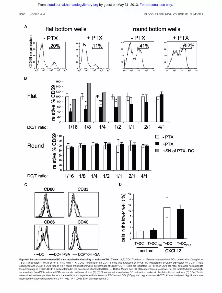
Figure 5. Pertussis toxin–treated DCs are impaired in the ability to activate CD4� T cells. (A,B) CD4�T cells (4 �104) were incubated with DCs, pulsed with 100 ng/mL ofTSST1, pretreated (�PTX) or not (�PTX) with PTX. CD69� expression on CD4� T cells was analyzed by FACS. (A) Histograms of CD69 expression on CD4� T cellscocultured with DCs at a DC/T ratio of 1:4 in round or flat-bottom wells; percentages of CD69�/CD4� T cells are indicated. (B) For each DC/T cell ratio, data were normalized tothe percentage of CD69�/CD4� T cells obtained in the cocultures of untreated DCs ( 100%). Means and SD of 3 experiments are shown. For the indicated ratio, overnightsupernatants from PTX-pretreated DCs were added to the cocultures (u) (C) Flow cytometric analysis of DC maturation markers in the flat-bottom cocultures. (D) CD4� T cellswere added to the upper chamber of a transwell system together with untreated or PTX-treated DCs (DCPTX) and migration toward CXCL12 was analyzed. Significance wasassessed by Student unpaired t test (*P � .05; **P � .005). Error bars represent SD.
3586 NOBILE et al BLOOD, 1 APRIL 2008 � VOLUME 111, NUMBER 7
For personal use only. by guest on May 31, 2013. bloodjournal.hematologylibrary.orgFrom

showing that chemokines produced during antigen-specific interac-tion of DCs with CD4� T cells specifically induced mobilityof immature DCs.
Time lapse and confocal microscopy images of DCs that havebeen interacting for 6 to 18 hours with T cells in the presence ofsuperantigens show striking changes of the DCs’ morphology,which correlated with the acquisition of their migratory ability. Inthese conditions, 25% to 30% of the immature DCs elongated,changing from a roundish shape of 10 to 15 �m in diameter to a“neuron-like” shape of 50 to 80 �m in length. These modificationswere also CC chemokine–dependent and were not observed whenimmature DCs were exposed to LPS or poly IC (data not shown)although these TLR ligands induced nonpolarized extension of thintransient dendrites around the cellular body of DCs. These elon-gated DCs established contact with both CD4� T cells and otherDCs (Figures 1,6,7) and tended to form a network of interdigitatingDCs after 4 to 6 hours of antigen-specific contact with T cells. Thischemokine-induced DC-DC interaction may favor the transfer ofmaterial between DCs, which has recently been shown to amplifyT cell responses.38
Morphologic changes observed in immature DCs were accom-panied by striking modifications of the actin cytoskeleton. Aspreviously reported,29,39 the majority of immature DCs displayed
big clusters of podosomes on their ventral face. When coculturedwith CD4� T cells and superantigen or with “active supernatants,”DCs showed actin filaments along the plasma membrane, as well aspolymerized actin-rich “cups” in the contact zones with T cells, butonly sparse podosomes on both ends (Figures 6,7). Podosomes arehighly dynamic structures consisting of a dense actin core sur-rounded by a ring of vinculin found on several cells from themyeloid lineage.40 These structures have been shown to beinvolved in cell migration, tissue invasiveness,41 and diapedesis.42
We show herein a strong inverse correlation between the presenceof podosomes and DC migration. Indeed, as reported by others,podosomes expressed by immature DCs may restrict their speed ofmigration by increasing their interaction with the substrate.25
In our study, we show for the first time that chemokines, whichinduce DC migration, also induce podosome disassembly. Severalmaturation-inducing agents such as LPS,29,43 PGE2, and TNF-�,25
have been shown to induce podosome dissolution, suggesting thatthis dissolution is an integral part of the DC maturation process.Results reported herein suggest that these 2 phenomena can bedissociated because PTX pretreatment of DCs blocks podosomedissolution but does not affect phenotypic DC maturation (Figure5C), whereas CC chemokines induce podosome dissolution with-out inducing DC maturation (Figure 7D).
Figure 6. Ag-specific interactions between CD4�
T cells and DCs induce podosome dissolution inDC. (A) Untreated or PTX-treated DCs were coculturedwith CD4�T cells on coverslips in the presence orabsence of TSST1. Cells were then fixed and labeledwith antivinculin Ab (green) and phalloidin (red). Bar, 10�m. (B) DC-presenting podosomes were quantified,plotted to the total number of cells present in the field,and presented as percentages. Means plus or minusSD of 4 independent experiments are shown; at least25 total cells were examined for each condition in eachexperiment. Significance was assessed by Studentunpaired t test (*P � .05; **P � .005).
T CELLS INDUCE DENDRITIC CELL MIGRATION 3587BLOOD, 1 APRIL 2008 � VOLUME 111, NUMBER 7
For personal use only. by guest on May 31, 2013. bloodjournal.hematologylibrary.orgFrom

Yet the resulting redistribution of the actin pool trapped inpodosomes toward different sites may be required for mature DCfunctions, such as the maturation of endosomes44 or the recruitmentof transmembrane receptors to the contact zone with interactingT lymphocytes.45
Chemokines can enhance and modulate T-cell activation byDCs in more than one fashion. DC presentation of cognate antigento CD4� T cells in lymph nodes induces the local production ofCCR5 ligands, which attract CD8� T cells nearby DC-CD4
conjugates, thus facilitating T-cell help.46 Chemokines alsomodulate T-cell response independently of their chemoattractantactivities47 (ie, they bind DC membranes inducing T-celladhesion48 and may induce DC maturation).26-28 Our datasuggest that chemokines produced during DC presentation ofcognate antigen to CD4� T cells may also increase T-cellresponse by modifying immature DCs.
Indeed, at low cell density PTX-pretreated DCs are less efficientthan untreated DCs at inducing T-cell activation (Figure 5). This
Figure 7. Chemokines induce podosome dissolution in DCs. (A) Untreated or PTX-treated DCs were exposed to the indicated supernatants, or to recombinantchemokines (CCL3, CCL4), and then labeled with antivinculin Ab (green) and phalloidin (red). Bar, 10 �m. (B,C) Cells presenting with podosomes were quantified as in Figure6. Means plus or minus SEM of 4 independent experiments are shown; at least 25 total cells were examined for each condition in each experiment. Significance was assessedby Student paired t test (*P � .05). (D) Flow cytometric analysis of DC maturation markers in response to chemokines.
3588 NOBILE et al BLOOD, 1 APRIL 2008 � VOLUME 111, NUMBER 7
For personal use only. by guest on May 31, 2013. bloodjournal.hematologylibrary.orgFrom

lower ability of PTX-treated DCs to induce T-cell activation maybe due to their inability to spread (Figures 6,7), thus offering lesssurface of contact with T lymphocytes. Alternatively, it may be dueto their defective mobility. This decreased activation of CD4�
T cells was not due to “bystander” effect of PTX released in themedium on T cells (Figure 5B,D). Moreover, it was not due to anabsence of secretion of chemokines known to attract T cells,because the same amount of CCL3 and CCL4 was present insupernatants from cocultures of PTX-treated DCs or untreated DCswith CD4� T cells and superantigen (data not shown). Finally, itwas not related to an inhibitory effect of PTX on DC maturationbecause PTX-treated DCs or untreated DCs cocultured with T cellsand superantigen show a comparable increased expression ofCD40, CD80, CD83, and CD86 (Figure 5C).
Altogether, our data suggest the following model: antigen-specific interaction between CD4� T cells and DCs induces thesecretion of chemokines by both cell types. This will attract moreimmature DCs, providing a source of “fresh” nonexhausted DCsthat still have the ability to capture antigen and produce IL-12.These findings may be physiologically relevant in different set-tings. First in inflammatory tissue, Ag-specific interactions ofmemory or effector T cells with DCs will induce production ofchemokines and cytokines that will attract and induce maturationof surrounding or circulating immature DCs. These newly recruitedDCs will capture the Ag (from the environment or from the matureDCs), migrate to the draining lymph node, and constitute a newpool of Ag-presenting DCs. Second, in the secondary lymphoidorgans, recruitment of some of the numerous resident DCs to sitesof antigen-specific DC–T cell interaction may be crucial for theinter-DC Ag transfer recently reported.38 This transfer of Ag toresident DCs would be induced by T-cell signals and accompaniedby a T cell–dependent maturation of these resident DCs that aremostly immature at the steady state.49 This would in turn increaseT-cell activation by amplifying Ag presentation through a largernetwork of resident DCs. Our data are compatible with such a
model because as shown herein, chemokines produced by antigen-specific DC-CD4� T cell conjugates induce mobility of DCs aswell as direct contact between DCs, potentially enabling transfer ofmaterial between DCs. Third, even in the absence of Ag transferbetween DCs, efficient self-presentation by newly recruited resi-dent DCs that will also receive T cell–derived activation signal mayincrease the activation of T cells responding to limiting amount offoreign antigen.50
We are currently developing in vivo models to test thesehypotheses.
Acknowledgments
We thank P. Guermonprez, C. Thery, A.M. Lennon and S. Huguesfor critical reading of the manuscript and/or discussion; F. Wahartefor assistance with microscopy imaging; and E. Labruyere (InstitutPasteur, Paris) for help with the Dunn Chamber assay.
This work was supported by grants from Institut Curie, Inserm,ARC (Association pour la Recherche contre le Cancer). C. N. is afellow of FRM (Fondation pour la Recherche Medicale).
Authorship
C.N. performed research, analyzed and interpreted data, anddrafted the manuscript; M.L., M.T., S.D., and F.M. performedresearch; K.C. analyzed data; G.O. contributed vital new reagentsor analytical tools; S.A. analyzed and interpreted data; and C.H.designed research, analyzed and interpreted data, and draftedthe manuscript.
Conflict-of-interest disclosure: The authors declare no compet-ing financial interests.
Correspondence: Dr Claire Hivroz, Inserm Unite 653, InstitutCurie, 26 Rue d’Ulm, 75248 Paris Cedex 05, France; e-mail:[email protected].
References
1. Banchereau J, Briere F, Caux C, et al. Immunobi-ology of dendritic cells. Annu Rev Immunol. 2000;18:767-811.
2. Albert ML, Jegathesan M, Darnell RB. Dendriticcell maturation is required for the cross-tolerization of CD8� T cells. Nat Immunol. 2001;2:1010-1017.
3. Alpan O, Bachelder E, Isil E, Arnheiter H, Matz-inger P. ‘Educated’ dendritic cells act as messen-gers from memory to naive T helper cells. NatImmunol. 2004;5:615-622.
4. Caux C, Massacrier C, Vanbervliet B, et al. Acti-vation of human dendritic cells through CD40cross-linking. J Exp Med. 1994;180:1263-1272.
5. Cella M, Scheidegger D, Palmer-Lehmann K,Lane P, Lanzavecchia A, Alber G. Ligation ofCD40 on dendritic cells triggers production ofhigh levels of interleukin-12 and enhances T cellstimulatory capacity: T-T help via APC activation.J Exp Med. 1996;184:747-752.
6. Bennett SR, Carbone FR, Karamalis F, FlavellRA, Miller JF, Heath WR. Help for cytotoxic-T-cellresponses is mediated by CD40 signalling. Na-ture. 1998;393:478-480.
7. Schoenberger SP, Toes RE, van der Voort EI, Of-fringa R, Melief CJ. T-cell help for cytotoxic T lym-phocytes is mediated by CD40-CD40L interac-tions. Nature. 1998;393:480-483.
8. Snijders A, Kalinski P, Hilkens CM, KapsenbergML. High-level IL-12 production by human den-dritic cells requires two signals. Int Immunol.1998;10:1593-1598.
9. Sporri R, Reis e Sousa C. Newly activated T cellspromote maturation of bystander dendritic cellsbut not IL-12 production. J Immunol. 2003;171:6406-6413.
10. Miro F, Nobile C, Blanchard N, et al. T Cell-dependent activation of dendritic cells requiresIL-12 and IFN-{gamma} signaling in T Cells. J Im-munol. 2006;177:3625-3634.
11. Smith CM, Wilson NS, Waithman J, et al. Cog-nate CD4(�) T cell licensing of dendritic cells inCD8(�) T cell immunity. Nat Immunol. 2004;5:1143-1148.
12. Randolph GJ, Angeli V, Swartz MA. Dendritic-celltrafficking to lymph nodes through lymphatic ves-sels. Nat Rev Immunol. 2005;5:617-628.
13. De Smedt T, Pajak B, Muraille E, et al. Regulationof dendritic cell numbers and maturation by lipo-polysaccharide in vivo. J Exp Med. 1996;184:1413-1424.
14. Robert C, Fuhlbrigge RC, Kieffer JD, et al. Inter-action of dendritic cells with skin endothelium: Anew perspective on immunosurveillance. J ExpMed. 1999;189:627-636.
15. Pendl GG, Robert C, Steinert M, et al. Immaturemouse dendritic cells enter inflamed tissue, a pro-cess that requires E- and P-selectin, but not P-selectin glycoprotein ligand 1. Blood. 2002;99:946-956.
16. Linder S, Hufner K, Wintergerst U, AepfelbacherM. Microtubule-dependent formation of podoso-mal adhesion structures in primary human macro-phages. J Cell Sci. 2000;113 Pt 23:4165-4176.
17. Zicha D, Dunn GA, Brown AF. A new direct-viewing chemotaxis chamber. J Cell Sci. 1991;99(Pt 4):769-775.
18. Benvenuti F, Lagaudriere-Gesbert C, GrandjeanI, et al. Dendritic cell maturation controls adhe-sion, synapse formation, and the duration of theinteractions with naive T lymphocytes. J Immunol.2004;172:292-301.
19. Sozzani S, Ghezzi S, Iannolo G, et al. Interleukin10 increases CCR5 expression and HIV infectionin human monocytes. J Exp Med. 1998;187:439-444.
20. Scandella E, Men Y, Legler DF, et al. CCL19/CCL21-triggered signal transduction and migra-tion of dendritic cells requires prostaglandin E2.Blood. 2004;103:1595-1601.
21. Alcami A, Symons JA, Collins PD, Williams TJ,Smith GL. Blockade of chemokine activity by asoluble chemokine binding protein from vacciniavirus. J Immunol. 1998;160:624-633.
22. Carfi A, Smith CA, Smolak PJ, McGrew J, WileyDC. Structure of a soluble secreted chemokineinhibitor vCCI (p35) from cowpox virus. Proc NatlAcad Sci U S A. 1999;96:12379-12383.
23. Dutt P, Wang JF, Groopman JE. Stromal cell-de-rived factor-1 alpha and stem cell factor/kit ligandshare signaling pathways in hemopoietic progeni-tors: a potential mechanism for cooperative in-duction of chemotaxis. J Immunol. 1998;161:3652-3658.
24. Calle Y, Burns S, Thrasher AJ, Jones GE. The
T CELLS INDUCE DENDRITIC CELL MIGRATION 3589BLOOD, 1 APRIL 2008 � VOLUME 111, NUMBER 7
For personal use only. by guest on May 31, 2013. bloodjournal.hematologylibrary.orgFrom

leukocyte podosome. Eur J Cell Biol. 2006;85:151-157.
25. van Helden SF, Krooshoop DJ, Broers KC, Ray-makers RA, Figdor CG, van Leeuwen FN. A criti-cal role for prostaglandin E2 in podosome disso-lution and induction of high-speed migrationduring dendritic cell maturation. J Immunol. 2006;177:1567-1574.
26. Trifilo MJ, Lane TE. The CC chemokine ligand 3regulates CD11c�CD11b�CD8alpha- dendriticcell maturation and activation following viral infec-tion of the central nervous system: implicationsfor a role in T cell activation. Virology. 2004;327:8-15.
27. Marsland BJ, Battig P, Bauer M, et al. CCL19 andCCL21 induce a potent proinflammatory differen-tiation program in licensed dendritic cells. Immu-nity. 2005;22:493-505.
28. Cappello P, Fraone T, Barberis L, et al. CC-chemokine ligand 16 induces a novel maturationprogram in human immature monocyte-deriveddendritic cells. J Immunol. 2006;177:6143-6151.
29. West MA, Wallin RP, Matthews SP, et al. En-hanced dendritic cell antigen capture via toll-likereceptor-induced actin remodeling. Science.2004;305:1153-1157.
30. Cyster JG. Chemokines and the homing of den-dritic cells to the T cell areas of lymphoid organs.J Exp Med. 1999;189:447-450.
31. Sallusto F, Lanzavecchia A. Understanding den-dritic cell and T-lymphocyte traffic through theanalysis of chemokine receptor expression. Im-munol Rev. 2000;177:134-140.
32. Behrens G, Li M, Smith CM, et al. Helper T cells,dendritic cells and CTL Immunity. Immunol CellBiol. 2004;82:84-90.
33. de Jong EC, Smits HH, Kapsenberg ML. Den-dritic cell-mediated T cell polarization. SpringerSemin Immunopathol. 2005;26:289-307.
34. Banchereau J, Steinman RM. Dendritic cells andthe control of immunity. Nature. 1998;392:245-252.
35. Sallusto F, Lanzavecchia A. Mobilizing dendriticcells for tolerance, priming, and chronic inflam-mation. J Exp Med. 1999;189:611-614.
36. Dieu MC, Vanbervliet B, Vicari A, et al. Selectiverecruitment of immature and mature dendriticcells by distinct chemokines expressed in differ-ent anatomic sites. J Exp Med. 1998;188:373-386.
37. Sallusto F, Schaerli P, Loetscher P, et al. Rapidand coordinated switch in chemokine receptorexpression during dendritic cell maturation. EurJ Immunol. 1998;28:2760-2769.
38. Allan RS, Waithman J, Bedoui S, et al. Migratorydendritic cells transfer antigen to a lymph node-resident dendritic cell population for efficient CTLpriming. Immunity. 2006;25:153-162.
39. Burns S, Thrasher AJ, Blundell MP, Machesky L,Jones GE. Configuration of human dendritic cellcytoskeleton by Rho GTPases, the WAS protein,and differentiation. Blood. 2001;98:1142-1149.
40. Marchisio PC, Cirillo D, Naldini L, Primavera MV,Teti A, Zambonin-Zallone A. Cell-substratum in-teraction of cultured avian osteoclasts is medi-ated by specific adhesion structures. J Cell Biol.1984;99:1696-1705.
41. Linder S, Aepfelbacher M. Podosomes: adhesionhot-spots of invasive cells. Trends Cell Biol. 2003;13:376-385.
42. Carman CV, Sage PT, Sciuto TE, et al. Transcel-
lular diapedesis is initiated by invasive podo-somes. Immunity. 2007;26:784-797.
43. Burns S, Hardy SJ, Buddle J, Yong KL, JonesGE, Thrasher AJ. Maturation of DC is associatedwith changes in motile characteristics and adher-ence. Cell Motil Cytoskeleton. 2004;57:118-132.
44. Boes M, Cuvillier A, Ploegh H. Membrane spe-cializations and endosome maturation in dendriticcells and B cells. Trends Cell Biol. 2004;14:175-183.
45. Vogt AB, Spindeldreher S, Kropshofer H. Cluster-ing of MHC-peptide complexes prior to their en-gagement in the immunological synapse: lipid raftand tetraspan microdomains. Immunol Rev.2002;189:136-151.
46. Castellino F, Huang AY, Altan-Bonnet G, Stoll S,Scheinecker C, Germain RN. Chemokines en-hance immunity by guiding naive CD8� T cells tosites of CD4� T cell-dendritic cell interaction. Na-ture. 2006;440:890-895.
47. Viola A, Contento RL, Molon B. T cells and theirpartners: The chemokine dating agency. TrendsImmunol. 2006;27:421-427.
48. Friedman RS, Jacobelli J, Krummel MF. Surface-bound chemokines capture and prime T cells forsynapse formation. Nat Immunol. 2006;7:1101-1108.
49. McIlroy D, Troadec C, Grassi F, et al. Investiga-tion of human spleen dendritic cell phenotype anddistribution reveals evidence of in vivo activationin a subset of organ donors. Blood. 2001;97:3470-3477.
50. Stefanova I, Dorfman JR, Germain RN. Self-recognition promotes the foreign antigen sensitiv-ity of naive T lymphocytes. Nature. 2002;420:429-434.
3590 NOBILE et al BLOOD, 1 APRIL 2008 � VOLUME 111, NUMBER 7
For personal use only. by guest on May 31, 2013. bloodjournal.hematologylibrary.orgFrom