A key role for poly(ADP-ribose) polymerase-1 activity during human dendritic cell maturation
9
of June 18, 2013. This information is current as Dendritic Cell Maturation Polymerase-1 Activity during Human A Key Role for Poly(ADP-Ribose) Alberto Chiarugi Lorenzo Borgognoni, Luca Massacesi, Flavio Moroni and Pimpinelli, Cipriani, Clara Ballerini, Tiziana Biagioli, Nicola Alessandra Aldinucci, Gianni Gerlini, Silvia Fossati, Giulia http://www.jimmunol.org/content/179/1/305 2007; 179:305-312; ; J Immunol References http://www.jimmunol.org/content/179/1/305.full#ref-list-1 , 21 of which you can access for free at: cites 66 articles This article Subscriptions http://jimmunol.org/subscriptions is online at: The Journal of Immunology Information about subscribing to Permissions http://www.aai.org/ji/copyright.html Submit copyright permission requests at: Email Alerts http://jimmunol.org/cgi/alerts/etoc Receive free email-alerts when new articles cite this article. Sign up at: Print ISSN: 0022-1767 Online ISSN: 1550-6606. Immunologists All rights reserved. Copyright © 2007 by The American Association of 9650 Rockville Pike, Bethesda, MD 20814-3994. The American Association of Immunologists, Inc., is published twice each month by The Journal of Immunology by guest on June 18, 2013 http://www.jimmunol.org/ Downloaded from
Transcript of A key role for poly(ADP-ribose) polymerase-1 activity during human dendritic cell maturation

of June 18, 2013.This information is current as
Dendritic Cell MaturationPolymerase-1 Activity during Human A Key Role for Poly(ADP-Ribose)
Alberto ChiarugiLorenzo Borgognoni, Luca Massacesi, Flavio Moroni and
Pimpinelli,Cipriani, Clara Ballerini, Tiziana Biagioli, Nicola Alessandra Aldinucci, Gianni Gerlini, Silvia Fossati, Giulia
http://www.jimmunol.org/content/179/1/3052007; 179:305-312; ;J Immunol
Referenceshttp://www.jimmunol.org/content/179/1/305.full#ref-list-1
, 21 of which you can access for free at: cites 66 articlesThis article
Subscriptionshttp://jimmunol.org/subscriptions
is online at: The Journal of ImmunologyInformation about subscribing to
Permissionshttp://www.aai.org/ji/copyright.htmlSubmit copyright permission requests at:
Email Alertshttp://jimmunol.org/cgi/alerts/etocReceive free email-alerts when new articles cite this article. Sign up at:
Print ISSN: 0022-1767 Online ISSN: 1550-6606. Immunologists All rights reserved.Copyright © 2007 by The American Association of9650 Rockville Pike, Bethesda, MD 20814-3994.The American Association of Immunologists, Inc.,
is published twice each month byThe Journal of Immunology
by guest on June 18, 2013http://w
ww
.jimm
unol.org/D
ownloaded from

A Key Role for Poly(ADP-Ribose) Polymerase-1 Activityduring Human Dendritic Cell Maturation1
Alessandra Aldinucci,† Gianni Gerlini,‡§ Silvia Fossati,* Giulia Cipriani,* Clara Ballerini,†
Tiziana Biagioli,† Nicola Pimpinelli,‡ Lorenzo Borgognoni,§ Luca Massacesi,† Flavio Moroni,*and Alberto Chiarugi2*
Poly(ADP-ribose) (PAR) polymerase (PARP)-1 is a nuclear enzyme regulating protein that functions by targeting PARchains. Besides its classic role in DNA repair, PARP-1 is emerging as a key transcriptional regulator in different cell typesincluding the immune ones. In this study, we investigated the role of PARP-1 in human dendritic cell (DC) function. Wereport that both PARP-1 mRNA and protein levels significantly increased during in vitro DC differentiation from monocytes.Of note, inhibitors of PARP-1 such as phenanthridinone and thieno[2,3-c]isoquinolin-5-one reduced expression of CD86 andCD83 in a concentration-dependent manner, having no effects on expression of CD80 and HLA-DR in mature DCs. In thesame cultures, PARP-1 inhibitors also reduced production of IL-12 and IL-10. Addition of exogenous IL-12 to the culturemedium partially restored CD86 expression in DCs exposed to PARP-1 inhibitors. In line with the role of PAR formation inNF-�B-dependent transactivation, we also report that phenanthridinone and thieno[2,3-c]isoquinolin-5-one impaired NF-�Band AP-1 subunit DNA binding activity in cellular extract of activated DCs. Finally, we show that PARP-1 inhibitors reducedthe T cell allostimulatory activity of mature DCs, and that this reduction was prevented when DCs matured in the presenceof PARP-1 inhibitors plus IL-12. Of note, nonproliferating T cells exposed to PARP-1 inhibitor-challenged DCs couldundergo efficient proliferation when exposed to a subsequent activation stimulus such as anti-CD3 plus anti-CD-28. Together,data provide evidence for a key role of PARP-1 and poly ADP-ribosylation in DC immunocompetence and underscore therelevance of PARP-1 inhibitors to treatment of immune disorders. The Journal of Immunology, 2007, 179: 305–312.
P oly ADP-ribosylation is a posttranslational modificationof proteins operated by poly(ADP-ribose) (PAR)3 poly-merases (PARPs), a growing family of enzymes forming
chains of PAR from NAD (1, 2). PARP-1 is the oldest and bestcharacterized member of the PARP family, accounting for �80%of cellular PAR formation and targeting the negatively chargedPAR polymers to numerous nuclear proteins, affecting their func-tioning through steric hindrance and electrostatic repulsion. PARin turn is very rapidly degraded by PAR glycohydrolase, therebyallowing fast and dynamic signaling (3).
Although originally thought to be only involved in DNA re-pair, mounting evidence now points to PARP-1 as a key regu-lator of gene transcription. In particular, numerous reports dem-onstrate that PARP-1 and PAR formation positively regulatetranscription in several types of immune cells (4 –7), as well asmacrophage activation (8, 9), granulocyte migration (10, 11),
and T cell proliferation (6, 12, 13). Also, PARP-1 inhibitionincreases Ab class-switching (14), and alters Ab productionduring the autoimmune response in the CNS (15). Additionalreports underscore the active role of poly ADP-ribosylation inimmune activation of microglia and astrocytes (5, 7, 16). Thesefindings taken together point to binding of PARP-1 and PARsynthesis at the levels of transcription regulating complexes asprerequisites for efficient expression of proinflammatory genesand immune cell activation (4, 17).
In the last several years, dendritic cells (DCs) emerged asprofessional APCs able to present antigenic peptides in the con-text of MHC molecules to lymphocytes (18, 19). Upon Ag in-teraction, a maturation program is triggered in DCs that migratefrom peripheral tissues into afferent lymphatics to reach fullactivation into regional lymph nodes (18). This process is ac-companied by immunophenotypic changes characterized by up-regulation of MHC class I-class II and costimulatory molecules(i.e., CD80 and CD86) as well as by neo-expression of CD83,a hallmark of DC maturation (19). Given the ability of DCs toinduce immunity or tolerance, mostly depending on their acti-vation profile, a great deal of efforts is now being directed to thepharmacologic manipulation of DCs to boost the immune re-sponse to external Ags or to promote T cell tolerance duringautoaggressive immune reactions (20). The ability of DCs toproperly present tumor Ags, thereby favoring tumor vaccinationand eradication, is also intensively investigated and exploitedfor antitumor therapy (21). Hence, in light of the key role ofPARP-1 in transcriptional and functional activation of immunecells, as well as the availability of powerful chemical inhibitorsof the enzyme, this study seeks to determine whether PARP-1regulates DC immunocompetence.
*Department of Pharmacology, †Department of Neurological and Psychiatric Sci-ences, and ‡Department of Dermatological Sciences, University of Florence, and§Plastic Surgery Unit, Melanoma Referral Center, Santa Maria Annunziata Hospital,Florence, Italy
Received for publication June 6, 2006. Accepted for publication April 26, 2007.
The costs of publication of this article were defrayed in part by the payment of pagecharges. This article must therefore be hereby marked advertisement in accordancewith 18 U.S.C. Section 1734 solely to indicate this fact.1 This work was supported by grants from Associazione Italiana Sclerosi Multipla,The Italian Ministry of Scientific Research, and Ente Cassa di Risparmio di Firenze.2 Address correspondence and reprint requests to Dr. Alberto Chiarugi, Department ofPharmacology, University of Florence, Viale Pieraccini 6, 50139 Firenze, Italy.E-mail address: [email protected] Abbreviations used in this paper: PAR, poly(ADP-ribose); PARP, PAR polymerase;DC, dendritic cell; PHE, phenanthridinone; TIQ-A, thieno[2,3-c]isoquinolin-5-one.
Copyright © 2007 by The American Association of Immunologists, Inc. 0022-1767/07/$2.00
The Journal of Immunology
www.jimmunol.org
by guest on June 18, 2013http://w
ww
.jimm
unol.org/D
ownloaded from
Materials and MethodsDC differentiation and stimulation
PBMC were isolated from buffy coats by density gradient centrifugationusing Lymphoprep (Axis-Shield). Monocytes were isolated from PBMCusing MACS anti-CD14 microbeads and a Midi-MACS device (both fromMiltenyi Biotec). Cells were cultured in medium supplemented with GM-CSF (1000 U/ml; Chemicon International) and IL-4 (1000 U/ml; R&DSystems) for 7 days. DC activation was induced by LPS (1 �g/ml/day;Sigma-Aldrich). After 7 days, monocyte-derived DCs have been monitoredfor DC maturation markers by flow cytometry. The PARP-1 inhibitorsphenanthridinone (PHE), thieno[2,3-c]isoquinolin-5-one (TIQ-A), and theinactive analog UPF698 (22) have been directly added to the culture me-dium, while controls received vehicle.
Surface markers
DC surface markers have been evaluated by means of mouse anti-human-conjugated mAb (Immunotech). Cells have been analyzed on a four-colorEpics XL cytometer (Expo32 software; Beckman Coulter). Abs used wereagainst the following: CD80 (FITC), CD86 (PE), HLA-DR (ECD), CD83(PC5), CD14 (FITC), CD11c (PE), CD3 (PE), and CD4 (FITC). Cell vi-tality has been tested by means of propidium iodide (Molecular Probes).Briefly, staining has been done in PBS 1% FCS for 25 min at 4°C followedby two washes. Later, cells were analyzed by flow cytometry.
Phagocytic activity
DC macropinocytosis capability was examined as described (23). Briefly,2 � 105 not activated and LPS-activated DCs were resuspended in 10%FCS RPMI 1640 medium and equilibrated at 37° or 0°C for 10 min. Cellswere then pulsed with dextran-FITC (1 mg/ml, 40,000 m.w.; MolecularProbes) for 45 min at 37° or 0°C. After incubation, cells were washed fourtimes with cold PBS buffer containing 0.01% sodium azide (Sigma-Aldrich)and 1% FCS and analyzed by flow cytometry using propidium iodide to ex-clude dead cells. Background is represented by cells incubated at 0°C.
Western blotting
For Western blot analysis, cells were scraped, collected in Eppendorf tubes,centrifuged (1500 � g for 5 min at 4°C) and resuspended in lysis buffer (50mM Tris (pH 7.4), 1 mM EDTA, 1 mM PMSF, 4 �g/ml aprotinin andleupeptin, 1% SDS). Then 20–40 �g of protein/lane were loaded. After4–20% SDS-PAGE and blotting, membranes (Hybond-ECL; AmershamBiosciences) were blocked with PBS containing 0.1% Tween 20 (PBS-T)and 5% skimmed milk (PBS-T/5% milk) and then probed overnight withprimary Abs (1/1000 in PBS-T/5% milk). The anti-PARP-1 mAb (cloneC2-10) was from Alexis. The anti-phospho-p38 was from Cell SignalingTechnology. Membranes were then washed with PBS-T and incubated 1 hin PBS-T/5% milk containing the corresponding peroxidase-conjugatedsecondary Ab (1/2000). After washing in TPBS, ECL (Amersham Bio-sciences) was used to visualize the peroxidase-coated bands.
Immunocytochemistry
DCs were spun on poly-lysine-coated slide coverglass and then fixed with4% paraformaldehyde. Cells were then permeabilized and blocked with10% horse serum diluted in PBS/0.3% Triton X-100. Later on slides wereincubated for 3 h with 2% horse serum in PBS/0.3% Triton X-100 con-taining an anti-PARP-1 Ab (clone C2-10; Alexis). After washing, bindingwas revealed with a Cy2-conjugated anti-mouse Ab (1/200). Cells werecounterstained with Hoechst 33258 (2 �g/ml/10 min) and then mountedand visualized using a Nikon TE-2000-U fluorescence microscope and aCCD camera equipped with Metafluor-Metamorph software.
Evaluation of NF-�B DNA binding activity
The DNA binding activity of NF-�B was investigated in cells scraped,pelleted, and resuspended in buffer A (containing 10 mM HEPES (pH 7.8),10 mM KCl, 1 mM EDTA, 0.1 mM EGTA, 1 mM PMSF, and 4 �g/mlaprotinin and leupeptin). Cells were kept on ice for 15 min, vortexed every3 min, and then centrifuged at 5000 � g for 5 min at 4°C. The nuclearpellet was resuspended in 50 �l of buffer B, analogous to buffer A plus 400mM NaCl, and incubated for 10 min on ice. The mixture was centrifugedat 14,000 � g for 10 min at 4°C, and the supernatant aliquoted and storedat �80°C. The DNA binding activity was tested by incubating 10 �g ofprotein of the nuclear extract in 20 �l of a buffer containing 10 mM Tris(pH 7.4), 4% glycerol, 1 mM MgCl2, 0.5 mM EDTA, 1 mM DTT, 50 mMNaCl, 0.05 mg/ml polyinosinic-polycytidylic acid, and 10,000 cpm of spe-cific 32P-labeled oligonucleotide for 20 min at room temperature. The mix-ture was electrophoresed in 6% nondenaturing polyacrylamide gels that,
after drying, was exposed to x-ray film (Amersham Biosciences). The dou-ble-stranded oligonucleotide 5�-AGTTGAGGGGACTTTCCCAGGC-3�for NF-�B was used. For supershift experiments, 2 �l of the Abs raisedagainst p65/RelA (Santa Cruz Biotechnology) were added to the bindingmixture during incubation. DNA binding activity of p65, p50, c-Rel, andc-Fos was quantified by means of an ELISA kit (Active Motif), accordingto the manufacturer’s instruction.
Semiquantitative RT-PCR
Total RNA (1 �g) extracted with TRIzol (Invitrogen Life Technologies) wasreverse transcribed into DNA and subjected to PCR using the following soft-ware-designed oligonucleotide primers: PARP-1, 5�-CTGACATAGAGAAAAGGCTGG-3� (sense) and 5�-TCCTGGAGAAGGAAAGCTCT-3� (anti-sense); and �-actin, 5�-GGGTCAGAAGGATTCCTATG-3� (sense) and 5�-GGTCTCAAACATGATCTGGG-3� (antisense). The number of PCR cycles(94°C at 30 s, 58°C at 30 s, 72°C at 1 min, and 5 min for the last extension)for the amplification of reverse transcribed products, selected after determiningthe linear working range for the reaction, was 24 (�-actin) and 31 (PARP-1).PCR amplification products were separated on a 1.8% agarose gel.
MLR analysis
MLR was performed in a 96-well U-bottom plate (Nunc). Three differentdilutions (104, 5 � 103, 5 � 102 cells/well) of LPS-activated DCs, exposedor not during maturation and activation to PARP-1 inhibitors (30 �M),were cultured 4 days with allogeneic CD4� T cells (105 cells/well). Ex-periments were conducted in quadruplicate. At day 5, the proliferativeresponse was measured by [3H]thymidine incorporation test. [3H]Thymi-dine (1 �Ci/well; Amersham Biosciences) was added and pulsed the last8 h. Plates were then harvested (Tomtec MacIII) on glass fiber filter
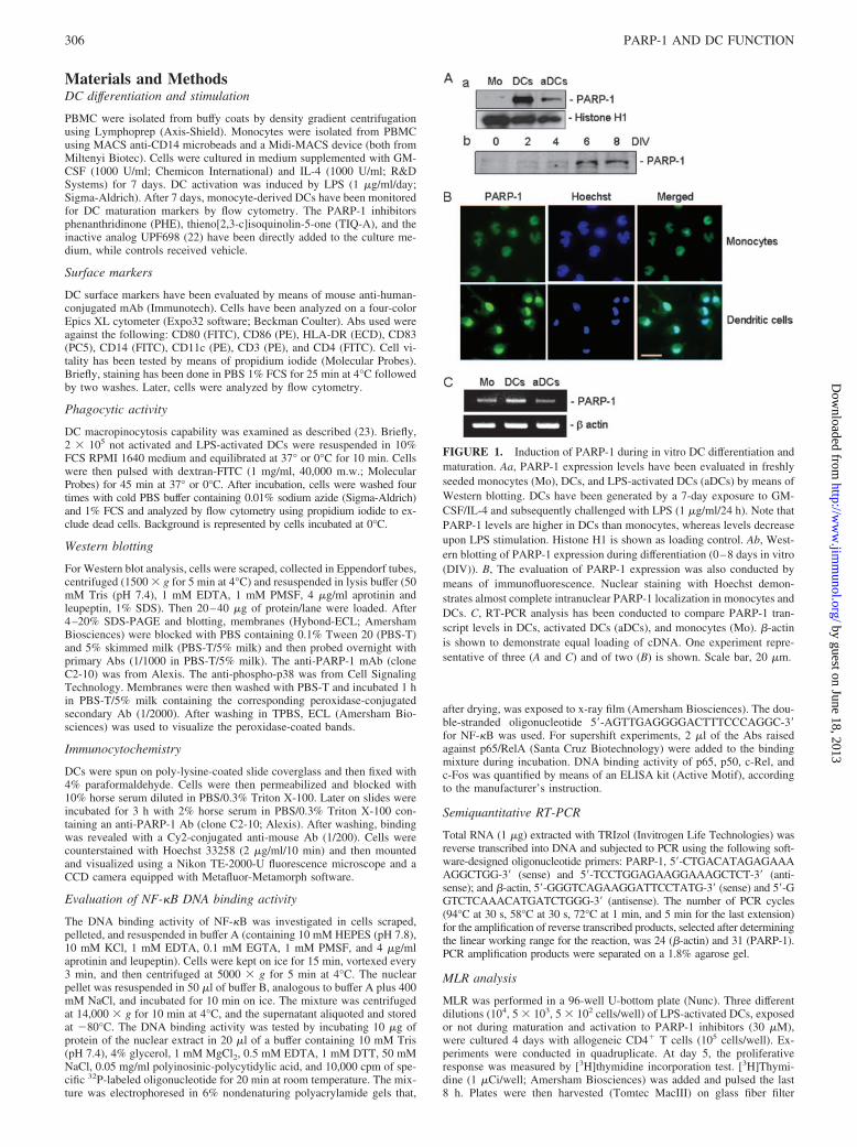
FIGURE 1. Induction of PARP-1 during in vitro DC differentiation andmaturation. Aa, PARP-1 expression levels have been evaluated in freshlyseeded monocytes (Mo), DCs, and LPS-activated DCs (aDCs) by means ofWestern blotting. DCs have been generated by a 7-day exposure to GM-CSF/IL-4 and subsequently challenged with LPS (1 �g/ml/24 h). Note thatPARP-1 levels are higher in DCs than monocytes, whereas levels decreaseupon LPS stimulation. Histone H1 is shown as loading control. Ab, West-ern blotting of PARP-1 expression during differentiation (0–8 days in vitro(DIV)). B, The evaluation of PARP-1 expression was also conducted bymeans of immunofluorescence. Nuclear staining with Hoechst demon-strates almost complete intranuclear PARP-1 localization in monocytes andDCs. C, RT-PCR analysis has been conducted to compare PARP-1 tran-script levels in DCs, activated DCs (aDCs), and monocytes (Mo). �-actinis shown to demonstrate equal loading of cDNA. One experiment repre-sentative of three (A and C) and of two (B) is shown. Scale bar, 20 �m.
306 PARP-1 AND DC FUNCTION
by guest on June 18, 2013http://w
ww
.jimm
unol.org/D
ownloaded from

(PerkinElmer), and thymidine uptake was measured by liquid scintillationin a MicroBeta 1450 Trimux counter (Wallac).
CD4� T cell separation
T cells were isolated from buffy coats of healthy donors with CD4� T cellisolation kit II (Miltenyi Biotec) and immediately used for MLR. Isolatedcells were checked for purity by flow cytometry, and purity level wasbetween 94 and 98%.
T cell restimulation
T cells were harvested at day 5 during MLR, washed three times in PBSbuffer, plated with fresh medium at 37°C for 24 h. Later, cells were countedand seeded at the density of 1 � 105/well in a 96-well flat-bottom platecoated with anti-CD3 (clone BB11; Euroclone) and anti-CD28 (clone BT3;Euroclone) mAbs (both at 1 �g/ml). Cell proliferation was evaluated by[3H]thymidine incorporation after 48 h. T cell viability was assessed bytrypan blue exclusion.
Cytokine measurement
Supernatants from LPS-activated (1 �g/ml/24 h) DCs were collected, andIL-12 and IL-10 were measured by means of an ELISA kit (BioSourceInternational).
ResultsPARP-1 expression during DC differentiation and maturation
We first sought to determine PARP-1 expression in in vitro-dif-ferentiated DCs. We found that levels of PARP-1 were higher inmonocyte-derived DCs exposed to GM-CSF/IL-4 for 7 days thanin freshly seeded monocytes. A 24-h challenge with LPS reducedPARP-1 expression in DCs (Fig. 1A, a). Time course analysisof PARP-1 expression during in vitro DC differentiationshowed that PARP-1 levels were barely detectable after 4 daysin vitro, although increased at 6 days and remained stable forthe following 2 days (Fig. 1A, b). Immunohistochemical anal-ysis confirmed these findings by showing a strong increase ofPARP-1 immunoreactivity in the nucleus of DCs with respect tothat found in the nucleus of monocytes (Fig. 1B). The findingthat PARP-1 expression levels changed during DC differentia-tion might be due to transcriptional and/or posttranscriptionalevents. We therefore determined whether PARP-1 transcriptlevels coherently changed with those of the protein. As shownin Fig. 1C, RT-PCR analysis showed that PARP-1 mRNA
FIGURE 2. Effect of PHE andTIQ-A on monocyte differentiationinto DCs. A, Evaluation of the effectof PHE and TIQ-A (both at 30 �M)on CD11c expression during mono-cyte differentiation into DCs (7 daysof exposure to GM-CSF/IL-4). B, Ef-fect of PHE and TIQ-A (both at 30�M) on CD14 expression duringmonocyte differentiation into DCs (7days of exposure to GM-CSF/IL-4).Note that neither compound affectsdown-regulation of CD14 nor thenumber of cells positive to propidiumiodide (PI). C, Phase contrast visual-ization of DCs at 7 days in vitroshows that PHE and TIQ-A (both at30 �M) did not change DC morphol-ogy. Scale bar, 20 �m. D, FACS anal-ysis shows that exposure to PHE andTIQ-A (both at 30 �M) reduces thepercentage of cells expressing CD86without affecting expression of cellspositive for HLA-DR, CD80, andCD83. �, p � 0.05 vs vehicle-treatedcontrol, determined by Student’s ttest. E, Evaluation of the effect ofPHE and TIQ-A on phagocytic activ-ity (see Materials and Methods) ofimmature DCs. One experiment rep-resentative of three is shown, exceptin D where data represent the mean �SEM of three experiments conductedin duplicate.
307The Journal of Immunology
by guest on June 18, 2013http://w
ww
.jimm
unol.org/D
ownloaded from

increased in DCs compared with monocytes and decreased uponLPS stimulation.
Effects of PARP-1 inhibitors on DC differentiation
Given the augmented expression of PARP-1 in DCs, we next won-dered whether the enzyme plays any role in monocyte differenti-ation into DCs. To address this issue, monocytes were exposed topotent inhibitors of PARP-1 activity such as PHE (24) or TIQ-A(22) at 30 �M, a concentration consistent with their IC50 onPARP-1 (22). Exposure to the two drugs did not affect the numberof CD11c� DCs at the end of the differentiation period (Fig. 2A).As shown in Fig. 2, A and B, these compounds neither increasedthe number of propidium iodide-positive cells (a prototypicalmarker of cell death), nor affected GM-CSF/IL-4-induced loss ofCD14 in DCs, a classic event during differentiation from mono-cytes. Accordingly, DCs exposed to PARP-1 inhibitors for 7 daysdid not differ morphologically from vehicle-treated DCs (Fig. 2Cand data not shown). On the contrary, PHE or TIQ-A selectivelyaltered membrane Ag expression. Specifically, the drugs reducedthe number of cells expressing CD86, although they did not affectthat of HLA-DR and CD80 (Fig. 2D). As expected, CD83 was nothighly expressed in immature DCs (Fig. 2D). Finally, chemicalinhibition of PARP-1 did not affect phagocytic activity of imma-ture DCs (Fig. 2E).
Effects of PARP-1 inhibitors on DC maturation
We then investigated the effect of pharmacologic inhibition ofPARP-1 on the immunophenotype of LPS-matured DCs. Upon24-h exposure to LPS (1 �g/ml), the number of CD86� cells in-creased from 28 � 3% to 78 � 13%. Similarly, that of CD83�
cells increased from 1 � 0.5% to 44 � 12% and that of cellsexpressing CD80 raised from 66 � 11% to 88 � 9%. Notably,exposure (8 days) to PHE and TIQ-A reduced the number of cellsexpressing CD86 (to 41 � 16% and 49 � 17%, respectively, p �0.05, determined using Student’s t test) and CD83 (to 28 � 13%and 24 � 12%, respectively, p � 0.05, Student’s t test) (Fig. 3).PHE and TIQ-A did not significantly change the expression ofHLA-DR and CD80 by mature DCs (Fig. 3). Phagocytic activity ofmature DCs was reduced compared with that of nonmature DCs,but again it was not affected by exposure to PARP-1 inhibitors(Fig. 3E).
The effect of PARP inhibitors on CD86 and CD83 expressionwas dose-dependent (Fig. 4A), whereas UPF698 (3,4-dihydro-5-benzoyl-isochinolin-2(1H)-one), an inactive PARP-1 inhibitorstructurally similar to PHE and TIQ-A (22), had no effect on theimmunophenotype (Fig. 4B). Exposure of DCs to PARP-1 inhib-itors only during the 24-h LPS challenge did not alter the LPS-induced expression of CD86, CD83, and CD80 (data not shown).
PARP-1 inhibitors reduce IL-12 and IL-10 cytokine productionby activated DCs with implication on the immunophenotype
We also sought to determine whether PHE and TIQ-A had anyeffect on IL-12 and IL-10, key cytokines produced by DCs. As
FIGURE 3. PHE and TIQ-A selectively decrease expression of CD86and CD83 in activated DCs. A–D, FACS analysis of the effect of PHE andTIQ-A (both at 30 �M) during DC differentiation and LPS activation onthe number of CD86-, CD83-, CD80-, and HLA-DR-positive DCs exposedto LPS (1 �g/ml/24 h). E, Evaluation of the effect of PHE and TIQ-A onphagocytic activity (see Materials and Methods) of mature DCs. Chemicalinhibition of PARP-1 does not affect phagocytic activity (see Materials andMethods). Results shown are representative experiments (see Results fordetails).
FIGURE 4. PARP-1 inhibition underlies the effects of PHE and TIQ-Aon activated DCs. A, PARP-1 inhibitors concentration-dependently (10–30�M) reduce expression of CD86 and CD83. B, PHE but not its inactiveanalog UPF698 (see Results for details) reduces the number of CD86- andCD83-positive LPS-activated DCs. Cells were exposed to drugs duringdifferentiation and activation. In A, one representative experiment of two isshown. In B, data represent the mean � SEM of two experiments con-ducted in duplicate. �, p � 0.05 vs vehicle-treated control, determinedusing Student’s t test.
308 PARP-1 AND DC FUNCTION
by guest on June 18, 2013http://w
ww
.jimm
unol.org/D
ownloaded from

shown in Fig. 5A, exposure to PARP-1 inhibitors during LPS stim-ulation significantly reduced IL-12 and IL-10 production by DCs.
It is well appreciated that autocrine and paracrine stimulation byproinflammatory cytokines including IL-12 concur to DC matura-tion (18, 20). We therefore investigated whether reduction of IL-12production by PARP-1 inhibitors was responsible for decreasedCD86 and CD83 expression. To this end, we exposed DC to LPSand PARP-1 inhibitors in the presence or absence of IL-12 directlyadded to the medium (5 ng/ml). Interestingly, addition of IL-12counteracted the effect of PARP-1 inhibitors on CD86 expression,while having no effect on expression of CD83, CD80, andHLA-DR (Fig. 5B).
Effect of PARP-1 inhibition on p38 kinase activation and DNAbinding activity of NF-�B and AP-1 subunits in mature DCs
Several reports point to p38 kinase as a key factor in DC differ-entiation and maturation. Notably, the immunophenotype of DCsexposed to a pharmacological inhibitor of p38 resembles that in-duced by PARP-1-inhibiting drugs (25–27). Under our experimen-tal settings, however, p38 was constitutively active and neitherLPS nor the PARP-1 inhibitors altered p38 activation levels (Fig.6A). A large body of evidence indicates that suppression ofPARP-1 activity reduces the inflammatory response because ofimpairment of transcription factor function involved in immuneactivation such as NF-�B and AP-1 (17, 28, 29). Accordingly,PARP-1 and PAR regulate NF-�B-dependent transactivation (30),whereas pharmacologic or genetic suppression of PARP-1 reducesNF-�B and AP-1 DNA binding activity and ensuing expression ofproinflammatory mediators (5–7, 9, 16, 31). We therefore deter-
mined the effect of PARP-1 inhibition on the DNA binding activityof NF-�B and AP-1 subunits in DCs. Constitutive binding ofNF-�B (p65 subunit) was present in extracts of immature DCs, andreduced by competitor cold oligoprobe (data not shown), indicat-ing specificity of binding. Pharmacological inhibition of PARP-1by PHE and TIQ-A did not alter the constitutive binding activity,although reduced that induced by LPS (1 h, 1 �g/ml) (Fig. 6B, a,and data not shown). The DNA binding activity of NF-�B andAP-1 subunits was also analyzed by means of ELISA. Data shownin Fig. 6B, b, confirm the findings on p65 obtained with the gel-shift assay. They also indicate that the binding activity of NF-�Bsubunit p50 but not that of c-Rel is reduced by chemicals inhibitingPARP-1, in line with previous findings with purified proteins (32).Likewise, the DNA binding activity of AP-1 subunit c-Fos wasdecreased in extracts of DCs challenged with PHE and TIQ-A(Fig. 6B, b, and data not shown).
Inhibitors of PARP-1 reduce allostimulatory capacity of DCs
Given the relevance of membrane Ags such as CD86 and CD83 toDC immunocompetence (33, 34), we then sought to determinewhether inhibition of PARP-1 in DCs impaired their allostimula-tory capacity. To this end, we measured proliferation of lympho-cytes exposed to different concentrations of mature DCs previouslyexposed or not to PHE or TIQ-A (see Materials and Methods). Inline with the immunophenotype studies, allogenic proliferationwas reduced when lymphocytes were mixed with DCs previouslychallenged with PARP-1 inhibitors (Fig. 6C, a). Given that IL-12
FIGURE 5. PARP-1 inhibitors reduce cyto-kine production by activated DCs. Effects on theimmunophenotype. A, Evaluation of the effect ofPHE and TIQ-A (both at 30 �M during differ-entiation and activation) on production of IL-12(Aa) and (Ab) IL-10 by mature DCs. B, Effect ofIL-12 (added to the medium at 5 ng/ml) on theexpression of CD86, CD83, CD80, andHLA-DR in DCs exposed to PHE and TIQ-A.Data represent the mean � SEM of three exper-iments conducted in duplicate. �, p � 0.05;��, p � 0.01 vs vehicle-treated control, usingStudent’s t test.
309The Journal of Immunology
by guest on June 18, 2013http://w
ww
.jimm
unol.org/D
ownloaded from

partially counteracted the effects of PARP-1 inhibitors on costimu-latory molecule expression (Fig. 5B), we analyzed the allostimu-latory activity of DCs exposed to PARP-1 inhibitors plus IL-12.Interestingly, the presence of IL-12 completely restored the abilityof PARP-1 inhibitor-exposed DCs to prompt T cell proliferation(Fig. 6C, a). To understand whether the T cells that did not pro-liferate could undergo efficient proliferation upon exposure to asubsequent stimulation, we re-exposed T cells to anti-CD3 plusanti-CD28 (see Materials and Methods). As shown in Fig. 6C, b,under these experimental conditions T cells underwent efficientproliferation regardless of a previous exposure to DCs matured inthe presence of PHE or TIQ-A (Fig. 6C, b).
DiscussionDuring the last several years a great deal of effort has been directedat promoting or repressing DC immunocompetence. A deeper in-sight in DC biology allowed designing of pharmacological strat-egies able to target functional checkpoints of DCs, such as differ-entiation, Ag uptake, and processing, migration, and maturation(20). In the present study, we show that PARP-1 expression isinduced during DC differentiation in vitro, whereas chemicals in-hibiting PARP-1 activity alter specific parameters of phenotypicand functional DC maturation. To our knowledge, this evidence isthe first that formation of PAR is a prerequisite for efficient stim-ulatory activity by DCs.
Our data are consistent with numerous reports indicatingPARP-1 as a key regulator of the immune response (4, 17). Spe-
cifically, the enzyme positively regulates activation of macro-phages (8, 9, 35), microglia (7, 36), granulocytes (37), lympho-cytes (6, 13, 14), as well as TNF-�-challenged endothelial cells(38). Also, parp-1 null mice are resistant to endotoxic shock (39),and PARP-1 inhibitors provide protection in models of pleuritis(40), arthritis (41), asthma (42), colitis (43, 44), allergic enceph-alomyelitis (6, 15, 45), and other autoimmune disorders (46). Inthis context, the present findings point to the negative regulation ofDCs by PARP-1 inhibitors as a key mechanism through which thisclass of drugs suppresses the inflammatory response. As for themolecular mechanisms though which PARP-1 inhibition impairsDC immunocompetence, we show that its enzymatic inhibitorsreduce NF-�B and AP-1 subunit DNA binding activity in matureDCs. Evidence that binding of c-Rel NF-�B subunit to its recog-nizing oligoprobe is not affected by chemicals inhibiting PARP-1indicates that the enzyme specifically regulates transcription factorDNA binding. Data are consistent with the regulation of NF-�B- andAP-1-dependent transactivation by PAR in macrophages and lympho-cytes (5–7, 36, 39, 47), as well as with the key role of these transcrip-tion factors in DC maturation and Ag presentation (48–51).
We originally report that PARP-1 mRNA and protein levelssignificantly increase during in vitro DC differentiation. Intrigu-ingly, a 10-fold increase in PARP-1 transcript levels has been re-ported during human lymphocyte activation (52), and a role forPARP-1 activity in cell differentiation has been proposed (53). Todate, factors regulating parp-1 expression at the promoter levelstill wait to be clearly understood. Different binding sites for YY1
FIGURE 6. Effect of PARP-1 inhibitorson p38 activation, NF-�B and AP-1 DNAbinding activity and T cell allostimulation byactivated DCs. A, Western blot analysis ofp38 phosphorylation under constitutive (C)condition and after LPS exposure with orwithout PARP-1 inhibitors. The constitutivephosphorylation levels are not affected bytreatments. Ba, Gels-shift analysis revealsthat LPS treatment (1 �g/ml/1 h) highly in-creases the constitutive DNA binding activ-ity of NF-�B subunit p65 in DC extracts. Incells exposed to PHE (30 �M, during differ-entiation and activation) the LPS-inducedbinding activity is reduced. Bb, ELISA eval-uation of the effect of PHE on the DNAbinding activity of p65, p50, c-Rel, and c-Fos under resting and LPS-activation condi-tions. Ca, MLR reveals that the T cell allo-stimulatory activity of activated DCs (5000or 500 DCs/well) exposed to PHE or TIQ-A(both 30 �M, during differentiation and ac-tivation) is reduced compared with activityin vehicle-treated controls. The presence ofIL-12 during maturation of DCs challengedwith PARP-1 inhibitors fully restores theirallostimulatory activity. Cb, T cells prolifer-ating after exposure to DCs and T cells thatdid not proliferate because of exposure toPHE- or TIQ-A-challenged DCs have beenexposed to the proliferative stimulus anti-CD3 plus anti-CD28 (see Materials andMethods). Proliferation does not differamong these T cell populations. One exper-iment representative of two is shown in Aand Ba. Data in Bb and C represent themean � SEM of at least three experimentsconducted in triplicate.
310 PARP-1 AND DC FUNCTION
by guest on June 18, 2013http://w
ww
.jimm
unol.org/D
ownloaded from

and Sp1 transcription factors, as well as multiple transcription ini-tiation sites are present in the parp-1 promoter region (54, 55).Genetic variants within this region may therefore affect PARP-1expression and inducibility. In this context, it is puzzling that spe-cific haplotypes of parp-1 promoter confer higher risk of autoim-mune disorders such as rheumatoid arthritis (56), celiac disease(57), and systemic lupus erythematosus (58). It is therefore tempt-ing to speculate that induction of PARP-1 in DCs and/or otherimmune cells participates in development of autoimmunity. As forthe decreased levels of PARP-1 in DCs exposed to LPS, we hy-pothesize that it could be due to as-yet unknown LPS-dependentspecific signaling and/or PARP-1-dependent autorepression of itsown promoter (55). Overall, these findings, along with evidencethat PARP-1 activity is increased in IFN-�-activated macrophages(8) and LPS-challenged microglia (7), suggest that induction ofpoly ADP-ribosylation plays a role in immune cell activation. Thisassumption is corroborated by our finding that chemical inhibitionof PARP-1 affects the immunophenotype of DCs. In particular, wereport that PARP-1 inhibitors reduce expression of CD86 andCD83, whereas they do not affect expression of HLA-DR andCD80 in activated DCs (Fig. 3). Data therefore suggest that polyADP-ribosylation is not a general regulator of transcription in DCsbut assists expression of specific genes. Remarkably, down-regu-lation of the immunomodulatory molecules CD86 and CD83 is inkeeping with the reduced proliferation of lymphocytes culturedwith DCs pre-exposed to PARP-1 inhibitors (Fig. 6).
The finding that PARP-1 inhibitors reduce production of IL-12and IL-10 by DCs indicates that poly ADP-ribosylation modulatesnot only the phenotypic but also the functional maturation of DCs.Interestingly, we show that addition of IL-12 restores CD86 ex-pression (Fig. 5B, b) as well as allostimulatory capacity (Fig. 6B,a) of DCs exposed to PARP-1 inhibitors. These results suggest thatreduction of DC immunocompetence by drugs inhibiting PARP-1is mainly due to reduced production of IL-12 and ensuing sup-pression of its autocrine/paracrine actions. Although c-Rel regu-lates IL-12 expression in DCs (59), results shown in Fig. 6B, b,suggest that impairment of its DNA binding activity does not un-derlie down-regulation of the cytokine by PARP-1 inhibitors. Con-versely, in keeping with the role of both p65 and p50 in IL-12 genetransactivation (60–62), our data point to the impairment of thesetwo NF-�B subunits as a mechanisms contributing to reduction ofIL-12. It is worth noting that classic anti-inflammatory drugs in-cluding corticosteroids reduce release of activating cytokines,whereas increase release of immunosuppressive cytokines fromDCs (20). The ability of PHE and TIQ-A to suppress production ofboth IL-12 and IL-10, therefore, points to PARP-1 and its inhib-itors as peculiar immunoregulators. On the contrary, PARP-1 in-hibitors share with well-known immunosuppressive compoundssuch as cyclosporine, FK506, and rapamycin the capability of re-ducing expression of only specific markers of DC maturation (20).Our data suggest that pharmacologic inhibition of PARP-1 mayrepresent a new strategy to generate tolerogenic DCs. Accordingly,we show that PHE and TIQ-A reduce the T cell allostimulatoryactivity of DCs. However, we also report that T cells that did notproliferate underwent efficient proliferation once exposed to anti-CD3 plus anti-CD28. These findings are in keeping with evidencethat DCs exposed to an inhibitor of NF-�B do not anergize T cells(63). Of note, in light of the ability of IL-10 to trigger long-lastingT cell anergy (64), the reduced production of this cytokine by DCsexposed to PARP-1 inhibitors (Fig. 5A, b) might contribute, atleast in part, to T cell proliferation upon exposure to anti-CD3 plusanti-CD28. Although it is known that PARP-1 inhibition represseslymphocyte proliferation (13), the fact that DCs were exposed tothe chemicals only during maturation, and that IL-12 restored their
stimulatory activity, rules out the possibility that suppression of Tcell proliferation occurred because of DC-dependent “drug-car-ryover.” Hence, data lend support to the hypothesis that PARP-1inhibitors compromise the T cell stimulatory capacity of DCs. Thepresent findings, along with evidence that suppression of PAR for-mation also affects immunocompetence of T and B lymphocytes(6, 13, 14), help to explain why inhibitors of PARP-1 exert wide-spread immunosuppression (65). Finally, although the effects ofPARP-1 inhibitors on DCs in vivo remains to be evaluated, weinfer that these compounds, currently tested in different clinicaltrials in humans (66), can be harnessed to develop innovative ther-apies aimed at promoting graft survival or combating autoimmunedisorders.
DisclosuresThe authors have no financial conflict of interest.
References1. D’Amours, D., S. Desnoyers, I. D’Silva, and G. G. Poirier. 1999. Poly(ADP-
ribosyl)ation reactions in the regulation of nuclear functions. Biochem. J. 342:249–268.
2. Ame, J.-C., C. Spenlehauer, and G. de Murcia. 2004. The PARP superfamily.Bioessays 26: 882–893.
3. Davidovich, L., M. Vodenicharov, E. B. Affar, and G. G. Poirier. 2001. Impor-tance of poly(ADP-ribose) glycohydrolase in the control of poly(ADP-ribose)metabolism. Exp. Cell Res. 268: 7–13.
4. Szabo, C. 1998. Role of poly(ADP-ribose) synthetase in inflammation. Eur.J. Pharmacol. 350: 1–19.
5. Ha, H. C., L. D. Hester, and S. H. Snyder. 2002. Poly(ADP-ribose) polymerase-1dependence of stress-induced transcription factors and associated gene expressionin glia. Proc. Natl. Acad. Sci. USA 99: 3270–3275.
6. Chiarugi, A. 2002. Inhibitors of poly(ADP-ribose) polymerase-1 suppress tran-scriptional activation in lymphocytes and ameliorate autoimmune encephalomy-elitis in rats. Br. J. Pharmacol. 137: 761–770.
7. Chiarugi, A., and M. A. Moskowitz. 2003. Poly(ADP-ribose) polymerase-1 ac-tivity promotes NF-�B-driven transcription and microglial activation: implicationfor neurodegenerative disorders. J. Neurochem. 85: 306–317.
8. Berton, G., C. Sorio, C. Laudanna, M. Menegazzi, A. Carcereri De Prati, andH. Suzuki. 1991. Activation of human-derived macrophages by interferon-y isaccompanied by increase of poly(ADP-ribose) polymerase activity. Biochim.Biophys. Acta 1091: 101–109.
9. Le Page, C., J. Sanceau, J. C. Drapier, and J. Wietzerbin. 1998. Inhibitors ofADP-ribosylation impair inducible nitric oxide synthase gene transcriptionthrough inhibition of NF-kB activation. Biochem. Biophys. Res. Commun. 243:451–457.
10. Ducrocq, S., N. Benjelloun, M. Plotkine, Y. Ben-Ari, and C. Charriaut-Marlangue.2000. Poly(ADP-ribose) synthase inhibition reduces ischemic injury and inflamma-tion in neonatal rat brain. J. Neurochem. 74: 2504–2511.
11. Mazzon, E., L. Dugo, S. A. De, J. H. Li, A. P. Caputi, J. Zhang, and S. Cuzzocrea.2002. Beneficial effects of GPI 6150, an inhibitor of poly(ADP-ribose) polymer-ase in a rat model of splanchnic artery occlusion and reperfusion. Shock 17:222–227.
12. King, S. L., R. McNerney, G. S. Whitley, and A. P. Johnstone. 1989. Differentialrole for ADP-ribosylation in gene expression during the activation of T lympho-cytes by various stimuli. Immunology 67: 258–262.
13. Weltin, D., K. Picard, K. Aupeix, M. Varin, D. Oth, J. Marchal, P. Dufour, andP. Bichoff. 1995. Immunosuppressive activities of 6(5H)-phenanthridinone, anew poly(ADP-ribose) polymerase inhibitor. Int. J. Immunopharmacol. 17:265–271.
14. Shockett, P., and J. Stavnezer. 1993. Inhibitors of poly(ADP-ribose) polymeraseincrease antibody class switching. J. Immunol. 151: 6962–6976.
15. Scott, G. S., R. B. Kean, T. Mikheeva, M. J. Fabis, J. G. Mabley, C. Szabo, andD. C. Hooper. 2004. The therapeutic effects of PJ34 [N-(6-oxo-5,6-dihydro-phenanthridin-2-yl)-N,N-dimethylacetamide.HCl], a selective inhibitor of poly-(ADP-ribose) polymerase, in experimental allergic encephalomyelitis are asso-ciated with immunomodulation. J. Pharmacol. Exp. Ther. 310: 1053–1061.
16. Ullrich, O., A. Diestel, I. Y. Eyupoglu, and R. Nitsch. 2001. Regulation of mi-croglial expression of integrins by poly(ADP-ribose) polymerase-1. Nat. CellBiol. 3: 1035–1042.
17. Szabo, C., and V. L. Dawson. 1998. Role of poly(ADP-ribose) synthetase ininflammation and ischaemia-reperfusion. Trends Pharmacol. Sci. 19: 287–298.
18. Rossi, M., and J. W. Young. 2005. Human dendritic cells: potent antigen-pre-senting cells at the crossroads of innate and adaptive immunity. J. Immunol. 175:1373–1381.
19. Banchereau, J., and R. M. Steinman. 1998. Dendritic cells and the control ofimmunity. Nature 392: 245–252.
20. Hackstein, H., and A. W. Thomson. 2004. Dendritic cells: emerging pharmaco-logical targets of immunosuppressive drugs. Nat. Rev. Immunol. 4: 24–34.
21. Banchereau, J., and A. K. Palucka. 2005. Dendritic cells as therapeutic vaccinesagainst cancer. Nat. Rev. Immunol. 5: 296–306.
311The Journal of Immunology
by guest on June 18, 2013http://w
ww
.jimm
unol.org/D
ownloaded from
22. Chiarugi, A., E. Meli, M. Calvani, R. Picca, R. Baronti, E. Camaioni,G. Costantino, M. Marinozzi, D. E. Pellegrini-Giampietro, R. Pellicciari, andF. Moroni. 2003. Novel isoquinolinone-derived inhibitors of poly(ADP-ribose)polymerase-1: pharmacological characterization and neuroprotective effects in anin vitro model of cerebral ischemia. J. Pharmacol. Exp. Ther. 305: 943–949.
23. Sallusto, F., M. Cella, C. Danieli, and A. Lanzavecchia. 1995. Dendritic cells usemacropinocytosis and the mannose receptor to concentrate macromolecules in themajor histocompatibility complex class II compartment: downregulation by cy-tokines and bacterial products. J. Exp. Med. 182: 389–400.
24. Banasik, M., H. Komura, M. Shimoyama, and K. Ueda. 1992. Specific inhibitorsof poly(ADP-ribose) synthetase and mono(ADP-ribosyl)transferase. J. Biol.Chem. 267: 1569–1575.
25. Arrighi, J. F., M. Rebsamen, F. Rousset, V. Kindler, and C. Hauser. 2001. Acritical role for p38 mitogen-activated protein kinase in the maturation of humanblood-derived dendritic cells induced by lipopolysaccharide, TNF-�, and contactsensitizers. J. Immunol. 166: 3837–3845.
26. Uchi, H., J. F. Arrighi, J. P. Aubry, M. Furue, and C. Hauser. 2002. The ses-quiterpene lactone parthenolide inhibits LPS- but not TNF-�-induced maturationof human monocyte-derived dendritic cells by inhibition of the p38 mitogen-activated protein kinase pathway. J. Allergy Clin. Immunol. 110: 269–276.
27. Nakahara, T., Y. Moroi, H. Uchi, and M. Furue. 2006. Differential role of MAPKsignaling in human dendritic cell maturation and Th1/Th2 engagement.J. Dermatol. Sci. 42: 1–11.
28. Chiarugi, A. 2002. PARP-1: killer or conspirator? The suicide hypothesis revis-ited. Trends Pharmacol. Sci. 23: 122–129.
29. Jagtap, P., and C. Szabo. 2005. Poly(ADP-ribose) polymerase and the therapeuticeffects of its inhibitors. Nat. Rev. Drug Discov. 4: 421–440.
30. Hassa, P. O., and M. O. Hottiger. 2002. The functional role of poly(ADP-ribose)polymerase 1 as novel coactivator of NF-�B in inflammatory disorders. Cell Mol.Life Sci. 59: 1534–1553.
31. Andreone, T. L., M. O’Connor, A. Denenberg, P. W. Hake, and B. Zingarelli.2003. Poly(ADP-ribose) polymerase-1 regulates activation of activator protein-1in murine fibroblasts. J. Immunol. 170: 2113–2120.
32. Chang, W.-J., and R. Alvarez-Gonzalez. 2001. The sequence-specific DNA bind-ing of NF-�B is reversibly regulated by automodification-reaction of poly(ADP-ribose) polymerase-1. J. Biol. Chem. 276: 47664–47670.
33. Lechmann, M., E. Zinser, A. Golka, and A. Steinkasserer. 2002. Role of CD83in the immunomodulation of dendritic cells. Int. Arch. Allergy Immunol. 129:113–118.
34. Sansom, D. M., C. N. Manzotti, and Y. Zheng. 2003. What’s the differencebetween CD80 and CD86? Trends Immunol. 24: 314–319.
35. Rapizzi, E., S. Fossati, F. Moroni, and A. Chiarugi. 2004. Inhibition of poly-(ADP-ribose) glycohydrolase by gallotannin selectively up-regulates expressionof proinflammatory genes. Mol. Pharmacol. 66: 890–898.
36. Ullrich, O., A. Diestel, I. Bechmann, M. Homberg, T. Grune, R. Hass, andR. Nitsch. 2001. Turnover of oxidatively damaged nuclear proteins in BV-2 mi-croglial cells is linked to their activation state by poly-ADP-ribose polymerase.FASEB J. 15: 1460–1462.
37. Szabo, C., Lim, L. H. R., Cuzzocrea, S., Getting, S. J., Zingarelli, B., Flower,R. J., Salzman, A. L., and Perretti, M. 1997. Inhibition of poly(ADP-ribose)synthetase attenuates neutrophil recruitment and exerts anti-inflammatory effects.J. Exp. Med. 186: 1041–1049.
38. Carrillo, A., Y. Monreal, P. Ramirez, L. Marin, P. Parrilla, F. J. Oliver, andJ. Yelamos. 2004. Transcription regulation of TNF-�-early response genes bypoly(ADP-ribose) polymerase-1 in murine heart endothelial cells. Nucleic AcidsRes. 32: 757–766.
39. Oliver, F. J., J. Menissier-de Murcia, C. Nacci, P. Decker, R. Andriantsitohania,S. Muller, G. de la Rubia, J. C. Stoclet, and G. de Murcia. 1999. Resistance toendotoxic shock as a consequence of defective NF-�B activation in poly(ADP-ribose) polymerase-1 deficient mice. EMBO J. 18: 4446–4454.
40. Cuzzocrea, S., B. Zingarelli, E. Gilad, P. Hake, A. L. Salzman, and C. Szabo.1997. Protective effect of melatonin in carrageenan-induced models of local in-flammation: relationship to its inhibitory effect on nitric oxide production and itsperoxynitrite scavenging activity. J. Pineal Res. 23: 106–116.
41. Miesel, R., M. Kurpisz, and H. Kroger. 1995. Modulation of inflammatory ar-thritis by inhibition of poly(ADP ribose) polymerase. Inflammation 19: 379–387.
42. Suzuki, Y., E. Masini, C. Mazzocca, S. Cuzzocrea, A. Ciampa, H. Suzuki, andD. Bani. 2004. Inhibition of poly(ADP-ribose) polymerase prevents allergen-induced asthma-like reaction in sensitized Guinea pigs. J. Pharmacol. Exp. Ther.311: 1241–1248.
43. Zingarelli, B., C. Szabo, and A. L. Salzman. 1999. Blockade of poly(ADP-ribose)synthetase inhibits neutrophil recruitment, oxidant generation, and mucosal injuryin murine colitis. Gastroenterology 116: 335–345.
44. Jijon, H. B., T. Churchill, D. Malfair, A. Wessler, L. D. Jewell, H. G. Parson, andK. Madsen. 2000. Inhibition of poly(ADP-ribose) polymerase attenuates inflam-mation in a model of chronic colitis. Am. J. Physiol. 279: G641–G651.
45. Scott, G. S., P. Hake, R. B. Kean, L. Virag, C. Szabo, and D. C. Hooper. 2001.Role of poly(ADP-ribose) synthetase activation in the development of experi-mental allergic encephalomyelitis. J. Neuroimmunol. 117: 78–86.
46. Masutani, M., H. Nakagama, and T. Sugimura. 2005. Poly(ADP-ribosyl)ation inrelation to cancer and autoimmune disease. Cell Mol. Life Sci. 62: 769–783.
47. Kameoka, M., K. Ota, T. Tetsuka, Y. Tanaka, A. Itaya, T. Okamoto, andK. Yoshihara. 2000. Evidence for regulation of NF-�B by poly(ADP-ribose)polymerase. Biochem. J. 346(Pt. 3): 641–649.
48. Thomas, J. M., J. L. Contreras, X. L. Jiang, D. E. Eckhoff, P. X. Wang,W. J. Hubbard, A. L. Lobashevsky, W. Wang, C. Asiedu, S. Stavrou, W. J. Cook,et al. 1999. Peritransplant tolerance induction in macaques: early events reflectingthe unique synergy between immunotoxin and deoxyspergualin. Transplantation68: 1660–1673.
49. Sanni, L. A., S. R. Thomas, B. N. Tattam, D. E. Moore, G. Chandrui, R. Stoker,and N. H. Hunt. 1998. Dramatic changes in oxidative tryptophan metabolismalong the kynurenine pathway in experimental cerebral and noncerebral malaria.Am. J. Pathol. 152: 611–619.
50. Min, W. P., D. Zhou, T. E. Ichim, X. Xia, X. Zhang, J. Yang, X. Huang,B. Garcia, P. Dutartre, A. M. Jevnikar, et al. 2003. Synergistic tolerance inducedby LF15-0195 and anti-CD45RB monoclonal antibody through suppressive den-dritic cells. Transplantation 75: 1160–1165.
51. Giannoukakis, N., C. A. Bonham, S. Qian, Z. Chen, L. Peng, J. Harnaha, W. Li,A. W. Thomson, J. J. Fung, P. D. Robbins, and L. Lu. 2000. Prolongation ofcardiac allograft survival using dendritic cells treated with NF-�B decoy oligode-oxyribonucleotides. Mol. Ther. 1: 430–437.
52. McNerney, R., M. Tavasolli, S. Shall, A. Brazinski, and A. P. Johnstone. 2001.Changes of mRNA levels of poly(ADP-ribose) polymerase during activation ofhuman lymphocytes. Biochim. Biophys. Acta 1009: 185–187.
53. Williams, G. T., and A. P. Johnstone. 1983. ADP-ribosyl transferase, rearrange-ment of DNA, and cell differentiation. Biosci. Rep. 3: 815–830.
54. Oei, S. L., and Y. Shi. 2001. Poly(ADP-ribosyl)ation of transcription factor YinYang 1 under conditions of DNA damage. Biochem. Biophys. Res. Commun. 285:27–31.
55. Oei, S. L., H. Herzog, M. Hirsch-Kauffmann, R. Schneider, B. Auer, andM. Schweiger. 1994. Transcriptional regulation and autoregulation of the humangene for ADP-ribosyltransferase. Mol. Cell Biochem. 138: 99–104.
56. Pascual, M., M. A. Lopez-Nevot, R. Caliz, M. A. Ferrer, A. Balsa,D. Pascual-Salcedo, and J. Martın. 2003. A poly(ADP-ribose) polymerase hap-lotype spanning the promoter region confers susceptibility to rheumatoid arthritis.Arthritis Rheum. 48: 638–641.
57. Rueda, B., B. P. Koeleman, M. A. Lopez-Nevot, E. Ortega, J. Maldonado,M. Lopez, I. Polanco, and J. Martin. 2005. Poly (ADP-ribose) polymerase-1haplotypes are associated with coeliac disease. Int. J. Immunogenet. 32: 245–248.
58. Chen, J. Y., C. M. Wang, S. C. Lu, Y. H. Chou, and S. F. Luo. 2006. Associationof apoptosis-related microsatellite polymorphisms on chromosome 1q in Taiwan-ese systemic lupus erythematosus patients. Clin. Exp. Immunol. 143: 281–287.
59. Grumont, R., H. Hochrein, M. O’Keeffe, R. Gugasyan, C. White, I. Caminschi,W. Cook, and S. Gerondakis. 2001. c-Rel regulates interleukin 12 p70 expressionin CD8� dendritic cells by specifically inducing p35 gene transcription. J. Exp.Med. 194: 1021–1032.
60. Goriely, S., C. Van Lint, R. Dadkhah, M. Libin, D. De Wit, D. Demonte,F. Willems, and M. Goldman. 2004. A defect in nucleosome remodeling preventsIL-12(p35) gene transcription in neonatal dendritic cells. J. Exp. Med. 199:1011–1016.
61. Petro, T. M. 2003. Modulation of IL-12 p35 and p40 promoter activity by smoke-less tobacco extract is associated with an effect upon activation of NF-�B but notIRF transcription factors. Int. Immunopharmacol. 3: 735–745.
62. Sanjabi, S., A. Hoffmann, H.-C. Liou, D. Baltimore, and S. T. Smale. 2000.Selective requirement for c-Rel during IL-12 p40 gene induction in macrophages.Proc. Natl. Acad. Sci. USA 97: 12705–12710.
63. Thompson, A. G., B. J. O’Sullivan, H. Beamish, and R. Thomas. 2004. T cellssignaled by NF-�B-dendritic cells are sensitized not anergic to subsequent acti-vation. J. Immunol. 173: 1671–1680.
64. Groux, H., M. Bigler, J. E. de Vries, and M. G. Roncarolo. 1996. Interleukin-10induces a long-term antigen-specific anergic state in human CD4� T cells. J. Exp.Med. 184: 19–29.
65. Virag, L., and C. Szabo. 2002. The therapeutic potential of poly(ADP-ribose)polymerase inhibitors. Pharmacol. Rev. 54: 375–429.
66. Woon, E. C., and M. D. Threadgill. 2005. Poly(ADP-ribose) polymerase inhibi-tion: where now? Curr. Med. Chem. 12: 2373–2392.
312 PARP-1 AND DC FUNCTION
by guest on June 18, 2013http://w
ww
.jimm
unol.org/D
ownloaded from


![Synthesis, [18F] radiolabeling, and evaluation of poly (ADP-ribose) polymerase-1 (PARP-1) inhibitors for in vivo imaging of PARP-1 using positron emission tomography](https://static.fdokumen.com/doc/165x107/6335c3a302a8c1a4ec01e906/synthesis-18f-radiolabeling-and-evaluation-of-poly-adp-ribose-polymerase-1.jpg)