Phenotypic Characterization of Five Dendritic Cell Subsets in Human Tonsils
11
Phenotypic Characterization of Five Dendritic Cell Subsets in Human Tonsils Kelly L. Summers, Barry D. Hock, Judith L. McKenzie, and Derek N. J. Hart From the Hematology/Immunology Research Group, Christchurch Hospital, Christchurch, New Zealand Heterogeneous expression of several antigens on the three currently defined tonsil dendritic cell (DC) sub- sets encouraged us to re-examine tonsil DCs using a new method that minimized DC differentiation and activation during their preparation. Three-color flow cytometry and dual-color immunohistology was used in conjunction with an extensive panel of antibodies to relevant DC-related antigens to analyze lin 2 HLA- DR 1 tonsil DCs. Here we identify , quantify , and locate five tonsil DC subsets based on their relative expres- sion of the HLA-DR, CD11c, CD13, and CD123 anti- gens. In situ localization identified four of these DC subsets as distinct interdigitating DC populations. These included three new interdigitating DC subsets defined as HLA-DR hi CD11c 1 DCs , HLA-DR mod CD11c 1 CD13 1 DCs, and HLA-DR mod CD11c 2 CD123 2 DCs, as well as the plasmacytoid DCs (HLA-DR mod CD11c 2 CD123 1 ). These subsets differed in their expression of DC-associated differentiation/activation antigens and co-stimulator molecules including CD83 , CMRF- 44, CMRF-56, 2-7, CD86, and 4-1BB ligand. The fifth HLA-DR mod CD11c 1 DC subset was identified as ger- minal center DCs , but contrary to previous reports they are redefined as lacking the CD13 antigen. The definition and extensive phenotypic analysis of these five DC subsets in human tonsil extends our under- standing of the complexity of DC biology. (Am J Pathol 2001, 159:285–295) Dendritic cells (DCs) have a central role in the initiation and regulation of immune responses in both lymphoid and nonlymphoid tissues. They share a number of com- mon features, notably MHC class II expression, in com- bination with an absence of the lineage-specific markers CD3, CD14, CD16, and CD19 (lin 2 ). 1–3 There is however considerable intra- and intertissue variation in the pheno- type, morphology, function, and tissue localization of dif- ferent DC populations. 1–7 After the identification of dis- tinct myeloid DC and lymphoid DC subsets in mice 3 there has been increasing interest in the characterization of human DC subsets. These have been categorized on the basis of differential expression of myeloid and lymphoid lineage-associated markers, as well as their responsive- ness to maturation or differentiation stimuli. 8 –13 There is evidence that myeloid DCs and lymphoid DCs direct immunogenic and tolerogenic responses, respectively. 13 It is unclear whether human DC subsets represent dis- tinct cell lineages 10,12 or differing maturation states. 9,14 As a result, the ontogeny and interrelationship of human DC subsets requires further investigation. Tonsils have been used as a readily available source of lymphoid tissue to characterize human DCs. 15 Three tonsil DC subsets have been identified: interdigitating DCs (IDCs), 16 plasmacytoid DCs, 17 and germinal center DCs (GCDCs). 18 These DC subsets were isolated after a period of in vitro culture, which is likely to alter cell phe- notype and morphology. 19 They were also positively se- lected from lin 2 cells based on their expression of the CD4, CD11c, or CD40 antigens, which would exclude any DC subsets lacking these antigens and would incor- porate all DC subsets expressing those antigens into one population. The observation that both IDCs and GCDCs were heterogeneous with regard to CD11c, CD83, HLA- DR, and CD13 intensity raised the possibility that these tonsil DC populations might contain additional subsets. This is the first study to analyze the composition of tonsil DC subsets within the entire lin 2 HLA-DR 1 DC population isolated using a new method that maintains the cells at 4°C to minimize changes in cellular differentiation/activation in- duced by the isolation procedure. We report the presence of five distinct DC subsets within human tonsils. The phe- notype of each tonsil DC subset was analyzed by three- color flow cytometry and two-color immunohistochemistry using an extensive panel of antigens relevant to DC lineage, activation state, and function. Materials and Methods Patients and Samples After approval by the Canterbury Ethics Committee and appropriate informed consent, tonsils were obtained from 68 patients undergoing routine tonsillectomies, which were excised in a noninflamed state. They were trans- Supported by New Zealand Lotteries Grant Board (to D. H.) and Canter- bury Medical Research Foundation (to K. S.). Accepted for publication March 27, 2001. Address reprint requests to Professor Derek N. J. Hart, Director, Mater Medical Research Institute, Aubigny Place, Raymond Terrace, South Brisbane 4101, QLD, Australia. E-mail: [email protected]. American Journal of Pathology, Vol. 159, No. 1, July 2001 Copyright © American Society for Investigative Pathology 285
Transcript of Phenotypic Characterization of Five Dendritic Cell Subsets in Human Tonsils

Phenotypic Characterization of Five Dendritic CellSubsets in Human Tonsils
Kelly L. Summers, Barry D. Hock,Judith L. McKenzie, and Derek N. J. HartFrom the Hematology/Immunology Research Group, Christchurch
Hospital, Christchurch, New Zealand
Heterogeneous expression of several antigens on thethree currently defined tonsil dendritic cell (DC) sub-sets encouraged us to re-examine tonsil DCs using anew method that minimized DC differentiation andactivation during their preparation. Three-color flowcytometry and dual-color immunohistology was usedin conjunction with an extensive panel of antibodiesto relevant DC-related antigens to analyze lin2 HLA-DR1 tonsil DCs. Here we identify, quantify, and locatefive tonsil DC subsets based on their relative expres-sion of the HLA-DR, CD11c, CD13, and CD123 anti-gens. In situ localization identified four of these DCsubsets as distinct interdigitating DC populations.These included three new interdigitating DC subsetsdefined as HLA-DRhi CD11c1 DCs, HLA-DRmod CD11c1
CD131 DCs, and HLA-DRmod CD11c2 CD1232 DCs, aswell as the plasmacytoid DCs (HLA-DRmod CD11c2
CD1231). These subsets differed in their expressionof DC-associated differentiation/activation antigensand co-stimulator molecules including CD83, CMRF-44, CMRF-56, 2-7, CD86, and 4-1BB ligand. The fifthHLA-DRmod CD11c1 DC subset was identified as ger-minal center DCs, but contrary to previous reportsthey are redefined as lacking the CD13 antigen. Thedefinition and extensive phenotypic analysis of thesefive DC subsets in human tonsil extends our under-standing of the complexity of DC biology. (Am JPathol 2001, 159:285–295)
Dendritic cells (DCs) have a central role in the initiationand regulation of immune responses in both lymphoidand nonlymphoid tissues. They share a number of com-mon features, notably MHC class II expression, in com-bination with an absence of the lineage-specific markersCD3, CD14, CD16, and CD19 (lin2).1–3 There is howeverconsiderable intra- and intertissue variation in the pheno-type, morphology, function, and tissue localization of dif-ferent DC populations.1–7 After the identification of dis-tinct myeloid DC and lymphoid DC subsets in mice3 therehas been increasing interest in the characterization ofhuman DC subsets. These have been categorized on thebasis of differential expression of myeloid and lymphoid
lineage-associated markers, as well as their responsive-ness to maturation or differentiation stimuli.8–13 There isevidence that myeloid DCs and lymphoid DCs directimmunogenic and tolerogenic responses, respectively.13
It is unclear whether human DC subsets represent dis-tinct cell lineages10,12 or differing maturation states.9,14
As a result, the ontogeny and interrelationship of humanDC subsets requires further investigation.
Tonsils have been used as a readily available sourceof lymphoid tissue to characterize human DCs.15 Threetonsil DC subsets have been identified: interdigitatingDCs (IDCs),16 plasmacytoid DCs,17 and germinal centerDCs (GCDCs).18 These DC subsets were isolated after aperiod of in vitro culture, which is likely to alter cell phe-notype and morphology.19 They were also positively se-lected from lin2 cells based on their expression of theCD4, CD11c, or CD40 antigens, which would excludeany DC subsets lacking these antigens and would incor-porate all DC subsets expressing those antigens into onepopulation. The observation that both IDCs and GCDCswere heterogeneous with regard to CD11c, CD83, HLA-DR, and CD13 intensity raised the possibility that thesetonsil DC populations might contain additional subsets.
This is the first study to analyze the composition of tonsilDC subsets within the entire lin2 HLA-DR1 DC populationisolated using a new method that maintains the cells at 4°Cto minimize changes in cellular differentiation/activation in-duced by the isolation procedure. We report the presenceof five distinct DC subsets within human tonsils. The phe-notype of each tonsil DC subset was analyzed by three-color flow cytometry and two-color immunohistochemistryusing an extensive panel of antigens relevant to DC lineage,activation state, and function.
Materials and Methods
Patients and Samples
After approval by the Canterbury Ethics Committee andappropriate informed consent, tonsils were obtained from68 patients undergoing routine tonsillectomies, whichwere excised in a noninflamed state. They were trans-
Supported by New Zealand Lotteries Grant Board (to D. H.) and Canter-bury Medical Research Foundation (to K. S.).
Accepted for publication March 27, 2001.
Address reprint requests to Professor Derek N. J. Hart, Director, MaterMedical Research Institute, Aubigny Place, Raymond Terrace, SouthBrisbane 4101, QLD, Australia. E-mail: [email protected].
American Journal of Pathology, Vol. 159, No. 1, July 2001
Copyright © American Society for Investigative Pathology
285

ported in sterile saline and processed immediately afterexcision. Tissue specimens for immunohistochemistrywere snap-frozen in Tissue-Tek OCT compound (SakuraFinetek, Torrance, CA) and stored at 280°C until re-quired. The remaining fresh tissue was used for DC iso-lation.
Monoclonal Antibodies (mAbs) and SolubleLigands
The mAbs and recombinant proteins used in this studyare listed in Table 1 and were used at optimal workingconcentration established by staining relevant controlcell populations.
Isolation of Fresh Tonsil DCs
Enzymatic digestion was avoided as it subjects the cellsto a period of incubation at 37°C and has been shownpreviously to release follicular DCs.15 Tonsil tissue wascarefully minced using scissors and dissociated using asyringe plunger. Released cells were passed through a40-grade mesh sieve into cold 10% fetal calf serum/RPMImedia (RPMI 1640 supplemented with 100 U/ml penicil-lin, 1 mmol/L glutamine, and 100 U/ml streptomycin).Cellular debris was allowed to settle for 5 minutes. Re-maining supernatant was filtered through a 70-mm nyloncell strainer (Falcon, Becton Dickinson Labware, NJ) andspun over a Ficoll-Hypaque density gradient (d 5 1.077)to obtain mononuclear cells. The DC isolation was con-tinued as previously described for blood DCs20 and sy-novial fluid DCs.21 Briefly, T cells were depleted by ro-setting with neuraminidase-treated sheep erythrocytesfor 1.5 to 16 hours at 4°C. Non-T cells were isolated overa Ficoll-Hypaque density gradient and labeled with amixture of mAbs (OKT3, CMRF-31, HuNK2, FMC-63)against the mature lineage-specific markers CD3 (Tcells), CD14 (monocytes), CD16 (NK cells), and CD19 (Bcells), respectively. In some cases, CD13 (WM-15) wasadded to the mAb mixture to isolate CD132 DCs orCD11c (KB90) was added to the mAb mixture to isolateCD11c2 DC subsets. Labeled cells were depleted bypositive selection using BioMag goat anti-mouse IgG-coated immunomagnetic beads (PerSeptive Biosystems,Framingham, MA) on a Dynamagnet (Dynal, Oslo, Nor-way) followed by sorting on a flow cytometer (FACS Van-tage; Becton Dickinson, Australia), which resulted in.97% pure unlabeled cells. The resulting cells lackedlineage-specific markers (lin2) and comprised the finalDC-enriched populations. DCs were identified within thelin2 populations as CD451, HLA-DR1 cells. May-Grun-swald Giemsa stain was used to analyze cell morphologyon cytospins.
Isolation of T Cells
T cells were obtained from tonsils and normal blood byhypotonic lysis of Er1 cells after rosetting. Contaminatingnon-T cells were labeled with a mAb mixture against
CD14, CD16, CD19, and HLA-DR and removed usingBioMag immunomagnetic beads and a Dynamagnet.This resulted in .97% pure CD31 T cells.
Mixed Leukocyte Reaction
Tonsil lin2 cells were sorted into CD11c1 and CD11c2
DC subsets and added at the indicated numbers to 2 3105 purified allogeneic CD31 blood T cells in triplicatewells. 3H-Thymidine (0.5 mCi) was added in the last 18hours of a 6-day culture and uptake measured on ascintillation counter.
Cell Immunofluorescence Staining
Freshly sorted tonsil lin2 cells were labeled with an ex-tensive panel of mAbs using standard two- and three-color immunofluorescence staining. Intracytoplasmicstaining using mAbs against p55, CD68, and Ig wasperformed using a Perm & Fix kit (Caltag Laboratories,Burlingame, CA). In the former case, the primary mAbwas detected with fluorescein isothiocyanate (FITC)-con-jugated sheep anti-mouse (SAM) IgG F(ab9)2 (SilenusLaboratories, Hawthorne, Australia), whereas the secondmAb was HLA-DR-PE-conjugated. Triple-color stainingwas used to analyze DC subsets on lin2 cells andCD132/lin2 cells using FITC-conjugated CD11c, PE/Cy5-conjugated HLA-DR and PE-SAM to detect the third an-tigen; or on CD11c2/lin2 cells using CD123 or CD4 plusFITC-SAM, PE/Cy5-conjugated HLA-DR and a PE-conju-gated third mAb. Isotype-matched controls were used foreach mAb. In all phenotypic analyses HLA-DR was usedas a marker to identify the DC population. A total of10,000 events was collected for each sample. Cells wereeither analyzed on the FACS immediately or were fixed in1% paraformaldehyde and stored at 4°C for next dayanalysis. The staining pattern and intensity appearedunaltered by cell fixation.
Tissue Immunohistochemistry
Cryostat sections of fresh tonsil tissue were dual-labeledas previously described.22 Alkaline phosphatase stainingwith a Fast Blue substrate (Sigma, St Louis, MO) detectedthe primary mAb (blue color), whereas immunoperoxi-dase staining with diaminobenzidine (DAKO Corporation,Carpinteria, CA) detected the second mAb (brown color).Double-stained cells were a grayish-green color. Stainedsections were analyzed by light microscopy (BX50;Olympus Optical Co. Ltd., Tokyo, Japan) and photo-graphed using the Olympus PM30 photomicrographysystem.
Tissue Immunofluorescence
Sections of fresh tonsil tissue were dual-labeled as de-scribed above for cell immunofluorescence staining.Briefly, after labeling the tissue for 30 minutes with anunconjugated mAb, excess mAb was removed using
286 Summers et alAJP July 2001, Vol. 159, No. 1

Table 1. List of mAbs and Recombinant Proteins Used in This Study
Antigen/molecule Clone Source
CD1a Na1/34 A. McMichael (Oxford, UK)CD3 OKT3 ATCC* (Rockville, MD)CD4 OKT4 ATCCCD11a TS1/22 ATCCCD11b OKM1 ATCCCD11c 3.9
KB90Caltag (Burlingame, CA)DAKO (Carpenteria, CA)
CD13 L138WM-15
Becton Dickinson (Sydney, Australia)K. Bradstock (Westmead, Australia)
CD14 CMRF31 Our laboratoryCD16 HuNK2 I. McKenzie (Melbourne, Australia)CD18 TS1/18 ATCCCD19 FMC63 H. Zola (Adelaide, Australia)CD20 L27 Becton Dickinson (Sydney, Australia)CD21 THB5 ATCCCD24 ALB9
BA1Vibe-3
Serotec (Oxford, UK)T. LeBien (MN, USA)W. Knapp (Vienna, Austria)
CD25 7G7 ATCCCD33 WM-54
My9LIB2L4F3
K. Bradstock (Westmead, Australia)3rd LDAW†
3rd LDAW3rd LDAW
CD38 OKT10 ATCCCD40 G28-5 ATCCCD45pan CMRF12 Our laboratoryCD45RA CMRF11 Our laboratoryCD45RB MT3 6th LDAWCD45RC 11G8 6th LDAWCD45RO UCHL1 ATCCCD50 CG106 D. Simmons (Oxford, UK)CD54 WCAM1 A. Boyd (Melbourne, Australia)CD58 TS/2914 ATCCCD68 KiM7 3rd LDAWCD70 BU69 D. Hardie (Birmingham, UK)CD72 BU41 D. Hardie (Birmingham, UK)CD79a JCB117 D. Mason (Oxford, UK)CD79a peptide Hm57 D. Mason (Oxford, UK)CD79b B29/127 D. Mason (Oxford, UK)CD80 L307
BB1Becton Dickinson (Sydney, Australia)P. Linsley (Seattle, WA)
CD83 HB15a Immunotech (Marseille Cedex, Fr)T. Tedder (Durham, NC)
CD86 BU63 Serotec (Oxford, UK)CD95 APO1 DAKO (Carpenteria, CA)CD123 Ancell (Bayport, MN)HLA-ABC W6/32 ATCCHLA-DP B7/21 F. Brodsky (San Francisco, CA)HLA-DQ L227 ATCCHLA-DR L243
L243HK14
ATCCBecton Dickinson (Sydney, Australia)Sigma (St Louis, MO)
B cell antigen FMC1 H. Zola (Adelaide, Australia)Plasma cell antigen VS38 D. Mason (Oxford, UK)Bcl-2 D. Mason (Oxford, UK)Fascin/p55 E. Langhoff (Hershey, PENN)2–7 3rd LDAW41BB AncellCMRF-44 Our laboratoryCMRF-56 Our laboratoryCTLA-4Ig P. Linsley (Seattle, WA)IgA,D,G,M,k,l Coulter (Sydney, Australia)IgG1 X63 ATCCIgG2a Sal5
UPC10H. Zola (Adelaide, Australia)Sigma (St Louis, MO)
IgG2b Sal4 H. Zola (Adelaide, Australia)IgM CMRF50 Our laboratory
*ATCC, American Type Culture Collection.†Leukocyte Differentiation Antigen Workshop.
Dendritic Cell Subsets in Human Tonsils 287AJP July 2001, Vol. 159, No. 1
three 5 minutes washes with phosphate-buffered saline(PBS) and bound mAb detected with FITC-SAM. Afteranother wash to remove excess FITC-SAM, 10% mouseserum/PBS was added to the tissue for 20 minutes toblock nonspecific staining of a PE-conjugated secondmAb. Tissue sections were analyzed using a fluorescentmicroscope (BX50, Olympus) and photographed usingthe Olympus PM30 photomicrography system.
Results
Characterization of lin2 Tonsil Cells Based onHLA-DR Density
Depletion of lin1 cells (CD3, CD14, CD16, and CD191)from the tonsil mononuclear cell fraction resulted in lin2
preparations that were highly enriched for DCs, as de-fined by HLA-DR expression (74.9% 6 11.5 SD, n 5 18).Based on the surface density of the HLA-DR molecule,tonsil DCs could be clearly subdivided into distinct HLA-DRhi- and HLA-DRmod-expressing populations (Figure1A). HLA-DRhi DCs represented a minor subset, account-ing for only 4.1% 6 1.8 SD (n 5 18) of the total HLA-DR1
lin2 DC population. All tonsil DCs expressed HLA-DQand HLA-ABC at a similar density (Table 2) and showedweaker nonspecific esterase activity than autologous ton-sil macrophages (data not shown).
HLA-DRneg lin2 cells were also isolated, which com-prised 24.2% 6 11.7 SD (n 5 18) of the total viable lin2
tonsil cells. These were lymphoid-sized cells (data notshown) of which only a proportion expressed CD45RA,RB, and RO isoforms. They were also heterogeneous intheir expression of CD2, CD11a/b/c, CD13, CD18, CD24(vib-e3), CD54, and CD68. All HLA-DRneg lin2 cells ex-pressed p55/fascin, HLA-ABC, and CD95. They lackedexpression of the MHC class II molecules HLA-DQ andHLA-DP, DC-associated activation markers, co-stimula-tor molecules, and the CD1a, CD4, CD8, CD25, CD33,CD123, bcl2, CD45RC, and CD58 antigens. MHC class IImolecules could not be induced on these cells with over-night culture in media (data not shown). Thus HLA-DRneg
lin2 cells did not seem to represent DC precursors andwere therefore excluded from further analyses.
Phenotype of All Tonsil DCs (lin2 HLA-DR1
Cells)
DC-Associated Markers
All tonsil DCs constitutively expressed the intracyto-plasmic p55/fascin protein but lacked the CD1a antigen.The majority of tonsil DCs expressed the cytoplasmicCD68 antigen (Figure 1B, Table 2).
Leukocyte Common Antigen
All tonsil DCs expressed the CD45 antigen but lackedits CD45RC isoform. There was differential expression ofthe CD45RA, RB, and RO isoforms between and withinDC subsets (Table 2).
T Cell, B Cell, and Myeloid Antigens
Because of the high number of B cells in the non-T cellfraction of tonsil, an extensive panel of B-cell-specificantigens was examined to confirm that DCs were notsubsets of B cells or plasma cells. Antigens expressed ontonsil B cells that were absent from DCs included CD10,CD20, CD21, CD24 (using ALB9 and BA-1 mAb), CD70,CD72, CD79a, CD79a peptide, and CD79b. In additionthe unclustered B cell antigen, FMC-1, and the plasmacell marker, VS38,23 were absent from all tonsil DC sub-sets. Furthermore neither cytoplasmic nor membranebound IgA, IgD, IgG, IgM, k, or l light chains weredetected. The carbohydrate-specific CD24 epitope de-tected by the mAb vib-e3, the CD123, CD2, CD4, and themyeloid CD13 antigen showed heterogeneous expres-sion on DCs. The CD8 and CD33 antigens were absenton all DC subsets, as confirmed by the lack of stainingusing four different CD33 mAbs (Figure 1C, Table 2).Culture in media for 16 hours, dramatically reduced CD4expression and induced CD13 expression on DCs (lin2
Figure 1. Expression of selected antigens on HLA-DRhi and HLA-DRmod
tonsil DC subsets. Flow cytometric analyses of fresh lin2 tonsil cells usingtest-FITC mAb and HLA-DR-PE. A: Intensity of HLA-DR divided tonsil lin2
cells into HLA-DRhi, HLA-DRmod, and HLA-DRneg cells. Dot plots of DC-associated markers (B), lineage markers (C), DC-associated activation mark-ers (D), and co-stimulator molecules (E). Quadrant markers are set on thebasis of isotype-matched control and HLA-DR staining to define the HLA-DRhi, HLA-DRmod, and HLA-DRneg populations. Data are from representativeexperiments of 18 performed.
288 Summers et alAJP July 2001, Vol. 159, No. 1

HLA-DR1 cells) (data not shown). This may reflect thedifferential survival of distinct DC subsets in culture.
CD95 and bcl2 Antigens
The CD95 (Fas) antigen was constitutively expressedon all tonsil DC subsets, and cytoplasmic bcl2 was ab-sent (Table 2).
Adhesion, Activation, and Co-Stimulator Molecules
The CD11a (LFA-1b), CD18 (LFA-1b), CD50 (ICAM-3),CD54 (ICAM-1), and CD40 molecules were expressedconstitutively on all DC subsets. In contrast the CD11b,CD25, CD58 (LFA-3), and CD80 antigens were not ex-pressed on any DC subsets. CD11c had heterogeneousexpression on HLA-DRmod DCs. The DC activation anti-gens CMRF-44 and 2-7 showed heterogeneous expres-sion on DCs. Expression of 4-1BB ligand, CD86, CMRF-56, and CD83 was restricted to HLA-DRhi DCs (Figure 1;B, D, and E, and Table 2). Expression of co-stimulator(CD40, CD80, CD86) and activation (CMRF-44, CMRF-56, CD83) antigens was up-regulated on HLA-DRhi DCsand a significant number of HLA-DRmod DCs after in vitroculture in media (data not shown). CMRF-441 lin2 cellsformed more stable clusters and were twice (mean, 2.33;n 5 5) as effective at stimulating allogeneic blood T cellsthan CMRF-442 lin2 tonsil cells (data not shown).
Identification of Three Tonsil DC Subsets Basedon HLA-DR and CD11c Expression
The above phenotypic analyses demonstrated that theCD11c antigen was constitutively expressed on HLA-DRhi DCs, but subdivided HLA-DRmod DCs into CD11c1
(17.7% 6 6.2 SD, n 5 7) and CD11c2 (81.4% 6 6.0 SD,n 5 7) subsets. CD11c2 DCs had the lowest densityexpression of HLA-DR (Figure 2A). Therefore three tonsilDC subsets could be clearly defined as 1) HLA-DRhi
CD11c1 DCs; 2) HLA-DRmod CD11c1 DCs; and 3) HLA-DRmod CD11c2 DCs.
Morphologically, fresh HLA-DRhi CD11c1 DCs werelarge cells with irregular-shaped nuclei, lightly basophiliccytoplasm containing vacuoles and dendritic processes(Figure 2C). HLA-DRmod CD11c1 DCs were medium-sized cells with an irregular shaped nucleus, cytoplasmicvacuoles, and lacking dendrites (Figure 2D). HLA-DRmod
CD11c2 DCs were small cells resembling lymphocyteswith a rounded nucleus, little cytoplasm and no dendriticprocesses (Figure 2E). The combined CD11c1 DC sub-sets stimulated a stronger (1.4 to 3.23) allogeneic T cellresponse than CD11c2 DCs (Figure 2B).
The phenotypes of these three tonsil DC subsets wereanalyzed further by triple labeling for CD68/HLA-DP/CD40, CD11c, and HLA-DR on purified lin2 cells. Thedensity of CD68 differed between the DC subsets withthe highest expression on HLA-DRmod CD11c2 DCs,whereas HLA-DRhi CD11c1 DCs and HLA-DRmod
Table 2. Selected Phenotype of Tonsil DC Subsets*
DC subsets
GCDC IDC
HLA-DRmod
CD11c1
CD132HLA-DRhi
CD11c1
HLA-DRmod CD11c1 HLA-DRmod
CD11c2
CD1231
HLA-DRmod
CD11c2
CD1232CD13hi CD13lo
p55 111 111 111 111 111 111CD1a 2 2 2 2 2 2HLA-DP 11 111 11 11 1 2CD68 1 1 11 1 111 111/2CMRF-44 2 111 2 1 2 2CMRF-56 2 11 2 2 2 2CD83 2 1 2 2 2 22–7 2 111 1 11 2 2CD45RA 1 1/2 2 1 11 1/2CD45RB 1 1/2 11 11 11 1/2CD45RO 2 1/2 111 11 2 2CD2 2 2 2 1 2 2CD4 11 11 11 11 111 2CD13 2 1/2 111 1 2 2CD24 (vib-e3) 2 1/2 2 2 1 2CD38 11 1/2 11 11 1 1/2CD40 11 111 11 11 1 1CD86 2 11 2 2 2 24-1BB ligand 2 11 2 2 2 2CD123 2 1/2 2 2 1 2
% of total DC 5.1 6 2.1 4.1 6 1.8 13.7 6 4.6 61.4 6 11.6 15.8 6 6.0
*Reactivity defined as percent positive relative to an isotype-matched negative control: 111, 11, 1, 100% positivity with decreasing density; 2,negative; 1/2, heterogeneous expression. Antigens expressed at equal density on all DC subsets were HLA-DQ, HLA-ABC, p55, CD11a, CD18,CD50, CD54, CD95. Antigens absent from all DC subsets were CD1a, CD3, CD8, CD10, CD11b, CD14, CD16, CD19, CD20, CD21, CD24 (ALB-9, BA-1), CD25, CD33, CD45RC, CD58, CD70, CD72, CD79, CD80, IgA, IgG, IgD, IgM, Igk, Igl, bcl2, plasma cell marker (VS38).
Dendritic Cell Subsets in Human Tonsils 289AJP July 2001, Vol. 159, No. 1

CD11c1 DCs expressed CD68 at an equally lower den-sity (Figure 2F, left). The highest density of HLA-DP wasfound on HLA-DRhi CD11c1 DCs with decreasing levelson HLA-DRmod CD11c1 DCs and HLA-DRmod CD11c2
DCs, respectively. HLA-DP was absent on a subset ofCD11c2 DCs (Figure 2F, middle). CD40 was present onall tonsil DC subsets with the highest density on HLA-DRhi
CD11c1 DCs followed by decreasing levels on HLA-DRmod CD11c1 DCs and HLA-DRmod CD11c2 DCs, re-spectively (Figure 2F, right, and Table 2).
Identification of Five Tonsil DC Subsets Basedon HLA-DR, CD11c, CD13, and CD123Expression
A more extensive phenotypic analysis was undertakenusing triple labeling on sorted lin2 cells. The resultantphenotypes and relative proportions of the five DC sub-sets identified are outlined in Table 2.
Phenotype of the HLA-DRhi CD11c1 DC Subset
Expression of the DC-associated activation markersCMRF-56 and CD83 and the co-stimulator moleculesCD86 and 4-1BB ligand were restricted to the HLA-DRhi
CD11c1 DC subset, with CD83 being expressed only atlow density. This DC subset expressed the highest den-sity of HLA-DP, the DC-activation markers CMRF-44 and
2-7 and the CD40 co-stimulator molecule. HLA-DRhi
CD11c1 DCs expressed CD4 at moderate density andCD68 at low density, whereas CD2 was not expressed.Differential expression of the CD123, CD45 isoforms,CD24 (vib-e3), CD38, and CD13 antigens may subdividethis population, however as the total number of HLA-DRhi
CD11c1 DCs was already low (,10% of the total DCpopulation) it was difficult to further characterize thesesubsets separately (Figure 1; B, D, and E, and Table 2).
Phenotyping of HLA-DRmod CD11c1 DCsReveals CD131 and CD132 Subsets
As shown in Figure 1C, HLA-DRmod DCs could be sub-divided based on their expression of CD13. Triple label-ing of sorted lin2 cells with CD13, CD11c, and HLA-DRconfirmed that CD13 expression was restricted to a sub-set of HLA-DRmod CD11c1 DCs (Figure 3A, top, andTable 2). CD131 DCs were the predominant subset com-prising 69.3% 6 5.5 SD (n 5 3) of the total HLA-DRmod
Figure 2. CD11c defines three tonsil DC subsets. A: Flow cytometric analysesof fresh lin2 tonsil cells using CD11c-FITC and HLA-DR-PE. CD11c waspresent on all HLA-DRhi DCs and divided HLA-DRmod DCs into CD11c1 andCD11c2 subsets. Boxes indicate these three DC subsets. B: Allogeneic MLRperformed using sorted CD11c1 DCs (HLA-DRhi/mod DCs) and CD11c2
(HLA-DRmod/neg) cells. Data are shown as mean 6 SEM and are from arepresentative experiment of four performed. C, D, and E: May-GrunswaldGiemsa stain of the three CD11c DC subsets. Original magnification, 31197.Scale bars, 10 mm. F: Flow cytometric analyses of fresh lin2 tonsil cellstriple-labeled with CD11c-FITC, HLA-DR-PE/Cy5, and test-PE mAb. Expres-sion of CD68, HLA-DP, and CD40 was examined on HLA-DRhi CD11c1 DCs(dashed line), HLA-DRmod CD11c1 DCs (thick straight line), and HLA-DRmod CD11c2 DCs (thin straight line). Isotype-matched controls areshown as dotted lines. Data are from representative experiments of fourperformed.
Figure 3. Phenotype of HLA-DRmod CD11c1 tonsil DCs. A: Gating on theHLA-DRmod lin2 tonsil cells shows heterogeneous expression of CD13 onCD11c1 DCs (top). Triple labeling of sorted lin2 tonsil cells with HLA-DR-PE/Cy5, CD13-PE, and test-FITC mAb determined antigen expression onHLA-DRmod CD11c1 CD131 DCs (thick straight line). Triple labeling ofsorted CD132 lin2 tonsil cells with HLA-DR-PE/Cy5, CD11c-FITC, andtest-PE mAb determined antigen expression on HLA-DRmod CD11c1 CD132
DCs (thin straight line). Isotype-matched controls were identical on bothpopulations and are shown as dotted lines. B: HLA-DRmod CD11c1 CD131
DCs could be subdivided into CD13hi- and CD13lo-expressing DCs. Triplelabeling of sorted lin2 tonsil cells with HLA-DR-PE/Cy5, CD13-PE, andtest-FITC mAb determined antigen expression on HLA-DRmod CD11c1
CD13hi and CD13lo DCs. Quadrants are set on isotype-matched controls.Data are from representative experiments of four performed.
290 Summers et alAJP July 2001, Vol. 159, No. 1

CD11c1 DC population. The phenotype of HLA-DRmod
CD11c1 CD131 DCs was analyzed by triple labelingsorted lin2 cells with HLA-DR, CD13, and test mAbs.HLA-DRmod CD11c1 CD132 DCs were analyzed by triplelabeling sorted CD132 lin2 cells with HLA-DR, CD11c,and test mAbs. Both CD131 and CD132 DC subsetsexpressed the HLA-DP, CD68, CD45RB, CD4, CD38,and CD40 antigens. Neither subset expressed theCMRF-56, CD83, CD24 (vib-e3), CD86, 4-1BB ligand, orCD123 antigens.
In contrast to the CD131 DC subset, HLA-DRmod
CD11c1 CD132 DCs lacked expression of the CD45ROand 2-7 antigens (Figure 3A, Table 2). HLA-DRmod
CD11c1 CD132 DCs expressed CD45RA but lackedCMRF-44 and CD2 expression. HLA-DRmod CD11c1
CD131 DCs could be further subdivided into CD13hi andCD13lo subsets, whereby only the CD13lo DCs ex-pressed CMRF-44 and CD45RA. Interestingly HLA-DRmod CD11c1 CD13lo DCs were the only tonsil DCsubset to express the CD2 antigen (Figure 3B, Table 2).
Phenotyping of HLA-DRmod CD11c2 DCs RevealsCD1231 and CD1232 Subsets
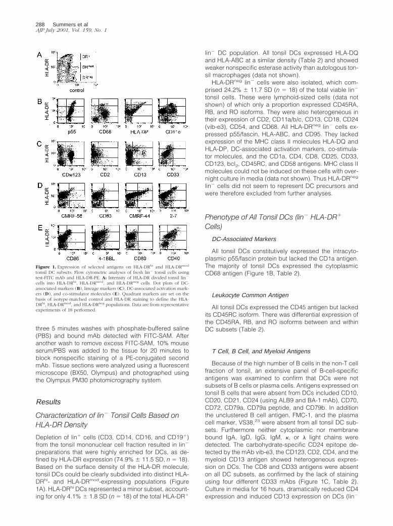
Triple labeling of sorted lin2 cells with CD11c, HLA-DR, and CD123 confirmed that HLA-DRmod CD11c2 DCswere a heterogeneous population based on CD123 ex-pression (Figures 1C and 4A). HLA-DRmod CD11c2
CD1231 DCs comprised the majority (78.5% 6 9.2 SD,n 5 4) of the total HLA-DRmod CD11c2 DC population.
The two CD11c2 DC subsets were analyzed by triplelabeling sorted CD11c2 lin2 tonsil cells with CD123,HLA-DR, and test mAbs. Both CD11c2 DC subsets ex-pressed low levels of CD40, whereas neither subset ex-pressed CMRF-44, CMRF-56, CD83, 2-7, CD86, 4-1BBligand, CD2, CD13, or CD45RO. Only the HLA-DRmod
CD11c2 CD1231 DCs expressed the CD4, HLA-DP, andCD24 (vib-e3) antigens. The CD68, CD45RA/RB, andCD38 antigens were expressed by CD1231 DCs, butshowed heterogeneous expression on the HLA-DRmod
CD11c2 CD1232 DCs (Figure 4B, Table 2).
Identification of Tonsil DC Subsets in Situ
HLA-DR staining was not useful as a single marker toidentify all tissue DCs or discriminate DC subsets be-cause of its extensive expression in tonsil tissue and thedifficulty of quantitative analyses in situ, respectively.However based on flow cytometric analyses, the entiretonsil DC population (including all DC subsets) could bebest identified in situ as either CD681/CD142 cells (Fig-ure 5A) or p551 cells (data not shown). There werelimitations to both methods however as the former com-bination excluded the minor HLA-DRmod CD11c2
CD1232 CD682 DC subset, whereas p55 overrepre-sented the number of DCs because of its expression onendothelial cells and HLA-DRneg lin2 tonsil cells. Bothmethods revealed similar findings in that DCs formed anextensive network of interconnecting cells throughout the
T-cell-enriched areas of tonsils. An additional populationof DCs was sparsely scattered within the follicles.
HLA-DRhi CD11c1 Tonsil DC Subset
HLA-DRhi CD11c1 DCs could be identified in freshtonsil tissue as either CD831/CD142/CD192 cells (Figure5B) or CMRF-561/CD192 cells (data not shown). This DCsubset was situated in close contact with CD81 (andCD41) T cells in the T cell areas of tonsils (Figure 5C).Although CD41 T cells dominate in paracortical areas, itwas difficult to decipher their interaction with adjacentCD41 DCs. To determine the percentage of HLA-DRhi
CD11c1 DCs within the total DC population accurately,triple labeling immunohistochemistry techniques wouldbe necessary. As this was not technically possible, dou-ble labeling of serial sections was used instead andsequential sections analyzed. The CD681/CD142 pheno-type was used to define tonsil DCs, CD831/CD142/CD192 to define HLA-DRhi CD11c1 DCs and CD681/CD831 to examine these activated DCs within the totalDC population. The number of activated HLA-DRhi
CD831 DCs comprised a minority of the total number ofCD681 paracortical DCs (Figure 5D), which is in similarproportions found in isolated DC preparations.
Figure 4. Phenotype of HLA-DRmod CD11c2 DCs. A: Flow cytometric anal-yses of fresh lin2 tonsil cells triple labeled with CD11c-FITC, HLA-DR-PE/Cy5, and CD123-PE. Gating on the CD11c2 DCs showed heterogeneousexpression of CD123. B: Flow cytometric analyses of CD11c2 lin2 tonsil cellstriple labeled with HLA-DR-PE/Cy5, CD123-FITC, and test-PE mAb deter-mined antigen expression on gated CD11c2 CD1231 DCs (thin straightline) and CD11c2 CD1232 DCs (thick straight line). Isotype-matchedcontrols are shown as dotted lines.
Dendritic Cell Subsets in Human Tonsils 291AJP July 2001, Vol. 159, No. 1

Double labeling using the CMRF-44 antigen plus theCD80 or CD86 co-stimulator molecules confirmed flowcytometric findings that all CMRF-441 DCs (HLA-DRhi
CD11c1 DCs and HLA-DRmod CD11c1 CD13lo DCs)lacked CD80 and only some CMRF-441 DCs (presum-ably the HLA-DRhi CD11c1 DCs) expressed the CD86antigen (data not shown).
HLA-DRmod CD11c1 Tonsil DC Subsets
The CD11c1 DCs (HLA-DRmod and HLA-DRhi DC sub-sets) were located in both the germinal center and T cellareas of tonsils (Figure 5E). CD11c1 GCDCs appeared tobe larger in size than CD11c1 paracortical DCs and, asopposed to the latter population, GCDCs lacked expres-sion of the CMRF-44 antigen (Figure 5F).
HLA-DRmod CD11c1 CD131 DCs were located in the Tcell areas, whereas HLA-DRmod CD11c1 CD132 DCswere located in tonsil follicles (Figure 5G). Thus HLA-DRmod CD11c1 CD132 DCs clearly represented GCDCs.Because of technical limitations with triple-color analysesin situ, it remained unclear whether some HLA-DRmod
CD11c1 CD132 DCs were also located in the T cellareas.
HLA-DRmod CD11c2 Tonsil DC Subsets
The vast majority of CD1231 cells lacked CD11c ex-pression and were located near high endothelial venulesin the T cell areas of tonsil (Figure 5H). Therefore GCDCsdo not express the CD123 antigen. Using a combinationof the CD68, CD11c, and CD123 antigens, CD11c2
CD1232 DCs could be identified as a minor populationwithin the T cell areas of tonsil tissue (Figure 5I). It was nottechnically possible to identify the CD11c2 CD1232
CD682 DC subset in situ because of the lack of definingantigens on these cells.
Discussion
In this study we demonstrate using a new method of DCpreparation that five distinct DC subsets are present inhuman tonsils (Figure 6). Extensive analysis of a largenumber of lin2 HLA-DR1 tonsil DC preparations revealedthe presence of HLA-DRhi CD11c1 DCs, HLA-DRmod
CD11c1 CD131 DCs, HLA-DRmod CD11c1 CD132 DCs,HLA-DRmod CD11c2 CD1231 DCs, and HLA-DRmod
CD11c2 CD1232 DCs subsets. In situ localization identi-fied four of these subsets as distinct IDC populations,which included plasmacytoid DCs17 and three new DCsubsets. We also confirmed the presence of GCDCs18
Figure 5. Identification of five tonsil DC subsets in situ. A: Majority of DCs(brown cells) were identified in the follicles and T cell areas as CD681
(brown)/CD142 (blue) cells. B: Subset 1: The HLA-DRhi CD11c1 DC subset(brown cells, arrow) could be identified as CD831 (brown)/CD32 (blue)cells. C: CD831 DCs (red cells) were located near CD81 (green) T cells. D:A small number of the total CD681 (green) DC population were CD831 (red)DCs (yellow cells). E: CD11c1 DCs (green cells) were located near CD31
(red) T cells in both the follicles and T cell areas. F: CD11c1 (green)CMRF-441 (red) DCs (yellow cells) were located in the T cell areas. G: Subset2: CD11c1 (green) CD131 (red) DCs (yellow cells) were located in the T cellareas. Subset 3: CD11c1 CD132 DCs (green cells) were located in the follicle.H: Subset 4: CD11c2 (green) CD1231 DCs (red cells) and (I) subset 5:CD11c2 CD1232 CD142 (red) CD681 DCs (green cells, arrows) werelocated in the T cell areas. Boxed F defines follicular areas. Original magni-fications: 3240 (A), 3573 (B and D), 3286 (C, E–I).
Figure 6. Five distinct subsets of DCs in human tonsils. In situ localizationidentified four of these subsets as distinct IDC populations, which included:1) plasmacytoid DCs (HLA-DRmod CD11c2 CD1231 DCs) and three new DCsubsets; 2) HLA-DRhi CD11c1 DCs; 3) HLA-DRmod CD11c1 CD131 DCs; and4) HLA-DRmod CD11c2 CD1232 DCs. GCDCs were also clarified as 5)HLA-DRmod CD11c1 CD132 DCs. The three CD11c1 DC subsets are likely torepresent different stages of DC maturation/activation. The CD11c1 DCs andCD11c2 DCs are thought to arise from separate myeloid and lymphoiddevelopmental pathways, respectively. IDC, interdigitating DCs; GCDC, ger-minal center DCs; HEV, high endothelial venule.
292 Summers et alAJP July 2001, Vol. 159, No. 1
but clarified their phenotype as a fifth HLA-DRmod
CD11c1 CD132 DC subset.A number of DC subsets with differing functional and
phenotypic characteristics have been identified in humanblood and tissues.7–10,14 Three tonsil DC subsets con-sisting of GCDCs,18 plasmacytoid DCs,17 and IDCs16
have been described previously. Our new methodologyhas the advantage of avoiding potential problems of an-tigenic changes associated with in vitro culture19 plus thebiases induced by positive selection. This combined witha very extensive phenotypic analysis established that theapparent heterogeneity in markers noted in these otherstudies16,18 could be extended into a more defined set offive DC subsets.
These five tonsil DC subsets each shared a number ofphenotypic features including expression of HLA-DQ,HLA-ABC, the DC-associated p55 marker,24 and CD95antigen. They also expressed HLA-DR, CD40, andCD45RB albeit at varying density between DC subsets,but lacked the CD33, CD45RC, and CD1a antigens. Theabsence of CD33 contradicts previous reports of low-level expression on GCDCs, which may relate to theabsence of cell culture as CD33 is inducible. In contrastto previous reports, we did not detect CD8016 or bcl2
25
within any tonsil DC subsets. This may again reflect adifference in isolation procedures because CD80 is in-duced by in vitro culture, which was used in previousstudies on tonsil DCs. Similar to blood DCs,8 high-densityexpression of cytoplasmic CD68 was associated withCD11c2 tonsil DCs. A separate population of HLA-DRneg
lin2 tonsil cells was also identified, as described previ-ously in normal blood26 and chronic arthritic synovialfluid.27 Although these cells expressed p55 and showedheterogeneous expression of CD45, CD13, and adhesionantigens, they did not express MHC class II antigenseither before or after in vitro culture. They therefore did notseem to represent DC precursor cells or a separate DCsubset.
The majority (;80%) of tonsil DCs are HLA-DRmod lin2
cells, which can be subdivided into morphologically dis-tinct smaller CD11c1 and more substantial CD11c2 DCsubsets. These are found in similar proportions to normalblood.9 The CD11c1 DCs can be further subdivided intoa small CD132 subset and a CD131 subset that arelocalized in the germinal center and T cell areas, respec-tively. The CD132 and CD131 DC subsets comprised5.1% 6 2.1 SD and 13.7 6 4.6 SD (n 5 4) of the total DCpopulation, respectively. HLA-DRmod CD11c1 CD132
and CD131 DCs shared a similar phenotype with theexception that only the CD131 DCs expressed the 2-7and CD45RO activation antigens. The CD131 DCs couldbe further subdivided based on CD13 intensity intoCD13hi and CD13lo subsets. These CD131 DC subsetsonly differed in their expression of the CMRF-44, CD2,and CD45RA antigens, which were present only on theCD13lo DC subset. HLA-DRmod CD11c1 CD132 DCswere the only subset to express the CD2 antigen.
In addition to location, the HLA-DRmod CD11c1 CD132
DC subset phenotypically resembled GCDCs as theylacked DC-associated activation and co-stimulator mol-ecules. The previously described GCDCs were isolated
based on a CD11c1 CD41 lin2 cell phenotype18 but, asoutlined in this study, this phenotype is common to threedistinct DC subsets located in both the T cell areas andgerminal centers, one of which expresses CD13. Thisfact, plus the observation that CD13 can be induced on asubpopulation of tonsil DCs after culture (data notshown), could explain the heterogeneous expression ofCD13 originally reported on GCDCs. Our study furtherextends their phenotype and shows that GCDCs havemany phenotypic similarities to mature/myeloid bloodDCs9,14 including low-density CD68 and lack of CD123expression. However, unlike CD11c1 blood DCs,9 theGCDCs identified in this study did not express the CD13or CD33 antigens.
Consistent with a low activation state, the HLA-DRmod
CD11c2 DC subset lacked expression of DC activationantigens, CD45RO, and co-stimulator molecules, al-though CD40 was expressed at low levels. The CD2 andCD13 antigens were also absent. Heterogeneous expres-sion of CD123 and CD4 enabled us to define two distinctHLA-DRmod CD11c2 DC subsets. The predominant HLA-DRmod CD11c2 DC subset (comprising 61.4% 6 11.6 SDof the total DC population) was CD1231, CD41, CD68hi,and closely resembled in phenotype, morphology, andlocalization near paracortical high endothelial venules28
the plasmacytoid tonsil DCs17 and immature CD11c2
DCs described in blood,9,10 bone marrow, and thymus.10
As a result, it has been suggested that this tonsil DCsubset may be derived from CD11c2 immature/lymphoidblood DCs.12,17 Our study further extends the phenotypeof plasmacytoid DCs and demonstrates that they expressp55, CD45RB, CD24 (vib-e3), low levels of HLA-DP, butlack DC activation markers and 4-1BB ligand.
HLA-DRmod CD11c2 CD1232 DCs were the only sub-set that lacked CD4 and HLA-DP expression. A smallnumber of these cells also lacked cytoplasmic CD68,CD45RA, CD45RB, and CD38. Interestingly a minorCD682 CD42 lin2 cell population had also been ob-served in a study of blood DCs.8 Unfortunately it was notpossible to identify these cells in tonsil sections becauseof their lack of suitable discriminatory markers.
The HLA-DRhi CD11c1 DC subset was the only subsetthat expressed the DC activation antigens, CD8329 andCMRF-5630 and the co-stimulator molecules, CD8631 and4-1BB ligand.32 This tonsil DC subset also expressed thehighest levels of the DC-associated activation markerCMRF-44,26,33 HLA-DP, CD40, and 2-7,19 which wenewly identify as a DC-associated activation marker. Mor-phologically this DC subset resembled activated bloodDCs.2 In tissue sections these activated DCs comprisedonly a minority of the total paracortical DC population,comparable to their low numbers present in DC isolates(4.1% 6 1.8 SD, n 5 18, of the total DC population. Theclose association of HLA-DRhi CD11c1 DCs to paracor-tical T cells and their low frequency resembles the char-acteristics of antigen-bearing DCs in lymph nodes thathave a rapid turnover after antigen presentationevents.34–36 The phenotypes of the HLA-DRhi CD11c1
DCs and HLA-DRmod CD11c1 CD132 DC subsets arebroadly similar, differing mainly in their expression ofactivation-associated antigens. As in vitro activation of
Dendritic Cell Subsets in Human Tonsils 293AJP July 2001, Vol. 159, No. 1

blood DCs induces similar changes,29–31,33 this raisesthe possibility that HLA-DRhi CD11c1 DCs represent amore activated form of HLA-DRmod CD11c1 CD132 DCs.
IDCs have been identified in tonsils as CD40hi lin2
cells, however the constitutive presence of CD40 on allfive DC subsets described in our study indicates thatthese IDCs incorporated several DC subsets. This isverified by the heterogeneous expression of the CD11cand CD83 antigens described on the former IDC popu-lation.16 Here we report that, in addition to plasmacytoidDCs (HLA-DRmod CD11c2 CD1231 DCs), three othernewly described DC subsets are located in the T cellareas of tonsils and can be defined as 1) HLA-DRhi
CD11c1 DCs, 2) HLA-DRmod CD11c1 CD132 DCs, and3) HLA-DRmod CD11c2 CD1232 DCs (Figure 6). Interest-ingly IDCs have been reported to stimulate activated Bcells16,37 suggesting that at least one DC subset may beinvolved in primary B cell responses.
There is accumulating evidence in mice that DC sub-sets arise from different lymphoid and myeloid develop-mental pathways.4,38 However, although the immatureDC subsets (ie, CD11c2, CD33lo, CD68hi, and CD123hi)and mature DC subsets (ie, CD11c1, CD33hi, CD68lo,and CD123lo) within human blood display a number oflymphoid and myeloid features, respectively4,8–10,14,38, itremains unclear whether they develop from distinct celllineages. A recent report provides evidence that, as op-posed to different maturation states, CD11c1 blood DCsrepresent multipotent myeloid precursor cells, whereasCD11c2 blood DCs represent lymphoid progenitors.12
The tonsil CD11c1 DC subsets described in this studyshare a number of features with CD11c1 mature/myeloidblood DCs being CD68lo and CD123lo/neg. They do how-ever, differ from each other in their expression of CD13,CD33, activation, and co-stimulator molecules. As eachof these antigens can be up-regulated after culture ontonsil DCs, one could speculate that the CD11c1 DCsubsets represent different maturation stages, ie,CD11c1 CD132 DRmod (early), CD11c1 CD131 DRmod
(intermediate), and CD11c1 CD131 DRhi (late). However,functional studies would be necessary to establish therelationship of these five tonsil DC subsets both to eachother and to other tissue DCs. Consistent with previousstudies on blood DC subsets, CD11c1 DCs,9 as well asactivated DCs,33,39 were the most potent stimulators ofallogeneic T cell responses in the tonsil.
The HLA-DRhi CD11c1 DCs and GCDCs seem to rep-resent highly immunogenic DCs that are actively present-ing antigens to paracortical and germinal center T and/orB cells, respectively. The other less-activated IDC sub-sets may represent DCs that have migrated to the drain-ing lymph node (tonsil) either without receiving up-regu-latory signals or else receiving negative regulatorysignals from interacting self T cells.40 These distinct DCsubsets may play different roles in regulating immunity ortolerance, although it remains unclear whether the type ofT cell response is driven by intrinsic properties of the DCsubset13 or by local environmental factors.41 Ultimately,targeting a specific DC subset may provide more effec-tive therapeutic regulation of immunological disorders.
Acknowledgments
We thank the ENT surgeons and nursing staff fromChristchurch, St George’s and Southern Cross Hospitalsfor providing us with tonsil specimens, Dr. Axel Heiser forhelpful suggestions, and Lisa Haring for providing assis-tance with flow cytometry.
References
1. Steinman RM: The dendritic cell system and its role in immunogenic-ity. Annu Rev Immunol 1991, 9:271–296
2. Hart DNJ: Dendritic cells: unique leucocyte populations which controlthe primary immune response. Blood 1997, 90:3245–3287
3. Banchereau J, Steinman RM: Dendritic cells and the control of immu-nity. Nature 1998, 392:245–252
4. Suss G, Shortman K: A subclass of dendritic cells kills CD4 T cells viaFas/fas-ligand-induced apoptosis. J Exp Med 1996, 183:1789–1796
5. Hill S, Coates JP, Kimber I, Knight SC: Differential function of dendriticcells isolated from blood and lymph nodes. Immunology 1994, 83:295–301
6. Pulendran B, Lingapappa J, Kennedy MK, Smith J, Teepe M, Ruden-sky A, Maliszewski CR, Maraskovsky E: Developmental pathways ofdendritic cells in vivo. J Immunol 1997, 159:2222–2231
7. Nestle FO, Zheng X-G, Thompson CB, Turka LA, Nickoloff BJ: Char-acterization of dermal dendritic cells obtained from normal humanskin reveals phenotypic and functionally distinctive subsets. J Immu-nol 1993, 151:6535–6545
8. Strobl H, Scheinecker C, Riedl E, Csmarits B, Bello-Fernandez C,Pickl WF, Majdic O, Knapp W: Identification of CD681lin2 peripheralblood cells with dendritic precursor characteristics. J Immunol 1998,161:740–748
9. O’Doherty U, Peng M, Gezelter S, Swiggard WJ, Betjes M, BhardwajN, Steinman RM: Human blood contained two subsets of dendriticcells, one immunologically mature and the other immature. Immunol-ogy 1994, 82:487–493
10. Olweus J, BitMansour A, Warnke R, Thompson PA, Carballido J,Picker LJ, Lund-Johansen F: Dendritic cell ontogeny: a human den-dritic cell lineage of myeloid origin. Proc Natl Acad Sci USA 1997,94:12551–12556
11. Kohrgruber N, Halanek N, Groger M, Winter D, Rappersberger K,Schmitt-Egenolf M, Stingl G, Maurer D: Survival, maturation, andfunction of CD11c2 and CD11c1 peripheral blood dendritic cells aredifferentially regulated by cytokines. J Immunol 1999, 163:3250–3259
12. Robinson S, Patterson S, English N, Davies S, Knight S, Reid C:Human peripheral blood contains two distinct lineages of dendriticcells. Eur J Immunol 1999, 29:2769–2778
13. Rissoan M, Soumelis V, Kadowaki N, Grouard G, Brieree F, de WaalMalefy R, Liu YJ: Reciprocal control of T helper cell and dendritic celldifferentiation. Science 1999, 283:1183–1186
14. Thomas R, Lipsky PE: Human peripheral blood dendritic cell subsets:isolation and characterization of precursor and mature antigen pre-senting cells. J Immunol 1994, 153:4016–4028
15. Hart DNJ, McKenzie JL: Isolation and characterisation of humantonsil dendritic cells. J Exp Med 1988, 168:157–170
16. Bjorck P, Flores-Romo L, Liu Y-J: Human interdigitating dendritic cellsdirectly stimulate CD40-activated naive B cells. Eur J Immunol 1997,27:1266–1274
17. Grouard G, Rissoan M-C, Filgueira L, Durand I, Banchereau J, LiuY-J: The enigmatic plasmacytoid T cells develop into dendritic cellswith interleukin (IL)-3 and CD40-ligand. J Exp Med 1997, 185:1101–1111
18. Grouard G, Durand I, Filgueira L, Banchereau J, Liu Y-J: Dendriticcells capable of stimulating T cells in germinal centres. Nature 1996,384:364–367
19. Teunissen MBM, Wormmeester DJ, Kreig SR, Peters PJ, Vogels IMC,Kapsenberg ML, Bos JD: Human epidermal Langerhans cells un-dergo profound morphologic and phenotypic changes during in vitroculture. J Invest Dermatol 1990, 94:166–173
20. Egner W, Andreesen R, Hart DNJ: Allostimulatory cells in fresh human
294 Summers et alAJP July 2001, Vol. 159, No. 1

blood: heterogeneity in antigen presenting cell populations. Trans-plantation 1993, 56:945–950
21. Summers K, O’Donnell J, Williams LA, Hart DNJ: Expression andfunction of CD80 and CD86 costimulator molecules on synovial den-dritic cells in chronic arthritic disease. Arthritis Rheum 1996, 39:1287–1291
22. Troy AJ, Summers KL, Davidson PJT, Atkinson CA, Hart DNJ: Minimalrecruitment and activation of dendritic cells within renal cell carci-noma. Clin Cancer Res 1998, 4:585–593
23. Turley H, Jones M, Erber W, Mayne K, de Waele M, Gatter K: VS38:a new monoclonal antibody for detecting plasma cell differentiation inroutine sections. J Clin Pathol 1994, 47:418–422
24. Mosialos G, Birkenbach M, Ayehunie S, Matsumura F, Pinkus GS,Kieff E, Langhoff E: Circulating human dendritic cells differentiallyexpress high levels of a 55-kd actin-bundling protein. Am J Pathol1996, 148:593–600
25. Bjorck P, Banchereau J, Flores-Romo L: CD40 ligation counteractsFas-induced apoptosis of human dendritic cells. Int Immunol 1997,9:365–372
26. Fearnley DB, McLellan AD, Mannering SI, Hock BD, Hart DNJ: Isola-tion of human blood dendritic cells using the CMRF-44 monoclonalantibody: implications for studies on antigen presenting cell functionand immunotherapy. Blood 1997, 89:3708–3716
27. Summers KL, Daniel PB, O’Donnell J, Hart DNJ: Dendritic cells insynovial fluid chronic inflammatory arthritis lack CD80 surface ex-pression. Clin Exp Immunol 1995, 100:81–89
28. Cella M, Jarrossay D, Facchietti F, Alebardi O, Nakajima H, Lanza-vecchia A, Colonna M: Plasmacytoid monocytes migrate to inflamedlymph nodes and produce large amounts of type I interferon. Nat Med1999, 8:919–929
29. Zhou LJ, Tedder TF: Human blood dendritic cells selectively expressCD83, a member of the immunoglobulin superfamily. J Immunol1995, 154:3821–3835
30. Hock BD, Fearnley DB, Boyce A, McLellan AD, Sorg RV, SummersKL, Hart DNJ: Human dendritic cells express a 95kDa activation/differentiation antigen defined by CMRF-56. Tissue Antigens 1999,53:320–334
31. McLellan AD, Starling GC, Williams LA, Hock BD, Hart DNJ: Activa-tion of human peripheral blood dendritic cells induces the CD86costimulatory molecule. Eur J Immunol 1995, 25:2064–2068
32. Zhou Z, Kim S, Hurtado J, Lee ZH, Kim KK, Pollok KE, Kwon BS:Characterization of human homologue of 4-1BB and its ligand. Im-munol Lett 1995, 45:67–73
33. Hock BD, Starling GC, Daniel PB, Hart DNJ: Characterisation ofCMRF-44, a novel monoclonal antibody to an activation antigen ex-pressed by the allostimulatory cells within peripheral blood, includingdendritic cells. Immunology 1994, 83:573–581
34. Matsuno K, Ezaki T, Kudo S, Uehara Y: A life stage of particle-ladenrat dendritic cells in vivo: their terminal division, active phagocytosisand translocation from the liver to the draining lymph. J Exp Med1996, 183:1865–1878
35. Hill S, Edwards AJ, Kimber I, Knight SC: Systemic migration of den-dritic cells during contract sensitization. Immunology 1990, 71:277–281
36. Ingulli E, Mondino A, Khoruts A, Jenkins MK: In vivo detection ofdendritic cell antigen presentation to CD41 T cells. J Exp Med 1997,185:2133–2141
37. Johansson B, Ingvarsson S, Bjorck P, Borrebaeck CA: Human inter-digitating dendritic cells induce isotype switching and IL-13-depen-dent IgM production in CD40-activated naive B cells. J Immunol2000, 164:1847–1854
38. Steinman RM, Pack M, Inaba K: Dendritic cells in the T-cell areas oflymphoid organs. Immunol Rev 1997, 156:25–37
39. Zhou L, Schwarting R, Smith HM, Tedder TF: A novel cell-surfacemolecule expressed by human interdigitating reticulum cells, Lang-erhans cells, and activated lymphocytes is a new member of the Igsuperfamily. J Immunol 1992, 149:735–742
40. McLellan AD, Heiser A, Hart DNJ: Induction of dendritic cell costimu-lator molecule expression is suppressed by T cells in the absence ofantigen-specific signalling: role of cluster formation, CD40 and HLA-class II for DC activation. Immunology 1999, 98:171–180
41. Vieira PL, de Jong EC, Wierenga EA, Kapsenberg ML, Kalinski P:Development of Th1-inducing capacity in myeloid dendritic cellsrequires environmental instruction. J Immunol 2000, 164:4507–4512
Dendritic Cell Subsets in Human Tonsils 295AJP July 2001, Vol. 159, No. 1