Phenotypic and molecular characterisation of Pseudomonas ...
233
Phenotypic and molecular characterisation of Pseudomonas aeruginosa infections from companion animals and potential reservoirs of antibacterial resistance in humans. Thesis submitted in accordance with the requirements of the University of Liverpool for the degree of Master of Philosophy by Andrea Catherine Scott May 2018
-
Upload
khangminh22 -
Category
Documents
-
view
1 -
download
0
Transcript of Phenotypic and molecular characterisation of Pseudomonas ...
Phenotypic and molecular characterisation of Pseudomonas
aeruginosa infections from companion animals and potential
reservoirs of antibacterial resistance in humans.
Thesis submitted in accordance with the requirements of the University of Liverpool
for the degree of Master of Philosophy by
Andrea Catherine Scott
May 2018
2
Acknowledgements
I would like to thank my primary supervisors Dr Joanne Fothergill and Dr Alan
Radford.
I also thank my secondary supervisors Dr Dorina Timofte, Dr Vanessa Schmidt, Dr
Gina Pinschbeck and Dr Neil McEwan and the Institute of Infection and Global
Health, University of Liverpool and Professor Craig Winstanley, along with the Small
Animal Teaching Hospital (SATH), Leahurst, University of Liverpool and the
Veterinary Diagnostic Laboratory (VDL), Leahurst.
Matthew Moore (PhD student at University of Liverpool) performed Bioinformatics
work for this project.
The sequencing in this project was performed as part of the International
Pseudomonas Consortium.
The Masters thesis of this work was supported through a University of
Liverpool/Wellcome Trust Research taster fellowship and internal funding.

3
Abbreviations
AMR – Antimicrobial resistance
BSAVA – British Small Animal Veterinary Association
BVA – British Veterinary Association
CDC - Centre for Disease Control and Prevention
CF – Cystic fibrosis
COPD – Chronic obstructive pulmonary disorder
CRE – Carbepenam-resistant enterobacteriacea
ECDPC - European Centre Disease Prevention and Control
FDA – US Food and Drug Administration
HAIs – Hospital acquired infections
ICEs – Integrative and conjugative elements
ICUs – Intensive care units
IDSA - Infectious Diseases Society of America
LPS – Lipopolysaccharide
LES –Liverpool epidemic strain
MDR - Multi drug resistant
MMR – Mis-match repair genes
MRSA - Methicillin-resistant Staphylococcus aureus
OM - Outer membrane
PCR – Polymerase chain reaction
PFGE – Pulse-field gel electrophoresis
PHE – Public Health England
VAP – Ventilator associated pneumonia
VMTH – Veterinary Medicine Teaching Hospital
WHO - World Health Organisation
WGS – Whole genome sequencing

4
Abstract........................................................................................................................6
Chapter1: Introduction.……………......................................................................................8
1.1 Pseudomonas aeruginosa..........................................................................................8
1.2 P.aeruginosa infections in humans….......................................................................13
1.3 Circulating P.aeruginosa strain types in humans and diversity of the population
structure ……………………..…………………………………………………………………........................18
1.4 Limited antimicrobial treatment options in treatment of P.aeruginosa infections
in humans.……………………………………..…..........................................................................23
1.5 P.aeruginosa infections in animals..........................................................................26
1.6 Antimicrobial treatments used for P.aeruginosa infections in animals................30
1.7 AMR in P.aeruginosa.……………………….………………………..……………………………...……34
1.8 Potential for cross-species transmission between humans and animals.............35
Chapter 2: Materials and Methods for Results Chapter 3 - Reservoirs of resistance:
polymyxin resistance in veterinary-associated isolates of P. aeruginosa……………….41
2.1 Bacterial isolates…..……............................................................................................41
2.2 Susceptibility testing.…..……………….........................................................................43
2.3 Clondiag Array Tube.……….………………………………………….......................................44
2.4 Genomic DNA extraction for Illumina sequencing...……………………….…………….….50
2.5 Whole Genome Sequencing (Illumina) of Bacterial Isolates ……………………………52
2.6 Genomics .……………………………………………………………………………………………………….53
2.7 Comprehensive Antibiotic Resistance Database sequence data analysis …………54
2.8 eBurst algorithm ……...…………………………….……………………..……………………………….55
2.9 Statistical Analysis……………………………………………………………………….……………….….56
Chapter 2: Materials and Methods for Results Chapter 4 - PCR characterisation of
antibiotic resistant determinants in P. aeruginosa from companion animals………..57
2.10 Bacterial isolates …………………………….................................................................57
2.11 Susceptibility testing …..…………………………….……………........................................60
2.12 DNA Extraction………..........................................................................................62
2.13 Polymerase Chain Reactions ……………………………………………………………….……....63
2.14 Primers ..……………………………………………………………………….…………………….……….66
2.15 Quantification and purity testing of genomic DNA by NanoDrop
spectrophotometer……………………………………………………………………………………………….66

5
2.16 DNA Sequencing ..……...………………………………………………….………..……………………67
2.17 Statistics ……………………………………………………………………………………………………….67
Chapter 3: Reservoirs of resistance: polymyxin resistance in veterinary-associated
isolates of Pseudomonas aeruginosa .…….……….……..................................................68
3.1 Introduction.……………….…………………….……………..................................................68
3.2 Aims.………………….…………………………………………………….........................................76
3.3 Results .……………………….……………………………........................................................77
3.4 Discussion .…………………………………………………………..............................................90
3.5 Conclusions.……………….……………………….……….....................................................98
Chapter 4: PCR characterisation of antibiotic resistant determinants in P. aeruginosa
from companion animals..…………………………………………,…….……………….….................99
4.1 Introduction.……….…………………………………………………..........................................99
4.2 Aims.……………………….……………………………….………….…………….............................128
4.3 Results .……………………….……………………………………….…….....................................129
4.4 Discussion.………………………………………………………………..…….……..........................165
4.5 Conclusions.……………….……………………………………….……………..............................170
Chapter 5: Discussion..…………………………………………………………………………………………171
References ..…………………………………………………………………………………………………….….182

6
Phenotypic and molecular characterisation of Pseudomonas aeruginosa infections
from companion animals and potential reservoirs of antibacterial resistance in
humans.
Andrea Catherine Scott - May 2018
Abstract
Antimicrobial resistance (AMR) is a multifactorial and complex issue, affecting
healthcare worldwide. This is an escalating concern, highlighted by European and
International bodies. AMR is not only a concern in human health but also animal
health. The close interface between people, pets, food production animals and the
environment exemplifies the importance that these two areas of health should not
be considered as independent factions. Pseudomonas aeruginosa is a formidable
pathogen due to its innate resistance making it naturally insensitive to many classes
of antimicrobials, alongside its ability to acquire further resistance mechanisms. P.
aeruginosa in the veterinary setting is a clinically relevant pathogen often
associated with disease.
In this project, I determined the susceptibility of a set of veterinary clinical P.
aeruginosa isolates (n=24) to polymyxin antibiotics, colistin (polymyxin E) and
polymyxin B, by broth micro dilution and showed that the MICs for polymyxin B to
be significantly higher than in a panel of human P. aeruginosa isolates (n= 37).
Resistance to other antibiotics used in human medicine was low but higher levels of
resistance were detected to ticarcillin (21%), and also to marbofloxacin (21%) and
enrofloxacin (33%), these being two widely used veterinary antibiotics. Using the
array tube typing method, the P. aeruginosa veterinary isolates were found to be
distributed throughout the P. aeruginosa population, with shared array types from
human infections such as keratitis and respiratory infections. Using whole genome
sequencing on a limited subset of isolates, the veterinary isolates were generally
clustered within the main P. aeruginosa population; however one isolate, did not

7
cluster with any other previously sequenced isolate. The genome sequencing also
revealed that isolates had mutations in genes associated with polymyxin resistance
and other antimicrobial resistance-related genes. These findings may suggest that
treating animal infections with antimicrobials could lead to resistance in isolates
capable of causing human infections.
The prevalence of resistance to beta-lactams, aminoglycosides and
fluoroquinolones in isolates from companion animals particularly in P. aeruginosa is
a relatively understudied area. Here, the resistance characteristics of a set of P.
aeruginosa isolates from companion animals (n=106), collected, at the Veterinary
Diagnostic laboratory (VDL) at the Small Animal Teaching Hospital, Leahurst
(University of Liverpool, UK) was investigated. Several resistance genes including
those relating to ESBLs (blaTEM, blaSHV and blaOXA) and those relating to
fluroquinolone resistance (qnr) were identified. In particular the identification of
qnrA and qnrB genes in P.aeruginosa isolates of companion animal origin is a novel
finding with no known reports to current date in the UK. Of the 8 confirmed
sequenced positive amplicons they comprised 7 different isolates all canine in
origin. Multidrug resistance was also demonstrated among some isolates.
These findings further support the likelihood that companion animals are an
understudied source of antimicrobial resistant P. aeruginosa isolates, and merit
sustained surveillance of the veterinary niche as a potential reservoir for resistant,
clinically-relevant bacteria.

8
Chapter 1 : Introduction
1.1 Pseudomonas aeruginosa
P. aeruginosa features and habitats
Pseudomonas aeruginosa is a Gram negative rod shaped aerobic bacterium that is
motile with a single polar flagella and can act as an opportunistic pathogen. Key
features of the bacterium are listed in Table 1.1 P. aeruginosa is metabolically
versatile and can grow both aerobically and as a facultative anaerobe, in addition to
utilising other metabolic systems (such as those using nitrate or benzoate) thus
contributing to its ability to be both ubiquitous and opportunistic in nature. These
factors also explain the organisms’ presence in numerous environments, including
within natural water sources such as lakes and marshes, in soil and also within both
the hospital and community environments (Khan & Cerniglia, 1994). P. aeruginosa
may be isolated from hospital respiratory ventilator equipment, antiseptics, sinks
and cleaning equipment (Trautmann et al, 2008; Trautmann et al, 2005). In the
non-hospital environment reservoirs have been reported in recreational water
venues such as poorly maintained swimming pools, hot tubs, whirlpools, spas
(Ratnam, 1986) and also in contact lens cleaning solution, (Dutta et al, 2012; Kaye et
al., 2017) soil and vegetables (Correa et al, 1991; Deredjian et al., 2014; Green et al,
1974; Khan & Cerniglia, 1994; Mena et al., 2008; Youenou et al, 2014).
Table 1.1 Key features of P. aeruginosa features
Description Gram negative rod shaped aerobic bacterium that is motile with a single polar
flagella and can act as an opportunistic pathogen
Diversity Found in diverse environments including water (Cabral, 2010), soil, sewage
(Slekovec et al., 2012) and also humans, animals and plants
Form Found in planktonic forms or biofilms (O’Tool et al, 2000)
Colony types Variable, smooth or mucoid in appearance;

9
Wild-type isolates found naturally from soil or water frequently produce a small,
rough colony. Clinical isolates are usually either a large, smooth colony type with
flat edges and elevated appearance. Or, they may have a mucoid appearance,
this colony type being associated with the over production of the polysaccharide
alginate and are typically found in those samples from the respiratory and
urinary tract (Cole et al, 2014; Distefano, 2015).
Pigment production P. aeruginosa can produce a variety of pigments; Pyocyanin a blue coloured
phenzine pigment;
Pyoverdine a siderophore, involved in iron metabolism, it is a yellow-green
brightly fluroscent pigment;
Pyorubin a red brown pigment;
Pyomelanin a brown pigment, melanin like compound synthesized from tyrosine.
(Lau et al 2004)
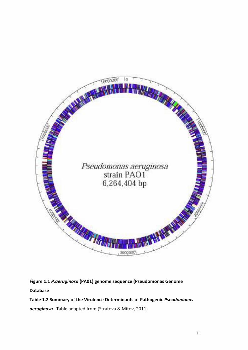
P. aeruginosa genome
P. aeruginosa has a comparatively large genome of approximately 6.3 million base
pairs (Mbp) (referred to in Figure 1.1). It is known to consist of a main conserved
core genome and variable accessory segment (Stover C-K., et al 2000). It has been
estimated that approximately 10% of its genome varies from strain to strain (Ozer
et al 2014) and is thus referred to as the accessory genome. Genetic elements
within the accessory genome of P. aeruginosa have been associated with
differences in virulence and antibiotic resistance (Kung et al , 2010; Ozer et al.,
2014). Virulence factors associated with P. aeruginosa include flagella, adhesion
and extracellular proteins, or secondary metabolites that have proteolytic/cytotoxic
activity (such as exotoxin A, elastase, proteases, pyocyanin, hemolysins) (Driscoll et
al, 2007). The known virulence factors of P. aeruginosa are reported in Table 1.2.
P. aeruginosa infection process and virulence
The P. aeruginosa infection process consists of three stages; bacterial adhesion and
colonization; local invasion; and disseminated systemic disease (Strateva & Mitov,
2011). The colonization phase, primarily involves the cell-associated virulence
factors (Table 1.2). The infectious stage (which may then develop to either an acute
infection or a chronic infection) is characterized by the production of extracellular

10
virulence factors. In acute infections high production of these factors are typical,
whereas inchronic infections lower amounts of these determinants are produced
(Strateva & Mitov, 2011).
P. aeruginosa virulence factors and quorum sensing
The virulence of P. aerurginosa depends on a varied number of both cell-associated
and extracellular factors (Table 1.2). The majority of P. aeruginosa infections are
both invasive and toxinogenic. Many of the extracellular virulence factors
(proteases, exotoxin A, pyocyanin, siderophores, haemolysins) are controlled by
quorum sensing (QS). This is a cell-to-cell signaling system that enables the bacteria
to produce these factors in a coordinated, cell-density-dependent manner and
overwhelm the host defense mechanisms during acute infection. The QS system can
also contribute to biofilm formation and so may participate in the pathogenesis of
chronic infection. Two-component sensor kinases such as RetS, LadS and GacS are
also recognized as controlling the production of virulence factors as well as the
switch from acute to chronic infection (Jimenez et al., 2012).
As mentioned, the production of many of the virulence factors is regulated by
quorum sensing (QS), a bacterial cell-to-cell communication mechanism. There
exists two well defined QS systems in P. aeruginosa, the las and rhl systems
(Pearson et al 1997; Pesci et al., 1997) although two other system have also been
identified (pqs and iqs). These rely on auto-inducer signal molecules (n N-acyl
homoserine lactone molecules, or other molecules whose production depends on S-
adenosylmethionine as a substrate). Auto-inducers are produced in the cell and
freely diffuse across the inner and outer membranes. These molecules accumulate
in the environment as the bacterial population density increases, and bacteria track
this information and collectively alter gene expression. QS controls genes that direct
activities that are beneficial when performed by groups of bacteria acting in
synchrony. The term “fitness” is used to describe this ability to adjust metabolism to
suit the environmental conditions, in order to survive and grow (Beceiro et al.,
2013; Millan et al., 2015). Processes controlled by QS include bioluminescence,
sporulation, competence, antibiotic production, biofilm formation, and virulence
factor secretion (Ruparell et al., 2016; Karatuna & Yagci, 2010)
11
Figure 1.1 P.aeruginosa (PA01) genome sequence (Pseudomonas Genome
Database
Table 1.2 Summary of the Virulence Determinants of Pathogenic Pseudomonas
aeruginosa Table adapted from (Strateva & Mitov, 2011)

12
Virulence Factors Cell
associated
factors
Extra cellular
factors
Adhesins
Type IV pili (N-methyl-phenylalanine pili)
Polysaccharide capsule (glycocalyx)
Alginate slime (biofilm)
Carbohydrate-binding
Proteins (lectins)
Adhesion Facilitation
Neuraminidase (sialidase)
Invasins
Elastases (LasB and LasA)
Alkaline protease
Haemolysins (phospholipase and rhamnolipid)
Cytotoxin (leukocidin)
Siderophores and siderophore uptake systems
Pyocyanin diffusible pigment
Motility/chemotaxis
Flagella (swimming motility)
Retractile pili (twitching motility)
Toxinogenesis
Exotoxin A
Lipopolysaccharide (endotoxin)
LecA and LecB lectins
Type III effector cytotoxins (ExoS, ExoU, ExoT,
ExoY)
Enterotoxin
Antiphagocytic surface properties
Capsules, slime layers
Lipopolysaccharide
Biofilm
Defense against serum bactericidal reaction
Slime layers, capsules, biofilm
Lipopolysaccharide
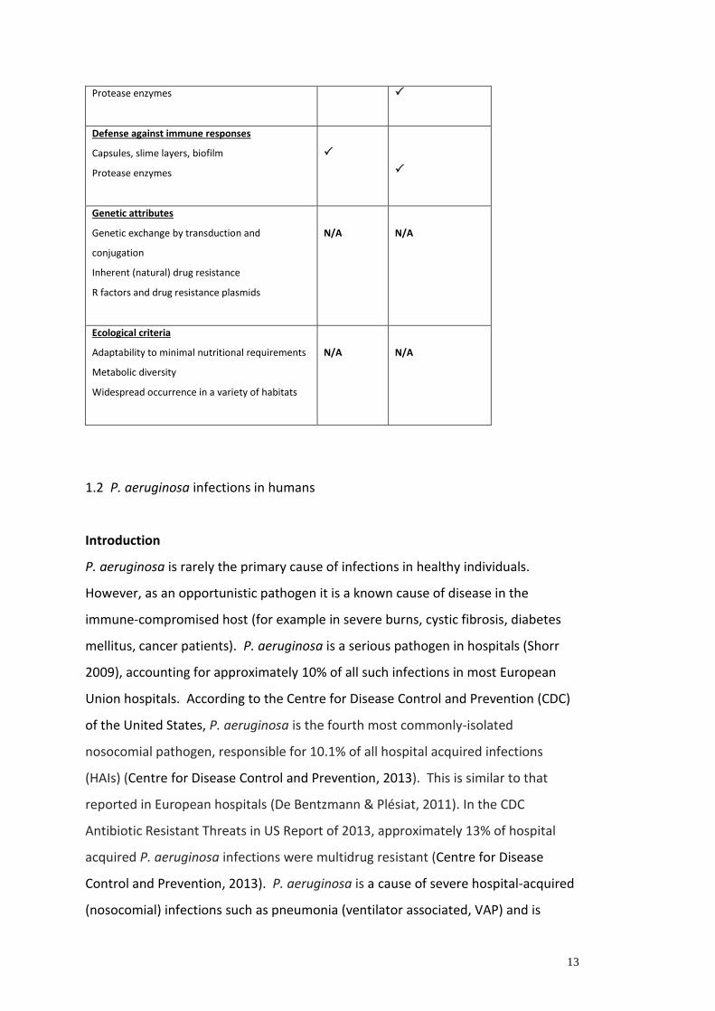
13
Protease enzymes
Defense against immune responses
Capsules, slime layers, biofilm
Protease enzymes
Genetic attributes
Genetic exchange by transduction and
conjugation
Inherent (natural) drug resistance
R factors and drug resistance plasmids
N/A
N/A
Ecological criteria
Adaptability to minimal nutritional requirements
Metabolic diversity
Widespread occurrence in a variety of habitats
N/A
N/A
1.2 P. aeruginosa infections in humans
Introduction
P. aeruginosa is rarely the primary cause of infections in healthy individuals.
However, as an opportunistic pathogen it is a known cause of disease in the
immune-compromised host (for example in severe burns, cystic fibrosis, diabetes
mellitus, cancer patients). P. aeruginosa is a serious pathogen in hospitals (Shorr
2009), accounting for approximately 10% of all such infections in most European
Union hospitals. According to the Centre for Disease Control and Prevention (CDC)
of the United States, P. aeruginosa is the fourth most commonly-isolated
nosocomial pathogen, responsible for 10.1% of all hospital acquired infections
(HAIs) (Centre for Disease Control and Prevention, 2013). This is similar to that
reported in European hospitals (De Bentzmann & Plésiat, 2011). In the CDC
Antibiotic Resistant Threats in US Report of 2013, approximately 13% of hospital
acquired P. aeruginosa infections were multidrug resistant (Centre for Disease
Control and Prevention, 2013). P. aeruginosa is a cause of severe hospital-acquired
(nosocomial) infections such as pneumonia (ventilator associated, VAP) and is

14
involved in the etiology of several diseases including bronchopneumonia, ocular,
burn and wound infections, bacteremia, endocarditis and meningitis. The organism
most commonly affects the lower respiratory system in humans and is an important
respiratory pathogen in patients with cystic fibrosis (CF) and also other chronic lung
diseases such as non-CF bronchiectasis and severe chronic obstructive pulmonary
disease (COPD) (Govan et al, 2007).
P. aeruginosa is a known cause of bacteraemia and this is associated with significant
mortality and morbidity. Mortality rates within the first 3-5 days of the onset of
bacteremia are high and fifty percent of isolates are resistant to the standard
antibiotics used in empirical treatment. Poorer clinical outcomes have also been
demonstrated with pneumonia caused by infections due to P. aeruginosa due to
associated multidrug resistance of the organism (Tam et al., 2010). P. aeruginosa is
reported as the most common Gram-negative pathogen causing nosocomial
pneumonia in the United States, and it is frequently implicated in hospital-acquired
urinary tract and bloodstream infections (Peleg et al, 2010; Mehrad et al, 2015)
Specific P. aeruginosa human infections including some epidemiology and
prevalences
Bacteremia
A UK government (UKGov, 2017) Public Health England (PHE) Infection Surveillance
report looking at bloodstream infections showed that the overall rate of
Pseudomonas as 6.2 per 100,000 population (July 2015) which was an 11% decrease
from 2007 (the analyses of this report were based on data relating to diagnoses of
Pseudomonas spp. and Stenotrophomonas spp. bacteramia 2007 – 2014 in England,
Wales and Northern Ireland. The most frequently identified Pseudomonas species
in blood isolates in 2014, according to the same surveillance report, was P.
aeruginosa at 81%. The proportion of which were reported as resistant to one of
the key antimicrobials in 2014 remained steady or increased slightly compared to

15
2013. A steady increase in reported resistance of Pseudomonas isolates to
piperacillin/tazobactam was observed reported 2010-2014 from 7%-10%. In
England, dual resistance to ciprofloxacin and ceftazidime was reported in 2% of
Pseudomonas spp. bacteraemia. Imipenem resistance increased the most from 13%
of isolates in 2013 to 16% isolates in 2014. In 2014 the proportion of resistant
Pseudomonas spp. bacteraemia isolates reported was 10% piperacillin/tazobactam,
11% ciprofloxacin, 7% ceftazidime, 10% meropenem, 16% imipenem and 4%
gentamicin.
Comparatively, a US study (Larru et al., 2016) evaluated the epidemiology and
antimicrobial resistance of bloodstream infections occurring during an 11-year
period in a large, tertiary care children's hospital in the United States and found
20% of these infections to be ESKAPE (includes P. aeruginosa) pathogen in origin.
No clinically significant increases in ceftazidime-resistant P. aeruginosa were
observed during the study, although MRSA rates did increase over time (Larru et al.,
2016).
Keratitis
In humans P. aeruginosa is a known problem in contact lens wearers, were it can be
a common cause of ulcerative keratitis (Stapleton et al, 1995). Ulcerative keratitis is
a rare, but potentially serious, complication of cosmetic contact lens wear. It has
the potential to affect vision and cause blindness (Austin et al, 2017; Zegans et al.,
2016). There are several different types of microbes that colonize lenses and can
lead to infection and inflammation, but one of the most common causes of
microbial keratitis remains P. aeruginosa. Its ability to produce proteases, to either
invade or kill corneal cells, and to coordinate expression of virulence factors via
quorum-sensing have been shown to be important during microbial keratitis.
Another important factor that contributes to the destruction of the cornea during
microbial keratitis is excessive activation of the host defense system. P. aeruginosa
can activate several pathways of the immune system and activation often involves

16
receptors on the corneal epithelial cells called toll-like receptors (Hazlett, 2004;
Willcox, 2007). P. aeruginosa has a tendency to adhere to the contact lens surface
and is transferred over scratched corneal epithelium penetrating the cornea's
deeper layers and leading corneal ulcers. Permanent blindness can be caused by a
severe infection. The lens, ocular environment, and storage case may offer an
appropriate survival niche for the organism to adhere to and colonize lens materials
during wear and storage (Hedayati, et al 2015).
A 2015 retrospective case control study (Vazirani et al, 2015) looked at ninety
episodes of P. aeruginosa keratitis from a tertiary care eye institution from 2007 -
2014 (23 multi-drug resistant as cases and 67 episodes drug-sensitive as controls).
Antimicrobial resistance in the MDR P.aeruginosa keratitis isolates lowest for both
colistin and imipenem at 56.52% each. Complications (such as corneal perforation,
cyanoacrylate glue application and keratoplasty) were more common in when
infections involved MDR isolate. Studies report that various factors, such as the use
of ocular lubricant, a compromised ocular surface, and bandage contact lens are
associated with MDR (multi-drug resistant) P. aeruginosa keratitis. An addition,
preservative-free lubricant ointment may act as a source or reservoir of infection in
this condition (Vazirani et al., 2015). A 2015 study by (Hedayati et al., 2015) looked
at contact lens wearers in Iran with 26 patients at a hospital from 2012-2013. With
a low number (8 samples) of the scraped ulcers being reported as sterile, and of the
positive cultures, 80% isolated P. aeruginosa. In this particular study 84% of the
microorganism cases were sensitive to ciprofloxacin, while imipenem, meropenem,
and ceftazidime were the second most effective antibiotics (susceptibilities tested
by disc diffusion method). The percentage of isolates sensitive to imipenem, were
somewhat higher in this study. Colistin susceptibility here was not tested however.
Fernandes et al (Fernandes et al 2016) specifically looked at extensively and pan
drug resistant P. aeruginosa keratitis. In their study (of 15 eyes from 13 different
patients, at an Eye Institute in India 2009-2013) 40% were sensitive only to
imipenem, 20% to colistin, 13.3% to neomycin and 6.7% each to imipenem and
colistin, impienem and ceftazidime and azithromycin respectively (susceptibilities
tested by disc diffusion method). The imipenem resistance in this study was higher

17
but it specifically looked at those isolates that were MDR from the Opthalmology
Microbiology Unit. By comparison a larger UK study (in the area of Kent) over a ten
year period to 2008 (Shalchi et al, 2011) looked at corneal scrapings isolate
sensitivity to chloramphenicol, cefuroxime, gentamicin and ciprofloxacin. This was
determined by microdilution (using Microscan System). The study assessed 476
scrapes from 440 patients. Bacterial keratitis accounted for 162 isolates (94.2%), of
which 99 (61.1%) were Gram-negative. There was a general increase in the number
of Gram-negative isolates with time. Testing showed widespread Gram-negative
resistance to chloramphenicol (74.1%), with reducing sensitivity over the time
frame of the study. There was 97.3% sensitivity to combination gentamicin and
cefuroxime, and 94.4% sensitivity to ciprofloxacin.
Cystic Fibrosis
In patients with CF, P. aeruginosa is the major pathogen and is the major cause of
morbidity and mortality (Hirsch & Tam, 2011; Tam et al., 2010). It has a high
prevalence and once established, a chronic infection with mucoid strains almost
inevitably occurs. Treatment strategies may involve controlling infection or
attempting to eradicate the organism in the early stages of infection. Inhaled
antibiotics are a mainstay of maintenance therapy while intravenous antibiotics are
usually required if pulmonary exacerbations develop.
Good hygiene measures are important in prevention and to limit cross-infection in
patients. A large variety of P. aeruginosa strains are seen to infect CF patients and it
is common for the same clone type to persist in the airways of CF patients (Marvig
et al 2014) (Johansen et al, 2008). P. aeruginosa populations within the lung in
infected CF patients are highly diverse and dynamic and extreme diversification
within a specific strain type during a chronic infection has been observed (Ashish et
al, 2012; Hogardt & Heesemann, 2010). There may be impaired mucociliary
clearance or dysfunction of antibacterial peptides, increased availability of bacterial
receptors, reduced ingestion of pathogens by CF cells and impaired defences
(related to low levels of molecules such as nitric oxide or glutathione). Conversion
to mucoid colony types and the formation of biofilms are successful mechanisms by
which the organism then enhances its survival and avoids host defences in the

18
established infection.
1.3 Circulating P. aeruginosa strain types in humans and diversity of the population
structure
The genome of P. aeruginosa is large and complex and its size ranges from 5.2-
7Mbp. It is known to consist of a main core genome and a variable accessory
segment. The core genome demonstrates low variability of nucleotide divergence
(0.5%) and multiple alleles at a few loci that are subject to diversifying selection
(Wiehlmann et al., 2007; Smith et al., 2005). The accessory portion of the genome
constitutes genomic islands and islets that are highly variable (Kung et al., 2010).
Typing of particular traits allows identification of bacterial isolates to the strain
level. MLST (multilocus sequence typing) is a typing scheme that uses sequence
data of DNA fragments of seven housekeeping genes (Wiehlmann et al., 2007).
MLST represents the genetic diversity of the 7 genes, which are all confined to the
core genome. MLST databases are published for multiple species of bacteria
including P. aeruginosa. The P.aeruginosa isolate that was first sequenced was
PA01, a reference strain widely used in laboratories and research studies (Curran at
al., 2004). This strain is 6.3Mbp and contains 5570 predicted open reading frames.
The P. aeruginosa genome contains a large number of genes predicted to encode
outer membrane proteins involved in adhesion, motility, antibiotic efflux, virulence
factors, export and environmental sensing by two-component systems. There are
also a large number of genes encoding transport systems and enzymes involved in
nutrient uptake and metabolism (Stover et al., 2000). The large genome allows P.
aeruginosa to be highly adaptable to many different environments and can utilize a
variety of carbon sources.
A great number of other P.aeruginosa isolates have also subsequently been genome
sequenced allowing important comparative genomic studies (Mathee et al., 2008;
Roy et al., 2010; Valot et al., 2015; Winstanley et al., 2009). P. aeruginosa has a
non-clonal structure but a few sequence types are widely distributed and frequently
encountered (Woodford et al., 2011) . These are referred to as clones. Some are

19
thought to be significant in the spread of resistance (Oliver et al., 2015).
Importantly the accessory genome is a source of genes encoding virulence factors
and resistance to multiple classes of antibiotics (Ballarini et al., 2012; Kos et al.,
2015; Kung et al., 2010; Talbot et al., 2006; Wiehlmann et al., 2007).
A panmictic population has been suggested for the population structure of P.
aeruginosa from a number of studies (Denamur, 1993; Picard et al., 1994).
Panmictic populations are characterised by extreme genetic diversity, whereby each
clinical isolate can be genetically distinct from another. Wiehlmann et al
(Wiehlmann et al., 2007) assessed 240 P. aeruginosa strains using DNA array tube
assay and reported that the majority of strains belonged to a few dominant clones
widespread in disease and environmental habitats. Overall the P. aeruginosa
population structure is considered non-clonal, that clinical isolates are
indistinguishable from environmental isolates, and that there are no specific clones
with a specific habitat (disease) selection (Pirnay et al., 2009). However, the study
reported the emergence, spread and persistence of multidrug resistant clones in
hospitals, predominantly in those wards such as intensive care units, whereby a
high antibiotic selection pressure may exist. Two serotypes, O11 and O12, are
highly associated with these epidemic strains (Farmer et al, 1982). Typing of these
strains supported a heterogeneous population in serotype O11 but those of
serotype O12 often appeared to lack significant diversity. There is also the
emergence of a number of ‘transmissible’ CF clones in P. aeruginosa, these have
been reported across the world (Scott & Pitt, 2004; Syrmis et al., 2004) and may
indicate the development of specific clones that have adapted to the CF airway
environment and have the ability to spread within CF patient populations
(Fothergill, et al., 2012). A study by Pirnay et al in 2009 (Pirnay et al., 2009) carried
out work to provide a reference evolutionary framework and to position the
emergent P. aeruginosa clones in the global population structure. The collection
consisted of 328 unrelated isolates (collected over the last 125 years from 69
localities in 30 countries on 5 continents). They included isolates from diverse
clinical (both human and animal) and environmental habitats. Pirnay et al (Pirnay et
al., 2009) confirmed from their data the non-clonal epidemic population structure

20
of P. aeruginosa. Their findings also indicated that there are no widespread CF
epidemic clones. They suggested that ‘CF strains are part of a successful and
ubiquitous core lineage that have infected CF patients from the natural
environment and spread through short to medium range transmission between
patients in CF clinics and holiday and rehabilitation camps’. They also reported of
the worldwide spread and persistence of the MDR clone O12. Inferring that the
excessive use of antibiotics has caused a worldwide preferential selection for
multiple resistant or pan-resistant P. aeruginosa strains (Pirnay et al., 2009;
Fothergill, et al., 2012).
It had generally been thought that individual CF patients acquired their infections
separately and harboured their own individual strain that were non transferable
between patients. However, Cheng et al (Cheng et al., 1996) reported the
development of a drug resistant strain in a CF children’s ward in Liverpool centre
and spread among patients, termed the Liverpool Epidemic Strain (Al-Aloul et al.,
2004; Ashish et al., 2012; Fothergill et al., 2012). Other epidemic strains have since
been reported in Manchester (A. M. Jones et al., 2001) and Midlands (Scott & Pitt,
2004) CF centers and worldwide (Aaron et al., 2014; Leão et al, 2010; Parkins et al.,
2014).
The LES has a number of characteristic features which include transmissibility and
superinfection, that is it has the ability to superinfect patients previously chronically
infected with a different strain of P. aeruginosa (McCallum et al., 2001). It has also
been reported to cause respiratory infections of both non-CF parents of the same
CF patient (McCallum et al, 2002). In addition to this a report in 2008 indicated that
the LES has also been associated with transmission to a pet cat (Mohan et al., 2008).
It has also been documented that LES isolates are significantly more antibiotic
resistant than other P. aeruginosa CF isolates and that resistance in LES isolates is
more likely to develop over time (Ashish et al., 2012; Salunkhe et al., 2005).
LES is known to be associated with increased morbidity and mortality (Al-Aloul et
al., 2004), increased resistance to antibiotics (Ashish et al., 2012) ,the ability to

21
superinfect patients already harbouring their unique strains of P. aeruginosa
(McCallum et al., 2001) and overproduction of important virulence factors
(Fothergill et al, 2012; Fothergill et al., 2007)
Due to the high transmissibility of the LES, Midlands and Manchester strains a policy
of microbiological surveillance for monitoring P. aeruginosa cross-infection of these
particular strains is in place. Along with the introduction of new infection control
measures in CF centres. Although much study and surveillance continues regarding
these genotypes in human population little is known of their dispersion among the
animal P. aeruginosa population.
Table 1.3 Summary of the antibacterial classes and the route of administration
commonly used in management of CF P. aeruginosa lung infections. Table
adapted from (Kanj & Kanafani, 2011).
Antibacterial Classes
Antibacterials and Route of administration commonly used in
management of CF
Antipseudomonal
penicillins
Ticarcillin administered iv.
Beta-Lactam/Beta-
Lactamase Inhibitor
Combinations
Ticarcillin-clavulanate and piperacillin- tazobactam administered iv.
Inhaled antibiotics may be used in the treatment of P. aeruginosa
pneumonia in particular CF patients, those used include colistin,
tobramycin and beta lactams,
Cephalosporins Cephalosporins considered good treatment of choice because of their
good activity and narrow spectrum of activity as compared to
carbapenems so Useful if isolate is susceptible ie Ceftazidime and
cefepime administered iv.

22
Monobactams Monobactams such as Aztreonam dosed iv. P. aeruginosa isolates that
produce metallo-beta lactamases may be susceptible to aztreonam.
Carbapenems Carbapenems such as meropenem, imipenem, doripenem administered
iv. The various carbapenems have different levels of activity against
Pseudomonas isolates. In vitro studies have, doripenem, like other
carbapenems, has minimal activity against metallo-β-lactamase–
producing Pseudomonas isolates compared with other carbapenems.
Whether these in vitro differences among carbapenems translate into
clinical out- come differences has not yet been determined.
Carbapenems are usually used in the empirical treatment of suspected
Pseudomonas species infections or when a polymicrobial infection is
considered a possibility. In view of their broad spectrum of activity and
the inherent risk of selecting for MDR organisms including P aeruginosa
and Acinetobacter species, antibiotic therapy should be de-escalated
when possible based on culture results.
Fluroquinolones Fluroquinolones such as ciprofloxacin and levofloxacin dosed iv.
Polymyxins Colistin (polymyxinE) The increasing rates of MDR Pseudomonas
isolates have prompted clinicians to turn to agents such as the
polymyxins that had previously fallen out of favour as a result of their
adverse effects (namely nephrotoxicity). The polymyxins are often used
as a salvage therapy when therapeutic choices are seriously limited.
Inhaled antibiotics may be used in the treatment of P. aeruginosa
pneumonia in particular CF patients, those used include colistin.
Aminoglycosides Inhaled antibiotics may be used in the treatment of P. aeruginosa
pneumonia in particular CF patients, those used include tobramycin.
Other Antibacterials
(Combination therapy)
Other antibacterials are generally not recommended for monotherapy
due to their high propensity to induce drug resistance.
They may be considered used usually in combination therapy with
other anti pseudomonal agents such as aminoglycosides, amikacin,
gentamycin, tobramycin or rifampicin.
(Kanj & Kanafani, 2011)

23
1.4 Limited antimicrobial treatment options in treatment of P. aeruginosa infections
in humans
P. aeruginosa is one of the so-named ESKAPE pathogens by the IDSA (Infectious
Diseases Society of America) and is categorized by the CDC as a serious level of
threat. These are significant antibiotic resistant threats for varying reasons
including a reduced availability of therapeutic options. P. aeruginosa is now
detailed on the WHO urgent list for priority pathogens for reasearch and
development of new antibiotics (WHO Feb 2017). ESKAPE pathogens are
highlighted as being of ever-growing relevance to antimicrobial chemotherapy in
future years (Boucher et al., 2009; Santajit & Indrawattana, 2016; Zilahi et al, 2016).
P. aeruginosa presents a serious therapeutic challenge for treatment of both
community-acquired and nosocomial infections, and selection of the appropriate
antibiotic to initiate therapy is essential to improve the clinical outcome.
Unfortunately, selection of the most appropriate antibiotic is complicated by the
ability of P. aeruginosa to develop resistance to multiple classes of antibacterial
agents, even during the course of treating an infection. Epidemiological outcome
studies have shown that infections caused by drug-resistant P. aeruginosa are
associated with significant increases in morbidity, mortality, need for surgical
intervention, length of hospital stay and chronic care, and overall cost of treating
the infection. Even more problematic is the development of resistance during the
course of therapy, a complication which has been shown to double the length of
hospitalization and overall cost of patient care. P. aeruginosa can develop
resistance to antibacterials either through the acquisition of resistance genes on
mobile genetic elements (i.e., plasmids) or through mutational processes that alter
the expression and/or function of chromosomally encoded mechanisms. Both
strategies for developing drug resistance can severely limit the therapeutic options
for treatment of serious infections (Cosgrove, 2006).

24
Antimicrobial resistance
Treatment of infectious diseases becomes more challenging particularly with the
ability of bacteria to rapidly mutate and adapt. This is apparent in P. aeruginosa, in
particular with its ability to develop a MDR (defined as non susceptibility to at least
one agent in three or more antimicrobial categories) (Magiorakos et al., 2012)
phenotype and also the potential passage of resistance mechanisms on mobile
genetic elements. Most notably with P. aeruginosa its ability to rapidly develop
resistance to multiple classes of antibiotics during the course of treating a patient
further complicates the challenge (Lister et al., 2009).
P. aeruginosa is a bacterium that possess a great and varied armory in its defences.
Among others; mediator activation via endotoxin release, exotoxins and enzymatic
products, designed to evade host defences, and chromosomal and plasmid-
mediated antibiotic resistance factors (El Solh & Alhajhusain, 2009). A multidrug-
resistant phenotype can arise in P. aeruginosa through the acquisition of multiple
imported resistance mechanisms on mobile genetic elements, a combination of
imported and chromosomally encoded resistance mechanisms, accumulation of
multiple chromosomal changes over time, and/or a single mutational event leading
to the overexpression of a multidrug resistance mechanism (such as an efflux pump)
(Lister et al 2009). Examples of specific gene mutations include ciprofloxacin
resistance being associated with a mutation in gyrA (Cambau et al., 1995; Lomholt
& Kilian, 2003) and mutations to topoisomerases II and IV that confer
fluoroquinolone resistance. The up-regulation of MexAB-OprM is known to
compromise the fluoroquinolones, penicillins, cephalosporins, and, to some degree,
meropenem (not imipenem). It also enhances resistance to many other drugs that
lack useful anti-pseudomonal activity. Up-regulation of other efflux systems, such
as MexCD-OprJ and MexEF-OprN, confers resistance to fluoroquinolones and some
β-lactams. The up-regulation of MexXY-OprM also affects aminoglycosides
(Hocquet et al., 2006; Morita et al, 2013). There are mutations in genes encoding
key regulators for example, mexT and ampR. Together, the traits affected by these
mutations have been termed ‘pathoadaptive’ traits (Winstanley et al, 2016).

25
As observed in chronic CF patients P. aeruginosa exhibits phenotypic diversity, often
characterized by the appearance of different colony morphology types. These may
include mucoid (hyperalginate producers), and small colony variants (Sousa &
Pereira, 2014). The small colony variants are typically slow growing and may exhibit
higher levels of resistance. They may adhere well to surfaces and be involved in
biofilm formation, which further contributes to treatment challenges. The
phenotypic variation of P. aeruginosa isolates of the same genotype can also be
seen in variation of antibiotic susceptibility profiles amongst isolates within a single
patient sample.
Hypermutators are known to occur within P. aeruginosa populatons. These are
bacteria with mutations in their DNA repair or mutation avoidance genes.
Hypermutators are found at a relatively higher prevalence in CF patients chronically
infected with P. aeruginosa (Ferroni et al., 2009; Lutz et al., 2013; Maciá et al,
2005). They are also found in other chronic lung disease such as severe COPD and
non-CF bronchiectasis (Parameswaran & Sethi, 2012; Sahuquillo-Arce et al, 2016).
Adaptation during chronic infections
P. aeruginosa evolves from a state of early, recurrent intermittent colonization of
the airways of patients with CF to a chronic infection state (Folkesson et al., 2012).
The genetic adaptation of P. aeruginosa in the chronic CF lung involves specific gene
mutations that can produce beneficial phenotypic changes to enable its persistence
in the lung. The mutations most commonly observed are those inactivating the
regulators mucA (alginate biosynthesis), lasR (quorum sensing) and mexZ (regulator
multidrug-efflux pump MexXY). Along with those inactivating the DNA mismatch
repair system (MRS) leading to hyper-mutator genotypes (Feliziani et al., 2010).
CF infections involving P. aeruginosa are dynamic and see a gradual development
from an acute early infection to a host-adapted pathogen of chronic infection
(Sousa et al., 2014). However, virulence has shown to be reduced in these isolates
that have become adapted to their niche environment. Lore, N.I. et al (Lorè et al.,
2012) showed that niche-specific selection in P. aeruginosa reduced its ability to

26
cause acute infections across a range of hosts while maintaining the capacity for
chronic infection in the CF host (Lorè et al., 2012).
Populations of P. aeruginosa in chronic CF lung infections typically exhibit high
phenotypic diversity. As this population diversity is dynamic over time, it makes
accurate diagnosis and treatment challenging. This phenotypic diversity includes
clinically important traits such as antibiotic resistance and toxin production. In such
highly diverse populations it is possible for false diagnoses (for example based upon
antimicrobial susceptibility testing using single/pairs of isolates). Given the already
limited efficacy of current antibiotics in these chronically infected CF patients,
research in this area is ongoing to help improve understanding of the evolution of
bacterial populations during chronic infections especially with the use of affordable
high throughput genome sequencing (Winstanley et al., 2016).
Chronic P.aeruginosa lung infection is an endobronchiolitis characterized by the
micro-colony mode of growth (biofilm). The most commonly used anti-
pseudomonal agents (Table 1.3) used are the extended-spectrum penicillins, beta-
lactams, aminoglycosides, cephalosporins, fluoroquinolones and polymxins (Høiby,
2011). However, the treatment of infections caused by multi-drug resistant
organisms is problematic as antibacterial options and the development of new
drugs for clinical use are limited. The drug development process is a long process
with numerous restrictions along the pipeline of antibiotic development. In
addition, no new novel agents with efficacy against the Gram negative pathogens in
particular, have been developed in the last 30 years (Briers & Lavigne., 2015; Coates
et al., 2011).
1.5 P.aeruginosa infections in animals
In the veterinary setting P.aeruginosa can be a cause of disease in a variety of
different species. It may be involved in ear, skin, respiratory, urinary, reproductive,
ocular and wound infections. Similar to its nature in humans P. aeruginosa is also

27
less common as a primary cause of disease in the healthy animal subject. In the
dog, P.aeruginosa is a frequently isolated pathogen in chronic otitis externa and
otitis media (Morris, 2004; Nuttall & Cole, 2007; Rubin et al., 2008; Steen &
Paterson, 2012) and may be found in chronic deep pyodermas (Hillier et al, 2006;
Rubin et al., 2008). In the canine patient P.aeruginosa related otitis can be
notoriously difficult to treat and is a painful condition. In these patients it is often
that the dog will have another underlying skin issue such as that of allergic skin
disease, termed atopy (Hillier et al., 2006) whereby the effective skin-barrier
function has become compromised. The recommended management of canine
otitis externa consists of identifying and treating the predisposing factors and
primary disease. Ear cleaning, flushing, appropriate topical therapy and if indicated,
systemic antimicrobial medications (Morris, 2004; Nuttall & Cole., 2007).
A number of other infections involving P.aeruginosa in animals are known and
reported in the literature, including that of contagious equine metritis (Atherton &
Pitt., 1982; Ensink et al., 1993) and in chronic equine wounds were the ability of
P.aeruginosa to form and survive within protective biofilms has been studied
(Percival et al, 2015; Westgate et al, 2011). Westgate et al (Westgate et al, 2011)
reported the first identification of biofilms in chronic wounds of horses. They looked
at 13 tissues samples from 18 chronic wounds and identified the most common
bacteria isolated from each wound, one of the most common from skin and wound
samples was P.aeruginosa (Westgate et al 2008).
There are reports of P. aeruginosa infections affecting chinchillas and the organism
has been isolated in high numbers in laboratory animals of this species (Wideman,
2006). In mink, P.aeruginosa is also a known cause of haemorrhagic pneumonia. In
particular, it is an acute and fatal disease in farmed mink (Salomonsen et al., 2013;
Shimizu et al., 1974; Wilson et al., 2015). A 2009 study (Pedersen et al., 2009)
reporting the useage of antimicrobials and occurrence of AMR among bacteria from
mink found that all the 39 P.aeruginosa samples in their study were sensitive
gentamicin and colistin (Pedersen et al., 2009).

28
In the veterinary environment P.aeruginosa in also encountered in various eye
conditions including the non-healing (so called ‘melting’) corneal ulcers and
bacterial ulcerative keratitis in horses and occasionally dogs (Delgado & Subtil,
2015; Hindley et al, 2016). A study by Hindley et al, 2016, looked at bacterial
isolates, from dogs with keratitis attending two Australian referral practices (2012-
2014). Positive cultures were obtained from 42 of the 71 ulcers sampled (59%),
with 45 bacteria isolated. The most commonly isolated bacteria were β-hemolytic
Streptococcus (14/45, 31%), Pseudomonas (14/45, 31%), and Staphylococcus
species (8/45, 18%). P. aeruginosa isolates were resistant to chloramphenicol,
cephalexin, and fusidic acid; however, >90% were susceptible to ciprofloxacin,
polymyxin B, and gentamicin. In comparison, a study of 97 dogs with bacterial
keratitis identified from teaching hospital medical records at the Universities of
Tennessee and Florida (1993 to 2003) were reviewed. The most commonly isolated
bacteria were Staphylococcus intermedius (29%), beta-hemolytic Streptococcus spp
(17%), and P. aeruginosa (21%). Here isolates of P. aeruginosa were susceptible to
tobramycin and gentamicin and had limited resistance to ciprofloxacin and
enrofloxacin. Among bacterial species isolated, there was no evidence of
development of increased AMR over time.
In horses, P. aeruginosa is one of the most commonly identified causative
organisms in ulcerative keratitis. Keller et al (Keller & Hendrix, 2005) documented
bacterial isolates and their antimicrobial susceptibilities from horses with ulcerative
keratitis treated at the University of Tennessee (1993 -2004). Of the 43 horses, 51
bacterial isolates were isolated. S. equi ssp. zooepidemicus and P. aeruginosa were
the most frequently isolated bacterial organisms in equine ulcerative keratitis. No
significant trends in aminoglycoside or fluoroquinolone resistance were noted
among these organisms. A retrospective study by Sauer et al (Sauer et al, 2003)
looked at all cases of equine bacterial ulcerative keratitis seen at the University of
Florida's VMTH (1991-2000). The 65 identified bacterial isolates were subjected to
sensitivity testing via disc diffusion and of these isolates, P. aeruginosa accounted
for 14 of the bacterial isolates (22%). A statistically significant increase in resistance
of P. aeruginosa isolates to the antibiotics gentamicin and tobramycin was found
29
between the isolates from 1992 to 1998 and those from 1999 to 2000 (Sauer et al.,
2003).
P. aeruginosa is reported as a pathogen in mastitis in goats. A recent study looking
at this condition in goats (Scaccabarozzi et al., 2015) showed caprine P. aeruginosa
isolates to be phenotypically diverse, able to cause both transient and chronic
infections and with both clinical and subclinical manifestations. P. aeruginosa also
initiated a more pronounced inflammatory response compared to other pathogens.
The study showed that the P. aeruginosa was able to persist during the dry period
through the subsequent lactation, demonstrating the difficult clearance of this
pathogen from the mammary gland (Scaccabarozzi et al., 2015). The multidrug
resistance patterns demonstrated by all of the isolates tested, contributes to the
difficulty to effectively cure the intra mammary infections. In the study the highest
levels of resistance were found to be against the most commonly used beta-lactam
antibiotics. There was also the suggestion of unique patterns of virulence-related
phenotypes with certain isolates possibly being better adapted to the udder
environment. For example an association between hemolysin production and
swimming motility may infer that some virulence factors would be capable of
making an isolate more adapted to the goat udder (Scaccabarozzi et al., 2015). P.
aeruginosa infections have also been reported in sheep causing mastitis (Wright et
al., 2015). This study reported two clonal P. aeruginosa isolates (PSE305 and
PSE306) from a mastitis infection outbreak, representing distinct colony
morphology variants with differences in phenotypic characteristics between the
two clones.
Examples of infections in animals involving P.aeruginosa are summarized in Table
1.4.

30
Table 1.4. Examples of infections in animals involving P. aeruginosa
Species
Examples of infections involving P.
aeruginosa in animals
Horses - Contagious equine metritis
- Non healing lower limb wounds
- Bacterial ulcerative keratitis
- Non healing corneal ulcers
Goats - Mastitis
Sheep
- Mastitis
Mink - Haemorrhagic pneumonia
Dogs - Otitis
- Deep pyodermas
- Bacterial keratitis
Chinchillas -Pneumonia
- Septicaemia
- Enteritis
- Otitis media and interna
1.6 Antimicrobial treatments used for P.aeruginosa infections in animals
Sulfonamides were the first antimicrobial to be introduced into food animal
medicine in the 1940s. The subsequent discoveries and availabilities of newer
antibiotics in the 1950’s has progressed to the widespread therapeutic usage of
antibacterials for management of infectious diseases in the majority of food animal
species. Antibiotics have also been given to food production animals for growth
promotion (in animal feeds) and prophylactic medication (Landers et al., 2012).

31
Non-therapeutic use of antibacterials in animals may include use as growth
promoters (added in low doses to the feed of farm animals, they improve their
growth performance) or prophylactic/metaphylactic purposes. For instance, an
animal may be treated with antibiotics post surgery or trauma. In companion
animal veterinary medicine, antibiotics are commonly used to control secondary
bacterial infection (for example during surgical procedures) and managing infection-
promoting disease conditions (such as in urolithiasis). Also, in herd/flock
management, whereby they may be given antibiotics if they are at risk of suffering
an outbreak of infectious disease due to exposure to disease agents or extremely
unfavorable host or environmental conditions. In poultry and livestock, mass
administration of antibiotics is often practiced when transporting or moving young
animals, during dry-cow therapy in dairy cows, and in preventing respiratory and
intestinal disease when animals have been subjected to severely stressful
conditions. With the emergence of antibiotic resistance, the use of growth
promoters has been banned on all/or particular classes of antibiotics, with different
countries having different lists of approved and banned growth promoter
antibiotics. In 2005, the EU banned antibiotics in animal feed as growth promoters
(Maron et al, 2013). A number of veterinary bodies currently implement guidelines
on antibacterial usage and prescribing promoting judicious use and antimicrobial
stewardship. For example, those guidelines by the British Small Animal Veterinary
Society (BSAVA 2017a; BSAVA 2017b) and the British Veterinary Association (BVA
2017).
In most countries, there are no reliable consumption figures on antimicrobial agents
administered to pet animals. Most national monitoring programs focus on food
animals and do not include data on antimicrobial resistance in companion animals.
Some long-term studies in Europe have described trends in antimicrobial resistance.
Several countries have national agencies for the purpose of monitoring
antimicrobial usage and rates of AMR in food animals, food and of people. For
example; in the USA National Antimicrobial Resistance Monitoring System (NARMS).
This is a collaborative project of state and local public health departments (FDA,

32
CDC and USDA). The Canadian Integrated Program for Antimicrobial Resistance
(CIPARS) in Canada; Observatoire National de Epidémiologie de la Résistance
Bactérienne aux Antibiotiques (ONERBA) in France and The Danish Integrated
Antimicrobial Resistance Monitoring and Research Programme (DANMAP) in
Denmark. DANMAP monitors sales for humans and animals annually, along with
rates of AMR in bacteria from people, food animals, and food products. The
component that monitors antibiotic usage in veterinary practice is VetStat, which
collects data from pharmacies, veterinary surgeons and feed mills (Bywater., 2004;
Chantziaras et al., 2014; Cogliani et al., 2011; Garcia-Migura et al., 2014; DANMAP,
2014; Bywater, 2004).
Polymyxin useage in veterinary species
It is difficult to identify usage of polymyxins within both companion and food
producing veterinary species as the small animal usage is not well documented.
In a report of antibiotics most commonly prescribed in animals in Europe (De Briyne
et al., 2013) a survey of 3004 practitioners from 25 European countries was
completed. It showed polymyxins were cited relatively infrequently by practitioners
for use in cats and dogs, however this was higher in cattle pigs and horses.
However, polymyxins were reported most in the treatment of diarrhoea in cattle
and calves (40% frequency of citation of this class of antibiotic for this indication in
this species). Polymyxins were also cited frequently as prescribed for diarrhoea
(including colibacillosis and dysentery) in pigs at 30%. Colistin is often used in
control of colibacillosis in poultry and pig production administered in the oral form.
Resistant isolates are sometimes received from pathogenic cases especially in
piglets. Colistin resistance was reported in E.coli and Salmonella enterica of poultry
and swine (De Briyne et al., 2013). However, in Europe the percentage resistance to
colistin in E.coli strains isolated from digestive tract microbiota of healthy animals
remains very low at <1%.

33
A surveillance study of carbapenem-resistant Enterobacteriacea (CRE) isolates from
a University hospital in Shanghai, China, reported the independent emergence of
colistin resistance in KPC (Klebsiella pneumonia carbapenemase) producing CRE
isolates without treatment with colistin (S. Chen et al., 2011). Wang et al reported
the IMP-45 (Imipenemase metallo-β-lactamase) producing MDR P.aeruginosa that
was of canine origin (Y. Wang et al., 2014). This is an important MBL (metallo beta
lactamase) gene, which can be disseminated horizontally among Gram-negative
bacteria including major pathogens of humans and animals. IMP type
carbapenemases represent one of the most clinically relevant MBL groups in terms
of epidemiological dissemination.
The main indications for polymyxin useage in the veterinary sector are infections
caused by Enterobacteriaceae in rabbits, pigs, poultry, cattle, sheep and goats (EMA
2017a; EMA 2017b). In addition, colistin is utilised in laying hens and cattle, sheep
and goats producing milk for human consumption. Most often, colistin products are
administered orally, as a drench, in feed, in drinking water or through milk replacer
diets. Combinations of colistin with other antimicrobials are available for group
treatments of food-producing animals in some European countries. Products for
parenteral and intra-mammary administration are also available, and Gram-
negative infections in ruminants including endotoxinaemia are claimed indications.
Polymyxin B is used for systemic treatment of endotoxaemia associated with severe
colic and other gastrointestinal diseases. Several proprietary forms of colistin and
polymyxin B are available for topical use in companion animals; prescription eye
and eardrops are available as colistin alone, or in combination with other
antimicrobials. Colistin tablets are available for calves for the prevention and
treatment of neonatal colibacillosis. In some EU Member States, veterinary
medicinal products containing colistin are not on the market and not used at all
(ESVAC, 2012). The 2010 and 2011 FDA (FDA, 2017) reports on sales of veterinary
antimicrobials show no sales of polymyxins, but there are reports of off label use of
polymyxin B in horses in cases of endotoxemia (Werners et al, 2005).

34
1.7 AMR in P. aeruginosa
Antimicrobial resistance is a multifactorial and complex issue, affecting healthcare
worldwide and is an escalating concern as a developing public health crisis.
European and International bodies have highlighted AMR as a global public health
issue of imperative importance. The World Health Organisation (WHO), the
European Centre Disease Prevention and Control (ECDPC) and Infectious Diseases
Society of America (IDSA) have included within their concerns and priorities,
emerging antimicrobial resistance mechanisms, surveillance and antibiotic
stewardship, considering within this, roles of both the veterinary and medical
communities. AMR is not only a concern in human health but also animal health.
The close interface between people, pets, food production animals and the
environment exemplifies the importance that these two areas of health should not
be considered as entirely independent with regards to AMR. Adopting a ‘one-
health’ approach in combatting this global problem is of imperative importance in
understanding and potentially controlling the advances of resistance among the
microbial population. P. aeruginosa is one of the so-named ESKAPE pathogens by
the IDSA and is categorized by the CDC (Centre for Disease Control and Prevention)
as a serious level of threat. These are significant antibiotic resistant threats for
varying reasons including a reduced availability of therapeutic options (Santajit &
Indrawattana, 2016).
Relevant β-lactams and aminoglycosides remain active against 70%–98% of P.
aeruginosa isolates in the United States and the United Kingdom (Lister et al., 2009;
Livermore, 2002). Nevertheless, resistance is more frequent in specific areas, most
notably, in hospital units for the management of patients with burns or CF, or in
ICUs. The high levels of aminoglycoside resistance in isolates from patients with CF
is apparent in both the United Kingdom and the United States (Poole, 2005a), and
also imipenem resistance among isolates from patients in ICUs. More generally,
there are higher levels of resistance among isolates from hospitalized patients,
compared with those from outpatients (da Silva Filho et al, 2001; Mesaros et al.,
2007; Pitt, 2002; Vicente et al., 2013; Winstanley et al 2016). Snapshot surveys

35
performed in 1982, 1993, and 1999 show little increase in resistance among P.
aeruginosa strains in the United Kingdom (Livermore, 2002); however, resistance to
ciprofloxacin in particular but, also to piperacillin and gentamicin, has recently risen
among such strains in the United States.
1.8 Potential for cross-species transmission between humans and animals
The occurrence and risks of P. aeruginosa transmission between animals and
humans is an area of limited study. There are a small number of reports in the
literature of cross transmission between humans and animals. In 2008 Mohan et al
(Mohan et al., 2008) documented a case report of a cat being infected by a
transmissible strain of P.aeruginosa from a CF owner. The pet cat presented with a
6wk history of serous nasal discharge and sneezing and required an extensive
antibiotic treatment course of 6wks marbofloxacin and was found to be harbouring
the LES of P.aeruginosa. The owner of the cat was an adult CF patient infected with
the LES, a known highly transmissible strain of P. aeruginosa and transmission was
deemed to be from human to animal in this instance.
Conversely, Maeda & Tazumi et al (Tazumi et al., 2009) looked at the potential for
transmission of bacteria between reptiles and a CF patient but concluded from their
case study there was no evidence of the CF patient’s reptile collection acting as a
source of any of his respiratory flora or vice versa. However, it did highlight
increased risk of transmission of pathogens to the reptiles in the direct care of CF
patients.
Animals being a potential source of P.aeruginosa infection to humans is highlighted
again in the work by Hirakawa et al (Hirakawa et al., 2010). This study reported that
P. aeruginosa was isolated at high rates from pet and laboratory chinchillas, with
typing analysis showing the isolates to be diversified however low antimicrobial
susceptibility was identified in several isolates. The isolates in the study were found
to contain the gene pilL that was previously identified only in highly virulent strains
of P. aeruginosa (Battle et al, 2008). Thus, it is necessary to take into account the
36
risk of infection from pets or laboratory chinchillas to humans. This risk can be even
more relevant in situations whereby pets are within the household of medical
patients. A report (Broughton et al, 2010) looking at zoonotic causes of infectious
peritonitis as complications in peritoneal dialysis patients, raises the importance of
considering companion animals (or the patients working with animals) as sources of
these infections. P. aeruginosa being one of the common causative agents in
infectious peritonitis of peritoneal dialysis patients.
Other species have also displayed human-bacterial transmission. A report in 2012
on interspecies transmission between a CF patient and their pet cat revealed
Bordetella bronchiseptica (BB) being transferred from a kitten to a CF patient
(Register et al, 2012). BB was isolated from routine sputum samples of an 11yr old
stable CF patient. There had been the recent acquisition of a kitten displaying acute
respiratory disease. Genetic characterization of the isolate and comparison with
other isolates of human and feline origin strongly suggested the kitten was the
source of infection. Genetic traits of the isolate were consistent with it being of
feline origin (Register et al., 2012). There has also been a report of a transplant
patient developing BB pneumonia from pet dogs (two cases out of 60 patients with
BB pneumonia contracted from pet dogs) in CF patient lung transplant recipients
(Ner et al., 2003). For these patients, the infection was fatal. Due to a lack of
research in this area, it is not known whether such occurences are a rare
phenomenom or an emerging risk as a route of infection. Gisel et al (Gisel et al,
2010) also documented a case of a kidney-pancreas transplant patient who suffered
postoperative complications requiring further surgery for adhesions and two weeks
postoperatively the patient developed pneumonia (failing to respond to repeated
courses of antibiotics). BB was isolated following diagnostics and it was discovered
that the patient's dogs had recently received a live-attenuated BB intranasal
vaccination. The patient recovered after 21 days of therapy with doxycycline based
upon susceptibility testing. However, in this study as polymerase chain reaction
testing was not performed, comparing the isolated strain to the vaccination strain,
the association here is presumptive and further confirmation would need to be
achieved.

37
There are reports of interspecies transmission, whereby studies suggest that pets
such as cats and dogs may be potential sources for methicillin-resistant
Staphylococcus aureus (MRSA) infection (Ferreira et al., 2011; Morris et l.,, 2010;
Morrow et al., 2014; Wang et al., 2008). A report of a case of MRSA infection in a
family that involved the pet cat reported the transmission of PVL-positive (Panton
Valentine leukocidin) MRSA increased virulence bacteria between a symptomatic
woman and both her asymptomatic family and their healthy pet cat (Wang et al.,
2008). However, it is unclear if the cat was source of patient’s infection or vice
versa. A further study reported PVL positive MRSA strains with identical resistance
patterns cultured from recurrent infections in a 51yr old patient, her healthy
husband, son and pet dog. Pulsed field gel electrophoresis showed all strains to be
indistinguishable (Weese et al., 2006). Furthermore a study in 2009 (Murphy et al.,
2009) documented the high prevalence of concurrent MRSA colonization and
identification of indistinguishable strains in humans and pet dogs and cats in the
same household. Investigations of six situations where MRSA was identified in one
or more animals in a household or veterinary facility were performed. MRSA was
isolated from 8 animals (5 dogs and 3 cats) with clinical infections, 1 cat that was in
contact with 2 infected cats and 14/88 (16%) of household contacts or veterinary
personnel (Murphy et al., 2009). An indistinguishable MRSA isolate was recovered
from at least one human that was in contact with each animal case. All isolates
were classified as Canadian epidemic MRSA-2, the predominant community-
associated MRSA clone in humans in Canada. They concluded that transmission of
MRSA between humans and animals, in both directions, was suspected (Murphy et
al., 2009; Weese et al., 2006). A recent study looking at the carriage of methicillin-
resistant staphylococci between humans and animals on a small farm by Loncaric,
et al (Loncaric et al., 2016) also reported of carriage of both MRSA and MRCoNS
(methicillin-resistant coagulase negative staphylococci) among humans and various
animals within a shared environment. The detection of strains with
indistinguishable molecular characteristics strongly suggested transmission of these
MRSA between the various animal species and humans. These studies highlight that
interspecies transmission of MRSA is possible. It is therefore likely that pets may be
38
considered as possible household reservoirs that can cause infection and
reinfection of humans.
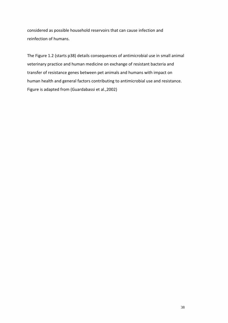
The Figure 1.2 (starts p38) details consequences of antimicrobial use in small animal
veterinary practice and human medicine on exchange of resistant bacteria and
transfer of resistance genes between pet animals and humans with impact on
human health and general factors contributing to antimicrobial use and resistance.
Figure is adapted from (Guardabassi et al.,2002)

39
Figure 1.2 Flow chart to show consequences of antimicrobial use in small animal veterinary practice and human medicine on exchange of resistant bacteria and transfer of resistance genes between pet animals and humans with impact on human health and general factors contributing to antimicrobial use and resistance.
Antimicrobial resistance in animals
Development and spread of resistant bacteria in pets
Limitation of therapeutic options in treatment of some disease in pets Transfer of
resistance genes
between pet animal
bacteria
Exchange of resistant bacteria between pets
and humans
Transfer of resistance genes
between pet animal and
human bacteria
Transfer of resistance genes between human
and bacteria
Limitation of therapeutic options in
treatment of some human
diseases
Development and spread of
resistant bacteria in humans
Antimicrobial use in human medicine
Pet to human transfer of
resistant bacteria
Human to human transfer of resistant bacteria
Human to pet transfer of resistant bacteria
Antimicrobial use
Antimicrobial
resistance
Human health impacts: -Reduced efficacy of antimicrobials used in humans -Increased healthcare costs -Increased human morbidity -Increased human mortality -Increased potential for carriage and dissemination of resistance genes
Antimicrobial residues in environment Dose and duration of treatment Gene transfer Clonal spread
Socioeconomic factors Travel Animal antimicrobial mis/over use Human antimicrobial mis/over use

40
Figure 1.3 details some examples of the reservoirs of antibacterial resistance involving both humans and animals.
Figure 1.3 Diagram to demonstrate the potential reservoirs of antibacterial resistance (blue arrows)
involving humans and animals and the selection pressures (red arrows).

41
Chapter 2: Materials and Methods
Materials and Methods for Results Chapter 3 - Reservoirs of resistance: polymyxin
resistance in veterinary-associated isolates of Pseudomonas aeruginosa:
2.1 Bacterial Isolates
24 P. aeruginosa isolates from 22 companion animal attending Leahurst Veterinary
Teaching Hospital (UK) in 2012 were collected for this study (Table 2.1). No prior
clinical details or antibiotic use for the cases were available. Human-associated P.
aeruginosa isolates (Table 2.1). from a previously published panel were used as a
comparison (De Soyza et al., 2013). Those isolates from this published panel that
were of non-human origin (eg. environmental origin) were excluded. All isolates
were stored in LB supplemented with glycerol and stored at -80 oC. When needed,
bacteria were grown on Columbia agar for 24hrs at 37oC.
Table 2.1 Veterinary and human bacterial isolates used in this study
Strain or Isolate Description (Origin/Source) Animal/Human Isolate Reference
1115 P.aeruginosa (Canine testicle/
scrotum swab)
Animal This study
1114 P.aeruginosa (Canine neck wound
swab)
Animal This study
1107 P.aeruginosa (Canine Bile sample) Animal This study
1098 P.aeruginosa ( Canine Vaginal
swab)
Animal This study
1095 P.aeruginosa (Canine Buccal
swab)
Animal This study
1090 P.aeruginosa (Canine Skin lesion
swab)
Animal This study
1070 P.aeruginosa (Canine neck swab) Animal This study
1055 P.aeruginosa (Canine Lip fold
swab)
Animal This study
994 P.aeruginosa ( Canine Right elbow
swab)
Animal This study
984 P.aeruginosa (Canine Right ear
swab)
Animal This study
982 P.aeruginosa (Canine Urine
sample)
Animal This study

42
978 P.aeruginosa (Canine Non-Healing
Wound swab)
Animal This study
970 P.aeruginosa (Canine Wound
swab)
Animal This study
969 P.aeruginosa (Canine
Oropharyngeal tube swab)
Animal This study
897 P.aeruginosa ( Canine ear swab) Animal This study
886-1 &-2 P.aeruginosa (Canine nail pus
swab)
Animal This study
992 P.aeruginosa ( Feline
Oropharyngeal tube stoma swab)
Animal This study
856 P.aeruginosa ( Equine Abdominal
incision swab)
Animal This study
903 P.aeruginosa ( Equine Urine
sample)
Animal This study
823 P.aeruginosa ( Canine Left ear
swab)
Animal This study
811 P.aeruginosa ( Equine nasal swab) Animal This study
467-L P.aeruginosa (Canine Left Ear
swab)
Animal This study
467-R P.aeruginosa ( Canine Right Ear
swab)
Animal This study
AES-1R (AUST01) Paediatric CF (1992), Melbourne
Australia
Human De Soyza et al., 2013
AUS23 (AUST02) Adult CF (2007), Brisbane
Australia
Human De Soyza et al., 2013
AUS52 (AUST03) Adult CF (2008), Hobart Australia Human De Soyza et al., 2013
2192 Chronic CF patient, Boston MA Human De Soyza et al., 2013
AMT 0023-30 (AMT2330) Paediatric CF, Seattle WA Human De Soyza et al., 2013
AMT 0060-3 (AMT60-3) Paediatric CF, Seattle WA Human De Soyza et al., 2013
AMT 0060-2 (AMT60-2) Paediatric CF, Seattle WA Human De Soyza et al., 2013
AMT 0023-34 (AMT2334) Paediatric CF, Seattle WA Human De Soyza et al., 2013
U018a CF patient, Hobart Australia Human De Soyza et al., 2013
57P31PA Chronic obstructive pulmonary
disease, USA
Human De Soyza et al., 2013
IST27N CF patient, spontaneous non
mucoid variant, Lisbon Portugal
Human De Soyza et al., 2013
IST27mucoid (IST27muc) CF patient, Lisbon Portugal Human De Soyza et al., 2013
TB CF10839 (TB) CF, Germany Human De Soyza et al., 2013
UCBPP-PA14 (PA14) Human burn isolate Human De Soyza et al., 2013
NH57388A Denmark, CF Human De Soyza et al., 2013
A5803 Community acquired pneumonia Human De Soyza et al., 2013
39016 ICU (acute infection), Spain Human De Soyza et al., 2013
AMT 0060-1 (AMT60-1) Paediatric CF, Seattle WA Human De Soyza et al., 2013
15108/1 (5801) ICU (acute infection), Spain Human De Soyza et al., 2013
13121/1 (13121) ICU (acute infection), France Human De Soyza et al., 2013
LES400 CF, UK Human De Soyza et al., 2013

43
39177 Keratitis, Manchester UK Human De Soyza et al., 2013
CHA CF Human De Soyza et al., 2013
PAK Clinical non CF Human De Soyza et al., 2013
1709.12 Leuven Belgium, Non clinical CF
(2004)
Human De Soyza et al., 2013
LES 431 Non CF parent of CF patient, UK Human De Soyza et al., 2013
AA43 CF (late), Germany Human De Soyza et al., 2013
DK2 CF Denmark (earliest isolates
1973-2007)
Human De Soyza et al., 2013
C3719 CF, Manchester UK Human De Soyza et al., 2013
RP1 CF, Germany Human De Soyza et al., 2013
Mi162 Non CF burn, Ann Arbour, MI ,
1997
Human De Soyza et al., 2013
AA2 CF (early), Germany Human De Soyza et al., 2013
KK1 CF, Germany Human De Soyza et al., 2013
679 Non CF urine sample, male,
Wroclaw Poland, 2011
Human De Soyza et al., 2013
AA44 CF (late), Germany Human De Soyza et al., 2013
PA01 (ATCC 15692)
(Pa01esh)
Genome sequenced isolate Human De Soyza et al., 2013
NN2 CF, Germany Human De Soyza et al., 2013
LES B58 (B58) CF, UK (1988) Human De Soyza et al., 2013
2.2 Susceptibility Testing
Minimum Inhibitory Concentration (MIC) Assays
For all isolates, minimum inhibitory concentration (MIC) for polymyxin B and colistin
from overnight broths of LB were performed using BSAC standardised methods
(BSAC) (Andrews, 2001). In brief, broth dilutions were performed in 96-well plates
with antibiotic concentrations from 128 ug/ml to 0.25 ug/ml. Positive and negative
controls were used throughout and all assays were performed in triplicate. When
isolates displayed high levels of resistance, additional assays using higher
concentrations up to 1024 ug/ml were performed. The plates were incubated at
37oC and assessed after 24 and 48 hrs of growth. Median values of the triplicate
MIC results were used to calculate resistance levels.
The susceptibility of each isolate was determined by the last concentration of
polymyxin B at which each isolate was unable to grow. For colistin, BSAC standard
44
breakpoints were used (4 ug/ml). This is consistent with the guidelines most
commonly used by human clinical microbiology diagnostic laboratories. However,
breakpoints for polymyxin B are not clearly defined. MIC values were interpreted
according to CLSI guidelines as BSAC and EUCAST do not provide breakpoint
guidelines for polymyxin B. CLSI recommended using breakpoints of S≤ 2 mg/L, I= 4
mg/L, R≥ 8 mg/L. It was considered that these breakpoints were justified for use in
this study as no alternatives were provided by BSAC/EUCAST. (BSAC, 2015).
MIC for Companion Animal Veterinary Antibiotic panel
Subcultures were made of the human and companion animal samples by passage
on blood agar or Columbia agar, cultured at 37oC overnight. Following incubation, a
few colonies were suspended in 5 mL sterile water solution to produce a uniform
turbidity of 0.5 McFarland. A 10 μL aliquot of this solution was then transferred to
11 mL Mueller–Hinton broth, and this broth then transferred to the Sensititre
COMPAN1F 96 well microtitre plate via the Trek Sensititre® AIM™ (TREK
Diagnostics, Cleveland, OH) automated inoculation delivery system. The plates
were incubated at 37 oC for 24 hrs and the plates read using Sensititre OptiRead™
Automated Fluorometric Plate Reading SystemTrek Sensititre® (TREK Diagnostic
Systems) to measure the MIC for inhibition of growth.
2.3 Clondiag Tube Array Genotyping for P. aeruginosa
Clondiag Tube Arrays (Alere Technologies) were used to type the isolates according
to the protocol. In brief, a sweep of each isolate was emulsified in 1 ml of sterile
diluted water (SDW) and centrifuged at 13000 rpm for 2 mins. The supernatant was
removed and resuspended in 200 uL of SDW, boiled for 5 mins and centrifuged at
13000 rpm for 2 mins. A master mix of Labelling buffer B1 and Labelling enzyme B2
was prepared. 5 ul of the supernatant was added to 5 ul of master mix. PCR was
performed (5 mins at 96 for 1 cycle, 50 cycles of 20 secs at 62 oC, 40 secs at 72 oC
and 60 secs at 96 oC. Following hybridization and washing, reagent C3 (containing
Horse Radish Peroxidase conjugate) was used to label the chip (the process is
depicted in Figure 3). Detection was performed using the Iconoscan, Iconoclust
Software (Alere Technologies). Tube Array images were transformed into array

45
types as previously described (Wiehlmann et al., 2007). In order to study the
veterinary isolates in the context of the wider population, an array type database of
>900 recorded P. aeruginosa strains was used (Cramer et al., 2012). For displaying
the wider P. aeruginosa population, the eBURST algorithm was applied (Feil et al,
2004). The number of isolates = 981, number of STs = 256, number of loci per
isolate =16 and number of resampling for bootstrapping = 1000 (for statistical
confidences).
Figure 2.1: Stages of the Clondiag Array Tube system adapted from Alere
Technologies.
(Alere, 2017a & Alere 2017b)
Layout of the AT chip

46
The AT consists of 77 oligonucleotides immobilised and embedded in a microchip in
the base of a tube (Wiehlmann et al., 2007). The layout is divided in to 3 sections, as
shown in Figure 2.2. These are; genomic islands, variable genes, and SNPs. The chip
contains 29 markers for a range of genomic islands and islets, 15 markers for
variable genes, a LES PS21 marker, and 16 markers relation to SNP loci. The markers
for the genomic islands, variable genes, and the LES PS21 marker are represented
by two spots on the chip, and the SNPs by four spots.
Interpretation of the AT chip
Analysis of the 13 SNP patterns at 7 conserved loci (ampC, citS, alkB2, fliCa, oprI,
oprL, and oriC) and the presence or absences of 3 variable genes encoding the type
III secretion virulence proteins ExoU and ExoS and the flagellin protein FliC provides
a genetic profile, which is initially read as a 16 digit binary code, and then translated
into a 4 digit hexidecimal code. The genomic islands and variable genes are
represented by two oligonucleotides and the presence of the spots on the chip
following hybridisation indicates whether the target is present. If the gene is
present the isolate is assigned a “1” for that locus. If the gene is not present it is
assigned a “0”. Each SNP is represented by four oligonucleotides: two on the left
which have sequences matching with PAO1 and two on the right with sequences
that do not match with PAO1 (these are considered a “mutant strain”). If the
hybridisation spots are stronger on the left (PAO1 sequence) the isolate is assigned
a “0” for that locus. If the hybridisation spots are stronger on the right (“mutant”
sequence) the isolate is assigned a “1” for that locus. Figure 5 demonstrates
examples of binding for both wild type (PAO1) and non-PAO1 (mutant) variants of
the hybridisation spots which were utilized to act as a guide when reading the AT
image.

47
Hexadecimal code conversion
The resulting hexadecimal binary code can then be converted to a 4 digit code as
shown in figure 6 and a genetic fingerprint is produced and assigned to the strain.
This code can then be compared to isolate information from several large,
previously described databases (Ballarini et al., 2012; De Soyza et al., 2013; Hall et
al., 2014; Wiehlmann et al., 2007). A combined dataset was created including
information from the isolates of the previous databases. The comparison allows for
further information to be acquired about the strain in question, in particular if it has
previously been assigned a clone type and environments from which isolates of the
same strain type have been previously collected.

48
C-45 C-46 C-47 PAGI-3-1 PAGI-3-8 PAGI-2-1
PAGI-2/3-1 PAGI-2/3-4 PAGI-2/3-5 PAGI-2/3-6
pKL-1 pKL-3 TB-C47-1 TB-C47-2 PAPI-1-Pili ch
PAPI-1-LuBiPr.
pKLC- unknown
pKLC-adhesion pKLC- metabol
Pyov. Rec. I
Pyov. Rec. IIa
Pyov. Rec. IIb
Pyov. Rec. III
Pyov. Rec. B
LES
PA0636 PA0722 PAGI-1 PA0980
PA0728 PA2185 Fla-island-1
Fla-2-orfA 47D7-1 PAPI-2-Actr
PA2221 PA3835 Fla-2orfI Fla-2orfj 47D7-2 PAPI-2-xF1753
ampC-7 fliCa fliCb exoS exoU
ampC-4 ampC-5 ampC-6
oprI ampC-1 ampC-3
citS-1 citS-2 oprI
fliCa fliCa alkB2
oriC oprL a oprL
Figure 2.2. Positions of SNPs, genomic islands, and variable genes on the AT chip. C45, C46, C47 – gene island in Clone C , PAGI – Pathogenicity Island, pKLC – pKLC102 plasmid in Clone C, pyov.rec 1,2a,2b,3 – pyoverdine type receptors I,II and III fpvA, pyov.rec B – pyoverdine type I receptor fpvB, LES PS21 marker, PA0636, PA0722, PA0728, PA2185, PA2221, PA3835 – PAO1 sequences, TB –C47-1, TB- C47-2 - TB, pKLC102 related gene island integrated in tRNA(Lys), fla-islands 1,2 orfA, orfI and orfJ – flagellin glycosylation islands. , fliCa, fliCb – Flagellin proteins, ExoU/ExoS – Type III secreted virulence factors, ampC – βlactamase, oprL – outer membrane lipoprotein, citS – citrate synthase, oprI – outer membrane lipoprotein, alkB2 – alkaline hydroxylase alkB2

49
Figure 2.3: Predetermined table for comparison of hybridisation patterns for the seven conserved loci used for AT chip interpretation.

50
Figure 2.4: Conversion of the hexadecimal binary code. Adapted from (Wiehlmann et al., 2007)
2.4 Genomic DNA extraction for Illumina sequencing
A selection of 7 isolates were selected to undergo whole genome sequencing (some
already carried out as part of preliminary unpublished work by Pottenger S,.&
Fothergill,J. 2014). Genomic DNA (gDNA) was extracted from these isolates using a
Promega Wizard Genomic DNA Purification Kit. The basic protocol was followed,
with some minor adjustments to improve yield and purity. Asterisks indicate where
the method used deviates from that provided by the manufacturer (experience of

51
lab has found these alterations gave better yields of DNA).
A single colony was picked and grown overnight in 5 ml LB broth in a shaking
incubator at 37°C and 180 rpm. To pellet the cells, 1 ml overnight culture was
centrifuged for 2 min at 14 000 rpm and the supernatant discarded. Cells were lysed
by adding 600 μl Nuclei Lysis Solution and mixing gently. The tubes were then
incubated at 80°C for 5 min and then cooled on ice for a further 5 min*. Once cool 3
μl RNase A Solution (Promega) was added and gently mixed. The tubes were then
incubated at 37°C for 1 h and then cooled on ice for 5 min*. Protein was
precipitated by adding 400 μl Protein Precipitation Solution* and vortexing
immediately. The tubes were kept on ice for 5 min and then centrifuged at 14 000
rpm for 5 min*. The supernatant was then transferred to a tube containing 600 μl
room temperature isopropanol and mixed by inverting the tube several times.
Following centrifugation at 14 000 rpm for 15 min* the supernatant was removed.
Once dry, 600 μl room temperature 70% ethanol (V/V) was added to each tube and
the samples were centrifuged at 14 000 rpm for 15 min*. The supernatant was then
removed and the tubes left to air dry completely. The DNA pellet was then
rehydrated by adding 100 μl DEPC-treated water and storing overnight at 4°C.
Quantification of genomic DNA by Qubit fluorometer
This process was carried out by colleague at University of Liverpool, Yasmin Hilliam.
Quantification of gDNA in samples was performed using a Qubit 3.0 fluorometer
and Qubit dsDNA broad range assay kit (Life Technologies). A working solution was
made using concentrated assay reagent and dilution buffer in a 1:200 dilution. 190
μl working solution was added to 10 μl of each of the two pre-diluted standards for
a total reaction volume of 200 μl. 2 μl sample was used per reaction, and made up
to a final volume of 200 μl with working solution. The tubes were vortexed for 2 – 3
seconds and then incubated at room temperature for 2 min before being inserted
into the Qubit fluorometer to measure fluorescence. Samples were required to
contain >20 ng μl-1 gDNA in order to be sent for sequencing.
52
Quantification and purity testing of genomic DNA by NanoDrop
spectrophotometer
This process was performed by colleague at University of Liverpool, Yasmin Hilliam.
Further quantification and purity testing of gDNA in samples was carried out using a
NanoDrop 1000 spectrophotometer (Thermo Scientific). Extracted DNA had
previously been eluted in DEPC-treated water and so the same was used as a blank
for NanoDrop measurements. 2 μl DEPC-treated water was carefully pipetted on to
the lower measurement pedestal of the spectrophotometer and the sampling arm
then closed to form a liquid column to allow the machine to make a blank
measurement. 2 μl sample was pipetted in the same manner on to the lower
measurement pedestal of the machine and measurements made. Both the upper
and lower measurement pedestals were wiped clean with a soft clean tissue
between sample measurements (Nanodrop, 2017a).
NanoDrop measurements provide a wide range of information on the absorbance
of the sample but the measurements most highly relating to the purity of the
sample are the ratios of sample absorbance at 260/280 nm and 260/230 nm.
260/280 ratio of absorbance assesses purity of DNA present in the sample. A ratio
of ~1.8 is generally accepted as “pure” for DNA samples. If the ratio is much lower
than 1.8 it may indicate the presence of protein, phenol or other contaminants that
absorb strongly at or near 280 nm. 260/230 ratio of absorbance is a secondary
measure of DNA purity and 260/230 ratios for “pure” samples are often higher than
the respective 260/280 ratios, often in the range of 1.8 – 2.2. If the ratio is lower
than expected, it may indicate the presence of contaminants which absorb at
230nm (Nanodrop, 2017b).
2.5 Whole Genome Sequencing (Illumina) of Bacterial Isolates
Genomic DNA from the seven polymyxin resistant isolates was extracted from
overnight cultures using the DNeasy Blood and Tissue Kit (QIAGEN). Genomic DNA
(500 ng) was mechanically fragmented for 40 sec using a Covaris M220 (Covaris,
Woburn MA, USA) with default settings. Fragmented DNA was transferred to PCR
tubes and library synthesis was performed with the Kapa Hyperprep kit (Kapa

53
biosystems, Wilmington MA, USA) according to manufacturer’s instructions. TruSeq
HT adapters (Illumina, SanDiego, CA, USA) were used to barcode the samples and
libraries were sequenced along with 41 other bacterial genomes in an Illumina
MiSeq 300 bp paired-end run at the Plateforme d’Analyses Génomiques of the
Institut de Biologie Intégrative et des Systèmes (Laval University, Quebec, Canada)
(Freschi et al., 2015). Resistant isolate genomes were assembled using the A5
assembler version (Coil et al., 2015) and annotated using prokka version 1.5
(Seemann, 2014).
2.6 Genomics
Genome assembly work was performed by Matthew Moore (University of
Liverpool). in collaboration with the Pseudomonas Genomics Consortium (PGC)
(Levesque RC, Kukavica-Ibrulj I, Jeukens J, Freschi L; L'Institut de biologie intégrative
et des systèmes (IBIS), Pavillon Charles-Eugène-Marchand 1030, avenue de la
Médecine, Labo 4142 Université Laval, Québec (QC), G1V 0A6 Canada).
The 7 polymyxin resistant isolate genomes were assembled using the A5 assembler
version (Coil et al, 2015) and annotated using prokka version 1.5 (Seemann, 2014).
All 7 polymyxin resistant genomes were aligned to reference genome PA01 and
separately, PA14 using bwa mem version 0.7.5a (McKenna et al, 2010). Resulting
.sam alignment files were sorted, converted to .bam format and duplicates marked
and removed using picardtools version 1.85. The Genome Analysis Toolkit (GATK)
version 3.3 was used to create indel targets, realign them and call variants using the
Unified Genotyper (UG) module. All variants were filtered using vcffilter version
GitHub (GitHub 2015) ‘DP >9’ and ‘QUAL >10’ to produce the final .vcf files.
Reference genomes PA01, PA14, PA7 and LESB58 and 338 deemed to represent the
wider population (Kos et al., 2015) were downloaded from the Pseudomonas
Database (Winsor et al., 2011). The core genome was defined and extracted by
Panseq (Laing et al., 2010) as 500bp fragments matching in all 342 genomes with
>=85% similarity. MEGA6 (Tamura et al, 2013) was used for all phylogenetic

54
analyses. Phylogeny was estimated using the maximum likelihood method (ML) and
tamura-nei substitution model from concatenated polymorphic sites within the
defined core genome. Inner node support was based on 100 bootstrap replicates.
The tree was drawn using figtree (Figtree, 2015).
Ortholog sequences for 31 polymyxin resistance associated genes were downloaded
from the Pseudomonas Database (Winsor et al., 2011). A custom blast database
was curated consisting of the 7 polymyxin resistant isolate genomes using the
BLAST+(55) makeblastdb software. Each polymyxin resistance associated gene was
aligned against the custom database using BLAST+ blastall. A python script was used
to extract matching regions and convert to amino acid sequences from the 7
polymyxin resistant isolate genomes.
2.7 Comprehensive Antibiotic Resistance Database (CARD) sequence data analysis
The CARD integrates disparate molecular and sequence data, provides a unique
organizing principle in the form of the Antibiotic Resistance Ontology (ARO), and
can quickly identify putative antibiotic resistance genes in new unannotated
genome sequences (CARD 2015). In addition to extensive browsing tools,
integrated within the CARD and available as tools on the website are BLAST
databases for all genes stored in the CARD. BLAST searches can include all genes or
subsets of CARD reflecting antibiotic resistance genes, antibiotic targets, and
antibiotic biosynthesis genes. The Resistance Gene Identifier (RGI) is a novel
analytical tool that provides a preliminary annotation of the submitted DNA
sequence(s) based upon the data available in the CARD. RGI accepts GenBank
accession or GI numbers, pasted sequences, or uploaded nucleotide sequence files
in FASTA format. Data with two or more FASTA sequences, such as whole-genome-
sequencing (WGS) assembly contigs, can be accepted (maximum size, 20 Mb). The
RGI analyzes the submitted sequences and provides a detailed output of predicted
antibiotic resistance genes and targeted drug classes. This includes resistance to
antibiotics via mutations in their targets or via dedicated antibiotic resistance gene

55
products (enzymes, protective proteins, and efflux systems). RGI results are then
summarized using a “resistance wheel,” with overall antibiotic resistance in the
center, antibiotic resistance classes in the middle, and individual antibiotic
resistance genes on the outer ring (McArthur et al., 2015; McArthur et al., 2013).
Seven of the sequenced isolates were uploaded in FASTA format and analysed using
this particular tool.
2.8 eBURST algorithm
The eBURST algorithm (eBurst, 2015) was devised as a way of displaying the
relationships between closely-related isolates of a bacterial species or population.
Using a model of bacterial evolution in which a founding genotype increases in
frequency in the population, and while doing so, begins to diversify to produce a
cluster of closely-related genotypes that are all descended from the founding
genotype. This cluster of related genotypes is referred to as a “clonal complex”.
The algorithm predicts descent of genotypes from the ancestor and displays the
output as a radial diagram. eBURST was originally devised and developed by Ed Feil
from the University of Bath (Feil et al, 2004). An enhanced version (eBurstv3) has
been developed with funding from the Wellcome Trust, and is hosted by
department of Infectious Disease Epidemiology Imperial College London. The
eBURST algorithm has mainly been applied to MLST data, although in this study it
has been used to display the relationships between genotypes identified through AT
genotyping. For use with the AT genotypes, the profiles were converted into a tab
delineated text file and uploaded to the eBURST site. For analysis, the programme
was set to estimate the relatedness based on 16 loci, with a minimum of one
identical loci for group definition, a single locus variant (SLV) count of 0 for
subgroup definition, and number of re-samplings for bootstrap support set to 1000.
The data is divided into groups of sequence types (STs) that have a level of similarity
in their SNP profiles. Within a single group all STs must be an SLV of at least one
other ST in the group. The primary founder of any group is defined as the ST that
differs from the largest number of other STs at only a single locus. In the case of two
STs having the same number of SLVs, the one with the greater number of double

56
locus variants (DLVs) is selected as the founding member. More than one group can
be displayed, appearing as a cluster, in a single eBURST diagram along with any
unlinked STs. The eBURST diagram shows the patterns of descent within a group in
a radial fashion with lines connecting the founder to each of its SLVs, and lines
connecting these STs to other STs varying from them at only one locus. The size of
the node representing an ST indicates the relative abundance of that ST within the
population.
2.9 Statistical Analysis:
SigmaPlot (version12.3) was used to make comparisions between the human and
veterinary polymyxin susceptibilities, using Fisher’s exact test. Non parametric test
were used on the basis that the results were not normally distributed.

57
Materials and Methods for Results Chapter 4 - PCR characterisation of antibiotic
resistant determinants in P. aeruginosa from companion animals:
2.10 Bacterial Isolates
Clinical P. aeruginosa (n=106) isolates from companion animal diagnostic samples
sent to Veterinary Diagnostic Laboratory at the Small Animal Teaching Hospital,
Leahurst (University of Liverpool, UK) between 2010-2015 were selected for this
study (Table 2.2). Selection of isolates for this study was made on the basis of
species (ie. any isolate that was from a companion animal species) and bacterial
species identification (all P. aeruginosa). Any duplicates were excluded. No clinical
data or prior knowledge of use of antibiotics in these cases was known. Overall,
106 isolates from companion animals (canine n=93, equine n=11, feline n=2) were
selected. The collection of isolates were stored at -80°C (Microbank, Pro-Lab
Diagnostics, Cheshire, UK). When required isolates were then transferred to
Mueller-Hinton nutrient agar (MHA, Oxoid, Basingstoke, UK) and incubated
overnight at 37oC in aerobic conditions. P. aeruginosa ATCC27853 was used as a
control strain.
Table 2.2 Clinical veterinary bacterial isolates used in this study from companion animals
Isolate Ref number Description Species
1
2
3
4
5
6
7
8
9
10
11
12
13
14
15
Chest drain fluid Canine
Right ear swab Canine
Skin swab Canine
Abdominal fluid Canine
Ear swab Canine
Vulval swab Canine
Urine Canine
Ear swab Canine
Ventral abdomen tissue Canine
Chin furuncle and foot swab Canine
Left stifle fluid Canine
Ear swab Canine
Ear swab Canine
Right ear swab Canine
Wound swab Right hind limb Canine

58
16
17
18
19
20
21
22
23
24
25
26
27
28
29
30
31
32
33
34
35
36
37
38
39
40
41
42
43
44
45
46
47
48
49
50
51
52
53
54
55
56
57
58
59
60
61
Left ear swab Canine
Nasal swab Equine
Left ear swab Canine
Abdominal incision swab Equine
Ear swab Canine
Urine Equine
Wound swab Canine
Urine Canine
Wound swab Canine
Swab right mandible (Non healing wound) Canine
Neck swab Canine
Urine Canine
Right ear swab Canine
Oesophagostomy tube stoma swab Feline
Right elbow wound swab Canine
Bile Canine
Vaginal swab Canine
Right ear swab Canine
Lip fold Canine
Right ear swab Canine
Buccal swab Canine
Skin lesion swab Canine
Left neck swab Canine
Testicle/scrotum swab Canine
Tissue Canine
Vaginal swab Canine
Wound swab Canine
Right ear swab Canine
Tracheal Canine
Left ear swab Canine
Right ear swab Canine
Tissue Canine
Right ear swab Canine
Right ear swab Canine
Bronchial aspirate Canine
Skin Canine
Urine Canine
Urinary catheter Canine
Right ear swab Canine
PEG tube stoma wound Canine
Left ear swab Canine
Bronchial aspirate Canine
Tissue Canine
Skin swab Canine
Wound swab Canine
Tracheal Canine

59
62
63
64
65
66
67
68
69
70
71
72
73
74
75
76
77
78
79
80
81
82
83
84
85
86
87
88
89
90
91
92
93
94
95
96
97
98
99
100
101
102
103
104
105
106
Wound swab Canine
Wound swab Canine
Right anal sac swab Canine
Catheter Equine
Tracheal wash Equine
Guttural pouch washes Equine
Tissue Canine
Right ear swab Canine
Wound serous discharge and fat tissue Canine
Urine Canine
Tissue Canine
Wound swab Canine
Right and Left BAL Feline
Left ear swab Canine
Urine catheter Canine
Maxillary sinus Equine
Colic incision swab Equine
Urine Canine
Urine Canine
Tissue Canine
Vulval fluid swab Canine
Urine Canine
Skin Canine
Left ear swab Canine
Right ear swab Canine
Ear swab Canine
Right ear swab Canine
Ear swab Canine
Left ear swab Canine
Right ear swab Canine
Urine Equine
Urine Canine
Midline swab Equine
Ear swab Canine
Ear swab Canine
Vulval swab Canine
TrWash ETTmu Canine
Ear swab Canine
Vaginal swab Canine
Urine Canine
Wound swab Canine
Urine Canine
Wound swab Canine
Right Ear Swab Canine
Urine Equine

60
Table 2.3 Clinical veterinary bacterial isolates used in this study from companion animals and identification of the sources of samples used.
Source of isolate Total No No from Equine
No from Canine
No from Feline
Ear 32 32 Skin (includes lip fold and neck swabs)
8 8
Wound/Nonhealing wound
14
Urine 14 2 12 Urine catheter 2 2 Catheter 1 1 Testicle/Scrotum swab 1 1 Vulval/Vaginal swab 6 6 Surgical wound/incision (including midline swab and colic incision)
4 1 3
Abdominal fluid 1 1 Foot swab 1 1 Stifle fluid 1 1 Tracheal/Tracheal wash 4 1 3 Guttural pouch 1 1 Maxillary sinus 1 1 Bronchial aspirate 2 2 Tissue swab (including abdominal tissue)
7 7
Anal sac swab 1 1 Buccal swab 1 1 Bile 1 1 Oesophogostomy tube stoma swab
1 1
Chest drain fluid 1 1 Bronchoalveolar lavage 1 1
2.11 Susceptibility Testing
Determination of Minimal Inhibitory Concentration (MIC) to a selection of
veterinary used antibacterials
Minimal Inhibitory Concentration (MIC) testing was performed using commercially
available Trek Sensititre COMPAN1F microtitre 96 well plates (Trek Diagnostics
Systems, West Sussex, UK), in accordance with manufacturer’s guidelines and as per
the method detailed earlier in Chapter 2 ‘MIC for Companion Animal Veterinary

61
Antibiotic panel’s – refer to page 31). P. aeruginosa ATCC27853 was used as the
quality control strain for MIC evaluations.
The MIC ranges were tested to a series of twenty two veterinary-used
antimicrobials (concentration ranges tested in brackets in mg/mL); ampicillin (0.25-
16), amoxicillin/clavulanic acid (4/2-32/16), trimethoprim/sulphamethoxazole
(0.5/9.5-2/38), penicillin (0.06-8), cefovecin (0.25-8), cefpodoxime (2-16),
erythromycin (0.5-4), oxacillin+2%NaCl (0.25-4), cefoxitin (2-16), clindamycin (0.5-
4), doxycycline (2-8), cefazolin (4-16), chloramphenicol (4-16), rifampicin (1-2),
amikacin (4-32), ceftiofur (2-16), enrofloxacin (0.25-2), gentamicin (1-8), imipenem
(1-8), marbofloxacin (0.25-2), ticarcillin (8-64), ticarcillin/clavulanic acid (8/2-64/2).
Three positive control wells were included on the plate. Results were interpreted
using standard EUCAST or CLSI breakpoint guidelines as appropriate.
Phenotypic detection of extended spectrum beta-lactamses (ESBL)
Detection of ESBL production was performed by double disc synergy test (DDST) (S.-
P. Lin, Liu, Lin, & Shi, 2012). Pairs of discs (MastDiscs, MAST Diagnostics Group Ltd,
Bootle, Merseyside, UK) containing the extended spectrum cephalosporins
cefotaxime, ceftazidime and cefpodoxime alone and in combination with clavulanic
acid (cefotaxime/clavulanic acid 30 mg/10 μg, ceftazidime/clavulanic acid 30 mg/10
μg and cefpodoxime/ clavulanic acid 30 mg/10 μg) were placed on the plates using
a multi-dispenser. Results were read after 24 hrs incubation at 37 °C. The test
isolate was regarded as an ESBL producer if the zone of inhibition around the
combination disc was at least 5mm larger (≥5mm larger) than that of the
cephalosporin alone (MAST group 2017).
Cloxacillin or boronic acid (250 μg/mL; both from Sigma-Aldrich Company Ltd.
Dorset, UK) was added to MHA to inhibit chromosomal cephalosporinase activity.
Following overnight culture, test isolates were suspended to 0.5 turbidity using
McFarland standards, and used to inoculate a Mueller-Hinton agar plate containing
the 250μg/mL cloxacillin or boronic acid (Lin et al., 2012) (Aghazadeh et al., 2014).
After plate drying, the discs were placed as described earlier.

62
Disc diffusion susceptibility testing against a human antibacterial panel
Antimicrobial susceptibility testing was performed by disc diffusion method to a
panel of clinically relevant commonly used human antibacterials containing
aztreonam (30 μg), meropenem (10 μg), cefepime (30μg), piperacillin (30 μg),
piperacillin (30 μg)/tazobactam (36 μg), and cefoxitin (30 μg) (all from Oxoid, Oxoid
Ltd, Thermo Fisher Scientific group, UK). Preparation of a standard bacterial
suspension equal to 0.5 McFarland was inoculated on MHA and the antibiotic discs
were placed on the plate using a multi-dispenser. Results were read after 24 hrs
incubation at 37 °C. The diameter of zone of growth inhibition were measured
interpretation performed by using the European Committee on Antimicrobial
Susceptibility Testing breakpoints (EUCAST 2015). Disc diffusion susceptibility
testing is a published, acceptable method for determining susceptibility and was
chosen in this instance as it enables large scale screening that wouldn’t be possible
in the time frame of this project if MICs were used for all antibiotics.
2.12 DNA Extraction
For DNA extraction a standard boil preparation method was followed. Subcultures
were made of the isolates by passage on MHA and incubation for 24 hrs at 37 °C. A
few bacterial colonies (two 5 μl sterile loops) were added to 1ml of sterile water in
1.5ml Eppendorf tube and mixed. The solution was then heated for 10 mins at 95
°C on a thermoplate, followed by centrifuging at 10,000 rpm for 2 mins. The
supernatant was removed to a fresh sterile Eppendorf tube for DNA use and labeled
appropriately for each isolate. This was stored at 4 °C for direct usage.
DNA Extraction – Wizard® Genomic DNA Purification Kit Method
Bacterial DNA was extracted from a selection of the P. aeruginosa isolates using a
commercially available Genomic DNA Purification kit (Wizard® Genomic DNA

63
Purification Kit, Promega Corporation, Madison, WI 53711-5399 USA) according to
the manufacturers guidelines.
1 ml of an overnight culture was added to a 1.5 ml microcentrifuge tube and
centrifuged at 13,000–16,000 × g for 2 minutes to pellet the cells. The supernatant
was removed. 3μl of RNase Solution was added to the cell lysate and the tube
inverted 2–5 times to mix. The sample was incubated at 37°C for 15–60 minutes
and then cooled to room temperature. A 200μl aliquot of Protein Precipitation
Solution was added to the RNase-treated cell lysate. This was then vortexed
vigorously at high speed for 20 seconds to mix the Protein Precipitation Solution
with the cell lysate. The sample was then incubated the on ice for 5 minutes. Then
centrifuged at 13,000–16,000 × g for 3 minutes. The supernatant (containing the
DNA) was transfered to a clean 1.5ml microcentrifuge tube containing 600 μl of
room temperature isopropanol. This was gently mixed by inversion until the
thread-like strands of DNA form a visible mass. Then centrifuged at 13,000–16,000
× g for 2 minutes. The supernatant was poured off and drained the tube. 600 μl of
room temperature 70% ethanol was added and the tube gently inverted several
times to wash the DNA pellet. After centrifuging at 13,000–16,000 × g for 2
minutes, aspirate the ethanol. The tube was drained on clean absorbent paper and
the pellet allowed to air dry for 10–15 minutes. Then adding 100 μl of DNA
Rehydration Solution to the tube and rehydrate the DNA by incubating the solution
overnight at room temperature or at 4 °C then stored the DNA at 2–8 °C.
2.13 Polymerase Chain Reaction (PCR)
DNA extraction was performed as previously described by boil preparation method.
PCR amplification (for both simplex and multiplex) was performed in a final volume
of 25 μl, using the primers as presented in Table 2.5.
The composition of PCR mixture was: 5 μl of a Master Mix, FIREPol® (5 x FIREPol®
Master Mix Ready to Load, Solis BioDyne, Estonia, a premixed ready-to-use solution
containing all reagents required for PCR - except template, primers and water).

64
In addition for each genes tested the forward and reverse primers (final primer
concentrations were 10pcm/μl PCR reaction), water and template DNA were added
in the quantities as listed in Table 2.3 for multiplex and Table 2.4 for simplex.
The PCR products were electrophoresed in 1% agarose gels containing PeqGREEN®
dye (VWR International Ltd, Leicestershire, England). The DNA fragments were
imaged with UV transilluminator and UViProMW software documentation system.
The DNA loading dye DNA/BsuRI (HaeIII) Marker 9, 50 mg (0.5
μg/μl)(ThermoScientific Molecular Biology), was placed on each gel run.
Table 2.4 Multiplex PCRs conditions
Genes Tested PCR Mixture PCR Conditions (using Applied BioSystems, Thermocycler 2720)
References
TEM, SHV, OXA 5 μl of FIREPol® Mastermix, 1μl of each forward and reverse primers, 12 μl water and 2 μl of each template DNA.
The amplification program for all of the aforementioned genes consisted of initial denaturation at 94 °C for 10 mins, annealing at 94 °C for 40 secs, 60 °C for 40 secs, extension at 72 °C for 1 min (for 30 cycles) and a final extension at 72 °C for 7 mins in a thermocycler.
(Dallenne et al., 2010)
GES, VEB, PER 5 μl of FIREPol® Mastermix, 0.75 μl of each forward and reverse primers, 13.5 μl water and 2 μl of each template DNA.
(Dallenne et al., 2010)
VIM, IMP, KPC 5 μl of FIREPol® Mastermix, 1.25 μl of each forward and reverse primers, 10.5 μl water and 2 μl of each template DNA.
(Dallenne et al., 2010)
qnrA, qnrB, qnrS 5 μl of FIREPol® Mastermix, 1 μl of each forward and reverse primers, 12 μl water and 2 μl of each template DNA.
initial denaturation at 94 °C for 10 mins, annealing at 94 °C for 1 min, 53 °C for 1 min, extension at 72 °C for 1 min (for 35 cycles) and a final extension at 72 °C for 10 mins in a thermocycler
(Robicsek et al., 2006)
The multiplexPCRs were not continued throughout due to misleading results (refer
to detail in Results Chapter 4) therefore it was decided to do all PCRs as simplex
method (details in Table 2.5).

65
Table 2.5 Simplex PCR conditions
Genes Tested PCR Mixture PCR Conditions (using Applied BioSystems, Thermocycler 2720)
References
blaTEM, blaSHV, blaOXA
5 μl of FIREPol® Mastermix, 1μl of each forward and reverse primers, 16 μl water and 2 μl of template DNA.
The amplification program for all of the aforementioned genes consisted of initial denaturation at 94 °C for 10 mins, annealing at 94 °C for 40 secs, 60 °C for 40 secs, extension at 72 °C for 1 min (for 30 cycles) and a final extension at 72 °C for 7 mins in a thermocycler.
(Dallenne et al., 2010)
blaGES, blaVEB, blaPER
5 μl of FIREPol® Mastermix, 0.75 μl of each forward and reverse primers, 16.5 μl water and 2 μl of template DNA.
(Dallenne et al., 2010)
blaVIM, blaIMP, blaKPC
5 μl of FIREPol® Mastermix, 1.75 μl of each forward and reverse primers, 15.5 μl water and 2 μl of template DNA.
(Dallenne et al., 2010)
group OXAs; OXA-1, OXA-2, OXA-10
5 μl of FIREPol® Mastermix, 0.5 μl of each forward and reverse primers, 18 μl water and 1μl of template DNA.
initial denaturation at 95 °C for 5 mins, annealing at 95 °C for 1 min, 55 °C for 40 secs, extension at 72 °C for 1 min (for 25 cycles) and a final extension at 72 °C for 10 mins in a thermocycler
(Poirel, et al, 2010)
qnrA, qnrB, qnrS 5 μl of FIREPol® Mastermix, 0.5 μl of each forward and reverse primers, 18 μl water and 1 μl of template DNA.
initial denaturation at 94 °C for 10 mins, annealing at 94 °C for 1 min, 55 °C for 40 secs, extension at 72 °C for 1 min (for 25 cycles) and a final extension at 72 °C for 10 mins in a thermocycler
(Robicsek et al., 2006)
AmpC 5 μl of FIREPol® Mastermix, 0.5 μl of each forward and reverse primers, 18 μl water and 1 μl of template DNA.
initial denaturation at 94 °C for 3 mins, annealing at 94 °C for 1 min, 60 °C for 40 secs, extension at 72 °C for 1 min (for 25 cycles) and a final extension at 72 °C for 5 mins in a thermocycler
(Rodríguez-Martínez et al, 2009)
CTX-MU, BEL-1, CS(integrons)
5 μl of FIREPol® Mastermix, 0.5 μl of each forward and reverse primers, 17 μl water and 2 μl of template DNA.
initial denaturation at 94 °C for 3 mins, annealing at 94 °C for 1min, 56.9 °C for 40 secs, extension at 72 °C for 1 min (for 25 cycles) and a final extension at 72 °C for 5 mins in a thermocycler
(Wedley et al., 2011) (Bogaerts, et al, 2007) (Zhao et al., 2003)
AAC(lb) cr variant 5 μl of FIREPol® Mastermix, 1 μl of each forward and reverse primers, 16 μl water and 2 μl of template DNA.
initial denaturation at 94 °C for 3 mins, annealing at 94 °C for 45 secs, 55 °C for 45 secs, extension at 72 °C for 45 secs (for 34 cycles) and a final extension at 72 °C for 5 mins in a thermocycler
(Hong et al., 2009; Park et al, 2006)

66
2.14 Primers Table 2.6 DNA sequencing primers used in this study. Primers purchased from Eurofins Genomics (Eurofins, Ebersberg, Germany)
2.15 Quantification and purity testing of genomic DNA by NanoDrop
spectrophotometer
Further quantification and purity testing of gDNA in samples was carried out using a
NanoDrop 1000 spectrophotometer (Thermo Scientific). Extracted DNA had
previously been eluted in DEPC-treated water and so the same was used as a blank
for NanoDrop measurements. 1 μl DEPC-treated water was carefully pipetted on to
Primer name
Sequence
Product size (bp)
Reference
TEM Fw TEM Rev
CATTTCCGTGTCGCCCTTATTC CGTTCATCCATAGTTGCCTGAC
800 (Dallenne et al., 2010)
SHV Fw SHV Rev
AGCCGCTTGAGCAAATTAAAC ATCCCGCAGATAAATCACCAC
713 (Dallenne et al., 2010)
OXA Fw OXA Rev
GGCACCAGATTCAACTTTCAAG GACCCCAAGTTTCCTGTAAGTG
564 (Dallenne et al., 2010)
GES Fw GES Rev VEB Fw VEB Rev PER Fw PER Rev VIM Fw VIM Rev IMP Fw IMP Rev KPC Fw KPC Rev qnrA Fw qnrA Rev qnrB Fw qnrB Rev qnrS Fw qnrS Rev OXA-1 Fw OXA-1 Rev OXA-2 Fw OXA-2 Rev OXA-10 Fw OXA-10 Rev CTX-MU Fw CTX-MU Rev BEL-1 Fw BEL-1 Rev CS-1 Fw CS-1 Rev AAC-61b Fw AAC-61b Rev AmpC Fw AmpC Rev
AGTCGGCTAGACCGGAAAG TTTGTCCGTGCTCAGGAT CATTTCCCGATGCAAAGCGT CGAAGTTTCTTTGGACTCTG GCTCCGATAATGAAAGCGT TTCGGCTTGACTCGGCTGA GATGGTGTTTGGTCGCATA CGAATGCGCAGCACCAG TTGACACTCCATTTACDG GATYGAGAATTAAGCCACYCT CATTCAAGGGCTTTCTTGCTGC ACGACGGCATAGTCATTTGC ATTTCTCACGCCAGGATTTG GATCGGCAAAGGTTAGGTCA GATCGTGAAAGCCAGAAAGG ACGATGCCTGGTAGTTGTCC ACGACATTCGTCAACTGCAA TAAATTGGCACCCTGTAGGC AGCCGTTAAAATTAAGCCS CTTGATTGAAGGGTTGGGCG GCCAAAGGCACGATAGTTGT GTCCGAGTTGACTGCCGG TCTTTCGAGTTACGGCATTAGC CCAATGATGCCCTCACTTTCC TGGGTRAARTARGTSACCAGAAYCAGCGG TGGGTRAARTARGTSACCAGAAYCAGCGG CGACAATGCCGCAGCTAACC CAGAAGCAATTAATAACGCCC GGCATCCAAGCACAAGC AAGCAGACTTGACTGAT TTGCGATGCTCTATGAGTGGCTA CTCGAATGCCTGGCGTGTTT ATGCAGCCAACGACAAAGG CGCCCTCGCGAGCGCGCTTC
399 648 520 390 139 538 516 469 417 908 700 760 593 482 1243
(Dallenne et al., 2010) (Dallenne et al., 2010) (Dallenne et al., 2010) (Dallenne et al., 2010) (Dallenne et al., 2010) (Dallenne et al., 2010) (Robicsek et al., 2006) (Robicsek et al., 2006) (Robicsek et al., 2006) (Poirel et al., 2010) (Poirel et al 2010) (Poirel et al., 2010) (Wedley et al., 2011) (Bogaerts et al., 2007) (Zhao et al., 2003) (Hong et al., 2009) (Rodríguez-Martínez et al., 2009)
67
the lower measurement pedestal of the spectrophotometer and the sampling arm
then closed to form a liquid column to allow the machine to make a blank
measurement. 1 μl sample was pipetted in the same manner on to the lower
measurement pedestal of the machine and measurements made. Both the upper
and lower measurement pedestals were wiped clean with a soft clean tissue
between sample measurements.
PCR products of adequate quantity were sent for sequencing to Source BioScience
(Source BioScience). The concentration of DNA required was dependent upon the
length of the sample sent. A concentration of 1 ng/ μl per 100 base pairs is required
for successful sequencing from putified PCR samples. Primers forwards and reverse
were also sent. These were required to be sent at 3.2 pmol/μl. For sequencing 5 μl
per reaction of both primer and DNA at the above specified concentrations was
required thus 15 μl of each was prepared.
2.16 DNA Sequencing
Genomic DNA from positive simplex PCR P. aeruginosa isolates was extracted using
a commercially available PCR clean up Gel extraction kit (NucleoSpin® Gel and PCR
Clean-up, Macherey-Nagel GmbH & Co, Duren, Germany) according to the
manufacturers guidelines.
The PCR amplicons were purified and sequenced on both strands. The DNA
sequences were analyzed with Staden software and compared with published DNA
sequences in the GenBank database https://blast.ncbi.nlm.nih.gov/Blast.cgi
2.17 Statistical Analysis
The 95% CIs were performed on proportions using the Wilson method
(http://epitools.ausvet.com.au)

68
Chapter 3: Results
3. Reservoirs of resistance: polymyxin resistance in veterinary-associated isolates
of Pseudomonas aeruginosa:
3.1 Introduction
Polymyxins are produced naturally by Bacillus polymyxa. They demonstrate
bactericidal activity against a wide range of species of Gram-negative bacilli
including Escherichia coli, Salmonella spp and P. aeruginosa. Polymyxins that are
utilised in clinical practice include polymyxin B and polymyxin E (colistin). These are
cationic peptides and the main difference between the two molecules is that
polymyxin B contains phenylalanine in position 6 (from the N-terminal), while
colistin contains D-leucine (Tran et al., 2016; Velkov et al., 2014). The outer
membrane normally serves as a protective barrier for Gram-negative bacteria
against antimicrobial peptides and disinfectants. The mechanism of action by which
polymyxins exert their effect is through electrostatic interaction with divalent
cations of the outer bacterial membrane. The polymyxin antibacterial peptides can
selectively bind to the lipopolysaccharide (LPS) of the outer cell membrane, the
membrane permeability is disturbed and this destabilization results in lysis of the
bacteria and ultimately leads to cell death (Chen et al., 2011; Yu., et al, 2015).
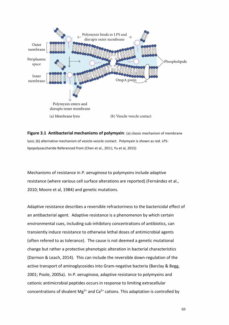
69
Figure 3.1 Antibacterial mechanisms of polymyxin: (a) classic mechanism of membrane
lysis; (b) alternative mechanism of vesicle-vesicle contact. Polymyxin is shown as red. LPS-
lipopolysaccharide Referenced from (Chen et al., 2011; Yu et al, 2015)
Mechanisms of resistance in P. aeruginosa to polymyxins include adaptive
resistance (where various cell surface alterations are reported) (Fernández et al.,
2010; Moore et al, 1984) and genetic mutations.
Adaptive resistance describes a reversible refractoriness to the bactericidal effect of
an antibacterial agent. Adaptive resistance is a phenomenon by which certain
environmental cues, including sub-inhibitory concentrations of antibiotics, can
transiently induce resistance to otherwise lethal doses of antimicrobial agents
(often refered to as tolerance). The cause is not deemed a genetic mutational
change but rather a protective phenotypic alteration in bacterial characteristics
(Darmon & Leach, 2014). This can include the reversible down-regulation of the
active transport of aminoglycosides into Gram-negative bacteria (Barclay & Begg,
2001; Poole, 2005a). In P. aeruginosa, adaptive resistance to polymyxins and
cationic antimicrobial peptides occurs in response to limiting extracellular
concentrations of divalent Mg2+ and Ca2+ cations. This adaptation is controlled by

70
the two-component regulators PhoP-PhoQ and PmrA-PmrB which up-regulate the
expression of the arnBCADTEF lipopolysaccharide (LPS) modification operon
(Fernández et al., 2010; Olaitan et al., 2014). The products of these arn genes
participate in the addition of 4-aminoarabinose to lipid A of LPS, thereby reducing
the net negative charge of LPS and limiting the interaction and self-promoted
uptake of polycationic antibiotics, such as polymyxins, host defense peptides, and
aminoglycosides, with the outer membrane (Fernández et al., 2010). However, this
adaptation to low divalent cations is unlikely to be clinically meaningful. Sub-
inhibitory concentrations of peptides can induce the expression of the pmrAB and
arnBCADTEF operons in P. aeruginosa. However, neither PhoP-PhoQ nor PmrA-
PmrB seems to be essential for this upregulation. Thus, an unidentified regulatory
system has been proposed to respond to peptides and promote the activation of
the arnBCADTEF and pmrAB operons independently of PhoP-PhoQ and PmrA-PmrB,
consequently increasing resistance to antimicrobial peptides (Fernández et al.,
2012; Gooderham et al., 2009; Macfarlane et al, 1999; McPhee et al, 2003). That is
the peptides themselves are able to induce adaptive resistance via LPS modification
(Fernández et al., 2010).
Genetic mutation is more stable and is inheritable. It is characterized by alterations
in the outer membrane. A study by Fernandez et al (Fernández et al., 2013)
reported a relatively small polymyxin B resistome involving 17
susceptibility/intrinsic resistance determinants. Their findings demonstrated that
the changes in susceptibility to polymyxin B and to colistin suggested evidence of
cross resistance. They showed that many of the same genes influence the
susceptibility of P. aeruginosa to different antibiotic classes and suggest that certain
genes may coordinate multidrug resistance possibly by affecting regulatory
pathways. In their study they determined the MICs of these strains to other
cationic antimicrobial peptides, including colistin. All of the tested transposon
mutants had altered susceptibility to colistin. The fold change in susceptibility of
the mutant was determined compared to that of wild-type PA14 by broth
microdilution for the polymyxins. In particular, it was of note that a 2 fold change in

71
MIC for polymyxin B also produced an identical 2 fold change in the MIC for colistin
(Fernández et al., 2013). This would suggest that the resistance genes of
P.aeruginosa appear to have the same effect on colistin as they do on polymyxin B
and may infer cross-resistance in the two polymyxins. This raises concern regarding
the use of polymyxins within the veterinary community whereby their use may help
drive development of polymyxin B/colistin resistant P. aeruginosa strains. These
could act as a potential source of resistant P.aeruginosa.
Several indications for polymyxin usage in veterinary medicine exist (as referenced
earlier in more detail in Chapter 1.1.5). Infections caused by Enterobacteriaceae in
rabbits, pigs, poultry, cattle, sheep and goats are a main indication for their use
(Bassetti et al 2015). In Europe, colistin and polymyxin B are used for treating
infections caused by Enterobacteriaceae in rabbits, broilers, veal, beef cattle, dairy
cattle and primarily pigs. In other areas of the world, polymyxins are still utilised as
growth promoters, however in Europe this usage has been banned since 2006
(Castanon, 2007; Millet et al., 2011). Polymyxin B is an antibiotic utilised in
veterinary medicine as a common first line topical therapy for treatment of otitis
externa in the form of proprietary ear drops product and may be used for systemic
treatment of endotoxaemia associated with severe colic and other gastrointestinal
diseases (Bassetti et al 2015). A 2013 European Medicines Agency report on colistin
usage in animals within the EU stated that across 19 EU/EEA European countries for
which sales data are available for 2010, polymyxins were the 5th most sold group of
antimicrobials (4.5%), after tetracyclines (39%), penicillins (23%), sulphonamides
(11%), and macrolides (9%) (Bassetti et al., 2015). However, this was based on food
producing animals (including horses) only and would not include companion
animals such as cats and dogs. The use of polymyxins specifically in companion
animals is not reported. However, several studies looking at the usage of
antimicrobial prescribing in small animal practice exist (Bassetti & Righi, 2015).
More recently there has been the development of a number of real time data
surveillance schemes that monitor illness and disease in pet animals using
veterinary practice and laboratory data SAVSNET (SAVSNET, 2017). Such
surveillance systems may prove useful for future analysis and development of
72
studies of antibiotic resistance data from companion animal across the UK (Jones et
al., 2014; Radford et al., 2011; Singleton et al., 2017).
Mechanisms of resistance in P. aeruginosa to polymyxins include adaptive
resistance and genetic mutations and generally characterized by alterations in the
outer membrane (Moore et al., 1984). A study by Fernandez et al (Fernández et al.,
2013) reported a relatively small polymyxin B resistome involving 17
susceptibility/intrinsic resistance determinants including evidence of cross
resistance between colistin and polymyxin B (Fernandez, 2013). This raises concern
regarding use of polymyxins within the veterinary community whereby their use
may help drive development of polymyxin B/colistin resistant P. aeruginosa strains.
Potentially selecting in veterinary medicine polymyxin-resistant isolates that may
subsequently be transferred to humans. These could then act as a source of
resistant P. aeruginosa populations (Moore et al., 2012).
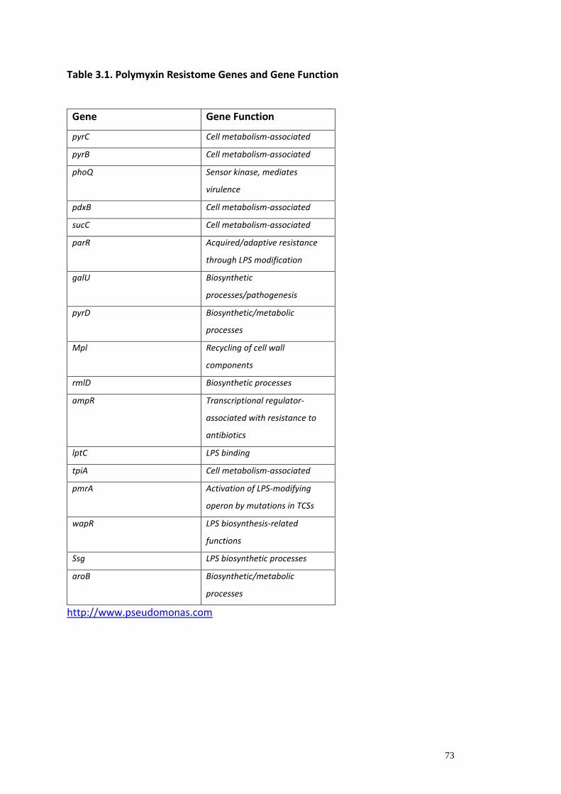
73
Table 3.1. Polymyxin Resistome Genes and Gene Function
Gene Gene Function
pyrC Cell metabolism-associated
pyrB Cell metabolism-associated
phoQ Sensor kinase, mediates
virulence
pdxB Cell metabolism-associated
sucC Cell metabolism-associated
parR Acquired/adaptive resistance
through LPS modification
galU Biosynthetic
processes/pathogenesis
pyrD Biosynthetic/metabolic
processes
Mpl Recycling of cell wall
components
rmlD Biosynthetic processes
ampR Transcriptional regulator-
associated with resistance to
antibiotics
lptC LPS binding
tpiA Cell metabolism-associated
pmrA Activation of LPS-modifying
operon by mutations in TCSs
wapR LPS biosynthesis-related
functions
Ssg LPS biosynthetic processes
aroB Biosynthetic/metabolic
processes
http://www.pseudomonas.com

74
Polymyxin Resistance Mechanisms
PhoP-PhoQ Two-Component System:
The OM usually serves as a permeability barrier to protect Gram-negative bacteria
from various antibacterial peptides and chemicals. The electrostatic interaction
between positively charged Dab residues on polymyxin and negatively charged
phosphate groups on lipid A of LPS are critical in its bactericidal activity. The
bacterial cell is able to reduce the initial electrostatic attraction by reducing net
negative charge of OM via lipid A modification, thereby increasing resistance to
polymyxins. The most common polymyxin-resistance mechanism in bacteria is
attributed to the ‘shielding’of phosphates on lipid A with positively charged groups.
This is mediated by PhoP-PhoQ regulatory system encoded by phoP locus (Yu., et al,
2015). Activated by PhoP-PhoQ, the PmrA-PmrB encoded by pmrCAB operon is the
major regulator to mediate the LPS modification in Gram-negative bacteria. PmrA-
dependent modification can occur on each of the three distinct LPS domains,
namely, lipid A, core polysaccharide, and O-antigen chain (McPhee et al., 2006;
Gellatly et al, 2012; McPhee et al., 2003).
Figure 3.2 Model describing the signals controlling expression of PhoP-PhoQ-
regulated determinants and the interaction between the PhoP-PhoQ and PmrA-
PmrB two-component systems, as well as some of the genes and phenotypes
governed by the PhoP-PhoQ system. Referenced from (Groisman, 2001).

75
MexAB-OprM efflux pump:
The MexA-MexB-OprM efflux system (Li et al., 1995) contributes to both intrinsic
and acquired resistance in P. aeruginosa. Reported studies with mutants that
overproduce or lack MexAB-OprM demonstrated that this efflux system extrudes
quinolones, macrolides, tetracycline, chloramphenicol, novobiocin, and most β-
lactams but not imipenem (Li et al., 1995; Masuda & Ohya, 1992; Srikumar et al,
1997). The MexAB-OprM efflux pump in P. aeruginosa has been proposed to confer
tolerance towards polymyxin E, due to the increase of mexAB-oprM expression in P.
aeruginosa upon polymyxin E exposure (Henrichfreise et al., 2007; Hirakata et al.,
2009; Li et al., 2000).
OM Protein:
Polymyxin resistance is reported to be associated with the expression of OM
proteins in bacteria. It has been suggested that the OM protein OprH, a membrane
stabilization protein, can promote resistance to polymyxin B in P. aeruginosa
(Macfarlane et al., 1999; Olaitan et al., 2014; Yu et al., 2015).
Plasmid mediated colistin resistance:
Liu et al 2015 (Liu et al., 2016) first reported the emergence of plasmid mediated
resistance gene, mcr-1 in E.coli and Klebsiella pneumonia isolates from pigs, meat
and human patients in China. In their study, the mcr-1 gene was present in 20.6% of
E. coli isolates from pigs, 14.9% of E. coli isolates from retail meat (pig and poultry)
and 1.4% of patients sampled (Liu et al., 2016). Occurrence of the mcr-1 gene has
been reported in E. coli, Salmonella and Klebsiella from Laos, Thailand, Tunisia,
Nigeria, South America and several European countries from people, pigs, poultry
and foodstuffs and in the UK (Anjum et al., 2016; Castanheira et al., 2016;
Falgenhauer et al., 2016; Kluytmans-van den bergh et al., 2016; Malhotra-Kumar et
al., 2016).

76
3.2 Aims
The significance of the role of companion animals as reservoirs of antimicrobial
resistance is an understudied area and merits further investigation. There is a risk of
transfer of resistant bacteria and/or resistance genes from animals to humans.
The overall aim of this study is to evaluate susceptibility of P. aeruginosa veterinary
isolates to polymyxin antibacterials and to determine whether the veterinary niche
represents a potential reservoir of resistant isolates.
Specific aims are to:
- Measure the susceptibility of a set of clinical P. aeruginosa isolates to
polymyxin antibiotics colistin (polymyxin E) and polymyxin B by broth micro
dilution.
- Compare these susceptibilities to a panel of human P. aeruginosa isolates.
- Determine the susceptibility of the P. aeruginosa isolates in the study to a
selection of antibacterials used in human clinical medicine by disc diffusion.
- Determine susceptibility of the P. aeruginosa isolates in the study to a
selection of commonly used veterinary antibacterials using an automated
microdilution system.
- Perform genotyping of the veterinary P. aeruginosa isolates using the
Clondiag Array Tube.
- Perform whole genome sequencing on a selection of resistant isolates to
determine genetic mutations in a defined subset of genes associated with
resistance to polymyxin antibacterials.

77
3.3 Results
Resistance to polymyxin antibiotics
Resistance of all P. aeruginosa isolates to polymyxin B and colistin were tested using
broth dilution MICs. For the veterinary isolates, 92 % displayed resistance to
polymyxin B and the remaining isolates were classed as displaying intermediate
resistance (Table 3.2), whilst 68% displayed resistance to colistin. For the human
isolates 33% displayed resistance to polymyxin B and just 3% (one isolate) to
colistin. Using the Fisher’s exact test the levels of resistance were signicantly higher
(ie. p<0.05) as compared to that of the human isolates (p = 0.033) for polymyxin B
and also significantly higher for colistin (p = 0.002).
Table 3.2 Minimum inhibitory concentration tests to show antimicrobial
resistance of P. aeruginosa in companion animal (n=24) and human (n=37)
isolates to polymyxin antimicrobials (calculated using median of triplicate MICs).
Isolate Antimicrobial Resistance
Breakpoint
(ug/mL)
Resistant
Strains
(n)
Resistant
Strains
(%)
Animal Polymyxin B ≥8 22 92
Animal Colistin >4 8 33
Human Polymyxin B ≥8 25 68
Human Colistin >4 1 3
Fisher’s exact test (significant = p<0.05): p value for polymyxin B MIC human v animal p=0.033 p value for colistin MIC human v animal p=0.002
The highest resistance was observed in isolate 970, from a canine wound, which
had an MIC of >100 mg/L (Figure 3.3). The level of resistance to colistin amongst the
veterinary isolates was 33% compared to only 3% in the human-associated panel
(Table 3.2). The isolates with the highest resistance to colistin were 856 and 1098
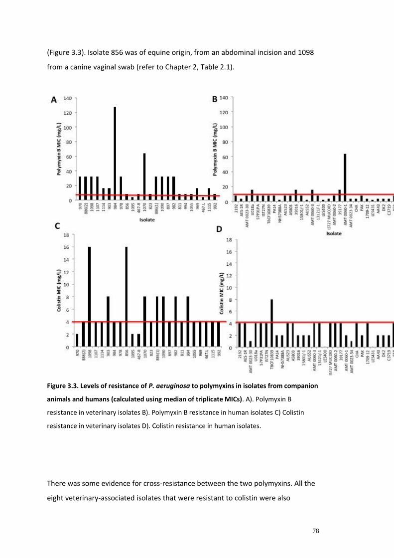
78
(Figure 3.3). Isolate 856 was of equine origin, from an abdominal incision and 1098
from a canine vaginal swab (refer to Chapter 2, Table 2.1).
Figure 3.3. Levels of resistance of P. aeruginosa to polymyxins in isolates from companion
animals and humans (calculated using median of triplicate MICs). A). Polymyxin B
resistance in veterinary isolates B). Polymyxin B resistance in human isolates C) Colistin
resistance in veterinary isolates D). Colistin resistance in human isolates.
There was some evidence for cross-resistance between the two polymyxins. All the
eight veterinary-associated isolates that were resistant to colistin were also

79
resistant to polymyxin B. The single human associated P. aeruginosa isolate, that
was resistant to colistin was also resistant to polymyxin B (Figure 3.3).
Resistance of veterinary-associated P. aeruginosa to other antibiotics
Disk diffusion susceptibility assays were performed using 5 commonly used
antibiotics in humans; ciprofloxacin, ceftazidime, tobramycin, meropenem and
tazobactam/piperacillin. No resistance was detected to ceftazidime, meropenem or
tobramycin. Two isolates were resistant to ciprofloxacin and a further three
displayed intermediate resistance. The two resistant isolates (1055 and 823) were
both of canine origin. Isolate 823 was completely resistant to ciprofloxacin with no
zone of inhibition (Figure 3.4). The isolates with intermediate resistance were 1095
(canine buccal swab), 467_L and 467_R (from the left and right ear of the same
dog). One isolate was resistant to TZP, isolate 823, from a canine ear. Isolate 823
had the lowest susceptibility of all the isolates tested. It was resistant to only
ciprofloxacin and tazobactam/piperacillin antibacterials (and intermediate to
meropenem).
Figure 3.4 Ciprofloxacin resistance in veterinary P. aeruginosa isolates (n=24). The
breakpoints are indicated by the red lines.

80
Using the Sensititre COMPAN1F mircotitre susceptibility assay, a panel of antibiotics
commonly used for veterinary clinical diagnostics was tested. Table 3.3 shows the
antibiotics with interpretation guidelines for P. aeruginosa.
Table 3.3 Antimicrobial resistance of P. aeruginosa isolated from companion
animals (n=24)
Antimicrobial Breakpoint
mg/L
Resistant
strains (n)
Intermediate
strains (n)
Sensitive
strains (n)
Resistant
strains (%)
Amikacin S=8, I=16),
R>16
0 0 24 0
Ceftiofur S=2, I=4, R>4 22 2 0 92
Enrofloxacin S=0.5, I=2,
R>2
8 13 3 33
Gentamicin S=4, I=8. R>8
1 0 23 4
Imipenem S=4, I=8. R>8
0 2 22 0
Marbofloxacin S=1, I=2. R>2
5 2 17 21
Ticarcillin S=64, R>64
5 0 19 21
Ticarc/clavulanic acid S=64, R>64 1 0 23 4
No resistance to amikacin was detected in the companion animal isolates and only
intermediate resistance was detected to imipenem. Four percent of isolates were
resistant to gentamicin and ticarcillin/clavulanic acid. Higher levels of resistance
were detected to ticarcillin (21%), marbofloxacin (21%) and enrofloxacin (33%). The
majority of isolates were resistant to ceftiofur (92%). The three most resistant
companion animal isolate to this panel of antibiotics were 823 (canine ear swab),
984 (canine ear swab) and 1055 (canine lip fold swab). Each of these isolates was
resistant to four of the antibiotics panel tested including enrofloxacin,
marbofloxacin and ticarcillin. These three isolates could be considered as multi
drug resistant (MDR) being non susceptible to 3 antimicrobial categories; ie. anti-
pseudomonal penicillin/beta lactamase inhibitor, fluroquinolones and a 3rd

81
generation cephalosporin. The companion animal isolates 467R and 467L (samples
from the same dog but Right and Left ear) were the least resistant.
The human isolates were also tested using the Sensititre COMPAN1F mircotitre
susceptibility assay. A similar trend in resistance was observed with the highest
resistance to ceftiofur and the lowest to amikacin. The human isolates showed
generally higher levels of resistance to the antibiotics tested (Table 3.4). The most
resistant human isolate to this panel was AUST03.
Table 3.4. Antimicrobial resistance of a human panel of P. aeruginosa strains (n=37)
Antimicrobial BreakPoints
mg/L
Resistant
strains (n)
Intermediate
strains (n)
Sensitive
strains (n)
Resistant
strains (%)
Amikacin S=8, I=16),
R>16
2 3 32 5.4
Ceftiofur S=2, I=4, R>4 35 2 0 95
Enrofloxacin S=0.5, I=2,
R>2
13 21 3 35
Gentamicin S=4, I=8. R>8
10 0 27 27
Impinenem S=4, I=8. R>8
8 7 22 22
Marbofloxacin S=1, I=2. R>2
13 0 24 35
Ticarcacillin S=64, R>64
8 0 29 22
Ticarc/clavulanic acid S=64, R>64
7 0 30 19

82
Circulating strain types of P. aeruginosa
The Clondiag Tube Array has been used to determine the population structure of P.
aeruginosa and a database of 955 isolates can be used to infer the wider population
(Wiehlmann et al., 2007). This typing technique was used to classify the companion
animal isolates. From the 24 isolates, 20 different array types were identified (Table
3.5).
Table 3.5. Clondiag Array Tube Hexidecimal codes of veterinary P. aeruginosa
isolates. SNP=single nucleotide polymorphism.
ANIMAL
ISOLATE
NUMBER
SNP
CODE
DETAILS OF
ISOLATES SHARING
THE SAME CLONE
TYPE AS THE
ANIMAL ISOLATES
1114 2C12 Water, Germany
467.L 0002 Same group as PA01
CF Germany.
Pneumonia, Switzerland.
1115 AF92 COPD, USA.
467.R 0002 CF Germany.
Pneumonia, Switzerland.
1098 B429 Keratitis, Great Britain.
River, Germany.
1095 AA0A Novel
978 6C12
CF, Italy.
COPD, USA
984 241A Acute infection, source
unknown.
Catheter, Germany.
823 241A Acute infection, source
unknown.
Catheter, Germany.
982 2FAA CF throat swab, Water,
Italy and Germany
1070 2D92 Novel
811 AA01 Novel
1107 2422 Novel
992 AF92 COPD, USA.
856 B420 Sputum, nose and water,
Germany.
969 840A Keratitis, Bristol.
903 B429 Keratitis, Great Britain
and River Germany.
83
994 0C12 CF sputum Germany.
897 848A Novel
970 0B9A Novel
886-1 2B92 Novel
886-2 2B92 Novel
1090 2O22 Novel
1055 0812 Found in CF patients,
bacteraemia and ICU,
France, Switzerland and
Austria.
Eight of these were novel array types not previously identified in the database. One
array type had previously been associated only with the environment from water).
However, the remaining array types had been previously associated with human
infections such as keratitis, cystic fibrosis and pneumonia lung infections and
catheter associated infections (Table 3.5). Using the database of array types to
generate a P. aeruginosa population structure, the isolates were mostly distributed
amongst the main P. aeruginosa clades. Five isolates were located as outliers,
isolates 811, 1107, 856 and the two isolates from the same dog, 467L and 467R
(Figure 3.5). The latter two isolates share an array type with the widely used
laboratory strain of P. aeruginosa, PAO1.
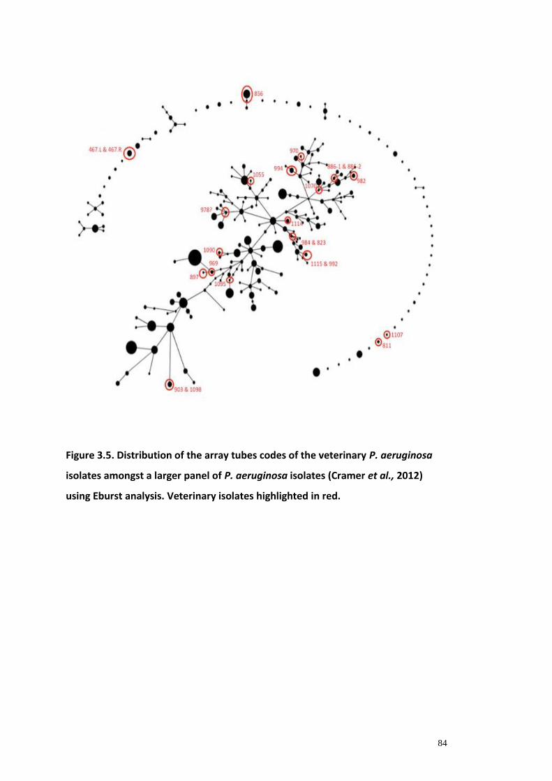
84
Figure 3.5. Distribution of the array tubes codes of the veterinary P. aeruginosa
isolates amongst a larger panel of P. aeruginosa isolates (Cramer et al., 2012)
using Eburst analysis. Veterinary isolates highlighted in red.

85
Genome sequencing and analysis of the polymyxin resistome
Seven isolates that displayed resistance to colistin and polymyxin B were selected
for whole genome sequencing. A phylogenetic tree of the veterinary isolates and
342 P. aeruginosa available genomes (Figure 3.6) shows that four of the isolates are
located in clade I and another two in clade II of the main P. aeruginosa population.
Although none of the isolates were found to be PA7-like, isolate 856 was diverse
from the main population and was located on a new arm of the phylogenetic tree
(Figure 3.6).
Figure 3.6. Maximum likelihood tree using the Tamura-Nei substitution model of P. aeruginosa core genome SNPs. The core genome is defined as polymorphic sites within 500bp fragments matching => 85% similarity in 342 genomes. The tree is based on 1336 phylogenetically informative polymorphic sites after gaps and N's were removed. Three clear groups are distinguishable: that containing PA14, PAO1 and PA7 (indicated by yellow coloured circles) respectively. Branches to PA7 and 856 (veterinary sequenced isolated indicated by blue coloured circles) have been truncated for clarity, so an impression of the real distances in the same tree has also been provided (the smaller diagram depicted below).

86
This genome had 125303 SNPs and 1884 indels compared to PAO1 and 130312
SNPs and 1985 indels in comparison to PA14 (Table 3.6). This was double the
number of SNPs and indels than any other isolate sequenced.
Table 3.6. Counts of overall variable sites, SNPs and Indels between each
polymyxin resistant isolate genome compared with PA01 and PA14 respectively,
by short read mapping and variant calling. Ts = transition and Tv = Transversion.
31 genes associated with resistance to polymyxins were analysed (Table 3.7 and
Table 3.8).
Multiple stop codons were identified in isolate 856, these causing potential loss of
function mutations. A stop codon in pyrC was identified in isolate 1098. Additional
amino acid modifications resulting in a change in hydrophobicity were identified in
all 7 genomes (Table 3.7).
Strain SNP count Indel count Total count Ts Tv Ts/Tv
vs PAO1 reference genome
903 56361 887 57248 43128 13233 3.26
856 125303 1884 127187 92796 32507 2.85
1098 57288 927 58215 43734 13554 3.23
982 29399 498 29897 21979 7420 2.96
1090 28373 538 28911 21404 6969 3.07
994 28454 507 28961 21415 7039 3.04
886_1 29138 532 29670 22016 7122 3.09
vs PA14 reference genome
903 35750 613 36363 27611 8139 3.39
856 130312 1985 132297 96630 33682 2.87
1098 42853 768 43621 32050 10803 2.97
982 59452 953 60405 45147 14305 3.16
1090 61403 1050 62453 46431 14972 3.1
994 60900 1004 61904 46129 14771 3.12
886_1 59378 950 60328 45211 14167 3.19

87
Table 3.7. Amino acid changes (presence of stop codons and changes in
hydrophobicity) identified in the 7 genome sequenced veterinary P. aeruginosa
isolates, for a selection of 31 genes analysed through whole genome sequencing
compared with PA01 and PA14 respectively, by short read mapping and variant
calling.
Function Amino
Acid/
Protein
856 886 903 994 982 1090 1098
Cell metabolism
associated
pyrC G423STOP G423STOP
pdxB V109K
D324A
V109K
aroB A200E A200E A200E A200E
A200E A200E
A200E
Acquired/adaptive
resistance through
LPS modification
parR L153R L153R
L153R
L153R
L153R
parS A115E
Recycling of cell
wall components
mpl A404P
E451STOP
Transcriptional
regulator associated
with resistance to
antibiotics
ampR E114A
M228R
R291STOP
E114A
E172V
E114A
M228R
E114A
M228R
Activation of LPS-
modifying operon by
mutations in TCSs
pmrA L71R L71R L71R L71R L71R
LPS biosynthesis-
related functions
wapR W114R
295STOP
Modification of lipid
A or Kdo with
aminoarabinose
arnC P23A
arnD A284D
arnF P137L
This showed the presence of resistance determinants including beta-lactamase
(PDC-1-7, amr), efflux (smeB, mex), fluoroquinolone resistance and elfamycin.
Isolate 856 had the least additional resistance genes however this was the most
divergent isolate through sequencing. In addition, the genome sequences were
analysed using the CARD database (refer to Figure 3.7).
Table 3.8. All amino acid changes to the selected 31 genes, identified in the 7
genome sequenced veterinary P. aeruginosa isolates for a selection of 31 genes

88
analysed through whole genome sequencing compared with PA01 and PA14
respectively, by short read mapping and variant calling.
Gene 856 886 903 994 982 1090 1098
pyrC D16G
T184S
Q247R
N297S
S355A
R368S
V402I
G423STOP
D16G
Q383E
D16G D16G D16G D16G
G130S
R363H
D16G
G423STOP
pyrB P272S
phoQ D90E
E267Q
V369L
pdxB Q369L A307V
T337N
Q365R
V109K
H183Y
P192T
A105V
L310M
A361T
E191G
D324A
Q365R
R179H
R247H
V109K
H183Y
P192T
parR T135A
L153R
L153R
S170N
P128S
L153R
S170N
L153R
S170N
L153R
S170N
Mpl S221T
I226S
Q347R
A404P
E451STOP
V358I V297M
A415V
V297M
rmlD V98I
ampR E114A
G283E
M228R
R291STOP
E114A
E172V
A208T
E114A
G283E
M228R
E114A
I251V
G283E
M228R
tpiA S47G
T78I
pmrA L71R L71R L71R L71R L71R
wapR A83T
T85A
W114R
295STOP
T85A R58H
R78K
T85A
T85A R78K
T85A
R58H
R78K
T85A
Ssg S35G A109V
aroB L54F
Q58E
V85A
E89G
A200E
V85A
A200E
V85A
A200E
V85A
A200E
S235L
I297T
V85A
A200E
V85A
A200E
I297T
V85A
A200E
amgS Q100R I260V I260V I260V
galU N338S
pyrD
sucC
lptC
arnA T42I
L50F
H199R
V250I
T297A
C312S
S313G
C312S
S313G
S509N
I551V
H59Y
F80Y
C312S
S313G
C312S
S313G
I551V
C312S
S313G
C312S
S313G
F80Y
C312S
S313G

89
V564I
arnB G120S
T143A
V153I
I231V
K286E
V302A
S321T
H332R
R363G
E376D
A316V
V302A
K286E
V302A
E376D
V302A
A316V
V302A
A316V
V302A V302A
K286E
E376D
arnC P23A
E35G
A265S
T316A
arnD E25D
F58L
V123T
G208S
A284D
F58L
arnE A32T
D33S
T26A
L56V
A84V
F87L
S114N
R28H
arnF A129T
P137L
V14M T106I
arnT C7W
L93F
L163F
A267S
P290A
L337Q
D440G
T443A
H447Y
R502Q
I509V
R502Q
I509V
C7W
H151Y
L337Q
I509V
T16S
E386D
R502Q A267S
R448H
R502Q
I509V
V266I
A282S
C7W
A214V
T443A
I509V
colS Q60K
V425I
cprR V27A
I59V
E183D
cprS V22I
A78E
D89G
V159I
V165I
T170A
E386D
T16S
parS E90K
A115E
V304I
H398R
H398R H398R
R243H
H398R H398R H398R H398R
pmrB
colR

90
Figure 3.7. Heat map of resistance genes present in 7 veterinary P. aeruginosa
isolates identified through whole genome sequencing. The shading of colour refers
to the percentage similarity between the resistance genes in the 7 isolates (the
darker colour representing highest % value (perfect match); mid-blue representing
a variant of the known AMR gene sequence and white for no sequence present.
Similar values are displayed as similar areas of colour.)
3.4 Discussion
In the veterinary setting P. aeruginosa can cause disease in a number of species. As
previously discussed in earlier Chapter 1 these include ears, skin, respiratory tract,
urinary tract, reproductive, ocular and wound infections. Similar to its nature in
humans, P. aeruginosa is also less common as a primary cause of disease in the
healthy animal. In canines, it is a frequently isolated pathogen in chronic otitis
externa, otitis media and chronic deep pyodermas (Morris, 2004; Nuttall & Cole,
isola
te_
98
2
isola
te_
109
0
isola
te_
99
4
iso
late
_8
86
.1
isola
te_
90
3
isola
te_
85
6
isola
te_
109
8
amrB_AF147719.1.gene3.p01
amrB_NC_002516.2.878882.p01
elfamycin resistant EF−Tu_NC_002516.2.881697.p01
elfamycin resistant EF−Tu_NC_002516.2.881718.p01
fluoroquinolone resistant parC_NC_002516.2.879741.p01
mexB_L11616.1.gene2.p01
mexD_NC_002516.2.881071.p01
mexD_U57969.1.gene2.p01
mexF_NC_002516.2.882884.p01
mexI_NC_002516.2.880346.p01
mexY_AB015853.1.gene2.p01
opmD_NC_002516.2.880347.p01
oprM_NC_002516.2.877851.p01
PDC−1_FJ666065.1.gene1.p1
PDC−1_NC_002516.2.878149.p01
PDC−2_FJ666064.1.gene1.p1
PDC−3_FJ666066.1.gene1.p1
PDC−4_FJ666067.1.gene1.p1
PDC−5_FJ666068.1.gene1.p1
PDC−6_FJ666069.1.gene1.p1
PDC−7_FJ666070.1.gene1.p1
rifampin resistant beta−subunit of RNA polymerase (rpoB)_NC_002516.2.881699.p01
smeB_AF173226.1.gene4.p01

91
2007). One of the commonly used first line therapies for canine otitis externa is the
effective commercial product Surolan® which contains polymyxin B as one of its
active ingredients. It is licenced for use in cats and dogs for several ear conditions
and skin infections.
A high proportion of the isolates within this companion animal sample set are of
canine origin and from skin (including wounds) or ear, sample sites. A recent report
looking at antimicrobial susceptibility profiles of bacterial isolates in the canine ear
in Australia identified P. aeruginosa as one of the 5 most commonly isolated
bacterial pathogen (Bugden, 2013). Of 3541 canine ear swabs, 35.5% isolated P.
aeruginosa. However, although they raised concerns of resistance levels in the
other Gram negative bacterial isolates (Escherichia coli and Proteus sp.) to
polymyxin B, the resistance in P. aeruginosa isolates was comparatively low to
polymyxin B (7%) and gentamicin (5%), although enrofloxacin was reasonably high
(36%) (Bugden, 2013). This is in contrast to the findings presented in our study
whereby polymyxin resistance levels of veterinary isolates was high. However, it is
of note that the samples are from a referral centre and as such the cases seen here
may be particularly extreme and difficult to treat having been sent from first
opinion general practice veterinary surgeons.
A number of other diseases involving P. aeruginosa in animals are known, including
that of contagious equine metritis (Allen et al., 2011) and in chronic equine wounds
where the ability of P. aeruginosa to form and survive within protective biofilms has
been reported (Freeman, 2009; Westgate 2011). Three of the samples within this
study were from horses; a urine sample, a nasal swab and an abdominal incision
wound and isolate 856 (equine abdominal incision swab) was shown to be the most
diverse from the main P. aeruginosa population. These findings add further
evidence that the veterinary niche is underrepresented in many population-wide
studies (Guardabassi et al., 2004).
Polymyxin B is used as a common treatment in veterinary medicine and it is feasible
that this longterm use in veterinary medicine has contributed to the increased

92
resistance. Therapeutics often use polymyxin B sulphate in the form of a topical
preparation (eg. Surolan®). A number of other formulations that include polymyxin
B for topical ocular solutions/ointments and ear preparations are available in other
countries (eg. Australia, Netherlands) but these are not marketed in the UK.
Polymyxin B in the form of polymyxin B sulphate administered intravenously has
previously been reported as a use in equine colics as a treatment of endotoxaemia.
Several studies document its efficacy (Barton et al., 2004; Werners et al., 2005). This
would be an off license use. Polymyxin B is on the list of substances essential for
the treatment of equidae for systemic treatment for endotoxaemia (antitoxigenic
effect, not antibacterial as such) associated with severe colic and other
gastrointestinal diseases (EU Agency, 2013; Barton et al., 2004; Richez & Burch,
2016). In the EU/EEA, colistin has been used in veterinary medicine since the 1950s
(Koyama et al., 1950), primarily for pigs including group treatments and prevention
of diarrhoea caused by E. coli and Salmonella spp., as first choice treatments for
neonatal diarrhoea in piglets (Timmerman et al., 2006) and veal calves (Pardon et
al., 2012) caused by E. coli as well as for the therapy of mild colibacillosis in poultry.
Colistin is not a therapeutic in use for companion animals in the UK although it has
previously been available as a licensed product for administration in various food
production animals for the indication of diarrhoea. However, in 2015 the
organisation RUMA (Responsible Use of Medicines in Agriculture Alliance)
announced that they would voluntarily restrict the use of colistin in UK livestock
(RUMA, 2015). This was following the reports of a new gene (MRC-1) that makes
common bacteria resistant to colistin, reports have been in E. coli (Castanheira et
al., 2016; Liu et al., 2016; Zhi et al., 2016a). There are now recent reports of
Salmonella and E. coli colistin resistance reported in the UK (Munja et al.,2016). It is
possible that the use of the polymyxins among veterinary species has driven
resistance within this niche of Pseudomonas isolates. Hence we have seen higher
levels of resistance in companion animal isolates as compared to that of the
comparative human panel.
Within the EU Member States, colistin and polymyxin B are authorised nationally.
The main indication for colistin in veterinary medicine is for preventing infection of

93
the gastrointestinal tract caused by non-invasive E. coli in pigs, poultry, cattle,
sheep, goats and rabbits. Colistin is also used in laying hens and cattle, sheep and
goats producing milk for human consumption. Colistin is also active against
endotoxins produced by some E. coli strains in the gastrointestinal tract. Typically,
colistin products are administered orally, in feed, in drinking water, as a drench, or
through milk replacer diets. Combinations of colistin with other antimicrobials are
available for group treatments of food-producing animals in some EU countries
(EMA, 2016b). Products for parenteral and intra-mammary administration are also
available, and infections due to Gram-negative bacteria in ruminants including
endotoxaemia are claimed indications.
As in human medicine, colistin and polymyxin B are registered for topical
administration to individual veterinary patients, except for food-producing animals
in the case of polymyxin B, in the absence of Meat Residue Limits. In companion
animals, prescription eye and eardrops are available with colistin alone, or in
combination with other antimicrobials. Colistin tablets are available for calves for
the prevention and treatment of neonatal colibacillosis. In some EU Member States,
veterinary medicinal products (VMPs) containing colistin are not on the market, i.e.
not commercialised (EMA/ESVAC, 2015).
Colistin products (polymyxin E) have never been marketed for use in animals in the
United States (US Food and Drug Administration, 2016). Sources from the FDA have
indicated that there is only one polymyxin B product (ophthalmic ointment,
combination of polymyxin B and oxytetracycline) approved for use in food-
producing species. In recent years, this product has been marketed in 2009 and
2012-2015, although it has been marketed in small quantities. Polymyxin B is also
available in the US as a component of approved ophthalmic products (for use in
dogs and cats) and otic products (for use in dogs). There is documented legal off-
label use in other non-food-producing species, such as horses. Sources from the
Public Health Agency of Canada have indicated that there are no approved colistin
products (polymyxin E) for use in animals in Canada (Public Health Agency of
Canada, 2016).

94
In 2013, the total sales of polymyxins in the 26 EU/EEA countries reporting data to
the ESVAC project (EMA/ESVAC, 2015), including tablets (but excluding topical
forms), polymyxins were the 5th most sold group of antimicrobials (6.1%), after
tetracyclines (36.7%), penicillins (24.5%), sulphonamides (9.6%), and macrolides
(7.4%). Total sales in weight summed up 495 tonnes. Of those 99.7% were for oral
forms as follows: 43.3% were oral solution (powder and liquid for use in drinking
water), 42.5% were premix (premixes for medicated feeding stuff) and 14.0% were
oral powder (powder to be administered with the feed or milk). Small amounts
were sold as: injectables (0.2%), tablets (0.1%) and intramammaries, intrauterines
and oral paste (less than 0.0% for each of the three forms). Of the group of
polymyxins, colistin represented more than 99.9% of the sales. In addition
combinations of colistin with other antimicrobials are authorised in some Member
States. The sales of those combination products represented less than 10% of the
overall sales of colistin (EMA/ESVAC, 2015). Colistin is used in aquaculture for the
prevention of Gram-negative infections (Xu et al., 2012), consumption data are not
available separately for this food production sector. In the Danish monitoring
programme (DANMAP), details on consumption do not refer to the use of colistin in
fish (DANMAP, 2012; EMA 2016).
Resistance to polymyxins has been attributed to modifications to the outer
membrane include modifications to lipid A and LPS as well as two-component
regulators.
The outer membrane normally serves as a protective barrier for Gram-negative
bacteria against the cationic antimicrobial peptides. The main difference between
the two molecules (polymyxin B and polymyxin E) being that polymyxin B contains
phenylalanine in position 6 (from the N-terminal), while colistin contains D-leucine
(Falagas & Kasiakou, 2005; Yu et al., 2015). Fernandez et al (Fernández et al., 2013)
reported a relatively small polymyxin B resistome involving 17
susceptibility/intrinsic resistance determinants. This study demonstrated
considerable cross-resistance in susceptibility to polymyxin B and to colistin. This
would suggest that the resistance genes of P.aeruginosa appear to have the same
95
effect on colistin as they do on polymyxin B and suggests some cross-resistance in
the two polymyxins. Seven of the veterinary isolates were genome sequenced and
mutations, including stop codons, were detected in genes previously implicated in
polymyxin resistance. Many of the sequences studied showed modifications in
genes leading to an altered amino acid, many of which had altered
hydrophobic/hydrophilic properties. Here, it is possible that a combination of
mutations leads to a change in phenotype through membrane remodelling.
A study by Manivannan et al (Manivannan et al, 2016) looking at a draft genome of
a MDR P. aeruginosa clinical isolate (from a female patient’s urine sample having
had ureteric reimplant) documented the presence of genes responsible for
virulence factors, quorum sensing and biofilm formation. The isolate was
extensively drug resistant and found to be resistant to penicillins, second and third
generation cephalosporins, aminoglycosides, carbapenems, tetracyclines,
quinolones, and trimethoprim-sulfamethoxazole and sensitive to only colistin, as
tested with the automated antibiotic susceptibility testing system Vitek®-2
(Manivannan et al., 2016). In their study, analysis indicated that the draft genome
has genes encoding for resistance to aminoglycosides, fosfomycin, beta-lactams,
sulfonamides, and trimethoprim (Manivannan et al., 2016).
A study by Lee et al (Lee et al., 2014), investigated genetic variations involved in the
acquisition and loss of colistin resistance in three clinical isogenic P. aeruginosa
isolates taken from a single patient and assessed their impacts on colistin
resistance.
Through the use of whole genome sequencing to identify single nucleotide
polymorphisms and insertions or deletions in two colistin-resistant isolates, which,
were then compared with a susceptible isolate. Their results showed 37 non-
synonymous mutations in 33 coding sequences detected in the colistin-resistant
isolates. One gene was significantly down-regulated in both colistin-resistant
isolates (a gene encoding erythronate-4-phosphate dehydrogenase). Eight genes
were up-regulated in the colistin-resistant isolates. Of these three encoded
hypothetical proteins and five were predicted to be involved in core biological
96
functions, encoding a cell wall-associated hydrolase, a response regulator EraR, a
sensor/response regulator hybrid, a glycosyltransferase and an arabinose efflux
permease (Lee et al., 2014). None of theses genes were identified in our polymyxin
study. They may be novel determinants that may be associated with the acquisition
of colistin resistance (Li., et al, 2014).
Using both the tube array typing method and genome sequencing on a limited
subset of isolates, the P. aeruginosa veterinary isolates were found to be
distributed thoughout the P. aeruginosa population. Using tube array, 9 of the
isolates had no previous description and were classified as novel. 15 isolates shared
array types with isolates previously found to cause human infections such as
pneumonia, cystic fibrosis and chronic obstructive pulmonary disease lung
infection, keratitis and bacteraemia and therefore highlight the ability of these
bacteria to cause human infections. Using whole genome sequencing, the
veterinary isolates were generally clustered within the main P. aeruginosa
population however one isolate, 856, did not cluster with any other previously
sequenced isolate. Through mapping to both PAO1 and PA14, the isolates had
double the number of SNPs and indels than any of the other isolates sequenced.
The isolate was of equine origin and this highlights the importance of studying
alternative and underrepresented niches in order to fully characterise the P.
aeruginosa pan genome.
In this study it is likely that polymyxin resistance involved chromosomal mutations
and there was no evidence of mcr-1 plasmid mediated colistin resistance. However,
a recent study discussed the emergence of plasmid-mediated colistin resistance
mechanism MCR-1 resistance in humans and animals (Liu et al., 2016). This study
was based in China and during a routine surveillance project on antimicrobial
resistance in commensal E.coli from food animals in China, a major increase of
colistin resistance was observed. When an E. coli strain, SHP45, possessing colistin
resistance that could be transferred to another strain, was isolated from a pig,
further analysis of possible plasmid-mediated polymyxin resistance was carried out.
This particular study demonstrated the emergence of the first plasmid-mediated

97
polymyxin resistance mechanism, MCR-1, in Enterobacteriaceae. The plasmid-
borne mcr-1 gene encodes a phosphoethanolamine transferase that mediates
addition of phosphoethanolamine to the lipid A moiety of the lipopolysaccharide
and thus transferring resistance to polymyxins (Liu et al., 2016). This is a gene that
to date has predominantly been identified in E. coli and also Klebsiella spp and
Salmonella spp. Since the first report by Liu et al 2016, further identifications
worldwide have emerged with reports in countries including in Algeria, Denmark,
France, Germany, Laos, the Netherlands, Malaysia, Nigeria, Switzerland and
Thailand (Castanheira et al., 2016; Doumith et al., 2016; Falgenhauer et al., 2016;
Kluytmans-van den bergh et al., 2016; Liu et al., 2016; Malhotra-Kumar et al., 2016;
Rolain et al., 2016; Liu et al., 2016; Zhi et al., 2016). The gene was also identified
from imported food products in Denmark – meat products (Hasman & Hammerum,
2015) and Switzerland, vegetables (Zurfluh, 2016). Plasmid-encoded carbapenem
and colistin resistance may be co-associated in E. coli (Falgenhauer et al., 2016).
Positive Salmonella enterica isolates of different serotypes were identified from
food samples in Portugal in 2011, and France in 2012 and 2016 (Figueiredo et al.,
2016; Lin et al., 2012; Zhi et al., 2016a). An epidemiological survey conducted in
France on a collection of ESBL-producing E. coli isolates recovered from the faeces
of diarrhoeic veal calves on farms from 2005 to 2014 showed a very high rate of
MCR-1-positive isolates (20.5%) (Haenni et al., 2015). Following these new reports
it was felt prudent to carry out further genomic assessment (courtesy of Matthew
Moore, University of Liverpool) of the seven P. aeruginosa isolates that had already
undergone WGS in this study. This additional work was performed by Matthew
Moore and Dr Jo Fothergill (University of Liverpool) after the main body of this
thesis work was completed (as this was only a one year funded Fellowship). The
seven isolates were screened for the presence of the mcr-1 determinant but none
were identified in this instance. As yet there does not appear to be reports of
plasmid encoded polymyxin resistance mechanism mcr-1 in P. aeruginosa isolates
(Baron et al., 2016).
In conclusion, all of these findings raise concerns regarding the use of polymyxins
within the veterinary community whereby their use may help drive development of

98
polymyxin B/colistin resistant P. aeruginosa strains. These could then potential act
as a source of resistant genetic material for the human associated P. aeruginosa
population. The findings suggest that future surveillance of polymyxin resistance in
animal associated P. aeruginosa strains should be recommended and highlights the
importance of improved correspondence between veterinary and medical
professions with regards to use of polymyxins. The veterinary setting, particular in
companion animals is an, often ignored, niche that provides close proximity
between humans and companion animals and therefore cross infection of resistant
organisms would be possible. The preservation of colistin as a “last resort”
successful anti-pseudomonal drug is of great concern (Kempf et al, 2016).
3.5 Conclusions
In this study, the increased resistance to polymyxin antibiotics, polymyxin B and
colistin in a panel of veterinary isolates from companion animals is described. To
the author’s knowledge, this is the first description of increased resistance to
colistin in this environmental niche. Resistance to other commonly used human
antibiotics was low.
Companion animals are an understudied source of antimicrobial resistant P.
aeruginosa isolates however, resistance to polymyxin amongst these isolates is
high. These findings merit sustained surveillance of the veterinary niche as a
potential reservoir for resistant, clinically-relevant bacteria.

99
4. PCR characterisation of antibiotic resistant determinants in P. aeruginosa from
companion animals
4.1 Introduction
AMR mechanisms in P. aeruginosa
P. aeruginosa can develop resistance to antibacterials either through the acquisition
of resistance genes on mobile genetic elements (i.e.integrative and conjugative
elements (ICEs)) or through mutational processes that alter the expression and/or
function of chromosomally encoded mechanisms. The main mechanisms associated
with antibiotic resistance are shown in Figure 4.1
Figure 4.1 Mechanisms of resistance in P. aeruginosa. Image from (Gullberg et al.,
2011)
100
Transmissible resistance refers to the ability to spread resistance genes via
horizontal gene transfer. ICEs (including plasmids ) are found in particular in
Enterobacteriaceae where they may play a role as important carriers of
antimicrobial resistance genes. Like plasmids, ICEs can exist in P. aeruginosa as
circular extrachromosomal elements and can impact on resistance to the beta-
lactams and aminoglycoside antibiotics. This is in contrast to resistance mechanisms
to fluoroquinolone among P. aeruginosa isolates which has mainly been associated
with chromosomal genes mutations such as gyrA, gyrB, parC and parE and/or
overexpression of multidrug efflux pumps (Fàbrega et al, 2009). Although the
plasmid-mediated qnr and the fluoroquinolone-modifying enzyme aac 6Ib-cr can
contribute to fluoroquinolone resistance among strains of Enterobacteriaceae
(Jacoby, 2005; Robicsek et al., 2006; Strahilevitz et al, 2009b), these two plasmid-
encoded mechanisms have not been found in clinical isolates of P. aeruginosa.
Resistance to the beta-lactams involves the production of inactivating beta-
lactamases and several families of these have been identified among clinical isolates
of P. aeruginosa (Lister et al., 2009). The most common imported beta-lactamases
found among P. aeruginosa isolates are penicillinases. The therapeutic impact of
these penicillinases is relatively limited since they do not impact the clinical efficacy
of extended-spectrum cephalosporins, monobactams, or carbapenems.
P. aeruginosa shows high intrinsic resistances to antibiotics and this can, in part, be
attributed to its’ low outer membrane permeability. The low permeability can
further contribute to other resistance mechanisms such as inducible
cephalosporinase or antibiotic efflux pumps therefore leading to highly resistant
bacteria. P. aeruginosa, in addition to being intrinsically resistant to several
antimicrobial agents, can acquire resistance to conventional anti-pseudomonal
antibiotics including anti-pseudomonal penicillins, ceftazidime, carbapenems,
aminoglycosides and ciprofloxacin.
Table 4.1 summarises the resistance mechanisms of P. aeruginosa.

101
Table 4.1. Examples of resistance mechanisms in Pseudomonas aeruginosa.
(Referenced from Gellatly et al., 2012).
Mechanism Example(s)
Efflux pumps MexAB–OprM, MexCD–OprJ, MexEF–OprN, MexXY–OprM (cephalosporins,
carbapenems, aminoglycosides, quinolones, ureidopenicillins)
Outer membrane
impermeability
OprF, OprD, OprB (carbapenems, aminoglycosides, quinolones)
β-lactamases AmpC (penicillins)
Targeted mutation DNA gyrase, DNA topoisomerase (quinolones)
MexZ (quinolones, cefepimes, aminoglycosides)
Horizontal transfer Metallo-β-lactamases, ESBLs (penicillins, cephalosporins, carbapenems)
Membrane changes Lipid A modification (aminoglycosides, polymyxins)
AmpC upregulation (penicillins)
ESBL, extended spectrum β-lactamase
Inherent and aquirred resistance can severely limit the therapeutic options for
treatment of serious infections (Lister et al., 2009).
Resistance types
Changes can be made to a bacteria's genetic inheritance in two ways; through
mutations that alter the pre-existing DNA of the cell—these alterations, base
changes and DNA deletions, insertions and inversions (Avison, 2005), change genes
already possessed but do not add new genes, to the cell genome. Alternatively,
resistance can also be obtained through the acquisition of new genetic material
(Bennett 2008).
1. Acquisition of additional genetic material
In bacteria, new genetic material can be acquired through mobile genetic elements
(ICEs) and can include plasmids, phages, transposons and integrons. The acquisition
of new genetic material is a naturally occurring process in bacteria and is facilitated

102
by the lack of a nucleus membrane protecting the genome. A study by Johnston et
al (Johnston., et al 2014) stated a total of 82 species having been shown to be
naturally transformable showing that acquisition through transformation is
common amongst many different species of bacteria. Gene transfer through
plasmids is one of the major routes for spreading antibiotic resistance in bacteria.
Many of the ICEs carrying antibiotic-resistant genes can be transferred between
different species of bacteria and therefore widespread resistance to a specific
antibiotic can develop rapidly. One example is a plasmid carrying a gene that
encodes the enzyme beta-lactamase. Beta-lactamases alters the structure of the
beta-lactam antibiotics (such as penicillin), rendering them inactive (Araujo et al.,
2016; Drawz & Bonomo, 2010). Outbreaks of life-threatening multi-drug resistant
superbugs such as New Delhi metallo-β-lactamase-1 (NDM-1) bacteria are often
due to the transfer of antibiotic resistance genes in ICEs (Abraham et al, 2014; Ji et
al, 2007; Liu et al., 2016; Poirel et al., 2010; Schmidt & Beutin, 1995).
Integrons are genetic elements first described at the end of the 1980s (Stokes &
Hall, 1989). Although most integrons were initially described in human clinical
isolates, they have now been identified in many non-clinical environments, such as
water and soil (Domingues et al, 2012; Nardelli et al., 2012). Integrons consist of
two conserved segments of DNA separated by a segment of variable length and a
sequence often carrying resistance determinants. Class 1, 2 and 3 integrons are
recognized (Domingues et al., 2012). Most resistance integrons belong to class 1,
which is the most frequent among isolates of human origin and has been found in P.
aeruginosa isolates from dogs (Lin et al., 2012; Rubin et al., 2008).
The class 1 integrons are important in the dissemination of resistance in Gram-
negative bacteria. Class 1 integrons being important in the worldwide problem of
antibiotic resistance, as they can capture and express diverse resistance genes and
may often be embedded in plasmids and transposons, facilitating their lateral
transfer into a wide range of pathogens (Gillings et al., 2008).
103
As described above, the acquisition of new genetic material requires gene transfer
from some outside source. Bacteria have three methods by which DNA may be
transferred from one cell to another; transformation, transduction and conjugation
(Bennett, 2009).
Resistance acquisition mechanisms:
i) Conjugation –
Conjugation is the process by which one bacterium transfers genetic material to
another through direct contact. During conjugation, one bacterium serves as the
donor of the genetic material, and the other serves as the recipient. Conjugative
plasmids, commonly present in bacterial cells (Sherley et al, 2003; Shintani et al,
2015) can be transferred to other cells by conjugation. This process often requires
sex pili to establish a mating-pair by promoting cell aggregation (Cabezón et al,
2015). Conjugation usually imposes a fitness cost on plasmid hosts as it requires
resources, alters cell physiology and determines viral susceptibility because sex pili
can serve as receptors for viruses (Cabezón et al., 2015).
This requires the participation of either a conjugative plasmid or a conjugative
transposon, each of which encodes a DNA transfer system that has evolved
specifically to mediate horizontal transfer of itself. Conjugation is responsible for
much of the horizontal gene transfer, particularly of resistance genes, seen among
prokaryotic cells, both Gram-negative and Gram-positive (Van Hoek et al., 2011).
ii) Transduction –
This involves gene transfer by bacterial ‘viruses’, called bacteriophages (‘phages’).
Phages tend to have very narrow host ranges. Transduction is the transfer of DNA
from one cell to another, mediated by a bacteriophage and occurs as a

104
consequence of rare errors in phage reproduction, when a small percentage of the
phage particles produced contain DNA sequences from the host cell, in place of or
in addition to the normal phage genome. Transduction is responsible for some of
the transfer of bacterial drug resistance genes (Munita et al, 2016). Fragments of
bacterial genomes or small bacterial plasmids can be moved from one bacterial cell
to another in this way. Survival of the DNA in the new cell requires either
recombination, prophage integration or the transferred DNA must be able to
replicate independently in its new host i.e. act as a plasmid (Van Hoek et al., 2011).
iii) Transformation –
Transformation requires no connection between the donor and recipient cells,
other than the DNA itself. The bacterial cell uptakes the DNA from its environment
and incorporates it into its own genome (Van Hoek et al., 2011).

105
Figure 4.2 The processes of bacterial, transformation(a), transduction(b) and
conjugation(c). (Furuya & Lowy, 2006)
a. Transformation occurs when DNA is released on lysis of an organism and is taken up by another
organism. The antibiotic-resistance gene can be integrated into the chromosome or plasmid of the
recipient cell.
b. In transduction, antibiotic-resistance genes are transferred from one bacterium to another by
means of bacteriophages and can be integrated into the chromosome of the recipient cell.
c. Conjugation occurs by direct contact between two bacteria: plasmids form a mating bridge across
the bacteria and DNA is exchanged, which can result in acquisition of antibiotic-resistance genes by
the recipient cell. Transposons are sequences of DNA that carry their own recombination enzymes
that allow for transposition from one location to another; transposons can carry antibiotic-resistance
genes. (Furuya & Lowy, 2006)

106
2 Intrinsic Resistance:
Intrinsic resistance is the ability of a bacterial species to resist activity of a particular
antibacterial agent through its inherent structural or functional features, which
allow tolerance of a particular drug or class of antibacterials (Munita et al., 2016).
This characteristic may be due to a lack of affinity of the drug for the bacterial
target. An example of such would be P. aeruginosa and the
sulphonamides/trimethoprim, tetracyclines and chloramphenicol whereby there is
a lack of uptake. Intrinsic resistance may also be due to inaccessibility of the drug
into the bacterial cell. This is the case for vancomycin and Gram-negative bacteria,
whereby the lack of uptake results from inability of vancomycin to penetrate the
outer membrane. Intrinsic resistance may be due to the extrusion of the drug by
chromosomally encoded active exporters or innate production of enzymes that
inactivate the drug. For example the production of beta lactamase enzymes by that
destroy beta lactam antibiotic such as ampicillin and destroy it before it can reach
the PBP (penicillin binding protein) target (Kong et al, 2010; Zeng & Lin, 2013) or
AmpC type beta lactamases which confer resistance to beta lactams. P. aeruginosa
also has constitutive expression of AmpC beta-lactamase (cephalasporinases) and
efflux pumps, combined with a low permeability of the outer membrane (Cabot et
al., 2011; Jacoby, 2009). The natural resistance of the species relates to the beta-
lactams: penicillin G; aminopenicillins, including those combined with beta-
lactamase inhibitors; first and second generation cephalosporins. Strains of a
particular bacterial species will exhibit the same mechanisms of innate resistance
and this is unaffected by prior antibiotic exposure (Beceiro et al., 2013; Schroeder et
al, 2017).
P. aeruginosa as discussed is intrinsically resistant to several antibiotics because of
the low permeability of its outer-membrane, the constitutive expression of various
efflux pumps, and the production of antibiotic-inactivating enzymes (e.g.
constitutive expression of AmpC beta-lactamase - cephalosporinases).
Furthermore, it also has a remarkable ability to develop or acquire further
mechanisms of resistance to multiple groups of antimicrobial agents, including

107
beta-lactams, aminoglycosides and fluoroquinolones (Mesaros et al., 2007a;
Mesaros et al., 2007b). P. aeruginosa exhibits many of the known mechanisms of
antimicrobial resistance; derepression of chromosomal AmpC cephalosporinase;
acquisition of integron-mediated beta-lactamases from different molecular classes
(carbenicillinases and extended-spectrum beta-lactamases belonging to class A,
class D oxacillinases and class B carbapenem-hydrolysing enzymes); diminished
outer membrane permeability (loss of OprD proteins); overexpression of active
efflux systems with wide substrate profiles; synthesis of aminoglycoside-modifying
enzymes (phosphoryltransferases, acetyltransferases and adenylyltransferases); and
structural alterations of topoisomerases II and IV determining quinolone resistance.
Concerningly, these mechanisms are often present simultaneously, thereby
conferring multi-resistant phenotypes (Strateva & Yordanov, 2009).
Membrane Permeability
P. aeruginosa, has a relatively impermeable outer membrane (OM) and expression
of numerous MDR efflux pumps that effectively reduce the intracellular
concentration of the given drug (Li et al., 1995). The OM is a semi-permeable
barrier to the uptake of antibiotics or nutrient molecules. Small hydrophilic
molecules (such as beta-lactam antibiotics) (refer to Figure 4.3. Blair et al, 2015)
have uptake restricted to a small portion of the OM (namely the water filled
channels of porin proteins) and as a result the entry of these molecules into the cell
is limited (Hancock, 1998). This low overall OM permeability is especially true of P.
aeruginosa, which is considered to be lower than some other Gram negatives. It is
estimated that the permeability of P. aeruginosa and Acinetobacter baumannii
outer membranes are only 1-8% of that E. coli and that non specific slow porins
OprF and OmpA-AB, respectively, restrict access of molecules larger than approx.
200Da, which is the size of a typical monosaccharide (Zgurskaya et al, 2015). The
major porin OprF is responsible for the large exclusion limit of the P. aeruginosa
OM. The porin OprD only mediates passage of zwitterionic carbapenem beta
lactams (eg. imipenem, meropenem) see Figure 4.5), as this porin contains a
binding site specifically for these dipeptides. Uptake across the OM of polycationic
antibiotics (eg. gentamicin, tobramycin and colistin) are mediated by a system

108
termed self-promoted uptake. This system involves the interactions of the
polycation with divalent cation binding site that are on the LPS cell surface and that
normally stabilise the OM. Since the polycationic antibiotic molecules are much
larger than the native divalent cations, they cause a disruption to the OM that
makes it become more permeable. – see Figures 4.4 and 4.5 (Lister et al., 2009).
Figure 4.3 Overview of intrinsic resistance mechanisms (Blair et al, 2015).
Depicting β-lactam antibiotics targeting a penicillin-binding protein (PBP). Antibiotic A can enter the
cell via a membrane-spanning porin protein, reach its target and inhibit peptidoglycan synthesis.
Antibiotic B can also enter the cell via a porin, but unlike Antibiotic A, it is efficiently removed by
efflux. Antibiotic C cannot cross the outer membrane and so is unable to access the target PBP (Blair
et al, 2015).

109
Figure 4.4 Mutational resistance to fluoroquinolones and carbapenems involving
chromosomally encoded mechanisms expressed by P. aeruginosa (Lister et al.,
2009).
Image (A) Depicts interactions of fluoroquinolones and carbapenems with susceptible P. aeruginosa expressing basal levels of AmpC, OprD, and non-mutated fluoroquinolone target genes (gyrA, gyrB, parC, and parE). Fluoroquinolone molecules pass through the outer membrane, peptidoglycan, periplasmic space, and cytoplasmic membrane and interact with DNA gyrase and topoisomerase IV (Topo IV) targets in the cytoplasm when these enzymes are complexed with DNA. The carbapenem molecules pass through the outer membrane-specific porin OprD and interact with their target PBPs (Penecillin-binding proteins), located on the outside of the cytoplasmic membrane. Image (B) Depicts chromosomally encoded mechanisms of resistance to fluoroquinolones and carbapenems. Fluoroquinolone resistance is mediated by (i) overexpression of RND efflux pumps extruding the drug molecules from the periplasmic and cytoplasmic spaces and/or (ii) mutational changes within the target genes. Locations of the QRDRs within target genes are highlighted in yellow. Carbapenem resistance is mediated primarily by (i) decreased production or loss of functional OprD in the outer membrane and/or (ii) overproduction of RND efflux pumps (with the exception of imipenem). Minor changes in susceptibility can be observed due to overexpression of AmpC, adding to the resistance potential (Lister et al., 2009)

110
Efflux Pumps
Efflux pumps are transport proteins involved in the extrusion of toxic substrates
from within cells into the external environment (Figure 4.5). These are known to be
important for intrinsic resistance of P. aeruginosa to antibiotics such as some
fluroquinolones, beta-lactams (except imipenem), tetracyclines and
chloramphenicol antibiotics. The active efflux systems of P. aeruginosa involve a
number of efflux systems including MexAB-OprM, MexCD-OprJ, MexEF-OprN and
MexXY-OprM (Webber & Piddock, 2003).
Figure 4.5. Three-component efflux pump (Livermore, 2002).
The pump itself (MexB, MexD, or MexF, according to the system) lies in the cytoplasmic membrane
and is attached via a linker lipoprotein (MexA, MexC, or MexE) to the exit portal (OprM, OprJ, or
OprN). Efflux system components appear in large roman type; other membrane components appear
in small italic type (Livermore, 2002).

111
3. Mutation of existing DNA:
This refers to the change by mutation of an existing gene product or control
mechanism. Changes in the bacterial genome through mutation may consequently
lead to a change in the nature of proteins expressed by the organism and this can
result in changes in antibacterial resistance. There are many mechanisms of
resistance through the accumulation of mutations. These are often through gene
‘loss of function’ mutations resulting from small nucleotide polymorphisms (SNPs)
or insertions/deletions of DNA. In P. aeruginosa, these mutations are very common
and contribute to the high resistance in many P. aeruginosa isolates. The
accumulation of mutations is also very common during chronic infections and
repeated exposure to antibiotics can help to select for mutated isolates with higher
resistance. These mutations can results in altered target sites and altered activity of
existing mechanisms such as efflux pumps. The target site may be altered by
mutation such that it evades the action of or no longer binds the antimicrobial
agent (e.g. ciprofloxacin). Mutations that result in alterations of an antibiotic's
target can also confer resistance, for example where a mutation in DNA gyrase
reduces the binding affinity of the enzyme for fluoroquinolones leading to
resistance (Breidenstein et al, 2011; Gellatly et al., 2012; Schweizer, 2003).
Mutations in bacterial DNA may change the target enzyme in a metabolic pathway
to therefore bypass the primary target (e.g. trimethoprim) (Munita et al., 2016; Van
Hoek et al., 2011). Bacteria may develop higher activity of efflux pumps that expel
the antimicrobial agent before it reaches an effective concentration (e.g
tetracyclines). Examples of these include, natural inducers, such as aminoglycosides
and other antibiotics targeting ribosomes, a mutation in mexZ, which normally
suppresses expression of mexXY , leads to the overexpression of the MexXY efflux
pump (Matsuo et al, 2004).

112
4. Further Mechanisms of Resistance
Tolerance and resistance of bacteria growing in biofilms:
Bacteria can grow as multicellular communities called biofilms, distinct from
planktonic growth. Biofilm-associated bacteria typically cause subacute (subacute
being not yet chronic >3mths, but has passed the acute phase <1mth. It may also
indicate that the condition is not as severe as the acute stage) and chronic
infections. They are clinically significant due to their persistence despite sustained
antimicrobial treatments and adequate host defenses. Biofilm bacteria are highly
resistant to a wide range of antimicrobial compounds and disinfectants, and the
mechanism underlying this resistance is multifactorial. Biofilms can be difficult to
eradicate when they cause biofilm-related diseases, e.g., implant infections, cystic
fibrosis, urinary tract infections, and periodontal diseases. A number of phenotypic
features of the biofilm can be involved in biofilm-specific tolerance and resistance.
Little is known about the molecular mechanisms involved (Olsen, 2015).
Antibiotic resistance typically refers to an increase in the minimum inhibitory
concentration (MIC) value of an antibiotic due to a permanent change in the
bacteria, e.g. mutation or resistance acquired through horizontal gene transfer.
However, antibiotic tolerance is the ability of cells to survive the effect of an
antibiotic due to a reversible phenotypic state. In biofilms, antimicrobial tolerance
is related to the mode of growth of the biofilm. This is in contrast to bacteria
growing in planktonic culture. A number of mechanisms have been reported to
operate together to produce a high total biofilm tolerance or resistance (Cao et al.,
2015; Sousa & Pereira, 2014; Strateva & Mitov, 2011). Biofilms are not regarded as
uniform cultures of physiologically identical cells and different mechanisms can
protect subpopulations of cells in the biofilm (O’Toole et al., 2000). Antibiotic
tolerance mechanisms in biofilms include failure of antibiotics to penetrate biofilms,
slow growth rate, altered metabolism, persister cells, oxygen gradients, and
extracellular biofilm matrix (Olsen, 2015).
113
Biofilms play an increasing role within the medical and veterinary community. Due
to the increased resistance of a biofilm, they can complicate chronic non-healing
wounds, implant/prosthesis infection and mastitis (Pye et al., 2013; Scaccabarozzi
et al., 2015; Westgate et al., 2011). With maturity, biofilms may become more
resistant to the effects of antimicrobials, which make the infection harder to treat
(Olsen, 2015; Sousa & Pereira, 2014). Many antibiotic therapies currently used to
treat bacterial infections are aimed at planktonic bacterial cells as opposed to cells
encased in a biofilm; this makes their treatment increasingly problematic. Without
adequate diagnostic and treatment protocols to treat veterinary biofilms, their
impact will remain a significant challenge.
Resistance to Beta-lactam Antibiotics
Beta-lactamases and Classification
β-lactam antibiotics are one of the most effective and commonly used antibacterials
with their useage approximating 60% of the global antibiotic use (Kong et al., 2010).
The Infectious Diseases Society of America (IDSA) reported that three of the top six
listed dangerous pathogens are β -lactam resistant bacteria (IDSA, 2017). Resistance
to β -lactam antibiotics may be through production of β -lactamase enzymes that
hydrolyse the β -lactam ring of the antibiotic or, changes of the penicillin binding
proteins that maintain the peptidoglycan structure in bacterial cell wall. Also via
alteration of porin channels and by initiation of efflux exporter proteins (Öztürk et
al, 2015).
The most widely used classification of the β-lactamases is the Ambler classification.
This divides β-lactamases into four classes (A, B, C and D) based upon their
homology of amino acid sequences (Table 4.2). Ambler class A is the active-site
serine β-lactamases; Ambler class B, is the metallo-β-lactamases that require a
bivalent metal ion (usually Zn 2+) for activity. Ambler class C refers to the serine β-

114
lactamases that were found to have dissimilar sequence to those already identified
as the class A enzymes. Within the designated class C, includes the ‘AmpC’ β-
lactamases. Ambler class D is a further classification of serine β-lactamases, referred
to as the OXA β-lactamases (Bush & Jacoby, 2010; Hall & Barlow, 2005; Weldhagen
et al., 2003).
The metallo-β-lactamases, are a group of enzymes that hydrolyse β-lactams through
an enzymatic process that is distinctly different from that of the serine β-
lactamases, and they are structurally unrelated to the serine β-lactamases. All
metallo-β-lactamases have been defined as class B β-lactamases. These are sub-
dived into three subgroups, B1, B2 and B3.6 (Hall & Barlow, 2005).
IMP and VIM are metallo—β-lactamases that rapidly hydrolyze penicillins,
cephalosporins, and carbapenems (not aztreonam) (Hall & Barlow, 2005).
Resistance to penicillins and cephalosporins usually accompanies production, but
carbapenem resistance may also require the loss of OprD. Various IMP enzymes
have been reported in Japan, Canada, Italy, France, Greece, and South
Korea,Taiwan (Deshpande et al., 2013; Docquier et al., 2003; Hayakawa et al., 2014;
Jeannot et al., 2012; Tada et al., 2016). VIM enzymes resemble IMP types with
regard to their hydrolytic properties, they but share only 30%–40% amino acid
identity (Livermore, 2002).
The genes for VIM and IMP enzymes, like those for OXA-ESBLs, are often carried as
cassettes within integrons, which are natural recombination systems that assemble
series of acquired genes behind a single promoter. This organization facilitates
gene recombination. The β-lactamase genes are often adjacent to aminoglycoside
6′-N acetyltransferase [aac(6′)-1b] determinants (Araujo et al., 2016; Galimand et al,
1993). A P. aeruginosa strain with this combination of cassettes is susceptible only
to polymyxins, ciprofloxacin, and, perhaps, aztreonam. If MexAB-OprM is up-
regulated by mutation, or if the isolate has topoisomerase and ampD mutations,
then only susceptibility to polymyxins remains (Livermore, 2002).

115
Table 4.2 Ambler Classification of beta-lactamases (Bush & Jacoby, 2010; B. G. Hall
& Barlow, 2005).
Class Active site Enzyme type
A Serine Narrow spectrum
A Serine Extended
spectrum
A Serine Carbapenemases
B Zinc Metallo beta
lactamases
(carbapenemases)
C Serine Cephalasporinases
D Serine OXA type
enzymes (ESBLs,
carbapenemases)
There currently exists a second classification scheme for the beta-lactamases which
classifies the beta lactamases on their hydrolytic properties. The functional
classification scheme is based on the 1995 proposal by Bush et al (Bush et al., 1995).
It takes into account substrate and inhibitor profiles to group the enzymes in ways
that can be correlated with their phenotype in clinical isolates. Major groupings
generally correlate with the more broadly based molecular classification. The
updated system includes group 1 (class C) cephalosporinases; group 2 (classes A and
D) broad-spectrum, inhibitor-resistant, and extended-spectrum beta-lactamases
and serine carbapenemases; and group 3 metallo-beta-lactamases. Several new
subgroups of each of the major groups are described, based on specific attributes of
individual enzymes. A list of attributes is also suggested for the description of a new
beta-lactamase, including the requisite microbiological properties, substrate and
inhibitor profiles, and molecular sequence data that provide an adequate
characterization for a new beta-lactam-hydrolyzing enzyme (Bush & Jacoby, 2010).

116
Table 4.3 shows the selection of genes used in this study and their relevant
classifications.
Table 4.3. A selection of genes used in this study known to be associated with
ESBLs and involved in transmissible resistance (Strateva & Yordanov, 2009)
Gene Description/Function Reference
blaTEM Ambler Class A beta lactamases and extended spectrum beta
lactamases
(Bush &
Jacoby, 2010;
Weldhagen et
al., 2003)
blaSHV Ambler Class A beta lactamases and extended spectrum beta
lactamases
(Bush &
Jacoby, 2010;
Weldhagen et
al., 2003)
blaOXA Ambler Class A beta lactamases and extended spectrum beta
lactamases
(Bush &
Jacoby, 2010)
blaGES Ambler Class A extended spectrum beta lactamases (Bush &
Jacoby, 2010;
Weldhagen et
al., 2003)
blaVEB Ambler Class A extended spectrum beta lactamases (Bush &
Jacoby, 2010;
Weldhagen et
al., 2003)
blaPER Ambler Class A extended spectrum beta lactamases (Weldhagen et
al., 2003)
blaKPC Mechanism of carbapenemase resistance, plasmid transferable
in Enterobacteracea
(Chen et al,
2012; Poirel et
al , 2010; Xu et
al., 2015)
blaVIM Ambler class B (metallo beta lactamase) beta lactamases,
plasmid transferable
(Bush &
Jacoby, 2010;
Rossolini et al,
2007)

117
blaIMP Ambler class B (metallo beta lactamase) beta lactamases,
plasmid transferable
Rossolini et al.,
2007 and
Bush & Jacoby
2010
blaOXA-1 Extended spectrum oxacillinases from Ambler Class D (Bush &
Jacoby, 2010)
blaOXA-2 Extended spectrum oxacillinases from Ambler Class D (Bush &
Jacoby, 2010)
blaOXA-10 Extended spectrum oxacillinases from Ambler Class D (Bush &
Jacoby, 2010)
BEL-1 Clavulanic acid inhibited Ambler Class A specific extended beta
lactamase, chromosome encoded, embedded in class1 integrons.
Activity inhibited by clav acid, tazobactam, cefoxitin,
moxobactam, imipenem. Hydrolyses most extended spectrum
cephalosprins and aztreonam.
(Bush &
Jacoby, 2010;
Weldhagen et
al., 2003)
CTX-M Ambler Class A , Extended spectrum beta-lactamase (Bush &
Jacoby, 2010;
Weldhagen et
al., 2003)
Amp-C Overexpression of naturally occurring Amp C is associated wit a
reduced susceptibility or resistance to extended spectrum
cephalosporins such as ceftazidime.
AmpC beta-lactamases (AmpC) are enzymes which convey
resistance to penicillins, second and third generation
cephalosporins and cephamycins. They also result in resistance
to combinations of these antibiotics and substances which are
actually intended to inhibit the effect of beta-lactamases. They
do not convey resistance to fourth generation cephalosporins.
The genes for these enzymes occur naturally in some bacteria
species as so-called chromosomal AmpC (e.g. in E. coli, but not in
Salmonella). The enzymes are only actually formed and only
become effective under certain conditions. The important thing
is the increasing number of AmpC genes localised outside the
chromosome on so-called plasmids, which is why they are often
referred to as ”plasmidic AmpC” (pAmpC). They ensure the
constant formation of the enzyme and lie on transmissible gene
(Rodríguez-
Martínez et
al., 2009)

118
sections. These can be exchanged between bacteria of the same
type or of different types (horizontal gene transfer).
CS integrons Integrons are naturally occurring genetic elements found as part
of the Tn21 transposon family or located on various broad host-
range plasmids. Principle importance in clinical isolates as they
mediate resistance.
(Bass et al.,
1999;
Lévesque et al,
1995)
(Lister et al., 2009)
Many acquired beta-lactamases have been noted in P. aeruginosa (Cuzon et al.,
2011; Godfrey & Bryan, 1984; Poole, 2005b; Sacha et al., 2008; Williams, 1991).
Clinical P. aeruginosa isolates producing class A extended-spectrum beta-
lactamases (ESBLs) such as TEM, SHV, PER, GES, VEB, BEL and CTX-M are
occasionally described (Weldhagen et al., 2003). The Ambler class B enzymes
(metallo-beta-lactamases, such as IMP – Imipenemase and VIM - Verona
imipenemase) are also spread among clinical P. aeruginosa isolates, especially in
Asia and Europe (Rossolini et al., 2007). Metallo-β-lactamases (MBL) are a set of
enzymes that catalyze the hydrolysis of a broad range of β-lactam drugs including
carbapenems. The enzyme mechanisms differ based on whether one or two zincs
are bound in the active site, which in turn, is dependent on the subclass of β-
lactamase. The dissemination of genes encoding these enzymes is widely reported
in P. aeruginosa making them an important cause of resistance (Palzkill, 2014). The
first plasmid-mediated MBL was reported in P. aeruginosa in Japan in 1991 (Butt et
al, 2005). Since then many reports from a number of countries have documented
the prevalence of MBLs (Akya et al.,2015; Marra et al., 2006; Pitout et al., 2005;
Pitout et al., 2008; Varaiya et al, 2008).
P. aeruginosa strains producing extended-spectrum oxacillinases from the Ambler
class D (i.e. OXA-10, OXA-2 and OXA-1 derivatives) have sporadically been described
but knowledge of their spread in the clinical setting remains poor (Bert et al 2002;
Farshadzadeh et al, 2014; Strateva & Yordanov, 2009).

119
Extended spectrum beta lactamases (ESBLs)
Extended spectrum beta-lactamase (ESBLs)-producing P. aeruginosa have spread
rapidly worldwide and pose a serious threat as a healthcare-associated infections
(Goossens & Grabein, 2005; Marcel et al., 2008; Microbiology et al., 2004). Since the
discovery of ESBLs in 1983, their prevalence has been reported in many regions of
the world and now comprises over three hundred variants (Rossolini et al., 2007).
Inactivating enzymes are prominent facilitators of resistance. Extended-spectrum
beta-lactamases (ESBLs) refer to enzymes that have activity against a broad
spectrum of beta-lactam antibiotics, thus making them ineffective. Bacteria which
produce these enzymes become resistant to important active substances such as
aminopenicillins (e.g. ampicillin), cephalosporins (including those of the third and
fourth generation) and monobactams (Chen et al., 2015; Shaikh et al., 2015). The
genes for these enzymes are found on transmissible gene sections that can be
exchanged between bacteria of the same type or of different types (horizontal gene
transfer). ESBLs are plasmid-mediated bacterial enzymes typically found in enteric
Gram-negative bacteria. Plasmids encode several beta lactamases (eg TEM-1, OXA-
1, OXA-2). This resistance can be detected in various bacteria species, in particular
enterobacteria (eg. Salmonella, Klebsiella and E.coli). Pathogens that produce ESBLs
are difficult to treat and are increasingly responsible for serious healthcare-
associated infections (HAIs) (Peleg & Hooper, 2011; Santajit & Indrawattana, 2016).
The prevalence of different ESBLs genotypes in Enterobacteriaceae varies in
different countries and regions (Bradford, 2001). The PER, VEB, GES TEM, SHV and
CTX genotypes are prevalent in Asian countries and regions. They have been
extensively reported in members of the family Enterobacteriaceae since the early
1980s In P. aeruginosa ESBL producers were described only more recently (Chen et
al., 2015; Crespo et al., 2004; Empel et al., 2007; Glupczynski et al., 2010; Kouda et
al., 2009; Laudy et al., 2017; Libisch et al., 2008; Patzer et al., 2009; Woodford et al.,
2008) These enzymes are either of the TEM and SHV types, which are also well

120
known in the Enterobacteriaceae; with the PER type, mostly originating from
Turkish isolates; the VEB type from Southeast Asia; and recently, the GES and IBC
types, which have been reported in France, Greece and South Africa (Chen et al.,
2015; Tawfik et al, 2012; Woodford et al., 2008)
In P. aeruginosa specifically, many acquired β-lactamases have been noted (Bert et
al., 2002; Z. Chen et al., 2015; Crespo et al., 2004; Empel et al., 2007; Glupczynski et
al., 2010; Kouda et al., 2009; Laudy et al., 2017; Libisch et al., 2008; Patzer et al.,
2009). Some of these are widely prevalent among isolates from southern Europe,
Turkey, and Southeast Asia, although they are not known to be widely prevalent in
the United Kingdom. β-lactamases that give wider resistance are emerging in P.
aeruginosa (Paterson & Bonomo, 2005; Shaikh et al., 2015), and include PER-1 β-
lactamase and the extended-spectrum OXA types. PER-1, a class A β-lactamase,
confers high-level resistance to ceftazidime, with susceptibility restored by the
addition of clavulanate, but it has little in vitro effect on piperacillin; carbapenems
are stable to PER-1 producers. PER-1 is frequently noted in P. aeruginosa from
Turkey and has also occasionally been reported in Europe (Empel et al., 2007;
Kolayli et al., 2005; Libisch et al., 2008; Llanes et al., 2006; Vahaboglu et al., 1997).
The OXA-ESBLs, are also, mainly are reported in P. aeruginosa from Turkey. A few
P. aeruginosa isolates from Turkey produce PER ESBLs, often together with potent
aminoglycoside-modifying enzymes (discussed in later section of this Chapter).
Since the first description of ESBL producing Enterobacteriaceae isolated from
hospitalized humans, many nosocomial outbreaks have been reported and
furthermore an increase in the detection of ESBL producing strains in the
community. Reports have also noted the dissemination of ESBL producing E. coli in
healthy food producing animals in several countries in Europe, and Asia, and USA in
food products including meat (Geser et al, 2012) fish and raw milk. There are
reports of ESBL producing P. aeruginosa in animal related isolates. There has been
the emergence of livestock associated ESBL-producing P. aeruginosa in cows,
poultry and pigs (Odumosu et al, 2016).

121
Plasmid mediated AmpC
AmpC β-lactamases are clinically important cephalosporinases encoded on the
chromosomes of many of the Enterobacteriaceae and a few other organisms, where
they mediate resistance to cephalothin, cefazolin, cefoxitin, most penicillins, and β-
lactamase inhibitor-β-lactam combinations. In many bacteria, AmpC enzymes are
inducible and can be expressed at high levels by mutation. Overexpression confers
resistance to broad-spectrum cephalosporins including cefotaxime, ceftazidime, and
ceftriaxone.
P. aeruginosa can be susceptible to carboxypenicillins, ceftazidime and aztreonam;
however, it can develop resistance to third generation cephalosporins. The most
frequent mechanism by which this occurs is through the constitutive hyper-
production of AmpC beta-lactamase (stable derepression) (Bagge et al, 2000). P.
aeruginosa produces an inducible chromosome-encoded AmpC beta-lactamase
(cephalosporinase). Usually the enzyme is produced in low quantities (‘low-level’
expression) and determines resistance to aminopenicillins and most of the early
cephalosporins (Langaee et al, 2000). However, chromosomal cephalosporinase
production in P. aeruginosa may increase from 100 to 1000 times in the presence of
inducing beta-lactams (especially imipenem) (Bagge et al, 2000). This resistance is
usually mediated by derepression of chromosomal beta-lactamase, caused by the
bla1 mutation. However, derepression of the chromosomal ampC beta lactamase
reduces susceptibility to penecillins and cephalosporins, the levels of resistance will
depend upon the degree of derepression.
Transmissible plasmids have acquired genes for AmpC enzymes, which
consequently can then appear in bacteria lacking or poorly expressing a
chromosomal blaAmpC gene. Resistance due to plasmid-mediated AmpC enzymes
is less common than extended-spectrum β-lactamase production in most parts of
the world but may be both harder to detect and broader in spectrum (Jacoby,
2009). Carbapenems can usually be used to treat infections due to AmpC-producing
bacteria, but carbapenem resistance can arise in some organisms by mutations that

122
reduce influx (outer membrane porin loss) or enhance efflux (efflux pump
activation) (Jacoby, 2009).
Figure 4.6. Mechanisms involved in regulation of ampC expression (Lister et al.,
2009). These figures represent the current knowledge obtained from studies with members of the Enterobacteriaceae and appear to parallel events in P. aeruginosa. (A) Wild-type basal expression of ampC. During normal cell wall recycling, 1,6-anhydromuropeptides are removed from the cell wall and transported into the cytoplasm via the AmpG permease. The 1,6-anhydromuropeptides are cleaved by AmpD to generate free tripeptides, which are later converted into UDP-MurNAc-pentapeptides. UDP-MurNAc-pentapeptide interacts with AmpR bound to the ampR-ampC intergenic region, creating a conformation that represses transcription of ampC. Low basal levels of AmpC are produced, and the enzyme is localized to the periplasmic space. (B) β-Lactam induction of ampC expression. Inducing β-lactams, such as cefoxitin and imipenem, cross the outer membrane through porins, enter the periplasmic space, and interact with target PBPs. An increase in pools of 1,6-anhydromuropeptides is observed, and AmpD is unable to efficiently process the higher levels of cell wall fragments. The anhydro-MurNAc-peptides (inducing peptides) replace UDP-MurNAc-pentapeptides (suppressing peptides) bound to AmpR, causing a conformational change in the protein. AmpR is converted into a transcriptional activator, ampC is expressed at higher levels, and levels of AmpC increase in the periplasmic space. When the amount of β-lactam decreases below “inducing levels,” the cytoplasmic pool of anhydro-MurNAc-peptides also decreases, and AmpD is able to efficiently cleave these peptides, restoring wild-type ampC expression, as shown in panel A. (C) AmpD-associated derepression of ampC expression. Mutations leading to the inactivation of AmpD or decreased expression of ampD impair the processing of cell wall recycled products and lead to increased levels of anhydro-MurNAc-peptides (inducing peptides) in the cytoplasm. As a result, the binding of inducing peptides to AmpR is favored, AmpR is “locked” in a conformation for transcriptional activation of ampC expression, and high-level constitutive expression of ampC is observed (Lister et al., 2009).

123
Polymyxins
The polymyxins, colistin (polymyxin E) and polymyxin B, are cationic polypeptide
antibiotics that disrupt the outer bacterial cell membrane by binding to the anionic
outer membrane and thereby causing bacterial cell death. This has been reviewed
extensively in Chapter 3.
Resistance to Fluroquinolones
Fluoroquinolones exhibit concentration-dependent bactericidal activity by inhibiting
the activity of DNA gyrase and topoisomerase enzymes essential for bacterial DNA
replication. After oral and parenteral administration, fluoroquinolones are widely
distributed in most extracellular and intracellular fluids and are concentrated in the
prostate, lungs, and bile. Most fluoroquinolones are metabolized in the liver and
excreted in urine, reaching high levels in urine. The fluoroquinolones are a group of
antibiotics with considerable application for use in veterinary dermatology. They
are most useful in the management of recurrent pyoderma and in chronic, deep
pyoderma with extensive scar tissue. In addition, fluouroquinolones frequently are
the drugs of choice for canine ear infections caused by P. aeruginosa. For many
years the only veterinary-licensed fluoroquinolone available was enrofloxacin. The
selection of a fluoroquinolone has now become wider with the availability of
orbifloxacin, difloxacin,marbofloxacin and pradofloxacin. Resistance to
fluroquinolones in P. aeruginosa, namely enrofloxacin is widely reported in
worldwide studies in dogs/cats predominantly in cases of canine otitis, from Canada
(Rubin et al., 2008), Denmark (Pedersen et al., 2007), USA (McKay et al., 2007),
Japan (Harada et al, 2012) and Brazil.
In veterinary species, fluroquinolones are frequently used in farm animal species
and avian and ‘exotic’ species along with small animals (cats and dogs) for various
respiratory conditions, mastitis, and skin or urinary tract infections. However, the

124
BVA (British Veterinary Association) advocates that the use of fluoroquinolones
(along with third and fourth generation cephalosporins and macrolides) should
always be carefully considered. It is strongly discouraged for use in groups or flocks
of animals except in very specific situations, and special attention should be given
to the risk of antimicrobial resistance. Use of these antimicrobials should be
reserved for the treatment of clinical conditions that have responded poorly, or are
expected to respond poorly, to other classes of antimicrobials. Off label use is
strongly discouraged (BVA, 2017).
Quinolone resistance can be both chromosome or plasmid mediated. Two major
mechanisms are described in P.aeruginosa strains: active efflux pump and structural
changes in target enzymes due to point mutations in chromosomal DNA gyrase
(gyrA, gyrB) and topoiso- merase IV (parC and parE). Mutations in the gyrA and gyrB
genes occur in a quinolone resistance determinant region (QRDR) near the active
enzyme site (Hooper, 2000). Resistance may also be plasmid-mediated (qnrA, qnrB,
qnrS and aac(6)Ib and the cr variant) whilst efflux systems have also been reported
as alternate mechanisms of resistance. The acquisition of certain plasmids can lead
to the production of enzymes that can also modify the aminoglycosides by variously
acetylating, adenylating or phosphorylating the antibiotic molecule (Galimand et al.,
1993; Park et al., 2006; Strateva & Yordanov, 2009). Plasmid mediated quinolone
resistance genes have been found in a variety of Enterobacteriaceae, especially E.
coli and species of Enterobacter, Klebsiella, and Salmonella. They have occasionally
been reported in P. aeruginosa, other Pseudomonas spp. (Cattoir et al., 2007; Hong
et al., 2009; Ogbolu et al, 2011; Strahilevitz et al., 2009a). Although most
prevalence studies have surveyed hospital isolates, animals have not been
neglected. Plasmid mediated quinolone resistance genes have been found in
samples of Enterobacter from domestic or wild birds, chicken, cattle, horses, cats,
dogs, reptiles and zoo animals (Ma et al., 2009; Mcmahon et al, 2015; Schink et al.,
2013; Schink et al, 2012; Verner-jeffreys et al., 2009; Yang et al., 2015).

125
Table 4.4. A selection of genes used in this study known to be associated with
fluroquinolones and involved in transmissible resistance
Gene Description/Function Ref
aac(6')-Ib-cr
aminoglycoside acetyltransferase enzyme capable of also
modifying ciprofloxacin plasmid transferable
(Galimand et
al., 1993)
qnrA Fluroquinolone resistance, protects DNA gyrase and
topoisomerase IV from quinolone inhibition plasmid transferable
(G. Jacoby et
al., 2008;
Strahilevitz et
al., 2009a)
qnrB Fluroquinolone resistance, protects DNA gyrase and
topoisomerase IV from quinolone inhibition, plasmid transferable
(G. Jacoby et
al., 2008;
Strahilevitz et
al., 2009a)
qnrS Fluroquinolone resistance, protect DNA gyrase and
topoisomerase IV from quinolone inhibition, plasmid transferable
(G. Jacoby et
al., 2008;
Strahilevitz et
al., 2009a)
(Lister et al., 2009)
Resistance to Aminoglycosides
Aminoglycosides are important antibacterial agents for the treatment of various
infections in humans and animals, although they are seldom the sole treatment
option. In veterinary medicine in the European Union (EU), aminoglycosides
account for 3.5% of the total sales of antimicrobials (EMA, 2017).
In P. aeruginosa specifically, many aminoglycoside-modifying enzymes have been
noted. Some of these are widely prevalent among isolates from southern Europe,
Turkey, and Southeast Asia, although they are not known to be widely prevalent in
the United Kingdom (Fàbrega et al., 2009; Hong et al., 2009). As discussed

126
previously, P. aeruginosa isolates from Turkey have been shown to produce both
PER enzymes and ESBLs, often together with potent aminoglycoside-modifying
enzymes (Nordmann & Naas, 1994). Acquired resistance occurs through several
mechanisms, but enzymatic inactivation of aminoglycosides is the most common
one. Resistance mechanisms differ between the aminoglycoside molecules and
between bacterial species. Cross-resistance to several aminoglycosides by a single
mechanism/plasmid CIEs does occur, but generally there is no complete cross-
resistance to all aminoglycosides by one mechanism. Mechanisms conferring
resistance to streptomycin and spectinomycin usually differ from those of the other
aminoglycosides (Ramirez et al., 2010). Aminoglycosides resistance has been found
in many different bacterial species, including those with zoonotic potential
(Garneau-tsodikova & Labby, 2016).
Resistance genes are often located on mobile elements facilitating their spread
between different bacterial species and between animals and humans. The same
resistance genes have been found in isolates from humans and animals (Ho et al.,
2010). In human medicine, gentamicin, tobramycin and amikacin are of
greater importance than the other aminoglycosides. Resistance to gentamicin,
tobramycin and amikacin is generally still considered scarce in veterinary organisms
and use of these aminoglycosides in animals is more often through local
administration (topical eye or ear medications) or by injection. Aminoglycosides are
important in human medicine for the treatment of MDR tuberculosis, Gram-
negative infections and enterococcal/streptococcal endocarditis and have been
categorized by WHO as critically important for human medicine (WHO CIA, 2017).
Aminoglycosides are, however, rarely the sole treatment option in either veterinary
or human medicine. Veterinary-authorised aminoglycosides useage should be
considered on the basis of (i) their importance in human medicine and (ii) the high
potential for transmission of resistance determinants between animals and humans
and the potential for co-selection of resistance.
Acquired resistance to aminoglycosides can be due to the production of
aminoglycoside-modifying enzymes encoded by horizontally acquired resistance

127
determinants, or by mutations that reduce aminoglycoside accumulation in the
bacterial cell. The most prevalent aminoglycoside-modifying enzymes found in P.
aeruginosa are the acetyl-transferases (AAC). Reduced aminoglycoside uptake
could be due to mutations causing lipopolysaccharide changes or up-regulation of
efflux systems based on the MexX–MexY linked-pump module. Unlike resistance
mediated by modifying enzymes, the spectrum of which can be variable depending
on the nature of the enzyme, resistance mediated by efflux systems tends to be
more broad spectrum (Galimand et al., 1993; Hocquet et al., 2006; Morita et al.,
2012).
Table 4.5 A selection of genes used in this study known to be associated with
aminoglycoside resistance and involved in transmissible resistance
Gene Description/Function Ref
aac(6′)-Ib-cr
Aminoglycoside resistance, plasmid transferable (Park et al.,
2006)

128
4.2 Aims
Resistance to beta-lactams, aminoglycosides and fluoroquinolones have been
widely reported through various mechanisms in human clinical isolates and in some
isolates of animal origin, predominantly the Enterobacteriaceae. However,
prevalence in isolates from companion animals particularly in P. aeruginosa is
relatively understudied. Here, the aim was to study the resistance characteristics of
a set of 106 P. aeruginosa isolates from companion animals, collected between
2010- 2015, at the Veterinary Diagnostic laboratory (VDL) at Leahurst Veterinary
Teaching Hospital (University of Liverpool, UK).
More specifically, the aim of the work presented in this chapter was to:
- Characterize the resistance of the isolates to beta-lactam antibiotics using disk
diffusion, MICs and double-disc synergy testing (DDST). Isolates were also screened
by PCR for a subset of genetic markers associated with beta-lactam resistance.
-Screen the isolates to study phenotypic resistance to aminoglycosides and the
presence of genes associated with the presence of aminoglycoside resistance
(aac(6′)-Ib-cr).
- Screening for genes (qnr A, qnr B, qnrS and aac(6′)-Ib-cr) associated with the
presence fluroquinolone resistance.

129
4.3 Results
The panel of isolates used were sourced from companion animals comprising 11
equine, 2 feline, and 93 canine samples (refer to Chaper 2 Methods, Table 2.2).
Samples originated from mostly ear infections, urine, wounds/non-healing wounds
including skin, vaginal/vulval, scrotal/testicle swabs and also limited respiratory
samples (tracheal washes, maxillary sinus, bronchoalveolar lavage) (refer to Chaper
2 Methods, Table 2.3). The inclusion criteria for isolates in this study were
performed on the basis of species (ie. any isolate that was from a companion animal
species) and bacterial species identification (all P. aeruginosa). Only a single isolate
from each sample was included in the analysis. It was accepted that in being a
Diagnostic Laboratory closely associated with the Small Animal Teaching Hospital,
Leahurst and so receiving a high number of samples from the referral centers
(equine and small animal Veterinary Teaching Hospitals) that the isolates from
these are more likely to be associated with disease more difficult to treat (hence
the patients being referred to a specialist center from a first opinion veterinary
practitioner).
Resistance to Beta-Lactam Antibiotics
There was expected high occurrence of resistance to third generation cephalosporin
ceftiofur (98.1% resistant ). The prevelance of fourth generation cephalosporin,
cefepime (Figure 4.9) was found to be 9.4%. Resistance to piperacillin,
piperacillin/tazobactam, ticarcillin and ticarcillin/clavulanate were 17%, 17.9%, 9.4%
and 5.7% respectively. The resistance to beta-lactam antibiotics is detailed in Table
4.6 and Table 4.7. Of the carbapenems; meropenam resistance (Figure 4.8) was
particularly low (3.7%) and also low for imipenam (8.5%) (Table 4.7), although
slightly higher in aztreonam (Figure 4.7) at 11%.

130
Table 4.6 Susceptibility testing of Pseudomonas aeruginosa isolates from companion animal (n=106) isolates against beta-lactam antibiotics using disk diffusion methodology (EUCAST 2015)
Antimicrobial Antibiotic Class Resistance
Breakpoin
t
S≥/<R
(mm)
Resistan
t Strains
(n)
Intermediat
e Strains
(n)
Sensitiv
e Strains
(n)
Resistant
Strains
(%)
Aztreonam Monobactam 50/16 12 94 0 11.3 [6.6,
18.8]
Meropenem Carbapenem 24/18 4 11 91 3.7 [1.5,
9.3]
Cefepime 4th Generation
Cephalosporin
19/19 10 - 96 9.4
[5.2,16.5]
Piperacillin Ureidopenecilli
n (4th
Generation B-
lactam)
18/18 18 - 88 17.0 [1.1,
25.3]
Piperacillin/tazobacta
m
Ureidopenecilli
n with B-
lactamase
inhibitor
18/18 19 - 87 17.9
[11.8,26.3
]
Table 4.7 Minimum inhibitory concentration tests to show antimicrobial resistance of P. aeruginosa in companion animal (n=106) isolates to a panel of beta-lactam antibiotics using the TREK COMPAN1F diagnostic plates assay.
Antimicrobial
Antibiotic class Breakpoint mg/L
MIC mg/mL 0.25 0.5 1 2 4 8 16 32 64 >64
MIC
50
MIC90
Resistant strains (%)
Ceftiofur Caphalosporin (veterinary)
S≤2, I=4, R≥8 1 1 104 8 8 98.1 [93, 99]
Imipenem
Ticarcacillin
Ticarc/clavul
acid
Carbapenem
Carboxypenecillin
Carboxypenecill
in and B-lactamase
inhibitor
S≤4, I=8, R>8
S≤64, R>64
S≤64/2, R>64/2
42 42 13 6 3
5 28 57 6 10
3 28 63 6 6
2
32
32
4
32
32
8.5 [5, 15]
9.4 [5, 17]
5.6 [3, 12]
Confidence intervals are 95% {Lower, Upper) Wilson method
Other antibacterials (amoxicillin/clavulanic acid, ampicillin, cefazolin, cefovecin, cefoxitine, cefpodoxime, chloramphenicol,
clindamycin, doxycycline, erythromycin, oxacillin, +2%NaCl, penicillin, rifampicin, trimethoprim sulfamethoxazole) included
in the Trek COMPAN1F plate were also tested but are not reported due to intrinsic P.aeruginosa resistance.

131
Figure 4.7 Susceptibility testing results of the P.aeruginosa isolates from companion animals (n=106) against Aztreonam 30mcg (DDST) (red line indicates ‘cut off’ value. Quality control used ATCC278539.
0
5
10
15
20
25
30
35
1 3 5 7 9
11
13
15
17
19
21
23
25
27
29
31
33
35
37
39
41
43
45
47
49
51
53
55
57
59
61
63
65
67
69
71
73
75
77
79
81
83
85
87
89
91
93
95
97
99
10
1
10
3
10
5
AT
CC
27
85
39
(QC
)
Zo
ne
of
Inh
ibit
ion
(m
m)
Isolate Number

132
Figure 4.8 Susceptibility testing results of the P.aeruginosa isolates from companion animals (n=106) against Meropenem 10mcg (DDST) (red line indicates ‘cut off’ value. Quality control used ATCC278539.
0
5
10
15
20
25
30
35
40
1 3 5 7 91
11
31
51
71
92
12
32
52
72
93
13
33
53
73
94
14
34
54
74
95
15
35
55
75
96
16
36
56
76
97
17
37
57
77
98
18
38
58
78
99
19
39
59
79
91
01
10
31
05
AT
CC
27
85
39
(QC
)
Zo
ne
of
Inh
ibit
ion
(m
m)
Isolate No.

133
Figure 4.9 Susceptibility testing results of the P.aeruginosa isolates from companion animals (n=106) against Cefepime 30mcg (DDST) (red line indicates ‘cut off’ value. Quality control used ATCC278539.
0
5
10
15
20
25
30
35
40
1 3 5 7 91
11
31
51
71
92
12
32
52
72
93
13
33
53
73
94
14
34
54
74
95
15
35
55
75
96
16
36
56
76
97
17
37
57
77
98
18
38
58
78
99
19
39
59
79
91
01
10
31
05
AT
CC
27
85
39
(QC
)
Zo
ne
of
Inh
ibit
ion
(m
m)
Isolate No.

134
Figure 4.10 Susceptibility testing results of the P.aeruginosa isolates from companion animals (n=106) against Piperacillin 30mcg (DDST) (red line indicates ‘cut off’ value. Quality control used ATCC278539.
0
5
10
15
20
25
30
35
1 3 5 7 91
11
31
51
71
92
12
32
52
72
93
13
33
53
73
94
14
34
54
74
95
15
35
55
75
96
16
36
56
76
97
17
37
57
77
98
18
38
58
78
99
19
39
59
79
91
01
10
31
05
AT
CC
27
85
39
(QC
)
Zo
ne
of
Inh
ibit
ion
(m
m)
Isolate No.

135
Figure 4.11 Susceptibility testing resultsof the P.aeruginosa isolates from companion animals (n=106) against Piperacillin/Tazobactam 36mcg (DDST) (red line indicates ‘cut off’ value. Quality control used ATCC278539 (labelled as isolate 107).
0
5
10
15
20
25
30
35
40
1 3 5 7 91
11
31
51
71
92
12
32
52
72
93
13
33
53
73
94
14
34
54
74
95
15
35
55
75
96
16
36
56
76
97
17
37
57
77
98
18
38
58
78
99
19
39
59
79
91
01
10
31
05
10
7
Zo
ne
of
Inh
ibit
ion
(mm
)
Isolate No.
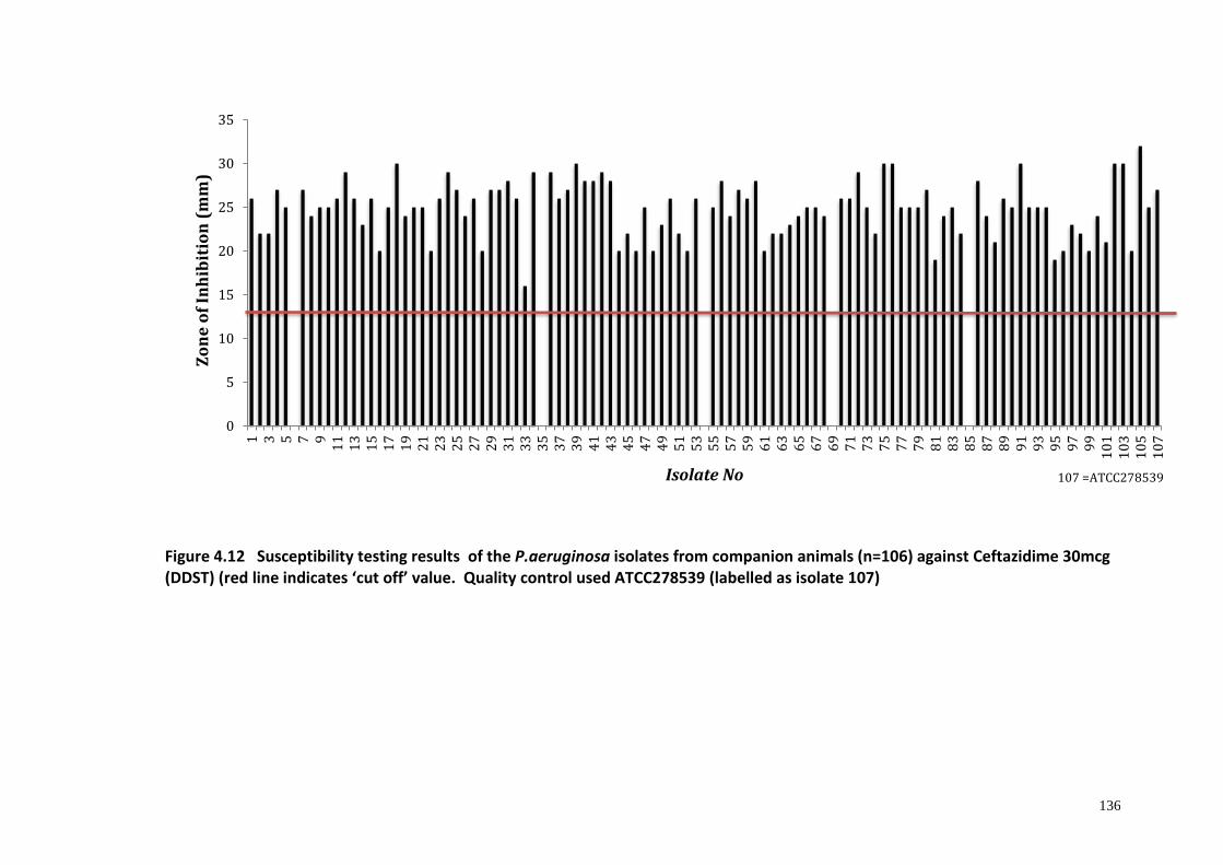
136
Figure 4.12 Susceptibility testing results of the P.aeruginosa isolates from companion animals (n=106) against Ceftazidime 30mcg (DDST) (red line indicates ‘cut off’ value. Quality control used ATCC278539 (labelled as isolate 107)
0
5
10
15
20
25
30
35
1 3 5 7 91
11
31
51
71
92
12
32
52
72
93
13
33
53
73
94
14
34
54
74
95
15
35
55
75
96
16
36
56
76
97
17
37
57
77
98
18
38
58
78
99
19
39
59
79
91
01
10
31
05
10
7
Zo
ne
of
Inh
ibit
ion
(m
m)
Isolate No 107 =ATCC278539

137
ESBL phenotyping results
Results are detailed for both methods in Table 4.8. The two methods did not
produce consistent results. Of the 106 isolates tested 3 were positive ESBL
producers by cloxacilliin method and 7 were positive by boronic acid method.
(begins page p138)

138
Table 4.8. ESBL testing results performed via two methods of the 106 P. aeruginosa veterinary isolates
Isolate ESBL Results (Cloxacillin method)
ESBL results (Boronic acid method)
Isolate ESBL Results (Cloxacillin method)
ESBL results (Boronic acid method)
Isolate ESBL Results (Cloxacillin method)
ESBL results (Boronic acid method)
1 - - 37 - - 73 - -
2 - - 38 - - 74 + -
3 - - 39 - - 75 - -
4 - - 40 - - 76 - -
5 - - 41 - - 77 - -
6 - - 42 - - 78 - -
7 - - 43 - + 79 - -
8 - - 44 - + 80 - -
9 - - 45 - + 81 - -
10 - - 46 - + 82 - -
11 - - 47 - - 83 - -
12 - - 48 - - 84 - -
13 - - 49 - - 85 - -
14 - - 50 - - 86 - +
15 - - 51 - - 87 - -
16 - - 52 - - 88 - -
17 - - 53 - - 89 - -
18 - - 54 . - 90 - -
19 - - 55 + - 91 - -
20 - - 56 - - 92 - -
21 - - 57 - - 93 - -
22 - - 58 - - 94 - -
23 - - 59 - - 95 - +
24 - - 60 - - 96 - -
25 - - 61 - - 97 - -
26 - - 62 - - 98 - -
27 - - 63 - - 99 - -
28 - - 64 - - 100 + -
29 - - 65 - - 101 - -
30 - - 66 - - 102 - -
31 - - 67 - - 103 - -
32 - - 68 - - 104 - -
33 - - 69 - - 105 - -
34 - - 70 - - 106 - -
35 - + 71 - -
36 - - 72 - -

139
Tables 4.9 -4.12 show details of PCR screening results for the ESBL-associated
resistance genes within the veterinary P. aeruginosa isoaltes for the genes blaTEM,
blaSHV, blaOXA; blaGES, blaVEB, blaPER; blaVIM, blaIMP, blaKPC and blaOXA-1, blaOXA-2, blaOXA-
10 respectively. Of the 106 isolates, 11 isolates (highlighted with grey shading in
Table 4.9) showed positive PCR product for ESBL-associated resistance gene blaTEM.
While eight isolates identified positive for blaSHV (Table 4.9) and three isolates
positive for blaOXA (Table 4.9). The three isolates that were identified as positive
resistance determinants for blaOXA (isolates 71, 72 and 73) – refer to Figure 4.12,
were also positive for the other ESBL-associated resistance genes blaTEM and blaSHV.
(begins page 140)

140
Table 4.9. Results of the PCR screening of ESBL-associated resistance genes within the veterinary P. aeruginosa isolates (n=106) - shows results for the genes blaTEM, blaSHV, blaOXA genes.
Isolate blaTEM blaSHV blaOXA Isolate blaTEM blaSHV blaOXA
1 - - - 54 + + -
2 - - - 55 - - -
3 - - - 56 - - -
4 - - - 57 - - -
5 - - - 58 + + -
6 - - - 59 - - -
7 - - - 60 - - -
8 - - - 61 - - -
9 - - - 62 - - -
10 - - - 63 - - -
11 - - - 64 - - -
12 - - - 65 - - -
13 + - - 66 - - -
14 - - - 67 - - -
15 - - - 68 - - -
16 - - - 69 - - -
17 - - - 70 + + +
18 - - - 71 + + +
19 - - - 72 + + +
20 - - - 73 - - -
21 - - - 74 - - -
22 - - - 75 - - -
23 - - - 76 - - -
24 - - - 77 - - -
25 - - - 78 - - -
26 - - - 79 - - -
27 - - - 80 - - -
28 - - - 81 - - -
29 - - - 82 - - -
30 - - - 83 - - -
31 - - - 84 - - -
32 - - - 85 + - -
33 - - - 86 - - -
34 - - - 87 - - -
35 - - - 88 - - -
36 - - - 89 - - -
37 - - - 90 - - -
38 - - - 91 - - -
39 - - - 92 - - -
40 - - - 93 - - -
41 - - - 94 - - -
42 + + - 95 - - -
43 + + - 96 - - -
44 + + - 97 - - -
45 + - - 98 - - -
46 - - - 99 - - -
47 - - - 100 - - -
48 - - - 101 - - -
49 - - - 102 - - -
50 - - - 103 - - -
51 - - - 104 - - -
52 - - - 105 - - -
53 - - - 106 - - -

141
The Figure 4.13 shows the PCR gel results for the blaOXA genes as reported.
Figure 4.13 Simplex PCR assay using primers to detect blaOXA gene performed on isolates
from isolates from the VDL, Leahurst. Isolates 70, 71, 72 appear to show strong
amplification of blaOXA-1. The DNA loading dye DNA/BsuRI (HaeIII) Marker 9, 50 mg (0.5
μg/μl)(ThermoScientific Molecular Biology), was placed on each gel run.
Table 4.10 details the results of the PCR screening of ESBL-associated resistance
genes within the veterinary P. aeruginosa isolates for the genes blaGES, blaVEB, blaPER.
Of the 106 isolates, five isolates (highlighted with grey shading in Table 4.10)
showed positive PCR product for ESBL-associated resistance gene blaGES. While four
isolates identified positive for blaPER (Table 4.10) there were no positives identified
for blaVEB. It is noted that although there was amplification of the multiple ESBL-
associated resistance genes as detailed here, some were feint and only weakly
positive but are still noted above in the table.

142
Table 4.10. Results of the PCR screening of ESBL-associated resistance genes within veterinary P. aeruginosa isolates (n=106) – shows the detection of blaGES, blaVEB, blaPER genes.
Isolate blaGES blaVEB blaPER Isolate blaGES blaVEB blaPER
1 - - - 54 + - -
2 - - - 55 - - -
3 - - - 56 - - -
4 - - - 57 - - -
5 + - - 58 - - -
6 - - - 59 - - -
7 - - - 60 - - -
8 - - + 61 - - -
9 - - - 62 - - -
10 - - - 63 - - -
11 - - - 64 - - -
12 - - - 65 - - -
13 - - - 66 - - -
14 - - + 67 - - -
15 - - - 68 - - -
16 - - - 69 - - -
17 - - - 70 - - -
18 - - - 71 - - -
19 - - - 72 - - -
20 - - - 73 - - -
21 - - - 74 - - -
22 - - - 75 - - -
23 - - - 76 - - -
24 - - - 77 - - -
25 - - - 78 - - -
26 - - - 79 - - -
27 - - - 80 - - -
28 - - - 81 - - -
29 - - - 82 - - -
30 - - - 83 - - -
31 - - - 84 - - -
32 - - + 85 - - -
33 - - - 86 - - -
34 - - - 87 - - -
35 - - - 88 - - -
36 - - - 89 - - -
37 - - - 90 - - -
38 - - - 91 - - -
39 - - - 92 - - -
40 - - - 93 - - -
41 - - - 94 - - -
42 - - + 95 - - -
43 - - - 96 - - -
44 - - - 97 - - -
45 - - - 98 - - -
46 - - - 99 - - -
47 + +
- - 100 - - -
48 - - 101 - - -
49 - - - 102 - - -
50 - - - 103 - - -
51 - - - 104 - - -
52 - - - 105 - - -
53 + - - 106 - - - *Although there was amplification of the multiple ESBL-associated resistance genes, some were feint and only weakly positive but are still noted above in table.

143
In the table 4.11 results of the PCR screening of ESBL-associated resistance genes of
the veterinary P. aeruginosa isolates for the blaVIM , blaIMP, blaKPC genes are shown.
Of the 106 isolates, 26 isolates (highlighted with grey shading in Table 4.11) showed
positive PCR product for ESBL-associated resistance gene blaGES, while six isolates
identified positive for blaKPC (Table 4.11). There were no positives identified for
blaIMP. It is noted that although there was amplification of the multiple ESBL-
associated resistance genes as detailed here, some were feint and only weakly
positive but are still noted above in the table. There was also the presence of
multiple unspcific bands produced on the gels making the resultant images difficult
to interpret. An example gel is depicted in Figure 4.13 and this issue is discussed
later in this Chapter. (Table 4.11 begins p144)
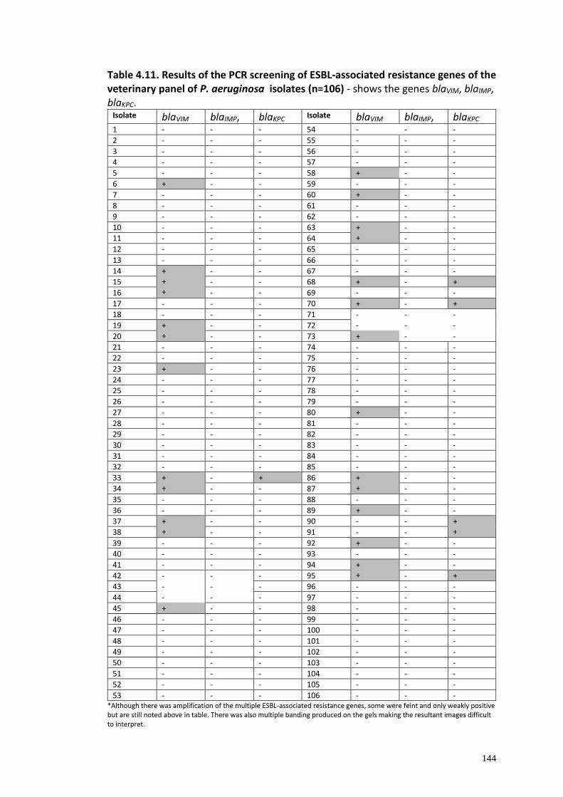
144
Table 4.11. Results of the PCR screening of ESBL-associated resistance genes of the veterinary panel of P. aeruginosa isolates (n=106) - shows the genes blaVIM, blaIMP, blaKPC.
Isolate blaVIM blaIMP, blaKPC Isolate blaVIM blaIMP, blaKPC
1 - - - 54 - - -
2 - - - 55 - - -
3 - - - 56 - - -
4 - - - 57 - - -
5 - - - 58 + - -
6 + - - 59 - - -
7 - - - 60 + - -
8 - - - 61 - - -
9 - - - 62 - - -
10 - - - 63 + +
- -
11 - - - 64 - -
12 - - - 65 - - -
13 - - - 66 - - -
14 + + +
- - 67 - - -
15 - - 68 + - +
16 - - 69 - - -
17 - - - 70 + - +
18 - - - 71 - - -
19 + +
- - 72 - - -
20 - - 73 + - -
21 - - - 74 - - -
22 - - - 75 - - -
23 + - - 76 - - -
24 - - - 77 - - -
25 - - - 78 - - -
26 - - - 79 - - -
27 - - - 80 + - -
28 - - - 81 - - -
29 - - - 82 - - -
30 - - - 83 - - -
31 - - - 84 - - -
32 - - - 85 - - -
33 + +
- + 86 + +
- -
34 - - 87 - -
35 - - - 88 - - -
36 - - - 89 + - -
37 + +
- - 90 - - + + 38 - - 91 - -
39 - - - 92 + - -
40 - - - 93 - - -
41 - - - 94 + +
- -
42 - - - 95 - +
43 - - - 96 - - -
44 - - - 97 - - -
45 + - - 98 - - -
46 - - - 99 - - -
47 - - - 100 - - -
48 - - - 101 - - -
49 - - - 102 - - -
50 - - - 103 - - -
51 - - - 104 - - -
52 - - - 105 - - -
53 - - - 106 - - - *Although there was amplification of the multiple ESBL-associated resistance genes, some were feint and only weakly positive but are still noted above in table. There was also multiple banding produced on the gels making the resultant images difficult to interpret.

145
Table 4.12 shows details the results of the PCR screening of ESBL-associated
resistance genes of the veterinary P. aeruginosa isolates for the genes blaOXA-1,
blaOXA-2, blaOXA-10. Of the 106 isolates, three isolates (highlighted with grey shading
in Table 4.12) showed positive PCR product for ESBL-associated resistance gene
blaOXA-2. There were no positives identified however for blaOXA-1 and blaOXA-10. It is
noted that although there was amplification of the multiple ESBL-associated
resistance genes as detailed here, some were feint and only weakly positive but are
still noted above in the table. (Table 4.12 begins page 146)

146
Table 4.12. Results of the PCR screening of ESBL-associated resistance genes within the larger veterinary panel of P. aeruginosa isolates (n-106) - shows the genes blaOXA-1, blaOXA-2, blaOXA-10.
Isolate blaOXA-1 blaOXA-2 blaOXA-10 Isolate blaOXA-1 blaOXA-2 blaOXA-10
1 - - - 54 - - -
2 - - - 55 - - -
3 - - - 56 - - -
4 - - - 57 - - -
5 - - - 58 - - -
6 - - - 59 - - -
7 - + +
- 60 - - -
8 - - 61 - - -
9 - - - 62 - - -
10 - - - 63 - - -
11 - - - 64 - - -
12 - - - 65 - - -
13 - - - 66 - - -
14 - - - 67 - - -
15 - - - 68 - - -
16 - - - 69 - - -
17 - - - 70 - - -
18 - - - 71 - - -
19 - - - 72 - - -
20 - - - 73 - - -
21 - - - 74 - - -
22 - - - 75 - - -
23 - - - 76 - - -
24 - - - 77 - - -
25 - - - 78 - - -
26 - - - 79 - - -
27 - - - 80 - - -
28 - - - 81 - - -
29 - - - 82 - - -
30 - - - 83 - - -
31 - - - 84 - - -
32 - - - 85 - - -
33 - - - 86 - - -
34 - + - 87 - - -
35 - - - 88 - - -
36 - - - 89 - - -
37 - - - 90 - - -
38 - - - 91 - - -
39 - - - 92 - - -
40 - - - 93 - - -
41 - - - 94 - - -
42 - - - 95 - - -
43 - - - 96 - - -
44 - - - 97 - - -
45 - - - 98 - - -
46 - - - 99 - - -
47 - - - 100 - - -
48 - - - 101 - - -
49 - - - 102 - - -
50 - - - 103 - - -
51 - - - 104 - - -
52 - - - 105 - - -
53 - - - 106 - - -
*Although there was amplification of the multiple ESBL-associated resistance genes, some were feint and only weakly positive but are still noted above in table.

147
The genes BEL-1 CTX-MU, AmpC and CS Integrons are not presented as all isolates
were PCR negative for these markers.
Multiplex Polymerase Chain Reaction (PCR) Assays
Following the initial multiplex PCR (refer to Chapter 2 for Methods and Results as
detailed above) on all 106 isolates for the previously described genes it was
concluded that the results of the multiplex PCR were not as clear to interpret as had
been expected. Many multiple faint positive bands or unexplained banding were
present in some isolates interfering with the reading of results. The primers chosen
were mostly previously used for Enterobacteriacea isolates by the VDL and
experience of their use with P.aeruginosa isolates was more limited. As there was
insufficient time and further funding on this project for carrying out repeated PCRs
with alternative gene primers or for performing a multitude of gradient PCRs to
refine PCR conditions, it was decided to repeat all as simplex PCRs but for a smaller
subset of isolates which had the potential to carry resistance genes based on their
susceptibility phenotype. The selection of this smaller subset of 32 isolates is
detailed below and listed in Table 4.13 (the results of these simplex PCRs on this
subset of isolates are presented under the section of work entitled: Findings from
Simplex Polymerase Chain Reaction (PCR) Assays).
Although the initial screening of 106 isolates via multiplex PCR for the TEM, SHV and
OXA set of genes did show some fairly clear positive/weak positive amplicons, for
the remainder of genes screened, the results were not easily interpretable due to
multiple non-specific banding (Figure 4.14).
An example of a gel that yielded potential unconfirmed positive amplicons is shown
in Figure 4.14.

148
Figure 4.14 PCR assay using primers to detect blaVIM gene performed on isolates from VDL,
Leahurst. Multiple isolates (86, 87, 89, 92, 94, 95) appear to show strong amplification of
blaVIM shown with the positive control and negative controls and multiple bands. The DNA
loading dye DNA/BsuRI (HaeIII) Marker 9, 50 mg (0.5 μg/μl)(ThermoScientific Molecular
Biology), was placed on each gel run.
This was thought to be due to multiplex PCR and so it was repeated but as simplex
presuming that this would improve the clarity of results. However, the resultant gel
images on the simplex PCRs that were repeated on the same 106 isolates the results
were generally overall difficult to interpret. The screening of all of the described
genes were then repeated but only on the narrowed down sample size of 32
isolates (as described earlier, along with how they were selected) and their DNA
extraction method was improved (refer to Materials and Methods; DNA Extraction),
due to time and cost constraints for this study this had to be limited. Improved
results were achieved following gradient PCRs to adjust the thermocycler conditions
for optimisation (conditions used as detailed in Table 4.13).
It is suggested that multiple or non specific PCR products can occur due to the
following potential reasons

149
Table 4.13 Potential causes of non specific PCR product
Possible causes of non specific product
Possible adjustments Any method adjustments made after gradient PCRs for optimisation
Premature replication Use a hot start polymerase, such as OneTaq Hot Start DNA Polymerase Set up reactions on ice using chilled components and add samples to thermocycler preheated to the denaturation temperature
N/A Already done in method
Primer annealing temperature too low
Increase annealing temperature
Annealing temperature increased
Incorrect Mg++ concentration
Adjust Mg++ in 0.2–1 mM increments
N/A as within a compound polymerase mastermix solution Firepol® so Mg non adjustable
Poor primer Check specific product literature for recommended primer design Verify that primers are non-complementary, both internally and to each other Increase length of primer Avoid GC-rich 3´ ends
N/A Insufficient time/funding to reorder alternative primers to trial
Excess primer Primer concentration can range from 0.05–1 µM in the reaction. Please see specific product literature for ideal conditions.
Primer concentrations were reduced
Contamination with exogenous DNA
Use positive displacement pipettes or non-aerosol tips Set-up dedicated work area and pipettor for reaction setup Wear gloves during reaction setup
All of this was already in place
Template conc too high For low complexity templates (i.e. plasAmid, lambda, BAC DNA), use 1 pg–10 ng of Dper 50 µl re
A lower conc dna used

150
Findings from Simplex Polymerase Chain Reaction (PCR) Assays
This further narrowed down subset of 32 isolates (Table 4.14) from the 106 were
selected on the basis of the criteria of being either ESBL positive phenotypes and/or
those that were resistant to ceftazidime (a third generation cephalosporin) plus any
of the antibiotics; aztreonam, meropenam (carbapenems), cefepime (fourth
generation cephalosporin), piperacillin or piperacllin/tazobactam (ESBLs). These
isolates underwent PCR following repeat subculture and DNA extraction using an
alternative extraction method (as referenced in Materials and Methods section -
Wizard® Genomic DNA Purification Kit Method). Resultant PCR products were
electrophoresed (in 1% agarose gels containing PeqGREEN® dye).
Table 4.14 A further narrowed down selection of isolates (n=32) from the 106 that were used for the characterisation study.
Isolate Source
4
6
22
28
33
42
43
44
45
46
47
48
52
53
54
55
63
64
69
70
74
72
81
82
Abdominal fluid
Vulval swab
Wound swab
Right ear swab
Right ear swab
Wound
Right ear swab
Tracheal
Left ear
Right ear swab
Tissue
Right ear swab
Urine
Urine catheter
Right ear swab
PEG tube Stoma wound
Wound
Right anal sac swab
Right ear swab
Wound serous discharge and fat tissue
Right and Left BAL
Tissue
Tissue
Vulval fluid swab
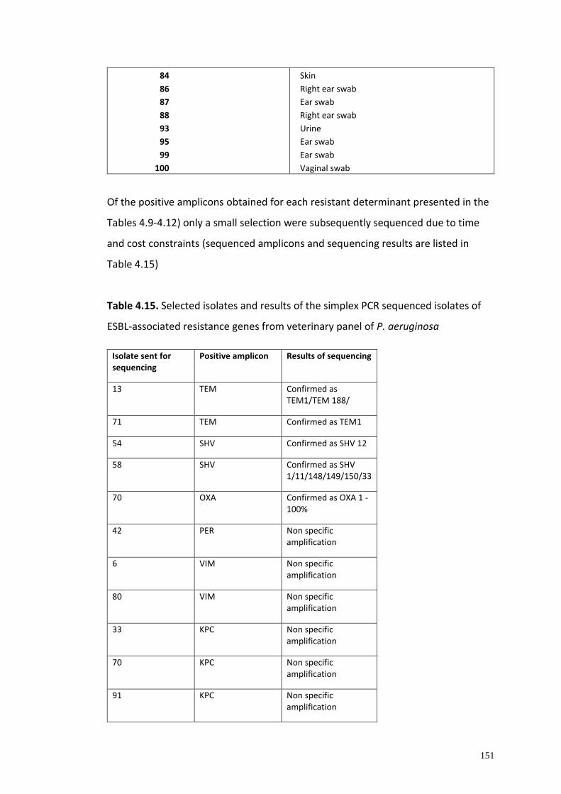
151
84
86
87
88
93
95
99
100
Skin
Right ear swab
Ear swab
Right ear swab
Urine
Ear swab
Ear swab
Vaginal swab
Of the positive amplicons obtained for each resistant determinant presented in the
Tables 4.9-4.12) only a small selection were subsequently sequenced due to time
and cost constraints (sequenced amplicons and sequencing results are listed in
Table 4.15)
Table 4.15. Selected isolates and results of the simplex PCR sequenced isolates of
ESBL-associated resistance genes from veterinary panel of P. aeruginosa
Isolate sent for sequencing
Positive amplicon Results of sequencing
13 TEM Confirmed as TEM1/TEM 188/
71 TEM Confirmed as TEM1
54 SHV Confirmed as SHV 12
58 SHV Confirmed as SHV 1/11/148/149/150/33
70 OXA Confirmed as OXA 1 - 100%
42 PER Non specific amplification
6 VIM Non specific amplification
80 VIM Non specific amplification
33 KPC Non specific amplification
70 KPC Non specific amplification
91 KPC Non specific amplification

152
The ESBL-associated resistance genes which generated a weak amplicon were not
included for sequencing.
The table below (Table 4.16) summarises the findings of isolates within the smaller
subset selected that were confirmed PCR positive and sequenced along with the
results of phenotypic antibacterial resistance testing for these isolates.
Table 4.16. Summary of the isolates and resistance genes confirmed by simplex
PCR and sequencing for selected veterinary P. aeruginosa isolates associated with
ESBL resistance (n=32)
Gene Simplex PCR positive isolates
Isolates confirmed by sequencing
Results of additional phenotypic antibacterial resistance to testing
blaTEM 6 isolates (43, 44, 45, 54, 55, 93)
Isolate 71 (canine urine) =blaTEM-1, Isolate 13 (canine ear swab)=blaTEM1/188
71 = Intermediate Resistance to aztreonam. Intermediate Resistant to enrofloxacin. * 13= Intermediate Resistance to aztreonam. Intermediate Resistance to enrofloxacin. * Susceptible to others.
blaSHV 4 isolates (43, 44, 54, 55)
Isolate 58 (canine tissue swab) = SHV 1/148/149/150/33 Isolate 54 (canine ear swab) =SHV 12
Intermediate Resistant to aztreonam. Intermediate Resistant to enrofloxacin. * Resistant to aztreonam, cefepime, piperacillin, pip/taz. Intermediate Resistant to Meropenam. Intermediate Resistant to Gentamicin. Resistant to enrofloxacin and Marbofloxacin. *
blaOXA 3 isolates (70, 71, 72)
Isolate 70 (canine wound serous discharge and fat tissue) = OXA-1
Intermediate Resistant to aztreonam. Resistant to cefepime, piperacillin, pip/taz. Intermediate Resistant to enrofloxacin. *

153
Resistance to aminoglycoside antibiotics
In order to determine the resistance to aminoglycosides, MICs to two clinically used
aminoglycoside antibiotics; amikacin and gentamicin were performed. From the
panel of 106 isolates, seven were found to be resistant to amikacin with an MIC of
≥16 mcg/ml. Nine isolates displayed intermediate resistance and 90 were found to
be susceptible. Of the seven amikacin resistant isolates, six were of canine origin
with one from equine origin. Of the 11 gentamicin resistant isolates, 10 were from
canine infections and one from equine (Table 4.17 and Table 4.18).
MIC resistance levels for amikacin and gentamicin were found to be at similar
levels, at 6.6% and 10.4% respectively (Table 4.17).
Table 4.17. Minimum inhibitory concentration testing of P. aeruginosa from
companion animal (n=106) isolates to aminogycosides
Antimicrobial
Breakpoint
mcg/L
MIC mcg/mL
0.25 0.5 1 2 4 8 16 32 64 >64
MIC50
MIC90
Resistant strains (%)
Amikacin S≤4, I=8, R≥16 90 9 6 1 4 4 6.6 [3, 13]
Gentamicin S≤2, I=4, R≥8
25 54 16 6 5
2
4
10.4 [0.176, 0.059]
Confidence intervals are 95% {Lower, Upper) Wilson method
Other antibacterials (amoxicillin/clavulanic acid, ampicillin, cefazolin, cefovecin, cefoxtine, cefpodoxime, chloramphenicol,
clindamycin, doxycycline, erythromycin, oxacillin, +2%NaCl, penicillin, rifampicin, trimethoprim sulfamethoxazole) included
in the Trek COMPAN1F plate were also tested but are not reported due to intrinsic P.aeruginosa resistance.
There were seven isolates that were resistant to amikacin and 11 were resistant to
gentamicin. One of these isolates (isolate 43, from canine ear swab) was confirmed
by sequencing as carrying the qnrB resistance gene (Table 4.22). Of the seven
amikacin resistant isolates six were similarly resistant to gentamicin. These
consisted of five isolates from canine infections and one from an equine sample.
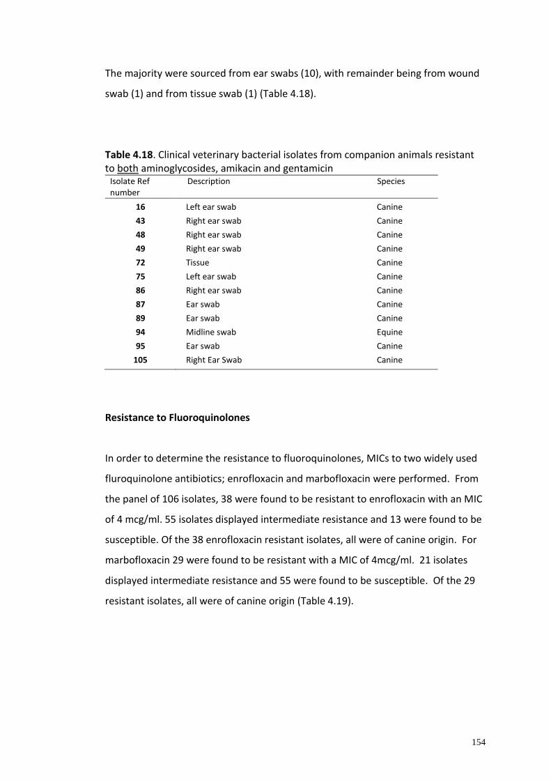
154
The majority were sourced from ear swabs (10), with remainder being from wound
swab (1) and from tissue swab (1) (Table 4.18).
Table 4.18. Clinical veterinary bacterial isolates from companion animals resistant to both aminoglycosides, amikacin and gentamicin
Isolate Ref number
Description Species
16
43
48
49
72
75
86
87
89
94
95
105
Left ear swab Canine
Right ear swab Canine
Right ear swab Canine
Right ear swab Canine
Tissue Canine
Left ear swab Canine
Right ear swab Canine
Ear swab Canine
Ear swab Canine
Midline swab Equine
Ear swab Canine
Right Ear Swab Canine
Resistance to Fluoroquinolones
In order to determine the resistance to fluoroquinolones, MICs to two widely used
fluroquinolone antibiotics; enrofloxacin and marbofloxacin were performed. From
the panel of 106 isolates, 38 were found to be resistant to enrofloxacin with an MIC
of 4 mcg/ml. 55 isolates displayed intermediate resistance and 13 were found to be
susceptible. Of the 38 enrofloxacin resistant isolates, all were of canine origin. For
marbofloxacin 29 were found to be resistant with a MIC of 4mcg/ml. 21 isolates
displayed intermediate resistance and 55 were found to be susceptible. Of the 29
resistant isolates, all were of canine origin (Table 4.19).
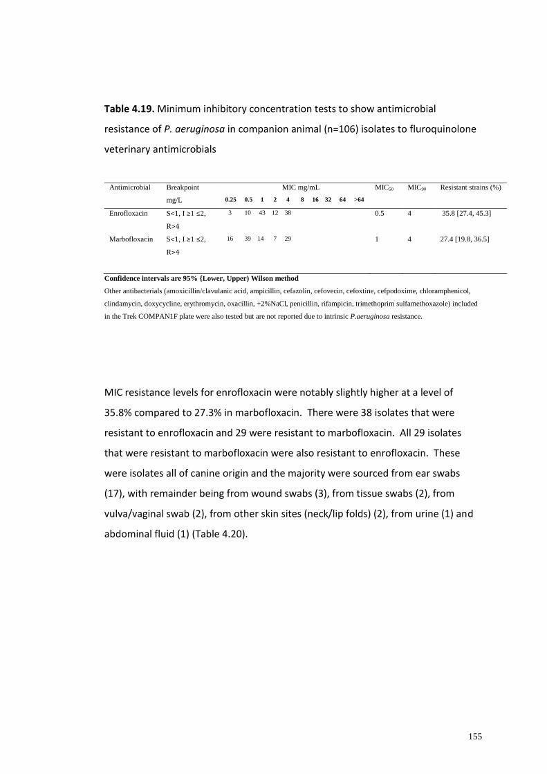
155
Table 4.19. Minimum inhibitory concentration tests to show antimicrobial
resistance of P. aeruginosa in companion animal (n=106) isolates to fluroquinolone
veterinary antimicrobials
Antimicrobial
Breakpoint
mg/L
MIC mg/mL
0.25 0.5 1 2 4 8 16 32 64 >64
MIC50
MIC90
Resistant strains (%)
Enrofloxacin S<1, I ≥1 ≤2,
R>4
3 10 43 12 38 0.5 4 35.8 [27.4, 45.3]
Marbofloxacin
S<1, I ≥1 ≤2,
R>4
16 39 14 7 29
1
4
27.4 [19.8, 36.5]
Confidence intervals are 95% {Lower, Upper) Wilson method
Other antibacterials (amoxicillin/clavulanic acid, ampicillin, cefazolin, cefovecin, cefoxtine, cefpodoxime, chloramphenicol,
clindamycin, doxycycline, erythromycin, oxacillin, +2%NaCl, penicillin, rifampicin, trimethoprim sulfamethoxazole) included
in the Trek COMPAN1F plate were also tested but are not reported due to intrinsic P.aeruginosa resistance.
MIC resistance levels for enrofloxacin were notably slightly higher at a level of
35.8% compared to 27.3% in marbofloxacin. There were 38 isolates that were
resistant to enrofloxacin and 29 were resistant to marbofloxacin. All 29 isolates
that were resistant to marbofloxacin were also resistant to enrofloxacin. These
were isolates all of canine origin and the majority were sourced from ear swabs
(17), with remainder being from wound swabs (3), from tissue swabs (2), from
vulva/vaginal swab (2), from other skin sites (neck/lip folds) (2), from urine (1) and
abdominal fluid (1) (Table 4.20).

156
Table 4.20. Clinical veterinary bacterial isolates from companion animals resistant to both fluroquinolones, enrofloxacin and marbofloxacin.
Isolate Ref number
Description Species
3
4
14
18
26
28
32
33
34
35
42
43
45
46
52
54
56
63
68
72
85
86
87
89
95
97
99
104
105
Skin swab Canine
Abdominal fluid Canine
Right ear swab Canine
Left ear swab Canine
Neck swab Canine
Right ear swab Canine
Vaginal swab Canine
Right ear swab Canine
Lip fold Canine
Right ear swab Canine
Wound Canine
Right ear swab Canine
Left ear swab Canine
Right ear swab Canine
Urine Canine
Right ear swab Canine
Left ear swab Canine
Wound swab Canine
Tissue Canine
Tissue Canine
Left ear swab Canine
Right ear swab Canine
Ear swab Canine
Ear swab Canine
Ear swab Canine
Vulval swab Canine
Ear swab Canine
Wound swab Canine
Right Ear Swab Canine
In order to determine whether the resistance phenotypes observed via MIC assays
were associated with the presence of fluroquinolone resistance determinants, PCR
for the genes qnrA, qnrB, qnrS and aac 6’lb-cr were performed. Six isolates showed
positive amplifications for resistance determinants qnrA (one – isolate 57) and qnrB
(five – isolates 42, 43, 44, 57, 70, 72) – refer to Figures 4.14 and 4.15. Of these
isolates three (isolates 42, 43, 72) were resistant to both fluroquinolones

157
enrofloxacin and marbofloxacin, one (isolate 57) was resistant to just enrofloxacin
alone and two demonstrated intermediate resistance to enrofloxacin (but were
susceptible to marbofloxacin). 16 isolates showed positive amplicons for resistance
determinants qnrS (Figure 4.21) and seven positive to aac(6′)-Ib -cr (Figure 4.22).
However, of these amplicons several were feint and considered only weakly
potential positives (detailed in Table 4.21). (begins on p158)

158
Table 4.21 Results of the PCR screening of fluroquinolone-associated resistance
genes within the larger veterinary panel of P. aeruginosa
Isolate qnrA qnrB qnrS aac(6’)-lb-cr
Isolate qnrA qnrB qnrS aac(6′)-Ib -cr
1 - - - - 54 + - - -
2 - - - - 55 - - + -
3 - - - - 56 - - - -
4 - - - - 57 + - - -
5 - - - - 58 - - + -
6 - - - - 59 - - - -
7 - - - - 60 - - + -
8 - - - - 61 - - - -
9 - - + - 62 - - - -
10 - - - - 63 - - + -
11 - - + - 64 - - - -
12 - - - - 65 - - - -
13 - - - - 66 - - - -
14 - - - - 67 - - - -
15 - - - - 68 - - - -
16 - - - - 69 - - - -
17 - - + - 70 - + - + + +
18 - - - - 71 - - -
19 - - - - 72 - + -
20 - - - - 73 - - - -
21 - - - - 74 - - + -
22 - - - - 75 - - - -
23 - - - - 76 - - - -
24 - - + - 77 - - - -
25 - - - - 78 - - - -
26 - - - - 79 - - + -
27 - - - - 80 - - - -
28 - - - - 81 - - - +
29 - - + +
- 82 - - - -
30 - - - 83 - - - -
31 - - - - 84 - - - -
32 - - - - 85 - - - -
33 - - - - 86 - - - -
34 - - - - 87 - - - -
35 - - - - 88 - - - -
36 - - - - 89 - - - -
37 - - - + 90 - - - -
38 - - - - 91 - - + -
39 - - - - 92 - - - -
40 - - + - 93 - - - -
41 - - - - 94 - - - -
42 - + + +
- + + +
95 - - - -
43 - - 96 - - - -
44 - - 97 - - - -
45 - - - - 98 - - - -
46 - - - - 99 - - + -
47 - - - - 100 - - - -
48 - - - - 101 - - - -
49 - - - - 102 - - + -
50 - - - - 103 - - - -
51 - - - - 104 - - - -
52 - - + - 105 - - - -
53 - - - - 106 - - - - *Although there was amplification of the multiple fluroquinolone-associated resistance genes, some were feint and only weakly positive but are still noted above in table.

159
The figures below depict some of the PCR gel results for the qnrA gene, qnrB, qnrS,
aac(6′)-Ib-cr genes as reported.
(a)
(b)
Figure 4.15 Simplex PCR assay using primers to detect qnrA gene performed on isolates
from VDL, Leahurst. An isolate (54) appears to show strong amplification of qnrA shown on
the first gel image (a), the second gel image (b) shows the positive control with positive
result. The DNA loading dye DNA/BsuRI (HaeIII) Marker 9, 50 mg (0.5
μg/μl)(ThermoScientific Molecular Biology), was placed on each gel run.

160
Figure 4.16 Simplex PCR assay using primers to detect qnrB gene performed on isolates
from VDL, Leahurst. Here isolates 42,43 and 44 appear to show strong amplification. The
PCR products were electrophoresed in 1% agarose gels containing PeqGREEN® dye (VWR
International Ltd, Leicestershire, England). The DNA fragments were imaged with UV
transilluminator and UViProMW software documentation system. The DNA loading dye
DNA/BsuRI (HaeIII) Marker 9, 50 mg (0.5 μg/μl)(ThermoScientific Molecular Biology), was
placed on each gel run.
Figure 4.17 Simplex PCR assay using primers to detect qnrS gene performed on isolates
from VDL, Leahurst. An isolate (99) appears to show strong amplification of qnrS shown on
the gel image. The DNA loading dye DNA/BsuRI (HaeIII) Marker 9, 50 mg (0.5
μg/μl)(ThermoScientific Molecular Biology), was placed on each gel run.

161
(a)
(b)
(c)
Figure 4.18 Simplex PCR assay using primers to detect aac(6′)-Ib -cr gene performed on
isolates from VDL, Leahurst. Three isolates (70, 71, 72) appear to show strong amplification
of aac(6′)-Ib -cr shown on the first gel image (a), the second gel image (b) shows the
positive control, the third gel image (c) shows 3 isolates (42, 43,44 showing strong
amplification of the aac(6′)-Ib -cr gene – the positive control is also identified. The DNA
loading dye DNA/BsuRI (HaeIII) Marker 9, 50 mg (0.5 μg/μl)(ThermoScientific Molecular
Biology), was placed on each gel run.

162
To verify the sequence identity a small selection (due to time and cost limitations)
of the amplified bands were sent for sequencing for each fluroquinolone-associated
resistance determinant, these are detailed in (Table 4.22) along with the result of
sequencing.
Table 4.22 Selected isolates and results of the PCR sequenced isolates of
fluroquinolone-associated resistance genes from veterinary panel of P. aeruginosa
Isolate sent for sequencing
Positive amplicon Results of sequencing
54 qnr A Confirmed as qnrA1
43 qnrB Confirmd as qnrB2
70 qnrB Non specific amplification
99 qnrS Didn’t reproduce a strong positive amplicon therefore not sent
72 aac -6’lb-cr Confirmed as
acetyltransferase AAC(6')-Ib-cr (aac(6')-Ib-cr)
*However this result was excluded because the initial Simplex PCR showed positive amplicon but the subsequent simplex PCR on the smaller selection of isolates did not produce positive PCR.
81 aac -6’lb -cr Non specific amplification

163
Detailed in Table 4.23 are the molecular and phenotypic characteristics of the
specific isolates with fluroquinolone associated resistance determinants as
confirmed via sequencing along with the number of positive amplicons identified on
PCR from the smaller subset (n 32) of veterinary P.aeruginosa isolates.
Table 4.23. Summary of the confirmed PCR positive and sequenced isolates associated with fluroquinolone resistance within the smaller (n 32) subset of P.aeruginosa veterinary isolates combined with the phenotypic characteristics
Gene Number of positive PCR isolates
Sequencing confirmed positive isolates
Results of additional phenotypic antibacterial resistance to testing
qnrA 2 isolates (54 and 55)
Isolate 54 (canine ear swab) = qnrA1
Intermediate Resistant to aztreonam. Resistant to cefepime, piperacillin, pip/taz. Intermediate Resistant to enrofloxacin. *
qnrB 2 isolates (43 and 44)
Isolate 43 (canine ear swab) = qnrB
Intermediate Resistant to aztreonam. Intermediate Resistant to Gentamicin. Resistant to enrofloxacin and marbofloxacin.
aac(6')-Ib-cr 7 isolates (4 stronger positive amplicons 43, 44, 45,100 and 3 weakly positive amplicons 42, 48, 54)
Isolate 72 (canine tissue swab) – aac 6’lb -cr
Resistant to aztreonam. Resistant to ticarcallin and ticacillin/clavulanate Resistant to enrofloxacin and marbofloxacin, Resistant to gentamicin. *
* Other antibacterials (amoxicillin/clavulanic acid, ampicillin, cefazolin, cefovecin, cefoxtine, cefpodoxime, chloramphenicol, clindamycin, doxycycline, erythromycin, oxacillin, +2%NaCl, penicillin, rifampicin, trimethoprim sulfamethoxazole) included in the Trek COMPAN1F plate were also tested but are not reported due to intrinsic P.aeruginosa resistance. (Nikaido 1994)

164
Multidrug resistance (MDR) in isolates from companion animals.
The PCR amplicons confirmed by DNA sequencing to encode for the screened
genes, originated from 8 different Pseudomonas isolates, with 5 of these isolates
harbouring more than one of the investigated resistance genes.
Isolates 43 (canine ear swab) and 44 (canine tracheal swab) were found to carry the
blaTEM, blaSHV and qnrB genes. Isolate 45 (canine ear swab) was found to carry both
the blaTEM and blaOXA genes. Isolate 54 (canine ear swab) and 55 (canine PEG tube
stoma wound) was found to carry the blaTEM, blaSHV and qnrA genes. Isolate 93
(canine urine) was found to carry just the blaTEM gene and isolates 84 (canine skin
swab) and 99 (canine ear swab) were both found to carry only the blaOXA gene.
Isolate 71 (canine urine sample) was found to be carrying TEM-1 gene with
intermediate resistance to aztreonam and enrofloxacin. Isolate 13 (canine ear
swab) was found to be carrying TEM-1/188 gene and similarly with intermediate
resistance to aztreonam and enrofloxacin. Isolate 58 (canine tissue sample) was
found to be carrying SHV-1/148/149/150/33 gene also with intermediate resistance
to aztreonam and enrofloxacin. Isolate 70 (canine wound sample) was found to be
carrying OXA-1 gene with resistance to cefepime, piperacillin and
pipercillin/tazobactam, along with intermediate resistance to aztreonam and
enrofloxacin. One particularly interesting isolate was isolate 54, from a canine ear
swabs and showing multidrug resistance characteristics. Isolate 54 was found to be
carrying SHV-12 gene and qnrA1 with resistance to aztreonam, enrofloxacin, to both
piperacillin and piperacillin/tazobactam and also to the 4th generation
cephalosporin cefepime, as such making it multidrug resistant (MDR). It also
demonstrated intermediate resistance to meropenam and gentamicin. Isolate 43
(canine ear swab) was found to harbour qnrB gene with resistance to enrofloxacin
and marbofloxacin. It also demonstrated intermediate resistance to aztreonam and
gentamicin.

165
Isolate 72 (canine tissue swab) was found to be carrying the aac(6′)-Ib -cr variant
resistant determinant by sequencing and was resistant to aztreonam, ticarcacillin,
ticacillin/clavulanate, enrofloxacin, marbofloxacin and gentamicin and therefore
was multidrug resistant. However, although this sequencing result is reported (in
Table 4.22) there was disparity in the PCR results for the isolate. Isolate 72 showed
a positive band on the PCR in the original simplex PCR (Table 4.21) but was then
negative in repeated simplex PCR for the smaller subset of isolates. As such this
isolate is being considered as negative for this gene in this isolate. There is the
potential possibility contamination may have resulted in misleading results in this
case.
4.4 Discussion
The true extent and importance of AMR in companion animals is not entirely
understood and limited surveillance contributes to this (P. H. Jones et al., 2014;
Radford et al., 2011; Scott Weese, 2008). As discussed previously in Chapter 1,
potential for interspecies human-animal transmission exists. Studies have
demonstrated that AMR is problematic in a number of important pathogens and
commensals, such as Staphylococci, Enterococci, E. coli and Salmonella, although in
P. aeruginosa this appears less well reported in animals as compared to humans.
Much of the literature has focussed on AMR in food producing animals. This area
may be a reservoir and source of AMR genes that could be transmitted to the
human population (Arnold et al., 2016; Weese, 2008). Studies have shown that
antimicrobial use in food animals contributes to the selection of antimicrobial
resistance and poses risks to humans due to transmission of resistant zoonotic
bacteria via the food chain and indirect transfer of resistance genes from animals to
humans (Aarestrup et al., 1998; Smith et al., 2002; Tang et al,. 2017; van Den
Bogaard & Stobberingh, 2000).
Companion animals are another potential reservoir source for AMR to develop and
is less well studied. The pet population is ever growing (PFMA, 2017) and in 2017 it
166
is estimated that 12m (44%) of households have pets giving a pet population of
around 54 million. 24% of households owning dogs and 17% of households owning
cats (PFMA, 2017). Several reports have highlighted the presence of resistance
genes in both farm (Timofte et al., 2014) and companion animals (Schmidt et al.,
2015; Timofte et al., 2011) including equines (Ahmed et al., 2012; Maddox et al.,
2011). Cats and dogs in particular, represent a potential source of spread of
antimicrobial resistance due to the extensive use of antimicrobial agents in these
animals and their close contact with humans. There are several studies looking at
AMR in companion animals (mainly in that of dogs and horses), predominantly
involving E.coli (Schmidt et al., 2015; Wedley et al., 2011). However, fewer studies
of AMR and P. aeruginosa isolates exist.
A number of antimicrobial classes are frequently used in small animal veterinary
medicine including penicillins, cephalosporins, macrolides, lincosamides, fusidic
acid, tetracyclines, chloramphenicol, potentiated sulphonamides, aminoglycosides
and fluoroquinolones. The most frequent causes of antimicrobial treatment in dogs
and cats are skin and wound infections, otitis externa, respiratory infections, and
urinary tract infections (UTI) (Radford et al., 2011; Singleton et al., 2017).
Due to the presence of several drug efflux systems and porins, P. aeruginosa is
intrinsically resistant to a wide range of antimicrobials. The results here confirmed
the intrinsic resistance to antibacterials amoxicillin/clavulanic acid, ampicillin,
cefazolin, cefovecin, cefoxitine, cefpodoxime, chloramphenicol, clindamycin,
doxycycline, erythromycin, oxacillin, +2%NaCl, penicillin, rifampicin, trimethoprim
sulfamethoxazole (these were included in the Trek COMPAN1F plate and were also
tested but results not reported).
There was high occurrence of resistance to the third generation cephalosporin
ceftiofur (98.1% resistant). Although the prevelance of resistance to fourth
generation cephalosporin, cefepime was found to be 9.4%. Beta-latam resistance
to piperacillin, piperacillin/tazobactam, ticarcillin and ticacillin/clavulanate were
17%, 17.9%, 9.6% and 5.6% respectively. Of the carbapenems; meropenam
resistance was particularly low (3.7%) and also low for imipenem (8.5%), although

167
slightly higher in aztreonam (11%). Resistance to aminoglycosides was low (6.6%
for amikacin and 10.4% for gentamicin).
The fluoroquinolones are some of the most commonly prescribed effective
antimicrobials against P. aeruginosa infections, in recent years bacterial resistance
to fluroquinolones has been promoted with overuse of these antibacterials (Jacoby,
2005) and as such, the monitoring of antimicrobial susceptibility is important for
selecting effective antimicrobial agents in the treatment of this disease.
Quinolones enter bacteria through porins or directly through the lipid and
cytoplasmic membrane and target DNA topoisomerases (Jacoby et al., 2008)
achieving their bactericidal effects through the inhibition of DNA gyrase and
topoisomerase IV. There are three plasmid-mediated quinolone resistance
determinants discussed in the literature. The qnr genes (qnrA, qnrB, qnrS), aac(6′)-
Ib -cr, and qepA. Qnr proteins are known to be a part of the pentapeptide repeat
family and protect DNA gyrase and topoisomerase IV from quinolone inhibition (Ma
et al., 2009). The aac(6′)-Ib-cr is a variant of aac(6′)-Ib. AAC (6')-Ib is an enzyme that
causes aminoglycoside resistance through acetylation, and a variant of this enzyme,
aac (6')-Ib-cr, is deemed responsible for plasmid-mediated quinolone resistance
(Ma et al., 2009).
Studies have largely shown that resistance to the fluroquinolones is mainly due to:
(i) the point mutations in the DNA gyrase (gyrA and gyrB) and topoisomerase IV
(parC and parE) genes, (ii) the presence of transferable plasmid-mediated quinolone
resistance (PMQR) determinants, and (iii) mutations in genes regulating the
expression of efflux pumps and decreased expression of outer membrane porins (D.
Lin et al., 2012). Plasmid-associated resistance to fluroquinolones can be mediated
by the production of qnr proteins, which preserve DNA gyrase and topoisomerase
IV from inhibition by quinolones (Strahilevitz et al., 2009a). The fluoroquinolones
are approved for indications such as respiratory tract, urinary tract infections and
soft tissue infections and in dogs and cats along with colibacillosis in poultry
(chickens and turkeys). Fluoroquinolone use is licenced for conditions such as
canine otitis externa and in rabbits, small mammals and reptiles for alimentary and
respiratory tract infections. The fluroquinolones are also licensed for veterinary use

168
in treatment of acute mastitis and respiratory infections in bovines and neonatal E.
coli gastroenteritis in calves. Other indications for which the fluoroquinolones have
been used in animal health include deep-seated infections, prostatitis, and other
bacterial infections resistant to standard antimicrobial therapy (Brown, 1996). In
this study, P. aeruginosa isolates showed 36% resistance to enrofloxacin and 27% to
marbofloxacin. With increasing utilization of fluoroquinolones in both human and
veterinary medicine, emerging resistance has become a significant concern (Cattoir
et al., 2007; Hong et al., 2009; Strahilevitz et al., 2009a; Sumrall et al, 2014).
A study of a University Hospital in North eastern Poland looked at a relatively small
number of P.aeruginosa human isolates (from 2002-2009) and reported 20% (5/25)
isolates had detectable qnrB by PCR (Michalska et al., 2014). The same study did
also identify aac(6′)-Ib in 28% (7/25) of isolates. Compared to none found in the
results of this work. Among Enterobacteriaceae screened for production of plasmid-
mediated fluoroquinolone resistance determinants, qnrB was reported as the most
prevalent gene (Kim et al 2009). However there is not a similar longitudinal study
to date to compare prevalence in P.aeruginosa or in veterinary isolates (for
comparison to human isolates). However, plasmid mediated quinolone resisatnce
(PMQR) genes (qnrA, qnrB, qnrS, qnrD and qepA) have been reported in veterinary
clinical isolates in China (Yang et al., 2015; Zhao et al., 2010). The qnrA1 and qnrB
determinants identified in P. aeruginosa isolates of this study are novel findings in
UK in companion animal isolates. Plasmid mediated quinolone resistance (PMQR)
determinants have been demonstrated among a variety of animal sources in the
Enterobactereaceae bacteria. A number of studies have reported presence of the
qnr genes in faeces from healthy animals or clinical isolates, including in chicken
faeces from poultry production systems, cattle faeces, pig farms, other food
production animals and companion animals (Ajayi et al, 2012; Huang et al., 2009;
Kuo et al., 2009; Ma et al., 2009) and appears to be distributed throughout the
world. Fluroquinolone resistance genes qnrA and qnrB are reported in P. putida in
isolates from imported shrimp (Tran et al, 2011) and in P. fluoroscens although none
currently reported in animal P. aeruginosa isolates. Cayci et al (Cayci et al., 2014),
169
performed screening for plasmid mediated quinolone resistance genes in P.
aeruginosa clinical (human) isolates, although no qnr genes were identified.
The qnrB1 has been reported present in an ESBL producing E.coli isolates from
Tunisia and in E.coli isolates from human and poultry origin in Ecuador based study
(Armas-Freire et al., 2015). In a study of AMR resistance risk factors in E.coli in
healthy Labrador retrievers (Schmidt et al., 2015) the qnr genes were not detected
in the sample set investigated in that study.
Bacterial resistance to fluroquinolones results from mutations in the quinolone
resistance determining regions of the drug targets, over expression of efflux pumps
and/or the more recently identified plasmid mediated low level resistance
mechanism (Cayci, Y.T 2014; Strahilevitz et al., 2009a).
In this study, whilst qnr determinants where confirmed, the aac(6′)-Ib-cr
determinants were not. The qnr and aac(6′)-Ib-cr determinants are widely reported
in human clinical isolates of Enterobacteriaceae with a worldwide distribution (Hong
et al., 2009; Ma et al., 2009; Park et al., 2006; Robicsek et al., 2006; Rodríguez-
Martínez et al., 2011; Sumrall et al., 2014; Xiao et al., 2012). There is suggestion of
transferability of resistant bacteria or mobile resistance determinants between
animals and humans potentially via the food chain or direct contact (Fey et al.,
2000; Manian, 2003). The quinolones are an antibacterial with widespread use in
livestock veterinary care and in treatment of companion animals in some countries,
including the UK. However, few reports on the occurrence of plasmid-mediated
quinolone resistant (PMQR) determinants among bacteria from companion animals
and food-producing animals have been published. Quinolones and β-lactams are
among the most commonly used antimicrobials in both human and veterinary
clinical medicine.
170
4.5 Conclusions
The findings in this work have identified several resistance genes including those
relating to ESBLs production (blaTEM, blaSHV and blaOXA) and those relating to
fluroquinolone resistance (qnr). In particular, the identification of qnrA and qnrB
genes in P.aeruginosa isolates of companion animal origin is a novel finding with no
known reports to current date in the UK. Whilst reports demonstate their
prevelance worldwide and in Enterobacteriaceae, reports on P aeruginosa of
companion animal origin remain non reported in literature. It would be
advantageous to have further time and funds on this study to be able to expand
upon the study sample size and also investigate the transmissibility of these
identified genes through conjugation studies and to further define the mechanisms
of resistance. In addition, genotyping of the resistant isolates to determine their
relative distribution within the wider P.aeruginosa population would be beneficial.
This study further confirms the likelihood that companion animals are an
understudied source of antimicrobial resistant P. aeruginosa isolates and merit
sustained surveillance of the veterinary niche as a potential reservoir for resistant,
clinically-relevant bacteria.
171
Chapter 5: General Discussion and Future Studies
Antimicrobial resistance is a multifactorial and complex issue affecting healthcare
worldwide. It is an escalating concern as a developing public health crisis and
European and International bodies have highlighted AMR as a global public health
issue of imperative importance. The World Health Organisation (WHO), the
European Centre Disease Prevention and Control (ECDPC) and Infectious Diseases
Society of America (IDSA) have included within their concerns and priorities,
emerging antimicrobial resistance mechanisms, surveillance and antibiotic
stewardship, applicable to both the veterinary and medical communities.
AMR is not only a concern in human health but also animal health. The close
interface between people, pets, food production animals and the environment
exemplifies the importance that these two areas of health should not be considered
as entirely independent factions, particularly in relation to AMR. Adopting a ‘one-
health’ approach in combatting this global problem is of great importance in
understanding and potentially controlling the advances of resistance among the
microbial population.
P. aeruginosa is one of the so-named ESKAPE pathogens by the IDSA and is
categorized by the CDC as a serious level of threat (CDC, 2013). It was also named
recently by the WHO as in the top three bacteria for which there is an urgent need
for new antibiotics for therapy (WHO, Feb 2017). The WHO specified in particular
carbapenem resistant P. aeruginosa in the WHO priority pathogens list for R&D of
new antibiotics as Priority 1 - Critical (WHO 2017; Important & Priority, 2017).
Increased resistance has resulted in limited therapeutic options. ESKAPE pathogens
are highlighted as being of ever-growing relevance to antimicrobial chemotherapy
in future years (Boucher et al., 2009; Santajit & Indrawattana, 2016). P. aeruginosa
is a formidable pathogen due to its innate resistant making it naturally insensitive to
many classes of antimicrobials, alongside its ability to acquire further resistance
mechanisms (Henrichfreise et al., 2007; Lambert, 2002). In addition to these
factors, it is known to form and survive in biofilms and is ubiquitous and
opportunistic nature.

172
This thesis reports the levels of antibacterial resistance in a panel of P. aeruginosa
isolates from companion animals including a high number of resistant isolates from
canine ear infections. It was shown that resistance to polymyxins (a class of
antimicrobials of particular importance in the management of P. aeruginosa lung
infections in patients with CF), particularly polymyxin B was significantly higher in
the panel of P. aeruginosa isolates taken from companion animals as compared to
that of a human panel. This study highlighted the importance of sustained
surveillance of the veterinary niche as source of antibacterial resistance.
Whole genome sequencing of a selection of polymyxin B resistant isolates from
animal sources was performed. These were found to be a unique and an interesting
source of genomic data. The sequencing revealed some of these resistant isolates
to be incredibly diverse. Notably isolate 856 (sample from an equine abdominal
incision) was diverse from the main population and was located on a distinct arm of
the phylogenetic tree (refer to Chapter 3, Figure 3.6). This genome had 125303
SNPs and 1884 indels compared to PAO1 and 130312 SNPs and 1985 indels in
comparison to PA14 (refer to Chapter 3, Figure 3.8). This was double the number of
SNPs and indels than any other isolate sequenced.
An abundance of P. aeruginosa genome sequencing data information already exists,
compiled predominantly from human isolates and environmental sources. P.
aeruginosa isolates derived from animal infections are currently under-represented
and this study highlights the importance of including such isolates. Increased
sequencing and analysis of veterinary-associated P. aeruginosa isolates may add
important additional data to the P. aeruginosa pan genome and population level
analyses. Using the Clondiag Tube Array, a commercially available strain typing
system, it was demonstrated that strain types identified from these companion
animals had also been associated with human infections and therefore there may
be a potential for transmission of infection through close contact. Indeed, there
have been case studies reporting this between human and animal (Deora et al,
2013; Ferreira et al., 2011; Mohan et al., 2008b; Register et al., 2012; Wannet et al,
173
2005) however this area is understudied and therefore the true risk is unknown. By
characterising and WGS a sample set of veterinary isolates in future studies it would
be potentially possible to offer novel findings in this area contributing to knowledge
of the P. aeruginosa population diversity in an understudied, under represented
field and potentially identifying it as a reservoir of antimicrobial resistance.
The results from this study suggest that a larger study is warranted. In such a study,
samples could be obtained on a random basis from a commercial UK veterinary
reference diagnostic laboratory (for example Idexx Veterinary Diagnostic
Laboratories – a commercial laboratory utilised by many veterinary practices) and
would therefore be from a variety of UK geographic areas, different species and
sites of origin. Characterisation through genomics and phenoptypic studies
(including virulence factor production, biofilm formation and antimicrobial
susceptibility testing) would provide a wealth of information which could both
contribute to knowledge of P. aeruginosa population biology as well as providing a
full characterisation of the features of isolates capable of causing infections in
particular species of animals. This information could contribute to appropriate
therapeutic choices and veterinary management of animals therefore ultimately
providing a rationale and basis for treatment for veterinary practitioners.
It is acknowledged that there are limitations in the interpreting the results of this
thesis. For instance, whilst the results of Chapter 3 showed the polymyxin
resistance to be higher in the veterinary clinical isolates as compared to the human
isolates and this was statistically significant (p < 0.05 – refer to Chapter 3 Results)
however these results being from a relatively small sample set may not necessarily
extrapolate to the wider population. It is also acknowledged that the two sample
sets used (animal versus human) were not of equal size. In further studies it would
be advantageous to acquire additional ‘background’ information such as that of the
clinical history from the cases isolates were sampled from. This may help to better
understand and postulate mechanisms of resistance development in the isolates. In
this work, the samples were those sent in from a referral university
practice/external veterinary practices directly to a Diagnostic Laboratory for

174
Microbiological assessment and then stored there, hence no clinical history would
be available. To know if any of the patients from which the isolates originated had
been previously treated with antibiotics would add value to understanding the full
significance of the levels of antibiotic resistance reported from this work. To collate
this prior clinical history in future study would be of benefit.
Limitations of the methods used, in particular that of the multiplex versus simplex
pcr where identified during the study. However, it is understood that part of these
issues may have been due to using some primers that were designed for use in
Enterobactereaccea and thermocycler protocols published for these, it is
acknowledged that to carry out prior optimisation PCRs to refine the conditions and
to have Pseudomonas specific designed primers for the regions being targeted
would have been an improvement that may have overcome these issues
encountered. Further improvements would have been to use WGS to identify
resistance detrminants, if funding would allow.
Whilst the larger sample set of 106 (Results reported in Chapter 4) is a statistically
better sample size, ultimately there was a reduced sample size of 32 used for
refining the results. These were chosen on basis of being ‘interesting’ isolates from
the initial screening ie they had identified some resistance in specific antibitics or
ESBL positive, however it is understood some bias would result in doing this. It is
also noted that the inconsistencies of utilising human clinical breakpoints ion
isolates of animla origin would not be ideal. However, some antibiotics especially
those not routinely used in veterinary medicine do not have established veterinary
breakpoints to use. VetCAST (a subcommittee of EUCAST) has been formed in 2015
and provides some guidance on this area of using available breakpoints.
Other findings as a result of the work performed within this thesis, has identified
several resistance genes including those relating to ESBLs production (blaTEM,
blaSHV and blaOXA) and those relating to fluroquinolone resistance (qnr) from P.
aeruginosa isolates of veterinary origin. In particular the identification of qnrA and
qnrB genes in P.aeruginosa isolates of companion animal origin is a novel finding
with no known reports to current date in the UK. Whilst reports demonstate their
prevelance worldwide and in Enterobacteriaceae, reports in P. aeruginosa of

175
companion animal origin remain non reported in literature, to the best of my
knowledge. These findings would suggest that further exploration of isolates of
animal origin could provide sources of resistant bacteria. That may have
implications as a niche reservoir of resistant bacteria to provide a source of cross-
resistance to human bacteria and also have implications in animal welfare and
disease treatment as well as human health. In addition further genotyping of
resistant animal isolates may provide novel information regarding their relative
distribution within the wider P.aeruginosa population.
There are studies in the literature, for example that by Haenni et al., (Haenni et al.,
2015), looking at population structure and antimicrobial susceptibility of P.
aeruginosa from animal infections in France. They found resistance phenotypes
were much more frequent in dogs (as compared to cows and horses), and multidrug
resistant P. aeruginosa appeared to emerge mainly in those suffering from otitis.
However, they noted such multidrug phenotypes are comparatively rare in other
animal species. It would be of interest to further expand this study to include a
greater number of P.aeruginosa isolates from a greater variety of animal species.
P. aeruginosa in the veterinary setting is a clinically relevant pathogen and a known
to be associated with disease. In companion animals, P. aeruginosa can cause
pyoderma, chronic otitis, ulcerative keratitis, wound infections, respiratory tract
infections and urinary tract infections in a number of species. A number of other
diseases involving P. aeruginosa in animals are known, including that of equine
metritis and in chronic equine wounds where the ability of P. aeruginosa to form
and survive within protective biofilms has been reported (Percival et al., 2015;
Westgate et al., 2011). Canine chronic otitis is a common presentation in small
animal veterinary medicine and a challenging infection to treat. Much is already
known regarding P. aeruginosa in human disease, in particular that of cystic fibrosis
(CF). From the extensive research carried out in the area of CF whereby these
patients suffer from long-term chronic lung infections caused by P. aeruginosa. It is
known that these are complicated and difficult to treat with routine antimicrobials.
This is, in part, attributed to the complex behaviour of P. aeruginosa in chronic

176
infections. During these chronic infections the bacteria diversify, leading to
populations with mixed levels of resistance or susceptibility to drugs. Further
complicated by the ability of P. aeruginosa to form complex biofilm communities
and coexist with many other bacterial species. The use of some antimicrobials may
even drive diversification and increased resistance to drugs. It is unknown if these
diverse populations occur in chronic infection of other species. Therefore, it would
be insightful to study the population biology and dynamics in chronic P. aeruginosa
infections of the canine ear by analysis of ear swab samples taken during therapy.
This could improve current knowledge of microbial population dynamics within
microbial communities in such infections and may inform diagnostic practice such
as testing “sweeps” of P. aeruginosa from a samples rather than a single isolate
when testing for antimicrobial susceptibility. By studying populations of P.
aeruginosa from animal infections it would be possible to assess the impact of
treatment strategies, fluctuations in the bacterial population biology and diversity,
and potential drivers of antimicrobial resistance. Cross resistance of antimicrobial
classes and transmission of resistant isolates between human and animal P.
aeruginosa are major issues and therefore merit continued study.
Little is known about P. aeruginosa carriage in seemingly healthy companion
animals and their owners. Suggested future avenues of study would be to
determine the levels of P. aeruginosa carriage in the human owners/contact animal
handlers of companion animals, particularly those canine patients with otitis. This
would allow the study of potential cross transmission routes between species. A US
study by Ferreira et al (Ferreira et al., 2011) looked at Methicillin-resistant
Staphylococcus aureus (MRSA) transmission between companion animals and
infected human patients presenting to outpatient medical care facilities. MRSA was
isolated from at least one companion animal in 4/49 (8.2%) households of MRSA-
infected outpatients compared to none of the pets of the 50 uninfected human
controls. Using PFGE, patient-pets MRSA isolates were identical for three pairs and
discordant for one pair. The results of this study suggest that companion animals of
MRSA-infected patients can be culture-positive for MRSA, thus representing a
potential source of infection or re-infection for humans. However, there is the
177
possibility that both parts became infected from different sources and direction of
transmission also cannot be determined. Weese et al (Weese et al., 2006) studied
the transmission of MRSA in veterinary clinics and in the households, after the
identification of a MRSA positive animal. The authors described six cases. MRSA was
isolated from 16% (14/88) of household contacts or veterinary personnel and in all
of the 6 cases it was possible to find at least one human isolate identical to the
animal (initial) one (Weese et al., 2006). Faires et al (Faires et al, 2009) evaluated
both the rate of MRSA transmission from infected animals to humans and vice-
versa. When the MRSA-infected animal was initially identified, at least one MRSA-
colonized person was identified in over one-quarter (6/22; 27.3%) of the study
households. By contrast, only one of the 8 (12.5%) study households of MRSA-
infected humans contained an MRSA-colonised pet (Faires et al, 2009). The high
prevalence of concurrent MRSA colonization as well as identification of
indistinguishable strains in humans and pet dogs and cats in the same household
suggested that interspecies transmission of MRSA is possible. Longitudinal studies
are required to identify factors associated with interspecies transmission. Similar
studies for investigation of such occurrence and factors involved for P.aeruginosa
would be similarly useful.
The close nature of contact of animals and humans in both the home and working
environments can allow for an opportunity of transmission of infection from human
to animal and vice versa. This is an area where relatively few studies exist (Deora et
al, 2013; Ferreira et al., 2011; Mohan et al., 2008b; Register et al., 2012; Wannet et
al, 2005). There is potential to expand this study by collating samples from
owners/handlers in contact with the corresponding pets for which canine chronic
ear swab samples are collected from a Small Animal Teaching Hospital could
provide a further interesting body of work. Potential results from such data could
help to identify risk factors and interventions with the aim of improving the health
of both humans and animals.
Antimicrobials are highly valuable in the prevention and treatment of infectious
diseases in dogs and cats, and the occurrence of antimicrobial resistance threatens

178
their efficacy and utility. Overuse of antimicrobials is an important contributor to
the occurrence of antimicrobial resistance. Few studies describing antimicrobial
use in small animal practice have been published, in particular the off license use of
antimicrobials. From the available evidence, antimicrobial use is common in small
animal practice, although the overall quantity prescribed to small animals is often
much smaller than in humans or food animal species (DANMAP 2015). In one
veterinary teaching hospital, the rate of antimicrobial prescriptions ranged from
168 to 235 prescriptions per 1,000 admissions to the hospital (Weese et al., 2006).
There is no specific recording scheme of the usage of antimicrobials for veterinary
use per country but the European Medicines Agency reports (EMA, 2016) sales of
antimicrobial agents in 29 European countries. Several epidemiological studies in
dogs have demonstrated that prior antimicrobial exposure was associated with
antimicrobial resistance in a number of bacterial species including E. coli isolates
from faeces (Murphy et al., 2009; Ogeer-Gyles et al., 2006; Schmidt et al., 2015;
Wedley et al., 2011) and opportunistic pathogens. Antimicrobial use needs to be
appropriate and prudent, considering factors such as when to use antimicrobials,
length of therapy and antimicrobial selection. Several veterinary professional
organizations have published guidelines on the prudent use of antimicrobials
(BSAVA 2017a). The PROTECT message developed out of an initiative of Small
Animal Medicine Society (SAMSoc) to review and promote responsible antibacterial
prescribing. This led to the PROTECT poster (BSAVA 2016b), produced by BSAVAs
(British Small Animal Veterinary Association) and SAMSoc. A 2017 study (Singleton
et al., 2017) describes antimicrobial agent prescription patterns over a 2 year period
in small animal veterinary practices involved in a real time surveillance network
scheme. The importance of guidelines or policies promoting responsible antibiotic
use was identified as being important in guiding practitioner prescribing (Singleton
et al., 2017).
There are documented areas within prescribing for small animal veterinary practice
that antimicrobial use by veterinary surgeons could be improved (Coyne et al.,
2016; Hughes et al., 2012; Mateus et al, 2014). These include eliminating the
common use of antimicrobials in elective, routine neutering procedures (eg.

179
ovariohysterectomy, castration) and reducing antimicrobial use in other clean
surgical procedures However, a study by Pratesi et al (Pratesi et al, 2015) looked at
the efficacy of postoperative antimicrobial use for clean orthopedic implant surgery
in dogs (a prospective randomized study in 100 consecutive cases) and showed that
overall postoperative infection rate was 12.9%. Infection occurred in 2 cases (4.3%)
administered postoperative oral antimicrobials and in 10 cases (21.3%) not
administered postoperative antimicrobials. Use of postoperative antimicrobials
was associated with a significant reduction in the risk of infection by 84% and risk of
infection was increased by 2% for each minute increase in anesthesia time. The
authors of the study concluded that administration of oral postoperative
antimicrobials had a protective effect. The BSAVA provides guidance in the use of
prophylaxis use of antibiotics and selection based on classification of wounds and
surgical wounds (BSAVA, 2017). Antimicrobial use could likely be reduced for
diseases such as feline lower urinary tract disease (FLUTD), feline upper respiratory
tract disease (URT) and canine infectious tracheobronchitis. These are three
particular conditions fairly commonly presented in veterinary first opinion practice
and, although in some instances may involve primary or secondary bacterial
pathogens, they are typically not primarily bacterial in origin. Traditional empirical
practice in the afore mentioned conditions has frequently been to ‘cover’ these
presentations with antibiotics.
Certain regulatory bodies already offer advice and protocols/prescribing pipeline for
antimicrobial use in veterinary practice, notably the BVA (BVA 2017) and the
BSAVA. The BSAVA provides guidelines to practicing veterinary surgeons (BSAVA
2017). PROTECT encourages a practice policy for empirical prescribing (whilst
awaiting cultures) that can optimize therapy, and minimize inappropriate use of
antibacterials. There are very strong arguments that antibaterials with restricted
use in human medicine (eg. imipenem, vancomycin) should not be used in animals.
These include amikacin, 3rd and 4th generation cephalosporins (except cefovecin)
and fluroquinolones. These antibacterials should only be used when other agents
are inappropriate and or ineffective and culture and sensitivity testing indicates that
they will be effective. Following the veterinary prescribing Cascade is advocated.

180
Using antimicrobials only when appropriate is important, as is antimicrobial
selection and application. A better understanding of the links between antibiotic
resistance in animals and humans and its transmission and relevant selection
pressures and drivers would also help guide the use of antibiotics within the
veterinary industry.
Overall it is apparent that resistance of many antibacterials and limited new
therapeutic options create a challenge in therapeutics of conditions in both humans
and animals. The potential for animals as a reservoir for the development of these
resistance antimicrobial genes has been eluded to earlier in this discussion but
there is also the capability and potential for rapid spread of these resistance genes
within this niche reservoir and furthermore the significant potential for
transmissibility including between humans and animals. Antimicrobial resistance in
animal P. aeruginosa should be closely monitored in the future, in line with possible
animal-to-human transfers between pets and owners. Among other conditions this
may be especially important for patients with cystic fibrosis, which often results
from unique P. aeruginosa strains acquired in the environment of the patient.
Animal welfare is of prime importance and whilst we need to preserve the
availability of antibiotics for treating animal infections, this needs to be done
responsibly to ensure that we do not promote further antimicrobial resistance.
In summary the findings of this work suggest that P. aeruginosa veterinary isolates
maybe an understudied reservoir of antibacterial resistance. The identification of
some novel findings using these two sets veterinary isolates studied in this body of
work demonstrate this. Whilst the study using the larger sample set has not been
exhaustive in its methods and further work (as discussed earlier in Chapter 3.2)
could have expanded upon these findings if time constraints had not limited this. A
further conclusion from this thesis has been that that any results should be verified
via multiple methods for clarity and confirmation purposes. In that whilst PCR
technique was used as a screening tool for identification of resistance determinants
present or absent in the sample sets, whole genome sequencing on all positive
amplicons (detailed in Results Chapter 4) would have been the’gold standard’

181
approach. As WGS becomes more widely available and affordable, it is hoped that
further funding will be secured in order to continue this work.
182
References/Bibliography
Aarestrup, F. M., Bager, F., Jensen, N. E., Madsen, M., Meyling, A., & Wegener, H. C.
(1998a). Resistance to antimicrobial agents used for animal therapy in
pathogenic-, zoonotic- and indicator bacteria isolated from different food
animals in Denmark: a baseline study for the Danish Integrated Antimicrobial
Resistance Monitoring Programme (DANMAP). Apmis, 106(8), 745–770.
http://doi.org/10.1111/j.1699-0463.1998.tb00222.x
Aarestrup, F. M., Bager, F., Jensen, N. E., Madsen, M., Meyling, A., & Wegener, H. C.
(1998b). Surveillance of antimicrobial resistance in bacteria isolated from food
animals to antimicrobial growth promoters and related therapeutic agents in
Denmark. Apmis, 106(6), 606–622. Retrieved from
http://www.ncbi.nlm.nih.gov/entrez/query.fcgi?cmd=Retrieve&db=PubMed&
dopt=Citation&list_uids=9725794
Aaron, S. D., Vandemheen, K. L., Giesbrecht-lewis, T., Tullis, E., Freitag, A., Paterson,
N., … Doucette, S. (2014). With Cystic Fibrosis, 304(19), 2145–2153.
Abraham, S., Wong, H. S., Turnidge, J., Johnson, J. R., & Trott, D. J. (2014).
Carbapenemase-producing bacteria in companion animals: A public health
concern on the horizon. Journal of Antimicrobial Chemotherapy, 69(5), 1155–
1157. http://doi.org/10.1093/jac/dkt518
Agency, E. M. (2013). Antimicrobial resistance - European Medicines Agency
provides advice on use of colistin and tigecycline in animals, 44(July), 0–2.
Aghazadeh, M., Hojabri, Z., Mahdian, R., Nahaei, M. R., Rahmati, M., Hojabri, T., …
Pajand, O. (2014). Role of efflux pumps: MexAB-OprM and MexXY(-OprA),
AmpC cephalosporinase and OprD porin in non-metallo-β-lactamase producing
Pseudomonas aeruginosa isolated from cystic fibrosis and burn patients.
Infection, Genetics and Evolution, 24, 187–192.
http://doi.org/10.1016/j.meegid.2014.03.018
Ahmed, M. O., Williams, N. J., Clegg, P. D., van Velkinburgh, J. C., Baptiste, K. E., &
Bennett, M. (2012). Analysis of Risk Factors Associated with Antibiotic-
Resistant Escherichia coli. Microbial Drug Resistance, 18(2), 161–168.

183
http://doi.org/10.1089/mdr.2011.0213
Ajayi, A. O., Oluduro, A. O., Olowe, O. A., Odeyemi, A. T., & Famurewa, O. (2012).
Plasmid-mediated Fluoroquinolone-resistance Qnr A and Qnr B Genes among
Escherichia coli from Cattle in Ado-Ekiti , Nigeria Genes Q nr A y Qnr B de la
Resistencia a la Fluoroquinolona Mediada por Plásmidos entre las Bacterias
Escherichia coli del Ganado e. West Indian Medicine Journal, 61(8), 784–788.
Akya, A., Salimi, A., Nomanpour, B., & Ahmadi, K. (2015). Prevalence and Clonal
Dissemination of Metallo-Beta-Lactamase-Producing Pseudomonas aeruginosa
in Kermanshah. Jundishapur Journal of Microbiology, 8(6), 0–4.
http://doi.org/10.5812/jjm.20980v2
Al-Aloul, M., Crawley, J., Winstanley, C., Hart, C. a, Ledson, M. J., & Walshaw, M. J.
(2004). Increased morbidity associated with chronic infection by an epidemic
Pseudomonas aeruginosa strain in CF patients. Thorax, 59(4), 334–336.
http://doi.org/10.1136/thx.2003.014258
Allen, J. L., Begg, a. P., & Browning, G. F. (2011). Outbreak of equine endometritis
caused by a genotypically identical strain of Pseudomonas aeruginosa. Journal
of Veterinary Diagnostic Investigation, 23(6), 1236–1239.
http://doi.org/10.1177/1040638711425589
Andrews, J. M. (2001). Determination of minimum inhibitory concentrations. The
Journal of Antimicrobial Chemotherapy, 48 Suppl 1, 5–16.
http://doi.org/10.1093/jac/48.suppl_1.5
Anjum, M. F., Duggett, N. A., AbuOun, M., Randall, L., Nunez-Garcia, J., Ellis, R. J., …
Teale, C. (2016). Colistin resistance in Salmonella and Escherichia coli isolates
from a pig farm in Great Britain. Journal of Antimicrobial Chemotherapy, 71(8),
2306–2313. http://doi.org/10.1093/jac/dkw149
Anton Y. Peleg, M.B., B.S., M.P.H. and David C. Hooper, M. . (2010). Hospital-
Acquired Infections Due to Gram-Negative Bacteria. The New England Journal
of Medicine, 362(19), 1804–1813.
http://doi.org/10.1056/NEJMra0904124.Hospital-Acquired
Araujo, B. F., Ferreira, M. L., De Campos, P. A., Royer, S., Batistão, D. W. D. F.,
Dantas, R. C. C., … Ribas, R. M. (2016). Clinical and molecular epidemiology of
multidrug-resistant p. aeruginosa carrying aac(6’)-ib-cr, qnrs1 and blaspm

184
genes in Brazil. PLoS ONE, 11(5), 1–15.
http://doi.org/10.1371/journal.pone.0155914
Armas-Freire, P. I., Trueba, G., Proaño-bolaños, C., Levy, K., Marrs, C. F., Cevallos,
W., & Eisenberg, J. N. S. (2015). Unexpected distribution of qnrB gene in E. coli
isolates from different origins in Ecuador. Int. Microbiol, 18(2), 85–90.
http://doi.org/10.2436/20.1501.01.237.Unexpected
Arnold, K. E., Williams, N. J., & Bennett, M. (2016). “Disperse abroad in the land”:
the role of wildlife in the dissemination of antimicrobial resistance. Biology
Letters, 12(8), 20160137. http://doi.org/10.1098/rsbl.2016.0137
Ashish, A., Shaw, M., Winstanley, C., Ledson, M. J., & Walshaw, M. J. (2012).
Increasing resistance of the Liverpool Epidemic Strain (LES) of Pseudomonas
aeruginosa (Psa) to antibiotics in cystic fibrosis (CF)-A cause for concern?
Journal of Cystic Fibrosis, 11(3), 173–179.
http://doi.org/10.1016/j.jcf.2011.11.004
Atherton, J. G., & Pitt, T. L. (1982). Types of Pseudomonas aeruginosa isolated from
horses. Equine Veterinary Journal, 14(4), 329–332.
http://doi.org/10.1111/j.2042-3306.1982.tb02446.x
Austin, A., Lietman, T., & Rose-Nussbaumer, J. (2017). Update on the Management
of Infectious Keratitis. Ophthalmology.
http://doi.org/10.1016/j.ophtha.2017.05.012
Avison, M. B. (2005). New approaches to combating antimicrobial drug resistance.
Genome Biology, 6(13), 243. http://doi.org/10.1186/gb-2005-6-13-243
Bagge, N., Ciofu, O., Skovgaard, L. T., & Høiby, N. (2000). Rapid development in vitro
and in vivo of resistance to ceftazidime in biofilm-growing Pseudomonas
aeruginosa due to chromosomal beta-lactamase. APMIS : Acta Pathologica,
Microbiologica, et Immunologica Scandinavica, 108(9), 589–600.
http://doi.org/10.1034/j.1600-0463.2000.d01-102.x
Ballarini, A., Scalet, G., Kos, M., Cramer, N., Wiehlmann, L., & Jousson, O. (2012).
Molecular typing and epidemiological investigation of clinical populations of
Pseudomonas aeruginosa using an oligonucleotide-microarray. BMC
Microbiology, 12(1), 152. http://doi.org/10.1186/1471-2180-12-152
Barclay, M. L., & Begg, E. J. (2001). Aminoglycoside adaptive resistance: importance

185
for effective dosage regimens. Drugs, 61(6), 713–21.
http://doi.org/10.2165/00003495-200161060-00001
Baron, S., Hadjadj, L., Rolain, J. M., & Olaitan, A. O. (2016). Molecular mechanisms
of polymyxin resistance: knowns and unknowns. International Journal of
Antimicrobial Agents, 48(6), 583–591.
http://doi.org/10.1016/j.ijantimicag.2016.06.023
Barton, M. H., Parviainen, a, & Norton, N. (2004). Polymyxin B protects horses
against induced endotoxaemia in vivo. Equine Veterinary Journal, 36(5), 397–
401. http://doi.org/10.2746/0425164044868350
Bass, L., Liebert, C. A., Lee, M. D., Summers, A. O., White, D. G., Thayer, S. G., &
Maurer, J. J. (1999). Incidence and characterization of integrons, genetic
elements mediating multiple-drug resistance, in avian Escherichia coli.
Antimicrobial Agents and Chemotherapy, 43(12), 2925–2929.
Bassetti, M., & Righi, E. (2015). Development of novel antibacterial drugs to combat
multiple resistant organisms. Langenbeck’s Archives of Surgery, 153–165.
http://doi.org/10.1007/s00423-015-1280-4
Battle, S. E., Meyer, F., Rello, J., Kung, V. L., & Hauser, A. R. (2008). Hybrid
pathogenicity island PAGI-5 contributes to the highly virulent phenotype of a
Pseudomonas aeruginosa isolate in mammals. Journal of Bacteriology, 190(21),
7130–7140. http://doi.org/10.1128/JB.00785-08
Beceiro, A., Tomás, M., & Bou, G. (2013). Antimicrobial resistance and virulence: A
successful or deleterious association in the bacterial world? Clinical
Microbiology Reviews, 26(2), 185–230. http://doi.org/10.1128/CMR.00059-12
Bennett, P. M. (2009). Plasmid encoded antibiotic resistance: acquisition and
transfer of antibiotic resistance genes in bacteria. British Journal of
Pharmacology, 153(S1), S347–S357. http://doi.org/10.1038/sj.bjp.0707607
Bergen, P., Landersdorfer, C., & Zhang, J. (2012). Pharmacokinetics and
pharmacodynamics of “old”polymyxins: what is new? … and Infectious Disease,
74(3), 213–223.
http://doi.org/10.1016/j.diagmicrobio.2012.07.010.Pharmacokinetics
Bert, F., Branger, C., & Lambert-Zechovsky, N. (2002). Identification of PSE and OXA
beta-lactamase genes in Pseudomonas aeruginosa using PCR-restriction

186
fragment length polymorphism. The Journal of Antimicrobial Chemotherapy,
50(1), 11–8. http://doi.org/10.1093/jac/dkf069
Blair, J. M. A., Webber, M. A., Baylay, A. J., Ogbolu, D. O., & Piddock, L. J. V. (2015).
Molecular mechanisms of antibiotic resistance. Nature Reviews Microbiology,
13(1), 42–51. http://doi.org/10.1038/nrmicro3380
Bogaerts, P., Bauraing, C., Deplano, A., & Glupczynski, Y. (2007). Emergence and
dissemination of BEL-1-producing Pseudomonas aeruginosa isolates in Belgium
[3]. Antimicrobial Agents and Chemotherapy, 51(4), 1584–1585.
http://doi.org/10.1128/AAC.01603-06
Boucher, H. W., Talbot, G. H., Bradley, J. S., Edwards, J. E., Gilbert, D., Rice, L. B., …
Bartlett, J. (2009). Bad Bugs, No Drugs: No ESKAPE! An Update from the
Infectious Diseases Society of America. Clinical Infectious Diseases, 48(1), 1–12.
http://doi.org/10.1086/595011
Bradford, P. (2001). Extended spectrum betalactamase in the 21 century:
characterization, epidemiology, and detection of this important resistant
threat. Clinical Microbiol Rev, 14(4), 933–951.
http://doi.org/10.1128/CMR.14.4.933
Breidenstein, E. B. M., de la Fuente-Núñez, C., & Hancock, R. E. W. (2011).
Pseudomonas aeruginosa: All roads lead to resistance. Trends in Microbiology,
19(8), 419–426. http://doi.org/10.1016/j.tim.2011.04.005
Briers, Y., & Lavigne, R. (2015). Breaking barriers: expansion of the use of endolysins
as novel antibacterials against Gram-negative bacteria. Future Microbiology,
10(3), 377–390. http://doi.org/10.2217/fmb.15.8
Broughton, A., Verger, C., & Goffin, E. (2010). Pets-related peritonitis in peritoneal
dialysis: Companion animals or trojan horses? Seminars in Dialysis, 23(3), 306–
316. http://doi.org/10.1111/j.1525-139X.2010.00726.x
Brown, S. A. (1996). Fluoroquinolones in animal health. Journal of Veterinary
Pharmacology and Therapeutics, 19(1), 1–14. http://doi.org/10.1111/j.1365-
2885.1996.tb00001.x
Bugden, D. L. (2013). Identification and antibiotic susceptibility of bacterial isolates
from dogs with otitis externa in Australia. Australian Veterinary Journal, 91(1-
2), 43–46. http://doi.org/10.1111/avj.12007

187
Bush, K., & Jacoby, G. A. (2010). Updated functional classification of ??-lactamases.
Antimicrobial Agents and Chemotherapy, 54(3), 969–976.
http://doi.org/10.1128/AAC.01009-09
Bush, K., Jacoby, G. A., & Medeiros, A. A. (1995). MINIREVIEW A Functional
Classification Scheme for  -Lactamases and Its Correlation with Molecular
Structure, 39(6), 1211–1233.
Butt, T., Usman, M., Ahmad, R. N., & Saif, I. (2005). Emergence of metallo-beta-
lactamase producing Pseudomonas aeruginosa in Pakistan. Journal of the
Pakistan Medical Association, 55(7), 302–304.
Bywater, R. J. (2004). Veterinary use of antimicrobials and emergence of resistance
in zoonotic and sentinel bacteria in the EU. Journal of Veterinary Medicine
Series B: Infectious Diseases and Veterinary Public Health, 51(8-9), 361–363.
http://doi.org/10.1111/j.1439-0450.2004.00791.x
Cabezón, E., Ripoll-Rozada, J., Peña, A., de la Cruz, F., & Arechaga, I. (2015). Towards
an integrated model of bacterial conjugation. FEMS Microbiology Reviews,
39(1), 81–95. http://doi.org/10.1111/1574-6976.12085
Cabot, G., Ocampo-Sosa, A. A., Tubau, F., Macia, M. D., Rodríguez, C., Moya, B., …
Oliver, A. (2011). Overexpression of AmpC and efflux pumps in Pseudomonas
aeruginosa isolates from bloodstream infections: Prevalence and impact on
resistance in a Spanish multicenter study. Antimicrobial Agents and
Chemotherapy, 55(5), 1906–1911. http://doi.org/10.1128/AAC.01645-10
Cabral, J. P. S. (2010). Water microbiology. Bacterial pathogens and water.
International Journal of Environmental Research and Public Health, 7(10),
3657–3703. http://doi.org/10.3390/ijerph7103657
Cambau, E., Perani, E., Dib, C., Petinon, C., Trias, J., & Jarlier, V. (1995). Role of
mutations in DNA gyrase genes in ciprofloxacin resistance of Pseudomonas
aeruginosa susceptible or resistant to imipenem. Antimicrob Agents
Chemother, 39(10), 2248–2252. Retrieved from
http://www.ncbi.nlm.nih.gov/pubmed/8619577
Cao, B., Christophersen, L., Thomsen, K., Sønderholm, M., Bjarnsholt, T., Jensen, P.
Ø., … Moser, C. (2015). Antibiotic penetration and bacterial killing in a

188
Pseudomonas aeruginosa biofilm model. Journal of Antimicrobial
Chemotherapy, 70(7), 2057–2063. http://doi.org/10.1093/jac/dkv058
Castanheira, M., Griffin, M. A., Deshpande, L. M., Mendes, R. E., Jones, R. N., &
Flamm, R. K. (2016). Detection of mcr-1 among Escherichia coli clinical isolates
collected worldwide as part of the SENTRY Antimicrobial Surveillance Program
during 2014-2015. Antimicrobial Agents and Chemotherapy, 316(8), 806–807.
http://doi.org/10.1128/AAC.01267-16
Castanon, J. I. R. (2007). History of the Use of Antibiotic as Growth Promoters in
European Poultry Feeds. Poultry Science, 86(11), 2466–2471.
http://doi.org/10.3382/ps.2007-00249
Cattoir, V., Poirel, L., Rotimi, V., Soussy, C. J., & Nordmann, P. (2007). Multiplex PCR
for detection of plasmid-mediated quinolone resistance qnr genes in ESBL-
producing enterobacterial isolates. Journal of Antimicrobial Chemotherapy,
60(2), 394–397. http://doi.org/10.1093/jac/dkm204
Chantziaras, I., Boyen, F., Callens, B., & Dewulf, J. (2014). Correlation between
veterinary antimicrobial use and antimicrobial resistance in food-producing
animals: A report on seven countries. Journal of Antimicrobial Chemotherapy,
69(3), 827–834. http://doi.org/10.1093/jac/dkt443
Chen, L. F., Anderson, D. J., & Paterson, D. L. (2012). Overview of the epidemiology
and the threat of Klebsiella pneumoniae carbapenemases (KPC) resistance.
Infection and Drug Resistance, 5, 133–141. http://doi.org/10.2147/IDR.S26613
Chen, S., Hu, F., Zhang, X., Xu, X., Liu, Y., Zhu, D., & Wang, H. (2011). Independent
emergence of colistin-resistant Enterobacteriaceae clinical isolates without
colistin treatment. Journal of Clinical Microbiology, 49(11), 4022–4023.
http://doi.org/10.1128/JCM.01233-11
Chen, Z., Niu, H., Chen, G., Li, M., Li, M., & Zhou, Y. (2015). Prevalence of ESBLs-
producing pseudomonas aeruginosa isolates from different wards in a Chinese
teaching hospital. International Journal of Clinical and Experimental Medicine,
8(10), 19400–19405.
Cheng, K., Smyth, R. L., Govan, J. R., Doherty, C., Winstanley, C., Denning, N., … Hart,
C. a. (1996). Spread of beta-lactam-resistant Pseudomonas aeruginosa in a
cystic fibrosis clinic. Lancet, 348(9028), 639–642.

189
http://doi.org/10.1016/S0140-6736(96)05169-0
Coates, A. R., Halls, G., & Hu, Y. (2011). Novel classes of antibiotics or more of the
same? British Journal of Pharmacology, 163(1), 184–194.
http://doi.org/10.1111/j.1476-5381.2011.01250.x
Cogliani, C., Goossens, H., & Greko, C. (2011). Restricting Antimicrobial Use in Food
Animals: Lessons from Europe. Microbe, 6(6), 274–279.
http://doi.org/10.1128/microbe.6.274.1
Coil, D., Jospin, G., & Darling, A. E. (2015). A5-miseq: An updated pipeline to
assemble microbial genomes from Illumina MiSeq data. Bioinformatics, 31(4),
587–589. http://doi.org/10.1093/bioinformatics/btu661
Cole, S. J., Records, A. R., Orr, M. W., Linden, S. B., & Lee, V. T. (2014). Catheter-
associated urinary tract infection by Pseudomonas aeruginosa is mediated by
exopolysaccharide-independent biofilms. Infection and Immunity, 82(5), 2048–
2058. http://doi.org/10.1128/IAI.01652-14
Correa, C. M. C., Tibana, A., & Gontijo Filho, P. P. (1991). Vegetables as a Source of
Infection with Pseudomonas-Aeruginosa in a University and Oncology Hospital
of Rio De Janeiro Brazil. Journal of Hospital Infection, 18(4), 301–306.
Cosgrove, S. E. (2006). The Relationship between Antimicrobial Resistance and
Patient Outcomes : Mortality , Length of Hospital Stay , and Health Care Costs,
21287(Suppl 2), 82–89.
Coyne, L. A., Latham, S. M., Williams, N. J., Dawson, S., Donald, I. J., Pearson, R. B., …
Pinchbeck, G. L. (2016). Understanding the culture of antimicrobial prescribing
in agriculture: A qualitative study of UK pig veterinary surgeons. Journal of
Antimicrobial Chemotherapy, 71(11), 3300–3312.
http://doi.org/10.1093/jac/dkw300
Cramer, N., Wiehlmann, L., Ciofu, O., Tamm, S., Høiby, N., & Tümmler, B. (2012).
Molecular Epidemiology of Chronic Pseudomonas aeruginosa Airway Infections
in Cystic Fibrosis. PLoS ONE, 7(11).
http://doi.org/10.1371/journal.pone.0050731
Crespo, M. P., Woodford, N., Sinclair, A., Kaufmann, M. E., Glover, J., Velez, J. D., …
Castan, C. R. (2004). Outbreak of Carbapenem-Resistant Pseudomonas
aeruginosa Producing VIM-8 , a Novel Metallo- β -Lactamase, in a Tertiary Care

190
Center in Cali , Colombia Outbreak of Carbapenem-Resistant Pseudomonas
aeruginosa Producing VIM-8, a Novel Metallo-Lactamase, 42(11), 5094–5101.
http://doi.org/10.1128/JCM.42.11.5094
Curran, B., Jonas, D., Grundmann, H., Pitt, T., & Dowson, C. G. (2004). Development
of a Multilocus Sequence Typing Scheme for the Opportunistic Pathogen
Pseudomonas aeruginosa Development of a Multilocus Sequence Typing
Scheme for the Opportunistic Pathogen Pseudomonas aeruginosa. Journal of
Clinical Microbiology, 42(12), 5644–5649.
http://doi.org/10.1128/JCM.42.12.5644
Cuzon, G., Naas, T., Villegas, M. V., Correa, A., Quinn, J. P., & Nordmann, P. (2011).
Wide dissemination of Pseudomonas aeruginosa producing β-lactamase bla
KPC-2 gene in Colombia. Antimicrobial Agents and Chemotherapy, 55(11),
5350–5353. http://doi.org/10.1128/AAC.00297-11
da Silva Filho, L. V, Levi, J. E., Bento, C. N., Rodrigues, J. C., & da Silvo Ramos, S. R.
(2001). Molecular epidemiology of Pseudomonas aeruginosa infections in a
cystic fibrosis outpatient clinic. J Med Microbiol, 50(3), 261–267. Retrieved
from
http://www.ncbi.nlm.nih.gov/entrez/query.fcgi?cmd=Retrieve&db=PubMed&
dopt=Citation&list_uids=11232773
Dallenne, C., da Costa, A., Decré, D., Favier, C., & Arlet, G. (2010). Development of a
set of multiplex PCR assays for the detection of genes encoding important β-
lactamases in Enterobacteriaceae. Journal of Antimicrobial Chemotherapy,
65(3), 490–495. http://doi.org/10.1093/jac/dkp498
Darmon, E., & Leach, D. R. F. (2014). Bacterial Genome Instability. Microbiology and
Molecular Biology Reviews, 78(1), 1–39. http://doi.org/10.1128/MMBR.00035-
13
De Bentzmann, S., & Plésiat, P. (2011). The Pseudomonas aeruginosa opportunistic
pathogen and human infections. Environmental Microbiology, 13(7), 1655–
1665. http://doi.org/10.1111/j.1462-2920.2011.02469.x
De Briyne, N., Atkinson, J., Borriello, S. P., & Pokludová, L. (2014). Antibiotics used
most commonly to treat animals in Europe. Veterinary Record, 175(13), 325–
325. http://doi.org/10.1136/vr.102462

191
De Briyne, N., Atkinson, J., Pokludová, L., Borriello, S. P., & Price, S. (2013). Factors
influencing antibiotic prescribing habits and use of sensitivity testing amongst
veterinarians in Europe. Veterinary Record, 173(19), 475–475.
http://doi.org/10.1136/vr.101454
De Soyza, A., Hall, A. J., Mahenthiralingam, E., Drevinek, P., Kaca, W., Drulis-Kawa,
Z., … Winstanley, C. (2013). Developing an international Pseudomonas
aeruginosa reference panel. MicrobiologyOpen, 2(6), 1010–1023.
http://doi.org/10.1002/mbo3.141
Delgado, E., & Subtil, S. (2015). Identification of multi-resistant strains in bacteria
associated with canine external ocular diseases, 930–937.
Denamur, E. (1993). The absence of correlation between allozyme and rrn RFLP
analysis indicates a high gene flow rate within human clinical Pseudomonas
aeruginosa isolates, 110, 275–280.
Deora, R., Carolina, N., & Street, E. M. (2013). Possible Transmission from a
Household Cat, 59(4), 246–250. http://doi.org/10.1111/j.1863-
2378.2011.01446.x.Bordetella
Deredjian, A., Colinon, C., Hien, E., Brothier, E., Youenou, B., Cournoyer, B., …
Nazaret, S. (2014). Low occurrence of Pseudomonas aeruginosa in agricultural
soils with and without organic amendment. Frontiers in Cellular and Infection
Microbiology, 4(April), 53. http://doi.org/10.3389/fcimb.2014.00053
Deshpande, L. M., Davies, T. A., Blandino, G., Nicoletti, G., Jones, R. N., &
Castanheira, M. (2013). IMP-33, a new IMP variant detected in Pseudomonas
aeruginosa from sicily. Antimicrobial Agents and Chemotherapy, 57(12), 6401–
6403. http://doi.org/10.1128/AAC.02532-12
Distefano, M. D. (2015). NIH Public Access, 154(5), 213–223.
http://doi.org/10.1007/978-1-62703-673-3
Docquier, J.-D., Riccio, M. L., Mugnaioli, C., Luzzaro, F., Endimiani, A., Toniolo, A., …
Rossolini, G. M. (2003). IMP-12, a new plasmid-encoded metallo-beta-
lactamase from a Pseudomonas putida clinical isolate. Antimicrobial Agents
and Chemotherapy, 47(5), 1522–8. http://doi.org/10.1128/aac.47.5.1522-
1528.2003
Domingues, S., da Silva, G. J., & Nielsen, K. M. (2012). Integrons: Vehicles and

192
pathways for horizontal dissemination in bacteria. Mobile Genetic Elements,
2(5), 211–223. http://doi.org/10.4161/mge.22967
Doumith, M., Godbole, G., Ashton, P., Larkin, L., Dallman, T., Day, M., … Woodford,
N. (2016). Detection of the plasmid-mediated mcr-1 gene conferring colistin
resistance in human and food isolates of Salmonella enterica and Escherichia
coli in England and Wales. Journal of Antimicrobial Chemotherapy, 71(8),
2300–2305. http://doi.org/10.1093/jac/dkw093
Drawz, S. M., & Bonomo, R. A. (2010). Three decades of beta-lactamase inhibitors.
Clinical Microbiology Reviews, 23(1), 160–201.
http://doi.org/10.1128/CMR.00037-09
Driscoll, J. A., Brody, S. L., & Kollef, M. H. (2007). The epidemiology, pathogenesis
and treatment of Pseudomonas aeruginosa infections. Drugs, 67(0012-6667
(Print)), 351–368. http://doi.org/10.2165/00003495-200767030-00003
Dutta, D., Cole, N., & Willcox, M. (2012). Factors influencing bacterial adhesion to
contact lenses. Molecular Vision, 18(January), 14–21.
El Solh, A. A., & Alhajhusain, A. (2009). Update on the treatment of Pseudomonas
aeruginosa pneumonia. Journal of Antimicrobial Chemotherapy, 64(2), 229–
238. http://doi.org/10.1093/jac/dkp201
Empel, J., Filczak, K., Mrówka, A., Hryniewicz, W., Livermore, D. M., & Gniadkowski,
M. (2007). Outbreak of Pseudomonas aeruginosa infections with PER-1
extended-spectrum β-lactamase in Warsaw, Poland: Further evidence for an
international clonal complex. Journal of Clinical Microbiology, 45(9), 2829–
2834. http://doi.org/10.1128/JCM.00997-07
Ensink, J. M., Van Klingeren, B., Houwers, D. J., Klein, W. R., & Vulto, A. G. (1993). In‐
vitro susceptibility to antimicrobial drugs of bacterial isolates from horses in
The Netherlands. Equine Veterinary Journal, 25(4), 309–313.
http://doi.org/10.1111/j.2042-3306.1993.tb02969.x
Fàbrega, A., Madurga, S., Giralt, E., & Vila, J. (2009). Mechanism of action of and
resistance to quinolones. Microbial Biotechnology, 2(1), 40–61.
http://doi.org/10.1111/j.1751-7915.2008.00063.x
Falagas, M. E., & Kasiakou, S. K. (2005). Colistin: the revival of polymyxins for the
management of multidrug-resistant gram-negative bacterial infections. Clinical

193
Infectious Diseases : An Official Publication of the Infectious Diseases Society of
America, 40(9), 1333–1341.
http://doi.org/10.1097/01.inf.0000174577.97635.7b
Falgenhauer, L., Waezsada, S. E., Yao, Y., Imirzalioglu, C., Käsbohrer, A., Roesler, U.,
… Chakraborty, T. (2016). Colistin resistance gene mcr-1 in extended-spectrum
β-lactamase-producing and carbapenemase-producing Gram-negative bacteria
in Germany. The Lancet Infectious Diseases, 16(3), 282–283.
http://doi.org/10.1016/S1473-3099(16)00009-8
Farmer, J. J., Weinstein, R. A., Zierdt, C. H., & Brokopp, C. D. (1982). Hospital
outbreaks caused by Pseudomonas aeruginosa: Importance of serogroup O11.
Journal of Clinical Microbiology, 16(2), 266–270.
Farshadzadeh, Z., Khosravi, A. D., Alavi, S. M., Parhizgari, N., & Hoveizavi, H. (2014).
Spread of extended-spectrum β-lactamase genes of blaOXA-10, blaPER-1 and
blaCTX-M in Pseudomonas aeruginosa strains isolated from burn patients.
Burns : Journal of the International Society for Burn Injuries, 40(8), 6–11.
http://doi.org/10.1016/j.burns.2014.02.008
Feil, E. J., Li, B. C., Aanensen, D. M., Hanage, W. P., & Spratt, B. G. (2004). eBURST:
Inferring Patterns of Evolutionary Descent among Clusters of Related Bacterial
Genotypes from Multilocus Sequence Typing Data. Journal of Bacteriology,
186(5), 1518–1530. http://doi.org/10.1128/JB.186.5.1518-1530.2004
Feliziani, S., Luján, A. M., Moyano, A. J., Sola, C., Bocco, J. L., Montanaro, P., …
Smania, A. M. (2010). Mucoidy, quorum sensing, mismatch repair and
antibiotic resistance in pseudomonas aeruginosa from cystic fibrosis chronic
airways infections. PLoS ONE, 5(9), 1–12.
http://doi.org/10.1371/journal.pone.0012669
Fernandes, M., Vira, D., Medikonda, R., & Kumar, N. (2016). Extensively and pan-
drug resistant Pseudomonas aeruginosa keratitis: clinical features, risk factors,
and outcome. Graefe’s Archive for Clinical and Experimental Ophthalmology,
254(2), 315–322. http://doi.org/10.1007/s00417-015-3208-7
Fernández, L., Álvarez-Ortega, C., Wiegand, I., Olivares, J., Kocíncová, D., Lam, J. S.,
… Hancock, R. E. W. (2013). Characterization of the polymyxin B resistome of
Pseudomonas aeruginosa. Antimicrobial Agents and Chemotherapy, 57(1),
194
110–119. http://doi.org/10.1128/AAC.01583-12
Fernández, L., Gooderham, W. J., Bains, M., McPhee, J. B., Wiegand, I., & Hancock,
R. E. W. (2010). Adaptive resistance to the “last hope” antibiotics polymyxin B
and colistin in Pseudomonas aeruginosa is mediated by the novel two-
component regulatory system ParR-ParS. Antimicrobial Agents and
Chemotherapy, 54(8), 3372–3382. http://doi.org/10.1128/AAC.00242-10
Fernández, L., Jenssen, H., Bains, M., Wiegand, I., Gooderham, W. J., & Hancock, R.
E. W. (2012). The two-component system CprRS senses cationic peptides and
triggers adaptive resistance in Pseudomonas aeruginosa independently of
ParRS. Antimicrobial Agents and Chemotherapy, 56(12), 6212–6222.
http://doi.org/10.1128/AAC.01530-12
Ferreira, J. P., Anderson, K. L., Correa, M. T., Lyman, R., Ruffin, F., Reller, L. B., &
Fowler, V. G. (2011). Transmission of mrsa between companion animals and
infected human patients presenting to outpatient medical care facilities. PLoS
ONE, 6(11), 1–6. http://doi.org/10.1371/journal.pone.0026978
Ferroni, A., Guillemot, D., Moumile, K., Bernede, C., Le Bourgeois, M.,
Waernessyckle, S., … Taddei, F. (2009). Effect of mutator P. aeruginosa on
antibiotic resistance acquisition and respiratory function in cystic fibrosis.
Pediatric Pulmonology, 44(8), 820–825. http://doi.org/10.1002/ppul.21076
Fey, P. D., Safranek, T. J., Rupp, M. E., Dunne, E. F., Ribot, E., Iwen, P. C., … Hinrichs,
S. H. (2000). Ceftriaxone-Resistant Salmonella Infection Acquired by a Child
from Cattle. New England Journal of Medicine, 342(17), 1242–1249.
http://doi.org/10.1056/NEJM200004273421703
Figueiredo, R., Card, R. M., Nunez, J., Pomba, C., Mendonça, N., Anjum, M. F., & Da
Silva, G. J. (2016). Detection of an mcr-1-encoding plasmid mediating colistin
resistance in Salmonella enterica from retail meat in Portugal. Journal of
Antimicrobial Chemotherapy, 71(8), 2338–2340.
http://doi.org/10.1093/jac/dkw240
Folkesson, A., Jelsbak, L., Yang, L., Johansen, H. K., Ciofu, O., Høiby, N., & Molin, S.
(2012). Adaptation of Pseudomonas aeruginosa to the cystic fibrosis airway: an
evolutionary perspective. Nature Reviews Microbiology, 10(12), 841–851.
http://doi.org/10.1038/nrmicro2907

195
Fothergill, J. L., Panagea, S., Hart, C. a, Walshaw, M. J., Pitt, T. L., & Winstanley, C.
(2007). Widespread pyocyanin over-production among isolates of a cystic
fibrosis epidemic strain. BMC Microbiology, 7, 45.
http://doi.org/10.1186/1471-2180-7-45
Fothergill, J. L., Walshaw, M. J., & Winstanley, C. (2012). Transmissible strains of
Pseudomonas aeruginosa in cystic fibrosis lung infections. European
Respiratory Journal, 40(1), 227–238.
http://doi.org/10.1183/09031936.00204411
Freschi, L., Jeukens, J., Kukavica-Ibrulj, I., Boyle, B., Dupont, M. J., Laroche, J., …
Levesque, R. C. (2015). Clinical utilization of genomics data produced by the
international Pseudomonas aeruginosa consortium. Frontiers in Microbiology,
6(SEP), 1–8. http://doi.org/10.3389/fmicb.2015.01036
Furuya, E. Y., & Lowy, F. D. (2006). Antimicrobial-resistant bacteria in the
community setting. Nature Reviews Microbiology, 4(1), 36–45.
http://doi.org/10.1038/nrmicro1325
Galimand, M., Lambert, T., Gerbaud, G. U. Y., & Courvalin, P. (1993).
Characterization of the aac ( 6 ’) -Ib Gene Encoding an Aminoglycoside 6 ' -N-
Acetyltransferase in Pseudomonas aeruginosa BM2656, 37(7), 1456–1462.
http://doi.org/10.1128/AAC.37.7.1456.Updated
Garcia-Migura, L., Hendriksen, R. S., Fraile, L., & Aarestrup, F. M. (2014).
Antimicrobial resistance of zoonotic and commensal bacteria in Europe: The
missing link between consumption and resistance in veterinary medicine.
Veterinary Microbiology, 170(1-2), 1–9.
http://doi.org/10.1016/j.vetmic.2014.01.013
Garneau-tsodikova, S., & Labby, K. J. (2016). Mechanisms of Resistance to
Aminoglycoside Antibiotics: Overview and Perspectives, 7(1), 11–27.
http://doi.org/10.1039/C5MD00344J.Mechanisms
Gellatly, S. L., Needham, B., Madera, L., Trent, M. S., & Hancock, R. E. W. (2012). The
Pseudomonas aeruginosa phoP-phoQ two-component regulatory system is
induced upon interaction with epithelial cells and controls cytotoxicity and
inflammation. Infection and Immunity, 80(9), 3122–3131.
http://doi.org/10.1128/IAI.00382-12

196
Geser, N., Stephan, R., & Hächler, H. (2012). Occurrence and characteristics of
extended-spectrum β-lactamase (ESBL) producing Enterobacteriaceae in food
producing animals, minced meat and raw milk. BMC Veterinary Research, 8(1),
21. http://doi.org/10.1186/1746-6148-8-21
Giedraitienė, A., Vitkauskienė, A., Naginienė, R., & Pavilonis, A. (2011). Antibiotic
resistance mechanisms of clinically important bacteria. Medicina (Kaunas,
Lithuania), 47(3), 137–46. http://doi.org/1103-01e [pii]
Gillings, M., Boucher, Y., Labbate, M., Holmes, A., Krishnan, S., Holley, M., & Stokes,
H. W. (2008). The evolution of class 1 integrons and the rise of antibiotic
resistance. Journal of Bacteriology, 190(14), 5095–5100.
http://doi.org/10.1128/JB.00152-08
Gisel, J. J., Brumble, L. M., & Johnson, M. M. (2010). Bordetella bronchiseptica
pneumonia in a kidney-pancreas transplant patient after exposure to recently
vaccinated dogs: Case report. Transplant Infectious Disease, 12(1), 73–76.
http://doi.org/10.1111/j.1399-3062.2009.00451.x
Glupczynski, Y., Bogaerts, P., Deplano, A., Berhin, C., Huang, T. D., van Eldere, J., &
Rodriguez-villalobos, H. (2010). Detection and characterization of class a
extended-spectrum-β-lactamase-producing Pseudomonas aeruginosa isolates
in Belgian hospitals. Journal of Antimicrobial Chemotherapy, 65(5), 866–871.
http://doi.org/10.1093/jac/dkq048
Godfrey, a J., & Bryan, L. E. (1984). Resistance of Pseudomonas aeruginosa to new
beta-lactamase-resistant beta-lactams. Antimicrobial Agents and
Chemotherapy, 26(4), 485–488. http://doi.org/10.1128/AAC.26.4.485
Gooderham, W. J., & Hancock, R. E. W. (2009). Regulation of virulence and
antibiotic resistance by two-component regulatory systems in Pseudomonas
aeruginosa. FEMS Microbiology Reviews, 33(2), 279–294.
http://doi.org/10.1111/j.1574-6976.2008.00135.x
Goossens, H., & Grabein, B. (2005). Prevalence and antimicrobial susceptibility data
for extended-spectrum ??-lactamase- and AmpC-producing
Enterobacteriaceae from the MYSTIC Program in Europe and the United States
(1997-2004). Diagnostic Microbiology and Infectious Disease, 53(4), 257–264.
http://doi.org/10.1016/j.diagmicrobio.2005.10.001

197
Govan, J. R., Brown, A. R., & Jones, A. M. (2007). Evolving epidemiology of
Pseudomonas aeruginosa and the Burkholderia cepacia complex in cystic
fibrosis lung infection. Future Microbiology, 2(2), 153–164.
http://doi.org/10.2217/17460913.2.2.153
Green, S. K., Schroth, M. N., Cho, J. J., Kominos, S. K., & Vitanza-jack, V. B. (1974).
Agricultural plants and soil as a reservoir for Pseudomonas aeruginosa. Applied
Microbiology, 28(6), 987–91. Retrieved from
http://www.ncbi.nlm.nih.gov/pubmed/4217591\nhttp://www.pubmedcentral.
nih.gov/articlerender.fcgi?artid=PMC186868\nhttp://www.pubmedcentral.nih
.gov/articlerender.fcgi?artid=186868&tool=pmcentrez&rendertype=abstract
Groisman, E. A. (2001). The Pleiotropic Two-Component Regulatory System PhoP-
PhoQ MINIREVIEW The Pleiotropic Two-Component Regulatory System PhoP-
PhoQ. Journal of Bacteriology, 183(6), 1835–1842.
http://doi.org/10.1128/JB.183.6.1835
Guardabassi, L., Schwarz, S., & Lloyd, D. H. (2004). Pet animals as reservoirs of
antimicrobial-resistant bacteria. Journal of Antimicrobial Chemotherapy, 54(2),
321–332. http://doi.org/10.1093/jac/dkh332
Gullberg, E., Cao, S., Berg, O. G., Ilbäck, C., Sandegren, L., Hughes, D., & Andersson,
D. I. (2011). Selection of resistant bacteria at very low antibiotic
concentrations. PLoS Pathogens, 7(7), 1–9.
http://doi.org/10.1371/journal.ppat.1002158
Gutu, A. D., Sgambati, N., Strasbourger, P., Brannon, M. K., Jacobs, M. A., Haugen,
E., … Moskowitz, S. M. (2013). Polymyxin resistance of Pseudomonas
aeruginosa phoQ mutants is dependent on additional two-component
regulatory systems. Antimicrobial Agents and Chemotherapy, 57(5), 2204–
2215. http://doi.org/10.1128/AAC.02353-12
Haenni, M., Hocquet, D., Ponsin, C., Cholley, P., Guyeux, C., Madec, J.-Y., &
Bertrand, X. (2015). Population structure and antimicrobial susceptibility of
Pseudomonas aeruginosa from animal infections in France. BMC Veterinary
Research, 11(1), 1–5. http://doi.org/10.1186/s12917-015-0324-x
Hall, A. J., Fothergill, J. L., McNamara, P. S., Southern, K. W., & Winstanley, C.
(2014). Turnover of strains and intraclonal variation amongst Pseudomonas

198
aeruginosa isolates from paediatric CF patients. Diagnostic Microbiology and
Infectious Disease, 80(4), 324–326.
http://doi.org/10.1016/j.diagmicrobio.2014.09.007
Hall, B. G., & Barlow, M. (2005). Revised Ambler classification of β-lactamases [1].
Journal of Antimicrobial Chemotherapy, 55(6), 1050–1051.
http://doi.org/10.1093/jac/dki130
Hancock, R. E. W. (1998). Resistance Mechanisms in Pseudomonas aeruginosa and
Other Nonfermentative Gram-Negative Bacteria. Clin Infect Dis, 27, S93–S99.
http://doi.org/10.1086/514909
Harada, K., Arima, S., Niina, A., Kataoka, Y., & Takahashi, T. (2012). Characterization
of Pseudomonas aeruginosa isolates from dogs and cats in Japan: Current
status of antimicrobial resistance and prevailing resistance mechanisms.
Microbiology and Immunology, 56(2), 123–127. http://doi.org/10.1111/j.1348-
0421.2011.00416.x
Hasman, H., & Hammerum, A. M. (2015). Hansen Fet al. Detection of mcr-1
encoding plasmid-mediated colistin-resistant Escherichia coli isolates from
human bloodstream infection and imported …. Euro Surveill, 1–5.
Hayakawa, K., Miyoshi-Akiyama, T., Kirikae, T., Nagamatsu, M., Shimada, K., Mezaki,
K., … Ohmagari, N. (2014). Molecular and epidemiological characterization of
IMP-Type metallo-β-lactamase-producing enterobacter cloacae in a large
tertiary care hospital in Japan. Antimicrobial Agents and Chemotherapy, 58(6),
3441–3450. http://doi.org/10.1128/AAC.02652-13
Hazlett, L. D. (2004). Corneal response to Pseudomonas aeruginosa infection.
Progress in Retinal and Eye Research, 23(1), 1–30.
http://doi.org/10.1016/j.preteyeres.2003.10.002
Hedayati, H., Ghaderpanah, M., Rasoulinejad, S. A., & Montazeri, M. (2015). Clinical
Presentation and Antibiotic Susceptibility of Contact Lens Associated Microbial
Keratitis. Journal of Pathogens, 2015, 1–5.
http://doi.org/10.1155/2015/152767
Henrichfreise, B., Wiegand, I., Pfister, W., & Wiedemann, B. (2007). Resistance
mechanisms of multiresistant Pseudomonas aeruginosa strains from Germany
and correlation with hypermutation. Antimicrobial Agents and Chemotherapy,

199
51(11), 4062–4070. http://doi.org/10.1128/AAC.00148-07
Hillier, A., Alcorn, J. R., Cole, L. K., & Kowalski, J. J. (2006). Pyoderma caused by
Pseudomonas aeruginosa infection in dogs: 20 Cases. Veterinary Dermatology,
17(6), 432–439. http://doi.org/10.1111/j.1365-3164.2006.00550.x
Hindley, K. E., Groth, A. D., King, M., Graham, K., & Billson, F. M. (2016). Bacterial
isolates, antimicrobial susceptibility, and clinical characteristics of bacterial
keratitis in dogs presenting to referral practice in Australia. Veterinary
Ophthalmology, 19(5), 418–426. http://doi.org/10.1111/vop.12325
Hirakata, Y., Kondo, A., Hoshino, K., Yano, H., Arai, K., Hirotani, A., … Kaku, M.
(2009). Efflux pump inhibitors reduce the invasiveness of Pseudomonas
aeruginosa. International Journal of Antimicrobial Agents, 34(4), 343–346.
http://doi.org/10.1016/j.ijantimicag.2009.06.007
Hirakawa, Y., Sasaki, H., Kawamoto, E., Ishikawa, H., Matsumoto, T., Aoyama, N., …
Amao, H. (2010). Prevalence and analysis of Pseudomonas aeruginosa in
chinchillas. BMC Veterinary Research, 6(1), 52. http://doi.org/10.1186/1746-
6148-6-52
Hirsch, E. B., & Tam, V. H. (2011). Impact of multidrug-resistant Pseudomonas
aeruginosa infection on patient outcomes. Expert Review of
Pharmacoeconomics & Outcomes Research, 10, 1–18.
http://doi.org/10.1586/erp.10.49.Impact
Ho, P. L., Wong, R. C., Lo, S. W., Chow, K. H., Wong, S. S., & Que, T. L. (2010).
Genetic identity of aminoglycoside-resistance genes in Escherichia coli isolates
from human and animal sources. Journal of Medical Microbiology, 59(6), 702–
707. http://doi.org/10.1099/jmm.0.015032-0
Hocquet, D., Nordmann, P., Garch, F. El, Plésiat, P., Cabanne, L., & Ple, P. (2006).
Involvement of the MexXY-OprM Efflux System in Emergence of Cefepime
Resistance in Clinical Strains of Pseudomonas aeruginosa Involvement of the
MexXY-OprM Efflux System in Emergence of Cefepime Resistance in Clinical
Strains of Pseudomonas aeruginosa. Society, 50(4), 1347–1351.
http://doi.org/10.1128/AAC.50.4.1347
Hogardt, M., & Heesemann, J. (2010). Adaptation of Pseudomonas aeruginosa
during persistence in the cystic fibrosis lung. International Journal of Medical
200
Microbiology, 300(8), 557–562. http://doi.org/10.1016/j.ijmm.2010.08.008
Høiby, N. (2011). Recent advances in the treatment of Pseudomonas
aeruginosainfections in cystic fibrosis. BMC Medicine, 9(1), 32.
http://doi.org/10.1186/1741-7015-9-32
Hong, B. K., Chi, H. P., Chung, J. K., Kim, E. C., Jacoby, G. A., & Hooper, D. C. (2009).
Prevalence of plasmid-mediated quinolone resistance determinants over a 9-
year period. Antimicrobial Agents and Chemotherapy, 53(2), 639–645.
http://doi.org/10.1128/AAC.01051-08
Hooper, D. C. (2000). Mechanisms of Action and Resistance of Older and Newer
Fluoroquinolones. Clinical Infectious Diseases, 31(Supplement_2), S24–S28.
http://doi.org/10.1086/314056
Huang, S.-Y., Dai, L., Xia, L.-N., Du, X.-D., Qi, Y.-H., Liu, H.-B., … Shen, J.-Z. (2009).
Increased prevalence of plasmid-mediated quinolone resistance determinants
in chicken escherichia coli isolates from 2001 to 2007. Foodborne Pathogens
and Disease, 6(10), 1203–1209. http://doi.org/10.1089/fpd.2009.0348
Hughes, L. A., Williams, N., Clegg, P., Callaby, R., Nuttall, T., Coyne, K., … Dawson, S.
(2012). Cross-sectional survey of antimicrobial prescribing patterns in UK small
animal veterinary practice. Preventive Veterinary Medicine, 104(3-4), 309–316.
http://doi.org/10.1016/j.prevetmed.2011.12.003
Important, C., Important, H., Priority, H., & Priority, H. (2005). for Human Medicine (
WHO CIA list ).
Jacoby, G. A. (2005). Mechanisms of resistance to quinolones. Clinical Infectious
Diseases : An Official Publication of the Infectious Diseases Society of America,
41 Suppl 2(Supplement_2), S120–6. http://doi.org/10.1086/428052
Jacoby, G. A. (2009). AmpC ??-Lactamases. Clinical Microbiology Reviews, 22(1),
161–182. http://doi.org/10.1128/CMR.00036-08
Jacoby, G., Cattoir, V., Hooper, D., Martínez-Martínez, L., Nordmann, P., Pascual, A.,
… Wang, M. (2008). Qnr Gene Nomenclature. Antimicrobial Agents and
Chemotherapy, 52(7), 2297–2299. http://doi.org/10.1128/AAC.00147-08
Jeannot, K., Poirel, L., Robert-Nicoud, M., Cholley, P., Nordmann, P., & Plésiat, P.
(2012). IMP-29, a novel IMP-Type metallo-β-lactamase in pseudomonas
aeruginosa. Antimicrobial Agents and Chemotherapy, 56(4), 2187–2190.
201
http://doi.org/10.1128/AAC.05838-11
Ji, Y. L., Sheng, H., Keun, S. S., Yong, H. P., & Hovde, C. J. (2007). Characterization of
an Escherichia coli O157:H7 plasmid O157 deletion mutant and its survival and
persistence in cattle. Applied and Environmental Microbiology, 73(7), 2037–
2047. http://doi.org/10.1128/AEM.02643-06
Johansen, H. K., Moskowitz, S. M., Ciofu, O., Pressler, T., & Høiby, N. (2008). Spread
of colistin resistant non-mucoid Pseudomonas aeruginosa among chronically
infected Danish cystic fibrosis patients. Journal of Cystic Fibrosis, 7(5), 391–397.
http://doi.org/10.1016/j.jcf.2008.02.003
Jones, A. M., Govan, J. R. W., Doherty, C. J., Dodd, M. E., Isalska, B. J., Stanbridge, T.
N., & Webb, A. K. (2001). Spread of a multiresistant strain of Pseudomonas
aeruginosa in an adult cystic fibrosis clinic Superinfection with a transmissible
strain of Pseudomonas aeruginosa in adults with cystic fibrosis chronically
colonised by P aeruginosa For personal use . Onl. The Lancet, 358, 557–558.
Jones, P. H., Dawson, S., Gaskell, R. M., Coyne, K. P., Tierney, Setzkorn, C., … Noble,
P. J. M. (2014). Surveillance of diarrhoea in small animal practice through the
Small Animal Veterinary Surveillance Network (SAVSNET). Veterinary Journal,
201(3), 412–418. http://doi.org/10.1016/j.tvjl.2014.05.044
Kanj, S. S., & Kanafani, Z. A. (2011). Current concepts in antimicrobial therapy
against resistant gram-negative organisms: extended-spectrum beta-
lactamase-producing Enterobacteriaceae, carbapenem-resistant
Enterobacteriaceae, and multidrug-resistant Pseudomonas aeruginosa. Mayo
Clinic Proceedings, 86(3), 250–9. http://doi.org/10.4065/mcp.2010.0674
Karatuna, O., & Yagci, A. (2010). Analysis of quorum sensing-dependent virulence
factor production and its relationship with antimicrobial susceptibility in
Pseudomonas aeruginosa respiratory isolates. Clinical Microbiology and
Infection, 16(12), 1770–1775. http://doi.org/10.1111/j.1469-
0691.2010.03177.x
Kaye, R., Kaye, A., Sueke, H., Neal, T., Winstanley, C., Horsburgh, M., & Kaye, S.
(2017). Recurrent Bacterial Keratitis, 4136–4139.
http://doi.org/10.1167/iovs.13-12130
Keller, R. L., & Hendrix, D. V. H. (2005). Bacterial isolates and antimicrobial

202
susceptibilities in equine bacterial ulcerative keratitis (1993--2004). Equine
Veterinary Journal, 37(3), 207–211.
http://doi.org/10.2746/0425164054530731
Kempf, I., Jouy, E., & Chauvin, C. (2016). Colistin use and colistin resistance in
bacteria from animals. International Journal of Antimicrobial Agents, 48(6),
598–606. http://doi.org/10.1016/j.ijantimicag.2016.09.016
Khan, A. A., & Cerniglia, C. E. (1994). Detection of Pseudomonas aeruginosa from
clinical and environmental samples by amplification of the exotoxin A gene
using PCR. Appl. Environ. Microbiol., 60(10), 3739–3745. Retrieved from
http://www.ncbi.nlm.nih.gov/entrez/query.fcgi?cmd=Retrieve&db=PubMed&
dopt=Citation&list_uids=7986047
Kluytmans-van den bergh, M., Huizinga, P., Bonten, M. J., Bos, M., De Bruyne, K.,
Friedrich, A. W., … Kluytmans, J. A. (2016). Presence of mcr-1-positive
Enterobacteriaceae in retail chicken meat but not in humans in the
Netherlands since 2009. Eurosurveillance, 21(9), 1–7.
http://doi.org/10.2807/1560-7917.ES.2016.21.9.30149
Kolayli, F., Gacar, G., Karadenizli, A., Sanic, A., & Vahaboglu, H. (2005). PER-1 is still
widespread in Turkish hospitals among Pseudomonas aeruginosa and
Acinetobacter spp. FEMS Microbiology Letters, 249(2), 241–245.
http://doi.org/10.1016/j.femsle.2005.06.012
Kong, K.-F., Schneper, L., & Mathee, K. (2010). NIH Public Access. Apmis, 118(1), 1–
36. http://doi.org/10.1111/j.1600-0463.2009.02563.x.Beta-lactam
Kos, V. N., Draspe, M., McLaughlin, R. E., Whiteaker, J. D., Roy, P. H., Alm, R. A., …
Gardner, H. (2015). The resistome of Pseudomonas seudomonas aeruginosa in
relationship to phenotypic susceptibility. Antimicrobial Agents and
Chemotherapy, 59(1), 427–436. http://doi.org/10.1128/AAC.03954-14
Kouda, S., Ohara, M., Onodera, M., Fujiue, Y., Sasaki, M., Kohara, T., … Sugai, M.
(2009). Increased prevalence and clonal dissemination of multidrug-resistant
Pseudomonas aeruginosa with the blaIMP-1 gene cassette in Hiroshima.
Journal of Antimicrobial Chemotherapy, 64(1), 46–51.
http://doi.org/10.1093/jac/dkp142
Kung, V. L., Ozer, E. A., & Hauser, A. R. (2010). The Accessory Genome of
203
Pseudomonas aeruginosa. Microbiology and Molecular Biology Reviews, 74(4),
621–641. http://doi.org/10.1128/MMBR.00027-10
Kuo, H.-C., Chou, C.-C., Tu, C., Gong, S.-R., Han, C.-L., Liao, J.-W., … Lee, C.-N. (2009).
Characterization of plasmid-mediated quinolone resistance by the qnrS gene in
Escherichia coli isolated from healthy chickens and pigs. Veterinarni Medicina,
54(10), 473–482.
Laing, C., Buchanan, C., Taboada, E. N., Zhang, Y., Kropinski, A., Villegas, A., …
Gannon, V. P. (2010). Pan-genome sequence analysis using Panseq: an online
tool for the rapid analysis of core and accessory genomic regions. BMC
Bioinformatics, 11(1), 461. http://doi.org/10.1186/1471-2105-11-461
Lambert, P. a. (2002). Mechanisms of antibiotic resistance in Pseudomonas
aeruginosa. Journal of the Royal Society of Medicine, 95 Suppl 4(Figure 1), 22–
26.
Landers, T. F., Cohen, B., Wittum, T. E., & Larson, E. L. (2012). A Review of Antibiotic
Use in Food Animals: Perspective, Policy, and Potential. Public Health Reports,
127(1), 4–22. http://doi.org/10.1177/003335491212700103
Langaee, T. Y., Gagnon, L., & Huletsky, A. (2000). Inactivation of the ampD gene in
Pseudomonas aeruginosa leads to moderate-basal-level and hyperinducible
AmpC β-lactamase expression. Antimicrobial Agents and Chemotherapy, 44(3),
583–589. http://doi.org/10.1128/AAC.44.3.583-589.2000
Larru, B., Sulieman, S. E., Localio, R., Ross, R. K., Sharland, M., Zaoutis, T. E., &
Gerber, J. S. (2016). Frontline clinician knowledge of antimicrobial prescribing
in an academic tertiary children’s hospital: A point prevalence study. Journal of
the Pediatric Infectious Diseases Society, 5(4), 462–464.
http://doi.org/10.1093/jpids/piv026
Lau, G. W., Hassett, D. J., Ran, H., & Kong, F. (2004). The role of pyocyanin in
Pseudomonas aeruginosa infection. Trends in Molecular Medicine, 10(12),
599–606. http://doi.org/10.1016/j.molmed.2004.10.002
Laudy, A. E., Róg, P., Smolinska-Król, K., Ćmiel, M., Søoczynska, A., Patzer, J., … Tyski,
S. (2017). Prevalence of ESBL-producing Pseudomonas aeruginosa isolates in
Warsaw, Poland, detected by various phenotypic and genotypic methods. PLoS
ONE, 12(6), 1–15. http://doi.org/10.1371/journal.pone.0180121

204
Leão, R. S., Carvalho-assef, A. P. D., Ferreira, A. G., Folescu, T. W., & Barth, A. L.
(2010). Comparison of the worldwide transmissible, 1079–1081.
Lee, J. Y., Na, I. Y., Park, Y. K., & Ko, K. S. (2014). Genomic variations between
colistin-susceptible and-resistant pseudomonas aeruginosa clinical isolates and
their effects on colistin resistance. Journal of Antimicrobial Chemotherapy,
69(5), 1248–1256. http://doi.org/10.1093/jac/dkt531
Lévesque, C., Piché, L., Larose, C., Roy, P. H., & Roy, P. H. (1995). PCR mapping of
integrons reveals several novel combinations of resistance genes . These
include : PCR Mapping of Integrons Reveals Several Novel Combinations of
Resistance Genes, 39(1), 185–191.
http://doi.org/10.1128/AAC.39.1.185.Updated
Levy, S. B. (2002). Factors impacting on the problem of antibiotic resistance. Levy,
Stuart B, 49(1), 25–30. http://doi.org/10.1093/jac/49.1.25
Li, X. Z., Barré, N., & Poole, K. (2000). Influence of the MexA-MexB-oprM multidrug
efflux system on expression of the MexC-MexD-oprJ and MexE-MexF-oprN
multidrug efflux systems in Pseudomonas aeruginosa. The Journal of
Antimicrobial Chemotherapy, 46(6), 885–893.
Li, X. Z., Nikaido, H., & Poole, K. (1995). Role of MexA-MexB-OprM in antibiotic
efflux in Pseudomonas aeruginosa. Antimicrobial Agents and Chemotherapy,
39(9), 1948–1953. http://doi.org/10.1128/AAC.39.9.1948
Libisch, B., Poirel, L., Lepsanovic, Z., Mirovic, V., Balogh, B., Pászti, J., … Nordmann,
P. (2008). Identification of PER-1 extended-spectrum β-lactamase producing
Pseudomonas aeruginosa clinical isolates of the international clonal complex
CC11 from Hungary and Serbia. FEMS Immunology and Medical Microbiology,
54(3), 330–338. http://doi.org/10.1111/j.1574-695X.2008.00483.x
Lin, D., Foley, S. L., Qi, Y., Han, J., Ji, C., Li, R., Wang, Y. (2012). Characterization of
antimicrobial resistance of Pseudomonas aeruginosa isolated from canine
infections. Journal of Applied Microbiology, 113(1), 16–23.
http://doi.org/10.1111/j.1365-2672.2012.05304.x
Lin, S.-P., Liu, M.-F., Lin, C.-F., & Shi, Z.-Y. (2012). Phenotypic detection and
polymerase chain reaction screening of extended-spectrum β-lactamases
produced by Pseudomonas aeruginosa isolates. Journal of Microbiology,

205
Immunology, and Infection = Wei Mian Yu Gan Ran Za Zhi, 45(3), 200–7.
http://doi.org/10.1016/j.jmii.2011.11.015
Lister, P. D., Wolter, D. J., & Hanson, N. D. (2009). Antibacterial-resistant
Pseudomonas aeruginosa: Clinical impact and complex regulation of
chromosomally encoded resistance mechanisms. Clinical Microbiology
Reviews, 22(4), 582–610. http://doi.org/10.1128/CMR.00040-09
Liu, Y. Y., Wang, Y., Walsh, T. R., Yi, L. X., Zhang, R., Spencer, J., … Shen, J. (2016).
Emergence of plasmid-mediated colistin resistance mechanism MCR-1 in
animals and human beings in China: A microbiological and molecular biological
study. The Lancet Infectious Diseases, 16(2), 161–168.
http://doi.org/10.1016/S1473-3099(15)00424-7
Livermore, D. M. (2002). Multiple mechanisms of antimicrobial resistance in
Pseudomonas aeruginosa: our worst nightmare? Clinical Infectious Diseases :
An Official Publication of the Infectious Diseases Society of America, 34(5),
634–640. http://doi.org/10.1086/338782
Llanes, C., Neuwirth, C., Garch, F. El, Hocquet, D., Ple, P., & Bacte, L. De. (2006).
Genetic analysis of a multiresistant strain of Pseudomonas aeruginosa.
Microbiology.
Lomholt, J. a, & Kilian, M. (2003). Ciprofloxacin susceptibility of Pseudomonas
aeruginosa isolates from keratitis. The British Journal of Ophthalmology, 87,
1238–40. http://doi.org/10.1136/bjo.87.10.1238
Loncaric, I., Künzel, F., Klang, A., Wagner, R., Licka, T., Grunert, T., … Monecke, S.
(2016). Carriage of meticillin-resistant staphylococci between humans and
animals on a small farm. Veterinary Dermatology, 27(3), 191–e48.
http://doi.org/10.1111/vde.12321
Lorè, N. I., Cigana, C., De Fino, I., Riva, C., Juhas, M., Schwager, S., … Bragonzi, A.
(2012). Cystic Fibrosis-Niche Adaptation of Pseudomonas aeruginosa Reduces
Virulence in Multiple Infection Hosts. PLoS ONE, 7(4), e35648.
http://doi.org/10.1371/journal.pone.0035648
Lutz, L., Leao, R. S., Ferreira, A. G., Pereira, D. C., Raupp, C., Pitt, T., … Barth, A. L.
(2013). Hypermutable Pseudomonas aeruginosa in cystic fibrosis patients from
two Brazilian cities. Journal of Clinical Microbiology, 51(3), 927–930.

206
http://doi.org/10.1128/JCM.02638-12
Ma, J., Zeng, Z., Chen, Z., Xu, X., Wang, X., Deng, Y., … Wang, M. (2009). High
prevalence of plasmid-mediated quinolone resistance determinants qnr,
aac(6′)-Ib-cr, and qepA among ceftiofur-resistant Enterobacteriaceae isolates
from companion and food-producing animals. Antimicrobial Agents and
Chemotherapy, 53(2), 519–524. http://doi.org/10.1128/AAC.00886-08
Macfarlane, E. L. a, Kwasnicka, A., Ochs, M. M., & Hancock, R. E. W. (1999). PhoP-
PhoQ homologues in Pseudomonas aeruginosa regulate expression of the
outer-membrane protein OprH and polymyxin B resistance. Molecular
Microbiology, 34(2), 305–316. http://doi.org/10.1046/j.1365-
2958.1999.01600.x
Maciá, M. D., Blanquer, D., Togores, B., Pérez, J. L., & Oliver, A. (2005).
Hypermutation Is a Key Factor in Development of Multiple-Antimicrobial
Resistance in Pseudomonas aeruginosa Strains Causing Chronic Lung Infections
Hypermutation Is a Key Factor in Development of Multiple-Antimicrobial
Resistance in Pseudomonas aeruginosa. Society, 49(8), 3382–3386.
http://doi.org/10.1128/AAC.49.8.3382
Maddox, T. W., Williams, N. J., Clegg, P. D., O’Donnell, A. J., Dawson, S., &
Pinchbeck, G. L. (2011). Longitudinal study of antimicrobial-resistant
commensal Escherichia coli in the faeces of horses in an equine hospital.
Preventive Veterinary Medicine, 100(2), 134–145.
http://doi.org/10.1016/j.prevetmed.2011.02.006
Magiorakos, A. P., Srinivasan, A., Carey, R. B., Carmeli, Y., Falagas, M. E., Giske, C. G.,
Monnet, D. L. (2012). Multidrug-resistant, extensively drug-resistant and
pandrug-resistant bacteria: An international expert proposal for interim
standard definitions for acquired resistance. Clinical Microbiology and
Infection, 18(3), 268–281. http://doi.org/10.1111/j.1469-0691.2011.03570.x
Malhotra-Kumar, S., Xavier, B. B., Das, A. J., Lammens, C., Butaye, P., & Goossens, H.
(2016). Colistin resistance gene mcr-1 harboured on a multidrug resistant
plasmid. The Lancet Infectious Diseases, 16(3), 283–284.
http://doi.org/10.1016/S1473-3099(16)00012-8
Manian, F. a. (2003). Asymptomatic nasal carriage of mupirocin-resistant,

207
methicillin-resistant Staphylococcus aureus (MRSA) in a pet dog associated
with MRSA infection in household contacts. Clinical Infectious Diseases : An
Official Publication of the Infectious Diseases Society of America, 36(2), e26–
e28. http://doi.org/10.1086/344772
Manivannan, B., Mahalingam, N., Jadhao, S., Mishra, A., & Nilawe, P. (2016). Draft
Genome Sequence of a Clinically Isolated Extensively Drug- Resistant
Pseudomonas aeruginosa Strain, 4(2), 2015–2016.
http://doi.org/10.1128/genomeA.00162-16.Copyright
Marcel, J. P., Alfa, M., Baquero, F., Etienne, J., Goossens, H., Harbarth, S., … Jarlier,
V. (2008). Healthcare-associated infections: Think globally, act locally. Clinical
Microbiology and Infection, 14(10), 895–907. http://doi.org/10.1111/j.1469-
0691.2008.02074.x
Maron, D., Smith, T. J., & Nachman, K. E. (2013). Restrictions on antimicrobial use in
food animal production: an international regulatory and economic survey.
Globalization and Health, 9(1), 48. http://doi.org/10.1186/1744-8603-9-48
Marra, A. R., Pereira, C. A. P., Gales, A. C., Menezes, L. C., Cal, R. G. R., Souza, M. A.
De, … Einstein, A. (2006). Bloodstream Infections with Metallo-  -Lactamase-
Producing Pseudomonas aeruginosa : Epidemiology , Microbiology , and
Clinical Outcomes. Antimicrobial Agents and Chemotherapy, 50(1), 388–390.
http://doi.org/10.1128/AAC.50.1.388
Marvig, R. L., Sommer, L. M., Molin, S., & Johansen, H. K. (2014). Convergent
evolution and adaptation of Pseudomonas aeruginosa within patients with
cystic fibrosis. Nature Genetics, 47(1), 57–64. http://doi.org/10.1038/ng.3148
Masuda, N., & Ohya, S. (1992). Cross-resistance to meropenem, cephems, and
quinolones in Pseudomonas aeruginosa. Antimicrobial Agents and
Chemotherapy, 36(9), 1847–1851. http://doi.org/10.1128/aac.
Mateus, A. L. P., Brodbelt, D. C., Barber, N., & Stärk, K. D. C. (2014). Qualitative
study of factors associated with antimicrobial usage in seven small animal
veterinary practices in the UK. Preventive Veterinary Medicine, 117(1), 68–78.
http://doi.org/10.1016/j.prevetmed.2014.05.007
Mathee, K., Narasimhan, G., Valdes, C., Qiu, X., Matewish, J. M., Koehrsen, M., …

208
Lory, S. (2008). Dynamics of Pseudomonas aeruginosa genome evolution.
Proceedings of the National Academy of Sciences, 105(8), 3100–3105.
http://doi.org/10.1073/pnas.0711982105
Matsuo, Y., Eda, S., Gotoh, N., Yoshihara, E., & Nakae, T. (2004). MexZ-mediated
regulation of mexXY multidrug efflux pump expression in Pseudomonas
aeruginosa by binding on the mexZ-mexX intergenic DNA. FEMS Microbiology
Letters, 238(1), 23–28. http://doi.org/10.1016/j.femsle.2004.07.010
McArthur, A. G., Waglechner, N., Nizam, F., Yan, A., Azad, M. A., Baylay, A. J., …
Wright, G. D. (2013). The comprehensive antibiotic resistance database.
Antimicrobial Agents and Chemotherapy, 57(7), 3348–3357.
http://doi.org/10.1128/AAC.00419-13
McArthur, A. G., & Wright, G. D. (2015). Bioinformatics of antimicrobial resistance in
the age of molecular epidemiology. Current Opinion in Microbiology, 27, 45–
50. http://doi.org/10.1016/j.mib.2015.07.004
McCallum, S. J., Corkill, J., Gallagher, M., Ledson, M. J., Anthony Hart, C., &
Walshaw, M. J. (2001). Superinfection with a transmissible strain of
Pseudomonas aeruginosa in adults with cystic fibrosis chronically colonised by
P. aeruginosa. The Lancet, 358(9281), 558–560. http://doi.org/10.1016/S0140-
6736(01)05715-4
McKay, L., Rose, C. D. S., Matousek, J. L., Schmeitzel, L. S., Gibson, N. M., & Gaskin, J.
M. (2007). Antimicrobial testing of selected fluoroquinolones against
Pseudomonas aeruginosa isolated from canine otitis. Journal of the American
Animal Hospital Association, 43(6), 307–312.
Mcmahon, T., Zijl, P. C. M. Van, & Gilad, A. A. (2015). NIH Public Access, 27(3), 320–
331. http://doi.org/10.1002/nbm.3066.Non-invasive
McPhee, J. B., Bains, M., Winsor, G., Lewenza, S., Kwasnicka, A., Brazas, M. D., …
Hancock, R. E. W. (2006). Contribution of the PhoP-PhoQ and PmrA-PmrB two-
component regulatory systems to Mg2+-induced gene regulation in
Pseudomonas aeruginosa. Journal of Bacteriology, 188(11), 3995–4006.
http://doi.org/10.1128/JB.00053-06
McPhee, J. B., Lewenza, S., & Hancock, R. E. W. (2003). Cationic antimicrobial
peptides activate a two-component regulatory system, PmrA-PmrB, that

209
regulates resistance to polymyxin B and cationic antimicrobial peptides in
Pseudomonas aeruginosa. Molecular Microbiology, 50(1), 205–217.
http://doi.org/10.1046/j.1365-2958.2003.03673.x
Mehrad, B., Clark, N. M., Zhanel, G. G., & Lynch, J. P. (2015). Antimicrobial
resistance in hospital-acquired gram-negative bacterial infections. Chest,
147(5), 1413–1421. http://doi.org/10.1378/chest.14-2171
Mena, A., Smith, E. E., Burns, J. L., Speert, D. P., Moskowitz, S. M., Perez, J. L., &
Oliver, A. (2008). Genetic adaptation of Pseudomonas aeruginosa to the
airways of cystic fibrosis patients is catalyzed by hypermutation. Journal of
Bacteriology, 190(24), 7910–7917. http://doi.org/10.1128/JB.01147-08
Mesaros, N., Glupczynski, Y., Avrain, L., Caceres, N. E., Tulkens, P. M., & Van
Bambeke, F. (2007). A combined phenotypic and genotypic method for the
detection of Mex efflux pumps in Pseudomonas aeruginosa. Journal of
Antimicrobial Chemotherapy, 59(3), 378–386.
http://doi.org/10.1093/jac/dkl504
Mesaros, N., Nordmann, P., Plesiat, P., Roussel-Delvallez, M., Van Eldere, J.,
Glupczynski, Y.,Van Bambeke, F. (2007). Pseudomonas aeruginosa: Resistance
and therapeutic options at the dawn of the 2d millenium. Antibiotiques, 9(3),
189–198. Retrieved from
http://www.embase.com/search/results?subaction=viewrecord&from=export
&id=L47518817\nhttp://dx.doi.org/10.1016/S1294-5501(07)91378-
3\nhttp://sfx.library.uu.nl/utrecht?sid=EMBASE&issn=12945501&id=doi:10.10
16/S1294-5501(07)91378-3&atitle=Pseudomonas+aerugino
Mesaros, N., Nordmann, P., Plésiat, P., Roussel-Delvallez, M., Van Eldere, J.,
Glupczynski, Y., … Van Bambeke, F. (2007). Pseudomonas aeruginosa:
Resistance and therapeutic options at the turn of the new millennium. Clinical
Microbiology and Infection, 13(6), 560–578. http://doi.org/10.1111/j.1469-
0691.2007.01681.x
Michalska, A. D., Sacha, P. T., Ojdana, D., Wieczorek, A., & Tryniszewska, E. (2014).
Prevalence of resistance to aminoglycosides and fluoroquinolones among
Pseudomonas aeruginosa strains in a University Hospital in Northeastern
Poland. Brazilian Journal of Microbiology, 45(4), 1455–1458.

210
http://doi.org/10.1590/S1517-83822014000400041
Microbiology, C., Goossens, H., Eraksoy, H., Unal, S., Grabein, B., Masterton, R., …
Information, T. (2004). Research note, 10(8), 1290–1292.
Millan, A. S., Santos-Lopez, A., Ortega-Huedo, R., Bernabe-Balas, C., Kennedy, S. P.,
& Gonzalez-Zorn, B. (2015). Small-plasmid-mediated antibiotic resistance is
enhanced by increases in plasmid copy number and bacterial fitness.
Antimicrobial Agents and Chemotherapy, 59(6), 3335–3341.
http://doi.org/10.1128/AAC.00235-15
Millet, S., & Maertens, L. (2011). The European ban on antibiotic growth promoters
in animal feed: From challenges to opportunities. Veterinary Journal, 187(2),
143–144. http://doi.org/10.1016/j.tvjl.2010.05.001
Mohan, K., Fothergill, J. L., Storrar, J., Ledson, M. J., Winstanley, C., & Walshaw, M.
J. (2008a). Transmission of Pseudomonas aeruginosa epidemic strain from a
patient with cystic fibrosis to a pet cat. Thorax, 63(9), 839–840.
http://doi.org/10.1136/thx.2007.092486
Mohan, K., Fothergill, J. L., Storrar, J., Ledson, M. J., Winstanley, C., & Walshaw, M.
J. (2008b). Transmission of Pseudomonas aeruginosa epidemic strain from a
patient with cystic fibrosis to a pet cat. Thorax, 63(9), 839–840.
http://doi.org/10.1136/thx.2007.092486
Moore, J. E., & Elborn, J. S. (2012). Implications for colistin use in patients with
cystic fibrosis (CF). Letter in response to “ Prophylactic and metaphylactic
antimicrobial use in Belgian fattening pig herds” by Callens et al. [Prev. Vet.
Med. 106 (2012) 53-62]. Preventive Veterinary Medicine, 107(3-4), 286–287.
http://doi.org/10.1016/j.prevetmed.2012.09.006
Moore, R. a, Chan, L., & Hancock, R. E. (1984). Evidence for two distinct mechanisms
of resistance to polymyxin B in Pseudomonas aeruginosa. Antimicrobial Agents
and Chemotherapy, 26(4), 539–545. http://doi.org/10.1128/AAC.26.4.539
Morita, Y., Tomida, J., & Kawamura, Y. (2012). Mexxy multidrug efflux system of
Pseudomonas aeruginosa. Frontiers in Microbiology, 3(NOV), 1–13.
http://doi.org/10.3389/fmicb.2012.00408
Morita, Y., Tomida, J., & Kawamura, Y. (2013). Responses of Pseudomonas
aeruginosa to antimicrobials. Frontiers in Microbiology, 4(JAN), 1–8.

211
http://doi.org/10.3389/fmicb.2013.00422
Morris, D. O. (2004). Medical therapy of otitis externa and otitis media. Veterinary
Clinics of North America - Small Animal Practice, 34(2), 541–555.
http://doi.org/10.1016/j.cvsm.2003.10.009
Morris, D. O., Boston, R. C., O’Shea, K., & Rankin, S. C. (2010). The prevalence of
carriage of meticillin-resistant staphylococci by veterinary dermatology
practice staff and their respective pets. Veterinary Dermatology, 21(4), 400–
407. http://doi.org/10.1111/j.1365-3164.2010.00866.x
Morrow, C. B., Raraigh, K. S., Green, D. M., Blackman, S. M., Cutting, G. R., &
Collaco, J. M. (2014). Cat and Dog Exposure and Respiratory Morbidities in
Cystic Fibrosis. The Journal of Pediatrics, 165(4), 830–835.e2.
http://doi.org/10.1016/j.jpeds.2014.05.046
Muller, C., Plésiat, P., & Jeannot, K. (2011). A two-component regulatory system
interconnects resistance to polymyxins, aminoglycosides, fluoroquinolones,
and β-lactams in Pseudomonas aeruginosa. Antimicrobial Agents and
Chemotherapy, 55(3), 1211–1221. http://doi.org/10.1128/AAC.01252-10
Munita, J. M., Arias, C. A., Unit, A. R., & Santiago, A. De. (2016). HHS Public Access.
Mechanisms of Antibiotic Resistance, 4(2), 1–37.
http://doi.org/10.1128/microbiolspec.VMBF-0016-2015.Mechanisms
Murphy, C., Reid-Smith, R. J., Prescott, J. F., Bonnett, B. N., Poppe, C., Boerlin, P., …
McEwen, S. A. (2009). Occurrence of antimicrobial resistant bacteria in healthy
dogs and cats presented to private veterinary hospitals in southern Ontario: A
preliminary study. The Canadian Veterinary Journal. La Revue Vétérinaire
Canadienne, 50(10), 1047–1053.
Nadal Jimenez, P., Koch, G., Thompson, J. A., Xavier, K. B., Cool, R. H., & Quax, W. J.
(2012). The Multiple Signaling Systems Regulating Virulence in Pseudomonas
aeruginosa. Microbiology and Molecular Biology Reviews, 76(1), 46–65.
http://doi.org/10.1128/MMBR.05007-11
Nardelli, M., Scalzo, P. M., Ramírez, M. S., Quiroga, M. P., Cassini, M. H., & Centrón,
D. (2012). Class 1 integrons in environments with different degrees of
urbanization. PLoS ONE, 7(6). http://doi.org/10.1371/journal.pone.0039223
Ner, Z., Ross, L. a, Horn, M. V, Keens, T. G., MacLaughlin, E. F., Starnes, V. a, & Woo,

212
M. S. (2003). Bordetella bronchiseptica infection in pediatric lung transplant
recipients. Pediatric Transplantation, 7(5), 413–417.
http://doi.org/10.1034/j.1399-3046.2003.00074.x
Nordmann, P., & Naas, T. (1994). Sequence analysis of PER-1 extended-spectrum
beta-lactamase from Pseudomonas aeruginosa and comparison with class A
beta-lactamases. Antimicrobial Agents and Chemotherapy, 38(1), 104–14.
http://doi.org/10.1128/AAC.38.1.104
Nuttall, T., & Cole, L. K. (2007). Evidence-based veterinary dermatology: A
systematic review of interventions for treatment of Pseudomonas otitis in
dogs. Veterinary Dermatology, 18(2), 69–77. http://doi.org/10.1111/j.1365-
3164.2007.00575.x
O’Toole, G., Kaplan, H. B., & Kolter, R. (2000). Biofilm Formation as Microbial
Development. Annual Review of Microbiology, 54(1), 49–79.
http://doi.org/10.1146/annurev.micro.54.1.49
Odumosu, B. T., Ajetunmobi, O., Dada-Adegbola, H., & Odutayo, I. (2016). Antibiotic
susceptibility pattern and analysis of plasmid profiles of Pseudomonas
aeruginosa from human, animal and plant sources. SpringerPlus, 5(1), 1381.
http://doi.org/10.1186/s40064-016-3073-9
Ogbolu, D. O., Daini, O. A., Ogunledun, A., Alli, A. O., & Webber, M. A. (2011). High
levels of multidrug resistance in clinical isolates of Gram-negative pathogens
from Nigeria. International Journal of Antimicrobial Agents, 37(1), 62–66.
http://doi.org/10.1016/j.ijantimicag.2010.08.019
Ogeer-Gyles, J., Mathews, K., Weese, J. S., Prescott, J. F., & Boerlin, P. (2006).
Evaluation of catheter-associated urinary tract infections and multi-drug-
resistant Escherichia coli isolates from the urine of dogs with indwelling urinary
catheters. Journal of the American Veterinary Medical Association, 229(10),
1584–1590. http://doi.org/10.2460/javma.229.10.1584
Olaitan, A. O., Morand, S., & Rolain, J.-M. (2014). Mechanisms of polymyxin
resistance: acquired and intrinsic resistance in bacteria. Frontiers in
Microbiology, 5(November), 1–18. http://doi.org/10.3389/fmicb.2014.00643
Oliver, A., Mulet, X., López-Causapé, C., & Juan, C. (2015). The increasing threat of
Pseudomonas aeruginosa high-risk clones. Drug Resistance Updates, 21-22,

213
41–59. http://doi.org/10.1016/j.drup.2015.08.002
Olsen, I. (2015). Biofilm-specific antibiotic tolerance and resistance. European
Journal of Clinical Microbiology and Infectious Diseases, 34(5), 877–886.
http://doi.org/10.1007/s10096-015-2323-z
Ozer, E. A., Allen, J. P., & Hauser, A. R. (2014). Characterization of the core and
accessory genomes of Pseudomonas aeruginosa using bioinformatic tools
Spine and AGEnt. BMC Genomics, 15(1), 737. http://doi.org/10.1186/1471-
2164-15-737
Öztürk, H., Ozkirimli, E., & Özgür, A. (2015). Classification of beta-lactamases and
Penicillin Binding Proteins using ligand-centric network models. PLoS ONE,
10(2), 1–23. http://doi.org/10.1371/journal.pone.0117874
Palzkill, T. (2014). NIH Public Access, 91–104. http://doi.org/10.1111/j.1749-
6632.2012.06796.x.Metallo-
Parameswaran, G. I., & Sethi, S. (2012). Pseudomonas infection in chronic
obstructive pulmonary disease. Future Microbiology, 7(10), 1129–1132.
http://doi.org/10.2217/fmb.12.88
Park, C. H., Robicsek, A., Jacoby, G. A., Sahm, D., & Hooper, D. C. (2006). Prevalence
in the United States of aac(6???)-Ib-cr encoding a ciprofloxacin-modifying
enzyme. Antimicrobial Agents and Chemotherapy, 50(11), 3953–3955.
http://doi.org/10.1128/AAC.00915-06
Parkins, M. D., Glezerson, B. A., Sibley, C. D., Sibley, K. A., Duong, J., Purighalla, S., …
Rabin, H. R. (2014). Twenty-five-year outbreak of Pseudomonas aeruginosa
infecting individuals with cystic fibrosis: Identification of the prairie epidemic
strain. Journal of Clinical Microbiology, 52(4), 1127–1135.
http://doi.org/10.1128/JCM.03218-13
Paterson, D. L., & Bonomo, R. a. (2005). Extended-Spectrum  -Lactamases : a
Clinical Update. Society, 18(4), 657–686. http://doi.org/10.1128/CMR.18.4.657
Patzer, J. A., Walsh, T. R., Weeks, J., Dzierzanowska, D., & Toleman, M. A. (2009).
Emergence and persistence of integron structures harbouring VIM genes in the
Children’s Memorial Health Institute, Warsaw, Poland, 1998-2006. Journal of
Antimicrobial Chemotherapy, 63(2), 269–273.

214
http://doi.org/10.1093/jac/dkn512
Pearson, J. P., Pesci, E. C., & Iglewski, B. H. (1997). Roles of Pseudomonas
aeruginosa las and rhl quorum-sensing systems in control of elastase and
rhamnolipid biosynthesis genes . Roles of Pseudomonas aeruginosa las and rhl
Quorum-Sensing Systems in Control of Elastase and Rhamnolipid Biosynthesis
Genes Dow. Journal of Bacteriology, 179(18), 5756–5767.
http://doi.org/10.1128/JB.179.18.5756-5767.1997
Pedersen, K., Hammer, A. S., Sørensen, C. M., & Heuer, O. E. (2009). Usage of
antimicrobials and occurrence of antimicrobial resistance among bacteria from
mink. Veterinary Microbiology, 133(1-2), 115–122.
http://doi.org/10.1016/j.vetmic.2008.06.005
Pedersen, K., Pedersen, K., Jensen, H., Finster, K., Jensen, V. F., & Heuer, O. E.
(2007). Occurrence of antimicrobial resistance in bacteria from diagnostic
samples from dogs. Journal of Antimicrobial Chemotherapy, 60(4), 775–781.
http://doi.org/10.1093/jac/dkm269
Peleg, A. Y., & Hooper, D. C. (2011). Hospital-Acquired Infections Due to Gram-
Negative Bacteria. New England Journal of Medicine, 362(19), 1804–1813.
http://doi.org/10.1056/NEJMra0904124.Hospital-Acquired
Percival, S. L., McCarty, S. M., & Lipsky, B. (2015). Biofilms and Wounds: An
Overview of the Evidence. Advances in Wound Care, 4(7), 373–381.
http://doi.org/10.1089/wound.2014.0557
Pesci, E. C., Pearson, J. P., Seed, P. C., Pesci, E. C., Pearson, J. P., Seed, P. C., &
Iglewski, B. H. (1997). Regulation of las and rhl quorum sensing in
Pseudomonas aeruginosa Regulation of las and rhl Quorum Sensing in
Pseudomonas aeruginosa. Strain, 179(10), 3127–3132. http://doi.org/10 3127-
3132
Picard, B., Denamur, E., Barakat, A., Elion, J., Goullett, P., Microbiologie, L. De, …
Huchard, D. H. (2017). G en et ic heterogeneity of Pseudomonas aeruginosa c I
i n ica I isolates revealed by esterase electrophoretic polymorphism and
restriction fragment length polymorphism of the ribosomal RNA gene region,
40(1994), 313–322.
Pirnay, J. P., Bilocq, F., Pot, B., Cornelis, P., Zizi, M., Van Eldere, J., … De Vos, D.

215
(2009). Pseudomonas aeruginosa population structure revisited. PLoS ONE,
4(11). http://doi.org/10.1371/journal.pone.0007740
Pitout, J. D. D., Gregson, D. B., Poirel, L., Mcclure, J., & Le, P. (2005). Detection of.
Society, 43(7), 3129–3135. http://doi.org/10.1128/JCM.43.7.3129
Pitout, J. D. D., Revathi, G., Chow, B. L., Kabera, B., Kariuki, S., Nordmann, P., &
Poirel, L. (2008). Metallo-β-lactamase-producing Pseudomonas aeruginosa
isolated from a large tertiary centre in Kenya. Clinical Microbiology and
Infection, 14(8), 755–759. http://doi.org/10.1111/j.1469-0691.2008.02030.x
Pitt, T. L. (2002). Cross infection of cystic fibrosis patients with Pseudomonas
aeruginosa. Thorax, 57(11), 921. http://doi.org/10.1136/thorax.57.11.921
Poirel, L., Lagrutta, E., Taylor, P., Pham, J., & Nordmann, P. (2010). Emergence of
metallo-??-lactamase NDM-1-producing multidrug-resistant Escherichia coli in
Australia. Antimicrobial Agents and Chemotherapy, 54(11), 4914–4916.
http://doi.org/10.1128/AAC.00878-10
Poirel, L., Nordmann, P., Lagrutta, E., Cleary, T., & Munoz-Price, L. S. (2010).
Emergence of KPC-producing Pseudomonas aeruginosa in the United States.
Antimicrobial Agents and Chemotherapy, 54(7), 3072.
http://doi.org/10.1128/AAC.00513-10
Poole, K. (2005a). Aminoglycoside Resistance in Pseudomonas aeruginosa.
Antimicrob Agents Chemother, 49(2), 479–487.
http://doi.org/10.1128/AAC.49.2.479
Poole, K. (2005b). Efflux-mediated antimicrobial resistance. Journal of Antimicrobial
Chemotherapy (Vol. 56). http://doi.org/10.1093/jac/dki171
Pratesi, A., Moores, A. P., Downes, C., Grierson, J., & Maddox, T. W. (2015). Efficacy
of Postoperative Antimicrobial Use for Clean Orthopedic Implant Surgery in
Dogs: A Prospective Randomized Study in 100 Consecutive Cases. Veterinary
Surgery, 44(5), 653–660. http://doi.org/10.1111/vsu.12326
Pye, C. C., Yu, A. a., & Weese, J. S. (2013). Evaluation of biofilm production by
Pseudomonas aeruginosa from canine ears and the impact of biofilm on
antimicrobial susceptibility in vitro. Veterinary Dermatology, 24(4).
http://doi.org/10.1111/vde.12040
Radford, a. D., Noble, P. J., Coyne, K. P., Gaskell, R. M., Jones, P. H., Bryan, J. G. E., …

216
Dawson, S. (2011). Antibacterial prescribing patterns in small animal veterinary
practice identified via SAVSNET: the small animal veterinary surveillance
network. The Veterinary Record, 169(12), 310.
http://doi.org/10.1136/vr.d5062
Ratnam, S., Hogan, K., March, S. B., & Butler, R. W. (1986). Whirlpool-associated
follicilitis caused by Pseudomonas aeruginosa: Report of an outbreak and
review. J Clin Microbiol, 23(3), 655–659.
Register, K. B., Sukumar, N., Palavecino, E. L., Rubin, B. K., & Deora, R. (2012).
Bordetella bronchiseptica in a Paediatric Cystic Fibrosis Patient: Possible
Transmission from a Household Cat. Zoonoses and Public Health, 59(4), 246–
250. http://doi.org/10.1111/j.1863-2378.2011.01446.x
Report, C. (2002). from a patient with cystic fibrosis ( CF ) to non-CF relatives,
(February 2000), 559–561.
Richez, P., & Burch, D. G. S. (2016). Colistin in animals: a high risk for resistance
selection in Europe? Veterinary Record, 178(4), 101.2–102.
http://doi.org/10.1136/vr.i381
Robicsek, A., Jacoby, G. A., & Hooper, D. C. (2006). Review The worldwide
emergence of plasmid-mediated quinolone resistance. October, 629–640.
http://doi.org/doi:10.1016/S1473-3099(06)70599-0. PMID 17008172.%0A
Robicsek, A., Strahilevitz, J., Sahm, D. F., Jacoby, G. A., & Hooper, D. C. (2006). qnr
prevalence in ceftazidime-resistant Enterobacteriaceae isolates from the
United States. Antimicrobial Agents and Chemotherapy, 50(8), 2872–2874.
http://doi.org/10.1128/AAC.01647-05
Rodríguez-Martínez, J. M., Cano, M. E., Velasco, C., Martínez-Martínez, L., &
Pascual, Á. (2011). Plasmid-mediated quinolone resistance: An update. Journal
of Infection and Chemotherapy, 17(2), 149–182.
http://doi.org/10.1007/s10156-010-0120-2
Rodríguez-Martínez, J. M., Poirel, L., & Nordmann, P. (2009). Extended-spectrum
cephalosporinases in Pseudomonas aeruginosa. Antimicrobial Agents and
Chemotherapy, 53(5), 1766–1771. http://doi.org/10.1128/AAC.01410-08
Rolain, J.-M., Kempf, M., Leangapichart, T., Chabou, S., Olaitan, A. O., Le Page, S., …
Raoult, D. (2016). Plasmid-mediated mcr-1 gene in colistin-resistant clinical

217
isolates of Klebsiella pneumoniae in France and Laos. Antimicrobial Agents and
Chemotherapy, 60(11), AAC.00960–16. http://doi.org/10.1128/AAC.00960-16
Rossolini, G. M., Mantengoli, E., Docquier, J. D., Musmanno, R. A., & Coratza, G.
(2007). Epidemiology of infections caused by multiresistant Gram-negatives:
ESBLs, MBLs, panresistant strains. New Microbiologica, 30(3), 332–339.
Roy, P. H., Tetu, S. G., Larouche, A., Elbourne, L., Tremblay, S., Ren, Q., … Paulsen, I.
T. (2010). Complete genome sequence of the multiresistant taxonomic outlier
Pseudomonas aeruginosa PA7. PLoS ONE, 5(1), 1–10.
http://doi.org/10.1371/journal.pone.0008842
Rubin, J., Walker, R. D., Blickenstaff, K., Bodeis-Jones, S., & Zhao, S. (2008).
Antimicrobial resistance and genetic characterization of fluoroquinolone
resistance of Pseudomonas aeruginosa isolated from canine infections.
Veterinary Microbiology, 131(1-2), 164–172.
http://doi.org/10.1016/j.vetmic.2008.02.018
Ruparell, A., Dubern, J. F., Ortori, C. A., Harrison, F., Halliday, N. M., Emtage, A., …
Hardie, K. R. (2016). The fitness burden imposed by synthesising quorum
sensing signals. Scientific Reports, 6(1), 33101.
http://doi.org/10.1038/srep33101
Sacha, P., Wieczorek, P., Hauschild, T., Zórawski, M., Olszańska, D., & Tryniszewska,
E. (2008). Metallo-beta-lactamases of Pseudomonas aeruginosa--a novel
mechanism resistance to beta-lactam antibiotics. Folia Histochemica et
Cytobiologica, 46(2), 137–142. http://doi.org/10.2478/v10042-008-0020-9
Sahuquillo-Arce, J., Méndez, R., Hernández-Cabezas, A., & Menéndez, R. (2016).
Non-cystic fibrosis bronchiectasis: The long road to multidrug resistant
bacteria. Community Acquired Infection, 3(4), 110.
http://doi.org/10.4103/2225-6482.198491
Salomonsen, C. M., Boye, M., Høiby, N., Jensen, T. H., & Hammer, A. S. (2013).
Comparison of histological lesions in mink with acute hemorrhagic Pneumonia
associated with pseudomonas aeruginosa or Escherichia coli. Canadian Journal
of Veterinary Research, 77(3), 199–204.
Salunkhe, P., Smart, C. H. M., Morgan, A. W., Panagea, S., Walshaw, M. J., Hart, A.,
… Winstanley, C. (2005). A Cystic Fibrosis Epidemic Strain of. Journal of

218
Bacteriology, 187(14), 4908–4920. http://doi.org/10.1128/JB.187.14.4908
Santajit, S., & Indrawattana, N. (2016). Mechanisms of Antimicrobial Resistance in
ESKAPE Pathogens. BioMed Research International, 2016.
http://doi.org/10.1155/2016/2475067
Sauer, P., Andrew, S. E., Lassaline, M., Gelatt, K. N., & Denis, H. M. (2003). Changes
in antibiotic resistance in equine bacterial ulcerative keratitis (1991-2000): 65
Horses. Veterinary Ophthalmology, 6(4), 309–313.
http://doi.org/10.1111/j.1463-5224.2003.00312.x
Scaccabarozzi, L., Leoni, L., Ballarini, A., Barberio, A., Locatelli, C., Casula, A., …
Moroni, P. (2015). Pseudomonas aeruginosa in dairy goats: Genotypic and
phenotypic comparison of intramammary and environmental isolates. PLoS
ONE, 10(11). http://doi.org/10.1371/journal.pone.0142973
Schink, A. K., Kadlec, K., Hauschild, T., Brenner Michael, G., Dörner, J. C., Ludwig, C.,
… Schwarz, S. (2013). Susceptibility of canine and feline bacterial pathogens to
pradofloxacin and comparison with other fluoroquinolones approved for
companion animals. Veterinary Microbiology, 162(1), 119–126.
http://doi.org/10.1016/j.vetmic.2012.08.001
Schink, A. K., Kadlec, K., & Schwarz, S. (2012). Detection of qnr genes among
Escherichia coli isolates of animal origin and complete sequence of the
conjugative qnrB19-carrying plasmid pQNR2078. Journal of Antimicrobial
Chemotherapy, 67(5), 1099–1102. http://doi.org/10.1093/jac/dks024
Schmidt, H., & Beutin, L. (1995). Molecular analysis of the plasmid-encoded
hemolysin of Escherichia coli O157 : H7 strain EDL 933. Infection and Immunity,
63(3), 1055–1061.
Schmidt, S. (2009). Measuring absorptive capacity. Proceedings of the International
Conference on Intellectual Capital, Knowledge Management & Organizational
Learning, 20, 254–260. http://doi.org/10.1101/gr.107524.110.20
Schmidt, V. M., Pinchbeck, G. L., Nuttall, T., McEwan, N., Dawson, S., & Williams, N.
J. (2015). Antimicrobial resistance risk factors and characterisation of faecal E.
coli isolated from healthy Labrador retrievers in the United Kingdom.
Preventive Veterinary Medicine, 119(1-2), 31–40.
http://doi.org/10.1016/j.prevetmed.2015.01.013

219
Schroeder, M., Brooks, B. D., & Brooks, A. E. (2017). The complex relationship
between virulence and antibiotic resistance. Genes, 8(1).
http://doi.org/10.3390/genes8010039
Schweizer, H. P. (2003). Efflux as a mechanism of resistance to antimicrobials in
Pseudomonas aeruginosa and related bacteria: unanswered questions. Genet
Mol Res, 2(1), 48–62. http://doi.org/S01 [pii]
Scott, F. W., & Pitt, T. L. (2004). Identification and characterization of transmissible
Pseudomonas aeruginosa strains in cystic fibrosis patients in England and
Wales. Journal of Medical Microbiology, 53(7), 609–615.
http://doi.org/10.1099/jmm.0.45620-0
Scott Weese, J. (2008). Antimicrobial resistance in companion animals. Animal
Health Research Reviews, 9 (2)(2), 169–176.
http://doi.org/10.1017/S1466252308001485
Seemann, T. (2014). Prokka: Rapid prokaryotic genome annotation. Bioinformatics,
30(14), 2068–2069. http://doi.org/10.1093/bioinformatics/btu153
Shaikh, S., Fatima, J., Shakil, S., Rizvi, S. M. D., & Kamal, M. A. (2015). Antibiotic
resistance and extended spectrum beta-lactamases: Types, epidemiology and
treatment. Saudi Journal of Biological Sciences, 22(1), 90–101.
http://doi.org/10.1016/j.sjbs.2014.08.002
Shalchi, Z., Gurbaxani, A., Baker, M., & Nash, J. (2011). Antibiotic resistance in
microbial keratitis: Ten-year experience of corneal scrapes in the United
Kingdom. Ophthalmology, 118(11), 2161–2165.
http://doi.org/10.1016/j.ophtha.2011.04.021
Sherley, M., Gordon, D. M., & Collignon, P. J. (2003). Species differences in plasmid
carriage in the Enterobacteriaceae. Plasmid, 49(1), 79–85.
http://doi.org/10.1016/S0147-619X(02)00107-5
Shimizu, T., Homma, J. Y., Aoyama, T., Onodera, T., & Noda, H. (1974). Virulence of
Pseudomonas aeruginosa and spontaneous spread of Pseudomonas
pneumonia in a mink ranch. Infection and Immunity, 10(1), 16–20.
Shintani, M., Sanchez, Z. K., & Kimbara, K. (2015). Genomics of microbial plasmids:
Classification and identification based on replication and transfer systems and
host taxonomy. Frontiers in Microbiology, 6(MAR), 1–16.

220
http://doi.org/10.3389/fmicb.2015.00242
Singleton, D. A., Sánchez-Vizcaíno, F., Dawson, S., Jones, P. H., Noble, P. J. M.,
Pinchbeck, G. L., … Radford, A. D. (2017). Patterns of antimicrobial agent
prescription in a sentinel population of canine and feline veterinary practices in
the United Kingdom. Veterinary Journal, 224, 18–24.
http://doi.org/10.1016/j.tvjl.2017.03.010
Slekovec, C., Plantin, J., Cholley, P., Thouverez, M., Talon, D., Bertrand, X., &
Hocquet, D. (2012). Tracking Down Antibiotic-Resistant Pseudomonas
aeruginosa Isolates in a Wastewater Network. PLoS ONE, 7(12).
http://doi.org/10.1371/journal.pone.0049300
Smith, D. L., Harris, A. D., Johnson, J. a, Silbergeld, E. K., & Morris, J. G. (2002).
Animal antibiotic use has an early but important impact on the emergence of
antibiotic resistance in human commensal bacteria. Proceedings of the
National Academy of Sciences of the United States of America, 99, 6434–6439.
http://doi.org/10.1073/pnas.082188899
Smith, E. E., Sims, E. H., Spencer, D. H., Kaul, R., & Olson, M. V. (2005). Evidence for
diversifying selection at the pyoverdine locus of Pseudomonas aeruginosa.
Journal of Bacteriology, 187(6), 2138–2147.
http://doi.org/10.1128/JB.187.6.2138-2147.2005
Sousa, A., & Pereira, M. (2014). Pseudomonas aeruginosa Diversification during
Infection Development in Cystic Fibrosis Lungs—A Review. Pathogens, 3(3),
680–703. http://doi.org/10.3390/pathogens3030680
Srikumar, R., Li, X. Z., & Poole, K. (1997). Inner membrane efflux components are
responsible for beta-lactam specificity of multidrug efflux pumps in
Pseudomonas aeruginosa. Journal of Bacteriology, 179(24), 7875–81.
http://doi.org/10.1128/AAC.49.5.1782-1786.2005
Stapleton, F., Dart, J. K., Seal, D. V, & Matheson, M. (1995). Epidemiology of
Pseudomonas aeruginosa keratitis in contact lens wearers. Epidemiology and
Infection, 114(3), 395–402. http://doi.org/10.1017/S0950268800052109
Steen, S. I., & Paterson, S. (2012). The susceptibility of Pseudomonas spp. isolated
from dogs with otitis to topical ear cleaners. Journal of Small Animal Practice,
53(10), 599–603. http://doi.org/10.1111/j.1748-5827.2012.01262.x

221
Stokes, H. W., & Hall, R. M. (1989). A novel family of potentially mobile DNA
elements encoding site‐specific gene‐integration functions: integrons.
Molecular Microbiology, 3(12), 1669–1683. http://doi.org/10.1111/j.1365-
2958.1989.tb00153.x
Stover C-K., Pham X-Q., Erwin A-L., Mizoguchi S-D., Warrener P., H. M.-J., &
Brinkman F-S., Hufnagle W-O., Kowalik D-J., Lagrou M. Garber R-L., Goltry L.,
Tolentino E., Westbrock-Wadman S., Yuan Y., Brody L-L., Coulter S-N., Folger K-
R., Kas A., Larbig K., Lim R., Smith K., Spencer D., Wong G-K., Wu Z., Paulsen I-
T., Reizer J., Sa, O. M.-V. (2000). Complete genome sequence of Pseudomonas
aeruginosa PAO1, an opportunistic pathogen. Nature, 406(6799), 959–964.
http://doi.org/10.1038/35023079
Strahilevitz, J., Jacoby, G. A., Hooper, D. C., & Robicsek, A. (2009a). Plasmid-
mediated quinolone resistance: A multifaceted threat. Clinical Microbiology
Reviews, 22(4), 664–689. http://doi.org/10.1128/CMR.00016-09
Strahilevitz, J., Jacoby, G. A., Hooper, D. C., & Robicsek, A. (2009b). Plasmid-
mediated quinolone resistance: A multifaceted threat. Clinical Microbiology
Reviews, 22(4), 664–689. http://doi.org/10.1128/CMR.00016-09
Strateva, T., & Mitov, I. (2011). Contribution of an arsenal of virulence factors to
pathogenesis of Pseudomonas aeruginosa infections. Annals of Microbiology,
61(4), 717–732. http://doi.org/10.1007/s13213-011-0273-y
Strateva, T., & Yordanov, D. (2009). Pseudomonas aeruginosa - A phenomenon of
bacterial resistance. Journal of Medical Microbiology, 58(9), 1133–1148.
http://doi.org/10.1099/jmm.0.009142-0
Sumrall, E. T., Gallo, E. B., Aboderin, A. O., Lamikanra, A., & Okeke, I. N. (2014).
Dissemination of the transmissible quinolone-resistance gene qnrS1 by IncX
plasmids in Nigeria. PLoS ONE, 9(10), 1–10.
http://doi.org/10.1371/journal.pone.0110279
Syrmis, M. W., O’Carroll, M. R., Sloots, T. P., Coulter, C., Wainwright, C. E., Bell, S. C.,
& Nissen, M. D. (2004). Rapid genotyping of Pseudomonas aeruginosa isolates
harboured by adult and paediatric patients with cystic fibrosis using repetitive-
element-based PCR assays. Journal of Medical Microbiology, 53(11), 1089–
1096. http://doi.org/10.1099/jmm.0.45611-0

222
Tada, T., Nhung, P. H., Miyoshi-Akiyama, T., Shimada, K., Tsuchiya, M., Phuong, D.
M., … Kirikae, T. (2016). Multidrug-resistant sequence type 235 Pseudomonas
aeruginosa clinical isolates producing IMP-26 with increased carbapenem-
hydrolyzing activities in Vietnam. Antimicrobial Agents and Chemotherapy,
60(11), 6853–6858. http://doi.org/10.1128/AAC.01177-16
Talbot, G. H., Bradley, J., Edwards, J. E., Gilbert, D., Scheld, M., & Bartlett, J. G.
(2006). Bad Bugs Need Drugs: An Update on the Development Pipeline from
the Antimicrobial Availability Task Force of the Infectious Diseases Society of
America. Clinical Infectious Diseases, 42(5), 657–668.
http://doi.org/10.1086/499819
Tam, V. H., Rogers, C. A., Chang, K. T., Weston, J. S., Caeiro, J. P., & Garey, K. W.
(2010). Impact of multidrug-resistant Pseudomonas aeruginosa bacteremia on
patient outcomes. Antimicrobial Agents and Chemotherapy, 54(9), 3717–3722.
http://doi.org/10.1128/AAC.00207-10
Tamura, K., Stecher, G., Peterson, D., Filipski, A., & Kumar, S. (2013). MEGA6:
Molecular evolutionary genetics analysis version 6.0. Molecular Biology and
Evolution, 30(12), 2725–2729. http://doi.org/10.1093/molbev/mst197
Tang, Y., Fang, L., Xu, C., & Zhang, Q. (2017). Antibiotic resistance trends and
mechanisms in the foodborne pathogen, Campylobacter. Animal Health
Research Reviews, 1–12. http://doi.org/10.1017/S1466252317000135
Tawfik, A. F., Shibl, A. M., Aljohi, M. A., Altammami, M. A., & Al-Agamy, M. H.
(2012). Distribution of Ambler class A, B and D ??-lactamases among
Pseudomonas aeruginosa isolates. Burns, 38(6), 855–860.
http://doi.org/10.1016/j.burns.2012.01.005
Tazumi, a, Maeda, Y., Buckley, T., Millar, B., Goldsmith, C., Dooley, J., … Moore, J.
(2009). Molecular epidemiology of clinical isolates of Pseudomonas aeruginosa
isolated from horses in Ireland. Irish Veterinary Journal, 62(7), 456–459.
http://doi.org/10.1186/2046-0481-62-7-456
Timofte, D., Dandrieux, J., Wattret, A., Fick, J., & Williams, N. J. (2011). Detection of
extended-spectrum-β-lactamase-positive Escherichia coli in bile isolates from
two dogs with bacterial cholangiohepatitis. Journal of Clinical Microbiology,
49(9), 3411–3414. http://doi.org/10.1128/JCM.01045-11

223
Timofte, D., Maciuc, I. E., Evans, N. J., Williams, H., Wattret, A., Fick, J. C., &
Williams, N. J. (2014). Detection and molecular characterization of Escherichia
coli CTX-M-15 and Klebsiella pneumoniae SHV-12 ??-lactamases from bovine
mastitis isolates in the United Kingdom. Antimicrobial Agents and
Chemotherapy, 58(2), 789–794. http://doi.org/10.1128/AAC.00752-13
Toleman, M. A., Bennett, P. M., Walsh, T. R., Nemec, A., Dolzani, L., Brisse, S., …
Tolmasky, M. E. (2011). NIH Public Access. Sciences-New York, 13(6), 151–171.
http://doi.org/10.1016/j.drup.2010.08.003.Aminoglycoside
Tomás, M., Doumith, M., Warner, M., Turton, J. F., Beceiro, A., Bou, G., …
Woodford, N. (2010). Efflux pumps, OprD porin, AmpC beta-lactamase, and
multiresistance in Pseudomonas aeruginosa isolates from cystic fibrosis
patients. Antimicrobial Agents and Chemotherapy, 54(5), 2219–2224.
http://doi.org/10.1128/AAC.00816-09
Tran, Q. T., Nawaz, M. S., Deck, J., Nguyen, K. T., & Cerniglia, C. E. (2011). Plasmid-
mediated quinolone resistance in Pseudomonas putida isolates from imported
shrimp. Applied and Environmental Microbiology, 77(5), 1885–1887.
http://doi.org/10.1128/AEM.01176-10
Tran, T. B., Velkov, T., Nation, R. L., Forrest, A., Tsuji, B. T., Bergen, P. J., & Li, J.
(2016). Pharmacokinetics/pharmacodynamics of colistin and polymyxin B: are
we there yet? International Journal of Antimicrobial Agents, 48(6), 592–597.
http://doi.org/10.1016/j.ijantimicag.2016.09.010
Trautmann, M., Halder, S., Hoegel, J., Royer, H., & Haller, M. (2008). Point-of-use
water filtration reduces endemic Pseudomonas aeruginosa infections on a
surgical intensive care unit. American Journal of Infection Control, 36(6), 421–
429. http://doi.org/10.1016/j.ajic.2007.09.012
Trautmann, M., Lepper, P. M., & Haller, M. (2005). Ecology of Pseudomonas
aeruginosa in the intensive care unit and the evolving role of water outlets as a
reservoir of the organism. American Journal of Infection Control, 33(5 SUPPL.
1), 41–49. http://doi.org/10.1016/j.ajic.2005.03.006
Vahaboglu, H., Öztürk, R., Aygün, G., Coşkunkan, F., Yaman, A., Kaygusuz, A., …
Otkun, M. (1997). Widespread detection of per-1-type extended-spectrum β-
lactamases among nosocomial Acinetobacter and Pseudomonas aeruginosa

224
isolates in Turkey: A nationwide multicenter study. Antimicrobial Agents and
Chemotherapy, 41(10), 2265–2269.
Valot, B., Guyeux, C., Rolland, J. Y., Mazouzi, K., Bertrand, X., & Hocquet, D. (2015).
What it takes to be a Pseudomonas aeruginosa? The core genome of the
opportunistic pathogen updated. PLoS ONE, 10(5), 1–15.
http://doi.org/10.1371/journal.pone.0126468
van Den Bogaard, A., & Stobberingh, E. E. (2000). Epidemiology of resistance to
antibiotics. Link between animals and humans. International Journal of
Antimicrobial Agents, 14, 327–335.
Van Hoek, A. H. A. M., Mevius, D., Guerra, B., Mullany, P., Roberts, A. P., & Aarts, H.
J. M. (2011). Acquired antibiotic resistance genes: An overview. Frontiers in
Microbiology, 2(SEP), 1–27. http://doi.org/10.3389/fmicb.2011.00203
Varaiya, A., Kulkarni, N., Kulkarni, M., Bhalekar, P., & Dogra, J. (2008). Incidence of
metallo beta lactamase producing Pseudomonas aeruginosa in ICU patients.
Indian Journal of Medical Research, 127(4), 398–402.
Vazirani, J., Wurity, S., & Ali, M. H. (2015). Multidrug-resistant Pseudomonas
aeruginosa keratitis: Risk factors, clinical characteristics, and outcomes.
Ophthalmology, 122(10), 2110–2114.
http://doi.org/10.1016/j.ophtha.2015.06.007
Velkov, T., Roberts, K. D., Nation, R. L., Wang, J., Thompson, P. E., & Li, J. (2014).
Teaching “old” polymyxins new tricks: New-generation lipopeptides targeting
gram-negative “superbugs.” ACS Chemical Biology, 9(5), 1172–1177.
http://doi.org/10.1021/cb500080r
Verner-jeffreys, D. W., Welch, T. J., Schwarz, T., Pond, M. J., Woodward, M. J., Haig,
S. J., … Baker-Austin, C. (2009). High prevalence of multidrug-tolerant bacteria
and associated antimicrobial resistance genes isolated from ornamental fish
and their carriage water. PLoS ONE, 4(12).
http://doi.org/10.1371/journal.pone.0008388
Vicente, L., Ferreira, R., Ferreira, F. D. A., José, F., Reis, C., Carlos, M., … Ribeiro, J. D.
(2013). Review Article, 39(April), 495–512.
Wang, R., Lagakos, S. W., & Drazen, J. M. (2008). in a Family and Its Pet Cat, 4–5.
Wang, Y., Wang, X., Schwarz, S., Zhang, R., Lei, L., Liu, X., … Shen, J. (2014). IMP-45-
225
producing multidrug-resistant Pseudomonas aeruginosa of canine origin.
Journal of Antimicrobial Chemotherapy, 69(9), 2579–2581.
http://doi.org/10.1093/jac/dku133
Wannet, W. J. B., Duijkeren, E. Van, Wolfhagen, M. J. H. M., & Heck, M. E. O. C.
(2005). Transmission of a Panton-Valentine Staphylococcus aureus Strain
between Humans and a Dog Transmission of a Panton-Valentine Leucocidin-
Positive , Methicillin-Resistant Staphylococcus aureus Strain between Humans
and a Dog. Journal of Clinical Microbiology, 43(12), 8–11.
http://doi.org/10.1128/JCM.43.12.6209
Warne, R. T. (2011). An Investigation of, 235(5), 1–4.
http://doi.org/10.1177/1932202X11414821
Wayne, A., Mccarthy, R., & Lindenmayer, J. (2011). Therapeutic antibiotic use
patterns in dogs: Observations from a veterinary teaching hospital. Journal of
Small Animal Practice, 52(6), 310–318. http://doi.org/10.1111/j.1748-
5827.2011.01072.x
Webber, M. A., & Piddock, L. J. V. (2003). The importance of efflux pumps in
bacterial antibiotic resistance. Journal of Antimicrobial Chemotherapy, 51(1),
9–11. http://doi.org/10.1093/jac/dkg050
Wedley, a L., Maddox, T. W., Westgarth, C., Coyne, K. P., Pinchbeck, G. L., Williams,
N. J., & Dawson, S. (2011). Prevalence of antimicrobial-resistant Escherichia
coli in dogs in a cross-sectional, community-based study. The Veterinary
Record, 168(13), 354. http://doi.org/10.1136/vr.d1540
Weese, J. S., Dick, H., Willey, B. M., McGeer, A., Kreiswirth, B. N., Innis, B., & Low, D.
E. (2006). Suspected transmission of methicillin-resistant Staphylococcus
aureus between domestic pets and humans in veterinary clinics and in the
household. Veterinary Microbiology, 115(1-3), 148–155.
http://doi.org/10.1016/j.vetmic.2006.01.004
Weldhagen, G. F., Poirel, L., & Nordmann, P. (2003). Ambler Class A Extended-
Spectrum β -Lactamases in Pseudomonas aeruginosa : Novel Developments
and Clinical Impact MINIREVIEW Ambler Class A Extended-Spectrum  -
Lactamases in Pseudomonas aeruginosa : Novel Developments and Clinical

226
Impact. Antimicrobial Agents and Chemotherapy, 47(8), 2385–92.
http://doi.org/10.1128/AAC.47.8.2385
Werners, a H., Bull, S., & Fink-Gremmels, J. (2005). Endotoxaemia: a review with
implications for the horse. Equine Veterinary Journal, 37(4), 371–383.
http://doi.org/10.2746/0425164054529418
Westgate, S. J., Percival, S. L., Knottenbelt, D. C., Clegg, P. D., & Cochrane, C. a.
(2011). Microbiology of equine wounds and evidence of bacterial biofilms.
Veterinary Microbiology, 150(1-2), 152–159.
http://doi.org/10.1016/j.vetmic.2011.01.003
Wideman, W. L. (2006). Pseudomonas aeruginosa otitis media and interna in a
chinchilla ranch. Canadian Veterinary Journal, 47(8), 799–800.
Wiehlmann, L., Wagner, G., Cramer, N., Siebert, B., Gudowius, P., Morales, G., …
Tümmler, B. (2007). Population structure of Pseudomonas aeruginosa. Pnas,
104(19), 8101–6. http://doi.org/10.1073/pnas.0609213104
Willcox, M. D. P. (2007). Pseudomonas aeruginosa infection and inflammation
during contact lens wear: a review. Optometry and Vision Science, 84(4), 273–
278. http://doi.org/10.1097/OPX.0b013e3180439c3e
Williams, J. D. (1991). Mechanisms of beta-lactam resistance i n british isolates of.
Journal of Antimicrobial Chemotherapy, (May 1982).
Wilson, D. J., Baldwin, T. J., Whitehouse, C. H., & Hullinger, G. (2015). Causes of
mortality in farmed mink in the Intermountain West, North America. Journal of
Veterinary Diagnostic Investigation, 27(4), 470–475.
http://doi.org/10.1177/1040638715586438
Winsor, G. L., Lam, D. K. W., Fleming, L., Lo, R., Whiteside, M. D., Yu, N. Y., …
Brinkman, F. S. L. (2011). Pseudomonas Genome Database: Improved
comparative analysis and population genomics capability for Pseudomonas
genomes. Nucleic Acids Research, 39(SUPPL. 1), 596–600.
http://doi.org/10.1093/nar/gkq869
Winstanley, C., Brien, S. O., & Brockhurst, M. A. (2016). Pseudomonas aeruginosa
Evolutionary Adaptation and Diversification in Cystic Fibrosis Chronic Lung
Infections. Trends in Microbiology, xx(5), 1–11.
http://doi.org/10.1016/j.tim.2016.01.008

227
Winstanley, C., Langille, M. G. I., Fothergill, J. L., Kukavica-Ibrulj, I., Paradis-Bleau, C.,
Sanschagrin, F., … Levesque, R. C. (2009). Newly introduced genomic prophage
islands are critical determinants of in vivo competitiveness in the liverpool
epidemic strain of pseudomonas aeruginosa. Genome Research, 19(1), 12–23.
http://doi.org/10.1101/gr.086082.108
Winstanley, C., O’Brien, S., & Brockhurst, M. A. (2016). Pseudomonas aeruginosa
evolutionary adaptation and diversification in cystic fibrosis chronic lung
infections. Trends in Microbiology, 24(5), 327–337.
http://doi.org/10.1016/j.tim.2016.01.008
Woodford, N., Turton, J. F., & Livermore, D. M. (2011). Multiresistant Gram-
negative bacteria: The role of high-risk clones in the dissemination of antibiotic
resistance. FEMS Microbiology Reviews, 35(5), 736–755.
http://doi.org/10.1111/j.1574-6976.2011.00268.x
Woodford, N., Zhang, J., Kaufmann, M. E., Yarde, S., Tomas, M. del M., Faris, C., …
Livermore, D. M. (2008). Detection of Pseudomonas aeruginosa isolates
producing VEB-type extended-spectrum β-lactamases in the United Kingdom.
Journal of Antimicrobial Chemotherapy, 62(6), 1265–1268.
http://doi.org/10.1093/jac/dkn400
Wright, E. A., Di Lorenzo, V., Trappetti, C., Liciardi, M., Orru, G., Viti, C., …
Winstanley, C. (2015). Divergence of a strain of Pseudomonas aeruginosa
during an outbreak of ovine mastitis. Veterinary Microbiology, 175(1), 105–
113. http://doi.org/10.1016/j.vetmic.2014.11.011
Xiao, M., Wang, Y., Yang, Q. W., Fan, X., Brown, M., Kong, F., & Xu, Y. C. (2012).
Antimicrobial susceptibility of Pseudomonas aeruginosa in China: A review of
two multicentre surveillance programmes, and application of revised CLSI
susceptibility breakpoints. International Journal of Antimicrobial Agents, 40(5),
445–449. http://doi.org/10.1016/j.ijantimicag.2012.07.002
Xu, Y., Gu, B., Huang, M., Liu, H., Xu, T., Xia, W., & Wang, T. (2015). Epidemiology of
carbapenem resistant Enterobacteriaceae (CRE) during 2000-2012 in Asia.
Journal of Thoracic Disease, 7(3), 376–385. http://doi.org/10.3978/j.issn.2072-
1439.2014.12.33
Yang, X., Xing, B., Liang, C., Ye, Z., & Zhang, Y. (2015). Prevalence and

228
fluoroquinolone resistance of pseudomonas aeruginosa in a hospital of South
China. Int J Clin Exp Med, 8(1), 1386–1390. Retrieved from www.ijcem.com
Youenou, B., Brothier, E., & Nazaret, S. (2014). Diversity among strains of
Pseudomonas aeruginosa from manure and soil, evaluated by multiple locus
variable number tandem repeat analysis and antibiotic resistance profiles.
Research in Microbiology, 165(1), 2–13.
http://doi.org/10.1016/j.resmic.2013.10.004
Yu, Z., Qin, W., Lin, J., Fang, S., & Qiu, J. (2015). Antibacterial mechanisms of
polymyxin and bacterial resistance. BioMed Research International, 2015.
http://doi.org/10.1155/2015/679109
Zegans, M. E., DiGiandomenico, A., Ray, K., Naimie, A., Keller, A. E., Stover, C. K., …
Lietman, T. M. (2016). Association of Biofilm Formation, Psl Exopolysaccharide
Expression, and Clinical Outcomes in Pseudomonas aeruginosa Keratitis. JAMA
Ophthalmology, 134(4), 383.
http://doi.org/10.1001/jamaophthalmol.2015.5956
Zeng, X., & Lin, J. (2013). Beta-lactamase induction and cell wall metabolism in
Gram-negative bacteria. Frontiers in Microbiology, 4(MAY), 1–9.
http://doi.org/10.3389/fmicb.2013.00128
Zgurskaya, H. I., Weeks, J. W., Ntreh, A. T., Nickels, L. M., & Wolloscheck, D. (2015).
Mechanism of coupling drug transport reactions located in two different
membranes. Frontiers in Microbiology, 6(FEB), 1–13.
http://doi.org/10.3389/fmicb.2015.00100
Zhao, J., Chen, Z., Chen, S., Deng, Y., Liu, Y., Tian, W., … Liu, J. H. (2010). Prevalence
and dissemination of oqxAB in Escherichia coli isolates from animals,
farmworkers, and the environment. Antimicrobial Agents and Chemotherapy,
54(10), 4219–4224. http://doi.org/10.1128/AAC.00139-10
Zhao, S., Qaiyumi, S., Friedman, S., Singh, R., Foley, S. L., White, G., … White, D. G.
(2003). Characterization of Salmonella enterica Serotype Newport Isolated
from Humans and Food Animals Characterization of Salmonella enterica
Serotype Newport Isolated from Humans and Food Animals. Jornal of Clinical
Microbiology, 41(12), 5366–5371. http://doi.org/10.1128/JCM.41.12.5366
Zhi, C., Lv, L., Yu, L. F., Doi, Y., & Liu, J. H. (2016a). Dissemination of the mcr-1

229
colistin resistance gene. The Lancet Infectious Diseases, 16(3), 292–293.
http://doi.org/10.1016/S1473-3099(16)00063-3
Zhi, C., Lv, L., Yu, L. F., Doi, Y., & Liu, J. H. (2016b). Dissemination of the mcr-1
colistin resistance gene. The Lancet Infectious Diseases, 16(3), 292–293.
http://doi.org/10.1016/S1473-3099(16)00063-3
Zilahi, G., Artigas, A., & Martin-Loeches, I. (2016). What’s new in multidrug-resistant
pathogens in the ICU? Annals of Intensive Care, 6(1), 96.
http://doi.org/10.1186/s13613-016-0199-4
Zurfluh, K. (2016). Escherichia coli Isolates of Different Origins, 60(9), 5589–5591.
http://doi.org/10.1128/AAC.00935-16.Address
Website References
Alere 2017a, Alere Technologies. Accessed 20 November 2016. https://alere-
technologies.com/products/lab-solutions/platforms/arraytube-at.html
Alere 2017b, Alere Technologies. Accessed 20 November 2016. https://alere-
technologies.com/en/products/lab-solutions/assay-principles/isothermal-
amplification-labelling.html
BSAC 2015, British Society for Antimicrobial Chemotherapy. Methods for
Antimicrobial Susceptibility Testing. Accessed 20 November 2017.
http://bsacorguk/wp-content/uploads/2012/02/BSAC-disc-susceptibility-testing-
method-Jan-2015pdf
BSAVA 2017a, British Small Animal Veterinary Association Resources, Medicine
Guide, Prescribing cascade. 2017. accessed 20 November 2017.
https://www.bsava.com/Resources/Veterinary-resources/Medicines-
Guide/Prescribing-cascade
BSAVA 2017b, The BSAVA British Small Animal Veterinary Association, Resources,
PROTECT. 2017. accessed 20 November 2017

230
https://www.bsava.com/Resources/Veterinary-resources/PROTECT
BVA 2017, British Veterinary Association, Medicine Guide, Good Practice Guide.
2017. accessed 20 November 2017. https://www.bva.co.uk/Workplace-
guidance/Medicines/veterinary-medicines-good-practice-guide/
CARD 2017, CARD Comprehensive Antibiotic Resistance Database. Accessed July
2015. http://arpcard.mcmaster.ca
CDC 2013, Centre for Disease Control and Prevention, 2013, Antibiotic Resistance
Threats in the United States 2013, US Department of Health and Human Services,
accessed 16 November 2017. http://www.cdc.gov/drugresistance/threat-report-
2013/pdf/ar-threats-2013-508.pdf#page=69
DANMAP 2015, Danish Integrated Antimicrobial Resistance Monitoring and
Research Programme. July 2015. accessed 20 November 2017.
http://www.danmap.org/About%20Danmap.aspx
eBurst 2015, eBURST Algorithm. Accessed July 2015. http://eburst.mlst.net
EMA 2016a, European Medicines Agency, Sales of veterinary antimicrobial agents
in 29 European Countries in 2014. accessed 20 November 2017.
http://www.ema.europa.eu/docs/en_GB/document_library/Report/2016/10/WC50
0214217.pdf
EMA 2016b, European Medicines Agency, Updated advice on the use of colistin
products in animals within the European Union: development of resistance and
possible impact on human and animal health July 2016. accessed 20 November
2017.
http://www.ema.europa.eu/docs/en_GB/document_library/Scientific_guideline/20
16/07/WC500211080.pdf

231
EUCAST 2015, European Committee for Antimicrobial Sensitivity Testing Guidelines
2015. accessed 20 November 2017
http://www.eucast.org/fileadmin/src/media/PDFs/EUCAST_files/Breakpoint_tables
/v_5.0_Breakpoint_Table_01.pdf
FDA 2017, US Food and Drug Administration. 2010 Summary Report.
Antimicrobials Sold or Distributed for Use in Food Producing Animals. 28 October
2011. Accessed 26 November 2017.
https://www.fda.gov/downloads/ForIndustry/UserFees/AnimalDrugUserFeeActAD
UFA/UCM277657.pdf
Figtree 2015, Molecularevolution.org. Accessed 23 July 2015.
http://www.molecularevolution.org/software/phylogenetics/figtree
GitHub 2015, GitHub. Accessed 23 July 2015. https://github.com/ekg/vcflib
GOV 2014 Government UK; UK Veterinary Antimicrobial Resistance and Sales
Surveillance Report 2014. accessed 12 January 2018.
https://www.gov.uk/government/uploads/system/uploads/attachment_data/file/4
77788/Optimised_version_-_VARSS_Report_2014__Sales___Resistance_.pdf
MAST group 2017, The Importance and Detection of AmpC and ESBL Enzymes.
accessed 20 November 2017.
http://www.mastgrp.com/%5CIdentification%20strips%20discs%20and%20rings%5
CReferences%5CLivermore%20poster%20v2.0.pdf
Nanodrop 2017a, Product User Guide. Thermofisher Scientific. Accessed 20
November 2016. http://www.nanodrop.com/library/nd-1000-v3.7-users-manual-
8.5x11.pdf
Nanodrop 2017b, Nanodrop Nucleic Acid Purity. Thermofisher Scientific. Accessed
20 November 2016. http://www.nanodrop.com/Library/T009-NanoDrop%201000-
&-NanoDrop%208000-Nucleic-Acid-Purity-Ratios.pdf

232
PFMA 2017a, Pet Food Manufacturers Association. Pet Population 2017 . accessed
26 November 2017. https://www.pfma.org.uk/pet-population-2017
Pseudomonas Genome Database 2017, P.aeruginosa (PA01) genome sequence.
accessed 20 November 2016. http://beta.pseudomonas.com
RUMA 2015 Responsible Use of Medicines in Agriculture Alliance (RUMA);
RUMA announces voluntary restrictions on colistin use in UK livestock. accessed 12
January 2018. http://www.ruma.org.uk/ruma-announces-voluntary-restrictions-
on-colistin-use-in-uk-livestock/
SAVSNET Small Animal Veterinary Surveillance Network, British Small Animal
Veterinary Association (BSAVA) and the University of Liverpool. accessed 19
November 2017. http://www.savsnet.co.uk
Source BioScience Sanger Sequencing, 2017. Source BioScience Nottingham UK.
2017 accessed 19 November 2015.
http://www.lifesciences.sourcebioscience.com/genomic-services/sanger-
sequencing-service/
UKGov 2017, GOV.UK – Guidance – Gram-Negative bacteria: prevention,
surveillance and epidemiology. 01 August 2017. accessed 20 February 2016.
https://www.gov.uk/guidance/gram-negative-bacteria-prevention-surveillance-
and-epidemiology
WHO 2017, Publication; Global priority list of antibiotic-resistant bacteria to guide
research, discovery, and development of new antibiotics. The World Health
Organisation, February 2017. accessed 20 November 2017.
http://www.who.int/medicines/publications/global-priority-list-antibiotic-resistant-
bacteria/en/
WHO CIA 2017, Publication; Critically Important Antibiotics for Human Medicine.

233
5th edition. The World Health Organisation, February 2017. accessed 21 December
2017
http://www.who.int/foodsafety/publications/antimicrobials-fifth/en/
WHO Feb 2017, Publication: Global Priority list of antibiotic-resistant bacteria to
guide research, discovery and development of new antibiotics. The World Health
Organisation, February 2017. accessed 22 December 2017
http://www.who.int/medicines/publications/WHO-PPL-Short_Summary_25Feb-
ET_NM_WHO.pdf