Characterization of Pseudomonas syringae pv. syringae from ...
21
Int. J. Phytopathol. 09 (02) 2020. 71-91 DOI: 10.33687/phytopath.009.02.3270 71 Available Online at EScience Press International Journal of Phytopathology ISSN: 2312-9344 (Online), 2313-1241 (Print) https://esciencepress.net/journals/phytopath CHARACTERIZATION OF PSEUDOMONAS SYRINGAE PV. SYRINGAE FROM DISEASED STONE FRUITS IN KYRGYZSTAN AND TESTING OF BIOLOGICAL AGENTS AGAINST PATHOGEN Tinatin Doolotkeldieva, Saikal Bobushova Plant Protection Department, Faculty of Agriculture, Kyrgyz-Turkish Manas University, Bishkek city, 720044, Kyrgyzstan. A R T I C L E I N F O A B S T R A C T Article history Received: May 08, 2020 Revised: July 07, 2020 Accepted: August 18, 2020 The plant diseases caused by the Pseudomonas syringae сomplex bacteria are economically important and occur worldwide on various plants, and it is as a pathogen that has not been the object of studies and little is known about its epidemiology in Kyrgyzstan. The conventional phenotypic (LOPAT, API tests) and PCR-assisted isolation were used for the identification of Pseudomonas syringae pv. syringaе isolates from the affected organs of local stone fruits, such as peach (Prunus persica), cherry (Prunus subgen), apricot (Prunus armeniaca), and plum (Prunus salicina) samples taken from the Chy, Issuk-Kul, and Batken regions of the country. 16S rRNA gene amplification was performed with primers 27F (5'-AGA GTT TGA TCC TGG CTC AG -3') and 907R (5 '–CCG TCA ATT CCT TTG AGT TT-3') for the identification of obtained P.syringae pv. syringaе isolates. From 40 primary isolates of Gram-negative rod-shaped bacteria, 12 were identified as Pseudomonas syringae pv. syringae, while the remaining isolates were identified as bacteria from Stenotrophomonas, Xanthomonas, Erwinia genera. The antagonist bio control agent— Streptomyces bacteria strains were screened and selected against the bacterial canker pathogen in in vitro experiments and on apricot seedlings in vivo conditions. Obtained results could encourage to develop a local bio-product based on this bioagent for spraying stone fruits with the initial manifestation of disease symptoms and to conduct preventive treatments in the fall and spring to increase the plant's resistance to pathogens. Keywords Stone fruits Bacterial canker Pseudomonas syringae pv. syringaе Bio control agent Streptomyces bacteria Corresponding Author: Tinatin Doolotkeldieva Email: [email protected] © The Author(s) 2020. INTRODUCTION The plant diseases caused by the Pseudomonas syringae сomplex bacteria are economically important and occur worldwide on various plants. Among plant pathogenic bacteria, Pseudomonas, pv. syringae can cause diseases in more than 180 plant species including annual and perennial plants, fruit trees, ornamentals, and vegetables (Little et al., 1998; Agrios, 2005; Lamichhane et al., 2014; Lamichhane et al., 2015). The phytopatogenic pseudomonads cause plant diseases with different symptoms, including cankers, diebacks, blossom, twig, leaf or kernel blights, leaf spots (P. syringae different pathovars), soft and brown rots (P. viridiflava, P. marginalis different pathovars), tumors or galls (P. savastanoi pathovars), mushroom blights (P. tolasii, P. agarici pathovars), and tomato (P. syringae pv. tomato) (Koike et al., 2017; Braun-Kiewnick and Sands, 2001; Choi et al., 2016; Ahmed et al., 2018). Meanwhile, other authors noted that P. syringae strains form an overlapping continuum of host range potential with equal representation of narrow, moderate, and broad host ranges (Morris et al., 2019). Several examples
-
Upload
khangminh22 -
Category
Documents
-
view
0 -
download
0
Transcript of Characterization of Pseudomonas syringae pv. syringae from ...
Int. J. Phytopathol. 09 (02) 2020. 71-91 DOI: 10.33687/phytopath.009.02.3270
71
Available Online at EScience Press
International Journal of Phytopathology ISSN: 2312-9344 (Online), 2313-1241 (Print)
https://esciencepress.net/journals/phytopath
CHARACTERIZATION OF PSEUDOMONAS SYRINGAE PV. SYRINGAE FROM DISEASED STONE FRUITS IN KYRGYZSTAN AND TESTING OF BIOLOGICAL AGENTS AGAINST
PATHOGEN
Tinatin Doolotkeldieva, Saikal Bobushova Plant Protection Department, Faculty of Agriculture, Kyrgyz-Turkish Manas University, Bishkek city, 720044, Kyrgyzstan.
A R T I C L E I N F O A B S T R A C T
Article history Received: May 08, 2020 Revised: July 07, 2020 Accepted: August 18, 2020
The plant diseases caused by the Pseudomonas syringae сomplex bacteria are economically important and occur worldwide on various plants, and it is as a pathogen that has not been the object of studies and little is known about its epidemiology in Kyrgyzstan. The conventional phenotypic (LOPAT, API tests) and PCR-assisted isolation were used for the identification of Pseudomonas syringae pv. syringaе isolates from the affected organs of local stone fruits, such as peach (Prunus persica), cherry (Prunus subgen), apricot (Prunus armeniaca), and plum (Prunus salicina) samples taken from the Chy, Issuk-Kul, and Batken regions of the country. 16S rRNA gene amplification was performed with primers 27F (5'-AGA GTT TGA TCC TGG CTC AG -3') and 907R (5 '–CCG TCA ATT CCT TTG AGT TT-3') for the identification of obtained P.syringae pv. syringaе isolates. From 40 primary isolates of Gram-negative rod-shaped bacteria, 12 were identified as Pseudomonas syringae pv. syringae, while the remaining isolates were identified as bacteria from Stenotrophomonas, Xanthomonas, Erwinia genera. The antagonist bio control agent—Streptomyces bacteria strains were screened and selected against the bacterial canker pathogen in in vitro experiments and on apricot seedlings in vivo conditions. Obtained results could encourage to develop a local bio-product based on this bioagent for spraying stone fruits with the initial manifestation of disease symptoms and to conduct preventive treatments in the fall and spring to increase the plant's resistance to pathogens.
Keywords Stone fruits Bacterial canker Pseudomonas syringae pv. syringaе Bio control agent Streptomyces bacteria
Corresponding Author: Tinatin Doolotkeldieva Email: [email protected] © The Author(s) 2020.
INTRODUCTION
The plant diseases caused by the Pseudomonas syringae
сomplex bacteria are economically important and occur
worldwide on various plants. Among plant pathogenic
bacteria, Pseudomonas, pv. syringae can cause diseases in
more than 180 plant species including annual and
perennial plants, fruit trees, ornamentals, and vegetables
(Little et al., 1998; Agrios, 2005; Lamichhane et al., 2014;
Lamichhane et al., 2015). The phytopatogenic
pseudomonads cause plant diseases with different
symptoms, including cankers, diebacks, blossom, twig,
leaf or kernel blights, leaf spots (P. syringae different
pathovars), soft and brown rots (P. viridiflava, P.
marginalis different pathovars), tumors or galls (P.
savastanoi pathovars), mushroom blights (P. tolasii, P.
agarici pathovars), and tomato (P. syringae pv. tomato)
(Koike et al., 2017; Braun-Kiewnick and Sands, 2001;
Choi et al., 2016; Ahmed et al., 2018).
Meanwhile, other authors noted that P. syringae strains
form an overlapping continuum of host range potential
with equal representation of narrow, moderate, and
broad host ranges (Morris et al., 2019). Several examples
Int. J. Phytopathol. 09 (02) 2020. 71-91 DOI: 10.33687/phytopath.009.02.3270
72
have been observed for the overlapping of this pathogen
bacteria, in that sugar beet and cucurbits are considered
as common hosts for P. syringae strains (Morris et al.,
2000; Sedighian et al., 2014). P. syringae pv. garcae is not
a coffee pathogen but can affect coffee seedling
(Destéfano et al., 2010). P. syringae pv. lachrymans is a
pathogen of the ornamental tree White Bird of Paradise
(Strelitzia augusta), which has shown a virulence on
zucchini (Polizzi et al., 2005).
P. syringae bacteria as the ubiquitous pathogen of
numerous plant species has a special name—P. syringae
sensu lato—proposed by (Morris et al., 2013). This group
consists of a phylogenetic line containing related species
P. syringae (Mulet et al., 2010). The multilocus sequence
analysis (MLSA) (Berge et al., 2014) allowed to classify
the metapopulation of P. syringae strains involved in
various agricultural plant diseases (Lamichhane et al.,
2014; Lamichhane et al., 2015). P. syringae is composed
of plant pathogens divided into 60 pathovars (Young,
210) belonging to nine genomospecies, as determined by
DNA: DNA hybridization (Gardan et al., 1999).
The diagnostics of bacterial canker are commonly based
on isolation and the phenotypic characterization of the
causal agent, including pathogenicity (Bultreys and
Gheysen, 1999; Vicente et al., 2004). The phenotypic
tests LOPAT (Lelliott et al., 1966; Lelliott and Stead,
1987), GATTa ,and L-lactate utilizations (Latorre and
Jones, 1979) enable the determination of morphological,
physiological, and biochemical characteristics of the
bacteria. On King’s B medium, the majority of these
bacteria produce a fluorescent pigment visible under UV
light (King et al., 1954).These tests are used for the
identification of species and their discrimination into
pathovars and races.
To protect plants from diseases caused by P. syringae
pathogen, such bio-control bacteria like P. aeruginosa
strain D4, Bacillus stratosphericus strain FW3,
Paenibacillus polymyxa AC -1 were screened in vitro and
in vivo trials. Pseudomonas aeruginosa strain D4 and
Bacillus stratosphericus strain FW3 isolated from mine
tailings in South Korea have shown improved
antagonistic activity against P. syringae DC3000. The bio-
control efficiency of the potential strains on the S.
lycopersicum plant against P. syringae pathogen was
resulting in growth promotion and in vivo antagonistic
activity (Durairaj et al., 2018).
The biocontrol ability of a wild-type B. subtilis strain
6051 against the bacterial pathogen P. syringae pv.
tomato DC3000 infecting Arabidopsis roots was
evaluated in vitro and in soil. The minimum inhibitory
concentrations of B. subtilis were relatively high (25mg
mL -1) to be sufficient to kill P. syringae (Bais et al.,
2003). Paenibacillus polymyxa AC-1 rhizobacterium has
been examined for effects on bacterial phytopathogen P.
syringae and this biocontrol agent has inhibited the
growth of both P. syringae pv. tomato and P. syringae
pv. tabaci in a concentration-dependent manner in
vitro assays (Hong et al., 2016). P. fluorescens biocontrol
bacteria isolated from flowers of Actinidia deliciosa and
A. chinensis were tested against P. syringae pv. actinidiae
(Psa), the causal agent of kiwifruit bacterial canker and
were able to inhibit Psa growth in vitro and to reduce
disease incidence in planta (Donati et al., 2018).
Bacterial canker is the most harmful disease of the
cultivated varieties of apricots, plums, cherries, peaches,
and ornamental Prunus species in Kyrgyzstan. It causes
sunken patches of dead bark and small holes in leaves,
called “shothole”. P. syringae pv. syringae is a pathogen
that has not been the object of studies and little is known
about its epidemiology in Kyrgyzstan. Additional
problem is a limited number of measures are available to
control bacterial plant diseases. A better understanding
of the disease epidemiology would help limit its
incidence by providing more information on the correct
timing for the application of protective sprays.
This study aimed to isolate, identify, and screen the P.
syringae group bacteria using the widely used and still
highly sensitive culture approach, and by the PCR
analysis of 16S rRNA gene for the identification of
obtained natural isolates of P. syringae and select and
develop biocontrol agents for the protection of stone
fruit orchards from bacterial canker.
METHODS AND MATERIAL
Plant Sample Collection
In spring 2015–2017, early summer, and autumn
expeditions were organized to different stone fruit
growing regions of the country. The samples were taken
from different varieties of apricot (Prunus armeniaca),
peach (Prunus persica), plum (Prunus salicina), and
cherry (Prunus subgen) with disease symptoms in Chy,
Batken, and Issyk-Kul valleys to isolate the bacterial
canker pathogen isolates. An additional one hundred
samples (leaves, stems, spurs, and bark with gummy
ooze) from 14 orchards were collected (Table 1).
Int. J. Phytopathol. 09 (02) 2020. 71-91 DOI: 10.33687/phytopath.009.02.3270
73
Table 1. Samples taken from diseased plants.
Time of sampling Sampling place, GPS coordinates Plant species Plant organs
The end of April and May
2015, and October 2015
and 2017
Botanical Garden named after
Gareev, Bishkek city
42°52'12'' N 74°35'24'' E
Different varieties of apricots,
plums, and peaches
The affected
shoots, leaves,
barks ,and fruits
May 2015, and October
2015, 2016, and 2017
Bishkek city
42°52'12'' N 74°35'24'' E
Different varieties of apricots
and plums, cherries
The affected
shoots and leaves
The end of April and May
2015, and October 2015,
2016, and 2017
Chui region, Vorontsovka, Kokzhar
villages
42°49'11'' N 75°16'56.4'' E
Different varieties of apricots,
peaches, and plums, cherries
The affected
shoots, leaves,
barks, and fruits
October 2015, 2016, and
2017
Chui region, Ak-Beshim wild
apricot orchard
42°53'4.3'' N 74°40'41.5'' E
Wild varieties of apricots ,
cherries
The affected
shoots and leaves
October 2016, and May
2017
Issyk-Kul region, Fruit farming of
Balykchy, Bokonbaevo villages
42°7'3.7'' N 76°59'35.9'' E
42°27'44.9'' N 76°11'23.9'' E
Different varieties of apricots
The affected
shoots, leaves,
and barks
October 2016 and May
2017
Issyk-Kul region, fruit farming of
Tosor, Zharkynbaevo, Khajisai, and
Zhargylchak villages
42°9'56.5'' N 77°26'39.8'' E
42°8'17.5'' N 77°10'38.2'' E
Different varieties of apricots
The affected
shoots , leaves,
barks, and fruits
October 2016
Batken region, Apricot farming
villages
39°46'6.6'' N 69°27'50.3'' E
Different varieties of apricots
The affected
shoots leaves,
and fruits
Extraction of bacteria from primary materials
Samples were washed and cut in pieces in a sterile bag,
then placed in a suitable container such as a disposable
150 ml plastic cup with lid or a 200 ml Erlenmeyer flask.
Furthermore, 30 ml phosphate-buffered saline was added.
Subsequently, the container was placed on a rotary shaker
and incubated at 200 rev/min for 1h. For samples with
symptoms, an appropriate amount of macerate was
selected for the polymerase chain reaction (PCR) analysis.
As regards asymptomatic samples, extracted suspension
was concentrated by centrifugation, and 50 ml of
macerate was carefully poured either directly into the
centrifuge tube—leaving the pulp in a container—or pre-
filtered through filter paper and then centrifuged for 10
minutes, with an acceleration of 8000 g. The sample was
also frozen at -18 °C.
The supernatant was discarded without damage; the
pellet was resuspended in 1 ml of phosphate buffer and
transferred to a sterile microtube. The extract was then
used immediately for the selection tests: biochemical
and PCR analyses (Bultreys and Gheysen, 1999; Braun-
Kiewnick and Sands, 2001; Stefani and Loreti, 2014).
Mediums for isolation and characterization of
Pseudomonas syringae bacteria
Differential and semi-selective mediums were used for
the characterization of colonies based on the color,
texture, and other cultural features.
Pseudomonas Isolation Agar (Millipore, Sigma-Aldrich)
consists of: agar, 11.6 g/L; magnesium chloride 6H2O,
1.4 g/L; peptic digest of animal tissue, 16.0 g/L; casein
enzymic hyrolysate, 10.0 g/L. This medium was used for
primary isolation and identification of Pseudomonas
bacteria from plant samples.
King Agar B medium (Millipore, Sigma-Aldrich) consists
of: peptone 5 g; glycerol 10 ml; K2HPO4, 1.5 g;
MgSO4.7H2O-1.5 g; agar 20 g; distilled water 1 L; pH 7.0–
7.2; sterilized by autoclaving at 120 ℃ for 20 min. This
medium was used for density growth, fluorescing and
producing yellow-green pigment colonies.
Levan medium (Millipore, Sigma-Aldrich) consists of:
yeast extract-5 g; bactopeptone-5 g; NaCl, 5 g; sucrose 50
g; agar 20 g; distilled water 1 L; pH 7.0–7.2; sterilized by
autoclaving at 120 ℃ for 20 min. This medium was used
for levan or a polyfructan production by P. syringae.
Int. J. Phytopathol. 09 (02) 2020. 71-91 DOI: 10.33687/phytopath.009.02.3270
74
Glucose Peptone Agar (Millipore, Sigma-Aldrich)
consists of: peptic digest of animal tissue 20.0 g/L,
dextrose 10.0 g/L; sodium chloride 5.0 g/L; agar 15.0
g/L, final pH (at 25 °C) 7.2±0.2. This highly nutritious
medium was used to support the growth of fastidious
microorganisms, like P. syringae and for general
cultivation.
Nutrient agar (Millipore, Sigma-Aldrich). Ingredients
(g/L): Peptic digest of animal tissue 5.0 g
Beef extract 3.0 g, Agar 15.0 g. This composition was
used for the cultivation of isolated bacteria.
Isolation of the pure culture of Pseudomonas
syringae
In order to isolate the P. syringae isolates from prepared
primary materials as well as identify and store them in
vitro, the selective mediums such as Pseudomonas
Isolation Agar; King’s B medium (King et al., 1954),
Levan, Glucose peptone agar (GPA) and nutrient agar
(NA) with sucrose were used. A 30-50 μl dose of plant
extract suspension from the diseased parts was added to
the Petri dish and sequentially distributed over the
surface of the King’s B medium on three plates with a
spatula. The stroke method was also used for plating the
plant extract suspension on Pseudomonas Isolation Agar.
For this purpose, four decimal dilutions of plant extract
were prepared in the extraction buffer followed by 50-
100 μl of undiluted extract, before each dilution was
plated on its medium with the stroke method. In order to
control the quality of the medium, the reference strains of
bacterial canker pathogen were plated. After 28h of
incubation at 28 °C, Pseudomonas such as colonies were
purified twice on King’s B medium before being
characterized. All the strains were maintained in King’s B
medium at 4 °C for collection and other biochemical tests.
The classical biochemical tests
Biochemical tests were set for the study of phenotypic
properties of the P. syringae isolates based on LOPAT
(levan production, oxidase test, potato rot, arginine
dihydrolase, and tobacco hypersensitive reaction) and
GATTa's (gelatin liquefaction, aesculin hydrolysis,
tyrosinase activity, and Na-tartrate utilization) group
tests (Lelliott et al., 1966; Lelliott and Stead, 1987).
The hypersensitivity of plant tissue to Ps. syringae
In vitro bioassay tests to identify the hypersensitivity of
non-specific plant organisms to isolated bacterial canker
pathogens were performed. Tobacco plants (Nicotiana
tabacum var Hicks) were grown in a greenhouse for
eight to 12 weeks after transplanting. Fully expanded
upper leaves were inoculated by injecting bacterial
suspensions of 1 x 10⁸ viable bacteria/ml into the
intercellular spaces. The cells were washed using 3.4 ml
of saline and 15–20 μl was injected into the pulp of
leaves with a sterile syringe. As a control, the same
volume of sterile physiological saline was injected
(Johansson et al., 2015).
Artificial infection of pear, cherry, and plum fruits to
determine the Ps. syringae virulence
The most common and simple White method was used
for the identification of the degree or strength of the
virulence of the locally obtained isolates. In these tests,
the immature and mature pear, cherry, and plum fruits
were inoculated with P. syringae liquid culture (109 of
cells/ml), and the fruits were kept in a humid chamber.
In control, the fruits were inoculated with sterile water.
The results were estimated by a 5-point scale.
Temperature effects on the in vitro growth of Ps.
syringae isolates
The Ps. syringae isolates were cultivated in peptone meat
broth on the shaker (200 rev/min) at the different
temperatures of 15, 20, 28, and 40 ℃ for 48 hours of
incubation. The optical density of bacterial suspension
was measured at 525 nm (Janyway, UK) and a standard
curve was created. The number of bacteria was
subsequently counted.
Assessment of the antibiotic impact on the Ps.
syringae isolates
During this experiment, YGA and YPA mediums were
used. The molten medium was mixed with the bacterial
culture and poured into Petri dishes. After cooling, the
wells of 10 mm in diameter were made in the medium.
These wells were filled with antibiotic solutions
(erythromycin, ofloxacin, penicillin, and tetracycline) of
50 mg/ml, 100 mg/ml, 150 mg/ml, and 200 mg/ml
doses respectively, and incubated at 28 °С for 48 h. The
appearance of lysis zones around the wells proved the
sensitivity of pathogen cultures to various antibiotics.
In vitro determination of antibiotic activity of
antagonistic microorganisms against the pathogen
The agar blocks method was used. Biocontrol
Int. J. Phytopathol. 09 (02) 2020. 71-91 DOI: 10.33687/phytopath.009.02.3270
75
antagonistic microorganisms (Streptomyces sp.) were
plated onto the surface of the agar medium in a Petri
dish, having formed during the growth of a continuous
lawn. Cultures were incubated at a suitable temperature
for four to five days. Then a sterile cork drill (6-8 mm in
diameter) was used to cut from the layer agar blocks and
transferred to the surface of the agar medium,
inoculating only the test organism (P. syringae). The
appearance of lysis zones around the wells has proved
the sensitivity of pathogen cultures to Streptomyces
biocontrol strains. Total of six Streptomyces strains were
tested against P. syringae isolates; the tested
experiments were performed in triplicate.
Screening the antibiotic activity of antagonistic
microorganisms against the bacterial canker
pathogen on apricot seedlings
The potential of candidate antagonists to suppress the cell
production of P. syringae on infected leaves was tested on
susceptible local apricot seedlings. Seedlings were
sprayed with suspensions of P. syringae (1x 10⁶ ml-1) until
runoff and seedlings were incubated for five days at 85%
RH, 15 °C and with 16 h. of light per day. Thereafter, P.
syringae inoculated seedlings were sprayed with
antagonist suspensions (containing 1 x 10⁶ spores or cells
ml-1) or water (containing 0.01% Tween 80) as controls.
Two seedlings of both apricots were used for each
replicate of each treatment. Contact between leaves of
neighbouring plants was avoided. Seedlings were grown
for 9 to 12 days at 15 °C, with 16 h of light per day at 138
µE s-1 m-2. From both seedlings of each replicate, the
youngest five true leaves were carefully removed, put into
Duran bottles (100 ml) containing 35 ml of tap water with
0.01% Tween 80. Four, 10, 15 and 20 days after treatment
with antagonist cultures, five leaves of the apricot
seedling were removed for microscopy and planting on
the relevant mediums.
Molecular Identification
Sample preparation for polymerase chain reaction
King’s B medium-positive, fluorescent culture at a
concentration of 106 cells/ml in sterile distilled
water suspension was prepared and used
immediately or stored at -18 °C until the PCR
product was observed.
Polymerase chain reaction
Total DNA was isolated with a cell using DNeasy Blood
Tissue Kit according to standard protocols of QIAGEN,
Germany. Amplification was performed with a Multigene
Thermal Cycler (TC9600-G/TC, Labnet International),
using a 25 µl mixture containing 15 µl of PCR Master Mix
(Taq DNA polymerase, MgCl2, deoxy-ribonucleotide
triphosphate and reaction buffer), 2 µl of each primer, 1 µl
of template DNA, and 1 µl of H2O. The amplification
program was used as follows: 95 °C for 5 min, followed by
30 cycles of 1 minute denaturation at 95 °C, 1 minute
annealing at 55 °C, 1 minute extension at 72 °C, and a final
extension step of 5 minutes at 72 °C. The PCR products
were electrophoresed in a 2.0 % agarose gel and
visualized using the BioDoc-ItTM Imaging Systems (Ultra-
Violet Products Ltd.) after ethidium bromide staining. In
order to control contamination, we used a negative
control reaction and sterile water was added as a matrix.
Statistical analysis
In order to determine the difference between the disease
index in plants and the inoculation methods (direct
versus indirect), the variables were analyzed through a
two-way ANOVA analysis using the SPSS v. 23.0 software
(SPSS Inc.). The difference between the symptom score
and the two variables was also determined with the
same analysis. The effects were significant at P ≤ 0.05.
RESULTS
The survey results
For 2015–2016, the survey was carried out in the
Kokzhar, Vorontsovka, and Ak-Beshim villages and
Bishkek city of the Chy region. In spring and early
summer, observations revealed the following symptoms
on stems and spurs: sunken, dead areas of bark that
often accompanied by gummy ooze. If the infection had
spread all-round, the branches died rapidly. On leaves of
apricots and plums, small brown spots appeared which
were often round and fell out later through leave holes
(shothole). Bacterial canker can sometimes kill larger
branches or whole trees (Figure1, A and B). As such, the
infected fruit often develop depressed spots with dark
centers and sometimes have underlying gum pockets.
Ulcers and cracks on the bark reached up to one and a
half meters. The degree of damage was estimated on a 5-
point scale, and the lesions were about 2 points. While in
the plantations of wild apricots, (Ak-Beshim village) the
degree of damage did not exceed 1 point. In observed
sites P. syringae, pv. syringae isolates were obtained
from infected organs of pome trees like Peach (Prunus
persica), Cherry (Prunus subgen), Plum (Prunus salicina).
Int. J. Phytopathol. 09 (02) 2020. 71-91 DOI: 10.33687/phytopath.009.02.3270
76
The disease incident caused by P. syringae pv. syringae
has reached to 80-90±0.093% (P≤0.05) in these
analyzed trees. In addition, Xanthomonas sp. and
Stenotrophomonas sp. were isolated from diseased part
of trees, constantly accompanying the main pathogen.
Such types of disease as bacterial leaf shot hole, bacterial
fruit shot hole, bacterial canker, fruit necrosis and fruit
canker were dominated (Table 2).
Table 2. Disease outbreaks on pome plants caused by the P. syringae pv. syringae complex reported in 2015-2017 in
different regions of Kyrgyzstan.
Region year Host trees Disease Pathogen DI
(%)
Concomitant
bacterial species
Batken 2016 Apricot (Prunus armeniaca) Bacterial leaf spot P. s. pv. syringae 80 Erwinia sp.
Stenotrophomonas sp.
Batken 2016 Apricot (Prunus armeniaca) Stem necrosis P. s. pv. syringae 60 Stenotrophomonas sp.
Erwinia sp.
Batken 2016 Apricot (Prunus armeniaca) Fruit necrosis P. s. pv. syringae 60 Stenotrophomonas sp.
Pseudomonas sp.
Chui region 2015,
2016
Peach (Prunus persica) Bacterial leaf shot
hole
P. s. pv. syringae 80 Xanthomonas sp.
Erwinia sp.
Chui region 2015,
2016
Peach (Prunus persica) Bacterial Canker P. s. pv. syringae 60 Pseudomonas sp.
Xanthomonas sp.
Chui region 2015,
2016
Cherry (Prunus subgen) Bacterial leaf shot
hole
P. s. pv. syringae 80 Pseudomonas sp.
Stenotrophomonas sp.
Chui region, 2015,
2016
Cherry (Prunus subgen) Bacterial Canker P. s. pv. syringae 90 Pseudomonas sp.
Erwinia amylovora
Bishkek city 2015,
2016
Plum (Prunus salicina) Fruit necrosis P. s. pv. syringae 80 Xanthomonas sp.
Erwinia sp.
Bishkek city 2015,
2016
Plum (Prunus salicina) Fruit Canker Xanthomonas sp. 80 Xanthomonas sp.
Pseudomonas sp.
Issyk-Kul
region
2016,
2017
Apricot (Prunus armeniaca) Bacterial Canker on
the trunk, limbs
P. s. pv. syringae 90 Pseudomonas sp.
Erwinia sp.
Issyk-Kul
region
2016,
2017
Apricot (Prunus armeniaca) Fruit Canker P. s. pv. syringae 90 Pseudomonas sp.
Erwinia sp
Bishkek city 2015-
2017
Plum (Prunus salicina) Bacterial leaf shot
hole
P. s. pv. syringae 90 Pseudomonas sp.
Erwinia sp
Bishkek city 2015-
2017
Apricot (Prunus armeniaca) Bacterial Canker on
the trunk, limbs
P. s. pv. syringae 90 Pseudomonas sp.
Erwinia sp.
DI= Disease Incidence, P. s. pv. syringae = Pseudomonas syringae pv. syringae
During 2016–2017, in the Issyk-Kul region, the diseases
were found in old apricot trees (Prunus armeniaca), but
the greatest distribution was observed in Tosor village,
where almost every tree was affected by bacterial
canker, and even young seedlings were injured. When
cutting the damaged stem, the outer layers of the wood
were often stained a brown color in contrast to the
normal white, but the inner wood (xylem) was not
discolored except in the more severe cases (Figure 1C
and D). The degree of damage was estimated: in
orchards of Balykchi on a 3-point scale, Bokonbaevo on
1 point, and Tosor on 4 points, while Khadjisai and
Zhargylchak on 3 points.
The disease incidence caused by P. syringae pv. syringae
has reached up to 90% (P≤ 0.05) in the analyzed
diseased part of apricot trees. The symptoms of bacterial
canker on the trunk and limbs, fruit canker were
strongly differed in unarmed observation and dominated
among other symptoms. Along with the main causative
agent, other bacteria species like Pseudomonas sp.
Erwinia sp. were isolated into a pure culture; they can be
considered as saprophytic or facultative pathogens
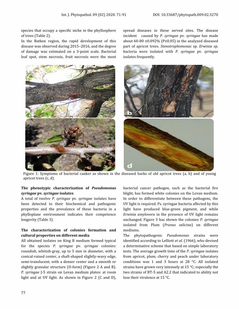
Int. J. Phytopathol. 09 (02) 2020. 71-91 DOI: 10.33687/phytopath.009.02.3270
77
species that occupy a specific niche in the phyllosphere
of trees (Table 2).
In the Batken region, the rapid development of this
disease was observed during 2015–2016, and the degree
of damage was estimated on a 3-point scale. Bacterial
leaf spot, stem necrosis, fruit necrosis were the most
spread diseases in these served sites. The disease
incident caused by P. syringae pv. syringae has made
about 60-80 ±0.092% (P≤0.05) in the analyzed diseased
part of apricot trees. Stenotrophomonas sp. Erwinia sp.
bacteria were isolated with P. syringae pv. syringae
isolates frequently.
Figure 1: Symptoms of bacterial canker as shown in the diseased barks of old apricot trees (a, b) and of young apricot trees (c, d).
The phenotypic characterization of Pseudomonas
syringae pv. syringae isolates
A total of twelve P. syringae pv. syringaе isolates have
been detected to their biochemical and pathogenic
properties and the prevalence of these bacteria in a
phylloplane environment indicates their competence
longevity (Table 3).
The characterization of colonies formation and
cultural properties on different media
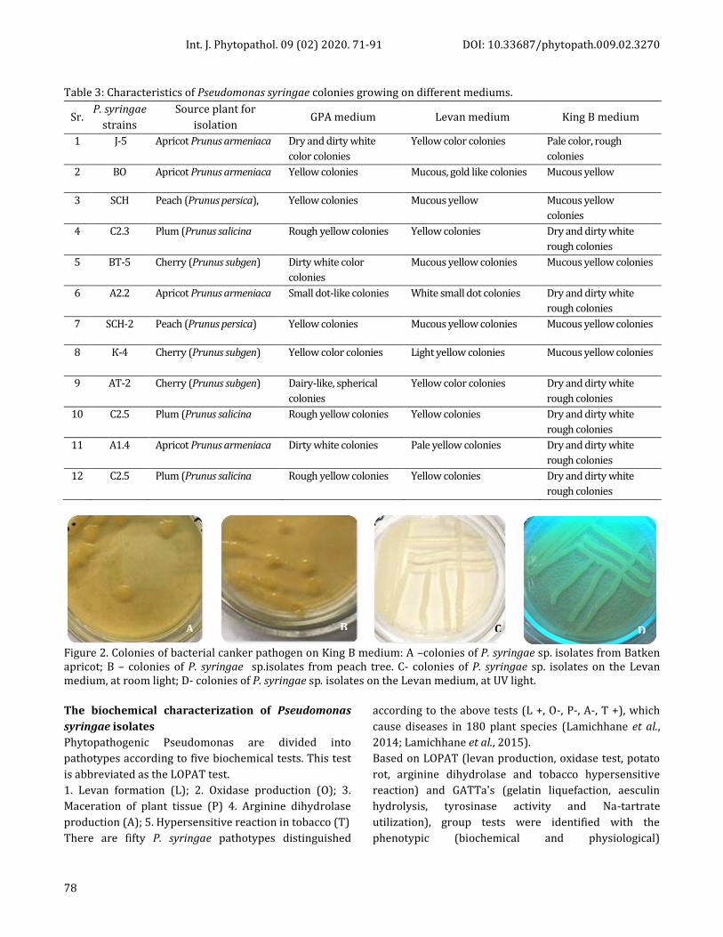
All obtained isolates on King B medium formed typical
for the species P. syringae pv. syringae colonies:
roundish, whitish-gray, up to 5 mm in diameter, with a
conical-raised center, a shaft-shaped slightly-wavy edge,
semi-translucent, with a denser center and a smooth or
slightly granular structure (O-form) (Figure 2 A and B).
P. syringae J-5 strain on Levan medium plates: at room
light and at UV light. As shown in Figure 2 (C and D),
bacterial cancer pathogen, such as the bacterial fire
blight, has formed white colonies on the Levan medium.
In order to differentiate between these pathogens, the
UV light is required. Ps. syringae bacteria affected by this
light have produced blue-green pigment, and while
Erwinia amylovora in the presence of UV light remains
unchanged. Figure 3 has shown the colonies P. syringae
isolated from Plum (Prunus salicina) on different
mediums.
The phytopathogenic Pseudomonas strains were
identified according to Lelliott et al. (1966), who devised
a determinative scheme that based on simple laboratory
tests. The average growth time of the P. syringae isolates
from apricot, plum, cherry and peach under laboratory
conditions was 1 and 3 hours at 28 °C. All isolated
strains have grown very intensely at 15 °C; especially the
two strains of BT-5 and A2.2 that indicated to ability not
lose their virulence at 15 °C.
Int. J. Phytopathol. 09 (02) 2020. 71-91 DOI: 10.33687/phytopath.009.02.3270
78
Table 3: Characteristics of Pseudomonas syringae colonies growing on different mediums.
Sr. P. syringae
strains
Source plant for
isolation GPA medium Levan medium King B medium
1 J-5 Apricot Prunus armeniaca Dry and dirty white
color colonies
Yellow color colonies Pale color, rough
colonies
2 BO Apricot Prunus armeniaca Yellow colonies Mucous, gold like colonies Mucous yellow
3 SCH Peach (Prunus persica), Yellow colonies Mucous yellow Mucous yellow
colonies
4 С2.3
Plum (Prunus salicina Rough yellow colonies Yellow colonies Dry and dirty white
rough colonies
5 BT-5 Cherry (Prunus subgen) Dirty white color
colonies
Mucous yellow colonies Mucous yellow colonies
6 А2.2
Apricot Prunus armeniaca Small dot-like colonies White small dot colonies Dry and dirty white
rough colonies
7 SCH-2 Peach (Prunus persica) Yellow colonies Mucous yellow colonies Mucous yellow colonies
8 К-4
Cherry (Prunus subgen) Yellow color colonies Light yellow colonies Mucous yellow colonies
9 АТ-2
Cherry (Prunus subgen) Dairy-like, spherical
colonies
Yellow color colonies Dry and dirty white
rough colonies
10 С2.5
Plum (Prunus salicina Rough yellow colonies Yellow colonies Dry and dirty white
rough colonies
11 А1.4 Apricot Prunus armeniaca Dirty white colonies Pale yellow colonies Dry and dirty white
rough colonies
12 С2.5
Plum (Prunus salicina Rough yellow colonies Yellow colonies Dry and dirty white
rough colonies
Figure 2. Colonies of bacterial canker pathogen on King B medium: A –colonies of P. syringae sp. isolates from Batken apricot; B – colonies of P. syringae sp.isolates from peach tree. С- colonies of P. syringae sp. isolates on the Levan medium, at room light; D- colonies of P. syringae sp. isolates on the Levan medium, at UV light.
The biochemical characterization of Pseudomonas
syringae isolates
Phytopathogenic Pseudomonas are divided into
pathotypes according to five biochemical tests. This test
is abbreviated as the LOPAT test.
1. Levan formation (L); 2. Oxidase production (O); 3.
Maceration of plant tissue (P) 4. Arginine dihydrolase
production (A); 5. Hypersensitive reaction in tobacco (T)
There are fifty P. syringae pathotypes distinguished
according to the above tests (L +, O-, P-, A-, T +), which
cause diseases in 180 plant species (Lamichhane et al.,
2014; Lamichhane et al., 2015).
Based on LOPAT (levan production, oxidase test, potato
rot, arginine dihydrolase and tobacco hypersensitive
reaction) and GATTa's (gelatin liquefaction, aesculin
hydrolysis, tyrosinase activity and Na-tartrate
utilization), group tests were identified with the
phenotypic (biochemical and physiological)
A B C D
Int. J. Phytopathol. 09 (02) 2020. 71-91 DOI: 10.33687/phytopath.009.02.3270
79
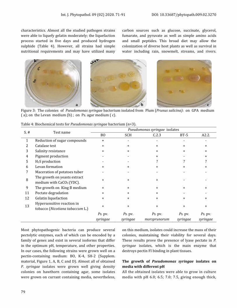
characteristics. Almost all the studied pathogen strains
were able to liquefy gelatin moderately; the liquefaction
process started in five days and produced hydrogen
sulphide (Table 4). However, all strains had simple
nutritional requirements and may have utilized many
carbon sources such as glucose, succinate, glycerol,
fumarate, and pyruvate as well as simple amino acids
and small peptides. This broad diet may allow the
colonization of diverse host plants as well as survival in
water including rain, snowmelt, streams, and rivers.
Figure 3: The colonies of Pseudomonas syringae bacterium isolated from Plum (Prunus salicina): on GPA medium ( a); on the Levan medium (b) ; on Ps. agar medium ( c).
Table 4: Biochemical tests for Pseudomonas syringae bacterium (n=3).
S. # Test name Pseudomonas syringae isolates
BO SCH С.2.3 BТ-5 А2.2.
1 Reduction of sugar compounds + - - - -
2 Catalase test + + + + +
3 Salinity resistance + + + + +
4 Pigment production - - + - +
5 H2S production - - ? ? ?
6 Levan formation + + + + +
7 Maceration of potatoes tuber - - - - -
8 The growth on yeasts extract
medium with CaCO3 (YDC). + + + - -
9 The growth on King B medium + + + + +
11 Pectate degradation + + + - -
12 Gelatin liquefaction + + + + +
13 Hypersensitive reaction in
tobacco (Nicotiana tabaccum L.) + + + + +
Ps. pv.
syringae
Ps. pv.
syringae
Ps. pv.
morsprunorum
Ps. pv.
syringae
Ps. pv.
syringae
Most phytopathogenic bacteria can produce several
pectolytic enzymes, each of which can be encoded by a
family of genes and exist in several isoforms that differ
in the optimum pH, temperature, and other properties.
In our cases, the following strains were grown well on a
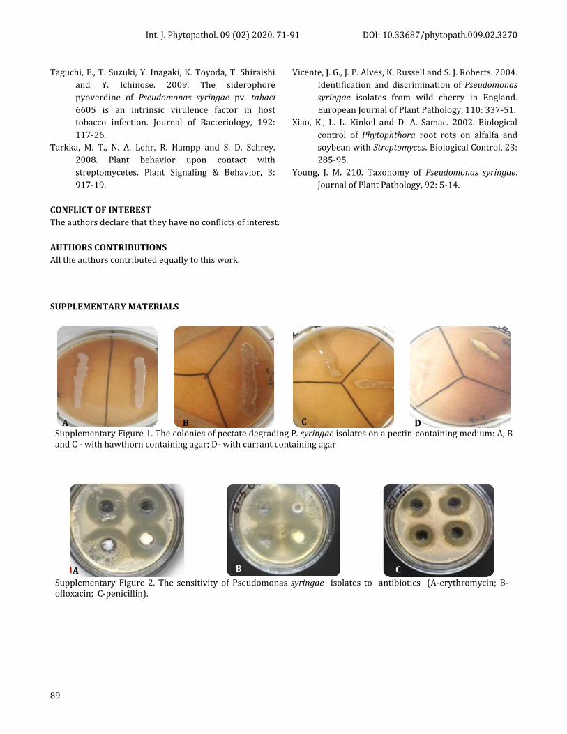
pectin-containing medium: BO, K-4, SH-2 (Supplem.
material, Figure 1, A, B, C and D). Almost all of obtained
P. syringae isolates were grown well giving density
colonies on hawthorn containing agar, some isolates
were grown on currant containing media, nevertheless,
on this medium, isolates could increase the mass of their
colonies, maintaining their viability for several days.
These results prove the presence of lyase pectate in P.
syringae isolates, which is the main enzyme that
destroys pectin FI binding in plant tissues.
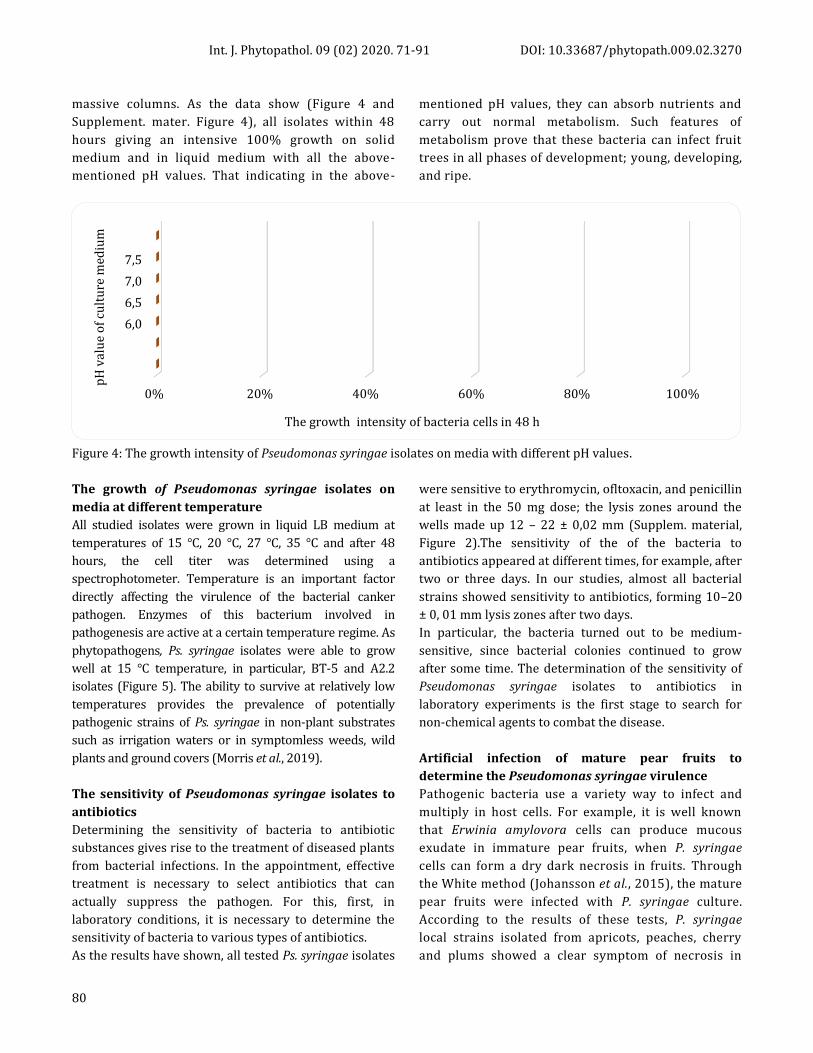
The growth of Pseudomonas syringae isolates on
media with different pH
All the obtained isolates were able to grow in culture
media with pH 6.0; 6.5; 7.0; 7.5, giving enough thick,
a b c
Int. J. Phytopathol. 09 (02) 2020. 71-91 DOI: 10.33687/phytopath.009.02.3270
80
massive columns. As the data show (Figure 4 and
Supplement. mater. Figure 4), all isolates within 48
hours giving an intensive 100% growth on solid
medium and in liquid medium with all the above-
mentioned pH values. That indicating in the above-
mentioned pH values, they can absorb nutrients and
carry out normal metabolism. Such features of
metabolism prove that these bacteria can infect fruit
trees in all phases of development; young, developing,
and ripe.
Figure 4: The growth intensity of Pseudomonas syringae isolates on media with different pH values.
The growth of Pseudomonas syringae isolates on
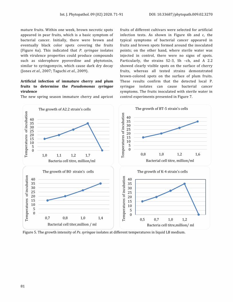
media at different temperature
All studied isolates were grown in liquid LB medium at
temperatures of 15 °C, 20 °C, 27 °C, 35 °C and after 48
hours, the cell titer was determined using a
spectrophotometer. Temperature is an important factor
directly affecting the virulence of the bacterial canker
pathogen. Enzymes of this bacterium involved in
pathogenesis are active at a certain temperature regime. As
phytopathogens, Ps. syringae isolates were able to grow
well at 15 °C temperature, in particular, BT-5 and A2.2
isolates (Figure 5). The ability to survive at relatively low
temperatures provides the prevalence of potentially
pathogenic strains of Ps. syringae in non-plant substrates
such as irrigation waters or in symptomless weeds, wild
plants and ground covers (Morris et al., 2019).
The sensitivity of Pseudomonas syringae isolates to
antibiotics
Determining the sensitivity of bacteria to antibiotic
substances gives rise to the treatment of diseased plants
from bacterial infections. In the appointment, effective
treatment is necessary to select antibiotics that can
actually suppress the pathogen. For this, first, in
laboratory conditions, it is necessary to determine the
sensitivity of bacteria to various types of antibiotics.
As the results have shown, all tested Ps. syringae isolates
were sensitive to erythromycin, ofltoxacin, and penicillin
at least in the 50 mg dose; the lysis zones around the
wells made up 12 – 22 ± 0,02 mm (Supplem. material,
Figure 2).The sensitivity of the of the bacteria to
antibiotics appeared at different times, for example, after
two or three days. In our studies, almost all bacterial
strains showed sensitivity to antibiotics, forming 10–20
± 0, 01 mm lysis zones after two days.
In particular, the bacteria turned out to be medium-
sensitive, since bacterial colonies continued to grow
after some time. The determination of the sensitivity of
Pseudomonas syringae isolates to antibiotics in
laboratory experiments is the first stage to search for
non-chemical agents to combat the disease.
Artificial infection of mature pear fruits to
determine the Pseudomonas syringae virulence
Pathogenic bacteria use a variety way to infect and
multiply in host cells. For example, it is well known
that Erwinia amylovora cells can produce mucous
exudate in immature pear fruits, when P. syringae
cells can form a dry dark necrosis in fruits. Through
the White method (Johansson et al., 2015), the mature
pear fruits were infected with P. syringae culture.
According to the results of these tests, P. syringae
local strains isolated from apricots, peaches, cherry
and plums showed a clear symptom of necrosis in
0% 20% 40% 60% 80% 100%
6,0
6,5
7,0
7,5
The growth intensity of bacteria cells in 48 h
рH
val
ue
of
cult
ure
med
ium
Int. J. Phytopathol. 09 (02) 2020. 71-91 DOI: 10.33687/phytopath.009.02.3270
81
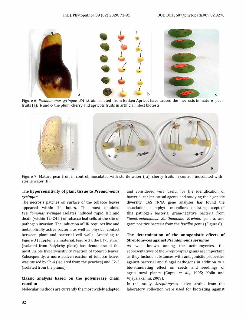
mature fruits. Within one week, brown necrotic spots
appeared in pear fruits, which is a basic symptom of
bacterial cancer. Initially, there were brown and
eventually black color spots covering the fruits
(Figure 6а). This indicated that P. syringae isolates
with virulence properties could produce compounds
such as siderophore pyoverdine and phytotoxin,
similar to syringomycin, which cause dark dry decay
(Jones et al., 2007; Taguchi et al., 2009).
Artificial infection of immature cherry and plum
fruits to determine the Pseudomonas syringae
virulence
The new spring season immature cherry and apricot
fruits of different cultivars were selected for artificial
infection tests. As shown in Figure 6b and c, the
typical symptoms of bacterial cancer appeared in
fruits and brown spots formed around the inoculated
points; on the other hand, where sterile water was
injected in control, there were no signs of spots.
Particularly, the strains S2-3, Sh –ch, and A 2.2
showed clearly visible spots on the surface of cherry
fruits, whereas all tested strains demonstrated
brown-colored spots on the surface of plum fruits.
These results confirm that the detected local P.
syringae isolates can cause bacterial cancer
symptoms. The fruits inoculated with sterile water in
control experiments presented in Figure 7.
Figure 5. The growth intensity of Ps. syringae isolates at different temperatures in liquid LB medium.
05
10152025303540
1,0 1,1 1,2 1,7Tem
per
atu
res
of
incu
bat
ion
Bacteria cell titre, million/ml
The growth of A2.2 strain's cells
05
10152025303540
0,8 1,0 1,2 1,6
Tem
per
atu
res
of
incu
bat
ion
Bacterial cell titre, million/ml
The growth of BT-5 strain's cells
05
10152025303540
0,7 0,8 1,0 1,4
Tem
per
atu
res
of
incu
bat
ion
Bacterial cell titer,million / ml
The growth of BO strain’s cells
05
10152025303540
0,5 0,7 1,0 1,2Tem
per
atu
res
of
incu
bat
ion
Bacteria cell titre,million/ ml
The growth of K-4 strain's cells
Int. J. Phytopathol. 09 (02) 2020. 71-91 DOI: 10.33687/phytopath.009.02.3270
82
Figure 6: Pseudomonas syringae BA strain isolated from Batken Apricot have caused the necrosis in mature pear fruits (a); b and c- the plum, cherry and apricots fruits in artificial infect biotests.
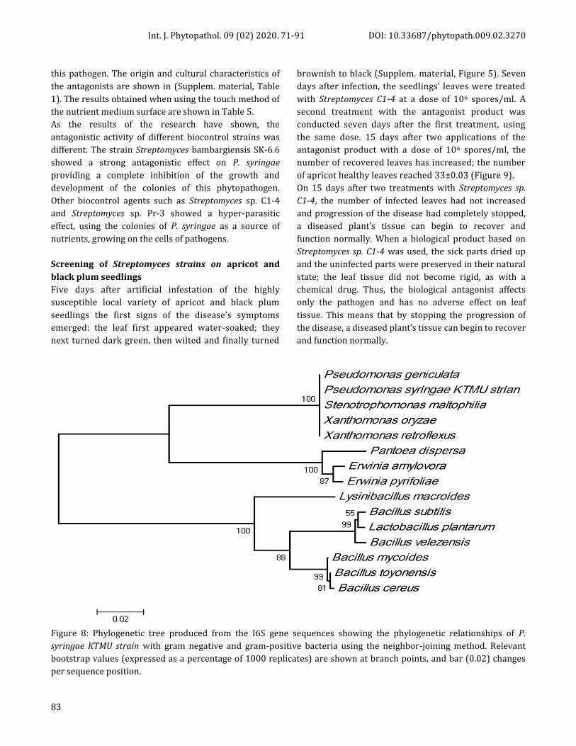
Figure 7: Mature pear fruit in control, inoculated with sterile water ( a); cherry fruits in control, inoculated with sterile water (b).
The hypersensitivity of plant tissue to Pseudomonas
syringae
The necrosis patches on surface of the tobacco leaves
appeared within 24 hours. The most obtained
Pseudomonas syringae isolates induced rapid HR and
death (within 12–24 h) of tobacco leaf cells at the site of
pathogen invasion. The induction of HR requires live and
metabolically active bacteria as well as physical contact
between plant and bacterial cell walls. According to
Figure 3 (Supplemen. material. Figure 3), the BT-5 strain
(isolated from Balykchy place) has demonstrated the
most visible hypersensitivity reaction of tobacco leaves.
Subsequently, a more active reaction of tobacco leaves
was caused by Sh-4 (isolated from the peaches) and C2-3
(isolated from the plums).
Classic analysis based on the polymerase chain
reaction
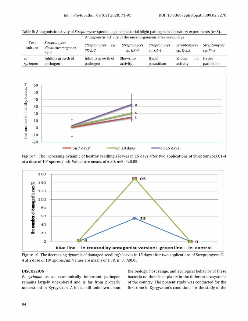
Molecular methods are currently the most widely adapted
and considered very useful for the identification of
bacterial canker causal agents and studying their genetic
diversity. 16S rRNA gene analyses has found the
association of epiphytic microflora consisting except of
this pathogen bacteria, gram-negative bacteria from
Stenotrophomonas, Xanthomonas, Erwinia, genera, and
gram positive bacteria from the Bacillus genus (Figure 8).
The determination of the antagonistic effects of
Streptomyces against Pseudomonas syringae
As well known among the actinomycetes, the
representatives of the Streptomyces genus are important,
as they include substances with antagonistic properties
against bacterial and fungal pathogens in addition to a
bio-stimulating effect on seeds and seedlings of
agricultural plants (Gupta et al., 1995; Kolla and
Vijayalakshmi, 2009).
In this study, Streptomyces active strains from the
laboratory collection were used for biotesting against
a b
a b
c
Int. J. Phytopathol. 09 (02) 2020. 71-91 DOI: 10.33687/phytopath.009.02.3270
83
this pathogen. The origin and cultural characteristics of
the antagonists are shown in (Supplem. material, Table
1). The results obtained when using the touch method of
the nutrient medium surface are shown in Table 5.
As the results of the research have shown, the
antagonistic activity of different biocontrol strains was
different. The strain Streptomyces bambargiensis SK-6.6
showed a strong antagonistic effect on P. syringae
providing a complete inhibition of the growth and
development of the colonies of this phytopathogen.
Other biocontrol agents such as Streptomyces sр. С1-4
and Streptomyces sр. Pr-3 showed a hyper-parasitic
effect, using the colonies of P. syringae as a source of
nutrients, growing on the cells of pathogens.
Screening of Streptomyces strains on apricot and
black plum seedlings
Five days after artificial infestation of the highly
susceptible local variety of apricot and black plum
seedlings the first signs of the disease’s symptoms
emerged: the leaf first appeared water-soaked; they
next turned dark green, then wilted and finally turned
brownish to black (Supplem. material, Figure 5). Seven
days after infection, the seedlings’ leaves were treated
with Streptomyces C1-4 at a dose of 106 spores/ml. A
second treatment with the antagonist product was
conducted seven days after the first treatment, using
the same dose. 15 days after two applications of the
antagonist product with a dose of 106 spores/ml, the
number of recovered leaves has increased; the number
of apricot healthy leaves reached 33±0.03 (Figure 9).
On 15 days after two treatments with Streptomyces sp.
C1-4, the number of infected leaves had not increased
and progression of the disease had completely stopped,
a diseased plant’s tissue can begin to recover and
function normally. When a biological product based on
Streptomyces sp. C1-4 was used, the sick parts dried up
and the uninfected parts were preserved in their natural
state; the leaf tissue did not become rigid, as with a
chemical drug. Thus, the biological antagonist affects
only the pathogen and has no adverse effect on leaf
tissue. This means that by stopping the progression of
the disease, a diseased plant’s tissue can begin to recover
and function normally.
Figure 8: Phylogenetic tree produced from the I6S gene sequences showing the phylogenetic relationships of P.
syringae KTMU strain with gram negative and gram-positive bacteria using the neighbor-joining method. Relevant
bootstrap values (expressed as a percentage of 1000 replicates) are shown at branch points, and bar (0.02) changes
per sequence position.
Int. J. Phytopathol. 09 (02) 2020. 71-91 DOI: 10.33687/phytopath.009.02.3270
84
Table 5: Antagonistic activity of Streptomyces sрecies against bacterial blight pathogen in laboratory experiments (n=3).
Test
culture
Antagonistic activity of the microorganisms after seven days
Streptomyces
diastochromogenes,
Sk-6
Streptomyces sр.
SK-2, 2
Streptomyces
sр. KK-4
Streptomyces
sр. С1-4
Streptomyces
sр. K-3.3
Streptomyces
sр. Pr-3
P.
syringae
Inhibits growth of
pathogen
Inhibits growth of
pathogen
Shows no
activity
Hyper
parasitism
Shows no
activity
Hyper
parasitism
Figure 9: The increasing dynamic of healthy seedling’s leaves in 15 days after two applications of Streptomyces C1-4
at a dose of 106 spores / ml; Values are means of ± SD, n=3, P≤0.05.
Figure 10: The decreasing dynamic of damaged seedling’s leaves in 15 days after two applications of Streptomyces C1-
4 at a dose of 106 spores/ml. Values are means of ± SD, n=3, P≤0.05.
DISCUSSION
P. syringae as an economically important pathogen
remains largely unexplored and is far from properly
understood in Kyrgyzstan. A lot is still unknown about
the biology, host range, and ecological behavior of these
bacteria on their host plants in the different ecosystems
of the country. The present study was conducted for the
first time in Kyrgyzstan's conditions for the study of the
bc
a
-20
-10
0
10
20
30
40
50
60
the
nu
mb
er o
f h
ealt
hy
lea
ves
, %
on 7 days" on 10 days on 15 days
Int. J. Phytopathol. 09 (02) 2020. 71-91 DOI: 10.33687/phytopath.009.02.3270
85
biology, ecology, and the prevalence of the pathogen in
different regions with different climatic conditions. The
monitoring survey for the symptomatic observation and
collection of samples has revealed that in old stone
plants planted over 30–40 years ago, the characteristic
symptoms for bacterial cancer were strongly exhibited
with large ulcer wounds and necrotic bark formations
with the release of the exudate amber formation on the
trunks and branches. The prolonged coexistence of the
pathogen and the host under certain climatic conditions
apparently could provide to both sides a survival status,
but with a gradual weakening of the host plant. The used
classical biochemical tests (LOPAT and GATTa's) have
allowed us to isolate pure cultures of pathogen
bacterium and identify the obtained isolates as a
pathogen—P. syringae. Additionally, 16Sr RNA gene
analysis has found the association of epiphytic
microflora consisting except this pathogen bacteria,
gram-negative bacteria from Erwinia, Xanthomanas,
Stenotrophomonas genera, and gram-positive bacteria
from the Bacillus genus. In such an association, this
pathogen in a dominant ratio has adapted to coexist with
its host plant and associated epiphytic bacteria in
different climatic zones of the country. In the
composition of the epiphytic microflora after the
pathogen, the bacteria of the Stenotrophomonas genus
was dominated. The role of this bacterium as a
saprophyte or facilitative parasite contributing to the
development of an outbreak must be clarified in future
studies. According to the scientists, these bacteria can be
found throughout the environment, particularly in close
association with plants. S. maltophilia frequently co-
occurs and forms multispecies biofilms with P.
aeruginosa (Ryan et al., 2009). As reported by studies
(Ercolani, 1991, 1985), that many phyllospheric bacteria
are non-spore-forming, gram-negative or gram-positive
heterotrophs, in general, the same species or group of
bacteria are present on a wide range of plants, which
was confirmed in our results. In addition, the studies
have concluded that these groups are well adapted to
the life in the phyllosphere, the conditions of which
change daily, seasonally, while the pathogens on the
diseased plants with obvious symptoms are
predominated in such communities (Hirano and Upper,
2000). Our results also approved the above reports.
The long coexistence of the pathogen-plant system in the
plant’s homeland is a prime example for this bacterium
since we found such a system between apricot and this
bacterium in Batken’s homeland, between peach
varieties and this bacterium, between plum, cherry
varieties and this bacterium in other regions; almost
every local stone fruit variety had pathogen such as P.
syringae. The composition of epiphytic microflora did
not differ much in the regions of the republic, and no
large differences were observed in the seasons of the
year.
The dominance of P. syringae in all climatic conditions
and seasons of the year indicates a wide range of the
temperature of its growth. The optimum temperature
turned out to be 28 ℃; at this temperature, the isolates
isolated from apricot, peach, cherry and plum in just two
to three hours, which gave a good growth of colonies on
media. At a temperature of 15 ℃, they had intensive
growth, which indicates that at this temperature, they do
not lose their virulence. Another adaptive nature
inherent in the isolated isolates is their ability to grow at
different pH values, ranging from 6.0 to 7.5. Therefore,
they affect plants in all phases of development, from
budding to ripe fruit, as well as all age stages, spanning
from seedlings to old, perennial trees. The results of
biotests on mature and immature fruits of different
stone fruits proved this ability. Unlike the Erwinia
bacteria, this pathogen causes dry rot in the fruit. The
presence of pectolytic enzymes detected by culturing
them on media containing hawthorn and currant
extracts proves that this pathogen is capable of
destroying plant tissues in case of damage.
As well known, the causative agent of bacterial canker P.
syringae bacteria affects more than 180 species of plants,
among them herbaceous plants, vegetables, fruit crops.
To limit the use of chemicals to protect plants from
bacteriosis caused by P. syringae a constant search of
safely to the environment and human health alternative
means is underway.
For this purpose, the antibacterial activity of the growth-
stimulating rhizospheric bacteria (PGPR) such as P.
aeruginosa, Bacillus stratosphericus, Paenibacillus
polymyxa, B. subtilis and others against P. syringae was
studied in vitro and in vivo experiments. Their
overwhelming disease activity against P. syringae was
reliably proven on tomatoes, kiwi fruit trees (Hong et al.,
2016; Donati et al., 2018).
The biocontrol potential of soil and rhizospheric
actinomycetes like Streptomyces in the regulation of pest
damage by Pseudomonas syringae in fruit trees remains
almost unexplored. According to studies, plant-
Int. J. Phytopathol. 09 (02) 2020. 71-91 DOI: 10.33687/phytopath.009.02.3270
86
associated actinomycetes play an important role in
plant health due to ability to produce a wide range of
secondary metabolites, like siderophores, various plant
phytohormones such as auxins, cytokinins and
gibberellins, and antibacterial and antifungal
compounds (Cassán et al., 2001; Bottini et al., 2004;
Solans et al., 2011). It has been proven that Streptomyces
species are capable of exerting antifungal and
antibacterial properties, protecting the plant root system
from pathogens, and also suppressing diseases in soils
(Gopalakrishnan et al., 2011; Xiao et al., 2002; Mendes et
al., 2011; Cha et al., 2015).
In contrast to studies where Streptomycetes were used
by introducing into the soil and around the root system
of diseased plants, we used the Streptomyces strains
against the bacterial canker pathogen by spraying the
leaf surface and the crown of diseased seedlings of stone
fruits, so biocontrol agents were introduced into
epiphytic plant associations.
When signs of the disease appear on the leaves, double
plentiful spraying of affected plants with a suspension of
Streptomyces C1-4 strain (106 spores/ml) with an
interval of 7 days prevented the development of the
disease. Fifteen days after the biologic agent was applied
twice, the plants fully recovered, new leaves and new
twigs appeared. These results indicate that the
Streptomyces C1-4 strain, introduced as a biological
agent into the phyllosphere of diseased plants was able
not only to colonize the epiphytic microflora, having an
antibacterial effect but also increased the protective
ability of the plants themselves, their potential to resist
the disease, which is consistent with the data of other
researchers (Conn et al., 2008; Tarkka et al., 2008; Kurth
et al., 2014), they noted that Streptomycetes can cause a
state of the increased protective ability of plants against
pathogens, thereby providing plants with induced
systemic resistance.
The growth of new shoots in a short period and the
complete restoration of damaged leaves, branches also
indicates to the growth-stimulating or phytohormonal
effect of this agent on plants, as noted in the studies of
(Cassán et al., 2001; Bottini et al., 2004). Thus, Streptomyces
strain C1-4 was selected after in vitro tests as an effective
antagonist and hyper-parasitic agent to P. syringae for
spraying the diseased leaves of apricot and black plum
seedlings. Streptomyces С1-4 showed a significant effect
against P. syringae after two applications. A suitable time
for this bio product’s application is spring, when the
symptoms of bacterial canker start to develop on the leaves
of trees and the air temperature is 18–23 °С.
CONCLUSIONS
Therefore, our results on diagnosis of this pathogen can
provide practical information useful for implementing
efficient control measures in the current cropping
season in the stone fruit gardens and in limiting future
disease outbreaks of bacterial cancer in different regions
of country. Also obtained results about capacity of the
pathogen to adapt to climatic conditions, sensitivity to
such biocontrol agents as Streptomyces species can
encourage to develop a local bio-product based on this
bioagent for spraying stone fruits with the initial
manifestation of disease symptoms and to conduct
preventive treatments in the fall and spring to increase
the plant's resistance to pathogens. Our studies are
suggesting the creation of induced plant immunity by
spraying the phyllosphere with biological agents.
ACKNOWLEDGEMENTS
This research was supported within the framework of
project “Surveillance and pathogen characterization of
bacterial canker of stone fruits using biochemical and
molecular methods and its bio-management” (PEREZ-
GUERRERO TRUST FUND FOR SOUTH-SOUTH COOPERATION, UNDP).
REFERENCES
Agrios, G. N. 2005. Plant Pathology. Elsevier Academic
Press: Burlington, Ma. USA.
Ahmed, R., M. Inam-ul-Haq, U. Shahzad, S. Hyder, S.
Shahzaman, A. Khan, H. Aatif, A. Ahmad and A.
Gondal. 2018. First report of bacterial canker
caused by Pseudomonas syringae pv.
morsprunorum race 1 on peach from Khyber
Pakhtunkhwa province of Pakistan. Plant disease,
102: 2027-27.
Bais, H. P., R. Fall and J. M. Vivanco. 2003. Biocontrol of
Bacillus subtilis against infection of Arabidopsis
roots by Pseudomonas syringae is facilitated by
biofilm formation and surfactin production. Plant
physiology, 134: 307-19.
Berge, O., C. L. Monteil, C. Bartoli, C. Chandeysson, C.
Guilbaud, D. C. Sands and C. E. Morris. 2014. A
user's guide to a data base of the diversity of
Pseudomonas syringae and its application to
classifying strains in this phylogenetic complex.
PLOS ONE, 9: e105547.
Int. J. Phytopathol. 09 (02) 2020. 71-91 DOI: 10.33687/phytopath.009.02.3270
87
Bottini, R., F. Cassan and P. Piccoli. 2004. Gibberellin
production by bacteria and its involvement in
plant growth promotion and yield increase.
Applied microbiology and biotechnology, 65: 497-
503.
Braun-Kiewnick, A. and D. C. Sands. 2001. Pseudomonas.
In: N W Schaad, J B Jones and W Chun (eds.),
Laboratory Guide for Identification of Plant
Pathogenic Bacteria. American Phytopathological
Society Press: St. Paul, Minesota, USA.
Bultreys, A. and I. Gheysen. 1999. Biological and
molecular detection of toxic lipodepsipeptide-
producing Pseudomonas syringae strains and PCR
identification in plants. Applied and
Environmental Microbiology, 65: 1904-09.
Cassán, F. D., C. D. Lucangeli, R. Bottini and P. N. Piccoli.
2001. Azospirillum spp. metabolize [ 17,17-2H2]
gibberellin A20 to [ 17,17-2H2] gibberellin A1 in
vivo in dy rice mutant seedlings. Plant and Cell
Physiology, 42: 763-67.
Cha, J.-Y., S. Han, H.-J. Hong, H. Cho, D. Kim, Y. Kwon, S.-K.
Kwon, M. Crüsemann, Y. Bok Lee, J. F. Kim, G.
Giaever, C. Nislow, B. S. Moore, L. S. Thomashow,
D. M. Weller and Y.-S. Kwak. 2015. Microbial and
biochemical basis of a Fusarium wilt suppressive
soil. The ISME Journal, 10: 119-29.
Choi, O., B. Kang, S. K. Cho, J. Park, Y. Lee, W.-I. Kim, J.
Marunga, I. Hwang and J. Kim. 2016. Identification
of Pseudomonas syringae pv. syringae causing
bacterial leaf blight of Miscanthus sinensis. Journal
of Plant Diseases and Protection, 124: 97-100.
Conn, V. M., A. R. Walker and C. M. M. Franco. 2008.
Endophytic actinobacteria induce defense
pathways in Arabidopsis thaliana. Molecular Plant-
Microbe Interactions, 21: 208-18.
Destéfano, S. A. L., L. M. R. Rodrigues, L. O. S. Beriam, F. R.
A. Patrício, R. A. Thomaziello and J. Rodrigues-
Neto. 2010. Bacterial leaf spot of coffee caused by
Pseudomonas syringae pv. tabaci in Brazil. Plant
Pathology, 59: 1162-63.
Donati, I., G. Buriani, A. Cellini, N. Raule and F. Spinelli.
2018. Screening of microbial biocoenosis of
Actinidia chinensis for the isolation of candidate
biological control agents against Pseudomonas
syringae pv. actinidiae. Acta Horticulturae: 239-46.
Durairaj, K., P. Velmurugan, J.-H. Park, W.-S. Chang, Y.-J.
Park, P. Senthilkumar, K.-M. Choi, J.-H. Lee and B.-
T. Oh. 2018. Characterization and assessment of
two biocontrol bacteria against Pseudomonas
syringae wilt in Solanum lycopersicum and its
genetic responses. Microbiological Research, 206:
43-49.
Ercolani, G. L. 1985. Factor analysis of fluctuation in
populations of Pseudomonas syringae pv.
savastanoi on the phylloplane of the olive.
Microbial Ecology, 11: 41-49.
Ercolani, G. L.. 1991. Distribution of epiphytic bacteria
on olive leaves and the influence of leaf age and
sampling time. Microbial Ecology, 21: 35-48.
Gardan, L., H. Shafik, S. Belouin, R. Broch, F. Grimont and
P. A. D. Grimont. 1999. DNA relatedness among
the pathovars of Pseudomonas syringae and
description of Pseudomonas tremae sp. nov. and
Pseudomonas cannabina sp. nov. (ex Sutic and
Dowson 1959). International Journal of
Systematic and Evolutionary Microbiology, 49:
469-78.
Gopalakrishnan, S., S. Pande, M. Sharma, P. Humayun, B.
K. Kiran, D. Sandeep, M. S. Vidya, K. Deepthi and O.
Rupela. 2011. Evaluation of actinomycete isolates
obtained from herbal vermicompost for the
biological control of Fusarium wilt of chickpea.
Crop Protection, 30: 1070-78.
Gupta, R., R. K. Saxena, P. Chaturvedi and J. S. Virdi. 1995.
Chitinase production by Streptomyces viridificans:
Its potential in fungal cell wall lysis. Journal of
Applied Bacteriology, 78: 378-83.
Hirano, S. S. and C. D. Upper. 2000. Bacteria in the leaf
ecosystem with emphasis on Pseudomonas
syringae- A pathogen, ice nucleus, and epiphyte.
Microbiology and Molecular Biology Reviews, 64:
624-53.
Hong, C. E., S. Y. Kwon and J. M. Park. 2016. Biocontrol
activity of Paenibacillus polymyxa AC-1 against
Pseudomonas syringae and its interaction with
Arabidopsis thaliana. Microbiological Research,
185: 13-21.
Johansson, O. N., A. K. Nilsson, M. B. Gustavsson, T.
Backhaus, M. X. Andersson and M. Ellerström.
2015. A quick and robust method for
quantification of the hypersensitive response in
plants. PeerJ, 3: e1469.
Jones, A. M., S. E. Lindow and M. C. Wildermuth. 2007.
Salicylic acid, yersiniabactin, and pyoverdin
production by the model phytopathogen
Pseudomonas syringae pv. tomato DC3000:
Int. J. Phytopathol. 09 (02) 2020. 71-91 DOI: 10.33687/phytopath.009.02.3270
88
synthesis, regulation, and impact on tomato and
Arabidopsis host plants. Journal of Bacteriology,
189: 6773-86.
King, E. O., M. K. Raney and D. E. Ward. 1954. Two simple
media for the demonstration of pyocianin and
fluorescin. Journal of Laboratory and Clinical
Medicine, 44: 301-07.
Koike, S. T., E. I. Alger, L. Ramos Sepulveda and C. T. Bull.
2017. First report of bacterial leaf spot caused by
Pseudomonas syringae pv. tomato on kale in
California. Plant disease, 101: 504-04.
Kolla, N. J. P. and M. Vijayalakshmi. 2009. Chitinase
production by Streptomyces sp. ANU 6277.
Brazilian Journal of Microbiology, 40: 725-33.
Kurth, F., S. Mailänder, M. Bönn, L. Feldhahn, S.
Herrmann, I. Große, F. Buscot, S. D. Schrey and M.
T. Tarkka. 2014. Streptomyces-induced resistance
against oak powdery mildew involves host plant
responses in defense, photosynthesis, and
secondary metabolism pathways. Molecular Plant-
Microbe Interactions, 27: 891-900.
Lamichhane, J. R., A. Messéan and C. E. Morris. 2015.
Insights into epidemiology and control of diseases
of annual plants caused by the Pseudomonas
syringae species complex. Journal of General Plant
Pathology, 81: 331-50.
Lamichhane, J. R., L. Varvaro, L. Parisi, J.-M. Audergon
and C. E. Morris. 2014. Disease and frost damage
of woody plants caused by Pseudomonas syringae
Advances in Agronomy. Elsevier. pp. 235-95.
Latorre, B. A. and A. L. Jones. 1979. Pseudomonas
morsprunorum,the cause of bacterial canker of
sour cherry in Michigan, and its epiphytic
association with P. syringae. Phytopathology, 69:
335-39.
Lelliott, R. A., E. Billing and A. C. Hayward. 1966. A
determinative scheme for the fluorescent plant
pathogenic pseudomonads. Journal of Applied
Bacteriology, 29: 470-89.
Lelliott, R. A. and D. E. Stead. 1987. Methods for the
Diagnosis of Bacterial Diseases of Plants.
Blackwell Scientific Publications: Oxford, Great
Britain.
Little, E. L., R. M. Bostock and B. C. Kirkpatrick. 1998.
Genetic characterization of Pseudomonas syringae
pv. syringae strains from stone fruits in California.
Applied and Environmental Microbiology, 64:
3818-23.
Mendes, R., M. Kruijt, I. de Bruijn, E. Dekkers, M. van der
Voort, J. H. M. Schneider, Y. M. Piceno, T. Z.
DeSantis, G. L. Andersen, P. A. H. M. Bakker and J.
M. Raaijmakers. 2011. Deciphering the
rhizosphere microbiome for disease suppressive
bacteria. Science, 332: 1097-100.
Morris, C. E., C. Glaux, X. Latour, L. Gardan, R. Samson
and M. Pitrat. 2000. The relationship of host
range, physiology, and genotype to virulence on
cantaloupe in Pseudomonas syringae from
cantaloupe blight epidemics in France.
Phytopathology, 90: 636-46.
Morris, C. E., J. R. Lamichhane, I. Nikolić, S. Stanković and
B. Moury. 2019. The overlapping continuum of
host range among strains in the Pseudomonas
syringae complex. Phytopathology Research, 1: 1-
16.
Morris, C. E., C. L. Monteil and O. Berge. 2013. The life
history of Pseudomonas syringae: Linking
agriculture to earth system processes. Annual
review of phytopathology, 51: 85-104.
Mulet, M., J. Lalucat and E. García-Valdés. 2010. DNA
sequence-based analysis of the Pseudomonas
species. Environmental Microbiology, 12: 1513-30.
Polizzi, G., I. Castello, G. Parlavecchio and G. Cirvilleri.
2005. First report of bacterial blight of Strelitzia
augusta caused by Pseudomonas syringae pv.
lachrymans. Plant disease, 89: 1010-10.
Ryan, R. P., S. Monchy, M. Cardinale, S. Taghavi, L.
Crossman, M. B. Avison, G. Berg, D. van der Lelie
and J. M. Dow. 2009. The versatility and
adaptation of bacteria from the genus
Stenotrophomonas. Nature Reviews Microbiology,
7: 514-25.
Sedighian, N., M. Shams-Bakhsh, E. Osdaghi and P.
Khodaygan. 2014. Etiology and host range of
bacterial leaf blight and necrosis of squash and
muskmelon in Iran. Journal of Plant Pathology, 96:
507-14.
Solans, M., G. Vobis, F. Cassán, V. Luna and L. G. Wall.
2011. Production of phytohormones by root-
associated saprophytic actinomycetes isolated
from the actinorhizal plant Ochetophila trinervis.
World Journal of Microbiology and Biotechnology,
27: 2195-202.
Stefani, E. and S. Loreti. 2014. Standard describes a
diagnostic protocol for Pseudomonas syringae pv.
actinidiae. EPPO Bulletin, 44: 360-75.
Int. J. Phytopathol. 09 (02) 2020. 71-91 DOI: 10.33687/phytopath.009.02.3270
89
Taguchi, F., T. Suzuki, Y. Inagaki, K. Toyoda, T. Shiraishi
and Y. Ichinose. 2009. The siderophore
pyoverdine of Pseudomonas syringae pv. tabaci
6605 is an intrinsic virulence factor in host
tobacco infection. Journal of Bacteriology, 192:
117-26.
Tarkka, M. T., N. A. Lehr, R. Hampp and S. D. Schrey.
2008. Plant behavior upon contact with
streptomycetes. Plant Signaling & Behavior, 3:
917-19.
Vicente, J. G., J. P. Alves, K. Russell and S. J. Roberts. 2004.
Identification and discrimination of Pseudomonas
syringae isolates from wild cherry in England.
European Journal of Plant Pathology, 110: 337-51.
Xiao, K., L. L. Kinkel and D. A. Samac. 2002. Biological
control of Phytophthora root rots on alfalfa and
soybean with Streptomyces. Biological Control, 23:
285-95.
Young, J. M. 210. Taxonomy of Pseudomonas syringae.
Journal of Plant Pathology, 92: 5-14.
CONFLICT OF INTEREST
The authors declare that they have no conflicts of interest.
AUTHORS CONTRIBUTIONS
All the authors contributed equally to this work.
SUPPLEMENTARY MATERIALS
Supplementary Figure 1. The colonies of pectate degrading P. syringae isolates on a pectin-containing medium: A, B and C - with hawthorn containing agar; D- with currant containing agar
Supplementary Figure 2. The sensitivity of Pseudomonas syringae isolates to antibiotics (A-erythromycin; B-ofloxacin; C-penicillin).
A
A
B
B
C
C
D
Int. J. Phytopathol. 09 (02) 2020. 71-91 DOI: 10.33687/phytopath.009.02.3270
90
Supplementary Figure 3. Hypersensitivity reaction in tobacco leaves.
Supplementary Figure 4. A) In 24 hours and B) 5 days after infection apricot seedlings with diseased symptoms on the leaves by spraying with Pseudomonas syringae (1x 10⁶ ml-1).
Supplementary Figure 5. Recovering leaves of apricot and black plum seedlings 30 days after second treatment by Streptomyces C1-4 at a dose of 106 spores/ml.
A B
Int. J. Phytopathol. 09 (02) 2020. 71-91 DOI: 10.33687/phytopath.009.02.3270
91
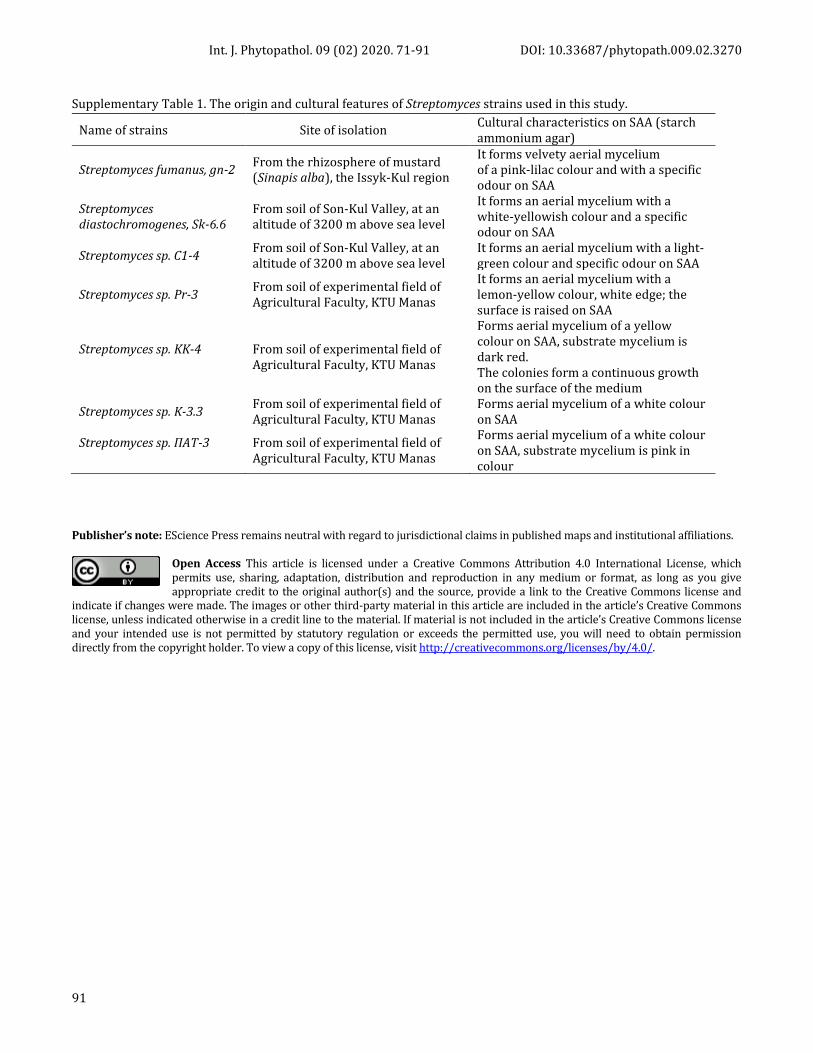
Supplementary Table 1. The origin and cultural features of Streptomyces strains used in this study.
Name of strains Site of isolation Cultural characteristics on SAA (starch ammonium agar)
Streptomyces fumanus, gn-2 From the rhizosphere of mustard (Sinapis alba), the Issyk-Kul region
It forms velvety aerial mycelium of a pink-lilac colour and with a specific odour on SAA
Streptomyces diastochromogenes, Sk-6.6
From soil of Son-Kul Valley, at an altitude of 3200 m above sea level
It forms an aerial mycelium with a white-yellowish colour and a specific odour on SAA
Streptomyces sр. С1-4 From soil of Son-Kul Valley, at an altitude of 3200 m above sea level
It forms an aerial mycelium with a light-green colour and specific odour on SAA
Streptomyces sр. Pr-3 From soil of experimental field of Agricultural Faculty, KTU Manas
It forms an aerial mycelium with a lemon-yellow colour, white edge; the surface is raised on SAA
Streptomyces sр. KK-4
From soil of experimental field of Agricultural Faculty, KTU Manas
Forms aerial mycelium of a yellow colour on SAA, substrate mycelium is dark red. The colonies form a continuous growth on the surface of the medium
Streptomyces sр. K-3.3 From soil of experimental field of Agricultural Faculty, KTU Manas
Forms aerial mycelium of a white colour on SAA
Streptomyces sр. ПАТ-3
From soil of experimental field of Agricultural Faculty, KTU Manas
Forms aerial mycelium of a white colour on SAA, substrate mycelium is pink in colour
Publisher’s note: EScience Press remains neutral with regard to jurisdictional claims in published maps and institutional affiliations.
Open Access This article is licensed under a Creative Commons Attribution 4.0 International License, which permits use, sharing, adaptation, distribution and reproduction in any medium or format, as long as you give appropriate credit to the original author(s) and the source, provide a link to the Creative Commons license and
indicate if changes were made. The images or other third-party material in this article are included in the article’s Creative Commons license, unless indicated otherwise in a credit line to the material. If material is not included in the article’s Creative Commons license and your intended use is not permitted by statutory regulation or exceeds the permitted use, you will need to obtain permission directly from the copyright holder. To view a copy of this license, visit http://creativecommons.org/licenses/by/4.0/.