Distinct subsets of dendritic cells resembling dermal DCs can be generated in vitro from monocytes,...
13
Journal of Immunological Methods 238 (2000) 119–131 www.elsevier.nl / locate / jim Distinct subsets of dendritic cells resembling dermal DCs can be generated in vitro from monocytes, in the presence of different serum supplements a, a,b c * Karine Duperrier , Assia Eljaafari , Colette Dezutter-Dambuyant , a c d c Christine Bardin , Christelle Jacquet , Koyo Yoneda , Daniel Schmitt , a a,b Lucette Gebuhrer , Dominique Rigal a Immunology HLA Departments, Etablissement de Transfusion Sanguine,1 –3 Rue du Vercors, 69007 Lyon, France b Jeune Equipe: ‘‘ Pathologie des Cellules Lymphoides’’, Universite Claude Bernard, Lyon, France c INSERM U346, Edouard Herriot Hospital, Lyon, France d Department of Dermatology, Faculty of Medicine, Kyoto University, Kyoto, Japan Received 23 September 1999; received in revised form 29 November 1999; accepted 17 January 2000 Abstract We recently demonstrated that dendritic cells (DCs) can be generated from monocytes in the presence of high concentrations of human serum (HS), provided the extra-cellular pH is maintained at plasma values. Because monocyte- derived DCs (Mo-DCs) can also be generated in the presence of fetal calf serum (FCS) or serum-free medium, we have investigated whether these different culture supplements influence DC generation. With this aim, purified monocytes were cultured with GM-CSF plus IL-4 for 6 days and were further exposed to TNF-a for 2 additional days, in the presence of HS, autologous plasma (AP), FCS, or X-VIVO 20, a serum-free medium. Our results show that good yields of functionally mature DCs can reproducibly be obtained in the presence of HS or AP, as assessed by CD83 and CD86 up-regulation, dextran-FITC uptake, allogeneic MLR assays and the induction of an autologous response. Interestingly, the effect of serum on DC generation was probably not only quantitative, but also qualitative, since (i) the majority of HS- or AP-cultured DCs expressed CD83 with very weak levels of CD1a, whereas CD831 DCs cultured in FCS or X-VIVO were mostly CD1a11; (ii) HS- and AP-cultured DCs were much more granular and heterogeneous than FCS- or X-VIVO-cultured DCs, and (iii) the presence of Birbeck-like granules was preferentially observed in HS- or AP-cultured DCs, as assessed by electron microscopy. That these different cells resemble dermal DCs (DDCs) was further supported by the observations that most of the cells displayed intracytoplasmic FXIIIa in the absence of Lag antigen, and expressed E-cadherin at very low levels. Altogether, our results indicate that starting from the same monocytic population, different subsets of DCs can be generated, Abbreviations: Ag, Antigens; AP, Autologous plasma; APC, Antigen presenting cell; DC, Dendritic cell; FACS, Fluorescence-activated cell sorter; FCS, Fetal calf serum; GM-CSF, Granulocyte macrophage-colony stimulating factor; HS, Human serum; IL, Interleukin; MLR, Mixed lymphocyte reaction; mAb, Monoclonal antibody; Mo-DC, Monocyte-derived dendritic cell; PBMC, Peripheral blood mononuclear cell; PBS, Phosphate-buffered saline; PHA, Phytohemagglutinin; TNF, Tumor necrosis factor *Corresponding author. Tel.: 133-4-7271-1722; fax: 133-4-7272-9348. E-mail address: [email protected] (K. Duperrier) 0022-1759 / 00 / $ – see front matter 2000 Elsevier Science B.V. All rights reserved. PII: S0022-1759(00)00147-2
-
Upload
independent -
Category
Documents
-
view
1 -
download
0
Transcript of Distinct subsets of dendritic cells resembling dermal DCs can be generated in vitro from monocytes,...

Journal of Immunological Methods 238 (2000) 119–131www.elsevier.nl / locate / jim
Distinct subsets of dendritic cells resembling dermal DCs canbe generated in vitro from monocytes, in the presence of
different serum supplementsa , a,b c*Karine Duperrier , Assia Eljaafari , Colette Dezutter-Dambuyant ,
a c d cChristine Bardin , Christelle Jacquet , Koyo Yoneda , Daniel Schmitt ,a a,bLucette Gebuhrer , Dominique Rigal
aImmunology HLA Departments, Etablissement de Transfusion Sanguine, 1 –3 Rue du Vercors, 69007 Lyon, FrancebJeune Equipe: ‘‘Pathologie des Cellules Lymphoides’’, Universite Claude Bernard, Lyon, France
cINSERM U346, Edouard Herriot Hospital, Lyon, FrancedDepartment of Dermatology, Faculty of Medicine, Kyoto University, Kyoto, Japan
Received 23 September 1999; received in revised form 29 November 1999; accepted 17 January 2000
Abstract
We recently demonstrated that dendritic cells (DCs) can be generated from monocytes in the presence of highconcentrations of human serum (HS), provided the extra-cellular pH is maintained at plasma values. Because monocyte-derived DCs (Mo-DCs) can also be generated in the presence of fetal calf serum (FCS) or serum-free medium, we haveinvestigated whether these different culture supplements influence DC generation. With this aim, purified monocytes werecultured with GM-CSF plus IL-4 for 6 days and were further exposed to TNF-a for 2 additional days, in the presence of HS,autologous plasma (AP), FCS, or X-VIVO 20, a serum-free medium. Our results show that good yields of functionallymature DCs can reproducibly be obtained in the presence of HS or AP, as assessed by CD83 and CD86 up-regulation,dextran-FITC uptake, allogeneic MLR assays and the induction of an autologous response. Interestingly, the effect of serumon DC generation was probably not only quantitative, but also qualitative, since (i) the majority of HS- or AP-cultured DCsexpressed CD83 with very weak levels of CD1a, whereas CD831 DCs cultured in FCS or X-VIVO were mostly CD1a11;(ii) HS- and AP-cultured DCs were much more granular and heterogeneous than FCS- or X-VIVO-cultured DCs, and (iii)the presence of Birbeck-like granules was preferentially observed in HS- or AP-cultured DCs, as assessed by electronmicroscopy. That these different cells resemble dermal DCs (DDCs) was further supported by the observations that most ofthe cells displayed intracytoplasmic FXIIIa in the absence of Lag antigen, and expressed E-cadherin at very low levels.Altogether, our results indicate that starting from the same monocytic population, different subsets of DCs can be generated,
Abbreviations: Ag, Antigens; AP, Autologous plasma; APC, Antigen presenting cell; DC, Dendritic cell; FACS, Fluorescence-activatedcell sorter; FCS, Fetal calf serum; GM-CSF, Granulocyte macrophage-colony stimulating factor; HS, Human serum; IL, Interleukin; MLR,Mixed lymphocyte reaction; mAb, Monoclonal antibody; Mo-DC, Monocyte-derived dendritic cell; PBMC, Peripheral blood mononuclearcell; PBS, Phosphate-buffered saline; PHA, Phytohemagglutinin; TNF, Tumor necrosis factor
*Corresponding author. Tel.: 133-4-7271-1722; fax: 133-4-7272-9348.E-mail address: [email protected] (K. Duperrier)
0022-1759/00/$ – see front matter 2000 Elsevier Science B.V. All rights reserved.PI I : S0022-1759( 00 )00147-2

120 K. Duperrier et al. / Journal of Immunological Methods 238 (2000) 119 –131
depending on the culture conditions. Thus, HS or AP favors the generation of fully mature DCs that resemble activateddermal DCs, whereas FCS, or X-VIVO preferentially leads to the generation of less mature CD1a11 dermal-like DCs. 2000 Elsevier Science B.V. All rights reserved.
Keywords: Serum; Monocytes; Dermal dendritic cells; Cellular therapy
1. Introduction one can also generate different subsets of DCs thatare more or less mature, starting from a singleprecursor i.e., the monocyte. But more interestingly,Antigen presenting cells (APCs) are known to playwe show that the serum supplement is one of thea role in the initiation and regulation of the immunefactors responsible for these differential effects.response to antigens (Ags) (Croft et al., 1992).
Among them, dendritic cells (DCs) are the only cellscapable of priming naive T cells (Steinman and
2. Materials and methodsNussenzweig, 1980; Steinman, 1991; Hart, 1997).Their potency in antigen presentation has led several
2.1. Media and reagentsinvestigators to use them as vaccine adjuvants intherapy against tumors. Hsu et al. (1996), first
The following culture media were used: RPMIreported strong immunity against B cell lymphomas1640 (Life Technologies, Eggenstein, Germany)in humans receiving idiotype-pulsed DCs. This suc-supplemented with glutamine (2 mM), penicillincess was confirmed by further studies in other(100 U/ml), streptomycin (100 mg/ml), andmalignancies (Holtl et al., 1998; Nestle et al., 1998).NaHCO (1.5 mg/ml); X-VIVOE 20 (Bio-Whit-However, the DC approach is hampered by the fact 3
taker, Walkersville, MD, USA). The sources of serumthat DCs comprise heterogeneous populations thatused were fetal calf serum (FCS), pooled human ABdiffer at the functional, morphological and/or pheno-serum (HS) and defibrinated human autologoustypic levels. Thus, immature DCs such as Langer-plasma (AP). All were heat inactivated, and added athans cells (Inaba et al., 1986; Romani et al., 1989),10% final concentration. Recombinant human GM-are likely to play a role as sentinels and are highlyCSF, IL-4 and TNF-a were purchased from R&Dspecialized in antigen capture, whereas mature DCsSystems (Abingdon, UK).are migratory cells with a major role in antigen
presentation (Sallusto and Lanzavecchia, 1994;Steinman et al., 1995). Within the mature DCs, 2.2. Cell preparationdifferent types of DCs have also been described.Thus, for example, the follicular DCs are located in Peripheral blood was harvested from normal heal-germinal centers (Grouard et al., 1996; Tew et al., thy donors. Then, whole blood was centrifuged (201997), whereas interstitial DCs move preferentially min at 200 g) in order to minimize contamination ofto T cell areas (Caux et al., 1997). More recently, peripheral blood mononuclear cells (PBMCs) withinside the same type of DCs, i.e., the dermal DCs platelets. The top layer containing most of the(DDCs), different subsets have been reported. These platelets and plasma was carefully removed beforesubsets may or may not display CD1a, CD14 or isolation of PBMCs by centrifugation (25 min at 600CMRF44, and are more or less mature, as reported g) in lymphocyte separation medium (Eurobio, Lesby Nestle et al. (1993) and McLellan et al. (1998). Ulis, France). After two washes with phosphateHowever, others have demonstrated that in vitro, it is buffered saline medium, a two-step discontinuouspossible to generate different sets of DCs, starting density gradient separation was performed in order tofrom different precursors, i.e., CD341 cells from isolate monocytes. With this aim, 50% (6 ml) andbone marrow (Caux et al., 1996) or CD141 blood 40% (3 ml) dilutions of stock iso-osmotic solution ofmonocytic cells (Bender et al., 1996; Pickl et al., Percoll (1.130 g/ml; Pharmacia LKB, Uppsala,1996; Romani et al., 1996), and here we show that Sweden) in Dulbecco’s calcium- and magnesium-

K. Duperrier et al. / Journal of Immunological Methods 238 (2000) 119 –131 121
free PBS containing 5% HS were layered sequential- from Immunotech (Marseille, France). For intra-cel-6ly. 40?10 PBMCs suspended in the Dulbecco’s lular Factor XIIIa and Lag antigen staining, cells
PBS/HS were layered over the gradient and cen- were fixed in 4% paraformaldehyde for 10 min, thentrifuged at 1000 g for 25 min at 48C. Low-density permeabilized with 0.25% saponin (Sigma, St. Louis,cells, mostly monocytes, were collected from the MO, USA) and 4% BSA for 1 h. Cells were theninterface of the two Percoll solutions and washed incubated for 30 min either with anti-FXIIIa (poly-twice with PBS. They were then resuspended in PBS clonal rabbit anti-serum, Nordic Immunological Lab-
7containing 10% HS in a volume of 60 ml per 10 oratories, Tilburg, The Netherlands) or Lag antibodytotal cells. Monocytes were finally isolated by nega- (murine monoclonal antibody, S. Imamura, Kyoto,tive selection, using a cocktail of hapten-conjugated Japan), then washed twice with 0.1% saponin. Stain-CD3, CD7, CD19, CD45RA, CD56, anti-IgE anti- ing was revealed with FITC-conjugated F(ab9)2
bodies and MACS microbeads coupled to the anti- fragment goat anti-rabbit anti-serum (Immunotech)hapten monoclonal antibody (Miltenyi Biotec, Paris, or anti-mouse IgG (Grassi et al., 1998).France). These magnetically labeled cells were de-pleted by retaining them on a MACS column in themagnetic field of the MidiMACS, whereas mono- 2.5. FITC-dextran internalizationcytes were eluted from the column using several
5washes. DCs (10 ) were resuspended in PBS–5% HS andcultured at 378C (or 08C for control) for 15 min.
2.3. Culture technique They were then incubated with 1 mg/ml FITC-dextran at 378C (or 08C for control) for 30 min. The
The monocytes were plated in tissue culture wells reaction was stopped with cold PBS containing 5%at 378C in humidified 5% CO in air. Medium was HS and 0.1% sodium azide. Cells were washed three2
supplemented with 200 U/ml rhGM-CSF and 500 times with cold PBS–HS–azide, and analyzed on aU/ml rhIL-4, in a 6 ml final volume. On days 3 and FACS Scan flow cytometer.5, cultures were fed by removing 3 ml medium andadding back 3 ml of fresh medium with cytokines.On day 6, all non-adherent cells were transferred into 2.6. Allogeneic and autologous MLRPTFE (Teflon) wells and cultured at a density of
55?10 cells per well in a 3 ml final volume in the DC stimulatory function was assessed either by5presence of rhTNF-a (200 U/ml) for 2 additional incubating 10 allogeneic T cells with graded num-
days. Cell viability was determined by trypan blue bers of irradiated DCs (30 Gy) in U-bottom mi-5exclusion assays. crowells, or by stimulating 10 autologous CD45RA
or CD45RO T cells with PHA (0.1 mg/ml) in the2.4. Cytofluorometric cell-surface phenotyping absence or presence of irradiated DCs. Cell prolifer-
3ation was measured by [ H]-thymidine incorporationCell staining was performed using FITC- or PE- on day 3 of culture for PHA-induced autologous
conjugated and affinity purified mouse monoclonal MLR or day 4 for allogeneic MLR.antibodies or unconjugated antibodies in combination
9with FITC-conjugated goat F(ab) anti-mouse Ig2
(FITC control) for indirect staining. Analyses were 2.7. Presentation of tetanus toxoidperformed using a FACS Scan flow cytometer(Becton Dickinson, Erembodegem-Aalst, Belgium). Presentation of tetanus toxoid (TT) (Calbiochem,The following mAbs were purchased from Becton Novabiochem, San Diego, CA, USA) was measured
5Dickinson: anti-CD14 (IgG ), anti-CD54 (IgG ), by co-culturing 10 autologous T cells with graded2 127anti-CD80 (IgG ), anti-CD86 (IgG ), anti-CD1a numbers of irradiated DCs pulsed with TT (5?101 1
3(IgG ), anti-HLA DR (IgG ) and anti-HLA-DQ M) or not, for 2 h. [ H]-Thymidine was added for 172 2
(IgG ). The anti-CD83 mAb (IgG ) was purchased h from day 5 to day 6.1 2

122K
.D
uperrieret
al./
Journalof
Imm
unologicalM
ethods238
(2000)119
–131
Fig. 1. Influence of serum on monocyte differentiation into immature DC. Monocytes were cultured in the presence of 10% HS, 10% AP, 10% FCS, or in X-VIVO 20, withGM-CSF plus IL-4 for 6 days (bold histograms). Cells were stained with conjugated MoAbs and analyzed by flow cytometry (fluorescence value on all viable cells arerepresented). As control, dotted histograms represent the fluorescence of monocytes at day 0 of culture.

K. Duperrier et al. / Journal of Immunological Methods 238 (2000) 119 –131 123
2.8. Electron microscopy IL-4 induced down-regulation of CD14, a monocytesurface marker, together with up-regulation of CD54
61?10 DCs obtained on day 8 of culture, were and HLA class II molecules, and neo-expression ofwashed in PBS and then fixed with 2% glutaral- CD80, thus confirming that monocytes differentiateddehyde in cacodylate buffer and post-fixed with into immature DC, whatever the culture conditions.osmium tetroxide (2%). After dehydratation through Interestingly, CD1a, a specific marker for Langer-a series of ethanol dilutions, cells were embedded in hans cells and/or immature DCs was highly up-Epoxy resin. Ultrathin sections of 50 to 100 cells regulated in FCS- or X-VIVO-cultured cells, but waswere examined on a JEOL 1200 EX electron micro- only weakly induced in DCs generated in the pres-scope (CMEABG, Universite de Lyon, Lyon, ence of HS or AP. Moreover, the levels of expressionFrance). of CD1a were quite different, since CD1a was
expressed at higher intensity in FCS- or X-VIVO-cultured DCs than in DCs cultured with HS or AP.
3. Results Whether this differential expression of CD1a wasquantitative only, or was due to the generation of
3.1. Influence of serum on differentiation of different subsets of DCs, was thus investigated.monocytes into immature dendritic cells
3.2. DC maturation is influenced by the source ofTo investigate the influence of serum on Mo-DC serum
generation, highly purified monocytes (.90%) werecultured at pH 7.4 (Eljaafari et al., 1998) with the Following exposure to GM-CSF plus IL-4, imma-relevant cytokines, i.e., GM-CSF plus IL-4 for 6 ture DCs were induced to mature upon TNF-adays, followed by TNF-a for 2 additional days, exposure for 2 additional days. The maturation ofeither in RPMI 1640 supplemented with 10% HS, DCs under different conditions of culture, was10% AP, or 10% FCS or in X-VIVO 20, a serum-free assessed by the increased expression of HLA-DRmedium. Differentiation of monocytes into DCs was and CD86 molecules, by the expression of CD83, athen analyzed by cytofluorometry. As shown in a marker of DC maturation, and by the down-regula-representative experiment (Fig. 1), GM-CSF plus tion of CD1a expression (Table 1). However, the
Table 1aInfluence of serum on day 6- and day 8-cultured DC phenotype and cell yield
Day of HLA-DR1 cells CD831 cells CD861 cells CD1a1 cell Dendritic cellculture
Mean6S.D. MFI Mean6S.D. MFI Mean6S.D. MFI Mean6S.D. MFI Yield (%) Viability (%)
A Day 6 99.660.4 1085 968.2 80 75.764.4 77 23.7612 59 30612 9563Day 8 99.660.4 2156 78.763.9 150 98.261.9 450 12.764.6 68 2064 9562
B Day 6 99.760.2 1045 9.265.6 72 84.3614 82 23.4615 57 3066 9562Day 8 99.860.2 2338 83.468.5 93 98.960.7 356 8.467.2 66 1864 9262
C Day 6 99.660.2 461 1.460.6 61 45.4615 59 74.9612 240 40618 9564Day 8 99.560.7 907 62.3612 77 9166.4 187 75.669.5 176 3668 9762
D Day 6 99.360.2 441 1.160.4 62 18.462.4 31 53616 187 40610 9663Day 8 98.861 671 3469.3 97 81.168.6 119 57.8612 106 22612 9262
a Monocytes were cultured for 6 days with GM-CSF plus IL-4. On day 6, non adherent cells were transferred into PTFE wells andcultured for an additional 2 days with TNF-a (day 8), in the presence of 10% HS (A), or 10% AP (B), or 10% FCS (C), or in X-VIVO-20serum-free medium (D). HLA-DR, CD83, CD86, CD1a staining were analyzed by flow cytometry. DC yield was calculated from thenumber of cells with dendritic morphology divided by that of starting monocytes and expressed as a percentage. Results are expressed asmeans of four to five different experiments.
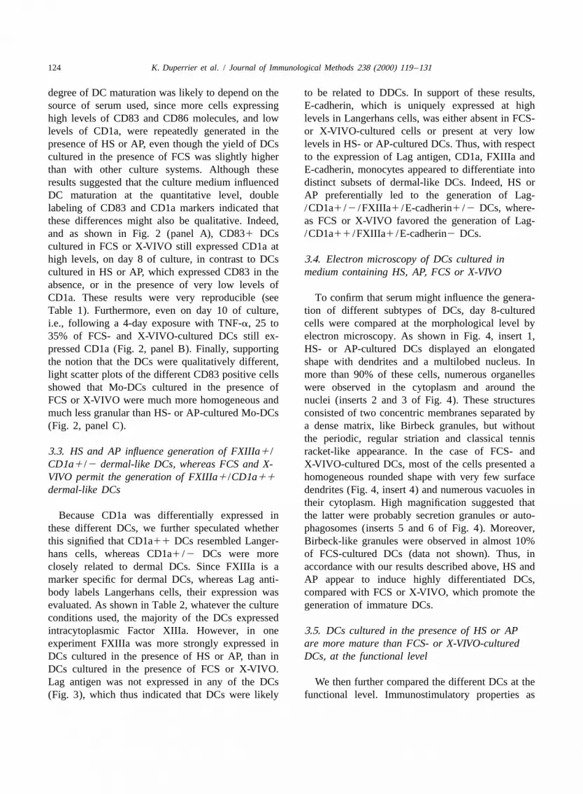
124 K. Duperrier et al. / Journal of Immunological Methods 238 (2000) 119 –131
degree of DC maturation was likely to depend on the to be related to DDCs. In support of these results,source of serum used, since more cells expressing E-cadherin, which is uniquely expressed at highhigh levels of CD83 and CD86 molecules, and low levels in Langerhans cells, was either absent in FCS-levels of CD1a, were repeatedly generated in the or X-VIVO-cultured cells or present at very lowpresence of HS or AP, even though the yield of DCs levels in HS- or AP-cultured DCs. Thus, with respectcultured in the presence of FCS was slightly higher to the expression of Lag antigen, CD1a, FXIIIa andthan with other culture systems. Although these E-cadherin, monocytes appeared to differentiate intoresults suggested that the culture medium influenced distinct subsets of dermal-like DCs. Indeed, HS orDC maturation at the quantitative level, double AP preferentially led to the generation of Lag-labeling of CD83 and CD1a markers indicated that /CD1a1 /2 /FXIIIa1 /E-cadherin1 /2 DCs, where-these differences might also be qualitative. Indeed, as FCS or X-VIVO favored the generation of Lag-and as shown in Fig. 2 (panel A), CD831 DCs /CD1a11 /FXIIIa1 /E-cadherin2 DCs.cultured in FCS or X-VIVO still expressed CD1a athigh levels, on day 8 of culture, in contrast to DCs 3.4. Electron microscopy of DCs cultured incultured in HS or AP, which expressed CD83 in the medium containing HS, AP, FCS or X-VIVOabsence, or in the presence of very low levels ofCD1a. These results were very reproducible (see To confirm that serum might influence the genera-Table 1). Furthermore, even on day 10 of culture, tion of different subtypes of DCs, day 8-culturedi.e., following a 4-day exposure with TNF-a, 25 to cells were compared at the morphological level by35% of FCS- and X-VIVO-cultured DCs still ex- electron microscopy. As shown in Fig. 4, insert 1,pressed CD1a (Fig. 2, panel B). Finally, supporting HS- or AP-cultured DCs displayed an elongatedthe notion that the DCs were qualitatively different, shape with dendrites and a multilobed nucleus. Inlight scatter plots of the different CD83 positive cells more than 90% of these cells, numerous organellesshowed that Mo-DCs cultured in the presence of were observed in the cytoplasm and around theFCS or X-VIVO were much more homogeneous and nuclei (inserts 2 and 3 of Fig. 4). These structuresmuch less granular than HS- or AP-cultured Mo-DCs consisted of two concentric membranes separated by(Fig. 2, panel C). a dense matrix, like Birbeck granules, but without
the periodic, regular striation and classical tennis3.3. HS and AP influence generation of FXIIIa1 / racket-like appearance. In the case of FCS- andCD1a1 /2 dermal-like DCs, whereas FCS and X- X-VIVO-cultured DCs, most of the cells presented aVIVO permit the generation of FXIIIa1 /CD1a11 homogeneous rounded shape with very few surfacedermal-like DCs dendrites (Fig. 4, insert 4) and numerous vacuoles in
their cytoplasm. High magnification suggested thatBecause CD1a was differentially expressed in the latter were probably secretion granules or auto-
these different DCs, we further speculated whether phagosomes (inserts 5 and 6 of Fig. 4). Moreover,this signified that CD1a11 DCs resembled Langer- Birbeck-like granules were observed in almost 10%hans cells, whereas CD1a1 /2 DCs were more of FCS-cultured DCs (data not shown). Thus, inclosely related to dermal DCs. Since FXIIIa is a accordance with our results described above, HS andmarker specific for dermal DCs, whereas Lag anti- AP appear to induce highly differentiated DCs,body labels Langerhans cells, their expression was compared with FCS or X-VIVO, which promote theevaluated. As shown in Table 2, whatever the culture generation of immature DCs.conditions used, the majority of the DCs expressedintracytoplasmic Factor XIIIa. However, in one 3.5. DCs cultured in the presence of HS or APexperiment FXIIIa was more strongly expressed in are more mature than FCS- or X-VIVO-culturedDCs cultured in the presence of HS or AP, than in DCs, at the functional levelDCs cultured in the presence of FCS or X-VIVO.Lag antigen was not expressed in any of the DCs We then further compared the different DCs at the(Fig. 3), which thus indicated that DCs were likely functional level. Immunostimulatory properties as

K. Duperrier et al. / Journal of Immunological Methods 238 (2000) 119 –131 125
Fig. 2. Comparative phenotypic analysis of cultured DCs. Monocytes were cultured for 6 days with GM-CSF plus IL-4 and for an additional2 days with TNF-a, in the presence of different sources of serum or in X-VIVO 20. Double labeling of CD1a (x axis) and CD83 ( y axis) wasperformed on day 8 (A) and day 10 (B) of DC culture. Cell size and granularity, represented, respectively, by forward scatter (FSC) and sidescatter (SSC), were assessed on day 8 cultured-DCs (C). Cytograms represent cells gated on the basis of their CD83 positive expression.

126 K. Duperrier et al. / Journal of Immunological Methods 238 (2000) 119 –131
Table 2 culture in FCS consistently resulted in the inductionaInfluence of serum on factor XIIIa expression of intense non specific autologous responses by T
FXIIIa1 cells lymphocytes (data not shown). Thus, these resultssuggest that DCs cultured in HS or AP are moreMean6S.D. MFImature than FCS- or X-VIVO-cultured DCs, at the
A 80.564.8 63functional level.B 84.162 55
C 85.9617 42D 69.3641 44 3.6. DCs cultured in the presence of HS or AP
a cannot capture Ag, following TNF-a exposureMonocytes were cultured in the presence of 10% HS (A), 10%AP (B), 10% FCS (C), or in X-VIVO 20 serum-free medium (D),with GM-CSF plus IL-4 for 6 days, and for an additional 2 days Because DCs are known to lose their capacity towith TNF-a. Cells were then stained with intra-cellular factor capture and process antigens during maturation, weXIIIa. Results are expressed as means of four different experi-
further analyzed this aspect. As shown in Fig. 6,ments.following exposure to TNF-a for 2 days, DCscultured in medium containing HS or AP wereunable to internalize dextran-FITC, as opposed to
well as the capacity of the DCs to function as DCs cultured in the presence of FCS or X-VIVO,accessory cells, were analyzed. Higher specific re- suggesting that HS- or AP-cultured DCs are the mostsponses to tetanus toxoid (Fig. 5A), and higher mature cells.allogeneic stimulatory activity (Fig. 5B) were ob-served in HS- and AP-cultured DCs. Furthermore,when DCs were compared for their accessory func- 4. Discussiontions, using naive and memory T cells as respondersand PHA as a stimulus, the results showed that HS- In this study we have evaluated the influence ofand AP-cultured DCs were more potent accessory HS, AP, FCS and X-VIVO 20, a serum-free medium,cells than FCS- or X-VIVO-cultured DCs, since such on the generation of DCs from monocytes, withcells were capable of inducing strong autologous respect to three specific DC features, i.e., phenotype,MLR responses (Fig. 5C). It should be noted that morphology and function. We found that DC matura-
Fig. 3. Expression of Lag antigen and E-cadherin on immature Mo-DCs. Monocytes were cultured for 6 days with GM-CSF plus IL-4, inthe presence of 10% HS, 10% AP, 10% FCS, or in X-VIVO 20 serum-free medium. Cells were analyzed for Lag antigen and E-cadherinexpression (bold histograms). Dotted histograms represent the fluorescence value of isotypic controls.

K. Duperrier et al. / Journal of Immunological Methods 238 (2000) 119 –131 127
Fig. 4. Influence of serum on DC morphology. Day 8-cultured cells were fixed and processed for electron microscopy, as described inMaterials and methods. HS-cultured DCs and FCS-cultured DCs are represented by photomicrographs 1 to 3 and 4 to 6, respectively. Thebar in photomicrographs 1 and 4 represents 2 mm. Inserts 2 and 3 represent high magnifications of Birbeck granule-like structures found inthe cytoplasm of the majority of HS-cultured DCs. Numerous autophagosomes were observed in the cytoplasm of FCS-cultured DCs, asshown in inserts 5 and 6. The bars in the inserts represents 200 nm.

128 K. Duperrier et al. / Journal of Immunological Methods 238 (2000) 119 –131
Fig. 5. Functional properties of DCs cultured in the presence of different sources of serum, or in X-VIVO 20 serum-free medium. (A)5Tetanus toxoid presentation was assessed by culturing 10 T lymphocytes with graded numbers of autologous day 8-cultured DCs, incubated
27in the presence of tetanus toxoid (5?10 M). The proliferative response was measured on day 5. (B) Allogeneic T cells were stimulated by3graded numbers of day 8-cultured DCs. The proliferative response in these cultures was measured after 4 days of culture, by [ H]-thymidine
incorporation, as described in Materials and methods. Background counts due to ongoing autologous MLR were subtracted in the allogeneicMLR and TT presentation assays. Results shown are representive of three separate experiments. Data are reported as mean values6standard
5deviation. (C) Accessory cell function of day 8-cultured DCs was assessed by culturing 10 CD45RO or CD45RA autologous T cells with33?10 day 8-cultured DCs, in the presence of PHA (0.1 mg/ml). The proliferative response was measured after 3 days of culture, by
3[ H]thymidine incorporation. Counts due to the proliferation of T cells alone in the presence of PHA were subtracted.

K. Duperrier et al. / Journal of Immunological Methods 238 (2000) 119 –131 129
Fig. 6. Influence of serum supplement on FITC-dextran uptake, on day 5 and day 8 of DC culture. DCs cultured in the presence of 10% HS,10% AP, 10% FCS or in X-VIVO 20, on day 5 and day 8, were tested for their ability to internalize FITC-dextran, as described in Materialsand methods. As a control, the dotted histograms represent the fluorescence values of FITC-dextran uptake at 08C.
tion is likely to be influenced by the source of serum CD1a1 /2 DCs, whereas FCS and X-VIVO pref-supplement used. Indeed, HS and AP promoted the erentially led to the generation of CD1a11 DCs.generation of highly mature DCs compared with FCS That these cells are distinct at the qualitative levelor X-VIVO, with respect to (i) CD83 and CD86 was also supported by electron microscopy whichup-regulation, two markers of DC maturation which showed that DCs cultured in the presence of HS orwere expressed at the highest rates and intensities, AP were highly differentiated and expressed Birbec-(ii) the abrogation of their capacity to internalize k-like granules, whereas FCS- and X-VIVO-cultureddextran-FITC, and thus to capture antigens, (iii) their DCs still exhibited phagocytic features. Indeed, theseT cell stimulatory activity, which was the most structures were observed in 90% or more of HS- orpotent and (iv) their morphology which, as assessed AP-cultured DCs, but only in 10% of FCS-culturedby electron microscopy, corresponded to that of fully DCs. Similar structures have also been observed bydifferentiated DCs. Interestingly, and in support of Grassi et al. (1998) in 11 to 36% of their monocyte-our results, Mannering et al. (1998) have observed derived DCs cultured in FCS. Since these structuresthat ex-vivo maturation of blood DCs is also im- are distinct from LC Birbeck granules, as indicatedproved by 10% HS, compared to FCS. by the non periodicity of their membranes, one may
Staining DCs with Factor XIIIa and Lag antigen assume that they are expressed in interstitial DCs,helped us to relate these cells to dermal DCs. Indeed, and that their expression is likely to increase with theFactor XIIIa which is characteristic of dermal DCs stage of DC maturation.(Nestle et al., 1993), was expressed by our Mo-DCs, The ability to generate different subsets of dermalwhatever the culture supplement used, whereas Lag DC in vitro is in accordance with the reports ofantigen which is expressed by Langerhans cells Nestle et al. (1993) and McLellan et al. (1998).(Kashihara et al., 1986), was absent in our cells. Indeed, both groups have described several subsetsMoreover, E-cadherin, which is present at high levels of ex-vivo isolated DDCs that differ with regards toin Langerhans cells, was either absent, or very the expression of CD1a and CD14 (Nestle et al.,weakly expressed in these DCs. Furthermore, with 1993), or CD83, CD86 and CMRF-44 (McLellan etregard to CD1a expression, the source of serum was al., 1998). Interestingly, DDCs that were the mostlikely to influence generation of distinct DC sub- mature and activated cells, at both the functional andtypes, since HS and AP favored the generation of phenotypic levels, were found to be migratory cells

130 K. Duperrier et al. / Journal of Immunological Methods 238 (2000) 119 –131
which preferentially located in direct contact with T Referenceslymphocytes. Thus, one could speculate from ourresults, that HS- and AP-cultured DCs resemble Bender, A., Sapp, M., Schuler, G., Steinman, R.M., Bhardwaj, N.,
1996. Improved methods for the generation of dendritic cellsactivated DDCs, since they were the most maturefrom nonproliferating progenitors in human blood. J. Im-cells.munol. Methods 196, 121.To further investigate whether a factor present in
Caux, C.,Vanbervliet, B., Massacrier, C., Dezutter-Dambuyant, C.,HS or AP could account for the improvement in DC de Saint-Vis, B., Jacquet, C., Yoneda, K., Imamura, S.,maturation, LPS and TNF-a, which have been Schmitt, D., Banchereau, J., 1996. CD341 hematopoieticdescribed to improve DC maturation (Suri and progenitors from human cord blood differentiate along two
independent dendritic cell pathways in response to GM-CSF1Austyn, 1998), were added to X-VIVO medium. TheTNF-a. J. Exp. Med. 184, 695.results obtained showed that the differentiation of
Caux, C., Massacrier, C., Vanbervliet, B., Dubois, B., Durand, I.,monocytes into DCs was inhibited (data not shown) Cella, M., Lanzavecchia, A., Banchereau, J., 1997. CD341since monocytes differentiated into macrophages, as hematopoietic progenitors from human cord blood differentiatepreviously reported by Steinbach et al. (1998). In along two independent dendritic cell pathways in response to
GM-CSF1TNF-a: II. Functional analysis. Blood 90, 1458.further titration experiments, we observed that theCroft, M., Duncan, D.D., Swain, S.L., 1992. Response of naiveinfluence of HS on DC maturation was related to the
antigen-specific CD41 T cells in vitro: characteristics andnature of the serum, since even when compared with antigen-presenting cell requirements. J. Exp. Med. 176, 1431.DCs cultured in the presence of 1% HS only, the Eljaafari, A., Duperrier, K., Mazet, S., Bardin, C., Bernaud, J.,expression and intensity of CD83 and CD86 were Durand, B., Gebuhrer, L., Betuel, H., Rigal, D., 1998. Genera-
tion of stable monocyte-derived dendritic cells in the presencemuch higher than when DCs were cultured in theof high concentrations of homologous or autologous serum:presence of 10% FCS (data not shown). Finally,influence of extra-cellular pH. Hum. Immunol. 59, 625.
when immature DCs were exposed to TNF-a for Grassi, F., Dezutter-Dambuyant, C., McIlroy, D., Jacquet, C.,either 2 or 4 days, the expression of CD83 increased Yoneda, K., Imamura, S., Boumsell, L., Schmitt, D., Autran,with the time of exposure to TNF-a, together with B., Debre, P., Hosmalin, A., 1998. Monocyte-derived dendritic
cells have a phenotype comparable to that of dermal dendriticdown-regulation of CD1a expression. However, evencells and display ultrastructural granules distinct from Birbeckon day 10 of culture, 25 to 35% of CD831 FCS- andgranules. J. Leukocyte Biol. 64, 484.
X-VIVO-cultured DCs still expressed CD1a (Fig. Grouard, G., Durand, I., Filgueira, L., Banchereau, J., Liu, Y.J.,2B). Taken together, these results suggest that quali- 1996. Dendritic cells capable of stimulating T cells in germinaltative rather than quantitative factors are likely to centers. Nature 384, 364.
Hart, D.N.J., 1997. Dendritic cells: unique leukocyte populationsaccount for the differences that were observedwhich control the primary immune response. Blood 90, 3245.between DCs cultured in HS, AP, FCS or X-VIVO.
Holtl, L., Rieser, C., Papesh, C., Ramoner, R., Bartsch, G.,In conclusion, we have shown that in vitro DC Thurnher, M., 1998. CD831 blood dendritic cells as a vaccine
generation from monocytes is not only dependent on for immunotherapy of metastatic renal-cell cancer. Lancet 352,cytokines, but also on culture supplements. Whereas 1358.
Hsu, F.J., Benike, C., Fagnoni, F., Liles, T.M., Czerwinski, D.,it has been described that different subsets of DCsTaidi, B., Engleman, E.G., Levy, R., 1996. Vaccination ofcan be generated depending on the source of pre-patients with B-cell lymphoma using autologous antigen-
cursor cells (CD34, CD14), we have shown that pulsed dendritic cells. Nat. Med. 2, 52.culture supplement variation can also lead to differ- Inaba, K., Schuler, G., Witmer, M.D., Valinsky, J., Atassi, B.,ent subsets of DCs, starting from the same precursor Steinman, R.M., 1986. Immunologic properties of purified
epidermal Langerhans cells. Distinct requirements for stimula-cell. Thus, in FCS or X-VIVO, CD1a11 immaturetion of unprimed and sensitized T lymphocytes. J. Exp. Med.dermal-like DCs were preferentially induced, where-164, 605.
as HS or AP favored the generation of very mature Kashihara, M., Ueda, M., Horiguchi, Y., Furukawa, F., Hanaoka,CD1a1 /2 dermal-like DCs. M., Imamura, S., 1986. A monoclonal antibody specifically
reactive to human Langerhans cells evidenced by immuno-goldrelabelling. J. Invest. Dermatol. 92, 217.
Mannering, S.I., McKenzie, J.L., Hart, D.N.J., 1998. OptimisationAcknowledgementsof the conditions for generating human DC initiated antigenspecific T lymphocyte lines in vitro. J. Immunol. Methods 219,
The authors thank Mrs. Grassi for help in intra- 69.cellular Factor XIIIa staining. McLellan, A.D., Heiser, A., Sorg, R.V., Fearnley, D.B., Hart,

K. Duperrier et al. / Journal of Immunological Methods 238 (2000) 119 –131 131
D.N.J., 1998. Dermal dendritic cells associated with T lym- Sallusto, F., Lanzavecchia, A., 1994. Efficient presentation ofphocytes in normal human skin display an activated pheno- soluble antigen by cultured human dendritic cells is main-type. J. Invest. Dermatol. 111, 841. tained by granulocyte /macrophage colony-stimulating factor
Nestle, F.O., Zheng, X.-G., Thompson, C.B., Turka, L.A., Nic- plus interleukin 4 and downregulated by tumor necrosis factorkoloff, B.J., 1993. Characterization of dermal dendritic cells a. J. Exp. Med. 179, 1109.
¨obtained from normal human skin reveals phenotypic and Steinbach, F., Krause, B., Blaß, S., Burmester, G.-R., Hiepe, F.,functionally distinctive subsets. J. Immunol. 151, 6535. 1998. Development of accessory phenotype and function
Nestle, F.O., Alijagic, S., Gilliet, M., Sun, Y., Grabbe, S., during the differentiation of monocyte-derived dendritic cells.Dummer, R., Burg, G., Schadendorf, D., 1998. Vaccination of Res. Immunol. 149, 627.melanoma patients with peptide- or tumor lysate-pulsed de- Steinman, R.M., Nussenzweig, M.C., 1980. Dendritic cells: fea-ndritic cells. Nat. Med. 4, 328. tures and functions. Immunol. Rev. 53, 127.
Pickl, W.F., Majdic, O., Kohl, P., Stockl, J., Riedl, E., Scheinecker, Steinman, R.M., 1991. The dendritic cell system and its role inC., Bello-Fernandez, C., Knapp, W., 1996. Molecular and immunogenicity. Annu. Rev. Immunol. 9, 271.functional characteristics of dendritic cells generated from Steinman, R.M., Hoffman, L., Pope, M., 1995. Maturation andhighly purified CD141 peripheral blood monocytes. J. Im- migration of cutaneous dendritic cells. J. Invest. Dermatol.munol. 157, 3850. 105, 2S.
Romani, N., Koide, S., Crowley, M., Witmer-Pack, M., Living- Suri, R.M., Austyn, J.M., 1998. Bacterial lipopolysaccharidestone, A.M., Fathman, C.G., Inaba, K., Steinman, R.M., 1989. contamination of commercial collagen preparations may medi-Presentation of exogenous protein antigens by dendritic cells ate dendritic cell maturation in culture. J. Immunol. Methodsto T cell clones: intact protein is presented best by immature 214, 149.epidermal Langerhans cells. J. Exp. Med. 169, 1169. Tew, J.G., Wu, J., Qin, D., Helm, S., Burton, G.J., Szakal, A.K.,
Romani, N., Reider, D., Heuer, M., Ebner, S., Kampgen, E., Eibl, 1997. Follicular dendritic cells and presentation of antigen andB., Niederwieser, D., Schuler, G., 1996. Generation of mature co-stimulatory signals to B cells. Immunol. Rev. 156, 39.dendritic cells from human blood. An improved method withspecial regard to clinical applicability. J. Immunol. Methods196, 137.