Human natural regulatory T cells subsets - Humboldt ...
99
Human natural regulatory T cells subsets: Functional characterization and T cell receptor repertoire analysis Dissertation zur Erlangung des akademischen Grades doctor rerum naturalium (Dr. rer. nat) Im Fach Biologie eingericht an der Mathematisch-Naturwissenschaftlichen Fakultät I der Humboldt-Universität zu Berlin vorgelegt von M. Sc., Hong Lei Präsident der Humboldt-Universität zu Berlin Prof. Dr. Jan-Hendrik Olbertz Dekan der Mathematisch-Naturwissenschaftlichen Fakultät I Prof. Stefan Hecht, Ph.D. Gutachter: 1. Prof. Dr. Hans-Dieter Volk 2. Prof. Dr. Richard Lucius 3. Prof. Dr. Petra Reinke Tag der mündlichen Prüfung: 04.03.2014
-
Upload
khangminh22 -
Category
Documents
-
view
3 -
download
0
Transcript of Human natural regulatory T cells subsets - Humboldt ...

Human natural regulatory T cells subsets: Functional characterization and T cell
receptor repertoire analysis
Dissertation
zur Erlangung des akademischen Grades
doctor rerum naturalium (Dr. rer. nat)
Im Fach Biologie
eingericht an der
Mathematisch-Naturwissenschaftlichen Fakultät I
der Humboldt-Universität zu Berlin
vorgelegt von
M. Sc., Hong Lei
Präsident der Humboldt-Universität zu Berlin
Prof. Dr. Jan-Hendrik Olbertz
Dekan der Mathematisch-Naturwissenschaftlichen Fakultät I
Prof. Stefan Hecht, Ph.D.
Gutachter: 1. Prof. Dr. Hans-Dieter Volk
2. Prof. Dr. Richard Lucius
3. Prof. Dr. Petra Reinke
Tag der mündlichen Prüfung: 04.03.2014

TABLE OF CONTENT
SUMMARY .......................................................................................................................... I
ZUSAMMENFASSUNG ..................................................................................................... II
ABBREVIATIONS ............................................................................................................. III
1. INTRODUCTION ........................................................................................................1
1.1 General aspects of Treg development and suppressive functionality .....................1
1.1.1 Innate and adaptive immune system ....................................................................1
1.1.2 General finding and development of Treg ............................................................2
1.1.3 Classification of thymus derived natural Treg and peripheral induced Treg..........4
1.1.4 Treg Suppression mechanism ..............................................................................5
1.1.5 Cross talk between Treg and non-T cells .............................................................7
1.2 Heterogeneous Treg for clinical translation .............................................................7
1.2.1 Treg for adoptive cell therapy ..............................................................................7
1.2.2 nTreg heterogeneity in periphery .........................................................................9
1.2.3 In vitro isolation and expansion of nTreg ........................................................... 10
1.3 T cell receptor repertoire of Treg and Tconv ........................................................ 11
1.3.1 T cell receptor (TCR) repertoire analysis methods ............................................. 11
1.3.2 Treg subsets and Tconv TCR repertoire in mice and human ............................... 11
2. AIMS OF THIS WORK ............................................................................................. 13
3. MATERIAL & METHODS ........................................................................................ 14
3.1 Materials.................................................................................................................. 14
3.1.1 Subjects ............................................................................................................. 14
3.1.2 Equipments and devices .................................................................................... 14
3.1.3 Medias, Buffers and Solutions ........................................................................... 14
3.1.4 Chemicals and Reagents .................................................................................... 15
3.1.5 Isolation and stimulation Kits ............................................................................ 15
3.1.6 Antibodies ......................................................................................................... 16
3.2 Methods ................................................................................................................... 17
3.2.1 PBMC isolation ................................................................................................. 17

3.2.2 Flow cytometric staining and analysis ............................................................... 18
3.2.3 Fluorescence Activated Cell Sorting (FACS) ..................................................... 21
3.2.4 FoxP3 demethylation assay ............................................................................... 22
3.2.5 Treg functional assay ......................................................................................... 22
3.2.6 Treg isolation and expansion ............................................................................. 25
3.2.7 DNA extraction and next-generation sequencing for TCR repertoire .................. 26
3.2.8 Computation analysis ........................................................................................ 26
3.2.9 Statistical analysis ............................................................................................. 27
4. RESULTS .................................................................................................................... 28
4.1 Phenotypic analysis of Treg from healthy donors and Tx patients ....................... 28
4.1.1 The majority of Treg express central-memory phenotype increasing with age .... 28
4.1.2 Treg up-regulated only CD137 while Tconv up-regulated both CD137 and
CD154 upon TCR stimulation ....................................................................................... 31
4.1.3 Treg in Tx patients contained more effector-memory cells and were more
susceptible to activation-induced cell death ................................................................... 35
4.2 Functional analysis of Treg subsets: central memory Treg showed enhanced
suppression efficiency than naïve Treg .......................................................................... 39
4.2.1 Freshly isolated TregCM showed enhanced suppression activity to early
activation of both CD4 Tconv and CD8 Tconv .............................................................. 39
4.2.2 Treg subsets showed similar suppression activity to both T cells and non-T cells
regarding proliferation of non-apoptotic responder cells ................................................ 45
4.2.3 Treg CM induced apoptosis to CD3- cells while TregN did not. ......................... 46
4.2.4 Enhanced suppression activity of TregCM may be related to particular cell-cell
interaction due to higher CTLA-4 expression on TregM than TregN ............................. 48
4.3 Expansion of Treg and subsets indicated the promising clinical translation for
cellular therapy ............................................................................................................... 49
4.3.1 Expansion with rapamycin could keep FoxP3 expression and suppression
function of Treg ............................................................................................................ 49
4.3.2 Enhanced expansion of Treg with G-Rex device, even closer to translation ....... 50
4.3.3 The shift from naïve to memory Treg phenotype after expansion is associated
with enhanced suppressive capacity .............................................................................. 52

4.4 TCR repertoire analysis indicated memory Treg derived from naïve ones rather
than Tconv ...................................................................................................................... 55
4.4.1 Next-Generation Sequencing (NGS) is a much powerful tool for TCR repertoire
analysis ......................................................................................................................... 55
4.4.2 Memory Treg seems to be derived from naïve Treg rather than Tconv suggesting
an antigen-driven process .............................................................................................. 57
5. DISCUSSION .............................................................................................................. 63
5.1 Phenotypic analysis of Treg from healthy ones and Tx patients ........................... 64
5.1.1 Similar subsets composition, but more memory cells in Treg than in Tconv ....... 64
5.1.2 Distinct activation patterns of Treg and Tconv subsets via TCR stimulation ....... 65
5.1.3 Treg subsets distribution in Tx patients .............................................................. 66
5.2 Functional analysis of Treg subsets with potential mechanism............................. 67
5.2.1 Enhanced suppression by TregCM and possible differentiation of Treg subsets . 67
5.2.2 Potential mechanism of enhanced suppression activity by memory Treg............ 67
5.2.3 Cross talk of Treg and other immune cells ......................................................... 68
5.3 Expansion of Treg in G-Rex10: new options for clinical translaiton .................... 68
5.3.1 Expansion of Treg subsets in vitro ..................................................................... 68
5.3.2 Expansion of Treg in G-Rex10 device ............................................................... 69
5.4 TCR repertoire analysis .......................................................................................... 70
5.4.1 Treg M derived from naïve Treg with clonal expansion ..................................... 70
5.4.2 Highest similarity of TCR repertoire between TregCM/EM ............................... 71
5.4.3 TCR Repertoire of human Treg and Tconv ........................................................ 71
6. REFERENCES ........................................................................................................... 74
7. APPENDIX ................................................................................................................. 89

Summary
I
SUMMARY
Regulatory T cells (Treg) offer new immunotherapeutic options to control undesired immune
reactions, but the heterogeinetiy of Treg raises the question which Treg population should be
used for clinical translation “from bench to bedside”, especially to solid organ transplantation
(SOT) patients. Based on this final goal, this project involves three main parts: i) investigating
Treg frequency and subsets distribution with age in healthy donors and transplant (Tx)
patients; ii) comparing the suppressive capacity of Treg subsets and expanding them in vitro
without losing functionality; iii) clarifyjing the differiation relationship of Treg subsets and
their relation to conventional T cells (Tconv) by T cell receptor (TCR) repertoire analysis.
From both healthy donors and Tx patients, an age-dependent shift from naïve Treg (TregN)
to memory Treg (TregM) was observed, dominated by the central-memory (CM) phenotype
over less abundant effector-memory (EM) population; compared to age matched healthy
donors, Treg in Tx patients contained less naive but more EM cells, moreover, they were pre-
activated by expression of CD137 due to the exposure to allo antigens, thus more susceptible
to activation-induced cell death. Regarding control of early Tconv activation, TregCM showed
enhanced suppressive capacity compared to TregN; furthermore, only TregCM could induce
apoptosis of responder cells especially in NK cells and B cells populations while TregN could
not, which may result from cell-cell interactions as TregM have mugh higher cytotoxic T-
lymphocyte antigen 4 (CTLA-4) expression than TregN. Following in vitro expansion of the
Treg subsets with rapamycin, however, TregN converted mainly into TregCM phenotype with
enhanced suppression activity comparable to the level of freshly isolated and expanded
TregCM. The poor proliferation capacity of TregEM might indicate EM as the terminal
differential stage. Expansion of Treg in the gas-permeable and GMP-compliant G-Rex device
could further enhance the expansion without losing phenotype and functionality. These data
suggest that expansion with total Treg is optimal for adoptive Treg therapy as the majority of
them are the highly potent TregCM.
Lastly, TCR repertoire study by next generation sequencing (NGS) indicated that TregM
derived from TregN rather than Tconv in an antigen-driven process. The highest similarity of
the TCR repertoires was observed between TregCM and TregEM. These data reveal new
insights for the first time into the distinct TCR repertoires of Treg subsets and Tconv in human
by NGS technology.

Zussamenfassung
II
ZUSAMMENFASSUNG
Regulatorische T-Zellen (Treg) eröffnen neue immuntherapeutische Wege zur Kontrolle
unerwünschter Immunreaktionen, jedoch wirft die Heterogenität dieser Zellen die Frage auf,
welche Treg-Population für die klinische Anwendung im Rahmen der „bench to bedside“ -
Anwendung, besonders im Bereich der Organtransplantation, am geeignetsten erscheint.
Darauf basierend werden in dieser Arbeit drei Fragestellungen bearbeitet: i) Bestimmung der
Häufigkeit von Tregs und deren Subpopulationen in verschiedenen Altersgruppen bei
Empfängern einer Organtransplantation (Tx) und einer gesunden Kontrollgruppe; ii) Vergleich
der Suppressorkapazität verschiedener Treg-Populationen und in vitro-Expansion der Zellen
unter Erhaltung ihrer Funktionalität; iii) Klärung der Differenzierungsmerkmale von Tregs
und deren Verknüpfung mit konventionellen T-Zellen (Tconv) mittels Analyse des T-Zell-
Rezeptor- (TCR) Repertoires.
Sowohl bei gesunden Probanden als auch bei Tx-Empfänger konnte eine altersabhängige
Verschiebung von naiven (TregN) hin zu dominant zentralen Gedächtnis-Zellen (TregCM)
beobachtet werden, Treg von Tx-Empfängern hatten mehr Effektor-Memory-Zellen (EM) und
sie waren mehr aktiviert. In Bezug auf die Kontrolle der frühen Tconv zeigen TregCM eine
erhöhte Suppressorkapazität im Vergleich zu TregN. Außerdem sind im Gegensatz zu TregN
nur TregCM dazu in der Lage, Apoptose bei Responderzellen zu induzieren. Der Grund
hierfür könnte in der stärkeren Expression von CTLA-4 auf TregM liegen. Die
Expansionskultur führte zur phänotypischen Veränderung der TregN, deren Umwandlung in
TregCM mit einer verbesserten Suppressoraktivität verbunden ist, welche mit der von frisch
isolierten und expandierten TregCM vergleichbar ist. Die schwache Proliferationskapazität
der TregEM könnte die Effektor-Memory-Zellen als begrenzende Phase der
Zelldifferenzierung ausweisen. Die Daten legen nahe, dass das Expandieren mit gesamt Treg
für die Adoptive-Treg-Therapie optimal sind, da sie der größte Anteil von ihnen die
hochpotenten TregCM sind.
TCR-Studien mittels Next Generation Sequencing zeigen weiter, dass TregM aus TregN
entstehen, anstatt aus Tconv, in einem Antigen-gesteuerten Prozess. Die größte Ähnlichkeit
des TCR-Repertoires wurde hierbei zwischen TregCM- und TregEM-Zellen nachgewiesen.
Diese Daten belegen erstmalig neue Erkenntnisse hinsichtlich der Unterschiede der TCR-
Repertoires von TregM und Tconv beim Menschen.

Abbreviations
III
ABBREVIATIONS
ADCC antibody dependent cellular cytotoxicity
APC antigen presenting cells
ATP adenosine triphosphate
BCR B cell receptor
BFA Brefeldin A
cAMP cyclic adenosine monophosphate
CD cluster of differentiation
CDC complement dependent cytotoxicity
cDNA complimentary DNA
CDR3 the third complementarity-determining region
CFSE arboxy fluorescein succinimidyl ester
CTL cytotoxic T lymphocyte
CTLA-4 Cytotoxic T-Lymphocyte Antigen 4
DC dendritic cells
FACS fluorescence activated cell sorting
FCS fetal calf serum
Foxp3 forkhead box 3
FSC forward scatter
GVHD graft versus host disease
hAB serum human serum AB
HLA human leucocyte antigen
HSCT hematopoetic stem cell transplantation
IDO Indolamin-2,3-Dioxygenase
IFN interferon
IL interleukin
iTreg induced regulatory T cells
IU international unit
L/D LIVE/DEAD
LFA-1 Lymphocyte function-associated antigen 1
mAb monoclonal antibody
MACS magnetic activated cell sorting
MFI mean fluorescence intensity
MH index Morisita-Horn similarity index
MHC major histocompatibility complex
NET neutrophils extracellular traps
NFAT Nuclear factor of activated T-cells
NF-ĸB nuclear factor kappa-light-chain-enhancer of activated B cells

Abbreviations
IV
NGS next generation sequencing
NK cells natural killer cells
nTreg natural regulator T cells
OL overlap
PBMC peripheral blood mononuclear cells
PCR polymerase chain reaction
PMA phorbol 12-myristate 13-acetate
PRRs pattern recognition receptors
SEM standard error mean
SOT solid organ transplant
SSC side scatter
ST stimulated
Tconv conventional T cells
TconvM memory conventional T cells
TconvN naïve conventional T cells
TCR T-cell receptor
TGF-ß tumor growth factor beta
Th1 type 1 T helper cells
TLR Toll-like receptor
TNF tumor necrosis factor
Tr1 type 1 regulatory T cells
TregCM central memory Treg cells
TregEM effector memory Treg cells
TregM memory regulatory T cells
TregN naïve regulatory T cells
TSDR Treg specific demethylation region
Tx transplantation
UN un-stimulated
V ß T-cell receptor variable ß chain

Introduction
1
1. INTRODUCTION
1.1 General aspects of Treg development and suppressive functionality
1.1.1 Innate and adaptive immune system
Human immune system is an extremely complex and powerful network, which functions like
a strong army to protect us from infections. It can not only augment immune responses upon
“foreign agents” like viruses; but also dampens reactions to “self-tissues”; resulting in a
formidably active but homeostatic “protecting” environment. This powerful network consists
of two distinct but intimately correlated systems: innate immune system and adaptive immune
system.
Briefly, when we got infections, innate immune system firstly recognizes the conserved
components in many microorganisms via pattern recognition receptors (PRRs), e.g. Toll-like
receptors (TLR) and works immediately in a general manner by mainly phagocytosis and
complement dependent cytotoxicity (CDC) (1-3). Leucocytes especially neutrophils,
macrophages, dendritic cells, NK cells and mast T cells are involved in these processes. As
the majority of phagocytes, neutrophils are one of the first “soldiers” arriving at an infection
site under chemotaxis (4). On one hand, they kill the invading microbes directly by
engulfment, secretion of anti-microbials and generation of neutrophils extracellular traps
(NET) (5); on the other hand, neutrophils also secrete cytokines to recruit and activate other
immune cells. In addition, macrophages present antigens to activate adaptive immune system
like the big “presenter”, dendritic cells, but also produce many chemicals like enzymes to
“digest” invading microbes directly (6).
However, innate immune system is far from enough for protecting organism from infections
because lots of pathogens could escape it. Luckily, we got second the protection layer,
adaptive immune system, which can be activated by innate immune responses thus “adapt”
stronger responses and reinforce it when same antigens are met again by “memory” in a
specific manner (7). Lymphocytes, especially B cells and T cells, are involved in these
responses. On one hand, cytotoxic T cells can kill infected cells directly by releasing
cytotoxins when they are activated by TCR binding to specific-antigen and MHC-I complex
and CD8 molecule; on the other hand, helper T cells can recognize antigens with MHC-II

Introduction
2
complex and get activated with releasing several cytokines to enhance the activity of
macrophages and cytotoxic T cells for direct killing. Another big player in adaptive immune
system is B cells(8). They recognize pathogens by their specific receptors on the cell surface
and uptake and process it into many peptides, which are subsequently presented by these B
cells with their MHC-II molecule to helper T cells, making these helper cells release
cytokines and again stimulate B cells to become plasma cells producing large amount of
antibody. These antibodies will again promote complement system by antibody-dependent
cell mediated cytotoxicity (ADCC) or direct phagocytosis. In addition, some of activated T or
B cells will become memory cells and response immediately next time when the same
antigens were met (8).
The tight collaboration of innate and adaptive immune system provides a powerful network
to protect us. More importantly, they can also discriminate “self” from “non-self” to minimize
possible self-reactive damage in several ways. In brief, self-reactive clones in lymphocytes
are neutralized mainly by clonal deletion, abortion and anergy when potential auto-reactive
cells encounter antigens or by its regulatory mechanism when those cells escape thymus
censorship (9, 10). In this regulatory mechanism, “suppressor T cells”, later called regulatory
T cells (Treg), are proved to be one of the most important regulators in keeping immune
homeostasis (11).
1.1.2 General finding and development of Treg
Treg are a specialized subpopulation of T cells that play a central role in maintaining
homeostasis within immune system. The concept of suppressor T cells was raised in early
1970s by Gershon and Kondo (12). They found that one population of T cells could not only
enhance immune activity, but also suppress it, keeping immune responses more balanced.
Massive researches regarding this T cell population, now called regulatory T cells, were
conducted subsequently all over the world.
Like many other seminal findings in science, the exploration of Treg with significant
importance in clinical adoptive cell therapy nowadays, also has ups and downs (13). From late
1960s to early 1980s, Treg attracted much interest as the concept of the “suppressor T cells”
was raised. During this period, it was found by many researchers that thymectomy of normal
mice and rats produced damage in “self” organs like ovaries and led to the development of
thyroiditis accompanied by autoantibodies production and so on (14-16). Type-1 diabetes was

Introduction
3
also found to be induced with same methods in both mice and rats later (17, 18). More
interestingly, inoculation of normal T cells from healthy mice or rats could prevent these
autoimmune diseases in syngeneic animals (19, 20). Taken together, Sakaguchi et al..
proposed that the coexistence of potential self-reactive CD4 T cells mediating autoimmune
diseases and other CD4 T cell population mainly suppressing them in periphery of healthy
mice and rats (21).
From mid 1980s to early 1990s, research of suppressor T cells was hindered by the big issue
regarding I-J region of mouse MHC gene and collapsed in this period. The I-J region was
believed to encode important molecules expressed by suppressor T cells and pivotal to the
suppression functionality (22). However, this region was not found in mouse MHC gene by
molecular biology techniques (23), suggesting the previous concept of “suppressor T cells”
might be false . Together with paucity of specific cell markers, research of suppressor T cells
went downhill (13).
In the second half of the nineties, a milestone of suppressor T cells research was achieved
mainly by the discovery of CD25, the α chain of interleukin 2 (IL2) receptor, which was
found as a useful surface marker for the suppressive T cells, later called regulatory T cells
(Treg) (24-26). Thus, Treg research was rising again with intensive further studies regarding
the pivotal functional role of IL2 and CD25 for Treg survival (27-29). Furthermore, the
discovery of CD25 also made isolation of Treg feasible, thus the investigation of Treg
function in vivo with adoptive Treg cell transfer became achievable.
Since 2000s, massive investigations regarding Treg development and function have been
conducted. Another mile stone was achieved by the discovery of transcriptional factor
forkhead box P3 (FoxP3), also called Scurfin, as an important regulator for Treg development
and function in 2003 (13). In the same year, several groups reported simultaneously that
FoxP3 mRNA could be expressed by CD4+CD25+ peripheral T cells and CD4+CD8-CD25+
thymocytes, but not by CD4+CD25- conventional T cells; moreover, ectopic expression of
Foxp3 conferred suppressor function on peripheral CD4+CD25- T cells (30-32). Later on,
more and more data have proved that FoxP3 is a master controller of Treg development in
thymus and function, thus the most reliable molecular Treg marker so far (33). Indentifying
FoxP3 as the specific Treg marker further broadened the way nowadays for Treg research and
benefited the clinical translation significantly as well.

Introduction
4
1.1.3 Classification of thymus derived natural Treg and peripheral induced Treg
Naturally occurring Treg (nTreg) derives in thymus. When a T cell precursor is produced in
bone marrow and transferred into thymus, it will receive very strict “training” or “education”
through several complicated selection processes. Only minority of the survivors can become
final Treg cell and enter into periphery. To recognize millions of antigens presented by major
histocompatibility complex (MHC) on the antigen-presenting cells, it is firstly essential for
T cells to have enormous T cell receptor (TCR) repertoire which are mainly produced by
stochastic V(D)J recombination of gene segments in the α and β chain of their TCR in the
thymus as well as the random insertion and deletion of nucleotides at the segment junction
sites. The clonal diversity of the TCR repertoire is further enhanced by different pairing of α
and β chain. Subsequently, the whole repertoire is shaped by positive and negative selections.
Positive selection selects single positive CD4 or CD8 T cells while negative selection further
deletes the clones with high affinity to self peptide and MHC complexes (34). These two
processes result in conventional T cells (Tconv) repertoire with low affinity to self-antigens
but high-affinity to foreign antigens (35). Some self-reactive cells with intermediately high
affinity can escape from negative selection and divert into nTreg cell lineage, which is mainly
responsible for maintaining homeostasis within immune system. The lineage-specific
transcription factor that determines the fate of Treg lineage is FoxP3 (36), its expression on
developing thymocytes seems to depend chiefly on the intensity of TCR stimulation (37, 38).
The demethylation of FoxP3 locus, Treg specific demethylation region (TSDR), depends on
the duration of TCR stimulation, thus TSDR of nTreg are totally demethylated (39).
Additionally, FoxP3+ Treg can also be induced from naïve Tconv with presence of cytokines
and low amount of antigens in periphery (40-42), that is so-called induced Treg (iTreg).
Unlike nTreg, TSDR of iTreg are not completely demethylated (43), although they also have
FoxP3 expression. As nTreg mainly prevent autoimmunity to keep immune tolerance, iTreg
are believed to mainly control chronic allergic inflammation and be one of the important
barriers for tumor cells (43). Besides Th3 cells, CD4 typ1 regulatory T (Tr1) cells are another
type of adaptive Treg produced out of thymus. They don’t express FoxP3, but secrete IL10
and express granzyme B to kill myeloid antigen presenting cells (44, 45). The basic
differentiation of different T cell lineages is shown in Figure 1. This project focuses on nTreg
research.
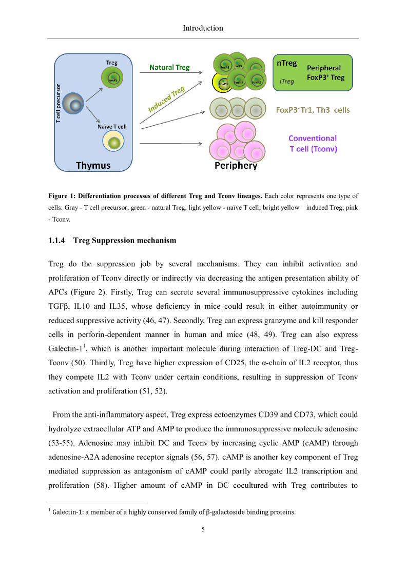
Introduction
5
Figure 1: Differentiation processes of different Treg and Tconv lineages. Each color represents one type of
cells: Gray - T cell precursor; green - natural Treg; light yellow - naïve T cell; bright yellow – induced Treg; pink
- Tconv.
1.1.4 Treg Suppression mechanism
Treg do the suppression job by several mechanisms. They can inhibit activation and
proliferation of Tconv directly or indirectly via decreasing the antigen presentation ability of
APCs (Figure 2). Firstly, Treg can secrete several immunosuppressive cytokines including
TGFβ, IL10 and IL35, whose deficiency in mice could result in either autoimmunity or
reduced suppressive activity (46, 47). Secondly, Treg can express granzyme and kill responder
cells in perforin-dependent manner in human and mice (48, 49). Treg can also express
Galectin-11, which is another important molecule during interaction of Treg-DC and Treg-
Tconv (50). Thirdly, Treg have higher expression of CD25, the α-chain of IL2 receptor, thus
they compete IL2 with Tconv under certain conditions, resulting in suppression of Tconv
activation and proliferation (51, 52).
From the anti-inflammatory aspect, Treg express ectoenzymes CD39 and CD73, which could
hydrolyze extracellular ATP and AMP to produce the immunosuppressive molecule adenosine
(53-55). Adenosine may inhibit DC and Tconv by increasing cyclic AMP (cAMP) through
adenosine-A2A adenosine receptor signals (56, 57). cAMP is another key component of Treg
mediated suppression as antagonism of cAMP could partly abrogate IL2 transcription and
proliferation (58). Higher amount of cAMP in DC cocultured with Treg contributes to
1 Galectin-1: a member of a highly conserved family of β-galactoside binding proteins.

Introduction
6
suppression of DC in a mice model (59). Furthermore, cAMP in suppressed Tconv induces
expression of the inducible cAMP early repressor (ICER), which acts as a repressor at IL2 and
IL4 gene loci (60). Additionally, Treg suppress particular TCR signaling pathways in Tconv
initially by suppression of calcium signaling, resulting in immediate inhibition of NFAT, NF-
ĸB activation and then IL2 transcription (61).
Figure 2: Different mechanisms of Treg-mediated Tconv suppression, adapted from Schmidt et al. (62).
Treg have different suppression mechanisms under different situations. They can secrete inhibitory cytokines
(TGFβ, IL10 and IL35) and kill Tconv by expressing granzymes in a perforin dependent manner. Treg also
suppress Tconv proliferation by IL2 consumption due to higher amount of CD25 expression. Treg can rapidly
suppress TCR-induced Ca2+, NFAT and NF-ĸB signal pathway. Treg can produce immunosuppressive molecule
adenosine and transfer cAMP to Tconv or APCs. Furthermore, they can decrease antigen presentation ability of
APCs via higher expression of CTLA-4.
Regarding antigen presenting cells (APC), Treg also suppress Tconv in an indirect manner
by inhibiting the antigen presentation ability of APC. Both murine and human Treg express
high amount of inhibitory molecule CTLA4 2 (63, 64), which compete for binding to
CD80/CD86 with costimulatory molecule CD28 partly depending on adhesion molecule LFA-
13, thus decrease the cell-cell contact between Tconv and APCs in vitro and in vivo (65-68).
On the other hand, Treg can also increase the expression of the enzyme indoleamine 2, 3-
dioxygenase (IDO) in DC via CTLA-4 induced signaling, resulting in starvation of Tconv and
2 CTLA4: Cytotoxic T-Lymphocyte Antigen 4, also known as CD152.
3 LFA-1: Lymphocyte function-associated antigen 1.

Introduction
7
arrest of cell cycle, as IDO can catalyze degradation of the essential amino acid, tryptophan
and also induce iTreg generation (69, 70).
1.1.5 Cross talk between Treg and non-T cells
In addition to the suppression of Tconv, Treg also interact with many other cell types
including DC, B cells, NK cells, macrophages, osteoblasts, mast cells and NK T cells (71).
Most importantly, Treg can decrease antigen presentation ability of DC as described
previously. Additionally, it is reported that Treg derived from the tumor environment are able
to lyse NK cells and CTL 4 in the granzyme B and perforin dependent manner (72). Recently,
Gasteiger et al. found that restraint of NK cell cytotoxicity by Treg is related with limiting of
IL2 availability (73). CD8 Treg have also been reported that they can down regulate the
immune responses of macrophages in mice and human (74, 75). Regarding bone cells, Treg
have been shown that they could suppress osteoclasts differentiation by secretion of cytokines
like TGF-β in vitro in 2007 (76, 77). Several years later, protection of local and systematic
bone destruction by Treg was observed as well in vivo by the same authors (78, 79), indicating
further crosstalk between skeletal system and immune system. Regarding B cells, although
they might be affected by immune suppressive cytokines secreted by Treg, it’s not clear so far
whether Treg are able to induce apoptosis or cell death of B cells in human.
1.2 Heterogeneous Treg for clinical translation
1.2.1 Treg for adoptive cell therapy
Since the discovery of CD25 as a new Treg surface marker in 1995 (24), worldwide
substantial efforts have been made in understanding the mechanism of Treg suppression and
exploring related clinical application for cellular therapy. Data from animal models have
proved that adoptive transfer of Treg can prevent several autoimmune diseases and block
GvHD 5 and allograft rejection after transplantation (80, 81). Therefore, regarding the
essential role of Treg in keeping immune tolerance, there are several different translational
therapy strategies from different aspects, as shown in Figure 3(82).
4 CTL: Cytotoxic T Lymphocyte, also known as CD8+ T cell or killer T cell
5 GvHD: Graft versus Host Diease.

Introduction
8
Figure 3: Treg immunotherapy, adapted from Wang et al. (82).
In vivo targeting of Treg for therapy has two aspects: i) in vivo depletion of Treg could be
used for treatment of cancer and chronic infection by injection of diphtheria toxin-IL-2 fusion
protein, which can bind to CD25, internalize cells (mostly Treg) and kill these cells, resulting
in augmenting anti-tumor and anti-viral function of Tconv (bottom left in Figure 3Figure 3)
(83); ii) in vivo activation and expansion of Treg with depletion or inhibition of Tconv by
using of rapamycin or CTLA-4-Ig fusion protein could induce tolerance to allograft and
reduce several autoimmune diseases (top left in Figure 3) (84). However, regarding adoptive
cellular therapy, nTreg can be isolated from peripheral blood, expanded in vitro to large
numbers with keeping the phenotype and suppression activity, then infused back to the
patients (top right in Figure 3); alternatively, due to the induction of Treg from naïve Tconv,
naïve Tconv could also be isolated and induced into iTreg in vitro, then expanded to large cell
numbers with keeping immunosuppressive capacity and infused back to patients, leading to

Introduction
9
the final tolerance to allograft or reduce autoimmune responses (bottom right in Figure 3)
(82).
So far, several proof of concept clinical studies on adoptive transfer of different Treg
population have been conducted after allergenic bone marrow transplantation, which have
proved that adoptive Treg therapy is safe and effective to treat several diseases (85-88).
However, regarding more clinical relevance in autoimmune diseases like type-1 diabetes and
solid organ transplantation (SOT), more new clinical trials are on the ways. Currently, we and
other five European institutions are collaborating on the “ONE study” to translate adoptive
Treg therapy into kidney transplantation. Thus, as Leslie et al. have proposed, Treg therapy
may “get their chance to shine” and achieve a new exciting milestone for human immune
tolerance induction in the future (89).
1.2.2 nTreg heterogeneity in periphery
Despite the promising prospect of Treg for adoptive cell therapy, there are still lots of open
questions regarding Treg heterogeneity and stability for real clinical translation. Similar with
Tconv, Treg also consist of naïve and memory sub-populations according to the expression of
CD45RA (90, 91). Furthermore, according to the report from Sakaguchi group, nTreg could
be divided into 3 subsets: CD45RA+Foxp3lo resting Treg (rTreg) and CD45RA–Foxp3hi
effector Treg (eTreg) and CD45RA-Foxp3lo cells, since rTreg and eTreg are
immunosuppressive in vitro but CD45RA-Foxp3lo cells are not, they are regarded as non-Treg,
also due to their secretion of IL17, IL2 and IFN-γ. Interestingly, rTreg can convent into eTreg
after T cell receptor (TCR) stimulation, but eTreg are more prone to apoptosis (92). In mice,
FoxP3 expression in Treg are proved to be unstable in vivo, they may even get lost due to
epigenetic modifications of the gene, thus these “unstable” Treg could differentiate into
memory effector cells as a huge “danger” for potential adoptive Treg transfer (93), which
makes Treg cell therapy even more complicated.
Regarding stability of Treg, Edinger group suggested naïve Treg as the most stable subset for
adoptive Treg therapy as they could keep FoxP3 expression after strong and repeated TCR
stimulation during in vitro expansion (94). However, majority of Treg express the “memory”
phenotype, particularly in patients. This raises the question: which Treg subset should be used
for adoptive cellular therapy with Treg in SOT patients? Thus, to translate Treg “from bench

Introduction
10
to bedside”, it is pivotal to investigate the heterogeneous composition of Treg population and
their functionality including stability more into details.
1.2.3 In vitro isolation and expansion of nTreg
Natural Treg are hypo-proliferative in vitro while Tconv are not, thus expansion of pure nTreg
population to large numbers with keeping FoxP3 expression and suppression function is the
biggest obstacle for clinical translation. Regarding isolation of pure nTreg, the discovery of
CD25 made the isolation feasible and the involvement of CD127 enhanced the purity
significantly. This is due to CD127 expression is inversely correlates with FoxP3 expression
and Treg suppressive capacity. Therefore, sorting of CD4+CD25hiCD127lo cells becomes
another isolation strategy for Treg (95). Except from the commonly used isolation methods
like activated cell separation (MACS) and Fluorescence Activated Cell Sorting (FACS), IBA
GmbH (Goettingen, Germany) has manufactured streptamer® reagents, including anti-CD4-
Fab-Streptamer, anti-CD25-Fab-Streptamer and anti-CD45RA-Fab-Streptamer, for isolation
of Treg by several labeling and positive selection processes. After each selection, tagged cells
are liberated from the magnetically tagged Fab-Streptamers by incubation with D-biotin,
which is a competing Streptactin ligand and can cause dissociation of the Fab-Streptamer
label from the cell surface. This isolation method provides a new option for Treg isolation
(81).
Expansion of Treg in vitro without losing their phenotype and immune suppressive capacity
is also essential for successful adoptive Treg therapy due to the low number of circulating
Treg in periphery. From our data, only 0.5-1% total peripheral blood mononuclear cells
(PBMC) are Treg by expressing the phenotype “CD4+CD25hiFoxP3+CD127lo” (Results 4.1.1).
Several groups have proved that culturing nTreg in vitro with anti-CD3 and anti-CD28
monoclonal antibody-coated beads and high dose of IL-2 in presence of rapamycin can
successfully expand them keeping high purity and suppression function (96, 97). However,
since FoxP3 expression may get lost after strong and repeated stimulation during expansion
(94), new strategy of Treg expansion with moderate stimulation conditions or in more
effective cell culture devices are required for the future Treg expansion.

Introduction
11
1.3 T cell receptor repertoire of Treg and Tconv
1.3.1 T cell receptor (TCR) repertoire analysis methods
As mentioned in 1.1.3, due to several selection processes, the T cell receptor (TCR) repertoire
is highly diverse, 2.5×10⁷ for human naïve T cells (98). To analyze such huge and diverse
TCR repertoire, the conventional methods are mainly focused on measuring the length of the
third complementarity-determining region (CDR3), the most variable region of TCRβ-chain
(99). This analysis method includes several molecular and biological techniques.
Complimentary DNA (cDNA) is generated from isolated RNA through reverse transcription
process. CDR3-encoding mRNA are then amplified by PCR using specific V and C primers
with combination of either fluorescent C or J primer or others like radiographic isotope. The
product are later separated by electrophoresis and visualized by, for instance, fluorescent
sequencing equipment. Bands of different fluorescence intensity pattern are finally observed,
indicating total population of CDR3-encoding mRNA sharing same V-gene or V-J gene but
with different length (100). Thus, analysis of CDR3 length distribution provides a basic
perception of repertoire variations between different sub-populations and over time. It has
been widely used in understanding the diversity of TCR repertoire in different cell types and
at different infection periods (101-103).
Although we could get basic interpretation of TCR repertoire composition of a cell
population by analysis of CDR3 length distribution, the specific clone information at a
sequence level is still missing. Therefore, a new and more powerful technology named next
generation sequencing (NGS) is generated and applied to analysis TCR repertoire at a
sequence level in this century, which provides us the opportunity to simultaneously analyze
single-gene disorders (104, 105).
1.3.2 Treg subsets and Tconv TCR repertoire in mice and human
In addition to the essential role in clinical diagnosis and monitoring during infections, TCR
repertoire analysis is also important in studying development of distinct cell lineages and
differential status of one specific cell type. Regarding T cells development, in accord with
positive and negative selection processes in thymus, several groups have showed TCR
repertoire of murine nTreg and Tconv are mostly distinct (106-108). Hindley et al. have
analyzed TCR repertoire of tumor-infiltrating Tconv and nTreg from mice and found no

Introduction
12
overlap between them, indicating nTreg and Tconv TCR repertoire are influenced differently
even in the same tumor microenvironment (107); Relland et al. have also found that TCR
repertoire of nTreg and Tconv for same foreign antigen are distinct using CDR3 length
distribution analysis by TCR α-chain spectratyping in mice (108). Interestingly, our group
have found formerly that antigen specific iTreg and Tconv do share same clones for their TCR
repertoire, indicating same origins of iTreg as Tconv, but distinct as nTreg (109). However,
TCR data from human nTreg and Tconv are very limited (110), and no data has been available
so far using next-generation sequencing. Regarding differential relations of several Treg
subsets including naïve and memory cells, TCR repertoire data are also missing.

Aims of this work
13
2. AIMS OF THIS WORK
Due to the promising immune tolerance induction by Treg in several animal models (111-115)
and the safety confirmation of Treg therapy in human (85), adoptive cellular therapy with
Treg to solid organ transplantation patients (SOT patients) is the final goal of this project.
However, to achieve this, more knowledge of Treg cell product composition and function is
still needed. Recently, the existence of naïve and memory cells in natural Treg population has
been shown and naïve Treg showed superior or equivalent capacity regarding in vitro
expansion and suppression activity to memory Treg (90, 94). However, majority of Treg
express the “memory” phenotype, particularly in SOT patients. This raises the question which
Treg subset should be used. Thus, to translate Treg “from bench to bedside”, it is pivotal to
investigate the heterogeneous composition of Treg population and their functionality with
differentiation relationship. Therefore, three main topics should be addressed in this study:
i. Study the heterogeneous composition of Treg in healthy donors and Tx patients.
Define total Treg population and subset composition from healthy donors based on age
and gender
Compare subsets composition and correlations in Treg versus Tconv
Compare activation patterns of Treg and Tconv subsets upon in vitro TCR-stimulation
Compare subsets distribution and activation pattern of Treg in renal transplantation
recipients with healthy donors
ii. Compare the functional difference of Treg subsets.
Compare suppression activity of Treg subsets regarding activation, proliferation and
apoptosis induction of responder cells
Study the cross talk between Treg subset and other immune cells
Impact of expansion on Treg subsets
iii. Analyze the relationship of Treg subsets with Tconv using TCR repertoire analysis.
Study the differentiation relationship of Treg subsets
Study the relationship of Treg and Tconv by TCR repertoire
These data that are of importance to decide whether we can use total Treg cell preparation,
composed of different subset, or enrichment of particular Treg subset (e.g. naïve cells), are
required for optimal adoptive Treg therapy in SOT patients. The TCR repertoire data of this
study will shed light on differentiation relationship of human nTreg subsets and Tconv.

Materials & Methods
14
3. MATERIAL & METHODS
3.1 Materials
3.1.1 Subjects
Blood samples for Treg phenotypic analysis were collected from healthy donors and renal
transplant recipients (Tx patients) after getting written informed consent and approval by the
Charité University Medicine Berlin ethics committee (Institutional Review Board). For the
study of functionality and TCR repertoires, buffy coat samples of healthy adults from the
German Red Cross (DRK) were used.
3.1.2 Equipments and devices
Table 1: List of main equipments and devices
Equipment and Device Company
Flow Cytometer, FACS LSRII BD Bioscience
FACS (Cell-Sorter), FACSAria BD
Quadro MACSTM Separation Unit
Octo MACSTM Separation Unit
MACSiMAG Separator
Miltenyi Biotec
Cell counter CASY® Innovatis
CO2 Incubator Sanyo
G-Rex10 gas permeable culture device Wilson Wolf Manufacturing Corporation, USA
FACS-Diva Software BD BioScience
FlowJo Tree Star
GraphPad Prism GraphPad Software
3.1.3 Medias, Buffers and Solutions
Table 2: List of Medias, buffers and solutions
Medium or Buffer Description
RMPI medium:
Rosewell Park Memorial Institute Medium
(RPMI) 1640 (Gibco BRL, USA), supplemented
with 100 U/mL penicillin and 0.1 mg/ml

Materials & Methods
15
streptomycin and either 10% human AB serum
(Lonza, USA) or 10% foetal calf serum (FCS,
Sigma Aldrich, Germany)
Proleukin medium X-Vivo 15 Medium, supplemented with 10%
human AB Serum (Lonza, USA) and 500 IU/mL
Proleukin with or without 100nm rapamycin
PBS/BSA-buffer: 0.5% Bovine serum albumin (BSA Boehringer-
Mannheim, Germany) in PBS-buffer
MACS buffer 2 mM ethylene diaminetetraacetic acid (EDTA) in
PBS/BSA buffer
FACS buffer 2 mM ethylene diaminetetraacetic acid (EDTA) in
PBS/BSA buffer with 0.1% sodium azide
Fix/Permeabilization buffer for intracellular
staining
eBioscience
Annexin V binding buffer Biolegend
3.1.4 Chemicals and Reagents
Table 3: List of chemicals and reagents
Chemical or reagent Company
Brefeldin A (Bref-A), (5mg/ml in 70% ethanol) Sigma-Aldrich, Germany
Carboxyfluoresceindiacetat (CFDA), 5mM in
Dimethylsulfoxid (DMSO)
Molecular Probes, Netherlands
Monensin BD Bioscience, Germany
EDTA , (2mM in PBS-Buffer) Merck, Germany
4,6-Diamidin-2-Phenylindol-Dihydrochlorid
(DAPI), (1g/ml in PBS-Buffer)
Roche, Germany
Paraformaldehyde (PFA) Sigma Aldrich, Schnelldorf
Phorbol 12-Myristat 13-Acetat (PMA) Sigma Aldrich, Schnelldorf
Ionomycin Sigma Aldrich, Schnelldorf
Rapamycin Alexis Biochemicals
3.1.5 Isolation and stimulation Kits
Table 4: List of isolation and stimulation kits

Materials & Methods
16
Name of Kit Company
CD4+CD25+ regulatory T cell Isolation Kit MiltenyiBiotec
CD4+CD127dim/- regulatory T cell Isolation Kit MiltenyiBiotec
CD4+CD45RA+ regulatory T cell Isolation Kit MiltenyiBiotec
T cell Activation/ Expansion kit MiltenyiBiotec
Treg Expansion Kit MiltenyiBiotec
Human CD4 micro beads MiltenyiBiotec
LIVE/DEAD Fixable Aqua dead cell staining kit Invitrogen
QIAamp DNA Mini Kit Qiagen, Germany
QIAamp DNA Micro Kit Qiagen, Germany
Treg suppression inspector MiltenyiBiotec
Fast Immune Human Regulatory T Cell Function
Kit
BD
3.1.6 Antibodies
Table 5: List of used antibodies
Antibody (Clone) Conjugate Company
CD14 (MHCD1430) Pacific Orange Invitrogen
CD3 (UCHT1) PE-Cy7 BD BioScience
CD3 (SK7) Pacific Blue BD BioScience
CD4 (RPA-T4) Alexa 700 BD BioScience
CD8 (APC-Cy7) SK1 BD BioScience
CD25 (M-A251) PE BD BioScience
CD127 (eBioRDR5) APC-eFlour 780 eBioScience
FoxP3 (259D/C7) Alexa 488 BD BioScience
Mouse IgG1 k ( MOPC-21) Alexa 488 BD BioScience
Helios (22F6) Alexa 647 BioLegend
CD45RA (2H4LDH11LDB9) ECD Beckman Coulter, Krefeld
CD45RO (APC) UCHL1 BD BioScience
CD49d (MZ18-24A9) APC MiltenyiBiotech
CD62L (DREG-56) PerCP-Cy5.5 BioLegend
CD31 (WM59) PE-Cy7 BioLegend
CD152, CTLA-4 (BNI3) PE-Cy5 BD BioScience
CD154 (CD40L) (24-31) Pacific Blue BioLegend
CD137 (4-1BB) PE-Cy5 BD BioScience

Materials & Methods
17
Annexin V Alexa 647 Biolegend
IFNγ (4S. B3) APC Cy7 Biolegend
IL-10 (JES3 19F1) PE BD BioScience
IL-17 (BL168) Pacific Blue Biolegend
IL-2 (MQ1-17H12) PerCP-Cy5.5 BD BioScience
IL4 (8D4-8) PE BioLegend
TNFα (MAb11) PE-Cy7 BioLegend
FoxP3 (259D/C7) Alexa 488 BD BioScience
Mouse IgG1 k ( MOPC-21) Alexa 488 BD BioScience
Helios (22F6) Alexa 647 BioLegend
CD45RA (2H4LDH11LDB9) ECD Beckman Coulter, Krefeld
CD45RO (APC) UCHL1 BD BioScience
CD49d (MZ18-24A9) APC MiltenyiBiotech
3.2 Methods
3.2.1 PBMC isolation
Peripheral blood mononuclear cells (PBMC) were separated from erythrocytes, granulocytes
and plasma in whole blood using density gradient centrifugation with Biocoll. First, fresh
heparin blood was diluted 1:1 by PBS; diluted blood was added carefully to Biocoll solution
with a ratio of 2:1. Centrifugation was performed with 340 × g for 30 minutes at room
temperature (RT) without brake. Due to different density of different cells, erythrocytes and
granulocytes, PBMC were enriched to the interface between plasma (upper layer with
thrombocytes) and Biocoll (Granulocytes and erythrocytes were under Biocoll), which were
then transferred carefully into a new tube and washed with 50 ml PBS (340 × g, RT, 30 min).
Cell pellets were re-suspended in another 50ml PBS and centrifuged again together with
supernatant from the last wash. Lastly, cell pellets of one donor from all tubes were pooled
and re-suspend in PBS. Subsequent cell counting was performed with CASY® cell counter
according to the manual.
PBMC isolation from Buffycoat was performed in a shorter procedure. One Buffycoat
(around 80-100ml) was put into 4 50ml-tubes evenly and added PBS to a final 35ml in each
tube. This diluted blood was transferred subsequently to cover 15 ml Biocoll in a new tube
with care. Centrifugation was done at 800 × g for 20 minutes at room temperature (RT)

Materials & Methods
18
without brake. Cells in the interface were transferred and washed twice afterwards as
described before.
3.2.2 Flow cytometric staining and analysis
Flow cytometric staining contains surface staining for cell surface molecules and intracellular
staining for transcription markers and cytokines. For the surface staining, 250 µl whole blood
or 106 PBMCs were put in a 5ml FACS-tube and washed twice with 1ml PBS/BSA buffer
(4°C, 340 × g, 6 min). Discard supernatant and re-suspend cells in 50 µl staining buffer.
Fluorescent dyes conjugated antibodies for surface markers were mixed with staining buffer
to a final volume of 50 µl per sample, which was added to each tube, mixed, and incubated
with cells on ice for 20 minutes away from light. Wash the cells with 1 ml FACS buffer (4 °C,
340 × g, 6 min), discard supernatant and re-suspend cells in around 100 µl buffer, which were
either analysed on flow cytometry (LSR II) immediately or fixed and permeabilized for
intracellular staining.
Intracellular staining was done mainly by the Foxp3 staining protocol from eBioscience.
Briefly, after surface staining, cells were fixed with 1 ml freshly prepared Fix/Perm buffer
(Dilute Fix/Perm concentrate to 1:4 with Fix/Perm dilution buffer), vertex and incubate at
room temperature for 30 min away from light. Cells were washed once with FACS buffer
first, then twice with permeabilizaiton buffer (Dilute 10× concentrate to 1:10 with water).
Prepare 50 µl antibodies mixture to each sample for intracellular staining with diluted
permeabilizaiton buffer. Add 50 µl antibodies mixture to each tube and make a final 100 µl
volume for staining at room temperature for 30-60 min away from light. Cells were
subsequently washed with 1 ml diluted permeabilizaiton buffer and re-suspended in 100 µl
buffer. Flow cytometric analysis was performed afterwards on LSR II with FACS DIVA
software.
Due to potential influence of Ficoll separation, we compared the staining with the fresh
whole blood and the Ficoll-isolated PBMC for Treg phenotype, gating strategy is shown in
Figure 4. Firstly, dead cells and monocytes were excluded by Live/Dead aqua and CD14
together in a dump channel. Secondly, we gated roughly on lymphocyte population by
forward scatter (FSC) and side scatter (SSC), doublets were excluded further by FSC-A and
FSC-H. T cells were gated as CD3+ cells and CD4 T cells as CD3+CD4+ cells. To obtain pure
Treg population, we used CD127 for further gating and defined Treg as

Materials & Methods
19
CD4+CD25hiFoxP3+CD127lo cells, CD127lo cells on total live PBMC was used as a template
for gating. Staining between whole blood and PBMC were comparable; however, for
intracellular FoxP3 staining, the positive population was clearer from PBMC staining
compared to whole blood (Figure 4)
Figure 4: Phenotypic staining of total Treg in fresh whole blood and PBMC. Stepwise phenotypic gating
strategy was showed in fresh whole blood and PBMC from one representative donor.
3.2.2.1 FACS panel for Treg phenotypes

Materials & Methods
20
Table 6: Antibodies mixture for one sample
Panel 1: Surface staining AB [μl] Panel 2: Surface staining AB [μl]
L/D (PO) 0.5 CD14 (PO) 5
CD3 (PECy7) 4 L/D (PO) 0.5
CD4 (Alexa 700) 2 CD3 (PB) 4
CD25 (PE) 5 CD4 (Alexa 700) 2
CD127 (APC-Alexa 780) 2 CD25 (PE) 5
CD49d (APC) 2 CD127 (APC-Alexa 780) 3
CD45RA (ECD) 2 CD45RA (ECD) 2
/ / CD31 (PeCy7) 5
/ / CD62L (PerCP-Cy5.5) 1
Intracellular staining AB [μl] Intracellular staining AB [μl]
Foxp3 (A488) 10 Foxp3 (A488) 10
CTLA-4 (PE-Cy5) 3 Helios (A647) 5
3.2.2.2 FACS panel for Treg activation
Cells were stimulated with CD2/3/28 T cell activation beads with a ratio of 1 cell to 1 bead for
24 hours. After initiating the stimulation for 2 hours, 3µg/ml Brefeldin A was added.
Table 7: Antibodies for Treg activation panel
Surface staining AB [μl]
L/D (PO) 0.5
CD3 (PECy7) 4
CD4 (Alexa 700) 2
CD25 (PE) 5
CD127 (APC-Alexa 780) 2
CD49d (APC) 2
CD45RA (ECD) 2
Intracellular staining AB [μl]
Foxp3 (A488) 10
CD137 (PE-Cy5) 5
CD154 (PB) 1

Materials & Methods
21
3.2.2.3 FACS panel for cytokines
Cells were stimulated with 50ng/ml PMA and 600ng/ml Ionomycin for 6 hours, the last 4
hours were accompanied by 2µM Monensin and 7.5µg/ml Brefeldin A.
Table 8: Antibodies for cytokines
Surface staining AB [μl]
L/D (PO) 0.5
CD4 (ECD) 3
Intracellular staining AB [μl]
Foxp3 (A488) 10
Helios (A647) 5
IL2 (PerCP-Cy5.5) 4
IL10 (PE) 10
IL17 (PB) 2
IFNg (Alexa 700) 1
TNFa (Pe-Cy7) 0.5
IL2 (PerCP-Cy5.5) 4
3.2.3 Fluorescence Activated Cell Sorting (FACS)
PBMC were first sorted for CD4 T cells by positive selection with magnetic activated cell
separation (MACS). Briefly, cells were washed with 10 ml MACS buffer twice, discard
supernatant, then 200 µl human CD4 microbeads were added to 800 µl cells suspension (1: 5),
vortex and incubate in the fridge for 15 minutes, then wash them once. Transfer cells from
one buffy coat to 2 pre-rinsed LS columns; wash them for 3 times with 3ml MACS buffer.
Lastly, the columns were taken out of magnet and put on new Falcon tubes; each column was
immediately eluted with 5 ml MACS buffer. Merge eluate from one donor together and
centrifuge the cells, i.e., CD4+ fraction.
108 MACS sorted CD4+ T cells were stained with 15µl CD4 (Alexa 700), 50µl CD25 (PE),
50µl CD45RA (FITC) and 10µl CD62L (PerCP_Cy5.5) in a final 1ml buffer for 20 minutes in
the fridge. Cells were washed and filtered. DAPI was added to cell suspension before sorting
with ratio of 3:100. CD4+CD25- fraction was sorted as Tconv; CD4+CD25hiCD45RA+ fraction
was sorted as naïve Treg; memory counterpart as CD4+CD25hiCD45RA- cells were further
separated into CD62L+ (central memory) and CD62L- (effector memory) fractions.

Materials & Methods
22
3.2.4 FoxP3 demethylation assay
The FoxP3 demethylation assay was performed mainly as described before with minor
modification (116, 117). Briefly, Genomic DNA of Treg and Tconv subsets were extracted
with QIAamp DNA Blood Mini Kit (Qiagen, Germany) and performed for bisulfate
conversion subsequently according to the manual (EpiTect, Qiagen). Generally, 60ng
bisulfate-treated DNA were used for real-time PCR in a final volume of 20 µl containing
FastStart Universal Probe Master, Lamda DNA, methylation or non-methylation-specific
probe and primers from Epiontis Company. When more bisulfate-treated DNA (maximal
240ng) was added, the final volume of reaction was also scaled up with the same
concentration of other reagents. Final proportion of demethylated FoxP3 locus of TSDR was
calculated by dividing the demethylated copy number by the total genomic FoxP3 copy
number.
3.2.5 Treg functional assay
3.2.5.1 Suppression assay of activation
The suppression assay was mainly performed as published previously with minor
modifications (118). Briefly, 105 autologous PBMC were used as responder cells; they were
cultured either alone or with 2.5×10⁴ or 105 Treg, final volume in each well was adjusted to
200µl with RMPI medium. CD2/3/28 beads were added into each well with a ratio of 1 cell to
1 bead. Lastly, add 3µl CD154APC to each well to stain transiently expressed CD154 on cell
surface. The whole 96-well plate was centrifuged shortly at room temperature with 500 ×g for
1 minute to spin down the cells. Subsequent activation was done in CO2 Incubator for 6 hours
away from light. Samples without stimulation or with Treg alone were used as controls. 2
repetitions were performed for every condition.
After activation, the cells were washed and stained with 0.5µl Live/Dead Aqua, 8µl CD3
PerCP-Cy5.5/ CD4 FITC/ CD25 PE, 2µl CD69 PE Cy7, 1µl CD8 APC_Cy7 and 1µl
CD45RA ECD to a final 50µl volume (20minutes in the fridge, dark). Then the cells were
washed and analyzed on LSR II. The gating strategy is showed in Figure 5. Suppression of
CD69 or CD154 expression was calculated as (A-B) / A, where A is the mean frequency of
positive cells in the sample with responder cells alone and B is the mean frequency of positive
cells in the sample cultured with different Treg subset.

Materials & Methods
23
Figure 5: Gating strategy for the suppression assay. Gating strategy is shown from one representative donor
with responder cells cultured alone. Treg are excluded from the analysis by gating on CD25lo cells; responder
cells co-cultured with TregCM are used to set this gating as shown in the smaller dot plot.
3.2.5.2 Suppression assay of proliferation and apoptosis
Autologous PBMC were used as responder cells and labeled with 2 µM CFDA-SE for 3
minutes, staining was stopped completely by adding 2ml ice-cold FCS for 1 minute. Cells
were washed with RMPI medium twice afterwards. The CFSE labeled PBMC were cultured
either alone or with Treg at a ratio of 1:1. However, the total number of all cells in each well
was 100, 000 and final volume was 200µl with X-vivo medium. 105 Treg suppression
inspectors were also added to each well for stimulation. Autologous Tconv were cultured with
responder cells as well for controls. 2-3 repetitions were performed for every condition. After
3-4 days, the culture is stopped according to the proliferation of the responder cells.
Supernatant of each well was collected and freezed for cytokines detection. Cells were
washed with FACS buffer twice, then by Annixin V binding buffer once, discard supernatant
and re-suspend cells in Annixin V binding buffer. Stain them with 2µl CD3 PerCP, 1µl CD4
Alexa 700, 1µl CD8 APC_Cy7, 1µl Annexin V APC in a final 50µl for 20 minutes at room

Materials & Methods
24
temperature. Add 200µl Annixin V binding buffer to each well to stop the staining, 2µl DAPI
was added before analyzing samples on LSR II.
Treg were excluded from the analysis by gating on CFSE+ cells. DAPI+Annexin V+ cells
were regarded as dead cells with DAPI-Annexin V+ cells as apoptotic cells. Non-apoptotic
cells with DAPI-Annexin V- populaiton was further analyzed for proliferation by CFDA-SE
dilution as shown in Figure 6.
Figure 6: Flow cytometric gating for proliferation and apoptosis assay was showed from a representative
donor. (A) Unlabeled cells were used as the control to discriminate Treg from CFDA-SE labeled responder cells.
(B) CFDA-SE labeled responder cells were analyzed with DAPI and Annexin V expression. DAPI+Annexin V+
cells were regarded as dead cells and DAPI-Annexin V+ cells were apoptotic cells. Non-apoptotic cells with
DAPI-Annexin V- populaiton was further analyzed for proliferation by CFDA-SE dilution. (C) Responder cells
were divided into non-T cells by CD3 expression and CD4 and CD8 T cells. All 3 sub-populations undergo same
analysis as total PBMC for apoptosis and proliferation.

Materials & Methods
25
The percentage of inhibition of proliferation was calculated as (A-B)/A ×100, where A was
mean percentage of proliferating cells in responders alone and B was mean percentage of
proliferating cells in responders with Treg or Tconv.
Calculation of percentage of apoptosis was done as B’/A’ ×100, where A’ was mean
percentage of apoptotic cells in responders alone and B’ was mean percentage of apoptotic
cells in responders with Treg or Tconv; i.e. >100 means apoptosis induction.
3.2.6 Treg isolation and expansion
3.2.6.1 Treg isolation
Treg were isolated mainly with the isolation kits from Miltenyi according to the manual by
MACS. Taking CD4+CD127dim/- regulatory T cell isolation kit as an example, the procedure is
as following: non-CD4 cells and CD127hi cells were deleted by a negative selection with a
cocktail antibody of these cells. If starting with 107 cells, cells were mixed with 10µl biotin-
antibody-cocktail and incubated for 10 min in fridge, then 30µl MACS buffer and 20µl anti-
biotin-microbeads were added and incubated for another 15 minutes in fridge. Cells were
washed and re-suspended in 500µl buffer, which were transferred to a pre-rinsed LD column
and washed twice with 1ml MACS buffer. Negative fraction was then labeled with 10µl CD25
microbeads in a final 100µl volume. After incubation for 15 minutes in fridge and washing,
cells were transferred onto MS column and washed 3 times with 500µl buffer; positive
fraction was flushed out into second pre-rinsed MS column with 1 ml buffer out of the
magnet. The second column was put back to magnet again and washed 3 times with 500µl
buffer. Final eluate from the second MS column was final fraction containing Treg. At last,
centrifuge the eluate and re-suspend cells in X-vivo medium.
3.2.6.2 Treg expansion in plate
Treg expansion was mainly done according to the manufacture of human Treg expansion kit
(Miltenyi). Briefly, 105 cells were place into one well of 96-well plate with 100 µl X-vivo
medium plus 10% human AB serum, 500 IU Proleukin and 100nm Rapamycin. Cells were
stimulated with CD3/28 beads with a ratio of 4 beads to 1 cell. Fresh medium were added
every 2-3 days and cells were transferred into 24-well plate at day 14. After 3-wk expansion,
beads were removed and cells were resting for 4 days.

Materials & Methods
26
Regarding removing the beads, re-suspend cells in buffer at a density of up to 2×10⁷ cells
per 1 mL and vortex thoroughly; place the tube (1.5ml or 5ml) in the magnetic field of the
MACSiMAG Separator for 3-5 min. Retaining the tube in the magnet, carefully remove the
supernatant containing the MACSiBead-depleted cells and place in a new tube. If necessary,
wash the cells and repeat it once.
3.2.6.3 Treg expansion in G-rex10 device
5×10⁵ or 106 Treg were placed in 20 ml expansion medium in a G-Rex10 device and
stimulated with CD3/28 beads with a ratio of 4 beads to 1 cell. 300IU additional proleukin
was added every 10 days. After a 3-wk expansion, beads were removed and cells were resting
for 4 days.
3.2.7 DNA extraction and next-generation sequencing for TCR repertoire
Total genomic DNA of TregN, CM, EM and Tconv from 6 healthy donors and TregN, M,
TconvN, M from additional 4 healthy donors were extracted using QIAamp DNA Blood Mini
Kit (QIAGEN). The CDR3 in the TCR-β chain of all these DNA sampls was amplified and
sequenced on the ImmunoSEQ platform at Adaptive Biotechnologies using next generation
sequencing technology (105, 119).
3.2.8 Computation analysis
The calculation of Shannon entropy (SE) in this study is mainly the same as we described
before (30). The SE was normalized, i.e. divided by the natural logarithm (Ln) of the total
number of distinct sequences in each sample. Thus, the normalized SE ranges from 0 and 1,
where “0” indicates a population dominated by only one clone and “1” indicates the highest
diversity, e.g. a cell population with many clones with similar frequencies.
The Moristita-Horn similarity (MH) index is a method to quantify the similarity of two
populations such as TCR repertoires (31), it was applied in this research and calculated as
described before (32).

Materials & Methods
27
3.2.9 Statistical analysis
Statistical tests were performed using GraphPad Prism 5.0 (GraphPad Software, La Jolla, CA,
USA). Significance between two groups was determined by paired-t-test if normal
distribution is confirmed or Wilcoxon matched pairs test if not. 1-way ANOVA or repeated
measures ANOVA analysis with Tukey correction were applied for comparison with more
than 3 groups, correlation was determined by Spearman test. Details are noted in each figure
legend. Significant difference was defined by the value of probability (p): *P<0.05; **P<0.01;
***P<0.001.

Results
28
4. RESULTS
4.1 Phenotypic analysis of Treg from healthy donors and Tx patients
To the first aim, 3 FACS panels were established to evaluate the phenotype and activation
pattern of Treg. Peripheral blood samples from 60 healthy donors (21 men; 39 women) and 22
renal transplant recipients were collected for this study.
4.1.1 The majority of Treg express central-memory phenotype increasing with age
4.1.1.1 Treg frequency and subsets distribution regarding age and gender
First, we addressed the question whether age or gender has an impact on the distribution of
Treg subsets, which might influence the quality of a putative adoptive Treg therapy product.
The total Treg (CD4+CD25hiFoxP3+CD127lo T cells) frequency in CD4 T cells and the Treg
subset composition from 60 healthy donors were analyzed. The gating strategy is shown in
Figure 7A. No significant impact of age on the frequency of total Treg in CD4 T cells was
found in adults (Figure 7B). There was also no significant difference of the Treg proportion
between male and female (Figure 7C). Regarding the heterogeneous composition of Treg in
Figure 7A, we performed further analysis on Treg subsets in different age decades. As
expected, the proportion of naïve cells (CD45RA+CD62L+) within the total Treg decreases
with increasing age, reaching a plateau at the age period of 40-49 (Figure 7D). Further
analysis of TregM subsets reveals that the majority (66.97% on average) of total Treg belongs
to the central-memory (CM, CD45RA-CD62L+) phenotype with a clear impact of age (Figure
7 E), but after the age of 40, the proportion becomes very stable. By contrast, the minority of
Treg (12.28% on average) show the effector-memory (EM, CD45RA-CD62L-) phenotype
without significant influence by age in adults (Figure 7F). In addition, Treg of healthy donors
contained no terminally differentiated cells (TEMRA) (CD45RA+CD62L-) at all (Figure 7G).
Expression of other important Treg markers are also shown in Figure 7G, e.g., 70.25% of
total Treg are CD49d- , 93.60% are CTLA4+ and 78.80% are Helios+ .
Compared to Tconv, it is noted that Treg contain significantly less naïve and EM, but more
CM pooulation than the respective Tconv (Figure 7H-J). Thus, the majority of Treg exhibit the
CM phenotype, circulating mainly between blood and lymphoid organs.

Results
29

Results
30
Figure 7: Majority of regulatory T cells express central-memory phenotype increasing with age. (A) Flow
cytometric gating scheme for total Treg and subsets: Starting from lymphocytes by FSC and SSC gating,
doublets were excluded by FSC-H versus FSC-A gating and dead cells were excluded by live/dead aqua. Cells
were further gated on CD3+CD4+, then CD25hiFoxP3+. CD127 was used to define Treg population as
CD4+CD25hiFoxP3+CD127lo T cells, gating of this marker was based on total lymphocytes as showed in the
small dot plot. Treg and Tconv were further analyzed into subsets. (B, C) There is no significant impact of age
and sex on frequency of total Treg in CD4 T cells. (D) Proportion of TregN in total Treg population decreases
with age. (E) Majority of Treg express CM phenotype with age. (F) Minority of Treg are EM cells without
significant impact from age. (G) Summary of Treg subsets from all donors. (H-J) Treg contain less naive and EM
but more CM subsets compared to respective Tconv. Data are from 60 healthy donors from the age of 19 to 87
(21 men; 39 women), distribution of TregCM and TregEM by age is from 36 of total. One-way AVOVA with
Tukey correction was used for Figure (B, D, E, F, G); unpaired-t-test was applied for Figure B; Wilcoxon
matched pairs test was used for Figure (H-J). (Lei et al. in Preparation)
4.1.1.2 Each Treg subset is weakly positively correlated with Tconv subset regarding the
proportion
Compared with the respective Tconv subsets, it was found that the proportion of naïve Treg
was positively correlated with that of naive Tconv (Spearman r=0.5627) (Figure 8). CM and
EM Treg were also weakly correlated with their respective Tconv counterparts. These results
suggest some relation of memory generation between both CD4 populations, Treg and Tconv,
as period of antigens exposition or homeostatic expansion over age.
Figure 8: Frequency of NAÏVE, CM and EM cells in Treg were positively correlated with their frequency
in donor-matched respective Tconv subsets. (A) Frequency of naïve cells in total Tconv and Treg were showed
as values in X-axis and Y-axis separately. (B, C) Frequency of central memory (CM) and effector memory (EM)
cells in Tconv and Treg were also compared. Correlation of same cell type in Treg and Tconv was analyzed with
Spearman test.

Results
31
4.1.2 Treg up-regulated only CD137 while Tconv up-regulated both CD137 and CD154
upon TCR stimulation
CD137, also called 4-1BB, is a member of the TNFR super family with co-stimulatory
function; 4-1BB stimulation plays an important role in cell survival, proliferation and function
in CD4 and CD8 T cells (120). CD154, also named CD40L, is another important activation
marker for conventional T cells and its expression can be used to define antigen-reactive
T cells (121). T cells activated by CD2/3/28 beads could up-regulate the expression of these
two activation markers. Therefore, the expression of CD137 and CD154 were applied to
assess the activation status of T cells in the study.
4.1.2.1 Total Treg up-regulated only CD137 expression
Very recently, Schoenbrunn et al. demonstrated a different expression pattern of Treg versus
Tconv. Tconv up-regulate CD154 and partly also CD137, while Treg selectively up-regulate
CD137 only (122). We have confirmed these data in all our healthy donors. Different
activation patterns in Treg and Tconv was showed in Figure 9. Total PBMC were poly-
clonally stimulated. Treg and Tconv were further analyzed for expression of CD137 and
CD154: i) around 60% of Treg up-regulated CD137 expression with absence of CD154
(Figure 9B, C), which was regarded as pure activated antigen-reactive Treg; ii) almost no
CD154+CD137- cells were found in Treg population; iii) <5% of Treg up-regulating both
CD137 and CD154 were regarded as “contaminated” cells (Figure 9B, E) because of low
expression of Helios and low demethylation status of FoxP3 region. These “contaminated”
cells might be induced Treg, as they express the phenotype (109). Activated Tconv, expressed
CD154+ cells (10% of CD154+CD137- and 40% of CD154+CD137+ in Figure 9D, E) and 20%
of CD137+CD154- cells (Figure 9C). In summary, Treg, as gated by our strategy (mostly
nTreg), do not up-regulate CD154 but CD137 following TCR activation.

Results
32
Figure 9: Treg up-regulated only CD137 while Tconv up-regulated both CD137 and CD154 upon TCR
stimulation. (A) Schema of activation assay, red cells represent Tconv, green cells with Foxp3 inside represent
Treg and orange cells mean all other cell types in PBMC. (B) CD137 and CD154 expression from one
representative donor was showed; un-stimulated cells were used as the control. (C) Significant higher proportion
of CD137+ cells was found in Treg than Tconv. (D) Tconv expressed CD154 while Treg did not. (E) Large
amount of Tconv were also CD137+CD154+ cells. (n=30, Wilcoxon matched pairs test.)
4.1.2.2 Memory Treg contained higher CD137+ cells, but less CD137+CD154+ cells than
naive Treg upon TCR stimulation
Due to the functional comparison of Treg subsets that would be performed later, here
activation patterns in naïve Treg (Treg N), memory Treg (Treg M) and Tconv subsets
(Naive/Memory) were compared in Figure 10. Naïve and memory cells were discriminated by
further CD45RA expression. We found slightly but significantly higher expression of

Results
33
CD137+CD154- cells, suggesting higher activation level in memory Treg. Interestingly, the
proportion of double positive cells (CD137+CD154+) was even slightly lower in memory than
naïve Treg (Figure 10A, B).
Figure 10: Distinct activation patterns in Treg and Tconv subsets. Treg and Tconv population were further
separated into naïve (CD45RA+) and memory (CD45RA-) cells and expression of CD137 and CD154 on
the subpopulations were compared. (A, B) Treg M contained slightly higher expression of CD137 while Treg
N contained more double positive cells. (C-E) Expression of CD137 and CD154 as single positive and double
positive cells on Tconv N/M were compared: Tconv M showed slightly higher fraction of CD137+CD154+ cells
than Tconv N did. (F, G) Tconv M contained more CD137+ cells than Tconv N while no difference was found
between them regarding CD154 expression. (n=30, Wilcoxon matched pairs test.) (Lei et al. in Preparation)

Results
34
Regarding Tconv, majority of activated Tconv were CD137+CD154+ and significantly higher
fraction of double positive cells were found in memory Tconv (mean 37.19%) than naïve
Tconv (mean 33.70%) (Figure 10D). As for total CD137+ cells (% CD137+CD154- plus %
CD137+CD154+) in both populations, the result was almost the same (Figure 10F). However,
lower proportion of CD154+CD137- cells were found in memory Tconv than naïve ones
(Figure 10E). When only CD154+ cells (CD154+CD137- plus CD154+CD137+) were taken
into account, no significant difference was found between Tconv N and Tconv M (Figure
10G).
4.1.2.3 FoxP3+Helios+ cells may define more pure Treg population regarding CD154
expression
Figure 11: Helios expression is restricted to Treg fraction defined by no CD154 expression. (A-C) Dot plots
of CD4 T cells from one representative donor were showed, gray area represents unstimulated sample. (D)
FoxP3+Helios- cells are able to express CD154 while FoxP3+Helios+ are not. (n=30, Wilcoxon matched pairs
test.)
Helios is an Ikaros transcription factor family member and could up-regulates FoxP3 by
binding to the foxp3 promoter (123). Here, when we used CD154 as an exclusion marker for

Results
35
Treg, almost no CD154+ cells were found in FoxP3+Helios+ cells while around 15% of cells
expressing CD154 were found in FoxP3+Helios- population (Figure 11).
4.1.3 Treg in Tx patients contained more effector-memory cells and were more
susceptible to activation-induced cell death
4.1.3.1 Tx patients contained more TregEM than healthy donors with similar age
Figure 12: Tx patients contained more effector-memory and TEMRA cells than healthy donors in Treg
population with same age period. (A) Increased Treg frequency was found in stable patients above 4 years post

Results
36
renal transplantation than it in healthy donors (HC). (B) Decreased proportion of naïve cells in total Treg in Tx
patients than HC donors with same age period (> 40 years). (C) Similar frequency of central memory cells was
found in these Tx patients and HC donors. (D) Tx patients contained increased number of effector memory cells
than HC donors. (E) Higher frequency of TEMRA (CD45RA+CD62L-) cells was found in some Tx patients. All
HC donors and Tx patients compared here are above 40 years old. (N_HC donors=21, N_TX patients=20,
Mann Whitney test.) (Lei et al. in Preparation)
Due to the important role of Treg in immune tolerance, Treg frequency and phenotype in Tx
patients was also compared with that in healthy donors. In Tx patients post renal
transplantation more than 4 years, increased proportion of Treg has been observed. As the
proportion of both naïve and memory cells in Treg were quite stable in healthy donors older
than 40 years (Figure 7D, E, F), only probands above 40 years old were selected from both
groups to make it comparable. Significantly less naïve Treg was found in Tx patients
compared to healthy ones (Figure 12B); on the other hand, more TregEM were observed in Tx
patients (Figure 12D) although there was no significant difference regarding TregCM
proportion (Figure 12C). Interestingly, Small proportion (1.1%) of TEMRA (CD45RA+CD62L-)
cells were found from Tx patients while not at all was observed from healthy donors (Figure
12E).
4.1.3.2 Treg in Tx patients were pre-activated in periphery while Tconv were not
without in vitro stimulation
Activation pattern of Treg and Tconv from Tx patients were analyzed the same way as we did
for healthy donors. Interestingly, some pre-activation pattern was observed in Treg (Figure
13A) from Tx patients, rather than in Tconv (Figure 13B) without any TCR stimulation. In
un-stimulated samples from Tx patients, >20% of CD137+CD154- Treg were observed while
almost no positive Treg were found in healthy donors, indicating some pre-activation in Tx
patients, e.g. by allo-antigens exposure. However, we didn’t see this pre-activation in Tconv
from the same patients (Figure 13C). Regarding the specific Tconv marker (CD154)
expression, we did see up-regulation of CD154 expression in Tconv upon in vitro stimulation
(Figure 13D). CD137 expression in Tconv was also up-regulated in Tconv after stimulation
(Figure 13C), suggesting that our in vitro stimulation and FACS staining themselves worked
well. This early activation in Treg rather than Tconv may relate to the active suppression to
allo-reactivity of Treg in these patients more than 4 years post renal transplantation.

Results
37
Figure 13: Peripheral Treg in Tx patients rather than healthy donors were pre-activated. (A) CD137 and
CD154 expression in Treg and Tconv in one representative unstimulated Tx patient. (B) CD137 and CD154
expression in same stimulated patient. (C) Tx patients have significantly higher CD137+CD154- cells in Treg
population than healthy donors without in-vitro stimulation. (D) Tonv cells in Tx patients up-regulate CD154
expression upon in-vitro stimulation. (N_Healthy=30, N_Tx patients=22, Mann Whitney test was used for
“HC_UN Treg” and “Tx_UN Treg”; Wilcoxon matched pairs test was for “Tx_UN Treg” versus “Tx_UN
Tconv” and “Tx_UN Tconv” versus “TX_ST Tconv”. “UN” means un-stimulated and “ST” means stimulated.

Results
38
4.1.3.3 Peripheral Treg in Tx patients are more susceptible to activation-induced cell
death
Besides the pre-activation of Treg in Tx patients, total Treg proportion in CD4 T cells was
compared before and after in vitro stimulation. Surprisingly, a dramatic decrease of Treg
proportion was found in Tx patients after TCR stimulation (Figure 14A). However, this
phenomenon was not observed before and after same stimulation in healthy donors (Figure
14B). Together with pre-activation and more effector-memory cells in Treg from Tx patients,
these data suggest that Treg in Tx patients are more pre-activated in vivo, representing a
sensitive balance of activation and regulation.
Figure 14: Peripheral Treg in Tx patients are more susceptible to activation-induced cell death. (A) Treg
frequency in Tx patients dropped dramatically after in-vitro poly clonal stimulation. (B) Treg frequency in
healthy donors did not change significantly before and after same stimulation. (N_HC=30, N_Tx=22, Wilcoxon
matched pairs test)
Taken together, we have studied Treg frequency and heterogeneity in both healthy donors
and Tx patients. Similar subsets composition of naïve, CM and EM cells as Tconv was found
in Treg population, but Treg contained more CM cells than respective Tconv in healthy
donors. Based on different age decades, stable frequency of total Treg in CD4+ T cells was
found without significant gender and age differences in healthy donors. Proportion of TregN
decreased while TregCM increased with age, however, above 40 years old, the frequency
change upon age decades was not significant any more.
Treg and Tconv have different activation patterns upon poly-clonal stimulation: Treg showed
mainly CD137+CD154- pattern while Tconv showed mainly CD154+CD137- or
CD154+CD137+ pattern. However, both memory Treg and memory Tconv have more CD137

Results
39
expression than their naïve counterpart, which may be important for Treg and Tconv memory
functionality and survival.
In Tx patients expressing > 4 years stable renal graft function, increased Treg frequency
indicated dominate regulation to some extent. May be due to the allo-antigens experience, less
naïve cells and more EM cells were observed compared with healthy donors with same age
period. Interestingly, a pre-activation status in Treg was also observed only from Tx patients
rather than healthy donors, which may be related to active suppression to allo-reactivity of
Treg in these Tx patients with stable renal graft function. Furthermore, dramatic decreased
Treg frequency upon TCR stimulation may indicate high susceptibility of these cells in Tx
patients to activation-induced cell death.
4.2 Functional analysis of Treg subsets: central memory Treg show enhanced
suppression efficiency than naïve Treg
The counterpart of Treg, Tconv were also composed of N, CM and EM subsets (124), whose
contributions to recall response have been intensively discussed for decades. In CD4 Tconv,
TCM proliferate extensively whereas TEM show less proliferation in response to recall antigens
and have shorter telomeres (125). In CD8 T cells, human TEM cells also display immediate
cytotoxic activity at early memory phase of infection while TCM populations acquire effector
function after short-term stimulation and make a markedly greater contribution to recall
responses from later memory time points because of their increased proliferative potential
(124, 126-131). However, the contribution of the subsets in Treg is still rarely known up to
now. Therefore, further studies on the suppressive functionality of the three Treg subsets were
conducted in this part.
4.2.1 Freshly isolated TregCM showed enhanced suppression activity to early
activation of both CD4 Tconv and CD8 Tconv
4.2.1.1 TregCM contained higher FoxP3 demethylation level than TregN
Treg subsets as Naive, CM and EM were sorted by MACS and FACS as described in the
method 3.2.3. Gating for FACS sorting was showed in Figure 15. CD25hiCD45RA+ cells were

Results
40
sorted as naïve Treg; CD25hiCD45RA- cells in P5 were further gated into CD62L+ cells as
TregCM; CD62L- cells as TregEM.
Figure 15: FACS sorting strategy was showed for Treg N, CM and EM subpopulations.
TregN, CM and EM were sorted, the purity of which were controlled by phenotype
evaluation with FACS staining and demethylation of FoxP3 locus (TSDR). The TSDR
experiment was performed by Katrin Vogt (Institute of Medical Immunology, Charité
University Medicine Berlin).
The main phenotype evaluation of the freshly sorted Treg subsets was shown in Figure 16A,
B by expressing both FoxP3 and Helios, an Ikaros transcription factor family member that
might up-regulate FoxP3 by binding to the FoxP3 promoter (33). From the FoxP3
demethylation of TSDR in Treg subsets (Figure 16C), TregCM revealed a higher FoxP3
demethylation of TSDR than TregN, reaching almost 100% (Figure 16C).

Results
41
Figure 16: Central-memory Treg contained higher FoxP3 demethylation level of TSDR than naïve Treg.
(A) Flow cytometric gating for sorted Treg from a representative donor. (B) > 80% of all TregN, TregCM and
TregEM express FoxP3 and Helios. Gray area represents isotype control. (C) Higher FoxP3 demethylation status
was observed in TregCM than TregN subset. Results are form 6 donors and data of Treg EM are from 4 of them.
Statistics between TregN and TregCM was done with paired t test.
4.2.1.2 TregCM showed enhanced suppression activity to early activation of both CD4
Tconv and CD8 Tconv
Suppression activity of Treg subsets was performed by using suppression of activation
markers (CD69, CD154) expression on stimulated Tconv, which could predict the suppressive
ability for cells proliferation and cytokines production at 4 days (118). Gating strategy of the
whole analysis was described in chapter 3.2.5.1 of the methods part (Figure 5). One
representative example of CD69 and CD154 expression on responder cells with or without
Treg subset is shown in Figure 17A. Suppression of CD69 expression on CD4 T cells
(expression range: 72.2%-89.2%) by TregN, TregCM and TregEM is shown in Figure 17B.
Suppression of CD154 expression on CD4 T cells (expression range: 52.10%-78.70%) is

Results
42
shown in Figure 17C. Interestingly, in this early activation stage, TregCM showed enhanced
suppression of CD69 and CD154 expression compared to TregN on CD4 T cells, similar
suppression tendency was also observed on CD8 T cells (CD69 expression range in CD8+
T cells: 46.6%-77.6%) (Figure 17D), but no significant difference was found between
TregCM and TregEM in all cases. We did not analyze suppression of CD154 on CD8 T cells
because the expression was very low even without Treg (< 2%).
Figure 17: TregCM showed enhanced suppression of early activation in CD4 and CD8 Tconv than TregN.
(A) Representative examples of CD69 and CD154 expression on CD4 T cells with or without Treg subset, gray
area indicated un-stimulated cells. (B, C) Suppression of CD69 and CD154 expression on CD4 T cells by
different Treg subsets. (D) Suppression of CD69 expression on CD8 T cells. Data were from 9 donors with 2-3
repetitions for every subset in each donor. Mean values of repetitions were showed and analyzed for statistics
(Repeated measures ANOVA with Tukey correction). (Lei et al. in Preparation)

Results
43
4.2.1.3 Suppression of Tconv by TregCM was dose-dependent and mainly focused on
T cells, rather than CD3- cells
Figure 18: Suppression of early activation by TregCM was dose-dependent and mainly seen on T cells. (A-
B) Stronger suppression of CD69 and CD154 expression in CD4 Tconv by high dose (HD) TregCM was
observed compared to low dose (LD) Treg. (C) Dose dependent suppression of CD69 by TregCM was also found
in CD8 Tconv. (D) Suppression by TregN was stronger in CD4 Tconv compared to respective CD8 Tconv, but
not in CD3- cells. (E, F) Same suppression tendency to T cells was also observed by TregCM and EM. (n=8,
Paired t test). (Lei et al. in Preparation)
Dose dependency by Treg subsets to the suppression of early activation in Tconv was
observed (Figure 18A-C). Interestingly, slightly higher suppression of early activation was

Results
44
found in CD4 T cells than in CD8 T cells by all 3 Treg subsets (Figure 18D, E, F). However,
no suppression was observed regarding CD69 expression on non-T cells.
Due to similar subsets composition of Treg and Tconv, Tconv are also divided into naïve and
memory subpopulation by gating in the analysis. However, all Treg subsets (N, CM, EM)
showed comparable suppression to the activation of both naïve and memory Tconv via TCR
stimulation (Figure 19).
Figure 19: Treg subsets showed comparable suppression to activation of naïve and memory Tconv. (A-C)
Suppression of CD69 expression on naïve and memory CD4 Tconv by Treg N, CM, EM were shown separately.
(D-F) Comparable suppression of CD154 expression was observed on CD4 Tconv subsets. (G-I) Comparable
suppression of CD69 expression was also observed on CD8 Tconv subsets (Wilcoxon matched pairs test).

Results
45
4.2.2 Treg subsets showed similar suppression activity to both T cells and non-T cells
regarding proliferation of non-apoptotic responder cells
Figure 20: No Significant difference was found among Treg subsets regarding inhibition of the
proliferation of non-apoptotic responder cells. (A) Proliferation of responder cells was showed in responder
cells alone, or co-cultured with Treg subsets respectively from one representative donor. (B-D) No significant
difference was found between any pair of Treg subsets regarding proliferation of CD4 T cells or CD8 T cells. (E)
Treg subsets also inhibit proliferation of CD3- cells and TregCM showed stronger suppression than TregEM.
Data were from 6 donors and Treg EM was from 5 of them with 2-3 repetitions for each donor (Paired -t-test).

Results
46
Proliferation and apoptosis of responder cells co-cultured with Treg were examined as
described in chapter 3.2.5.2 of the methods part. Regarding inhibition of the proliferation of
non-apoptotic cells, no significant difference was found among TregN, CM, EM (Figure 20A,
B). Similar phenomenon was observed in CD4 and CD8 T cells population (Figure 20C, D) as
well. In addition, suppression of proliferation was also found here in non-T cells population,
which should be mainly B cells and NK cells as lymphocytes were gated first (Figure 20E).
4.2.3 Treg CM induced apoptosis to CD3- cells while TregN did not.
Figure 21: Apoptosis of total PBMC induced by Treg CM was mainly seen on CD3- cells. (A) TregCM could
induce apoptosis to total PBMC while TregN or Treg EM could not. (B, C) No apoptosis induction was found on
either CD4 T cells or CD8 T cells by any Treg subset. (D) Only TregCM induced apoptosis in CD3- cells.
“Control” was responder cells alone. (n=6, Treg EM are from 5 of the total 6 donors. Wilcoxon signed rank test
was for comparison of any subset and “control”; paired-t-test was for comparison of TregN and TregCM.)
The apoptosis induction of responder cells by different Treg subsets was studied. First of all,
apoptosis in total PBMC was caused only by TregCM rather than TregN (Figure 21A). When

Results
47
total lymphocytes were divided into CD4 T cells, CD8 T cells and non-T cells (CD3-) by
gating, no significant apoptosis induction was found by any Treg subset in CD4 and CD8 T
cells (Figure 21B, C), only apoptosis of CD3- cells was observed by TregCM rather than
TregN (Figure 21D), indicating that apoptosis of total PBMC induced by TregCM was mainly
focused on CD3- cells.
Furthermore, when apoptotic cells during proliferating process and un-proliferating process
were separated, the apoptosis induction by TregCM was mainly observed in un-proliferating
process (Figure 22).
Figure 22: Apoptosis induced by Treg CM mainly occurred in non-proliferating cells. (A) DAPI- responder
cells were further analyzed for Annexin V expression versus proliferation. Apoptosis in proliferating cells were
showed in P1 and in un-proliferating cells were showed in P2. Regarding apoptosis induction in proliferating
cells, mean % of P1 in responders alone was regarded as A’; mean % of P1 in responders with Treg was B’;
B’/A’ ×100 generated final “% apoptosis” in proliferating cells as Y-axis indicated in Figure B. Same calculation
in P2 was performed to show “% apoptosis” in un-proliferating cells in Figure C (n=6, Treg EM are from 5 of
the total 6 donors). (Lei et al. in Preparation)

Results
48
4.2.4 Enhanced suppression activity of TregCM may be related to particular cell-cell
interaction due to higher CTLA-4 expression on TregM than TregN
Based on the results from the enhanced suppression of early activation and apoptosis
induction by TregCM than TregN, the mechanism behind was also explored partly. CTLA-4
expression on memory Treg and naïve Treg were compared because of its highly important
role in cell-cell contact suppression mechanism. As what we expected, memory Treg did
express higher level of CTLA-4 than naïve Treg (Figure 23).
Figure 23: Memory Treg express higher CTLA-4 than naïve Treg. (A) Treg in peripheral blood were
separated into naïve and memory sub-populations by CD45RA expression, shown from one representative donor.
(B) CTLA-4 expression was compared in naïve and memory Treg. Gray area represents expression in Tconv. (C)
Significantly higher geometric mean fluorescence intensity (MFI) of CTLA-4 was observed in memory Treg.
(n=12, paired-t-test).
In summary, TregCM showed a stronger expression of FoxP3 and their demethylation level
of the FoxP3 TSDR was even higher than the one of TregN. Most importantly, they expressed
significantly stronger suppressive capacity to prevent early activation of Tconv compared to
TregN and similar ability to inhibit proliferation of Tconv. Moreover, TregCM rather than

Results
49
TregN were able to induce apoptosis of responder cells; the apoptosis was mainly seen on
CD3- cells (mainly B cells and NK cells). Lastly, the enhanced regulation of memory Treg
may be related to CTLA-4 dependent cell-cell contact mechanism. Thus, Treg could not only
suppress activation of T cells, but also inhibit proliferation of both T cells and non-T cells and
induce apoptosis in non-T cells, in parallel. Treg CM showed significantly stronger capacity
than TregN in these processes.
4.3 Expansion of Treg and subsets indicated promising clinical translation for Treg
therapy
4.3.1 Expansion with rapamycin could keep FoxP3 expression and suppression
function of Treg
Due to the low frequency of Treg in peripheral blood, expansion of them in vitro without
losing their suppression activity is a big issue for clinical translation of adoptive Treg therapy.
Rapamycin, a mTOR inhibitor, plays a key role in achieving this. After 3-wk expansion with
rapamycin, the expansion rate of Treg was less effective (Figure 24A). However, expanded
Treg with rapamycin did keep most of FoxP3 and Helios expression while cells without
rapamycin lost them dramatically (Figure 24B). In contrast to Tconv, almost no IL2, IL17 and
IFNg production were found in expanded Treg with rapamycin after PMA/Ionomycin
stimulation, although small amount of TNFα was detected. By contrast, in expanded Treg
without rapamycin, >20% IL2 and IFNg producing cells and >50% TNFα producing cells
were observed (Figure 24C).

Results
50
Figure 24: Expansion of Treg with rapamycin could keep Foxp3 expression and inhibit effecter cytokine
production. (A) Expansion of Treg with rapamycin led to slightly lower expansion. (B) Expansion of Treg with
rapamycin could keep Foxp3 expression. (C) Expansion with rapamycin could minimize regeneration of effector
cytokine producing cells. (n=6, paired-t-test).
4.3.2 Enhanced expansion of Treg with G-Rex device, even closer to translation
Culturing Treg with poly-clonal stimulation in conventional cell culture plates could achieve
around 20 fold of expansion stably. But this is still not enough for clinical translation as from
preclinical data a >50 fold expansion of Treg from 50-100 ml blood is required. G-Rex device
is a gas-permeable and GMP-compliant cell culture device. Expansion of Treg in this G-
Rex10 device could enhance expansion to at least 50 fold (Figure 25A) and even higher with
increased starting cell numbers. Regarding phenotype, Treg marker (FoxP3 and Helios)
expression in expanded cells from plate, and G-Rex10 device were comparable, i.e. >70% of
the cells expressed FoxP3 and half of them expressed Helios (Figure 25B, C).

Results
51
Figure 25: Expansion of Treg in G-Rex10 device could significantly increase the expansion without losing
FoxP3 expression. (A) Higher expansion of Treg in G-Rex10 device. (B, C) Both of the expansion could keep
FoxP3 expression and half amount of Helios expression. Expansion data in plate were from 6 donors and in G-
Rex10 were from 4 of them (Paired-t-test).
Similar to the cells expanded in plates, small amounts of IFNg and TNFα but no IL2 and
IL17 were detected in 3-wk expanded Treg in G-Rex 10 device upon in vitro PMA /
Ionomycin stimulation (Figure 26). Therefore, expansion of Treg in the GMP-compliant G-
Rex device contributes significantly to Treg expansion, thus promotes the clinical translation
of Treg therapy moving forward.

Results
52
Figure 26: Prevention of effector cytokines production after expansion in G-Rex10 device was similar to
expansion in plates. Cytokine production of expanded Treg in G-Rex10 from one representative donor was
shown. Unstimulated cells (UN) were used as the control for gating on stimulated ones (ST).
4.3.3 Shift from naïve to memory Treg phenotype after expansion is associated with
enhanced suppressive capacity
Freshly isolated TregCM show a more enhanced suppressive capacity than TregN regarding
activation, proliferation and apoptosis induction of responder cells (Results 4.2). Functional
analysis of Treg subsets were further performed after expansion with polyclonal stimulation in
presence of rapamycin and IL-2 for 3 weeks. Firstly, no significant difference was found
between TregN and TregCM regarding expansion, but proliferation of TregEM was very poor
(Figure 27A), which was in agreement with a previous report (132). Therefore, we only
focused on the comparison of TregN and TregCM. After 3-wk expansion, naïve Treg lost most
of the CD45RA expression (Figure 27B) and diverted into CM phenotype (Figure 27C); Most
TregCM were still in CM phase although few of them diverted into TregEM (Figure26C, D).
These subsets could keep more than 70% of FoxP3 expression after expansion (Figure 27E).
Zooming into these FoxP3+ cells in each cell type, naïve Treg kept all their Helios expression
as compared with freshly sorted cells; however, TregCM lost >50% Helios expression (Figure
27F). Interestingly, expanded TregN obtained enhanced suppressive activity to the level of
isolated and expanded TregCM.

Results
53
Figure 27: Naïve Treg converted into TregCM show enhanced suppression activity after expansion with
rapamycin, while TregEM proliferate poorly. (A) Similar expansion of TregN and TregCM is observed after
3-wk expansion with rapamycin and proleukin, but proliferation of TregEM is very poor. (B) TregN lost naïve
phenotype dramatically after expansion. (C) >80% expanded TregN expressed CM phenotype while majority of
TregCM still kept CM phenotype. (D) Small proportion of expanded TregN and TregCM converted into EM
phenotype. (E) TregN and TregCM kept >70% FoxP3 expression after expansion. (F) Expanded TregCM lost >
50% Helios expression while TregN did not. (G, H) Expanded TregN acquired enhanced suppression activity
compared to freshly isolated ones. Data are form 5 donors while expansion of Treg EM is from 4 of them
(paired-t-test). (Lei et al. in Preparation)

Results
54
In short, expansion of Treg with rapamycin could keep FoxP3 expression and their
suppression activity; moreover, expansion in gas-permeable and GMP-compliant device G-
Rex 10 could enhance significantly expansion to at least 50 fold using poly clonal stimulation
without losing FoxP3 expression. As for Treg subset, expanded naïve Treg converted mainly
into TregCM keeping FoxP3 and Helios expression with enhanced suppression activity while
expanded TregCM kept FoxP3 expression but lost some Helios expression, nevertheless, they
still showed equivalent suppressive activity before and after expansion, but proliferation of
TregEM was very poor.
Together with the previous data, we concluded that the majority of Treg in peripheral blood
from healthy donors and Tx patients stayed in central-memory phase, showing most efficient
suppression regarding early activation, proliferation and apoptosis induction of other immune
cells (mainly T cells, B cells, NK cells). Consequently, TregN converted into TregCM and did
more efficiently the regulation job. Cell-cell contact suppression was one of the mechanisms
behind this enhanced suppression efficiency, due to higher expression of CTLA-4 on memory
Treg than naïve ones. However, TregEM might be the terminal differentiation phase as they
proliferated very poorly. This hypothesis was strengthened once more by subsets distribution
in Tx patients that they were pre-activated expressing more EM phenotype and more
susceptible to activation-induced cell death.

Results
55
4.4 TCR repertoire analysis indicated memory Treg derived from naïve ones rather
than Tconv
Enormous T cell receptor (TCR) repertoire clonal diversity is essential for T cells to recognize
millions of antigens to exert immune responses. Thus, apart from playing a significant role in
monitoring infections and diseases, analysis of TCR repertoire is also an important method to
study the relationship between different cell lineages or cell types in the process of
differentiation.
Based on the third aim of this project, we would figure out whether Treg might be derived
from naïve ones in an antigen-driven process or induced from Tconv, as shown for TGFβ
(109). In order to clarify the differentiation relationship between Treg subsets and Tconv,
Treg (N, CM, EM) and Tconv from 6 healthy donors and Treg (N, M) and Tconv (N, M) from
additional 4 healthy donors were prepared for DNA extraction, subsequently applied to
TCRβ-chain repertoire analysis in this study using next-generation sequencing (NGS) and
bioinformatic tools. The bioinformatics computing of TCR repertoire data with Shannon
entropy and the Morisita-Horn similarity (MH) index has been done by Leon Kuchenbecker
and Prof. Avidan U. Neumann (Berlin-Brandenburg Center for Regenerative Therapies,
Charité University Medicine Berlin) for collaboration.
4.4.1 Next-Generation Sequencing (NGS) is a much powerful tool for TCR repertoire
analysis
Third complementarity-determining region (CDR3) is the most variable region of TCRβ-
chain, thus analysis of TCRβ-chain repertoire is always focused on CDR3, which contain
sequences encoded by V, D, J gene segments. The conventional and classic way is to use
TCRβ-chain spectratyping for measuring CDR3 length.
The distribution of CDR3 length in every V-Family of TCR β-chain from 3 representative
donors is shown in Figure 28. Normal distribution in TregN indicates polyclonal composition
of the repertoire; in contrast, abnormal distribution in TregCM especially TregEM suggest
clonal expansion in those Vβ families, e.g. Vβ4 and Vβ9 in TregEM. However, CDR3 length
distribution could not provide direct sequence information at clonal level. Consequently, a

Results
56
new technology for TCR repertoire analysis is essentially required to visualize clonal
relationship of cell subsets.
Figure 28: CDR3 length distortion of Vβ families of Treg subsets indicates indirectly clonal expansion of
memory Treg. CDR3 length distribution in all V-families of TregN, TregCM, TregEM from 3 donors were
shown separately. Different color represents different donor.
Next generation sequencing (NGS) is the powerful new technology to analysis TCR
repertoire at a sequence level with V-gene and J-gene composition. Treg subsets and Tconv
from total 10 donors were analyzed for their TCR repertoire by this high-through put
sequencing at Adaptive Biotechnologies (USA). The average obtained total reads from each

Results
57
cell type were 660,000 with average unique sequences of 198,000. Sequence composition of
naïve Treg from one representative donor was shown as a 3D image in Figure 29 showing all
V and J-genes. Poly clonal distribution was observed with highest frequency of a single clone
as low as 1.1%.
Figure 29: Poly clonal distribution of TCR repertoire from TregN was observed by using NGS with
frequencies of single clones lower than 1.1%.
4.4.2 Memory Treg seems to be derived from naïve Treg rather than Tconv suggesting
an antigen-driven process
4.4.2.1 Clonal expansion in memory Treg compared to naïve ones
The frequencies of the top 1,000 clones (defined by unique V, J and CDR3 sequence) of each
cell type from one representative donor is shown in Figure 30A according to the rank. Clones
with much higher frequencies are observed in TregCM or EM compared to TregN, indicating
clonal expansion in the memory Treg repertoire. Additionally, the Shannon entropy (SE) is
one of the most common and robust tools for measuring diversity and complexity of a data
set, thus widely used in analyzing immunoglobulin repertoire diversity (35). A reduced SE in
memory Treg confirms clonal expansion with statistical significance (Figure 30B).
To determine the source of expanded memory Treg clones – from Tconv (induced Treg) or
from TregN in an antigen-driven process, we analyzed all the obtained distinct sequences

Results
58
from Treg subsets, e.g., all the unique sequences of TregN and TregCM from one
representative donor are shown in Figure 30C with their frequencies in each subset. As an
example, the clone highlighted in Figure 30C has a frequency of 0.12% in TregN while in
TregCM it has a frequency of 4.17%, suggesting approximately 35-fold expansion of this
specific clone in TregCM. The same clonal expansion was analyzed in all memory-naïve pairs
from all 10 donors, as shown in Figure 30D. It reached up to 200-fold clonal expansion in the
memory Treg subset (CM or EM) and up to 500-fold in total memory Treg repertoire
compared to the TregN pool.
Figure 30: Visible clonal expansion is observed in memory Treg compared to naïve ones by NGS. (A)
Frequency of the top 1,000 clones of the TCR repertoires in each cell type is shown according to the rank from
one representative donor. (B) Significantly decreased normalized Shannon Entropy (SE) in memory Treg than
naïve ones indicate strongly clonal expansion in memory phase. (C) Clones in TregN and TregCM were plotted
according to their frequency in TregN and TregCM. Green rhombus represents the clone detected only in TregN;
blue triangle represents the clone detected only in TregCM and cross represents shared clone between TregN and
CM. (D) Highest clonal expansion from naïve Treg repertoires are shown both in memory Treg subset (CM or
EM) and total TregM population from 10 donors. (Lei et al. in Preparation)

Results
59
4.4.2.2 Memory Treg seem to be derived from naïve ones rather than Tconv with highest
similarity within CM/EM
In addition to the shared clones with highest expansion rates from naïve to memory Treg
repertoire, the total number of overlapping clones from all sequences in our groups were
compared, including TregCM with EM, memory Treg subsets (CM or EM) with TregN, and
memory Treg subsets with Tconv from same donors as shown in Figure 31A; the percentage
of overlapping clones was calculated as fraction of clones in the total number of clones in the
latter cell population for each pair (Figure 31A). Interestingly, there is an overlap of more than
25% between TregCM and TregEM. Although the TCR repertoire of TregN is very diverse
and the individual clones therefore very rare, we observed an overlap of more than 2.5%
between TregN and the memory Treg repertoire (CM or EM) at the applied sequencing depth.
Only a very small fraction of clones can be found both in the Tconv repertoire as well as in
the memory Treg (TregCM or TregEM) repertoire (Figure 31A). Furthermore, when we
search for triple overlapping clones detected in all three Treg subsets (N, CM, EM), there is an
overlap of around 1.5% between TregN, CM and EM, but almost no triple overlap is found in
the groups of TregCM, EM and Tconv (Figure 31B).
The Morisita-Horn similarity (MH) index is another method to compare two TCR repertoires
(36, 37). The MH index of the same groups in Figure 31A reveals a significant similarity of
memory Treg subsets (CM or EM) with TregN, while almost no similarity with Tconv is
observed (Figure 31C). Alike to the comparison by overlap, the highest similarity is observed
between TregCM and EM. This data suggests that memory Treg seem to be derived from
TregN, rather than Tconv.
Regarding the highest similarity of TregCM and EM, rank of overlapping clones in the
repertoires of both populations from one representative donor is shown in Figure 31D.
Interestingly, these clones are not just top rank ones; instead, they are distributed evenly with
both high and low frequencies, indicating shared and similar memory development in
TregCM and EM differentiation phases. Moreover, when we look at the rank of the triple
overlapping clones in TregN, CM and EM in the CM and EM TCR repertoires, the majority
of these clones show up around the diagonal, suggesting a similar expansion level in both
memory phases. However, a small proportion of triplets present in the top ranks of EM, rather
than CM repertoires, indicating a potentially advanced clonal expansion in EM. However, no

Results
60
significant decreased Shannon entropy was obtained in TregEM compared to TregCM,
although the tendency was observed (Figure 31E).
Figure 31: Memory Treg seem to be derived from naïve ones rather than Tconv with highest similarity
between TregCM and TregEM. (A) Intersections of clones between TregCM and EM, memory Treg subset
(CM or EM) and TregN or Tconv are shown separately as % double overlap in total clones of the latter cell type.
(B) Triple overlapping clones are still observed in 3 Treg subsets (TregCM / EM / N), but not in the group of

Results
61
“TregCM / EM / Tconv”. % triple overlap is proportion of shared clones in total clones of the last T cell type.
(C) Morisita-Horn index of “TregCM / EM”, “Treg (CM, EM) / TregN” and “Treg (CM, EM) / Tconv” reveal
highest similarity of repertoire of CM and EM, almost no similarity is shown between the repertoires of
memoryTreg and Tconv. (D) Distribution of double overlapping clones between “TregCM / EM” and triple
overlapping ones in “TregN / CM / EM” are shown according to their rank in TregCM and TregEM from a
representative donor. Cross represents shared clones between TregCM and TregEM; cross with a circle
represents triply the overlapping clone in TregN, CM and EM. (E) Similar SE values of TregCM/EM. (Lei et al.
in Preparation)
Although no similarity was observed between TregM and Tconv TCR repertoire, small
similarity of the repertoire between TregN and Tconv is found in Figure 32A. Similarly, very
small amount of overlapped clones (< 0.3%) is also found between TregN and Tconv;
however, no triple overlap among TregN, TregM and Tconv could be observed (Figure 32B),
i.e. potential overlap of clones between naïve Treg and Tconv are not detectable in memory
Treg repertoire, suggesting possible contamination of highly diverse naïve Treg with Tconv by
sorting. Nevertheless, expanded clones in memory Treg are totally different with Tconv
repertoire, thus “self-reactive”. In addition, memory Treg repertoire is much more clonal
expanded than respective memory Tconv due to lower Shannon entropy (Figure 32C).

Results
62
Figure 32: Small overlapped clones between TregN and Tconv were not detectable in more clonal
expanded TregM. (A) Small similarity of TregN and Tconv was observed. (B) Small double overlapped clones
were found between TregN and Tconv, but no triple overlap among TregN, TregM and Tconv could be observed.
(C) Similar clonal diversity of TregN and TconvN, but more clonal expansion in TregM compared to Tconv M.
(n=8, Wilcoxon matched pairs test).
To sum up, the NGS data on TCR repertoires sequences suggest a distinct usage of TCR
between Treg and Tconv in human. Similar to TconvM, clonal expansion was also observed
among TregM while TregN showed a polyclonal repertoire (top clones with frequency <1%).
The strong overlap between TregN and TregCM and even more between TregCM and
TregEM as well as the absence of overlap between TregCM/EM and Tconv suggest that
TregM are more likely derived from TregN rather than Tconv (e.g. as so called induced Treg).
Additionally, diversity of the TCR repertoire of TregM seems to be lower than the respective
TconvM in human although both naïve repertoires are equally diverse.

Discussion
63
5. DISCUSSION
According to the first aim of this study, heterogeneous composition and activation pattern of
Treg in healthy donors and Tx patients were analyzed. In both cases, Treg population consist
of naïve (CD45RA+CD62L+), central-memory (CM, CD45RA-CD62L+) and effector-memory
(EM, CD45RA-CD62L-) sub types although 1.1% of TEMRA (CD45RA+CD62L-) cells are
found in Treg form Tx patients. Central memory cells are the majority of Treg in both cases.
Age and gender have no significant influence on total Treg frequency; however, proportion of
naïve and CM subsets are affected reversely by age. Furthermore, this subset composition in
Treg is the same as their counterpart, Tconv; however, their activation patterns are distinct:
Treg up-regulate only CD137 expression whereas Tconv up-regulate both CD137 and CD154
upon in vitro upon TCR stimulation. Interestingly, due to long exposure to allo-antigens, Treg
from Tx patients are pre-activated with CD137 expression in absence of in-vitro stimulation
and more susceptible to activation induced cell death; together with other observations that
Treg in Tx patients contain less naïve but more EM cells than age-matched healthy donors;
these data indicate that Treg in Tx patients are further differentiated and effector-memory
might be the terminal phase.
To the second aim of this work, functional analysis of Treg subsets were performed from
different aspects and CM was the most efficient phase for Treg to do regulation job. Firstly,
TregCM showed significantly higher suppression activity in controlling early activation of
CD4 and CD8 T cells than naïve Treg. In a longer (4 days) proliferation assay, TregN and
TregCM showed equivalent inhibition to proliferation of non-apoptotic PBMC, but only
TregCM could induce apoptosis to autologous PBMC whereas TregN could not. Moreover,
cross-talk between Treg and other immune cells was found as inhibition of proliferation was
observed from both T cells and non-T cells (mainly NK cells and B cells) and significant
apoptosis induction was mainly seen on non-T cells. After 3-wk expansion of Treg subsets
with rapamycin, TregN converted into CM phenotypically with enhanced suppression activity
whereas most TregCM still stay in CM phase with similar suppression activity and small
proportion of it diverted into EM phase. Poor expansion of TregEM indicates again the
possible terminal differentiation phase for EM. These data imply that expansion with total
Treg is optimal for adoptive Treg therapy as the majority of them belong to highly potent CM
phenotype; additionally, Treg expansion in a newly developed gas-permeable and GMP-

Discussion
64
compliant rapid culture device, G-Rex10, provides promising prospect for adoptive Treg
therapy as >50 fold of expansion could be achieved stably by that.
To the third aim of this work, the NGS data on TCR repertoires sequences suggest a distinct
usage of TCR between Treg and Tconv. Similar to TconvM, clonal expansion was also
observed among TregM while TregN showed a polyclonal repertoire (top clones with
frequency <1%). The strong overlap between TregN and TregCM and even more between
TregCM and TregEM as well as the absence of overlap between TregCM/EM and Tconv
suggest that TregM are more likely derived from TregN rather than Tconv (e.g. as so called
induced Treg).
5.1 Phenotypic analysis of Treg from healthy donors and Tx patients
5.1.1 Similar subsets composition, but more memory cells in Treg than in Tconv
Regarding Treg subsets composition in peripheral blood, similar to CD4 Tconv, Treg also
consist of naïve, central-memory and effector memory subsets (10, 11, 38); moreover, each
subset in Treg was weakly but positively correlated with the same subset in donor-matched
Tconv, indicating their possible in parallel development, which have been proposed recently
by Duhen et al. (138). But unlike Tconv (particularly CD8 but also some CD4) (39), no TEMRA
cells are found in Treg populations from healthy adults as shown in Figure 7G. Furthermore,
we could show that the majority of memory Treg belongs to the CM phenotype. Although we
did not find an impact of age or gender on total Treg counts, there is a significant inverse
relation between loss of TregN and increase of TregM with age as recently described (12, 40,
41). We could show for the first time that TregCM but not TregEM significantly increase with
age. Enrichment of TregCM reaches a plateau at the age >40 years, which is similar to the
very recent publication on total memory Treg by Schlossberger et al. (41). Moreover,
compared to the respective Tconv, Treg contain much higher frequencies of CM cells. This
data suggests that memory Treg population development might be the result of (auto) antigen
exposure during the first decades of age that is obviously stronger than exposure for non-self
antigens to Tconv. Interestingly, autoimmunity rarely manifests after the age of 50 years,
suggesting that auto-regulatory processes are decided within the first half of life. In contrast to
Treg; however, memory Tconv further expand in elderly (>50 years) donors as the result of
chronic/repetitive non-self antigens exposure.

Discussion
65
5.1.2 Distinct activation patterns of Treg and Tconv subsets via TCR stimulation
Regarding the activation pattern, different activation patterns are found in Treg with
CD137+CD154- cells and Tconv with CD137+CD154+ or CD154+CD137- cells as others have
reported with antigens stimulation (122). Furthermore, when Treg subsets are included in the
analysis, more CD137+CD154- cells and less CD137+CD154+ cells are found in TregM than
TregN. According to the recent publication, CD137+CD154- cells represent an antigen-
reactive Treg population with higher purity due to higher demethylation level than CD154+
cells (122). Additionally, CD154 was important for naïve T cells polarization (133) and was
also regarded as an specific marker for antigen specific Tconv (121). Thus, TregM with higher
CD137 but no CD154 expression may indicate a pure Treg population while TregN contain
small proportion of double positive cells, which could be contaminated with some Tconv as
CD154 works more as an activation marker for Tconv rather than Treg; however, in mice, up-
regulation of CD154 expression and its transduced signals promote FoxP3+ Treg generation
and development in thymus (134). Therefore, it is possible that the CD154 expression in
peripheral naïve Treg may have similar effect for FoxP3 induction. Higher expression of
CD137 in TconvM and TregM might relate with its important role in cell survival,
proliferation and function in CD4 cells (120). Higher expression of both CD137 and CD154
in TconvM than TconvN may also indicate the faster and stronger reactivity of TconvM upon
infections. Moreover, according to what Choi et al. have found, CD137 signaling is required
to neutralize suppressive function of activated (memory) rather than resting (naïve) Treg
(135), thus higher expression of this molecule on TregM may relate with their stronger
suppression as we will discuss later.
Helios is another important Treg marker as the transcription factor that binds to FoxP3
promoter and up-regulate its expression (123). In this study, FoxP3+Helios+ cells show no
CD154 expression, whereas FoxP3+Helios- cells have around 10% CD154+ cells, indicating
again they might be “contaminated” with Tconv. These data support that FoxP3+Helios+ cells
define rather pure Treg population as a recent publication (136). However, Helios expression
might not be very stable for Treg as we found later that expanded Treg could lose some of the
expression, but still keep their suppressive capacity.

Discussion
66
5.1.3 Treg subsets distribution in Tx patients
The final goal of this research is to translate Treg “from bench to bedside” especially into
SOT patients. Therefore, it is important to first study the distribution and characteristics of
Treg subsets in Tx patients. Our data have shown that Tx patients post above 4 years
transplantation have slightly increased total Treg proportion than healthy donors, indicating
tolerance induction to some extent. Less naïve but more EM cells are found in Tx patients
than age-matched healthy donors with even 1.1% of TEMRA cells (CD45RA+CD62L-), which
were only found before in CD8 T cells as the terminal differentiated memory cells and very
susceptible to apoptosis with high levels of perforin and Fas ligand (137); the increase of
TEMRA cells in CD8 T cells in Tx patients were even associated with acute kidney rejection
(138). Therefore, increase of EM cells, even TEMRA cells suggest a further differentiate status
of Treg in Tx patients due to long term exposure to allo-antigens.
Activation patterns of Treg subsets in Tx patients showed that frequency of total Treg
decreased dramatically upon in vitro stimulation (Figure 14A); while that is not the case in
healthy donors. When CD137 expression was studied, a pre-activation pattern was found in
only Treg population rather than respective Tconv population in Tx patients. The early
activation of Treg in these patients should be result of exposure to allo-antigens; and this pre-
activation may also be required for providing more efficient suppression functionality as we
find later that Treg do the regulation job more efficiently in the CM phase. Nevertheless,
together with increased EM cells and presence of few TEMRA cells, pre-activation pattern and
susceptibility to activation induced cell death of Treg in Tx patients all reveal that these Treg
are pre-activated, thus prone to exhaustion and apoptosis. These data suggest some potential
limitations of the Treg from Tx patients in keeping their immune tolerance after
transplantations.
In short, the age-dependent shift to memory Treg from both healthy donors and Tx patients
raises at least three questions that are of interest for both basic knowledge on Treg biology
and adoptive Treg therapy in elderly patients as it might be impacted: i) are memory Treg
(particularly the major fraction of TregCM) comparable to TregN regarding their regulatory
capacity?; ii) are memory Treg stable after in vitro expansion – a prerequisite for adoptive T
cell therapy?; iii) are memory Treg derived from TregN or converted Tconv, so called induced
Treg? Data from this study gives at least partial answers to all three questions in the following
three parts.

Discussion
67
5.2 Functional analysis of Treg subsets with potential mechanism
5.2.1 Enhanced suppression by TregCM and possible differentiation of Treg subsets
CD69 is one of the earliest activation markers for CD4 and CD8 T cells upon stimulation.
Once expressed, it works also as a co-stimulatory factor for further activation and
proliferation. Additionally, CD69 is also expressed by other immune cells like NK cells and B
cells. Thus, it is an important molecule to study the activation and differentiation status of
immune cells (139, 140). CD154, another important T cells activation marker (121, 141),
works as a co-stimulatory molecule of the tumor necrosis factor (TNF) family. It is involved
in immune responses regarding B cells activation and germinal centre formation; development
of specific or lineage-committed T cells is also reported to critically depend on CD154
expression (133). Therefore, monitoring CD69 and CD154 expression can be used as a new
method to study the early activation status of immune cells (118).
The majority of Treg in adult healthy donors belong to the TregCM subset, increasing with
age up to 40 years but only few cells expressing the TregEM phenotype. Therefore, we
focused on TregCM functionality but found similarities between the two TregM subsets.
TregCM showed a stronger expression of FoxP3 and their demethylation level of the FoxP3
locus (TSDR) was even higher than the one of TregN. Most importantly, they expressed
significantly stronger suppressive capacity to prevent early activation of Tconv regarding
CD69 and CD154 expression compared to TregN and similar ability to inhibit proliferation of
Tconv. Moreover, TregCM but not TregN were able to induce apoptosis of responder cells;
and the apoptosis mainly occurred in un-proliferating cells rather than proliferating ones,
indicating that suppression of proliferation and induction of apoptosis in responder cells could
be two separate suppression mechanisms for Treg and occur in parallel in keeping immune
homeostasis.
Interestingly, similar advantages of DRhiCD45RA- Treg and antigen-specific memory Treg
suppression were also shown by others very recently (142-144), suggesting memory Treg
containing stronger suppression activity than naïve ones at least in some aspects.
5.2.2 Potential mechanism of enhanced suppression activity by memory Treg
Since central memory is the most efficient phase for Treg to exert their suppression, what is
the mechanism behind? To this end, cytotoxic T-lymphocyte–associated protein-4 (CTLA-4)

Discussion
68
expression on naïve and memory Treg was compared and more expression was found on
TregM. As CTLA-4 competes for B7 family molecules CD80/CD86 and inhibits co-
stimulation of neighboring T cells (145), higher expression of CTLA-4 on TregM should
contribute partly to the enhanced suppression activity. Besides this, Booth et al. also found
TregM contained higher expression of CD39 than naïve cells indicating more production of
adenosine (146). Galectin-1, another suppression mechanism, belongs to galactoside–binding
proteins family and can function as a key regulator for Treg suppression; expression of this
molecule on Treg is up-regulated upon TCR stimulation (50). Therefore, expression of
galectin-1 in TregM should also be higher than TregN, thus contributing to the apoptosis
induction by TregCM rather than TregN.
5.2.3 Cross talk of Treg and other immune cells
Besides inhibition of T cells proliferation, suppression to proliferation of non-T cells is also
observed in this study, indicating cross talk of Treg and other immune cell as others have
reported before (71). As the gating in this study was first set mainly on lymphocytes, the non-
T cells (CD3- population) should be mainly NK cells and B cells. In the 6-hour stimulation
assay, suppression of activation by CD69 expression on these cells is not observed, but in the
4-day proliferation assay, suppression to the proliferation of CD3- population (NK cells and B
cells) is observed, which is even stronger than suppression to T cell proliferation. Moreover,
apoptosis induced by TregCM to total PBMC is mainly seen on this non-T cell part, indicating
indirectly that NK cells and B cells might be more susceptible than T cells to Treg
suppression regarding cell proliferation and apoptosis. To confirm this, co-culture of Treg and
B cells or NK cells directly could be performed with assessment of proliferation and apoptosis
induction after wards.
5.3 Expansion of Treg in G-Rex10: New options for clinical translation
5.3.1 Expansion of Treg subsets in vitro
To get sufficient number of Treg for adoptive therapy, in vitro expansion is a prerequisite for
adoptive Treg therapy. Recently, it could be shown by several groups that expansion of human
nTreg is feasible by cross-linking CD3/CD28 and IL-2 in the presence of the mTOR inhibitor
to prevent conversion and the loss of regulatory activity (147, 148). Here we could show that
Discussion
69
TregCM are comparably expanded as TregN. Both subsets kept their regulatory potency and
FoxP3 expression. Remarkably, almost all TregN switched to the TregCM phenotype by
expansion and reached comparable regulatory potency, supporting the advantage of the
TregCM phenotype. Total peripheral naïve T cells were reported to divert completely into
memory cells after 10 days upon poly-clonal stimulation(124). Additionally, Chai et al. have
reported before that expanded Treg showed higher suppression than freshly isolated ones
(149), which could also be explained by enhanced suppression activity of originally naïve
Treg upon TCR stimulation during expansion. Interestingly, regarding the FoxP3 and Helios
expression in expanded Treg subsets, naïve Treg kept following both expansion and the
switch to the TregCM phenotype while TregCM kept the TregCM phenotype as well as the
FoxP3 expression, but lost >50% of the Helios expression, suggesting a rather moderate
stability of TregCM upon stimulation since Helios binds to the FoxP3 promoter and up-
regulates its expression (123). However, these expanded TregCM still showed equivalent
suppressive activity as Du et al.. have also reported recently (150), suggesting Helios
expression for Treg suppressive capacity might not be as important as it is for FoxP3 up-
regulation and Treg stability (123, 151).
A smaller proportion of TregN and TregCM also switch to CD45RA-CD62L-(EM) after 3-wk
expansion. Due to poor proliferative capacity and inefficiency of CD4+CD25hiCD62L- cells in
protecting from lethal acute graft-versus-host disease (GvHD) in vivo (152), EM might be the
terminal differentiation stage for Treg. More discussion on the subsets differentiation will
continue in next part together with TCR repertoire data.
5.3.2 Expansion of Treg in G-Rex10 device
Expansion of Treg with rapamycin could keep most of the Treg phenotype and suppression
function in our hands. However, limited expansion fold is always a big challenge for Treg
therapy. Using the newly developed cell culture device, G-Rex10, could enhance the
expansion to at least 50 fold stably without losing phenotype and suppression function.
Chakraborty et al. have also reported recently that expanded Treg with G-Rex device could
suppress division of CD8 T cells in vitro and GvHD in vivo in a mouse model (153). Together
with the GMP compliancy of this air permeable cell culture device, expansion of Treg in G-
Rex will facilitate clinical translation of Treg therapy without doubt.

Discussion
70
5.4 TCR repertoire analysis
To answer the third question if memory Treg derived from TregN or converted from Tconv
like induced Treg, TCR repertoire analysis of Treg and Tconv was performed. Conventional
tools to do this are mainly focused on CDR3 length distribution rather than real sequence
analysis. Development of next-generation sequencing (NGS) technology makes the repertoire
analysis at sequence level achievable. In this study, analysis of repertoire from Treg and
Tconv subsets from 10 donors shed some light on differentiation relation of these cells during
thymus and peripheral development.
5.4.1 Treg M derived from naïve Treg with clonal expansion
First of all, an average of 660,000 reads was obtained from each sample by NGS technology.
These data revealed further clonal expansion in memory Treg compared to naïve ones with
enhanced frequencies of the clones. Due to the influence of different sample size, Shannon
Entropy (SE) was used to access the diversity of the repertoires in sense of how flat versus
oligoclonal the clone type distribution was. Significantly decreased SE value in TregM than
TregN reveals oligo-clonal distribution in TregM at a statistical level. However, to focus on
each specific clone defined by same CDR3 sequence and V, J genes, clonal expansion of same
clone in both naïve and memory populations was showed and the highest expansion of one
clone from naïve to memory Treg reached 500 fold in peripheral blood, which was showed for
the first time in human.
Besides highest expanded clones in the overlap between two population repertoires,
proportion of total overlap between different populations also support that TregM contain
significantly higher overlap of the repertoire with TregN than Tconv. However, this is only
proportion of absolute overlap; taking the possible influence from different sample sizes into
consideration, the MH index 6 was applied with more statistical relevance. Similarly
significant higher similarity was found in the group of “TregM / TregN” than the group of
“TregM / Tconv”, indicating once more memory Treg should derive from naïve ones rather
than Tconv.
6 MH index: Morisita-Horn similarity index

Discussion
71
5.4.2 Highest similarity of TCR repertoire between TregCM/EM
However, in both comparison of overlap and MH index, highest overlap and similarity was
found between Treg CM and EM. Although higher clonal expansion in TregEM was found
than TregCM in Figure 30A, when we tried to analyze that in a more statistical way with
Shannon entropy, no significant difference was found although reduced SE was observed in 3
of the total 4 donors (Figure 31E). However, it should be noted that we calculate the SE value
by strictly defining each clone containing exactly same V-gene, J-gene and a.a. sequence7;
significantly reduced SE in TregEM repertoire would be observed compared to TregCM (data
not shown) if we used the SE value provided by Adaptive Biotechnologies (USA), which was
computed based on all detected clones without excluding those sequences containing
“undefined” V-gene or J-gene. To be on the safe side, the former computation was applied in
this study.
In addition, rank of shared clones in Treg CM and EM are evenly distributed with both high
and low frequencies. However, rank of the triple overlap of TregN, CM and EM show that
they are not only clones with similar rank in both CM and EM (clones around the diagonal in
Figure 31D) , but also clones with high rank in TregEM. All of these indicate Treg EM could
be the possible terminal phase for Treg differentiation with highest clone expansion; however,
due to the largely distributed shared clones between TregCM and EM, we still could not
exclude the possible turnover between them.
5.4.3 TCR Repertoire of human Treg and Tconv
As the distinct TCR repertoire between TregM and Tconv in human have been clarified
before, the TregN repertoire was also compared with Tconv and very slight overlap was
found, i.e. < 0.3% Tconv repertoire were overlapped in TregN repertoire (Figure 32);
however, these overlapped clones were not expanded or detected in TregM repertoire (Figure
32B). Thus, the small overlap between TregN and Tconv could be explained to potential
sorting contaminations as TregN have lower CD25 expression or to the very high diversity of
TregN repertoire and sensitivity of NGS technology. Nevertheless, as majority of human Treg
population express memory phenotype, these NGS data suggest a distinct usage of TCR
7 a.a. sequence: amino acid sequence

Discussion
72
between Treg and Tconv in human, which is in agreement with the similar data in mice (106,
107, 154) and in human by other methods (110).
Regarding clonal diversity revealed by Shannon entropy, similar diversity was found
between TregN and TconvN repertoire as other authors have speculated and showed before
(155-157). However, when the TCR repertoire of TregM and TconvM were compared,
significantly reduced diversity was found in TregM repertoire, indicating their advanced
clonal expansion than donor-matched TconvM repertoire. To our knowledge, this is revealed
in human for the first time. However, the limited repertoire of TregM might have an impact on
the efficacy of polyclonal Treg therapy, particularly in elderly patients, as target specificities
(allo, auto) might be missing
From the methodological aspect, the adoption of Shannon entropy and MH index in the TCR
repertoire analysis shed light on the relevance at a statistical level. The analysis of
overlapping clonotypes as function of ranks can provide even more hints on relations between
different sub-population.
To sum up, from the first part of the results, heterogeneous composition of Treg with N, CM,
EM subsets were found in both healthy donors and Tx patients. The age-dependent shift from
TregN to TregM was mainly occurring into CM rather than EM phase before 40 years old and
Treg contained significantly higher CM than respective Tconv, indicating memory Treg
development might be the result of (auto) antigen exposure during the first decades of age that
was obviously stronger than exposure for non-self antigens to Tconv. Treg from Tx patients
were even more activated with expressing CD137 and contained more EM cells; furthermore,
they were susceptible to activation induced cell death, suggesting the limitations of these cells
in keeping immune tolerance.
The second part of the data indicated that TregCM contained enhanced suppression activity
than TregN in controlling an early activation of T cells and inducing apoptosis to both T cells
and non-T cells, which may relate with cell-cell contact suppression. For clinical translation,
expansion of Treg with rapamycin in the air permeable and GMP compliant device, G-Rex10,
significantly increased the expansion fold to 50 stably without losing Treg phenotype and
function. Following in vitro expansion of subsets, the majority of TregN switch to TregCM
phenotype associated with enhanced suppressive capacity to the level of TregCM, indicating

Discussion
73
expansion of total Treg are optimal for Treg therapy as majority of them express CM
phenotype. Poor proliferation of TregEM hinted the terminal differentiation stage.
Lastly, TCR repertoire data from NGS suggest that TregM derived from naïve one rather
than Tconv with highest clonal expansion of 500 fold in an antigen-driven process. Highest
similarity was found between TregCM and EM. However, the limited repertoire of TregM
might have an impact on the efficacy of polyclonal Treg therapy, particularly in elderly
patients, as target specificities (allo, auto) might be missing.
In short, these data shed some light on human Treg subsets distribution, functionality and
differentiation. The enhanced suppressive capacity of freshly isolated TregCM and the
stability after expansion have significant importance for clinical translation of adoptive Treg
therapy, implying that expansion with total Treg is optimal for adoptive Treg therapy since the
majority of them belong to CM phenotype. From the basic immunological aspect, these data
shows for the first time that peripheral human Treg M derive from naïve ones rather than
Tconv with the highest similarity found between CM and EM repertoires and TregEM might
be the terminal differential phase.

References
74
6. REFERENCES
1. Medzhitov, R. 2007. Recognition of microorganisms and activation of the immune response. Nature 449: 819-826.
2. Rus, H., C. Cudrici, and F. Niculescu. 2005. The role of the complement system in innate immunity. Immunologic research 33: 103-112.
3. May, R. C., and L. M. Machesky. 2001. Phagocytosis and the actin cytoskeleton. Journal of cell science 114: 1061-1077.
4. Caielli, S., J. Banchereau, and V. Pascual. 2012. Neutrophils come of age in chronic inflammation. Current opinion in immunology 24: 671-677.
5. Hickey, M. J., and P. Kubes. 2009. Intravascular immunity: the host-pathogen encounter in blood vessels. Nature reviews. Immunology 9: 364-375.
6. Wynn, T. A., A. Chawla, and J. W. Pollard. 2013. Macrophage biology in development, homeostasis and disease. Nature 496: 445-455.
7. Pancer, Z., and M. D. Cooper. 2006. The evolution of adaptive immunity. Annual review of immunology 24: 497-518.
8. Janeway, C. A., P. Travers, M. Walport, and M. J. Shlomchik. 2005. Immunobiology: the Immune System in Health and Disease. Garland Science, New York.
9. Miller, J. F. 1993. Self-nonself discrimination and tolerance in T and B lymphocytes. Immunologic research 12: 115-130.
10. Xing, Y., and K. A. Hogquist. 2012. T-cell tolerance: central and peripheral. Cold Spring Harbor perspectives in biology 4.
11. Ohkura, N., Y. Kitagawa, and S. Sakaguchi. 2013. Development and Maintenance of Regulatory T cells. Immunity 38: 414-423.
12. Gershon, R. K., and K. Kondo. 1970. Cell interactions in the induction of tolerance: the role of thymic lymphocytes. Immunology 18: 723-737.
13. Sakaguchi, S. 2011. Regulatory T cells: history and perspective. Methods in molecular biology (Clifton, N.J.) 707: 3-17.
14. Nishizuka, Y., and T. Sakakura. 1969. Thymus and reproduction: sex-linked dysgenesia of the gonad after neonatal thymectomy in mice. Science (New York, N.Y.) 166: 753-755.
15. Kojima, A., and R. T. Prehn. 1981. Genetic susceptibility to post-thymectomy autoimmune diseases in mice. Immunogenetics 14: 15-27.

References
75
16. Penhale, W. J., A. Farmer, R. P. McKenna, and W. J. Irvine. 1973. Spontaneous thyroiditis in thymectomized and irradiated Wistar rats. Clinical and experimental immunology 15: 225-236.
17. Penhale, W. J., P. A. Stumbles, C. R. Huxtable, R. J. Sutherland, and D. W. Pethick. 1990. Induction of diabetes in PVG/c strain rats by manipulation of the immune system. Autoimmunity 7: 169-179.
18. Fowell, D., and D. Mason. 1993. Evidence that the T cell repertoire of normal rats contains cells with the potential to cause diabetes. Characterization of the CD4+ T cell subset that inhibits this autoimmune potential. The Journal of experimental medicine 177: 627-636.
19. Sakaguchi, S., T. Takahashi, and Y. Nishizuka. 1982. Study on cellular events in post-thymectomy autoimmune oophoritis in mice. II. Requirement of Lyt-1 cells in normal female mice for the prevention of oophoritis. The Journal of experimental medicine 156: 1577-1586.
20. Penhale, W. J., W. J. Irvine, J. R. Inglis, and A. Farmer. 1976. Thyroiditis in T cell-depleted rats: suppression of the autoallergic response by reconstitution with normal lymphoid cells. Clinical and experimental immunology 25: 6-16.
21. Sakaguchi, S., K. Fukuma, K. Kuribayashi, and T. Masuda. 1985. Organ-specific autoimmune diseases induced in mice by elimination of T cell subset. I. Evidence for the active participation of T cells in natural self-tolerance; deficit of a T cell subset as a possible cause of autoimmune disease. The Journal of experimental medicine 161: 72-87.
22. Green, D. R., P. M. Flood, and R. K. Gershon. 1983. Immunoregulatory T-cell pathways. Annual review of immunology 1: 439-463.
23. Kronenberg, M., M. Steinmetz, J. Kobori, E. Kraig, J. A. Kapp, C. W. Pierce, C. M. Sorensen, G. Suzuki, T. Tada, and L. Hood. 1983. RNA transcripts for I-J polypeptides are apparently not encoded between the I-A and I-E subregions of the murine major histocompatibility complex. Proceedings of the National Academy of Sciences of the United States of America 80: 5704-5708.
24. Sakaguchi, S., N. Sakaguchi, M. Asano, M. Itoh, and M. Toda. 1995. Immunologic self-tolerance maintained by activated T cells expressing IL-2 receptor alpha-chains (CD25). Breakdown of a single mechanism of self-tolerance causes various autoimmune diseases. Journal of immunology (Baltimore, Md. : 1950) 155: 1151-1164.
25. Itoh, M., T. Takahashi, N. Sakaguchi, Y. Kuniyasu, J. Shimizu, F. Otsuka, and S. Sakaguchi. 1999. Thymus and autoimmunity: production of CD25+CD4+ naturally anergic and suppressive T cells as a key function of the thymus in maintaining immunologic self-tolerance. Journal of immunology (Baltimore, Md. : 1950) 162: 5317-5326.

References
76
26. Suri-Payer, E., A. Z. Amar, A. M. Thornton, and E. M. Shevach. 1998. CD4+CD25+ T cells inhibit both the induction and effector function of autoreactive T cells and represent a unique lineage of immunoregulatory cells. Journal of immunology (Baltimore, Md. : 1950) 160: 1212-1218.
27. Willerford, D. M., J. Chen, J. A. Ferry, L. Davidson, A. Ma, and F. W. Alt. 1995. Interleukin-2 receptor alpha chain regulates the size and content of the peripheral lymphoid compartment. Immunity 3: 521-530.
28. Suzuki, H., Y. W. Zhou, M. Kato, T. W. Mak, and I. Nakashima. 1999. Normal regulatory alpha/beta T cells effectively eliminate abnormally activated T cells lacking the interleukin 2 receptor beta in vivo. The Journal of experimental medicine 190: 1561-1572.
29. Takahashi, T., Y. Kuniyasu, M. Toda, N. Sakaguchi, M. Itoh, M. Iwata, J. Shimizu, and S. Sakaguchi. 1998. Immunologic self-tolerance maintained by CD25+CD4+ naturally anergic and suppressive T cells: induction of autoimmune disease by breaking their anergic/suppressive state. International immunology 10: 1969-1980.
30. Hori, S., T. Nomura, and S. Sakaguchi. 2003. Control of regulatory T cell development by the transcription factor Foxp3. Science (New York, N.Y.) 299: 1057-1061.
31. Fontenot, J. D., M. A. Gavin, and A. Y. Rudensky. 2003. Foxp3 programs the development and function of CD4+CD25+ regulatory T cells. Nature immunology 4: 330-336.
32. Khattri, R., T. Cox, S. A. Yasayko, and F. Ramsdell. 2003. An essential role for Scurfin in CD4+CD25+ T regulatory cells. Nature immunology 4: 337-342.
33. Roncador, G., P. J. Brown, L. Maestre, S. Hue, J. L. Martinez-Torrecuadrada, K. L. Ling, S. Pratap, C. Toms, B. C. Fox, V. Cerundolo, F. Powrie, and A. H. Banham. 2005. Analysis of FOXP3 protein expression in human CD4+CD25+ regulatory T cells at the single-cell level. European journal of immunology 35: 1681-1691.
34. Finkel, T. H., J. C. Cambier, R. T. Kubo, W. K. Born, P. Marrack, and J. W. Kappler. 1989. The thymus has two functionally distinct populations of immature alpha beta + T cells: one population is deleted by ligation of alpha beta TCR. In Cell, United States. 1047-1054.
35. Morris, G. P., and P. M. Allen. 2012. How the TCR balances sensitivity and specificity for the recognition of self and pathogens. Nature immunology 13: 121-128.
36. Ohkura, N., Y. Kitagawa, and S. Sakaguchi. 2013. Development and maintenance of regulatory T cells. Immunity 38: 414-423.
37. Kawahata, K., Y. Misaki, M. Yamauchi, S. Tsunekawa, K. Setoguchi, J. Miyazaki, and K. Yamamoto. 2002. Generation of CD4(+)CD25(+) regulatory T cells from autoreactive T cells simultaneously with their negative selection in the thymus and from nonautoreactive T cells by endogenous TCR expression. Journal of immunology (Baltimore, Md. : 1950) 168: 4399-4405.

References
77
38. Jordan, M. S., A. Boesteanu, A. J. Reed, A. L. Petrone, A. E. Holenbeck, M. A. Lerman, A. Naji, and A. J. Caton. 2001. Thymic selection of CD4+CD25+ regulatory T cells induced by an agonist self-peptide. Nature immunology 2: 301-306.
39. Ohkura, N., M. Hamaguchi, H. Morikawa, K. Sugimura, A. Tanaka, Y. Ito, M. Osaki, Y. Tanaka, R. Yamashita, N. Nakano, J. Huehn, H. J. Fehling, T. Sparwasser, K. Nakai, and S. Sakaguchi. 2012. T cell receptor stimulation-induced epigenetic changes and Foxp3 expression are independent and complementary events required for Treg cell development. Immunity 37: 785-799.
40. Furtado, G. C., M. A. Curotto de Lafaille, N. Kutchukhidze, and J. J. Lafaille. 2002. Interleukin 2 signaling is required for CD4(+) regulatory T cell function. The Journal of experimental medicine 196: 851-857.
41. Apostolou, I., and H. von Boehmer. 2004. In vivo instruction of suppressor commitment in naive T cells. In The Journal of experimental medicine, United States. 1401-1408.
42. Curotto de Lafaille, M. A., A. C. Lino, N. Kutchukhidze, and J. J. Lafaille. 2004. CD25- T cells generate CD25+Foxp3+ regulatory T cells by peripheral expansion. In Journal of immunology (Baltimore, Md. : 1950), United States. 7259-7268.
43. Curotto de Lafaille, M. A., and J. J. Lafaille. 2009. Natural and adaptive foxp3+ regulatory T cells: more of the same or a division of labor? Immunity 30: 626-635.
44. Groux, H., A. O'Garra, M. Bigler, M. Rouleau, S. Antonenko, J. E. de Vries, and M. G. Roncarolo. 1997. A CD4+ T-cell subset inhibits antigen-specific T-cell responses and prevents colitis. Nature 389: 737-742.
45. Magnani, C. F., G. Alberigo, R. Bacchetta, G. Serafini, M. Andreani, M. G. Roncarolo, and S. Gregori. 2011. Killing of myeloid APCs via HLA class I, CD2 and CD226 defines a novel mechanism of suppression by human Tr1 cells. European journal of immunology 41: 1652-1662.
46. Li, M. O., S. Sanjabi, and R. A. Flavell. 2006. Transforming growth factor-beta controls development, homeostasis, and tolerance of T cells by regulatory T cell-dependent and -independent mechanisms. Immunity 25: 455-471.
47. Izcue, A., J. L. Coombes, and F. Powrie. 2009. Regulatory lymphocytes and intestinal inflammation. Annual review of immunology 27: 313-338.
48. Grossman, W. J., J. W. Verbsky, W. Barchet, M. Colonna, J. P. Atkinson, and T. J. Ley. 2004. Human T regulatory cells can use the perforin pathway to cause autologous target cell death. In Immunity, United States. 589-601.
49. Gondek, D. C., V. Devries, E. C. Nowak, L. F. Lu, K. A. Bennett, Z. A. Scott, and R. J. Noelle. 2008. Transplantation survival is maintained by granzyme B+ regulatory cells and adaptive regulatory T cells. Journal of immunology (Baltimore, Md. : 1950) 181: 4752-4760.

References
78
50. Garin, M. I., C. C. Chu, D. Golshayan, E. Cernuda-Morollon, R. Wait, and R. I. Lechler. 2007. Galectin-1: a key effector of regulation mediated by CD4+CD25+ T cells. Blood 109: 2058-2065.
51. de la Rosa, M., S. Rutz, H. Dorninger, and A. Scheffold. 2004. Interleukin-2 is essential for CD4+CD25+ regulatory T cell function. European journal of immunology 34: 2480-2488.
52. Baecher-Allan, C., J. A. Brown, G. J. Freeman, and D. A. Hafler. 2001. CD4+CD25high regulatory cells in human peripheral blood. Journal of immunology (Baltimore, Md. : 1950) 167: 1245-1253.
53. Borsellino, G., M. Kleinewietfeld, D. Di Mitri, A. Sternjak, A. Diamantini, R. Giometto, S. Hopner, D. Centonze, G. Bernardi, M. L. Dell'Acqua, P. M. Rossini, L. Battistini, O. Rotzschke, and K. Falk. 2007. Expression of ectonucleotidase CD39 by Foxp3+ Treg cells: hydrolysis of extracellular ATP and immune suppression. Blood 110: 1225-1232.
54. Kobie, J. J., P. R. Shah, L. Yang, J. A. Rebhahn, D. J. Fowell, and T. R. Mosmann. 2006. T regulatory and primed uncommitted CD4 T cells express CD73, which suppresses effector CD4 T cells by converting 5'-adenosine monophosphate to adenosine. Journal of immunology (Baltimore, Md. : 1950) 177: 6780-6786.
55. Mandapathil, M., B. Hilldorfer, M. J. Szczepanski, M. Czystowska, M. Szajnik, J. Ren, S. Lang, E. K. Jackson, E. Gorelik, and T. L. Whiteside. 2010. Generation and accumulation of immunosuppressive adenosine by human CD4+CD25highFOXP3+ regulatory T cells. The Journal of biological chemistry 285: 7176-7186.
56. Ernst, P. B., J. C. Garrison, and L. F. Thompson. 2010. Much ado about adenosine: adenosine synthesis and function in regulatory T cell biology. Journal of immunology (Baltimore, Md. : 1950) 185: 1993-1998.
57. Zarek, P. E., C. T. Huang, E. R. Lutz, J. Kowalski, M. R. Horton, J. Linden, C. G. Drake, and J. D. Powell. 2008. A2A receptor signaling promotes peripheral tolerance by inducing T-cell anergy and the generation of adaptive regulatory T cells. Blood 111: 251-259.
58. Bopp, T., C. Becker, M. Klein, S. Klein-Hessling, A. Palmetshofer, E. Serfling, V. Heib, M. Becker, J. Kubach, S. Schmitt, S. Stoll, H. Schild, M. S. Staege, M. Stassen, H. Jonuleit, and E. Schmitt. 2007. Cyclic adenosine monophosphate is a key component of regulatory T cell-mediated suppression. The Journal of experimental medicine 204: 1303-1310.
59. Fassbender, M., B. Gerlitzki, N. Ullrich, C. Lupp, M. Klein, M. P. Radsak, E. Schmitt, T. Bopp, and H. Schild. 2010. Cyclic adenosine monophosphate and IL-10 coordinately contribute to nTreg cell-mediated suppression of dendritic cell activation. Cellular immunology 265: 91-96.

References
79
60. Bodor, J., J. Bodorova, and R. E. Gress. 2000. Suppression of T cell function: a potential role for transcriptional repressor ICER. Journal of leukocyte biology 67: 774-779.
61. Schmidt, A., N. Oberle, E. M. Weiss, D. Vobis, S. Frischbutter, R. Baumgrass, C. S. Falk, M. Haag, B. Brugger, H. Lin, G. W. Mayr, P. Reichardt, M. Gunzer, E. Suri-Payer, and P. H. Krammer. 2011. Human regulatory T cells rapidly suppress T cell receptor-induced Ca(2+), NF-kappaB, and NFAT signaling in conventional T cells. Science signaling 4: ra90.
62. Schmidt, A., N. Oberle, and P. H. Krammer. 2012. Molecular mechanisms of treg-mediated T cell suppression. Frontiers in immunology 3: 51.
63. Read, S., V. Malmstrom, and F. Powrie. 2000. Cytotoxic T lymphocyte-associated antigen 4 plays an essential role in the function of CD25(+)CD4(+) regulatory cells that control intestinal inflammation. The Journal of experimental medicine 192: 295-302.
64. Dieckmann, D., H. Plottner, S. Berchtold, T. Berger, and G. Schuler. 2001. Ex vivo isolation and characterization of CD4(+)CD25(+) T cells with regulatory properties from human blood. The Journal of experimental medicine 193: 1303-1310.
65. van der Merwe, P. A., D. L. Bodian, S. Daenke, P. Linsley, and S. J. Davis. 1997. CD80 (B7-1) binds both CD28 and CTLA-4 with a low affinity and very fast kinetics. The Journal of experimental medicine 185: 393-403.
66. Schneider, H., J. Downey, A. Smith, B. H. Zinselmeyer, C. Rush, J. M. Brewer, B. Wei, N. Hogg, P. Garside, and C. E. Rudd. 2006. Reversal of the TCR stop signal by CTLA-4. Science (New York, N.Y.) 313: 1972-1975.
67. Oderup, C., L. Cederbom, A. Makowska, C. M. Cilio, and F. Ivars. 2006. Cytotoxic T lymphocyte antigen-4-dependent down-modulation of costimulatory molecules on dendritic cells in CD4+ CD25+ regulatory T-cell-mediated suppression. Immunology 118: 240-249.
68. Wing, K., Y. Onishi, P. Prieto-Martin, T. Yamaguchi, M. Miyara, Z. Fehervari, T. Nomura, and S. Sakaguchi. 2008. CTLA-4 control over Foxp3+ regulatory T cell function. Science (New York, N.Y.) 322: 271-275.
69. Fallarino, F., U. Grohmann, K. W. Hwang, C. Orabona, C. Vacca, R. Bianchi, M. L. Belladonna, M. C. Fioretti, M. L. Alegre, and P. Puccetti. 2003. Modulation of tryptophan catabolism by regulatory T cells. Nature immunology 4: 1206-1212.
70. Curti, A., S. Pandolfi, B. Valzasina, M. Aluigi, A. Isidori, E. Ferri, V. Salvestrini, G. Bonanno, S. Rutella, I. Durelli, A. L. Horenstein, F. Fiore, M. Massaia, M. P. Colombo, M. Baccarani, and R. M. Lemoli. 2007. Modulation of tryptophan catabolism by human leukemic cells results in the conversion of CD25- into CD25+ T regulatory cells. Blood 109: 2871-2877.

References
80
71. Shevach, E. M. 2009. Mechanisms of foxp3+ T regulatory cell-mediated suppression. Immunity 30: 636-645.
72. Cao, X., S. F. Cai, T. A. Fehniger, J. Song, L. I. Collins, D. R. Piwnica-Worms, and T. J. Ley. 2007. Granzyme B and perforin are important for regulatory T cell-mediated suppression of tumor clearance. In Immunity, United States. 635-646.
73. Gasteiger, G., S. Hemmers, M. A. Firth, A. Le Floc'h, M. Huse, J. C. Sun, and A. Y. Rudensky. 2013. IL-2-dependent tuning of NK cell sensitivity for target cells is controlled by regulatory T cells. The Journal of experimental medicine.
74. Bryniarski, K., M. Szczepanik, M. Ptak, E. Sikora, M. Majewska-Szczepanik, and W. Ptak. 2013. Down-regulation of macrophage immune activity by natural CD8+ regulatory T cells. Folia biologica 61: 65-72.
75. Taams, L. S., J. M. van Amelsfort, M. M. Tiemessen, K. M. Jacobs, E. C. de Jong, A. N. Akbar, J. W. Bijlsma, and F. P. Lafeber. 2005. Modulation of monocyte/macrophage function by human CD4+CD25+ regulatory T cells. Human immunology 66: 222-230.
76. Kim, Y. G., C. K. Lee, S. S. Nah, S. H. Mun, B. Yoo, and H. B. Moon. 2007. Human CD4+CD25+ regulatory T cells inhibit the differentiation of osteoclasts from peripheral blood mononuclear cells. Biochemical and biophysical research communications 357: 1046-1052.
77. Zaiss, M. M., R. Axmann, J. Zwerina, K. Polzer, E. Guckel, A. Skapenko, H. Schulze-Koops, N. Horwood, A. Cope, and G. Schett. 2007. Treg cells suppress osteoclast formation: a new link between the immune system and bone. Arthritis and rheumatism 56: 4104-4112.
78. Zaiss, M. M., B. Frey, A. Hess, J. Zwerina, J. Luther, F. Nimmerjahn, K. Engelke, G. Kollias, T. Hunig, G. Schett, and J. P. David. 2010. Regulatory T cells protect from local and systemic bone destruction in arthritis. Journal of immunology (Baltimore, Md. : 1950) 184: 7238-7246.
79. Zaiss, M. M., K. Sarter, A. Hess, K. Engelke, C. Bohm, F. Nimmerjahn, R. Voll, G. Schett, and J. P. David. 2010. Increased bone density and resistance to ovariectomy-induced bone loss in FoxP3-transgenic mice based on impaired osteoclast differentiation. Arthritis and rheumatism 62: 2328-2338.
80. Roncarolo, M. G., and M. Battaglia. 2007. Regulatory T-cell immunotherapy for tolerance to self antigens and alloantigens in humans. Nature reviews. Immunology 7: 585-598.
81. Safinia, N., J. Leech, M. Hernandez-Fuentes, R. Lechler, and G. Lombardi. 2013. Promoting transplantation tolerance; adoptive regulatory T cell therapy. Clinical and experimental immunology 172: 158-168.
82. Wang, X., L. Lu, and S. Jiang. 2011. Regulatory T cells: customizing for the clinic. Science translational medicine 3: 83ps19.

References
81
83. Rasku, M. A., A. L. Clem, S. Telang, B. Taft, K. Gettings, H. Gragg, D. Cramer, S. C. Lear, K. M. McMasters, D. M. Miller, and J. Chesney. 2008. Transient T cell depletion causes regression of melanoma metastases. Journal of translational medicine 6: 12.
84. Schiff, M. 2011. Abatacept treatment for rheumatoid arthritis. Rheumatology (Oxford, England) 50: 437-449.
85. Brunstein, C. G., J. S. Miller, Q. Cao, D. H. McKenna, K. L. Hippen, J. Curtsinger, T. Defor, B. L. Levine, C. H. June, P. Rubinstein, P. B. McGlave, B. R. Blazar, and J. E. Wagner. 2011. Infusion of ex vivo expanded T regulatory cells in adults transplanted with umbilical cord blood: safety profile and detection kinetics. In Blood, United States. 1061-1070.
86. Di Ianni, M., F. Falzetti, A. Carotti, A. Terenzi, F. Castellino, E. Bonifacio, B. Del Papa, T. Zei, R. I. Ostini, D. Cecchini, T. Aloisi, K. Perruccio, L. Ruggeri, C. Balucani, A. Pierini, P. Sportoletti, C. Aristei, B. Falini, Y. Reisner, A. Velardi, F. Aversa, and M. F. Martelli. 2011. Tregs prevent GVHD and promote immune reconstitution in HLA-haploidentical transplantation. Blood 117: 3921-3928.
87. Trzonkowski, P., M. Bieniaszewska, J. Juscinska, A. Dobyszuk, A. Krzystyniak, N. Marek, J. Mysliwska, and A. Hellmann. 2009. First-in-man clinical results of the treatment of patients with graft versus host disease with human ex vivo expanded CD4+CD25+CD127- T regulatory cells. Clinical immunology (Orlando, Fla.) 133: 22-26.
88. Serafini, G., M. Andreani, M. Testi, M. Battarra, A. Bontadini, E. Biral, K. Fleischhauer, S. Marktel, G. Lucarelli, M. G. Roncarolo, and R. Bacchetta. 2009. Type 1 regulatory T cells are associated with persistent split erythroid/lymphoid chimerism after allogeneic hematopoietic stem cell transplantation for thalassemia. Haematologica 94: 1415-1426.
89. Leslie, M. 2011. Immunology. Regulatory T cells get their chance to shine. In Science (New York, N.Y.), United States. 1020-1021.
90. Seddiki, N., B. Santner-Nanan, S. G. Tangye, S. I. Alexander, M. Solomon, S. Lee, R. Nanan, and B. Fazekas de Saint Groth. 2006. Persistence of naive CD45RA+ regulatory T cells in adult life. In Blood, United States. 2830-2838.
91. Valmori, D., A. Merlo, N. E. Souleimanian, C. S. Hesdorffer, and M. Ayyoub. 2005. A peripheral circulating compartment of natural naive CD4 Tregs. The Journal of clinical investigation 115: 1953-1962.
92. Miyara, M., Y. Yoshioka, A. Kitoh, T. Shima, K. Wing, A. Niwa, C. Parizot, C. Taflin, T. Heike, D. Valeyre, A. Mathian, T. Nakahata, T. Yamaguchi, T. Nomura, M. Ono, Z. Amoura, G. Gorochov, and S. Sakaguchi. 2009. Functional delineation and differentiation dynamics of human CD4+ T cells expressing the FoxP3 transcription factor. In Immunity, United States. 899-911.
93. Zhou, X., S. L. Bailey-Bucktrout, L. T. Jeker, C. Penaranda, M. Martinez-Llordella, M. Ashby, M. Nakayama, W. Rosenthal, and J. A. Bluestone. 2009. Instability of the

References
82
transcription factor Foxp3 leads to the generation of pathogenic memory T cells in vivo. Nature immunology 10: 1000-1007.
94. Hoffmann, P., R. Eder, T. J. Boeld, K. Doser, B. Piseshka, R. Andreesen, and M. Edinger. 2006. Only the CD45RA+ subpopulation of CD4+CD25high T cells gives rise to homogeneous regulatory T-cell lines upon in vitro expansion. In Blood, United States. 4260-4267.
95. Liu, W., A. L. Putnam, Z. Xu-Yu, G. L. Szot, M. R. Lee, S. Zhu, P. A. Gottlieb, P. Kapranov, T. R. Gingeras, B. Fazekas de St Groth, C. Clayberger, D. M. Soper, S. F. Ziegler, and J. A. Bluestone. 2006. CD127 expression inversely correlates with FoxP3 and suppressive function of human CD4+ T reg cells. The Journal of experimental medicine 203: 1701-1711.
96. Godfrey, W. R., Y. G. Ge, D. J. Spoden, B. L. Levine, C. H. June, B. R. Blazar, and S. B. Porter. 2004. In vitro-expanded human CD4(+)CD25(+) T-regulatory cells can markedly inhibit allogeneic dendritic cell-stimulated MLR cultures. Blood 104: 453-461.
97. Trzonkowski, P., M. Szarynska, J. Mysliwska, and A. Mysliwski. 2009. Ex vivo expansion of CD4(+)CD25(+) T regulatory cells for immunosuppressive therapy. Cytometry. Part A : the journal of the International Society for Analytical Cytology 75: 175-188.
98. Arstila, T. P., A. Casrouge, V. Baron, J. Even, J. Kanellopoulos, and P. Kourilsky. 1999. A direct estimate of the human alphabeta T cell receptor diversity. In Science (New York, N.Y.), United States. 958-961.
99. Yao, X. S., Y. Diao, W. B. Sun, J. M. Luo, M. Qin, and X. Y. Tang. 2007. Analysis of the CDR3 length repertoire and the diversity of TCR alpha chain in human peripheral blood T lymphocytes. Cell Mol Immunol 4: 215-220.
100. Miqueu, P., M. Guillet, N. Degauque, J. C. Dore, J. P. Soulillou, and S. Brouard. 2007. Statistical analysis of CDR3 length distributions for the assessment of T and B cell repertoire biases. In Mol Immunol, England. 1057-1064.
101. Naesens, M., and M. M. Sarwal. 2010. Harnessing the diversity of the human T-cell repertoire: a monitoring tool for transplantation tolerance? European journal of immunology 40: 2986-2989.
102. Babel, N., G. Brestrich, L. P. Gondek, A. Sattler, M. W. Wlodarski, N. Poliak, N. Bethke, A. Thiel, M. H. Hammer, P. Reinke, and J. P. Maciejewski. 2009. Clonotype analysis of cytomegalovirus-specific cytotoxic T lymphocytes. Journal of the American Society of Nephrology : JASN 20: 344-352.
103. Wlodarski, M. W., Z. Nearman, A. Jankowska, N. Babel, J. Powers, P. Leahy, H. D. Volk, and J. P. Maciejewski. 2008. Phenotypic differences between healthy effector CTL and leukemic LGL cells support the notion of antigen-triggered clonal transformation in T-LGL leukemia. Journal of leukocyte biology 83: 589-601.

References
83
104. Martin, J., A. Cervero, P. Mir, J. A. Martinez-Conejero, A. Pellicer, and C. Simon. 2013. The impact of next-generation sequencing technology on preimplantation genetic diagnosis and screening. Fertility and sterility 99: 1054-1061.e1053.
105. Robins, H. S., P. V. Campregher, S. K. Srivastava, A. Wacher, C. J. Turtle, O. Kahsai, S. R. Riddell, E. H. Warren, and C. S. Carlson. 2009. Comprehensive assessment of T-cell receptor beta-chain diversity in alphabeta T cells. In Blood, United States. 4099-4107.
106. Hsieh, C. S., Y. Zheng, Y. Liang, J. D. Fontenot, and A. Y. Rudensky. 2006. An intersection between the self-reactive regulatory and nonregulatory T cell receptor repertoires. In Nature immunology, United States. 401-410.
107. Hindley, J. P., C. Ferreira, E. Jones, S. N. Lauder, K. Ladell, K. K. Wynn, G. J. Betts, Y. Singh, D. A. Price, A. J. Godkin, J. Dyson, and A. Gallimore. 2011. Analysis of the T-cell receptor repertoires of tumor-infiltrating conventional and regulatory T cells reveals no evidence for conversion in carcinogen-induced tumors. In Cancer research, United States. 736-746.
108. Relland, L. M., J. B. Williams, G. N. Relland, D. Haribhai, J. Ziegelbauer, M. Yassai, J. Gorski, and C. B. Williams. 2012. The TCR repertoires of regulatory and conventional T cells specific for the same foreign antigen are distinct. In Journal of immunology (Baltimore, Md. : 1950), United States. 3566-3574.
109. Schwele, S., A. M. Fischer, G. Brestrich, M. W. Wlodarski, L. Wagner, M. Schmueck, A. Roemhild, S. Thomas, M. H. Hammer, N. Babel, A. Kurtz, J. P. Maciejewski, P. Reinke, and H. D. Volk. 2012. Cytomegalovirus-specific regulatory and effector T cells share TCR clonality--possible relation to repetitive CMV infections. American journal of transplantation : official journal of the American Society of Transplantation and the American Society of Transplant Surgeons 12: 669-681.
110. Fazilleau, N., H. Bachelez, M. L. Gougeon, and M. Viguier. 2007. Cutting edge: size and diversity of CD4+CD25high Foxp3+ regulatory T cell repertoire in humans: evidence for similarities and partial overlapping with CD4+CD25- T cells. In Journal of immunology (Baltimore, Md. : 1950), United States. 3412-3416.
111. Taylor, P. A., C. J. Lees, and B. R. Blazar. 2002. The infusion of ex vivo activated and expanded CD4(+)CD25(+) immune regulatory cells inhibits graft-versus-host disease lethality. Blood 99: 3493-3499.
112. Siepert, A., S. Ahrlich, K. Vogt, C. Appelt, K. Stanko, A. Kuhl, J. van den Brandt, H. M. Reichardt, H. Nizze, M. Lehmann, M. Tiedge, H. D. Volk, B. Sawitzki, and P. Reinke. 2012. Permanent CNI treatment for prevention of renal allograft rejection in sensitized hosts can be replaced by regulatory T cells. American journal of transplantation : official journal of the American Society of Transplantation and the American Society of Transplant Surgeons 12: 2384-2394.
113. Taylor, P. A., R. J. Noelle, and B. R. Blazar. 2001. CD4(+)CD25(+) immune regulatory cells are required for induction of tolerance to alloantigen via costimulatory blockade. The Journal of experimental medicine 193: 1311-1318.

References
84
114. Matthews, J. B., E. Ramos, and J. A. Bluestone. 2003. Clinical trials of transplant tolerance: slow but steady progress. American journal of transplantation : official journal of the American Society of Transplantation and the American Society of Transplant Surgeons 3: 794-803.
115. Sakaguchi, S., N. Sakaguchi, J. Shimizu, S. Yamazaki, T. Sakihama, M. Itoh, Y. Kuniyasu, T. Nomura, M. Toda, and T. Takahashi. 2001. Immunologic tolerance maintained by CD25+ CD4+ regulatory T cells: their common role in controlling autoimmunity, tumor immunity, and transplantation tolerance. In Immunol Rev, Denmark. 18-32.
116. Appel, H., P. Wu, R. Scheer, C. Kedor, B. Sawitzki, A. Thiel, A. Radbruch, J. Sieper, and U. Syrbe. 2011. Synovial and peripheral blood CD4+FoxP3+ T cells in spondyloarthritis. The Journal of rheumatology 38: 2445-2451.
117. Wieczorek, G., A. Asemissen, F. Model, I. Turbachova, S. Floess, V. Liebenberg, U. Baron, D. Stauch, K. Kotsch, J. Pratschke, A. Hamann, C. Loddenkemper, H. Stein, H. D. Volk, U. Hoffmuller, A. Grutzkau, A. Mustea, J. Huehn, C. Scheibenbogen, and S. Olek. 2009. Quantitative DNA methylation analysis of FOXP3 as a new method for counting regulatory T cells in peripheral blood and solid tissue. Cancer research 69: 599-608.
118. Canavan, J. B., B. Afzali, C. Scotta, H. Fazekasova, F. C. Edozie, T. T. Macdonald, M. P. Hernandez-Fuentes, G. Lombardi, and G. M. Lord. 2012. A rapid diagnostic test for human regulatory T-cell function to enable regulatory T-cell therapy. In Blood, United States. e57-66.
119. Robins, H., S. Srivastava, P. Campregher, C. Turtle, J. Andriesen, S. Riddell, C. Carlson, and E. Warren. 2010. Overlap and Effective Size of the Human CD8(+) T Cell Receptor Repertoire. Science translational medicine 2.
120. Cannons, J. L., P. Lau, B. Ghumman, M. A. DeBenedette, H. Yagita, K. Okumura, and T. H. Watts. 2001. 4-1BB ligand induces cell division, sustains survival, and enhances effector function of CD4 and CD8 T cells with similar efficacy. Journal of immunology (Baltimore, Md. : 1950) 167: 1313-1324.
121. Frentsch, M., O. Arbach, D. Kirchhoff, B. Moewes, M. Worm, M. Rothe, A. Scheffold, and A. Thiel. 2005. Direct access to CD4+ T cells specific for defined antigens according to CD154 expression. In Nat Med, United States. 1118-1124.
122. Schoenbrunn, A., M. Frentsch, S. Kohler, J. Keye, H. Dooms, B. Moewes, J. Dong, C. Loddenkemper, J. Sieper, P. Wu, C. Romagnani, N. Matzmohr, and A. Thiel. 2012. A converse 4-1BB and CD40 ligand expression pattern delineates activated regulatory T cells (Treg) and conventional T cells enabling direct isolation of alloantigen-reactive natural Foxp3+ Treg. In Journal of immunology (Baltimore, Md. : 1950), United States. 5985-5994.
123. Getnet, D., J. F. Grosso, M. V. Goldberg, T. J. Harris, H. R. Yen, T. C. Bruno, N. M. Durham, E. L. Hipkiss, K. J. Pyle, S. Wada, F. Pan, D. M. Pardoll, and C. G. Drake.

References
85
2010. A role for the transcription factor Helios in human CD4(+)CD25(+) regulatory T cells. In Mol Immunol. 2010 Elsevier Ltd, England. 1595-1600.
124. Sallusto, F., D. Lenig, R. Forster, M. Lipp, and A. Lanzavecchia. 1999. Two subsets of memory T lymphocytes with distinct homing potentials and effector functions. Nature 401: 708-712.
125. Hengel, R. L., V. Thaker, M. V. Pavlick, J. A. Metcalf, G. Dennis, Jr., J. Yang, R. A. Lempicki, I. Sereti, and H. C. Lane. 2003. Cutting edge: L-selectin (CD62L) expression distinguishes small resting memory CD4+ T cells that preferentially respond to recall antigen. Journal of immunology (Baltimore, Md. : 1950) 170: 28-32.
126. Wherry, E. J., V. Teichgraber, T. C. Becker, D. Masopust, S. M. Kaech, R. Antia, U. H. von Andrian, and R. Ahmed. 2003. Lineage relationship and protective immunity of memory CD8 T cell subsets. In Nature immunology, United States. 225-234.
127. Masopust, D., V. Vezys, A. L. Marzo, and L. Lefrancois. 2001. Preferential localization of effector memory cells in nonlymphoid tissue. In Science (New York, N.Y.), United States. 2413-2417.
128. Kedzierska, K., V. Venturi, K. Field, M. P. Davenport, S. J. Turner, and P. C. Doherty. 2006. Early establishment of diverse T cell receptor profiles for influenza-specific CD8(+)CD62L(hi) memory T cells. In Proceedings of the National Academy of Sciences of the United States of America, United States. 9184-9189.
129. Schlub, T. E., V. P. Badovinac, J. T. Sabel, J. T. Harty, and M. P. Davenport. 2010. Predicting CD62L expression during the CD8+ T-cell response in vivo. In Immunol Cell Biol, England. 157-164.
130. Roberts, A. D., and D. L. Woodland. 2004. Cutting edge: effector memory CD8+ T cells play a prominent role in recall responses to secondary viral infection in the lung. Journal of immunology (Baltimore, Md. : 1950) 172: 6533-6537.
131. Roberts, A. D., K. H. Ely, and D. L. Woodland. 2005. Differential contributions of central and effector memory T cells to recall responses. In The Journal of experimental medicine, United States. 123-133.
132. Fu, S., A. C. Yopp, X. Mao, D. Chen, N. Zhang, M. Mao, Y. Ding, and J. S. Bromberg. 2004. CD4+ CD25+ CD62+ T-regulatory cell subset has optimal suppressive and proliferative potential. American journal of transplantation : official journal of the American Society of Transplantation and the American Society of Transplant Surgeons 4: 65-78.
133. Seijkens, T., D. Engel, M. Tjwa, and E. Lutgens. 2010. The role of CD154 in haematopoietic development. Thrombosis and haemostasis 104: 693-701.
134. Spence, P. J., and E. A. Green. 2008. Foxp3+ regulatory T cells promiscuously accept thymic signals critical for their development. Proceedings of the National Academy of Sciences of the United States of America 105: 973-978.

References
86
135. Choi, B. K., J. S. Bae, E. M. Choi, W. J. Kang, S. Sakaguchi, D. S. Vinay, and B. S. Kwon. 2004. 4-1BB-dependent inhibition of immunosuppression by activated CD4+CD25+ T cells. In Journal of leukocyte biology, United States. 785-791.
136. Alexander, T., A. Sattler, L. Templin, S. Kohler, C. Gross, A. Meisel, B. Sawitzki, G. R. Burmester, R. Arnold, A. Radbruch, A. Thiel, and F. Hiepe. 2013. Foxp3+ Helios+ regulatory T cells are expanded in active systemic lupus erythematosus. Annals of the rheumatic diseases 72: 1549-1558.
137. D'Asaro, M., F. Dieli, N. Caccamo, M. Musso, F. Porretto, and A. Salerno. 2006. Increase of CCR7- CD45RA+ CD8 T cells (TEMRA) in chronic graft-versus-host disease. Leukemia 20: 545-547.
138. Betjes, M. G., R. W. Meijers, E. A. de Wit, W. Weimar, and N. H. Litjens. 2012. Terminally differentiated CD8+ Temra cells are associated with the risk for acute kidney allograft rejection. Transplantation 94: 63-69.
139. Ziegler, S. F., F. Ramsdell, and M. R. Alderson. 1994. The activation antigen CD69. Stem cells (Dayton, Ohio) 12: 456-465.
140. Marzio, R., J. Mauel, and S. Betz-Corradin. 1999. CD69 and regulation of the immune function. Immunopharmacology and immunotoxicology 21: 565-582.
141. Leibold, J., J. Feucht, A. Halder, S. Stevanovic, S. Kayser, R. Handgretinger, and T. Feuchtinger. 2012. Induction of Thelper1-driven antiviral T-cell lines for adoptive immunotherapy is determined by differential expression of IFN-gamma and T-cell activation markers. Journal of immunotherapy (Hagerstown, Md. : 1997) 35: 661-669.
142. Schaier, M., N. Seissler, E. Schmitt, S. Meuer, F. Hug, M. Zeier, and A. Steinborn. 2012. DR(high+)CD45RA(-)-Tregs potentially affect the suppressive activity of the total Treg pool in renal transplant patients. PloS one 7: e34208.
143. Chen, T., G. Darrasse-Jeze, A. S. Bergot, T. Courau, G. Churlaud, K. Valdivia, J. L. Strominger, M. G. Ruocco, G. Chaouat, and D. Klatzmann. 2013. Self-Specific Memory Regulatory T Cells Protect Embryos at Implantation in Mice. Journal of immunology (Baltimore, Md. : 1950).
144. Brincks, E. L., A. D. Roberts, T. Cookenham, S. Sell, J. E. Kohlmeier, M. A. Blackman, and D. L. Woodland. 2013. Antigen-Specific Memory Regulatory CD4+Foxp3+ T Cells Control Memory Responses to Influenza Virus Infection. The Journal of Immunology.
145. Takahashi, T., T. Tagami, S. Yamazaki, T. Uede, J. Shimizu, N. Sakaguchi, T. W. Mak, and S. Sakaguchi. 2000. Immunologic self-tolerance maintained by CD25(+)CD4(+) regulatory T cells constitutively expressing cytotoxic T lymphocyte-associated antigen 4. The Journal of experimental medicine 192: 303-310.
146. Booth, N. J., A. J. McQuaid, T. Sobande, S. Kissane, E. Agius, S. E. Jackson, M. Salmon, F. Falciani, K. Yong, M. H. Rustin, A. N. Akbar, and M. Vukmanovic-Stejic. 2010. Different proliferative potential and migratory characteristics of human CD4+

References
87
regulatory T cells that express either CD45RA or CD45RO. In Journal of immunology (Baltimore, Md. : 1950), United States. 4317-4326.
147. Battaglia, M., A. Stabilini, B. Migliavacca, J. Horejs-Hoeck, T. Kaupper, and M. G. Roncarolo. 2006. Rapamycin promotes expansion of functional CD4+CD25+FOXP3+ regulatory T cells of both healthy subjects and type 1 diabetic patients. Journal of immunology (Baltimore, Md. : 1950) 177: 8338-8347.
148. Hippen, K. L., S. C. Merkel, D. K. Schirm, C. Nelson, N. C. Tennis, J. L. Riley, C. H. June, J. S. Miller, J. E. Wagner, and B. R. Blazar. 2011. Generation and large-scale expansion of human inducible regulatory T cells that suppress graft-versus-host disease. American journal of transplantation : official journal of the American Society of Transplantation and the American Society of Transplant Surgeons 11: 1148-1157.
149. Chai, J. G., D. Coe, D. Chen, E. Simpson, J. Dyson, and D. Scott. 2008. In vitro expansion improves in vivo regulation by CD4+CD25+ regulatory T cells. Journal of immunology (Baltimore, Md. : 1950) 180: 858-869.
150. Du, W., Y. W. Shen, W. H. Lee, D. Wang, S. Paz, F. Kandeel, and C. P. Liu. 2013. Foxp3(+) Treg Expanded from Patients with Established Diabetes Reduce Helios Expression while Retaining Normal Function Compared to Healthy Individuals. In PloS one, United States. e56209.
151. Grzanka, J., D. Leveson-Gower, K. Golab, X. J. Wang, N. Marek-Trzonkowska, A. Krzystyniak, A. Wardowska, J. M. Mills, P. Trzonkowski, and P. Witkowski. 2013. FoxP3, Helios, and SATB1: Roles and relationships in regulatory T cells. International immunopharmacology.
152. Ermann, J., P. Hoffmann, M. Edinger, S. Dutt, F. G. Blankenberg, J. P. Higgins, R. S. Negrin, C. G. Fathman, and S. Strober. 2005. Only the CD62L+ subpopulation of CD4+CD25+ regulatory T cells protects from lethal acute GVHD. In Blood, United States. 2220-2226.
153. Chakraborty, R., A. Mahendravada, S. K. Perna, C. M. Rooney, H. E. Heslop, J. F. Vera, B. Savoldo, and G. Dotti. 2013. Robust and cost effective expansion of human regulatory T cells highly functional in a xenograft model of graft-versus-host disease. In Haematologica, Italy. 533-537.
154. Wong, J., R. Obst, M. Correia-Neves, G. Losyev, D. Mathis, and C. Benoist. 2007. Adaptation of TCR repertoires to self-peptides in regulatory and nonregulatory CD4+ T cells. In Journal of immunology (Baltimore, Md. : 1950), United States. 7032-7041.
155. Pacholczyk, R., H. Ignatowicz, P. Kraj, and L. Ignatowicz. 2006. Origin and T cell receptor diversity of Foxp3+CD4+CD25+ T cells. In Immunity, United States. 249-259.
156. Hsieh, C. S., Y. Liang, A. J. Tyznik, S. G. Self, D. Liggitt, and A. Y. Rudensky. 2004. Recognition of the peripheral self by naturally arising CD25+ CD4+ T cell receptors. In Immunity, United States. 267-277.

References
88
157. Kasow, K. A., X. Chen, J. Knowles, D. Wichlan, R. Handgretinger, and J. M. Riberdy. 2004. Human CD4+CD25+ regulatory T cells share equally complex and comparable repertoires with CD4+CD25- counterparts. Journal of immunology (Baltimore, Md. : 1950) 172: 6123-6128.

Appendix
89
7. APPENDIX
Publications list
Lei H., Kuchenbecker L., Vogt K., Sawitzki B., Streitz M., Landwehr-Kenzel S., Juelke J.,
Babel N., Neumann A., Reinke P., Volk H.D. (2013) Human central memory regulatory
T cells – antigen-driven increase with age and enhanced regulatory efficiency
(In submission)
Landwehr-Kenzel S., Issa F., Luu S.H., Schmück M., Lei H., Zobel A., Thiel A., Babel N.,
Wood K., Volk H.D., Reinke P. Novel GMP-compliant Protocol Employing Allogeneic B Cell
Bank to Induce Clonal Expansion of even Low-Abundance Alloantigen-Specific Natural
Regulatory T cells, Am J Transplant. 2014 Mar;14 (3):594-606

Appendix
90
Selbstständigkeitserklärung
Hiermit erkläre ich, Hong Lei, dass ich die vorliegende Arbeit “Human natural regulatory T
cells subsets: Functional characterization and T cell receptor repertoire analysis” selbstständig
verfasst und keine anderen als die angegebenen Quellen und Hilfsmittel verwendet habe.
Ich versichere, dass diese Arbeit in dieser oder anderer Form noch keiner anderen
Prüfungsbehörde vorgelegt wurde.
Hong Lei

Appendix
91
Acknowledgements
I would like to thank my supervisors, Prof. Dr. Hans-Dieter Volk and Prof. Dr. Petra Reinke,
for giving me the opportunity to work in this project at Charité. I deeply appreciate your
wisdom, patience and kindness throughout my Ph.D. studies.
I would like to thank China Scholarship Council (CSC) for supporting me financially. I also
thank Berlin-Brandenburg Center for Regenerative Therapies (BCRT) for providing me
additional financial support.
As an associate member of Berlin-Brandenburg School for Regenerative Therapies (BSRT), I
would like to thank the coordinator Dr. Sabine Bartosch and Janet Klonower for arranging all
the courses.
I especially appreciate all the important collaborators Prof. Dr. Avidan U. Neumann, PD Dr.
Nina Babel, Leon Kuchenbecker, Prof. Dr. Birgit Sawitzki and Katrin Vogt for your support
and help in this project.
I would like to thank all the members of AG Volk: Mathias Streitz for helping me adapting to
the lab and learning techniques; Kerstin Juelke for discussions and assistance in experiments;
Sybille Landwehr-Kenzel for the assistance in the project; Sybill Thomas and Joanna
Frontzek for the former Treg work, Anke Jurisch, Christine Wachlin, Amelia Roesel, Annika
Fischer, Michael Schmueck, Carolin Giannini, Levent Akyüz, Mohamed Abou El-Enein and
André Sollwedel. It has been a great pleasure working with you. I also thank Petra Katz and
Hilke Schmidts for your kind assistance in the institute.
I thank Dr. Désirée Kunkel from Flow Cytometry Core Lab of the BCRT (BCRT-FCL) for
assistance of cell sorting in this project.
Special thanks to all the blood donors and patients involved in this project.
Last, I would like to thank my parents for the support. Special and deep appreciation to my
husband and my son, thank you for all your support and bringing me so much fun during this
period!