REGULATORY RESEARCH PERSPECTIVES
31
Impact on Public Health January 2003 Volume 3, Issue 1 REGULATORY RESEARCH PERSPECTIVES On the Chemical Causation of Methyl Deficiency and its Attendant Pathologies Lionel A. Poirier 1 , Luis A. Herrera 2 , and Carolyn K. Wise 1 1 FDA’s National Center for Toxicological Research (NCTR), Jefferson, Arkansas 72079, 2 Investigacion Biomedica en Cancer, IB-INCan, Box 70-228, Mexico City 04510, Mexico (LAH) Abstract: It is now fairly well established that physiological methyl group deficiency and abnormal DNA methylation are involved in the etiology of several pathological processes. The proceedings of a recent meeting on this subject held at the NIH campus in Bethesda, MD, the "Trans-HHS Workshop: Diet, DNA Methylation Processes and Health", have recently been described [Journal of Nutrition, 132 (8S):2329S-2484S, 2002]. The pathologies considered at the workshop included: cancer, atheroscle- rosis, birth defects, aging, diabetes and pancreatic toxicity, and hepatotoxicity. Two major causes of the diseases related to methyl group insufficiency were considered at this conference: dietary defi- ciency and genetic polymorphism. A third prospective cause, chemicals, was not treated in depth. The purpose of this article is to explore the evidence that exogenous chemicals, like dietary imbal- ances and genetic defects, may produce the abnormal methylation processes linked to the etiology of disease. The pathologies considered are cancer, pancreatic toxicity and diabetes, atherosclerosis, birth defects, and neurotoxicity. Four categories of toxic chemicals are considered: the antimetab- olites of the methyl donors, the anticonvulsants, polyhalogenated compounds, and metals. The re- sults indicate that a number of such agents appear to exert their pathological effects, at least in part, through abnormal methylation processes. Introduction mal DNA methylation in various toxicity and diabetes (6,7), athero- physiological conditions involved in sclerosis (8), aging (9,10), birth de- In August 2001, a meeting enti- growth, normal development and fects (11-13), and neurological dis- tled, “Trans-HHS Workshop: Diet, differentiation, and disease preven- turbances (14,15). Many of these DNA Methylation Processes and tion. The underlying premise of the pathologies could be induced by Health” was held at the NIH, which meeting was that abnormal meth- dietary deficiencies (16,17), genetic explored the relation between ab- ylation was a common feature of alterations (18-20) and chemicals normal methylation and several dis- many diseases and that derange- (3,21,22) . The Workshop was prin- ease processes (1). It considered in ment in methylation reactions could cipally focused on the nutritional some depth the role of dietary fac- foster the development of several causation of disease but did con- tors in DNA methylation processes diseases by similar or common sider, to a lesser extent, genetic al- and attempted to delineate the mechanisms. These pathologies (Continued on page 2) causes and mechanisms of abnor- included cancer (2-5), pancreatic
-
Upload
khangminh22 -
Category
Documents
-
view
3 -
download
0
Transcript of REGULATORY RESEARCH PERSPECTIVES

Impact on Public Health
January 2003
Volume 3, Issue 1 REGULATORY RESEARCH PERSPECTIVES
On the Chemical Causation of Methyl Deficiency and its
Attendant Pathologies
Lionel A. Poirier 1, Luis A. Herrera 2, and Carolyn K. Wise 1
1 FDA’s National Center for Toxicological Research (NCTR), Jefferson, Arkansas 72079,2 Investigacion Biomedica en Cancer, IB-INCan, Box 70-228, Mexico City 04510, Mexico (LAH)
Abstract: It is now fairly well established that physiological methyl group deficiency and abnormal DNA methylation are involved in the etiology of several pathological processes. The proceedings of a recent meeting on this subject held at the NIH campus in Bethesda, MD, the "Trans-HHS Workshop: Diet, DNA Methylation Processes and Health", have recently been described [Journal of Nutrition, 132 (8S):2329S-2484S, 2002]. The pathologies considered at the workshop included: cancer, atherosclerosis, birth defects, aging, diabetes and pancreatic toxicity, and hepatotoxicity. Two major causes of the diseases related to methyl group insufficiency were considered at this conference: dietary deficiency and genetic polymorphism. A third prospective cause, chemicals, was not treated in depth. The purpose of this article is to explore the evidence that exogenous chemicals, like dietary imbalances and genetic defects, may produce the abnormal methylation processes linked to the etiology of disease. The pathologies considered are cancer, pancreatic toxicity and diabetes, atherosclerosis, birth defects, and neurotoxicity. Four categories of toxic chemicals are considered: the antimetabolites of the methyl donors, the anticonvulsants, polyhalogenated compounds, and metals. The results indicate that a number of such agents appear to exert their pathological effects, at least in part, through abnormal methylation processes.
Introduction mal DNA methylation in various toxicity and diabetes (6,7), atherophysiological conditions involved in sclerosis (8), aging (9,10), birth de
In August 2001, a meeting enti- growth, normal development and fects (11-13), and neurological distled, “Trans-HHS Workshop: Diet, differentiation, and disease preven- turbances (14,15). Many of these DNA Methylation Processes and tion. The underlying premise of the pathologies could be induced by Health” was held at the NIH, which meeting was that abnormal meth- dietary deficiencies (16,17), genetic explored the relation between ab- ylation was a common feature of alterations (18-20) and chemicals normal methylation and several dis- many diseases and that derange- (3,21,22) . The Workshop was prinease processes (1). It considered in ment in methylation reactions could cipally focused on the nutritional some depth the role of dietary fac- foster the development of several causation of disease but did con-tors in DNA methylation processes diseases by similar or common sider, to a lesser extent, genetic al-and attempted to delineate the mechanisms. These pathologies
(Continued on page 2) causes and mechanisms of abnor- included cancer (2-5), pancreatic

Page 2
Volume 3, Issue 1 Regulatory Research Perspectives
(Continued from page 1) terations (18-20) especially in conjunction with dietary imbalances (18). Because of the Workshop's focus on diet, the chemical causation of methyl deficiency and of the diseases associated with abnormal methylation processes did not receive much emphasis (1). However, three excellent reviews (2,23,24) have recently described the possible role of abnormal DNA methylation as a major factor in chemical carcinogenesis by epigenetic agents. The present review attempts to summarize evidence that specific categories of epigenetic carcinogens exert a pattern of pathological effects caused, at least in part, by abnormal methyl group metabolism and abnormal DNA methylation. At the core of any consideration
of abnormal methyl metabolism in the etiology of disease are the complex metabolic interrelations between the various sources and uses of the methyl intermediates (Figure 1). While all such schema are of necessity incomplete, the main dietary and metabolic features relative to disease etiology can be illustrated. There are four major dietary sources of methyl groups (Figure 1). Two, methionine and choline, provide preformed methyl groups; two, the vitamins B12 and folic acid, are used in the de novo synthesis of methyl groups in vivo (Figure 1). Deficiencies of one or more of these four essential nutrients have been shown to exert pathologic effects in humans and/or experimental animals (1). Such pathologies include: cancer (25,26), atherosclerosis (27,28), pancreatic toxicity and dia-
betes (29,30), birth defects (31,32), neurological disturbances (33,34) and abnormal neurological development (33,34), and hepatotoxicity (35,36). While each of these nutrients exerts important roles other than those listed in Figure 1, they do share one metabolic feature in common: the biosynthesis of Sadenosylmethionine (SAM). SAM is the body's chief physiological methyl group donor. It is used to methylate a host of substrates, including proteins, phospholipids, RNA, and DNA (37-39). DNA is the macromolecule on which most attention is focused in studies on abnormal methylation processes and disease. The de-methylated metabolic product of SAM, S-adenosylhomocysteine (SAH) is produced in all SAM-dependent methylations and is a
(Continued on page 3)
MTX
THFADHFA DNA,TMP Methionine SAM RNA, Protein
Me-B12 Betaine
-N 5CH3 Choline N 5,10 CH2
- THFA Homocysteine SAH
THFA Me-DNA,dUMP -RNA, -Protein
B6 SAM
Cystathionine Adenosine Abbreviations: SAM S-adenosylmethionine B6 Vitamin B-6 SAH S-adenosylhomocysteine B12 Vitamin B-12 THFA Tetrahydrofolic acid Me- Methyl-DHFA Dihydrofolic acid MTX Methotrexate dUMP Deoxyuridine monophosphate CH3- Methyl group TMP Thymidine monophosphate
Figure 1. The metabolism of methionine and related compounds.

Page 3
Regulatory Research Perspectives
(Continued from page 2) strong competitive inhibitor of all methylases studied (38,40). Two other compounds deserve special mention because of their close association with SAM and with methylation reactions: homocysteine (HCys) and vitamin B6. In humans, hyperhomocysteinemia has long been associated with the development of atherosclerosis (41). In recent studies, homocysteinemia has been associated with the development both of birth defects (42,43) and of cancer (44). The accumulation of HCys in blood can result from nutritional deficiencies of folate and vitamin B12, as well as of vitamin B6 (45-49). The effect of B6 on HCys is thought to be due to its role as a coenzyme in the enzyme cystathionine ß synthetase (CBS) (46,50). Hence a deficiency of vitamin B6 is becoming increasingly associated with high risks of atherosclerosis (49,51,52), cancer (53,54), and birth defects (55,56). While
several mechanisms have been brought forth to explain the pathological effects of HCys (57,58), it is its role as a source of SAH that has received most of the recent attention (30,59,60). By reversing the normal direction of the enzyme SAH hydrolase, high levels of HCys may produce high intracellular levels of SAH and, consequently, a hypo-methylating environment (60-62). Alterations in the levels or activities of enzymes associated with the formation and utilization of SAM have also exerted pathologic effects resembling those produced by methyl group insufficiency caused by methyl-deficient diets (Table I). For example, alterations in the polymorphism or expression of the enzyme methylenetetrahydrofolate reductase (MTHFR), the enzyme actually responsible for the formation of labile methyl groups in vivo (Figure 1) have been associated with increased risk of cancer (depending upon tissue and dietary
Volume 3, Issue 1
Table I Associations between disease and methyl
insufficiency caused by diet or enzyme defects
PARAMETER
AGENTS
DIET ENZYME DEFECT
Disease
Cancer + (2-5,16,25,53,54) + (4,18,63,68-70)
Pancreatic toxicity + (6,29) + (66,67)
Atherosclerosis + (8,26-28) + (40,41,79)
Birth defects + (11,12,31,32,55,56) + (42,64,65)
Neurotoxicity + (14,15,33,34) + (73)
Biochemical alterations
SAM/SAH + (3,18,19,33,34) + (19,20,30,73,74)
Homocysteine + (13,18,19,44-46,48,51,52,80,81) + (19,79)
DNA methylation + (3,11,16,17,19) + (19,20)
MTase + (82)
factors) (4,18,63) and birth defects (64,65) . Abnormal MTHFR activity in diabetics has been associated with the more advanced stages of the disease rather than with its inception (30,66,67). The enzyme catechol -O -methy l t ransferase (COMT) uses SAM to methylate catechols, including the estrogen metabolites 2- and 4-hydroxyestradiol. In most studies, the low activity polymorphic form of COMT (met/met) is associated with increased risk of breast cancer formation compared to that observed with the high activity polymorphic form of this enzyme (val/val) (6870). However, in some populations, part icular ly post-menopausal women with a low body mass index (BMI), the low activity polymorph is associated with decreased risk of developing breast cancer (71,72). One hypothesis suggested for these observations is that in the sensitive women the less active form of the enzyme allows more of the tumorigenic estrogens to remain in circulation, while in the protected women, the less active form of COMT places less of a drain on the available SAM. Finally, diminished activity of methionine adenosyltransferase, the enzyme that actually synthesizes SAM, is associated both with demyelination of nervous tissue in man (73) and with hepatotoxicity in rodents (74). Because of time constraints, the recent meeting "Trans-HHS Workshop: Diet, DNA Methylation Processes and Health" was unable to adequately consider chemicals as prospective causes of the methyl deficiency-related diseases. This was a bit of a paradox, for early studies utilizing the chronic administration of toxic chemicals, particularly carcinogens, were among the first to link an insufficiency of "labile" methyl groups with multiple pathologies (75,76). Particularly noteworthy in this regard were the synergistic hepatotoxic and hepatocarcinogenic effects by aflatoxin and choline deficiency (75) and the hepatotoxicity, hepatocarcinogenicity and
(Continued on page 4)
Volume 3, Issue 1 Page 4 Regulatory Research Perspectives
(Continued from page 3) pancreatotoxicity in rats of the methionine antagonist ethionine (76). Interestingly, in 1953 J. A. and E. C. Miller suggested, "The Methyl Deficiency Hypothesis" as a possible explanation for the carcinogenic activity of the aminoazo dye hepatocarc inogen N-,N-dimethyl -4aminoazobenzene (77). Twenty years later, direct biochemical evidence that physiological methyl insufficiency resulted from the chronic feeding of a hepatocarcinogen was published (78). The current evidence, from the Bethesda Workshop as well as from other sources, shows that five biochemical parameters related to methylation: SAM, SAH, HCys, DNA, and gene methylation may be reasonably used to assess overall methylation status in vivo and in particular organs (1). The major diseases linked to abnormal methylation reactions and treated at the Bethesda Workshop were: cancer (25,25), atherosclerosis (8), pancreatic toxicity and diabetes (6,7), birth defects (11-13), neurological disturbances and abnormal neurological development (14,15), hepatotoxicity (21,22), and aging (9,10). The effect of diet and genetic alterations on methylation status and on five related pathologies is summarized in Table I. The aim of this manuscript is to provide evidence that specific categories of chemicals cause the same pathologies that result from an insufficient supply of physiological methyl groups produced by dietary deficiencies or by enzymatic changes resulting from genetic polymorphisms or gene expression. Since the body of literature in this area is both broad and diffuse, some practical limits had to be set on the agents and the parameters to be evaluated. The major categories of chemicals to be considered are 1) the antimetabolites of the methylation reactions, 2) the anti-convulsants, 3) the polyhalogenated organics, and 4) the metals. As was described above with the dietary and genetic causes of abnormal methyl group metabolism, the bio-
logical endpoints to be focused upon will be cancer, pancreatic toxicity and diabetes, atherosclerosis, birth defects, and neurotoxicity. Hepatotoxicity has been excluded as a separate pathologic category since the hepatotoxic chemicals whose effects on methyl metabolism have been investigated are almost invariably hepatocarcinogens as well. Aging has also been excluded as a separate category because, apart from the five pathology endpoints that will be discussed, there are few studies linking the chemical prolongation of life with improvements in methylation status. The biochemical parameters altered by the chemicals will include SAM and SAH, HCys, DNA methylation, and DNA methyltransferase (MTase). Except in the cases of cancer and aging, there are relatively few studies in which alterations in methyl metabolism and DNA methylation have been examined in the target tissue. Hence, the general, rather than the tissue-specific, effects of toxic agents on methylation reactions will often be considered.
Section II: The Antimetabolites
Ever since the early studies on vitamins, antimetabolites have demonstrated their usefulness in determining the essentiality and mechanism of action of various nutrients (83). Later (84,85), antimetabolites of specific nutrients and their metabolites found great applicability as chemotherapeutic agents against cancer. The pathological and biochemical effects of four antimetabolites, ethionine, azacytidine, methotrexate (MTX), and fluorodeoxyuridine (FUDR), which inhibit the formation and/or use of physiological methyl groups, often resemble those associated with methyl group deficiencies produced by dietary deficiency or enzyme polymorphisms (Table II). Of these, it is the methionine antagonist ethionine whose reported effects appear to resemble most closely those produced by a dietary methyl group deficiency
(Table II). Ethionine is a good hepatocarcinogen in both rats and mice (76,86-88), and its carcinogenic activity is inhibited by methionine (76) and enhanced by dietary methyl group deficiency (86). Ethionine is also toxic to the pancreas of rats and other species (76,89) and is a part of a regimen that results in the hepatization of pancreatic acinar cells in hamsters (90). Ethionine pancreatotoxicity in rats is marked by destruction of the exocrine pancreas, is enhanced by choline deficiency, and can be prevented by methionine (89). Ethionine treatment has also caused birth anomalies in rats and chicks (76,91). Ethionine is an effective competitive inhibitor of methionine in the synthesis of SAM (76,92,93), especially in the liver. It produces a hypomethylating environment in the liver, both by reducing the hepatic contents of SAM, as well as by the accumulation of the ethyl analogue of SAM, S - adenosy le th i on ine (SAE) (76,92,93). SAE is an effective inhibitor of several SAM-dependent methylases, including MTase (94). As expected, both the acute (94) and the chronic (95,96) treatment of rats with ethionine results in global DNA hypomethylation. SAE, like ethionine, transforms rat liver epithelial cells in culture (97). The administration of ethionine has also resulted in increased hepatic levels of HCys (93). Both in vivo and in cell culture, azacytidine is an excellent DNA hypomethylating agent (Table II) (98101). Azacytidine is incorporated into DNA as an analogue of cytidine and, through its subsequent chemical interaction with MTase, prevents the normal methylation of DNA (99,101). Azacytidine has exhibited tumorigenic activity in several animal studies (Table II) (102-105). It has also shown cell transforming activity in cell culture (105-108). Azacytidine is teratogenic (Table II) and has produced neural tube defects in developing rat embryos in vitro (109,110). While we could find no evidence of any direct effect of
(Continued on page 5)

Volume 3, Issue 1 Regulatory Research Perspectives Page 5
TABLE II THE ANTIMETABOLITES
PARAMETER AGENTS
ETHIONINE AZACYTIDINE MTX FUDR
Disease
Cancer + (76,86-88) + (102-105) +/- (111-114) +/- (141-145)
Pancreatic toxicity + (76,89,90) +/- (115-117)
Atherosclerosis +/- (118,119)
Birth defects + (76,91) + (105,109,110) + (120-122) + (147-149)
Neurotoxicity +/- (124-131) + (152-154)
Biochemical alterations
SAM/SAH + (76,92,93) + (47,127,128,133)
Homocysteine + (93) + (134-136,138)
DNA methylation + (94-96) + (98-100,105) +/- (137-140) - (140)
MTase + (94) + (99,102)
(Continued from page 4) azacytidine on SAM, SAH or HCys, it did reverse the chemopreventive effects of SAM during the early stages of liver tumor promotion by phenobarbital in rats (100).
Unlike ethionine and azacytidine, the one-carbon antimetabolites MTX and FUDR have not been shown to inhibit SAM-related enzymes. Their pattern of biological effects might thus be expected to differ from those of the former two antimetabolites. This is somewhat the case with MTX. MTX is a very well established chemotherapeutic drug, which exerts its activity principally by the inhibition of the enzyme dihydrofolate reductase leading to a deficiency of reduced folate intermediates (84). MTX has not shown marked carcinogenicity either in humans or in experimental animals (Table II). IARC considers the evidence for carcinogenicity both towards humans and towards experimental animals to be inadequate (111,112). In experimental studies, MTX appeared to exert co
carcinogenic activity towards the buccal pouches of hamsters treated with benzpyrene (113) and towards the mammary glands of male rats receiving procarbazine (114). In one study, MTX has caused, significant but moderate, pancreatic toxicity in mice, as evidenced by fatty metamorphosis and diminished levels of amylase, lipase and other enzymes; but other studies showed no clear evidence of such toxicity either in humans or in rats (115-117). Similarly, in its effects on heart disease, methotrexate treatment caused varying effects: In cholesterol-fed rabbits, MTX treatment protected against the formation of aortic artherosclerotic plaques, but in patients with rheumatoid arthritis, it increased cardiovascular comorbity associated with atherosclerosis (118,119) (Table I). MTX is a well-known teratogen in both humans and experimental animals (120-122) (Table II), with cleft palates as a major presenting lesion. In addition, MTX enhanced the formation of neural tube defects (NTDs) in the
embryos of pregnant mice treated with valproic acid (123). MTX therapy exerts significant effects on the nervous system (Table II), but these vary with the disease treated and the administration protocols used (124-131). When used to treat acute lymphocytic leukemia or nonHodgkin’s lymphoma, MTX caused neurologic problems, neuropsychological deficits, white matter changes in the brain, leukoencephalopathy, and demyelination (125-129). However, MTX has been used with some benefit to treat two other disorders associated with demyelination: multiple sclerosis (MS) (130) and chronic inflammatory demyelinating polyneuropathy (131). In one study (127), the leukoencephalopathy caused by MTX treatment of ALL patients could be associated with decreased SAM levels, increased SAH and decreased SAM/SAH ratios in the cerebrospinal fluid (CSF). In another study (128), the demyelination produced by MTX was accompanied by low
(Continued on page 6)

Volume 3, Issue 1
Page 6 Regulatory Research Perspectives
(Continued from page 5) levels of SAM, methionine and 5methyltetrahydrofolic acid in the CSF. Consistent with the effects of MTX on SAM in the CSF and demyelination is the observation that treatment with the methyl donors methionine, betaine and SAM induced remyelination in patients with inborn errors of folate and one-carbon metabolism (132). In addition to the clinical studies described, MTX has also altered the SAM/SAH ratios in the livers of rats and mice (47,133). Increases in plasma and CSF levels of HCys have been described for patients treated with high doses of MTX (134-136). As are its effects on some other parameters, the effects of MTX on DNA methylation are mixed (Table II). MTX treatment decreased the extent of DNA methylation in the brains and livers of rats (137,138). On the other hand, MTX reversed the hypomethylation seen in the peripheral blood mononuclear cells of patients with arthritis and, like other chemothera-
peutic inhibitors of DNA synthesis, induced DNA hypermethylation in cells (139,140). FUDR is an analogue of deoxyuridine that exerts its chemotherapeutic activity through the inhibition by its metabolite FdUMP of the enzyme thymidylate synthetase (85). Cell transformation by FUDR has been described with both hamster and mouse fibroblast cells in culture (141,142), but no solid studies appear to have been published demonstrating the carcinogenic activity of this compound in vivo. An early report with newborn Swiss mice appeared to show the production by FUDR of lung adenomas in this strain (143), but a larger study using lung adenoma formation in Strain A mice failed to demonstrate significant tumorigenicity by this compound (144). FUDR enhanced skin tumor formation in mice treated with 3-methylcholanthrene (145), but this experiment did not use control groups treated with FUDR only. We could find no significant effect of
FUDR on pancreatic changes or on atherosclerosis. It does, however, exhibit significant teratogenic activity in rats, with cleft palates being among the major anomalies described (Table II) (146-149). The effects of FUDR on SAM, SAH and HCys levels either in animals or in cell culture do not appear to have been widely investigated. However, like MTX and several other cancer chemotherapy agents, FUDR, at cytotoxic levels, caused hypermethylation of DNA in human cancer cells in culture (140).
Very recent studies have described another antimetabolite of methyl metabolism with carcinogenic activity (150,151). The compound is diethanolamine (DEA). DEA acts as an antagonist of choline, and its chronic administration causes an increased incidence of liver cancer in B6C3F1 mice (150,151). The administration of DEA to mice for four weeks led to markedly diminished hepatic levels
(Continued on page 7)
TABLE III THE ANTICONVULSANTS
PARAMETER AGENTS PHENOBARBITOL PHENYTOIN DIAZEPAM OTHER
Disease
Cancer + (157-160) + (161,162) + (163,164) + (163,165)
Pancreatic toxicity + (177,214,215) + (167) + (168-174)
Atherosclerosis -/+ (178,179,181,182,184) -/+ (182,184) - (180) -/+ (181,182,184)
Birth defects + (185-188) + (185-187,189-191) + (186,187,192) + (161,185,186,188190,193,194)
Neurotoxicity + (196-198) + (199)
Biochemical alterations
SAM/SAH + (200-202) + (202,203) + (204) + (193,205,206)
Homocysteine + (155,209) + (155,208,209) + (208-210)
DNA methylation + (201,211-213) + (205)
MTase

Page 7
Volume 3, Issue 1 Regulatory Research Perspectives
(Continued from page 6) of the methyl intermediates choline, phosphocholine, and SAM, as well as to elevated levels of SAH (150). DEA may thus provide another tool in detailing the respective contribution of various methylation processes in the development of disease.
Section III The Anticonvulsants
Introduction and Cancer: After the antimetabolites of the methyl group donors, it is probably the anticonvulsants that constitute the category of agent whose correlation with methyl metabolism has been most widely investigated. The major reason for this is the widely recognized antagonism between the anticonvulsants and folic acid (155,156), which has often resulted in fetal abnormalities in epileptic mothers treated with anticonvulsants. In addition, the anti-convulsants, as a chemical class, have frequently been tested as potential carcinogens or tumor promoting agents (Table III) (157-166). In its assessment of the sporadic tumors arising in patients treated with one or more of the anticonvulsants, IARC had generally categorized the information supporting the human carcinogenicity of these compounds as "inadequate" or "limited" (161,164). However, in standard animal carcinogenicity studies, a number of anticonvulsants of varying structures have demonstrated carcinogenic or liver tumor promoting activity in mice and rats (157-166). In fact, it was the use of phenobarbital as a tumor promoter that provided the first direct experimental evidence of the applicability of the two-stage model of carcinogenesis in the rodent liver (166). Several studies since then have amply confirmed the tumorigencity of phenobarbital and of other anticonvulsants in rodents. Pancreatic toxicity and diabetes: Pancreatotoxic effects of the anti-convulsants have been described in both humans and in experimental animals (Table III). Pancreatitis in
epileptic patients has occasionally been observed following treatment with phenytoin (167), carbamazepine (168-171) and valproic acid (172-174). Phenobarbital treatment of rats has been reported to diminish replication of the beta-islet cells (175) and to increase the levels of amylase and lipase in blood, signs of pancreatic injury (176). The combination of KBr and phenobarbital to treat epileptic dogs has led to the formation of pancreatitis in this species (177) Atherosclerosis: With regard to the formation of heart disease and atherosclerosis, the anticonvulsants present a mixed picture (Table III). In most studies, both in humans and in experimental animals, administration of anticonvulsants has resulted in an improved distribution of the lipids associated with atherosclerosis. Thus, phenobarbital treatment increased the ratio of HDL cholesterol to total serum cholesterol in human subjects and decreased the serum cholesterol levels and the formation of atheromata in rabbits fed an atherogenic, cholesterol-containing diet (178,179). Diazepam also protected against the formation of atherosclerotic lesions in roosters fed an atherogenic diet (180), while treatment of epileptic children with valproic acid lowered the total cholesterol and the ratio of total cholesterol to HDL cholesterol in blood (181). A mini-review on the subject (182) described the improved ratios of HDL cholesterol to total cholesterol and noted the diminished mortality from coronary deaths in epileptic patients receiving long-term therapy with such anticonvulsants as phenobarbital, diphenylhydantoin and carbamazepine. On the other hand, anticonvulsant therapy has been shown to cause a rise in serum HCys levels (Table III) and to cause a rise in total cholesterol and/or LDL cholesterol in epileptic children (181). In addition, lipoprotein levels and carotid intimal thickness were reported to be elevated in epileptic patients receiving long-term medication with carbamazepine, pheny-
toin, phenobarbital or valproate (183,184). To quote Eiris et al. (181), "The literature on the effects of hepatic enzyme-inducing antiinflammatory drugs [AEDs] on serum lipid profiles and, by extension, on risk of atherosclerosis, is thus contradictory." Birth defects: The teratogenic activity of the anticonvulsants both in humans and in experimental animals is now well established (Table III) (155,185-195). The compounds exhibiting such activity include the three representative anticonvulsants listed in Table III: phenobarbital, phenytoin and diazepam, as well as carbamazepine and valproic acid. The principal developmental defects seen both in humans and in experimental animals were NTDs (185,189,192,193), although congenital heart malformations and facial clefts were also associated with the ingestion of barbiturates and phenytoin during pregnancy (189). Neurotoxicity: In three small, separate studies (196-198) neuropathologic changes were seen in the brains of patients treated with phenytoin; in each case the changes were accompanied by reversible demyelination of the tissue. In another clinical study, the progression of a demyelinating disease was exacerbated by treatment with valproic acid (199). SAM/SAH: Several studies have demonstrated the direct effects of anticonvulsants on SAM and SAH in the target tissues of experimental animals (Table III) (193,200-206). The chronic administration of phenobarbital under liver tumor-promoting conditions, as well as the similar chronic administration of phenytoin both gave rise to decreased levels of SAM and/or SAM/ SAH ratios in the livers of rats (200,201,203). In addition, phenytoin, phenobarbital and diazepam appeared to prevent both the seizures and the rise in SAM/SAH ratios in the brains of rodents treated with the convulsant methionine sulfoximine (202,204). The effects of valproic acid on the hepatic con
(Continued on page 8)

Page 8 Regulatory Research Perspectives Volume 3, Issue 1
TABLE IV POLYHALOGENATED COMPOUNDS
PARAMETER AGENTS CCl4 DDT DIOXIN OTHER
Disease
Cancer + (216,217) + (158,231-236) + (250-254) + (278,296)
Pancreatic toxicity + (218,219) + (241-243) + (252,255,256,258) + (279-281)
Atherosclerosis + (244) + (262-264) + (244,282,283)
Birth defects + (221,222) + (246,247) + (251,272-274) + (284-287)
Neurotoxicity + (223-226) + (232,248,249) + (224,225,274-277) + (285,288)
Biochemical alterations
SAM/SAH + (227) + (200)
Homocysteine + (227)
DNA methylation + (227) + (289-292)
MTase + (293)
(Continued from page 7) tents of SAM and SAH were varied (193,205,206). In one mouse study, an acute dose of valproate caused a sharp rise in the hepatic contents of SAM and SAH, along with a decrease in the SAM/SAH ratio (193). In a chronic study with pregnant rats, valproic acid administration led to altered hepatic contents of SAM and SAH, but no changes in the SAM/SAH ratios (205), while in another chronic study, valproic acid treatment led to elevated hepatic contents of SAM (206). There is much indirect evidence, as well, linking methylation status with the biological effects of the anticonvulsants. The antagonism between folate and the anticonvulsants in humans has already been noted (155,156). Methionine and SAM have both been shown to inhibit liver tumor promotion by phenobarbital in rodents (158-160). Phenytoin treatment of pregnant mice under a regime known to cause birth defects caused a significant drop in the MTHFR activity in
maternal liver (191,207). Treatment with folinic acid and a combination of vitamins B12 and B6 effectively inhibited the fetal malformation induced by valproic acid in mice (195). Folate, but not exogenous SAM, protected against the teratogenic effects of valproic acid in rats (194). Diminished levels of folate were seen in the plasma of mice treated with phenytoin (207) and in the livers of rats receiving valproate (206). Finally, the co-administration of the folate antimetabolite MTX enhanced the formation of neural tube defects produced in mice by valproic acid (123). Homocysteine: HCys is one metabolite whose blood levels are frequently elevated with anticonvulsant therapy (Table III) (155,208-210). In epileptic patients treated with phenobarbital, phenytoin, primidone or carbamazepine, both the fasting and the post-methionine load levels of HCys in serum were elevated compared to those of control subjects; correspondingly, their blood folate levels were depressed
(155,209,210). Valproic acid did not exert any significant effect in these studies, either on plasma HCys or on blood folate levels. Finally, epileptic patients who were homozygous for the 677T mutation in the enzyme MTHFR exhibited high HCys levels if they received the anticonvulsants phenytoin and carbamazepine, but not if they were treated with valproic acid (208). DNA methylation: Both global and gene-specific DNA hypomethylation has been observed in the livers of rats and mice during carcinogenesis or tumor promotion with phenobarbital (Table III) (201,211-213). Phenobarbital treatment induced hypo-methylation and increased expression of the hepatic oncogenes c-Haras and c-raf in mice and of c-myc, c -Ha-ras and c-K-ras in rats (201,211-213) . Similarly, the administration of a teratogenic dose of valproic acid to pregnant Wistar rats resulted in DNA hypomethylation in the fetal livers (205).
(Continued on page 9)
Page 9
Volume 3, Issue 1 Regulatory Research Perspectives
(Continued from page 8) Section IV
The Polyhalogenated Compounds
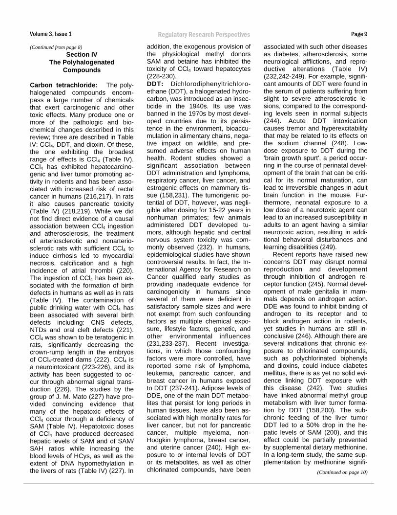
Carbon tetrachloride: The poly-halogenated compounds encompass a large number of chemicals that exert carcinogenic and other toxic effects. Many produce one or more of the pathologic and biochemical changes described in this review; three are described in Table IV: CCl4, DDT, and dioxin. Of these, the one exhibiting the broadest range of effects is CCl4 (Table IV). CCl4 has exhibited hepatocarcinogenic and liver tumor promoting activity in rodents and has been associated with increased risk of rectal cancer in humans (216,217). In rats it also causes pancreatic toxicity (Table IV) (218,219). While we did not find direct evidence of a causal association between CCl4 ingestion and atherosclerosis, the treatment of arteriosclerotic and nonarteriosclerotic rats with sufficient CCl4 to induce cirrhosis led to myocardial necrosis, calcification and a high incidence of atrial thrombi (220). The ingestion of CCl4 has been associated with the formation of birth defects in humans as well as in rats (Table IV). The contamination of public drinking water with CCl4 has been associated with several birth defects including: CNS defects, NTDs and oral cleft defects (221). CCl4 was shown to be teratogenic in rats, significantly decreasing the crown-rump length in the embryos of CCl4-treated dams (222). CCl4 is a neurointoxicant (223-226), and its activity has been suggested to occur through abnormal signal transduction (226). The studies by the group of J. M. Mato (227) have provided convincing evidence that many of the hepatoxic effects of CCl4 occur through a deficiency of SAM (Table IV). Hepatotoxic doses of CCl4 have produced decreased hepatic levels of SAM and of SAM/ SAH ratios while increasing the blood levels of HCys, as well as the extent of DNA hypomethylation in the livers of rats (Table IV) (227). In
addition, the exogenous provision of the physiological methyl donors SAM and betaine has inhibited the toxicity of CCl4 toward hepatocytes (228-230). DDT: Dichlorodiphenyltrichloroethane (DDT), a halogenated hydrocarbon, was introduced as an insecticide in the 1940s. Its use was banned in the 1970s by most developed countries due to its persistence in the environment, bioaccumulation in alimentary chains, negative impact on wildlife, and presumed adverse effects on human health. Rodent studies showed a significant association between DDT administration and lymphoma, respiratory cancer, liver cancer, and estrogenic effects on mammary tissue (158,231). The tumorigenic potential of DDT, however, was negligible after dosing for 15-22 years in nonhuman primates; few animals administered DDT developed tumors, although hepatic and central nervous system toxicity was commonly observed (232). In humans, epidemiological studies have shown controversial results. In fact, the International Agency for Research on Cancer qualified early studies as providing inadequate evidence for carcinogenicity in humans since several of them were deficient in satisfactory sample sizes and were not exempt from such confounding factors as multiple chemical exposure, lifestyle factors, genetic, and other environmental influences (231,233-237). Recent investigations, in which those confounding factors were more controlled, have reported some risk of lymphoma, leukemia, pancreatic cancer, and breast cancer in humans exposed to DDT (237-241). Adipose levels of DDE, one of the main DDT metabolites that persist for long periods in human tissues, have also been associated with high mortality rates for liver cancer, but not for pancreatic cancer, multiple myeloma, non-Hodgkin lymphoma, breast cancer, and uterine cancer (240). High exposure to or internal levels of DDT or its metabolites, as well as other chlorinated compounds, have been
associated with such other diseases as diabetes, atherosclerosis, some neurological afflictions, and reproductive alterations (Table IV) (232,242-249). For example, significant amounts of DDT were found in the serum of patients suffering from slight to severe atherosclerotic lesions, compared to the corresponding levels seen in normal subjects (244). Acute DDT intoxication causes tremor and hyperexcitability that may be related to its effects on the sodium channel (248). Low-dose exposure to DDT during the 'brain growth spurt', a period occurring in the course of perinatal development of the brain that can be critical for its normal maturation, can lead to irreversible changes in adult brain function in the mouse. Furthermore, neonatal exposure to a low dose of a neurotoxic agent can lead to an increased susceptibility in adults to an agent having a similar neurotoxic action, resulting in additional behavioral disturbances and learning disabilities (249). Recent reports have raised new concerns DDT may disrupt normal reproduction and development through inhibition of androgen receptor function (245). Normal development of male genitalia in mammals depends on androgen action. DDE was found to inhibit binding of androgen to its receptor and to block androgen action in rodents, yet studies in humans are still inconclusive (246). Although there are several indications that chronic exposure to chlorinated compounds, such as polychlorinated biphenyls and dioxins, could induce diabetes mellitus, there is as yet no solid evidence linking DDT exposure with this disease (242). Two studies have linked abnormal methyl group metabolism with liver tumor formation by DDT (158,200). The sub-chronic feeding of the liver tumor DDT led to a 50% drop in the hepatic levels of SAM (200), and this effect could be partially prevented by supplemental dietary methionine. In a long-term study, the same supplementation by methionine signifi
(Continued on page 10)

Volume 3, Issue 1
Page 10 Regulatory Research Perspectives
(Continued from page 9) cantly diminished the formation of metastatic hepatocellular carcinomas by DDT in diethylnitrosamineinitiated rats (158). We were unable to find any publications describing the effects of chronic DDT administration on the serum levels of HCys, on MTase activity or on DNA methylation. Dioxin: Dioxin appears to be a carcinogen both in humans and in experimental animals (Table IV) (250254). In humans, this increased tumor incidence was most notable with cancers of the rectum, the lung and the lymphohemopoietic system (251,252,254). In rodent species, liver cancer is the major tumor studied (250-252). In addition to its cancer-causing potential, dioxin induces many non-neoplastic pathologies (Table IV). Several studies have associated high human exposures to dioxin with the eventual formation of diabetes (252,254-257). Others observed either little or no correlation between diabetes and dioxin exposure (258-260). However, this apparent disparity may be attributable to differences in exposure levels in the populations concerned (252). As another manifestation of its pancreatotoxicity, dioxin induced hepatic transdifferentiation in the pancreas of hamsters (261). In addition, dioxin treatment in experimental animals produced hypoinsulinemia (255,259). The evidence in favor of a causal role of dioxin in atherosclerosis is indirect (262-266) and consists of: 1. the hyperlipidemia, resembling that of atherosclerosis, seen in dioxin-treated animals (262,265-268), 2. the elevated incidence of ischemic heart disease found in the populations exposed to high levels of dioxin (263,269), and 3. the shared risk factors previously described for cancer and atherosclerosis (264,270,271). Dioxin has produced a variety of birth defects, including spina bifida in humans and cleft palates in mice (251,272-274). Similarly, exposure to dioxin has resulted in peripheral neuropathy in humans (275,276) and polyneu-
ropathy in rats (277) (Table IV). In humans at least, the neuropathy has been proposed to result from an incipient diabetes (275,276), also associated with exposure to dioxin. The effects of dioxin on the methylation parameters considered in this review do not appear to have been described in the scientific literature to a significant degree. Others: A number of additional polyhalogenated compounds have exhibited carcinogenic activity (274,278). Many have demonstrated the pattern of pathologic effects and the alterations in methylation processes seen with the previously considered carcinogens. For example, pancreatitis has resulted from methylene chloride poisoning in humans and chloroform injection into dogs (279,280). The serum levels of polychlorinated biphenyls in pregnant women with diabetes were found to be significantly higher than in the corresponding pregnant control subjects (281). Similarly, the blood levels of total organochlorine residues were highest in severely atherosclerotic patients compared to the corresponding levels in control subjects or moderately atherosclerotic patients (244). In different studies, polychlorinated biphenyls diminished blood levels of HDL in one animal model for atherosclerosis (282) and caused vascular endothelial cell dysfunction in another (283). The developmental toxicity of the organohalides has been amply demonstrated in experimental animals. PBB treatment of pregnant animals led to the formation of cleft palate in developing mouse embryos and to decreased crown-rump lengths in male rat embryos with eventual behavioral toxicity in the resulting rat pups (284,285). Chloroform inhalation by pregnant dams produced fetal anomalies in both rats and mice (286,287); these included decreased fetal crown-rump lengths in both rats and mice, delayed ossification and missing ribs in rats and cleft palates in mice. Polybrominated biphenyls have exhibited neurotoxic effects both in developing rats and in humans
(285,288). In a series of recent studies
(289-292), the group of Pereira has examined the effects on DNA methylation by the carcinogen tetrachloroethylene (TCE), its metabolites dichloroacetic acid (DCA) and trichloroacetic acid (TCA), and several related compounds in the livers of mice. The subchronic administration of chloroform and CHBrCl2
caused global DNA hypomethylation in livers of B6C3F1 mice; the similar administration of chloroform, CHBrCl2, CHBr2Cl and CHBr3 led to the hypomethyaltion of the promoter region of the c-myc oncogene (292). In the model system employed, TCE, DCA and TCA all promoted liver tumor formation in initiated B6C3F1 mice and produced both global and gene-specific hypo-methylation of hepatic DNA (290,291,293). The genes shown to be hypomethylated following treatment with DCE, DCA, and TCA are the protooncogenes c-jun and the cmyc, with hypomethylation of both genes shown to occur in their promoter regions (289,291,293). The hypomethylation of both oncogenes was accompanied by increased gene expression (290). Liver tumor promotion by DCA and TCA led to increased MTase activity in the resulting liver adenomas and to decreased MTase activity in the adjacent non-tumorous tissue (293). The possible involvement of SAM in the hypomethylation produced by subchronic doses of DCA, TCA and DCE was shown by the observations that the simultaneous administration of methionine reversed both the decrease in c-myc and c-jun methylation and the accompanying increase in the expression of these genes produced by DCA and TCA (291). Additional links between abnormal methyl metabolism and the toxicity of the polyhalogenated carcinogens are seen in the formation of methylmercapto metabolites of PCBs (294) and of the inhibition by both carbon tetrachloride and chloroform of methionine biosynthesis in a B12-dependent auxotroph of E.
(Continued on page 11)
Page 11
Volume 3, Issue 1 Regulatory Research Perspectives
(Continued from page 10) coli (295).
Section V The Metals
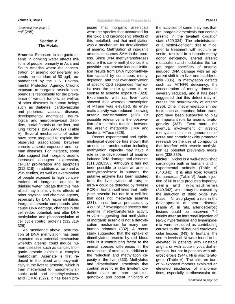
Arsenic: Exposure to inorganic arsenic in drinking water affects millions of people, primarily in Asia and South America where the concentration of arsenic considerably exceeds the standard of 50 µg/l, recommended by the U.S. Environmental Protection Agency. Chronic exposure to inorganic arsenic compounds is responsible for the prevalence of various tumors, as well as of other diseases in human beings such as diabetes, cardiovascular and peripheral vascular disease, developmental anomalies, neurological and neurobehavioral disorders, portal fibrosis of the liver, and lung fibrosis (242,297-312) (Table V). Several mechanisms of action have been proposed to explain the observed associations between chronic arsenic exposure and human diseases. For instance, some data suggest that inorganic arsenic increases oncogene expression, cellular proliferation and apoptosis (312-318). In addition, in vitro and in vivo studies, as well as examination of people exposed to high concentrations of inorganic arsenic in drinking water indicate that this metalloid may intensify toxic effects of other physical and chemical agents, especially by DNA repair inhibition. Inorganic arsenic compounds also cause DNA damage, changes in the cell redox potential, and alter DNA methylation and phosphorylation of cell cycle control proteins (312,318326). As mentioned above, perturbation of DNA methylation has been expected as a potential mechanism whereby arsenic could induce human diseases such as cancer. Inorganic arsenic exhibits a complex metabolism. Arsenate is first reduced in the blood and enzymatically in the liver to arsenite, which is then methylated to monomethylarsonic acid and dimethylarsinous acid (DMA) (327). It has been pro-
posed that inorganic arsenicals were the species that accounted for the toxic and carcinogenic effects of this metalloid, and that methylation was a mechanism for detoxification of arsenic. Methylation of inorganic arsenic consumes SAM in the process. Since DNA methyltransferases require this same methyl donor, it is possible that arsenic-induced initiation results from DNA hypomethylation caused by continuous methyl depletion, and that over-methylation of specific CpG sequences may exist over the entire genome in response to arsenite exposure (323). Experiments with rat liver cells showed that whereas transcription of MTase was elevated, its enzymatic activity was reduced following arsenic transformation (326). Of possible relevance is the observation of a chemical reaction between the arsenic metabolite DMA and bacterial MTase (328). Recent experimental and epidemiological evidence has shown that arsenic biotransformation including methylation capacity may have a role in the development of arsenic-induced DNA damage and diseases (311,329,330). Although it has not been possible to isolate an arsenic methyltransferase in humans, the putative enzyme has been isolated from the liver cytosol of rats; its mRNA could be detected by reverse PCR in human cell lines that methylate arsenite but not in a cell line that does not methylate arsenite (331). In non-human primates, only 4 out of 17 investigated species had arsenite methyltransferase activity in vitro suggesting that methylation of inorganic arsenic is not a detoxification mechanism for many nonhuman primates (332). A recent study suggested that the uptake of dimethylated arsenic by red blood cells is a contributing factor to the animal species differences in the metabolism of arsenic, in addition to the reduction and methylation capacity in the liver (333). Methylated and dimethylated arsenicals that contain arsenic in the trivalent oxidation state are more cytotoxic, genotoxic and potent inhibitors of
the activities of some enzymes than are inorganic arsenicals that contain arsenic in the trivalent oxidation state (329,334). The administration of a methyl-deficient diet to mice, prior to treatment with sodium arsenite, resulted in a hepatic methyl donor deficiency, altered arsenic metabolism and modulated the target organ specificity of arsenic-induced DNA damage with an apparent shift from liver and bladder to skin (335). In methylation defects such as MTHFR deficiency, the concentration of methyl donors is severely reduced, and it has been suggested that this defect may increase the neurotoxicity of arsenic (336). Other methyl-metabolism defects such as impaired folate transport have been suspected to play an important role for arsenic teratogenicity (337). Even more, the eventual involvement of arsenic methylation on the generation of acute and chronic toxicity promoted research on the use of compounds that interfere with arsenic methylation as potential preventive measures (338,339). Nickel: Nickel is a well-established carcinogen both in humans and in experimental animals (Table V) (340,341). It is also toxic towards the pancreas (Table V). Acute injection of Ni in rats produces hyperglycemia and hypoinsul inemia (340,342), which may be caused by the induction of nitric oxide synthase. Ni also played a role in the development of heart disease (Table V). In rats, arteriosclerotic lesions could be observed 7-9 weeks after an intrarenal injection of Ni3S2; hypertension and hyperlipidemia were excluded as contributing causes to the Ni-induced cardiovascular lesions (343). In humans, the serum levels of Ni were found to be elevated in patients with unstable angina or with acute myocardial infarction, but not in patients with atherosclerosis (344). Ni is also teratogenic (Table V). The children born of Ni-exposed mothers exhibited an elevated incidence of malformations, especially cardiovascular de
(Continued on page 12)

Volume 3, Issue 1
Page 12 Regulatory Research Perspectives
TABLE V THE METALS
PARAMETER AGENTS
As Ni Cd Zn Deficiency
Disease
Cancer + (311-313,316,319,452,453) + (340,341) +(353) + (385-392,454)
Pancreatic toxicity + (242,301-303,309,312) + (341,342,455) + (354-360,456) + (394-397,401,403,404)
Atherosclerosis + (264,298,299,307,308,312,322,453,457) + (343,344) + (361-368) + (398,399,406-411)
Birth defects + (337,453,458-460) + (345-347) +(353,373,374,421,461) + (412-419,421)
Neurotoxicity + (312,336,453) + (375-380) +/- (422-427)
Biochemical alterations
SAM/SAH + (326,327,335,462,463) - (349) + (428,429)
Homocysteine - (352) +/- (429,430)
DNA methylation + (313,315,316,323,326) + (350,351) + (429)
MTase + (328) + (350,351) + (384) + (384)
(Continued from page 11) fects (345). The progeny of mice and hamsters treated with Ni manifested several defects including exencephaly and cleft palate (346,347). While we could find no direct evidence of the neurotoxic effects of Ni, there was an association between high soil content of Ni, as well as Zn and Pb, in a high cluster population of multiple sclerosis cases (348). Compared to other carcinogens, Ni's effects on methylation processes appear somewhat paradoxical (349-352). In rats, Ni treatment reversed both the elevated urinary levels of FIGLU and the decreased hepatic contents of SAM produced by the chronic administration of a folate-deficient diet (349). In vitamin B12-deficient pigs, Ni decreased the elevated levels of serum HCys and
increased the serum and hepatic contents of vitamin B12 (352). In cell studies, Ni induced time-dependent changes in global and gene-specific DNA methylation; the Ni-induced DNA hypermethylation was observed even as the level of MTase was decreased (350,351). Cadmium: Cd is another metal that is deemed by IARC to be carcinogenic both to man and to animals (353). Cd exhibits multiple toxicities towards the pancreas (Table V). In rats and hamsters it causes hepatization of pancreatic acinar cells (354,355). In rats, subchronic doses of Cd were diabetogenic, causing hyperglycemia and decreased serum insulin levels in otherwise normal rats (356,357) and exacerbating the hyperglycemia and diabetic glomerulopathy in rats with pre-existing diabetes (358,359). In
mice, acute, nontoxic exposure to Cd inhibited pancreatic protease activities (360). The evidence linking Cd exposure with the development of atherosclerosis is quite strong (Table V). Chronic Cd administration led to the formation of atherosclerotic plaques and to hypertension in pigeons (361,362) and to thrombi, altered lipid profiles and atherosclerotic changes in rabbits (363,364). In another study with rabbits, Cd inhibited the atherogenic effects of a high cholesterol diet (365). In a series of human studies (366-368), the tissue levels of Cd correlated well with the degree of atherosclerosis in autopsied subjects, and a significantly elevated incidence of atherosclerosis was detected in subjects living in a specific region of the Netherlands con
(Continued on page 13)
Volume 3, Issue 1 Regulatory Research Perspectives Page 13
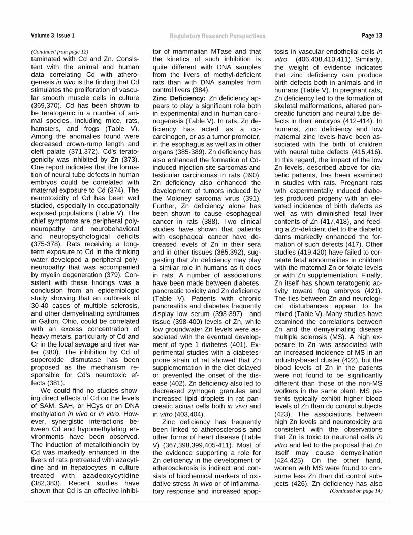
(Continued from page 12) taminated with Cd and Zn. Consistent with the animal and human data correlating Cd with atherogenesis in vivo is the finding that Cd stimulates the proliferation of vascular smooth muscle cells in culture (369,370). Cd has been shown to be teratogenic in a number of animal species, including mice, rats, hamsters, and frogs (Table V). Among the anomalies found were decreased crown-rump length and cleft palate (371,372). Cd's teratogenicity was inhibited by Zn (373). One report indicates that the formation of neural tube defects in human embryos could be correlated with maternal exposure to Cd (374). The neurotoxicity of Cd has been well studied, especially in occupationally exposed populations (Table V). The chief symptoms are peripheral polyneuropathy and neurobehavioral and neuropsychological deficits (375-378). Rats receiving a long-term exposure to Cd in the drinking water developed a peripheral polyneuropathy that was accompanied by myelin degeneration (379). Consistent with these findings was a conclusion from an epidemiologic study showing that an outbreak of 30-40 cases of multiple sclerosis, and other demyelinating syndromes in Galion, Ohio, could be correlated with an excess concentration of heavy metals, particularly of Cd and Cr in the local sewage and river water (380). The inhibition by Cd of superoxide dismutase has been proposed as the mechanism responsible for Cd's neurotoxic effects (381). We could find no studies showing direct effects of Cd on the levels of SAM, SAH, or HCys or on DNA methylation in vivo or in vitro. However, synergistic interactions between Cd and hypomethylating environments have been observed. The induction of metallothionein by Cd was markedly enhanced in the livers of rats pretreated with azacytidine and in hepatocytes in culture treated with azadeoxycytidine (382,383). Recent studies have shown that Cd is an effective inhibi
tor of mammalian MTase and that the kinetics of such inhibition is quite different with DNA samples from the livers of methyl-deficient rats than with DNA samples from control livers (384). Zinc Deficiency: Zn deficiency appears to play a significant role both in experimental and in human carcinogenesis (Table V). In rats, Zn deficiency has acted as a cocarcinogen, or as a tumor promoter, in the esophagus as well as in other organs (385-389). Zn deficiency has also enhanced the formation of Cdinduced injection site sarcomas and testicular carcinomas in rats (390). Zn deficiency also enhanced the development of tumors induced by the Moloney sarcoma virus (391). Further, Zn deficiency alone has been shown to cause esophageal cancer in rats (388). Two clinical studies have shown that patients with esophageal cancer have decreased levels of Zn in their sera and in other tissues (385,392), suggesting that Zn deficiency may play a similar role in humans as it does in rats. A number of associations have been made between diabetes, pancreatic toxicity and Zn deficiency (Table V). Patients with chronic pancreatitis and diabetes frequently display low serum (393-397) and tissue (398-400) levels of Zn, while low groundwater Zn levels were associated with the eventual development of type 1 diabetes (401). Experimental studies with a diabetes-prone strain of rat showed that Zn supplementation in the diet delayed or prevented the onset of the disease (402). Zn deficiency also led to decreased zymogen granules and increased lipid droplets in rat pancreatic acinar cells both in vivo and in vitro (403,404).
Zinc deficiency has frequently been linked to atherosclerosis and other forms of heart disease (Table V) (367,398,399,405-411). Most of the evidence supporting a role for Zn deficiency in the development of atherosclerosis is indirect and consists of biochemical markers of oxidative stress in vivo or of inflammatory response and increased apop
tosis in vascular endothelial cells in vitro (406,408,410,411). Similarly, the weight of evidence indicates that zinc deficiency can produce birth defects both in animals and in humans (Table V). In pregnant rats, Zn deficiency led to the formation of skeletal malformations, altered pancreatic function and neural tube defects in their embryos (412-414). In humans, zinc deficiency and low maternal zinc levels have been associated with the birth of children with neural tube defects (415,416). In this regard, the impact of the low Zn levels, described above for diabetic patients, has been examined in studies with rats. Pregnant rats with experimentally induced diabetes produced progeny with an elevated incidence of birth defects as well as with diminished fetal liver contents of Zn (417,418), and feeding a Zn-deficient diet to the diabetic dams markedly enhanced the formation of such defects (417). Other studies (419,420) have failed to correlate fetal abnormalities in children with the maternal Zn or folate levels or with Zn supplementation. Finally, Zn itself has shown teratogenic activity toward frog embryos (421). The ties between Zn and neurological disturbances appear to be mixed (Table V). Many studies have examined the correlations between Zn and the demyelinating disease multiple sclerosis (MS). A high exposure to Zn was associated with an increased incidence of MS in an industry-based cluster (422), but the blood levels of Zn in the patients were not found to be significantly different than those of the non-MS workers in the same plant. MS patients typically exhibit higher blood levels of Zn than do control subjects (423). The associations between high Zn levels and neurotoxicity are consistent with the observations that Zn is toxic to neuronal cells in vitro and led to the proposal that Zn itself may cause demyelination (424,425). On the other hand, women with MS were found to consume less Zn than did control subjects (426). Zn deficiency has also
(Continued on page 14)
Volume 3, Issue 1
Page 14 Regulatory Research Perspectives
(Continued from page 13) been shown to alter neurotransmitter activity in rats (427).
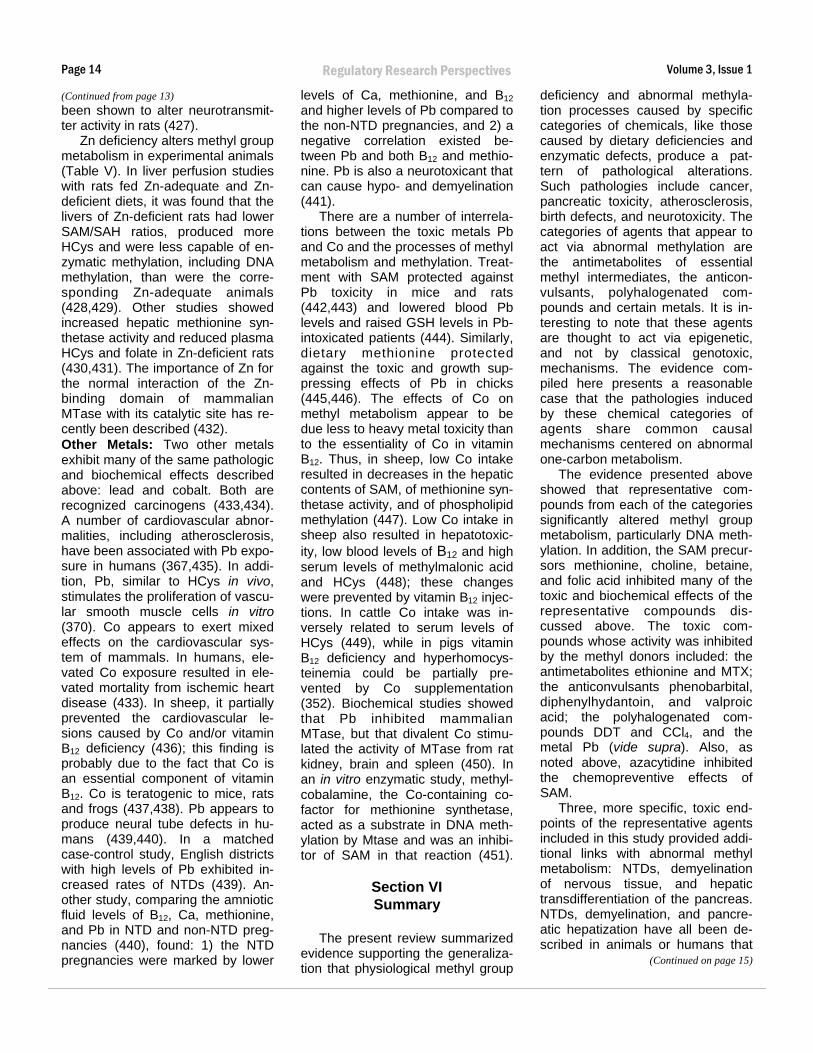
Zn deficiency alters methyl group metabolism in experimental animals (Table V). In liver perfusion studies with rats fed Zn-adequate and Zndeficient diets, it was found that the livers of Zn-deficient rats had lower SAM/SAH ratios, produced more HCys and were less capable of enzymatic methylation, including DNA methylation, than were the corresponding Zn-adequate animals (428,429). Other studies showed increased hepatic methionine synthetase activity and reduced plasma HCys and folate in Zn-deficient rats (430,431). The importance of Zn for the normal interaction of the Znbinding domain of mammalian MTase with its catalytic site has recently been described (432). Other Metals: Two other metals exhibit many of the same pathologic and biochemical effects described above: lead and cobalt. Both are recognized carcinogens (433,434). A number of cardiovascular abnormalities, including atherosclerosis, have been associated with Pb exposure in humans (367,435). In addition, Pb, similar to HCys in vivo, stimulates the proliferation of vascular smooth muscle cells in vitro (370). Co appears to exert mixed effects on the cardiovascular system of mammals. In humans, elevated Co exposure resulted in elevated mortality from ischemic heart disease (433). In sheep, it partially prevented the cardiovascular lesions caused by Co and/or vitamin B12 deficiency (436); this finding is probably due to the fact that Co is an essential component of vitamin B12. Co is teratogenic to mice, rats and frogs (437,438). Pb appears to produce neural tube defects in humans (439,440). In a matched case-control study, English districts with high levels of Pb exhibited increased rates of NTDs (439). Another study, comparing the amniotic fluid levels of B12, Ca, methionine, and Pb in NTD and non-NTD pregnancies (440), found: 1) the NTD pregnancies were marked by lower
levels of Ca, methionine, and B12
and higher levels of Pb compared to the non-NTD pregnancies, and 2) a negative correlation existed between Pb and both B12 and methionine. Pb is also a neurotoxicant that can cause hypo- and demyelination (441). There are a number of interrelations between the toxic metals Pb and Co and the processes of methyl metabolism and methylation. Treatment with SAM protected against Pb toxicity in mice and rats (442,443) and lowered blood Pb levels and raised GSH levels in Pbintoxicated patients (444). Similarly, dietary methionine protected against the toxic and growth suppressing effects of Pb in chicks (445,446). The effects of Co on methyl metabolism appear to be due less to heavy metal toxicity than to the essentiality of Co in vitamin B12. Thus, in sheep, low Co intake resulted in decreases in the hepatic contents of SAM, of methionine synthetase activity, and of phospholipid methylation (447). Low Co intake in sheep also resulted in hepatotoxicity, low blood levels of B12 and high serum levels of methylmalonic acid and HCys (448); these changes were prevented by vitamin B12 injections. In cattle Co intake was inversely related to serum levels of HCys (449), while in pigs vitamin B12 deficiency and hyperhomocysteinemia could be partially prevented by Co supplementation (352). Biochemical studies showed that Pb inhibited mammalian MTase, but that divalent Co stimulated the activity of MTase from rat kidney, brain and spleen (450). In an in vitro enzymatic study, methyl-cobalamine, the Co-containing cofactor for methionine synthetase, acted as a substrate in DNA methylation by Mtase and was an inhibitor of SAM in that reaction (451).
Section VI Summary
The present review summarized evidence supporting the generalization that physiological methyl group
deficiency and abnormal methylation processes caused by specific categories of chemicals, like those caused by dietary deficiencies and enzymatic defects, produce a pattern of pathological alterations. Such pathologies include cancer, pancreatic toxicity, atherosclerosis, birth defects, and neurotoxicity. The categories of agents that appear to act via abnormal methylation are the antimetabolites of essential methyl intermediates, the anticonvulsants, polyhalogenated compounds and certain metals. It is interesting to note that these agents are thought to act via epigenetic, and not by classical genotoxic, mechanisms. The evidence compiled here presents a reasonable case that the pathologies induced by these chemical categories of agents share common causal mechanisms centered on abnormal one-carbon metabolism. The evidence presented above showed that representative compounds from each of the categories significantly altered methyl group metabolism, particularly DNA methylation. In addition, the SAM precursors methionine, choline, betaine, and folic acid inhibited many of the toxic and biochemical effects of the representative compounds discussed above. The toxic compounds whose activity was inhibited by the methyl donors included: the antimetabolites ethionine and MTX; the anticonvulsants phenobarbital, diphenylhydantoin, and valproic acid; the polyhalogenated compounds DDT and CCl4, and the metal Pb (vide supra). Also, as noted above, azacytidine inhibited the chemopreventive effects of SAM. Three, more specific, toxic endpoints of the representative agents included in this study provided additional links with abnormal methyl metabolism: NTDs, demyelination of nervous tissue, and hepatic transdifferentiation of the pancreas. NTDs, demyelination, and pancreatic hepatization have all been described in animals or humans that
(Continued on page 15)

Page 15
Volume 3, Issue 1 Regulatory Research Perspectives
(Continued from page 14) were deficient in methyl donors due to diet, defective enzymes or disease (vide supra, (464,465)). The same biological effects have been noted in animals or humans exposed to the chemicals in the various categories of this report. NTDs have been observed following exposure to the anticonvulsants in general, to the polyhalogenated compounds CCl4 and dioxin, and finally to the metals Pb and Cd, as well as to Zn deficiency (vide supra). In addition, the antimetabolite MTX enhanced the formation of NTDs by valproic acid, while azacytidine itself produced NTDs in vitro (vide supra). Similarly, demyelination, particularly of peripheral nerves, has been observed following treatment with, or exposure to: methotrexate, the anticonvulsants, and the metals Pb, Cd and As ((466) vide supra). Hepatization of the pancreas has been induced in rats and hamsters by Cd, by dioxin, and by a regimen that included ethionine ((465) vide supra). The results compiled in the present review may provide useful lessons regarding the mechanism of action of the diverse agents examined. For example, ethionine and azacytidine clearly exhibit tumorigenic activity, while FUDR does not. Ethionine and azacytidine also inhibit SAM-dependent DNA methylation, while FUDR does not appear to do so. These observations indicate that the SAM-dependent methylation may be more directly involved in the carcinogenic process than is the inhibition of thymidylate synthetase and of DNA synthesis. In this regard, the fact that MTX acts to block both DNA synthesis and DNA methylation may explain why it has so seldom been found to exhibit tumorigenic activity. Further evidence on the role of SAM-dependent DNA methylation in carcinogenesis is seen in comparing the tissue specificities of ethionine and azacytidine. Azacytidine exerts its tumorigenicity and DNA hypo-methylating activity in several organs; to date, the carcinogenic and
DNA hypomethylating activity of ethionine has been confined to the liver. It is in the liver that ethionine is most readily converted to its hypo-methylating metabolite SAE.
Altered methyl metabolism in the liver appears to play a particularly important role in the toxicology of the anticonvulsants and the polyhalogenated compounds. In rodents, both categories of chemicals are hepatotoxic and generally exert their major carcinogenic effects in this organ (vide supra). Specific anticonvulsants and polyhalogenated compounds have been effective in decreasing SAM/SAH ratios and increasing the extent of DNA hypo-methylation in this organ. Two different mechanisms may be proposed for the diminished availability of SAM in the livers of animals treated with hepatotoxic agents: diminished synthesis and increased utilization. Defects in the expression of the liver-specific methionine adenosyltransferase MAT1A gene, and thus the activity of the corresponding enzyme, have been shown to be a major cause of SAM insufficiency caused by hepatotoxic agents (vide supra) (467,468). A second possible mechanism is the drain on hepatic SAM caused by many anticonvulsants and polyhalogenated compounds tested in rodents (vide supra, (469)); SAM is used for the synthesis of the phosphatidylcholine required in the induction of the hepatic cytochrome P450s caused by these agents. Such biosynthesis also results in the elevated formation of SAH. The demonstration of the validity of either mechanism is not without implications for the regulatory agencies. As has frequently been noted (470), liver tumor formation in rats and mice is responsible for a high proportion of the carcinogenic compounds detected by the NTP. Many of these compounds also appear to act as secondary agents or epigenetic carcinogens. Demonstration that the tumorigenic activity of such compounds towards the liver is dependent upon an altered methylation status in vivo would strengthen
the hypothesis that these compounds do in fact have a threshold of activity.
The hypomethylating environment produced in the liver by the anticonvulsants and the polyhalogenated compounds may affect other organs as well. A high degree of correspondence was seen between the decreased SAM/SAH ratios in liver and the rise in serum HCys by the same compounds (Tables III and IV). The elevated serum levels of HCys may thus contribute both to the formation of a hypomethylating environment and to the development of extrahepatic pathologies caused by the chronic administration of the anticonvulsants and the polyhalogenated compounds. Time did not permit a consideration of other categories of chemical agents, which also alter methyl group metabolism and cause pathological changes. These include some electrophilic carcinogens, nitrous oxide, sulfa drugs, and alcohol. The same constraints did not permit a consideration of alternate mechanisms of toxicity not centered on abnormal methylation processes. The authors are aware of the fact that in some biological systems, particularly in rodent liver, methyl deficiency leads to oxidative damage and, correspondingly, oxidative damage can lead to methyl insufficiency and abnormal DNA methylation ((468,471), vide supra). In this case the alternate potential mechanisms are not mutually exclusive. In conclusion, the results indicate that abnormal methylation processes caused by chemical agents, like those caused by dietary deficiencies or by metabolic defects, can be associated with a general pattern of specific pathological effects.

Page 16 Regulatory Research Perspectives Volume 3, Issue 1
References
1. Ross, S. l. and Poirier, L. A. 2002. Proceeding of the Trans-HHS Workshop on Diet, DNA Methylation Processes and Health. J. Nutrition 132: 2329S-2332S.
2. Robertson, K. D. and Jones, P. A. 2002. DNA Methylation: Past, Present and Future Directions. Carcinogenesis 21: 461-467.
3. Poirier, L. A. 2002. The Effects of Diet, Genetics and Chemicals on Toxicity and Aberrant DNA Methylation. J. Nutrition 132: 2336S-2339S.
4. Potter, J. D. 2002. Methyl Supply, Methyl Metabolizing Enzymes and Colorectal Neoplasia. J. Nutrition 132: 2410S-2412S.
5. Ziegler, R. G., Weinstein, S. J., and Fears, T. R. 2002. Nutritional and Genetic Inefficiencies in One-Carbon Metabolism and Cervical Cancer Risk. J. Nutrition 132: 2345S-2349S.
6. Longnecker, D. S. 2002. Abnormal Methyl Metabolism in Pancreatic Toxicity and Diabetes. J. Nutrition 132: 2373S-2376S.
7. Maier, S. and Olek, A. 2002. Diabetes: A Candidate Disease for Efficient DNA Methylation Profiling. J. Nutrition 132: 2440S-2443S.
8. Dong, C., Yoon, W., and Goldschmidt-Clermont, P. J. 2002. DNA Methylation and Atherosclerosis. J. Nutrition 132: 2406S-2409S.
9. Issa, J.P. 2002. Epigenetic Variation and Human Disease. J. Nutrition 132: 2388S-2392S.
10. Richardson, B. C. 2002. Role of DNA Methylation in the Regulation of Cell Function: Autoimmunity, Aging and Cancer. J. Nutrition 132: 2401S-2405S.
11. Cooney, C. A., Dave, A., and Wolff, G. L. 2002. Maternal Methyl Supplements in Mice Affect Epigenetic Variation and DNA Methylation of Offspring. J. Nutrition 132 in press: 2393S-2400S.
12. Green, N. S. 2002. Folic Acid Supplementation and Prevention of Birth Defects. J. Nutrition 132: 2356S-2360S.
13. Rader, J. I. 2002. Folic Acid Fortification, Folate Status, and Plasma Homocysteine. J. Nutrition 132: 2466S-2470S.
14. Ehrlich, M. 2002. DNA Hypomethylation, Cancer, the Immunodeficiency, Centromeric Region Instability, Facial Anomalies Syndrome, and Chromosomal Rearrangements. J. Nutrition 132: 2424S2429S.
15. Okano, M. and Li, E. 2002. Genetic Analyses of DNA Methyltransferse Genes in Mouse Model System. J. Nutrition 132: 2462S-2465S.
16. Choi, S. W. and Mason, J. B. 2002.
Folate Status: Effects on Pathways of Colorectal Carcinogenesis. J. Nutrition 132: 2413S-2418S.
17. Niculescu, M. D. and Zeisel, S. H. 2002. Diet, Methyl Donors and DNA Methylation: Interactions Between Dietary Folate, Methionine and Choline. J. Nutrition 132: 2333S-2335S.
18. Giovannucci, E. 2002. Epidemiologic Studies of Folate and Colorectal Neoplasia: A Review. J. Nutrition 132: 2350S-2355S.
19. James, S. J., Melnyk, S., Pogribna, M., Pogribny, I. P., and Caudill, M. A. 2002. Elevation in S-Adenosylhomocysteine and DNA Hypomethylation: Potential Epigenetic Mechanism for Homocysteine-Related Pathology. J. Nutrition 132: 2361S-2366S.
20. Finnell, R. H., Spiegelstein, O., Wlodarczyk, B., Triplett, A., Pogribny, I. P., Melnyk, S., and James, S. J. 2002. DNA Methylation in Folbp1 Knockout Mice Supplemented With Folic Acid During Gestation. J. Nutrition 132: 2457S-2461S.
21. Halsted, C. H., Villanueva, J. A., Devlin, A. M., and Chandler, C. J. 2002. Metabolic Interactions of Alcohol and Folate. J. Nutrition 132: 2367S-2372S.
22. Corrales, F. J., Perez-Mato, I., Sanchez del Pino, M. M., Ruiz, F., Castro, C., Garcia-Trevijano, E. R., Latasa, U., Martinez-Chantar, M. L., Martinez, A., Avila, M. A., and Mato, J. M. 2002. Regulation of Mammalian Liver Methonine Adenosyltransferase. J. Nutrition 132: 2377S2381S.
23. Sugimura, T. and Ushijima, T. 2000. Genetic and Epigenetic Alterations in Carcinogenesis. Mutation Res. 462: 235-246.
24. Goodman, J. I. and Watson, R. E. 2002. Altered DNA Methylation: a Secondary Mechanism Involved in Carcinogenesis. Annu. Rev. Pharmacol Toxicol. 42: 501-525.
25. Piyathilake, C. L. and Johanning, G. L. 2002. Cellular Vitamins, DNA Methylation and Cancer Risk. J. Nutrition 132: 2340S-2344S.
26. Newberne, P. M. 1986. Lipotropic Factors and Oncogenesis. Adv. Exp. Med. Biol. 206: 223-251.
27. Salmon, W. D. and Newberne, P. M. 1962. Cardiovascular Disease in Choline-Deficient Rats. Arch. Pathol. 73: 190-209.
28. Bunout, D., Petermann, M., Hirsch, S., de la Maza, P., Suazo, M., Barrera, G., and Kauffman, R. 2000. Low Serum Folate but Normal Homocysteine Levels in Patients With Atherosclerotic Vascular Disease and Matched Healthy Controls. Nutrition 16: 434-438.
29. Balaghi, M. and Wagner, C. 1995. Folate Deficiency Inhibits Pancreatic Amylase Secretion in Rats. Am. J.
Clin. Nutr. 61: 90-96. 30. Poirier, L. A., Brown, A. T., Fink, L.
M., Wise, C. K., Randolph, C. J., Delongchamp, R. R., and Fonseca, V . A . 2 0 0 1 . B l o o d S -Adenosylmethionine Concentrations and Lymphocyte Methylenetetrahydrofolate Reductase Activity in Diabetes Mellitus and Diabetic Nephropathy. Metabolism 50: 10141018.
31. Daly, L. E., Kirke, P. N., Molloy, A., Weir, D. G., and Scott, J. M. 1995. Folate Levels and Neural Tube Defects. Implications for Prevention. JAMA 274: 1698-1702.
32. Steen, M. T., Boddie, A. M., Fisher, A. J., Macmahon, W., Saxe, D., Sullivan, K. M., Dembure, P. P., and Elsas, L. J. 1998. Neural-Tube Defects Are Associated With Low Concentrations of Cobalamin (Vitamin B12) in Amniotic Fluid. Prenat. Diagn. 18: 545-555.
33. Tan, S. V. and Guiloff, R. J. 1998. Hypothesis on the Pathogenesis of Vacuolar Myelopathy, Dementia, and Peripheral Neuropathy in AIDS. J. Neurol. Neurosurg. Psychiatry 65: 23-28.
34. Bottiglieri, T. 1996. Folate, Vitamin B12, and Neuropsychiatric Disorders. Nutr. Rev. 54: 382-390.
35. Fehling, C., Jagerstad, M., Akesson, B., Axelsson, J., and Brun, A. 1978. Effects of Vitamin B12 Deficiency on Lipid Metabolism of the Rat Liver and Nervous System. Br. J. Nutr. 39: 501-513.
36. Banni, S., Corongiu, F. P., Dessi, M. A., Iannone, A., Lombardi, B., Tomasi, A., and Vannini, V. 1989. Free Radicals and Lipid Peroxidation in Liver of Rats Kept on a Diet Devoid of Choline. Free Radic. Res Commun. 7: 233-240.
37. Cantoni, G. L. 1975. Biological Methylation: Selected Aspects. Annu. Rev. Biochem. 44: 435-451.
38. Cantoni, G. L. 1985. The Role of S-Adenosylhomocysteine in the Biological Utilization of S- Adenosylmethionine. Prog. Clin. Biol. Res 198: 47-65.
39. Creusot, F. and Christman, J. K. 1981. Localization of DNA Methyl-transferase in the Chromatin of Friend Erythroleukemia Cells. Nucleic Acids Res 9: 5359-5381.
40. Cox, R., Prescott, C., and Irving, C. C. 1977. The Effect of S-Adenosylhomocysteine on DNA Methylation in Isolated Rat Liver Nuclei. Biochim. Biophys Acta 474: 493-499.
41. McCully, K. S. 1969. Vascular Pathology of Homocysteinemia: Implications for the Pathogenesis of Arteriosclerosis. Am. J. Pathol. 56: 111-128.
(Continued on page 17)

Page 17 Volume 3, Issue 1 Regulatory Research Perspectives (Continued from page 16)
42. Wenstrom, K. D., Johanning, G. L., Owen, J., Johnston, K. E., Acton, S., Cliver, S., and Tamura, T. 2000. Amniotic Fluid Homocysteine Levels, 5,10-Methylenetetrahydrafolate Reductase Genotypes, and Neural Tube Closure Sites. Am. J. Med. Genet 90: 6-11.
43. van der Put, N. M., van Straaten, H. W., Trijbels, F. J., and Blom, H. J. 2001. Folate, Homocysteine and Neural Tube Defects: an Overview. Exp. Biol. Med. (Maywood.) 226: 243-270.
44. Vollset, S. E., Refsum, H., Tverdal, A., Nygard, O., Nordrehaug, J. E., Tell, G. S., and Ueland, P. M. 2001. Plasma Total Homocysteine and Cardiovascular and Noncardiovascular Mortality: the Hordaland Homocysteine Study. Am. J. Clin. Nutr. 74: 130-136.
45. Title, L. M., Cummings, P. M., Giddens, K., Genest, J. J., Jr., and Nassar, B. A. 2000. Effect of Folic Acid and Antioxidant Vitamins on Endothelial Dysfunction in Patients With Coronary Artery Disease. J. Am. Coll. Cardiol. 36: 758-765.
46. Miller, J. W., Nadeau, M. R., Smith, J., Smith, D., and Selhub, J. 1994. Folate-Deficiency-Induced Homocysteinaemia in Rats: Disruption of S- Adenosylmethionine's Co-Ordinate Regulation of Homocysteine Metabolism. Biochem J. 298 ( Pt 2): 415-419.
47. Hilton, M. A., Hoffman, J. L., and Sparks, M. K. 1983. Effect of Methotrexate with 5-Methyltetrahydrofolate Rescue and Dietary Homocystine on Survival of Leukemic Mice and on Concentrations of Liver Adenosylamino Acids. Cancer Res. 43: 5210-5216.
48. Thomson, S. W., Heimburger, D. C., Cornwell, P. E., Turner, M. E., Sauberlich, H. E., Fox, L. M., and Butterworth, C. E. 2000. Correlates of Total Plasma Homocysteine: Folic Acid, Copper, and Cervical Dysplasia. Nutrition 16: 411-416.
49. Hofmann, M. A., Lalla, E., Lu, Y., Gleason, M. R., Wolf, B. M., Tanji, N., Ferran, L. J., Jr., Kohl, B., Rao, V., Kisiel, W., Stern, D. M., and Schmidt, A. M. 2001. Hyperhomocysteinemia Enhances Vascular Inflammation and Accelerates Atherosclerosis in a Murine Model. J. Clin. Invest. 107: 675-683.
50. She, Q. B., Hayakawa, T., and Tsuge, H. 1995. Alteration in the Phosphatidylcholine Biosynthesis of Rat Liver Microsomes Caused by Vitamin B6 Deficiency. Biosci. Biotechnol. Biochem 59: 163-167.
51. Robinson, K., Mayer, E. L., Miller, D. P., Green, R., van Lente, F., Gupta, A., Kottke-Marchant, K.,
Savon, S. R., Selhub, J., and Nissen, S. E. 1995. Hyperhomocysteinemia and Low Pyridoxal Phosphate. Common and Independent Reversible Risk Factors for Coronary Artery Disease. Circulation 92: 2825-2830.
52. Robinson, K., Gupta, A., Dennis, V., Arheart, K., Chaudhary, D., Green, R., Vigo, P., Mayer, E. L., Selhub, J., Kutner, M., and Jacobsen, D. W. 1996. Hyperhomocysteinemia Confers an Independent Increased Risk of Atherosclerosis in End-Stage Renal Disease and Is Closely Linked to Plasma Folate and Pyridoxine Concentrations. Circulation 94: 2743-2748.
53. Hartman, T. J., Woodson, K., Stol-zenberg-Solomon, R., Virtamo, J., Selhub, J., Barrett, M. J., and Al-banes, D. 2001. Association of the B-Vitamins Pyridoxal 5'-Phosphate (B(6)), B(12), and Folate with Lung Cancer Risk in Older Men. Am. J. Epidemiol. 153: 688-694.
54. Stolzenberg-Solomon, R. Z., Al-banes, D., Nieto, F. J., Hartman, T. J., Tangrea, J. A., Rautalahti, M., Sehlub, J., Virtamo, J., and Taylor, P. R. 1999. Pancreatic Cancer Risk and Nutrition-Related Methyl-Group Availability Indicators in Male Smokers. J. Natl. Cancer Inst. 91: 535541.
55. Davis, S. D., Nelson, T., and Shepard, T. H. 1970. Teratogenicity of Vitamin B6 Deficiency: Omphalocele, Skeletal and Neural Defects, and Splenic Hypoplasia. Science 169: 1329-1330.
56. Wong, W. Y., Eskes, T. K., Kuijpers-Jagtman, A. M., Spauwen, P. H., Steegers, E. A., Thomas, C. M., Hamel, B. C., Blom, H. J., and Stee-gers-Theunissen, R. P. 1999. Nonsyndromic Orofacial Clefts: Association with Maternal Hyperhomocysteinemia. Teratology 60: 253-257.
57. Jakubowski, H. 2001. Translational Accuracy of Aminoacyl-TRNA Synthetases: Implications for Atherosclerosis. J. Nutrition 131: 2983S-2987S.
58. Stuhlinger, M. C., Tsao, P. S., Her, J. H., Kimoto, M., Balint, R. F., and Cooke, J. P. 2001. Homocysteine Impairs the Nitric Oxide Synthase Pathway: Role of Asymmetric Di-methylarginine. Circulation 104: 2569-2575.
59. Kerins, D. M., Koury, M. J., Capdevila, A., Rana, S., and Wagner, C . 2 0 0 1 . P l a s m a S -Adenosylhomocysteine Is a More Sensitive Indicator of Cardiovascular Disease Than Plasma Homocysteine. Am. J. Clin. Nutr. 74: 723-729.
60. Yi, P., Melnyk, S., Pogribna, M., Pogribny, I. P., Hine, R. J., and James, S. J. 2000. Increase in
Plasma Homocysteine Associated With Parallel Increases in Plasma S-Adenosylhomocysteine and Lymphocyte DNA Hypomethylation. J. Biol. Chem. 275: 29318-29323.
61. Chiang, P. K. 1998. Biological Effects of Inhibitors of S-Adenosylhomocysteine Hydrolase. Pharmacol Ther. 77: 115-134.
62. Chiang, P. K. and Cantoni, G. L. 1979. Perturbation of Biochemical T r a n s m e t h y l a t i o n s b y 3 Deazaadenosine in Vivo. Biochem. Pharmacol. 28: 1897-1902.
63. Gershoni-Baruch, R., Dagan, E., Israeli, D., Kasinetz, L., Kadouri, E., and Friedman, E. 2000. Association of the C677T Polymorphism in the MTHFR Gene with Breast and/or Ovarian Cancer Risk in Jewish Women. Eur. J. Cancer 36: 23132316.
64. van der Put, N. M. and Blom, H. J. 2000. Neural Tube Defects and a Disturbed Folate Dependent Homocysteine Metabolism. Eur. J. Obstet. Gynecol. Reprod. Biol. 92: 57-61.
65. Hansen, D. K., Barbee, S. A., Grafton, T. F., Gu, Y., and Streck, R. D. 2001. Antisense Modulation of 5,10-Methylenetetrahydrofolate Reductase Expression Produces Neural Tube Defects in Mouse Embryos. Reprod. Toxicol. 15: 21-29.
66. Shpichinetsky, V., Raz, I., Friedlander, Y., Goldschmidt, N., Wexler, I. D., Ben-Yehuda, A., and Friedman, G. 2000. The Association Between Two Common Mutations C677T and A1298C in Human Methylenetetrahydrofolate Reductase Gene and the Risk for Diabetic Nephropathy in Type II Diabetic Patients. J. Nutrition 130: 2493-2497.
67. Shcherbak, N. S., Shutskaya, Z. V., Sheidina, A. M., Larionova, V. I., and Schwartz, E. I. 1999. Methylenetetrahydrofolate Reductase Gene Polymorphism as a Risk Factor for Diabetic Nephropathy in IDDM Patients. Mol. Genet Metab 68: 375378.
68. Thompson, P. A. and Ambrosone, C. 2000. Molecular Epidemiology of Genetic Polymorphisms in Estrogen Metabolizing Enzymes in Human Breast Cancer. J. Natl. Cancer Inst. Monogr 125-134.
69. Huang, C. S., Chern, H. D., Chang, K. J., Cheng, C. W., Hsu, S. M., and Shen, C. Y. 1999. Breast Cancer Risk Associated With Genotype Polymorphism of the Estrogen-Metabolizing Genes CYP17, CYP1A1, and COMT: a Multigenic Study on Cancer Susceptibility. Cancer Res. 59: 4870-4875.
70. Goodman, J. E., Lavigne, J. A., Wu, K., Helzlsouer, K. J., Strickland, P. T., Selhub, J., and Yager, J. D.
(Continued on page 18)

Volume 3, Issue 1 Page 18 Regulatory Research Perspectives (Continued from page 17)
2001. COMT Genotype, Micronutrients in the Folate Metabolic Pathway and Breast Cancer Risk. Carcinogenesis 22: 1661-1665.
71. Thompson, P. A., Shields, P. G., Freudenheim, J. L., Stone, A., Vena, J. E., Marshall, J. R., Graham, S., Laughlin, R., Nemoto, T., Kadlubar, F. F., and Ambrosone, C. B. 1998. Genetic Polymorphisms in Catechol-O-Methyltransferase, Menopausal Status, and Breast Cancer Risk. Cancer Res. 58: 21072110.
72. Mitrunen, K., Jourenkova, N., Kataja, V., Eskelinen, M., Kosma, V. M., Benhamou, S., Kang, D., Vainio, H., Uusitupa, M., and Hirvonen, A. 2001. Polymorphic Catechol-O-Methyltransferase Gene and Breast Cancer Risk. Cancer Epidemiol. Biomarkers Prev. 10: 635-640.
73. Chamberlin, M. E., Ubagai, T., Mudd, S. H., Wilson, W. G., Leonard, J. V., and Chou, J. Y. 1996. Demyelination of the Brain Is Associated With Methionine Adenosyltransferase I/III Deficiency. J. Clin. Invest. 98: 1021-1027.
74. Lu, S. C., Alvarez, L., Huang, Z. Z., Chen, L., An, W., Corrales, F. J., Avila, M. A., Kanel, G., and Mato, J. M. 2001. Methionine Adenosyltransferase 1A Knockout Mice Are Predisposed to Liver Injury and Exhibit Increased Expression of Genes Involved in Proliferation. Proc. Natl. Acad. Sci. U. S. A 98: 5560-5565.
75. Newberne, P. M., Carleton, W. W., and Wogan, G. N. 1964. Hepatomas in Rats and Hepatorenal Injury in Ducklings Fed Peanut Meal As a Major Source of Protein. Pathol. Vet. 1: 105-132.
76. Farber, E. 1963. Ethionine Carcinogenesis. Adv. Cancer Res. 7: 383474.
77. Miller, J. A. and Miller, E. C. 1953. The Carcinogenic Aminoazo Dyes. Adv. Caner Res. 1: 339-396.
78. Poirier, L. A. and Whitehead, V. M. 1973. Folate Deficiency and Elevated Formimioglutamate Excretion during Chronic Diethylnitrosamine Administration to Rats. Cancer Res. 33: 383-388.
79. Blom, H. J. 1998. Mutated 5,10Methylenetetrahydrofolate Reductase and Moderate Hyperhomocysteinaemia. Eur. J. Pediatr. 157 Suppl 2: S131-S134.
80. Verhoef, P., Stampfer, M. J., Buring, J. E., Gaziano, J. M., Allen, R. H., Stabler, S. P., Reynolds, R. D., Kok, F. J., Hennekens, C. H., and Willett, W. C. 1996. Homocysteine Metabolism and Risk of Myocardial Infarction: Relation with Vitamins B6, B12, and Folate. Am. J. Epidemiol. 143: 845-859.
81. Fortin, L. J. and Genest, J., Jr. 1995. Measurement of Homocyst(e) ine in the Prediction of Arteriosclerosis. Clin. Biochem 28: 155-162.
82. Pogribny, I. P., Basnakian, A. G., Miller, B. J., Lopatina, N. G., Poirier, L. A., and James, S. J. 1995. Breaks in Genomic DNA and Within the P53 Gene Are Associated With Hypomethylation in Livers of Folate/ Methyl-Deficient Rats. Cancer Res. 55: 1894-1901.
83. White, A., Handler, P., and Smith, E. L. 1964. Principles of Biochemistry 3rd Ed: 43-95.
84. Sirotnak, F. M. and DeGraw, J. I. 1984. Selective Antitumor Action of Folate Analogs. Folate Antagonists as Therapeutic Agents 43-95.
85. Heidelberger, C. 1965. Fluorinated Pyrimidines. Prog. Nucleic Acid Res Mol. Biol. 4: 1-50.
86. Shinozuka, H., Lombardi, B., Sell, S., and Iammarino, R. M. 1978. Enhancement of DL-Ethionine-Induced Liver Carcinogenesis in Rats Fed a Choline-Devoid Diet. J. Natl. Cancer Inst. 61: 813-817.
87. Allen, P. T. and Poirier, L. A. 1997. Suppression by Phenobarbital of Ethionine-Induced Hepatocellular Carcinoma Formation and Hepatic S-Adenosylethionine Levels. Carcinogenesis 18: 1103-1107.
88. Hoover, K. L., Hyde, C. L., Wenk, M. L., and Poirier, L. A. 1986. Ethionine Carcinogenesis in CD-1, BALB/ c and C3H Mice. Carcinogenesis 7: 1143-1148.
89. Lombardi, B. 1976. Influence of Dietary Factors on the Pancreatotoxicity of Ethionine. Am. J. Pathol. 84: 633-648.
90. Rao, M. S., Subbarao, V., Luetteke, N., and Scarpelli, D. G. 1983. Further Characterization of Carcinogen-Induced Hepatocytelike Cells in Hamster Pancreas. Am. J. Pathol. 110: 89-94.
91. Ruddick, J. A. and Runner, M. N. 1972. Ethionine as a Depressant of Synthesis of and Source of Label for DNA in Chick Embryos. Teratology 5: 353-360.
92. Shivapurkar, N. and Poirier, L. A. 1 9 8 5 . L e v e l s o f S -Adenosylmethionine and S-Adenosylethionine in Four Different Tissues of Male Weanling Rats during Subchronic Feeding of DL-Ethionine. Biochem. Pharmacol. 34: 373-375.
93. Svardal, A. M., Ueland, P. M., Aarsaether, N., Aarsland, A., and Berge, R. K. 1988. Differential Metabolic Response of Rat Liver, Kidney and Spleen to Ethionine Exposure, S-Adenosylamino Acids, Homocysteine and Reduced Glutathione in Tissues. Carcinogenesis 9: 227232.
94. Cox, R. and Irving, C. C. 1977. Inhi-
bition of DNA Methylation by S-Adenosylethionine with the Production of Methyl-Deficient DNA in Regenerating Rat Liver. Cancer Res. 37: 222-225.
95. Shivapurkar, N., Wilson, M. J., and Poirier, L. A. 1984. Hypomethylation of DNA in Ethionine-Fed Rats. Carcinogenesis 5: 989-992.
96. Kanduc, D., Ghoshal, A., Quagliariello, E., and Farber, E. 1988. DNA Hypomethylation in Ethionine-Induced Rat Preneoplastic Hepatocyte Nodules. Biochem Biophys Res Commun. 150: 739-744.
97. Brown, J. D., Wilson, M. J., and Poirier, L. A. 1983. Neoplastic Conversion of Rat Liver Epithelial Cells in Culture by Ethionine and S-Adenosylethionine. Carcinogenesis 4: 173-177.
98. Creusot, F., Acs, G., and Christ-man, J. K. 1982. Inhibition of DNA Methyltransferase and Induction of Friend Erythroleukemia Cell Differentiation by 5-Azacytidine and 5Aza-2'- Deoxycytidine. J. Biol. Chem. 257: 2041-2048.
99. Jones, P. A. 1986. DNA Methylation and Cancer. Cancer Res. 46: 461466.
100. Pascale, R., Simile, M. M., Ruggiu, M. E., Seddaiu, M. A., Satta, G., Sequenza, M. J., Daino, L., Vannini, M. G., Lai, P., and Feo, F. 1991. Reversal by 5-Azacytidine of the S-Adenosyl-L-Methionine-Induced Inhibition of the Development of Putative Preneoplastic Foci in Rat Liver Carcinogenesis. Cancer Lett. 56: 259-265.
101. Christman, J. K., Schneiderman, N., and Acs, G. 1985. Formation of Highly Stable Complexes Between 5-Azacytosine-Substituted DNA and Specific Non-Histone Nuclear Proteins. Implications for 5- Azacytidine-Mediated Effects on DNA Methylation and Gene Expression. J. Biol. Chem. 260: 4059-4068.
102. Stoner, G. D., Shimkin, M. B., Kniazeff, A. J., Weisburger, J. H., Weisburger, E. K., and Gori, G. B. 1973. Test for Carcinogenicity of Food Additives and Chemotherapeutic Agents by the Pulmonary Tumor Response in Strain A Mice. Cancer Res. 33: 3069-3085.
103. Vesely, J. and Cihak, A. 1973. High-Frequency Induction in Vivo of Mouse Leukemia in AKR Strain by 5- Azacytidine and 5-Iodo-2'-Deoxyuridine. Experientia 29: 11321133.
104. Carr, B. I., Reilly, J. G., Smith, S. S., Winberg, C., and Riggs, A. 1984. The Tumorigenicity of 5Azacytidine in the Male Fischer Rat. Carcinogenesis 5: 1583-1590.
105. IARC 1990. Azacytidine. IARC
(Continued on page 19)

Volume 3, Issue 1 Regulatory Research Perspectives Page 19
(Continued from page 18)
Monograph: Evaluation of Carcinogenic Risk in Humans: Overall Evaluations of Carcinogenicity. An Updating of IARC Monographs Volumes 1-42 47-63.
106. Landolph, J. R. and Jones, P. A. 1982. Mutagenicity of 5-Azacytidine and Related Nucleosides in C3H/10T 1/2 Clone 8 and V79 Cells. Cancer Res. 42: 817-823.
107. Benedict, W. F., Banerjee, A., Gardner, A., and Jones, P. A. 1977. Induction of Morphological Transformation in Mouse C3H/10T1/2 Clone 8 Cells and Chromosomal Damage in Hamster A(T1)C1-3 Cells by Cancer Chemotherapeutic Agents. Cancer Res. 37: 2202-2208.
108. McGeady, M. L., Jhappan, C., Ascione, R., and Vande Woude, G. F. 1983. In Vitro Methylation of Specific Regions of the Cloned Moloney Sarcoma Virus Genome Inhibits Its Transforming Activity. Mol. Cell Biol. 3: 305-314.
109. Takeuchi, I. K. and Takeuchi, Y. K. 1985. 5-Azacytidine-Induced Exencephaly in Mice. J. Anat. 140 ( Pt 3): 403-412.
110. Matsuda, M. and Yasutomi, M. 1992. Inhibition of Cephalic Neural Tube Closure by 5-Azacytidine in Neurulating Rat Embryos in Vitro. Anat. Embryol. (Berl) 185: 217-223.
111. IARC 1981. Methotrexate. IARC Monograph: Evaluation of Carcinogenic Risk in Humans: Overall Evaluations of Carcinogenicity. An Updating of IARC Monographs Volumes 1-42 Suppl 7: 267-292.
112. IARC 1987. Methotrexate. IARC Monograph: Evaluation of Carcinogenic Risk in Humans: Overall Evaluations of Carcinogenicity. An Updating of IARC Monographs Volumes 1-42 26: 241-243.
113. Shklar, G., Cataldo, E., and Fitzgerald, A. L. 1966. The Effect of Methotrexate on Chemical Carcinogenesis of Hamster Buccal Pouch. Cancer Res. 26: 2218-2224.
114. Rogers, A. E., Akhtar, R., and Zeisel, S. H. 1990. Procarbazine Carcinogenicity in Methotrexate-Treated or Lipotrope- Deficient Male Rats. Carcinogenesis 11: 14911495.
115. Woolley, P. V., III 1983. Hepatic and Pancreatic Damage Produced by Cytotoxic Drugs. Cancer Treat. Rev. 10: 117-137.
116. Celle, G., Dodero, M., Pannaccuilli, I., Fresco, G., Mansi, C., Savarino, V., and Bogliolo, G. 1975. Methotrexate Effects on Enteric Mucosa Liver and Pancreas of Normal and Bled Rats. Eur. J. Cancer 11: 317320.
117. Branski, D., Lebenthal, E., Freeman, A. I., Fisher, J. E., Hatch, T.
F., and Krasner, J. 1979. Methotrex-ate (MTX) Effect on Pancreatic Enzymes in Leukemic Mice. Dig. Dis. Sci. 24: 865-871.
118. Lee, W. M., Lee, K. T., and Reiner, J. M. 1980. Effects of Naproxen and Methotrexate on the Extent of Aortic Lesions and Cholesterol Concentrations in Cholesterol-Fed Rabbits. Exp. Mol. Pathol. 33: 349-352.
119. Landewe, R. B., van den Borne, B. E., Breedveld, F. C., and Dijkmans, B. A. 2000. Methotrexate Effects in Patients with Rheumatoid Arthritis with Cardiovascular Comorbidity. Lancet 355: 1616-1617.
120. Bawle, E. V., Conard, J. V., and Weiss, L. 1998. Adult and Two Children with Fetal Methotrexate Syndrome. Teratology 57: 51-55.
121. Alonso-Aperte, E. and Varela-Moreiras, G. 2000. Drugs-Nutrient Interactions: a Potential Problem during Adolescence. Eur. J. Clin. Nutr. 54 Suppl 1: S69-S74.
122. DeSesso, J. M. and Jordan, R. L. 1977. Drug-Induced Limb Dysplasias in Fetal Rabbits. Teratology 15: 199-211.
123. Elmazar, M. M. and Nau, H. 1992. Methotrexate Increases Valproic Acid-Induced Developmental Toxicity, in Particular Neural Tube Defects in Mice. Teratog. Carcinog. Mutagen. 12: 203-210.
124. Gilbert, M. R., Harding, B. L., and Grossman, S. A. 1989. Methotrexate Neurotoxicity: in Vitro Studies Using Cerebellar Explants From Rats. Cancer Res. 49: 2502-2505.
125. Harila-Saari, A. H., Huuskonen, U. E., Tolonen, U., Vainionpaa, L. K., and Lanning, B. M. 2001. Motor Nervous Pathway Function Is Impaired After Treatment of Childhood Acute Lymphoblastic Leukemia: a Study With Motor Evoked Potentials. Med. Pediatr. Oncol. 36: 345351.
126. Paakko, E., Harila-Saari, A., Vanionpaa, L., Himanen, S., Pyhtinen, J., and Lanning, M. 2000. White Matter Changes on MRI during Treatment in Children with Acute Lymphoblastic Leukemia: Correlation with Neuropsychological Findings. Med. Pediatr. Oncol. 35: 456461.
127. Kishi, T., Tanaka, Y., and Ueda, K. 2000. Evidence for Hypomethylation in Two Children with Acute Lymphoblastic Leukemia and Leukoencephalopathy. Cancer 89: 925-931.
128. Surtees, R., Clelland, J., and Hann, I. 1998. Demyelination and Single-Carbon Transfer Pathway Metabolites during the Treatment of Acute Lymphoblastic Leukemia: CSF Studies. J. Clin. Oncol. 16: 15051511.
129. Nowacki, P., Dolinska, D., Honczarenko, K., and Potemkowski, A.
1992. Impairment of Vertebral Canal Nervous Structures after Intrathecal Prophylaxis in Non-Hodgkin's Lymphomas. Neuropatol. Pol. 30: 325333.
130. Goodkin, D. E., Rudick, R. A., VanderBrug, M. S., Daughtry, M. M., Schwetz, K. M., Fischer, J., and Van Dyke, C. 1995. Low-Dose (7.5 Mg) Oral Methotrexate Reduces the Rate of Progression in Chronic Progressive Multiple Sclerosis. Ann. Neurol. 37: 30-40.
131. Ryan, M. M., Grattan-Smith, P. J., Procopis, P. G., Morgan, G., and Ouvrier, R. A. 2000. Childhood Chronic Inflammatory Demyelinating Polyneuropathy: Clinical Course and Long-Term Outcome. Neuromuscul. Disord. 10: 398-406.
132. Bottiglieri, T., Hyland, K., and Reynolds, E. H. 1994. The Clinical Potential of Ademetionine (S-Adenosylmethionine) in Neurological Disorders. Drugs 48: 137-152.
133. Svardal, A. M., Ueland, P. M., Berge, R. K., Aarsland, A., Aarsaether, N., Lonning, P. E., and Refsum, H. 1988. Effect of Methotrexate on Homocysteine and Other Sulfur Compounds in Tissues of Rats Fed a Normal or a Defined, Choline-Deficient Diet. Cancer Chemother. Pharmacol 21: 313-318.
134. Refsum, H., Ueland, P. M., and Kvinnsland, S. 1986. Acute and Long-Term Effects of High-Dose Methotrexate Treatment on Homocysteine in Plasma and Urine. Cancer Res. 46: 5385-5391.
135. Broxson, E. H., Jr., Stork, L. C., Allen, R. H., Stabler, S. P., and Kolhouse, J. F. 1989. Changes in Plasma Methionine and Total Homocysteine Levels in Patients Receiving Methotrexate Infusions. Cancer Res. 49: 5879-5883.
136. Quinn, C. T., Griener, J. C., Bottiglieri, T., Hyland, K., Farrow, A., and Kamen, B. A. 1997. Elevation of Homocysteine and Excitatory Amino Acid Neurotransmitters in the CSF of Children Who Receive Methotrexate for the Treatment of Cancer. J. Clin. Oncol. 15: 2800-2806.
137. Alonso-Aperte, E. and Varela-Moreiras, G. 1996. Brain Folates and DNA Methylation in Rats Fed a Choline Deficient Diet or Treated With Low Doses of Methotrexate. Int. J. Vitam. Nutr. Res 66: 232-236.
138. Varela-Moreiras, G., Ragel, C., and Perez de Miguelsanz, J. 1995. Choline Deficiency and Methotrexate Treatment Induces Marked but Reversible Changes in Hepatic Folate Concentrations, Serum Homocysteine and DNA Methylation Rates in Rats. J. Am. Coll. Nutr. 14: 480-485.
139. Kim, Y. I., Logan, J. W., Mason, J.
(Continued on page 20)

Page 20 Regulatory Research Perspectives Volume 3, Issue 1
(Continued from page 19)
B., and Roubenoff, R. 1996. DNA Hypomethylation in Inflammatory Arthritis: Reversal with Methotrexate. J. Lab Clin. Med. 128: 165-172.
140. Nyce, J. 1989. Drug-Induced DNA Hypermethylation and Drug Resistance in Human Tumors. Cancer Res. 49: 5829-5836.
141. Jones, P. A., Taderera, J. V., and Hawtrey, A. O. 1972. Transformation of Hamster Cells in Vitro. Eur. J. Cancer 8: 595-599.
142. Jones, P. A., Benedict, W. F., Baker, M. S., Mondal, S., Rapp, U., and Heidelberger, C. 1976. Oncog e n i c T r a n s f o r m a t i o n o f C3H/10T1/2 Clone 8 Mouse Embryo Cells by Halogenated Pyrimidine Nucleosides. Cancer Res. 36: 101107.
143. Poirier, L. A. 1965. The Carcinogenicity and Metabolism of Several Aromatic Amines and Their N-Oxidized Derivatives. Ph. D. Thesis: The University of Wisconsin
144. Poirier, L. A., Stoner, G. D., and Shimkin, M. B. 1975. Bioassay of Alkyl Halides and Nucleotide Base Analogs by Pulmonary Tumor Response in Strain A Mice. Cancer Res. 35: 1411-1415.
145. Arata, T., Tanaka, S., and Southam, C. M. 1968. Joint Oncogenic Action of Antimetabolites and 3Methylcholanthrene in Mice. J. Natl. Cancer Inst. 40: 623-627.
146. Singh, G. D. and Moxham, B. J. 1996. Mesenchymal Cell Activity During 5-Fluoro-2-Deoxyuridine-Induced Cleft Palate Formation in the Rat. Cleft Palate Craniofac. J. 33: 395-399.
147. Ferguson, M. W. 1981. Developmental Mechanisms in Normal and Abnormal Palate Formation With Particular Reference to the Aetiology, Pathogenesis and Prevention of Cleft Palate. Br. J. Orthod. 8: 115-137.
148. Ferguson, M. W. 1978. The Teratogenic Effects of 5-Fluoro-2Desoxyuridine (F.U.D.R.) on the Wistar Rat Fetus with Particular Reference to Cleft Palate. J. Anat. 126: 37-48.
149. Glover, T. W. and Howard-Peebles, P. N. 1983. The Combined Effects of FUdR Addition and Methionine Depletion on the X- Chromosome Fragile Site. Am. J. Hum. Genet 35: 117-122.
150. Lehman-McKeeman, L. D., Gam-sky, E. A., Hicks, S. M., Vassallo, J. D., Mar, M. H., and Zeisel, S. H. 2002. Diethanolamine Induces Hepatic Choline Deficiency in Mice. Toxicol. Sci. 67: 38-45.
151. NTP 1999. Toxicology and Carcinogenesis Studies of Diethanolamine in F344/N and B6C3F1 Mice
(Dermal Studies). National Toxicology Program, U. S. Department of Health and Human Services, Public Health Service, National Institutes of Health NTP TR478:
152. Bajetta, E., Biganzoli, L., Carnaghi, C., Di Bartolomeo, M., Spagnoli, I., Cassata, A., Galante, E., Mariani, L., Stampino, C. G., and Buzzoni, R. 1998. Oral Doxifluridine Plus Levo-leucovorin in Elderly Patients with Advanced Breast Cancer. Cancer 83: 1136-1141.
153. Fossa, S. D., Dahl, O., Hoel, R., Heier, M., and Loeb, M. 1985. Doxifluridine (5'-DFUrd) in Patients with Advanced Colorectal Carcinoma. A Phase II Study. Cancer Chemother. Pharmacol 15: 161-163.
154. Hurteloup, P., Armand, J. P., Cappelaere, P., Metz, R., Kerbrat, P., Keiling, R., Fumoleau, P., Fargeot, P., Schraub, S., and Bastit, P. 1986. Phase II Clinical Evaluation of Doxifluridine. Cancer Treat. Rep. 70: 731-737.
155. Apeland, T., Mansoor, M. A., and Strandjord, R. E. 2001. Antiepileptic Drugs As Independent Predictors of Plasma Total Homocysteine Levels. Epilepsy Res. 47: 27-35.
156. Lewis, D. P., Van Dyke, D. C., Stumbo, P. J., and Berg, M. J. 1998. Drug and Environmental Factors Associated With Adverse Pregnancy Outcomes. Part I: Antiepileptic Drugs, Contraceptives, Smoking, and Folate. Ann. Pharmacother. 32: 802-817.
157. IARC 1987. Phenobarbital. IARC Monograph: Evaluation of Carcinogenic Risk in Humans: Overall Evaluations of Carcinogenicity. An Updating of IARC Monographs Volumes 1-42 Suppl. 7: 313-316.
158. Shivapurkar, N., Hoover, K. L., and Poirier, L. A. 1986. Effect of Methionine and Choline on Liver Tumor Promotion by Phenobarbital and DDT in Diethylnitrosamine-Initiated Rats. Carcinogenesis 7: 547-550.
159. Pascale, R. M., Marras, V., Simile, M. M., Daino, L., Pinna, G., Bennati, S., Carta, M., Seddaiu, M. A., Massarelli, G., and Feo, F. 1992. Chemoprevention of Rat Liver Carcinogenesis by S-Adenosyl-L-Methionine: a Long-Term Study. Cancer Res. 52: 4979-4986.
160. Fullerton, F. R., Hoover, K., Mikol, Y. B., Creasia, D. A., and Poirier, L. A. 1990. The Inhibition by Methionine and Choline of Liver Carcinoma Formation in Male C3H Mice Dosed With Diethylnitrosamine and Fed Phenobarbital. Carcinogenesis 11: 1301-1305.
161. IARC 1987. Phenytoin. IARC Monograph: Evaluation of Carcinogenic Risk in Humans: Overall Evaluations of Carcinogenicity. An Updating of IARC Monographs Volumes
1-42 Suppl. 7: 319-321. 162. al Shammri, S., Guberman, A., and
Hsu, E. 1992. Neuroblastoma and Fetal Exposure to Phenytoin in a Child without Dysmorphic Features. Can. J. Neurol. Sci. 19: 243-245.
163. Diwan, b. A., Rice, J. M., and Ward, J. M. 1986. Tumor-Promoting Activity of Benzodiazepine Tranquilizers, Diazepam and Oxazepam, in Mouse Liver. Carcinogenesis 7: 789-794.
164. IARC 1987. Diazepam. IARC Monograph: Evaluation of Carcinogenic Risk in Humans: Overall Evaluations of Carcinogenicity. 26: 189191.
165. Watkins, J. R., Gough, A. W., McGuire, E. J., Goldenthal, E., and de la Iglesia, F. A. 1992. Calcium Valproate-Induced Uterine Adenocarcinomas in Wistar Rats. Toxicology 71: 35-47.
166. Peraino, C., Fry, R. J., and Staffeldt, E. 1971. Reduction and Enhancement by Phenobarbital of Hepatocarcinogenesis Induced in the Rat by 2-Acetylaminofluorene. Cancer Res. 31: 1506-1512.
167. Sepulveda Vildosola, A. C., Lopez, A. E., Yanez, L. P., Ramirez, C. R., Escobar, P. B., and Madrazo, D. l. G. 1999. Sodium Diphenylhydantoin as a Probable Cause of Pancreatitis. Rev. Gastroenterol Mex. 64: 186-189.
168. Tsao, C. Y. and Wright, F. S. 1993. Acute Chemical Pancreatitis Associated With Carbamazepine Intoxication. Epilepsia 34: 174-176.
169. Soman, M. and Swenson, C. 1985. A Possible Case of Carbamazepine-Induced Pancreatitis. Drug Intell. Clin. Pharm. 19: 925-927.
170. Pezzilli, R., Billi, P., Melandri, R., Broccoli, P. L., and Fontana, G. 1992. Anticonvulsant-Induced Chronic Pancreatitis. A Case Report. Ital. J. Gastroenterol 24: 245246.
171. Robbie, M. J., Scurry, J. P., and Stevenson, P. 1988. Carbamazepine-Induced Severe Systemic Hypersensitivity Reaction with Eosinophilia. Drug Intell. Clin. Pharm. 22: 783-784.
172. Asconape, J. J., Penry, J. K., Dreifuss, F. E., Riela, A., and Mirza, W. 1993. Valproate-Associated Pancreatitis. Epilepsia 34: 177-183.
173. Armour, D. J. and Veitch, G. B. 1988. Is Valproate Monotherapy a Practical Possibility in Chronically Uncontrolled Epilepsy? J. Clin. Pharm. Ther. 13: 53-64.
174. Ragucci, K. R. and Wells, B. J. 2001. Olanzapine-Induced Diabetic Ketoacidosis. Ann. Pharmacother. 35: 1556-1558.
175. Jones, H. B., Harbottle, S. J., and Bowdler, A. L. 1994. Assessment of
(Continued on page 21)

Page 21 Volume 3, Issue 1 Regulatory Research Perspectives (Continued from page 20)
the Labeling Index of Cohorts of the Pancreatic Islet Cell Population in Phenobarbitone-Treated Male Rats Using a Double Immunohistochemical Technique for 5-Bromo-2'-Deoxyuridine and Pancreatic Hormones. Arch Toxicol. 69: 5258.
176. Lavigne, J. G. and Marchand, C. 1972. Effects of Phenobarbital Pre-treatment on Rat Pancreas. Am. J. Physiol. 222: 360-364.
177. Gaskill, C. L. and Cribb, A. E. 2000. Pancreatitis Associated With Potas-sium Bromide/Phenobarbital Combi-nation Therapy in Epileptic Dogs. Can. Vet. J. 41: 555-558.
178. Luoma, P. V., Marniemi, J., and Sotaniemi, E. A. 1988. The Effects of Phenobarbital on Serum High Density Lipoprotein Subfractions and Apolipoproteins. Res. Commun. Chem. Pathol. Pharmacol. 62: 125128.
179. Salvador, R. A., Atkins, C., Haber, S., Kozma, C., and Conney, A. H. 1970. Effect of Phenobarbital and Chlorcyclizine on the Development of Atheromatosis in the CholesterolFed Rabbit. Biochem. Pharmacol. 19: 1975-1981.
180. Patel, D. J., Wong, H. Y., Newman, H. A., Nightingale, T. E., Frasinel, C., Johnson, F. B., Patel, S., and Coleman, B. 1982. Effect of Valium (Diazepam) on Experimental Atherosclerosis in Roosters. Artery 10: 237-249.
181. Eiris, J. M., Lojo, S., Del Rio, M. C., Novo, I., Bravo, M., Pavon, P., and Castro-Gago, M. 1995. Effects of Long-Term Treatment with Antiepi-leptic Drugs on Serum Lipid Levels in Children with Epilepsy. Neurology 45: 1155-1157.
182. Luoma, P. V. 1988. Microsomal Enzyme Induction, Lipoproteins and Atherosclerosis. Pharmacol Toxicol. 62: 243-249.
183. Schwaninger, M., Ringleb, P., An-necke, A., Winter, R., Kohl, B., Werle, E., Fiehn, W., Rieser, P. A., and Walter-Sack, I. 2000. Elevated Plasma Concentrations of Lipopro-tein (a) in Medicated Epileptic Pa-tients. J. Neurol. 247: 687-690.
184. Eiris, J., Novo-Rodriguez, M.I., Del Rio, M., Meseguer, P., Del Rio, M. C., and Gastro-Gago, M. 2000. The Effects on Lipid and Apolipotrotein Serum Levels of Long-term Car-bamazepine, Valproic Acid and Phenobarbital Therapy in Children with Epilepsy. Epilepsy Res. 41:1-7.
185. Hernandez-Diaz, S., Werler, M. M., Walker, A. M., and Mitchell, A. A. 2001. Neural Tube Defects in Relation to Use of Folic Acid Antagonists during Pregnancy. Am. J. Epidemiol. 153: 961-968.
186. Bruno, M. K. and Harden, C. L.
2002. Epilepsy in Pregnant Women. Curr. Treat. Options. Neurol. 4: 3140.
187. Lakos, P. and Czeizel, E. 1977. A Teratological Evaluation of Anticon-vulsant Drugs. Acta Paediatr. Acad. Sci. Hung. 18: 145-153.
188. Arpino, C., Brescianini, S., Robert, E., Castilla, E. E., Cocchi, G., Cornel, M. C., de Vigan, C., Lancaster, P. A., Merlob, P., Sumiyoshi, Y., Zampino, G., Renzi, C., Rosano, A., and Mastroiacovo, P. 2000. Teratogenic Effects of Antiepileptic Drugs: Use of an International Database on Malformations and Drug Exposure (MADRE). Epilepsia 41: 1436-1443.
189. Lindhout, D. and Omtzigt, J. G. 1992. Pregnancy and the Risk of Teratogenicity. Epilepsia 33 Suppl 4: S41-S48.
190. Gladstone, D. J., Bologa, M., Ma-guire, C., Pastuszak, A., and Koren, G. 1992. Course of Pregnancy and Fetal Outcome Following Maternal Exposure to Carbamazepine and Phenytoin: a Prospective Study. Reprod. Toxicol. 6: 257-261.
191. Hansen, D. K. and Billings, R. E. 1985. Phenytoin Teratogenicity and Effects on Embryonic and Maternal Folate Metabolism . Teratology 31: 363-371.
192. Nagele, R. G., Bush, K. T., Hunter, E. T., Kosciuk, M. C., and Lee, H. 1989. Biomechanical Basis of Diazepam-Induced Neural Tube Defects in Early Chick Embryos: a Morphometric Study. Teratology 40: 29-36.
193. Hishida, R. and Nau, H. 1998. VPAInduced Neural Tube Defects in Mice. I. Altered Metabolism of Sulfur Amino Acids and Glutathione. Teratog. Carcinog. Mutagen. 18: 49-61.
194. Ubeda-Martin, N., Alonso-Aperte, E., Achon, M., Varela-Moreiras, G., Puerta, J., and Perez, d. M. 1998. Morphological Changes Induced by Valproate and Its Administration Concomitant with Folinic Acid or SAdenosylmethionine in Pregnant Rats. Nutr. Hosp. 13: 41-49.
195. Elmazar, M. M., Thiel, R., and Nau, H. 1992. Effect of Supplementation With Folinic Acid, Vitamin B6, and Vitamin B12 on Valproic AcidInduced Teratogenesis in Mice. Fundam. Appl. Toxicol. 18: 389-394.
196. Ramirez, J. A., Mendell, J. R., War-molts, J. R., and Griggs, R. C. 1986. Phenytoin Neuropathy: Structural Changes in the Sural Nerve. Ann. Neurol. 19: 162-167.
197. Kim, S. S., Chang, K. H., Kim, S. T., Suh, D. C., Cheon, J. E., Jeong, S. W., Han, M. H., and Lee, S. K. 1999. Focal Lesion in the Splenium of the Corpus Callosum in Epileptic Patients: Antiepileptic Drug Toxicity? AJNR Am. J. Neuroradiol. 20: 125-129.
198. Behrens, S. and Pohlmann-Eden, B. 2001. Reversible Phenytoin-Induced Extrapontine Myelinolysis. Nervenarzt 72: 453-455.
199. Blindauer, K. A., Harrington, G., Morris, G. L., III, and Ho, K. C. 1998. Fulminant Progression of Demyelinating Disease after Valproate-Induced Encephalopathy. Neurology 51: 292-295.
200. Shivapurkar, N. and Poirier, L. A. 1982. Decreased Levels of S-Adenosylmethionine in the Livers of Rats Fed Phenobarbital and DDT. Carcinogenesis 3: 589-591.
201. Simile, M. M., Pascale, R., De Miglio, M. R., Nufris, A., Daino, L., Seddaiu, M. A., Gaspa, L., and Feo, F. 1994. Correlation Between SAdenosyl-L-Methionine Content and Production of C- Myc, C-Ha-Ras, and C-Ki-Ras MRNA Transcripts in the Early Stages of Rat Liver Carcinogenesis. Cancer Lett. 79: 9-16.
202. Schatz, R. A., Wilens, T. E., Tatter, S. B., and Sellinger, O. Z. 1986. Hypermethylation in the MSO-Epileptogenic Brain: Reversal by Dilantin or Phenobarbital. Biological Methylation and Drug Design 675678.
203. Carl, G. F., Smith, M. L., Furman, G. M., Eto, I., Schatz, R. A., and Krumdieck, C. L. 1991. Phenytoin Treatment and Folate Supplementation Affect Folate Concentrations and Methylation Capacity in Rats. J. Nutrition 121: 1214-1221.
204. Gill, M. W. and Schatz, R. A. 1985. The Effect of Diazepam on Brain Levels of S-Adenosyl-L-Methionine and S Adenosyl-L-Homocysteine: Possible Correlation with Protection from Methionine Sulfoximine Seizures. Res. Commun. Chem. Pathol. Pharmacol. 50: 349-363.
205. Alonso-Aperte, E., Ubeda, N., Achon, M., Perez-Miguelsanz, J., and Varela-Moreiras, G. 1999. Impaired Methionine Synthesis and Hypomethylation in Rats Exposed to Valproate During Gestation. Neurology 52: 750-756.
206. Carl, G. F. 1986. Effect of Chronic Valproate Treatment on Folate-Dependent Methyl Biosynthesis in the Rat. Neurochem. Res. 11: 671685.
207. Billings, R. E. 1984. Decreased Hepatic 5, 10-Methylenetetrahydrofolate Reductase Activity in Mice after Chronic Phenytoin Treatment. Mol. Pharmacol. 25: 459-466.
208. Yoo, J. H. and Hong, S. B. 1999. A Common Mutation in the Methylenetetrahydrofolate Reductase Gene Is a Determinant of Hyperhomocysteinemia in Epileptic Patients Receiving Anticonvulsants. Metabolism 48: 1047-1051.
(Continued on page 22)

Volume 3, Issue 1
Page 22 Regulatory Research Perspectives (Continued from page 21)
209. Apeland, T., Mansoor, M. A., Strandjord, R. E., and Kristensen, O. 2000. Homocysteine Concentrations and Methionine Loading in Patients on Antiepileptic Drugs. Acta Neurol. Scand. 101: 217-223.
210. Apeland, T., Mansoor, M. A., Strandjord, R. E., Vefring, H., and Kristensen, O. 2001. Folate, Homocysteine and Methionine Loading in Patients on Carbamazepine. Acta Neurol. Scand. 103: 294-299.
211. Pascale, R. M., Simile, M. M., Satta, G., Seddaiu, M. A., Daino, L., Pinna, G., Vinci, M. A., Gaspa, L., and Feo, F. 1991. Comparative Effects of L-M e t h i o n i n e , S -Adenosy l -L -Methionine and 5'- Methylthioadenosine on the Growth of Preneoplastic Lesions and DNA Methylation in Rat Liver During the Early Stages of Hepatocarcinogenesis. Anticancer Res 11: 1617-1624.
212. Counts, J. L., Sarmiento, J. I., Harbison, M. L., Downing, J. C., McClain, R. M., and Goodman, J. L. 1996. Cell Proliferation and Global Methylation Status Changes in Mouse Liver After Phenobarbital and/or Choline-Devoid, Methionine-Deficient Diet Administration. Carcinogenesis 17: 1251-1257.
213. Counts, J. L., McClain, R. M., and Goodman, J. I. 1997. Comparison of Effect of Tumor Promoter Treatments on DNA Methylation Status and Gene Expression in B6C3F1 and C57BL/6 Mouse Liver and in B6C3F1 Mouse Liver Tumors. Mol. Carcinog. 18: 97-106.
214. Fox, G. R. and Virgo, B. B. 1985. The Effects of Phenobarbital, Atropine, L-Alpha-Methyldopa, and DL Propranolol on Dieldrin-Induced Hyperglycemia in the Adult Rat. Toxicol. Appl. Pharmacol 78: 342350.
215. Lee, N. H., Kim, H. K., and Hong, S. S. 1972. Effects of Phenobarbital and Ethionine on Hepatic and Pancreatic Functions of Protein Deprived Rats. Arch Int. Pharmacodyn. Ther. 196: 70-82.
216. IARC 1979. Carbon Tetrachloride. IARC Monograph: Evaluation of Carcinogenic Risks in Human: 371399.
217. Dumas, S., Parent, M. E., Siemiatycki, J., and Brisson, J. 2000. Rectal Cancer and Occupational Risk Factors: a Hypothesis-Generating, Exposure-Based Case-Control Study. Int. J. Cancer 87: 874-879.
218. Ogasawara, T., Inagaki, T., Yamada, T., Ohara, H., Nakazawa, T., and Itoh, M. 1999. Impaired Pancreatic Exocrine Function in Rats With Carbon Tetrachloride- Induced Liver Cirrhosis. Int. J. Pancreatol. 26: 5967.
219. Takahashi, H., Imamura, M., Mikami, Y., and Yamauchi, H. 1999. Exacerbation of Acute Pancreatitis in the Presence of Chronic Liver Injury in Rats, With Special Reference to Therapeutic Efficacy of Prostaglandin E1. Pancreas 19: 199-204.
220. Wexler, B. C. and Greenberg, B. P. 1979. Effect of CCl4-Induced Cirrhosis on the Pathophysiologic Course of Acute Myocardial Infarction in Nonarteriosclerotic Vs Arteriosclerotic Male Rats. Atherosclerosis 32: 231-251.
221. Bove, F. J., Fulcomer, M. C., Klotz, J. B., Esmart, J., Dufficy, E. M., and Savrin, J. E. 1995. Public Drinking Water Contamination and Birth Outcomes. Am. J. Epidemiol. 141: 850862.
222. Schwetz, B. A., Leong, B. K., and Gehring, P. J. 1974. Embryo- and Fetotoxicity of Inhaled Carbon Tetrachloride, 1,1-Dichloroethane and Methyl Ethylketone in Rats. Toxicol. Appl. Pharmacol 28: 452-464.
223. Jirova, D., Sperlingova, I., Halaskova, M., Bendova, H., and Dabrowska, L. 1996. Immunotoxic Effects of Carbon Tetrachloride – the Effect on Morphology and Function of the Immune System in Mice. Cent. Eur. J. Public Health 4: 16-20.
224. Moser, V. C., Cheek, B. M., and MacPhail, R. C. 1995. A Multidisciplinary Approach to Toxicological Screening: III. Neurobehavioral Toxicity. J. Toxicol. Environ. Health 45: 173-210.
225. MacPhail, R. C., Berman, E., Elder, J. A., Kavlock, R. J., Moser, V. C., Narotsky, M. G., and Schlicht, M. 1995. A Multidisciplinary Approach to Toxicological Screening: IV. Comparison of Results. J. Toxicol. Environ. Health 45: 211-220.
226. Pentyala, S. N., Vig, P. J., Sekhon, B. S., and Desaiah, D. 1994. Effect of Carbon Tetrachloride on Inositol 1,4,5-Trisphosphate Dependent and Independent Regulation of Rat Brain Microsomal Ca2+ Flux. Cell Signal. 6: 561-567.
227. Varela-Moreiras, G., Alonso-Aperte, E., Rubio, M., Gasso, M., Deulofeu, R., Alvarez, L., Caballeria, J., Rodes, J., and Mato, J. M. 1995. Carbon Tetrachloride-Induced Hepatic Injury Is Associated With Global DNA Hypomethylation and Homocysteinemia: Effect of S-Adenosylmethionine Treatment. Hepatology 22: 1310-1315.
228. Corrales, F., Gimenez, A., Alvarez, L., Caballeria, J., Pajares, M. A., Andreu, H., Pares, A., Mato, J. M., and Rodes, J. 1992. S-Adenosylmethionine Treatment Prevents Carbon Tetrachloride-Induced S- Adenosylmethionine Synthetase Inactivation and Attenuates Liver
Injury. Hepatology 16: 1022-1027. 229. Tsuji, M., Kodama, K., and Oguchi,
K. 1990. Protective Effect of SAdenosyl-L-Methionine against CCl4-Induced Hepatotoxicity in Cultured Hepatocytes. Jpn. J. Pharmacol 52: 209-214.
230. Junnila, M., Barak, A. J., Beckenhauer, H. C., and Rahko, T. 1998. Betaine Reduces Hepatic Lipidosis Induced by Carbon Tetrachloride in Sprague-Dawley Rats. Vet. Hum. Toxicol. 40: 263-266.
231. IARC 1991. DDT and Associated Compounds. IARC Monograph: Evaluation of Carcinogenic Risks in Human: 179-249.
232. Schoeffner, D. J. and Thorgeirsson, U. P. 2000. Susceptibility of Nonhuman Primates to Carcinogens of Human Relevance. In Vivo 14: 149156.
233. Garabrant, D. H., Held, J., Langholz, B., Peters, J. M., and Mack, T. M. 1992. DDT and Related Compounds and Risk of Pancreatic Cancer. J. Natl. Cancer Inst. 84: 764771.
234. Morris, L., Blair, A., Gibson, R., Everett, G., Cantor, K., Schuman, L., Burmeister, L., Van Lier, S., and Dick, F. 1990. Pesticide Exposures and Other Agricultural Risk Factors for Leukemia among Men in Iowa and Minnesota. Cancer Res. 50: 6585-6591.
235. Lopez-Carrillo, L., Torres-Arreola, L., Torres-Sanchez, L., Espinosa-Torres, F., Jimenez, C., Cebrian, M., Waliszewski, S., and Saldate, O. 1996. Is DDT Use a Public Health Problem in Mexico? Environ. Health Perspectives 104: 584-588.
236. Lopez-Carrillo, L., Blair, A., Lopez-Cervantes, M., Cebrian, M., Rueda, C., Reyes, R., Mohar, A., and Bravo, J. 1997. Dichlorodiphenyltrichloroethane Serum Levels and Breast Cancer Risk: a Case-Control Study from Mexico. Cancer Res. 57: 3728-3732.
237. Jaga, K. and Brosius, D. 1999. Pesticide Exposure: Human Cancers on the Horizon. Rev. Environ. Health 14: 39-50.
238. Porta, M., Malats, N., Jariod, M., Grimalt, J. O., Rifa, J., Carrato, A., Guarner, L., Salas, A., Santiago-Silva, M., Corominas, J. M., Andreu, M., and Real, F. X. 1999. Serum Concentrations of Organochlorine Compounds and K-Ras Mutations in Exocrine Pancreatic Cancer. PANKRAS II Study Group. Lancet 354: 2125-2129.
239. Turusov, V., Rakitsky, V., and Tomatis, L. 2002. Dichlorodiphenyltrichloroethane (DDT): Ubiquity, Persistence, and Risks. Environ. Health Perspectives 110: 125-128.
(Continued on page 23)

Volume 3, Issue 1 Regulatory Research Perspectives Page 23
(Continued from page 22)
240. Cocco, P., Kazerouni, N., and Zahm, S. H. 2000. Cancer Mortality and Environmental Exposure to DDE in the United States. Environ. Health Perspectives 108: 1-4.
241. Dergham, S. T., Dugan, M. C., Arlauskas, P., Du, W., Vaitkevicius, V. K., Crissman, J. D., and Sarkar, F. H. 1997. Relationship of Family Cancer History to the Expression of P53, P21/WAF-1, HER-2/Neu, and K-Ras Mutation in Pancreatic Ade-nomcarcinoma. Int. J. Pancreatol. 21: 225-234.
242. Longnecker, M. P. and Daniels, J. L. 2001. Environmental Contaminants as Etiologic Factors for Diabetes. Environ. Health Perspectives 109 Suppl 6: 871-876.
243. Yau, D. T. and Mennear, J. H. 1977. The Inhibitory Effect of DDT on Insulin Secretion in Mice. Toxicol. Appl. Pharmacol 39: 81-88.
244. Pines, A., Cucos, S., Ever-Hadani, P., Melamed, E., Pollak, E., and Zevin-Pines, R. 1986. Levels of Some Organochlorine Residues in Blood of Patients with Arteriosclerotic Disease. Sci. Total Environ. 54: 135-155.
245. Maness, S. C., McDonnell, D. P., and Gaido, K. W. 1998. Inhibition of Androgen Receptor-Dependent Transcriptional Activity by DDT Isomers and Methoxychlor in HepG2 Human Hepatoma Cells. Toxicol. Appl. Pharmacol 151: 135-142.
246. Longnecker, M. P., Klebanoff, M. A., Brock, J. W., Zhou, H., Gray, K. A., Needham, L. L., and Wilcox, A. J. 2002. Maternal Serum Level of 1,1Dichloro-2,2-Bis(p-Chlorophenyl) Ethylene and Risk of Cryptorchidism, Hypospadias, and Polythelia among Male Offspring. Am. J. Epi-demiol. 155: 313-322.
247. Fry, D. M. and Toone, C. K. 1981. DDT-Induced Feminization of Gull Embryos. Science 213: 922-924.
248. Hong, J. S., Herr, D. W., Hudson, P. M., and Tilson, H. A. 1986. Neuro-chemical Effects of DDT in Rat Brain in Vivo. Arch Toxicol. Suppl 9: 14-26.
249. Eriksson, P. 1997. Developmental Neurotoxicity of Environmental Agents in the Neonate. Neurotoxicology 18: 719-726.
250. Goodman, D. G. and Sauer, R. M. 1992. Hepatotoxicity and Carcinogenicity in Female Sprague-Dawley Rats Treated With 2,3,7,8T e t r a c h l o r o d i b e n z o -p -D iox i n (TCDD): a Pathology Working Group Reevaluation. Regul. Toxicol. Pharmacol. 15: 245-252.
251. IARC 1977. Chlorinated Dibenzodioxins. IARC Monograph: Evaluation of Carcinogenic Risks in Human: 41-102.
252. IARC 1997. Polychlorinated Di-benzo-Para-Dioxins. IARC Monograph: Evaluation of Carcinogenic Risks in Human: 33-343.
253. Steenland, K., Piacitelli, L., Ded-dens, J., Fingerhut, M., and Chang, L. I. 1999. Cancer, Heart Disease, and Diabetes in Workers Exposed to 2,3,7,8 Tetrachlorodibenzo-p-Dioxin. J. Natl. Cancer Inst. 91: 779786.
254. Bertazzi, P. A., Consonni, D., Bachetti, S., Rubagotti, M., Baccarelli, A., Zocchetti, C., and Pesatori, A. C. 2001. Health Effects of Dioxin Exposure: a 20-Year Mortality Study. Am. J. Epidemiol. 153: 1031-1044.
255. Potter, C. L., Sipes, I. G., and Russell, D. H. 1983. Hypothyroxinemia and Hypothermia in Rats in Response to 2,3,7,8 Tetrachlorodibenzo-p-Dioxin Administration. Toxicol. Appl. Pharmacol 69: 89-95.
256. Kogevinas, M. 2001. Human Health Effects of Dioxins: Cancer, Repro-ductive and Endocrine System Ef-fects. Hum. Reprod. Update. 7: 331339.
257. Bertazzi, P. A., Consonni, D., Bachetti, S., Rubagotti, M., Baccarelli, A., Zocchetti, C., and Pesatori, A. C. 2001. Health Effects of Dioxin Exposure: a 20-Year Mortal-ity Study. Am. J. Epidemiol. 153: 1031-1044.
258. Jovanovich, L., Levin, S., and Khan, M. A. 1987. Significance of Mirex-Caused Hypoglycemia and Hyperlipidemia in Rats. J. Biochem Toxi-col. 2: 203-213.
259. Ebner, K., Matsumura, F., Enan, E., and Olsen, H. 1993. 2,3,7,8T e t r a c h l o r o d i b e n z o -p -D iox i n (TCDD) Alters Pancreatic Membrane Tyrosine Phosphorylation Following Acute Treatment. J. Biochem Toxicol. 8: 71-81.
260. Steenland, K., Nowlin, S., Ryan, B., and Adams, S. 1992. Use of Multi-ple-Cause Mortality Data in Epidemiologic Analyses: US Rate and Proportion Files Developed by the National Institute for Occupational Safety and Health and the National Cancer Institute. Am. J. Epidemiol. 136: 855-862.
261. Rao, M. S., Subbarao, V., and Scarpelli, D. G. 1988. Development of Hepatocytes in the Pancreas of Hamsters Treated With 2,3,7,8Tetrachlorodibenzo-p-Dioxin. J. Toxicol. Environ. Health 25: 201205.
262. Zinkl, J. G., Vos, J. G., Moore, J. A., and Gupta, B. N. 1973. Hematologic and Clinical Chemistry Effects of 2,3,7,8- Tetrachlorodibenzo-p-Dioxin in Laboratory Animals. Environ. Health Perspectives 5: 111-118.
263. Pesatori, A. C., Zocchetti, C., Guercilena, S., Consonni, D., Turrini, D.,
and Bertazzi, P. A. 1998. Dioxin Exposure and Non-Malignant Health Effects: a Mortality Study. Occup. Environ. Med. 55: 126-131.
264. Hansen, E. S. 1990. International Commission for Protection against Environmental Mutagens and Carcinogens. ICPEMC Working Paper 7/1/2. Shared Risk Factors for Cancer and Atherosclerosis--a Review of the Epidemiological Evidence. Mutat. Res 239: 163-179.
265. Brewster, D. W., Bombick, D. W., and Matsumura, F. 1988. Rabbit Serum Hypertriglyceridemia after Administration of 2,3,7,8- Tetrachlorodibenzo-p-Dioxin (TCDD). J. Toxicol. Environ. Health 25: 495-507.
266. Swift, L. L., Gasiewicz, T. A., Dunn, G. D., Soule, P. D., and Neal, R. A. 1981. Characterization of the Hyperlipidemia in Guinea Pigs Induced by 2,3,7,8-Tetrachlorodibenzo-p-Dioxin. Toxicol. Appl. Pharmacol 59: 489499.
267. McConnell, E. E., Moore, J. A., and Dalgard, D. W. 1978. Toxicity of 2,3,7,8-Tetrachlorodibenzo-p-Dioxin in Rhesus Monkeys (Macaca Mulatta) Following a Single Oral Dose. Toxicol. Appl. Pharmacol 43: 175187.
268. Lovati, M. R., Galbussera, M., Franceschini, G., Weber, G., Resi, L., Tanganelli, P., and Sirtori, C. R. 1984. Increased Plasma and Aortic Triglycerides in Rabbits after Acute Admin is t ra t ion o f 2 ,3 ,7 ,8Tetrachlorodibenzo-p-Dioxin. Toxicol. Appl. Pharmacol 75: 91-97.
269. Bertazzi, P. A., Consonni, D., Bachetti, S., Rubagotti, M., Baccarelli, A., Zocchetti, C., and Pesatori, A. C. 2001. Response to Smith and Lopiper. Am. J. Epidemiol. 153: 1048-1049.
270. Jones, T. D., Morris, M. D., and Basavaraju, S. R. 1998. Atherosclerotic Risks From Chemicals: Part II. A RASH Analysis of in Vitro and in Vivo Bioassay Data to Evaluate 45 Potentially Hazardous Compounds. Arch Environ. Contam Toxicol. 35: 165-177.
271. Basavaraju, S. R. and Jones, T. D. 1998. Atherosclerotic Risks From Chemicals: Part I. Toxicological Observations and Mechanisms of Atherosclerosis. Arch Environ. Con-tam Toxicol. 35: 152-164.
272. Takagi, T. N., Matsui, K. A., Yamashita, K., Ohmori, H., and Yasuda, M. 2000. Pathogenesis of Cleft Palate in Mouse Embryos Exposed to 2,3,7, 8- Tetrachlorodibenzo-p-Dioxin (TCDD). Teratog. Carcinog. Mutagen. 20: 73-86.
273. Holladay, S. D., Sharova, L., Smith, B. J., Gogal, R. M., Jr., Ward, D. L., and Blaylock, B. L. 2000. Nonspe
(Continued on page 24)

Page 24 Regulatory Research Perspectives Volume 3, Issue 1
(Continued from page 23)
cific Stimulation of the Maternal Immune System. I. Effects On Teratogen-Induced Fetal Malformations. Teratology 62: 413-419.
274. Birnbaum, L. S. 1995. Developmental Effects of Dioxins and Related Endocrine Disrupting Chemicals. Toxicol. Lett. 82-83: 743-750.
275. Michalek, J. E., Akhtar, F. Z., Arezzo, J. C., Garabrant, D. H., and Albers, J. W. 2001. Serum Dioxin and Peripheral Neuropathy in Veterans of Operation Ranch Hand. Neurotoxicology 22: 479-490.
276. Webb, K. B., Ayres, S. M., Mikes, J., and Evans, R. G. 1986. The Diagnosis of Dioxin-Associated Illness. Am. J. Prev. Med. 2: 103-108.
277. Grehl, H., Grahmann, F., Claus, D., and Neundorfer, B. 1993. Histologic Evidence for a Toxic Polyneuropathy Due to Exposure to 2,3,7,8T e t r a c h l o r o d i b e n z o -p -D iox in (TCDD) in Rats. Acta Neurol. Scand. 88: 354-357.
278. Hooper, K. and McDonald, T. A. 2000. The PBDEs: an Emerging Environmental Challenge and Another Reason for Breast-Milk Monitoring Programs. Environ. Health Perspectives 108: 387-392.
279. Chang, Y. L., Yang, C. C., Deng, J. F., Ger, J., Tsai, W. J., Wu, M. L., Liaw, H. C., and Liaw, S. J. 1999. Diverse Manifestations of Oral Methylene Chloride Poisoning: Report of 6 Cases. J. Toxicol. Clin. Toxicol. 37: 497-504.
280. Akuzawa, M., Morizono, M., Nagata, K., Hayano, S., Sakamoto, H., Yasuda, N., Okamoto, K., Kawasaki, Y., and Deguchi, E. 1994. Changes of Serum Amylase, Its Isozyme Fractions and Amylase-Creatinine Clearance Ratio in Dogs with Experimentally Induced Acute Pancreatitis. J. Vet. Med. Sci. 56: 269273.
281. Longnecker, M. P., Klebanoff, M. A., Brock, J. W., and Zhou, H. 2001. Polychlorinated Biphenyl Serum Levels in Pregnant Subjects with Diabetes. Diabetes Care 24: 10991101.
282. Carter, J. W. and Koo, S. I. 1988. Evaluation of the Polychlorobiphenyl Aroclor 1254 in an Animal Model of Atherosclerosis. Arch Environ. Con-tam Toxicol. 17: 307-312.
283. Slim, R., Hammock, B. D., Toborek, M., Robertson, L. W., Newman, J. W., Morisseau, C. H., Watkins, B. A., Saraswathi, V., and Hennig, B. 2001. The Role of Methyl-Linoleic Acid Epoxide and Diol Metabolites in the Amplified Toxicity of Linoleic Acid and Polychlorinated Biphenyls to Vascular Endothelial Cells. Toxicol. Appl. Pharmacol 171: 184-193.
284. Welsch, F. and Morgan, K. T. 1985.
Placental Transfer and Develop-mental Toxicity of 2,2',4,4',5,5'- Hexabromobiphenyl in B6C3F1 Mice. Toxicol. Appl. Pharmacol 81: 431442.
285. Henck, J. W., Mattsson, J. L., Rezabek, D. H., Carlson, C. L., and Rech, R. H. 1994. Developmental Neurotoxicity of Polybrominated Biphenyls. Neurotoxicol. Teratol. 16: 391-399.
286. Schwetz, B. A., Leong, B. K., and Gehring, P. J. 1974. Embryo- and Fetotoxicity of Inhaled Chloroform in Rats. Toxicol. Appl. Pharmacol 28: 442-451.
287. Murray, F. J., Schwetz, B. A., McBride, J. G., and Staples, R. E. 1979. Toxicity of Inhaled Chloroform in Pregnant Mice and Their Offspring. Toxicol. Appl. Pharmacol 50: 515-522.
288. Valciukas, J. A., Lilis, R., Anderson, H. A., Wolff, M. S., and Petrocci, M. 1979. The Neurotoxicity of Polybrominated Biphenyls: Results of a Medical Field Survey. Ann. N. Y. Acad. Sci. 320: 337-367.
289. Pereira, M. A., Kramer, P. M., Con-ran, P. B., and Tao, L. 2001. Effect of Chloroform on Dichloroacetic Acid and Trichloroacetic Acid- Induced Hypomethylation and Expression of the C-Myc Gene and on Their Promotion of Liver and Kidney Tumors in Mice. Carcinogenesis 22: 1511-1519.
290. Tao, L., Kramer, P. M., Ge, R., and Pereira, M. A. 1998. Effect of Dichloroacetic Acid and Trichloroacetic Acid on DNA Methylation in Liver and Tumors of Female B6C3F1 Mice. Toxicol. Sci. 43: 139-144.
291. Tao, L., Yang, S., Xie, M., Kramer, P. M., and Pereira, M. A. 2000. Effect of Trichloroethylene and Its Metabolites, Dichloroacetic Acid and Trichloroacetic Acid, on the Methylation and Expression of C-Jun and C-Myc Protooncogenes in Mouse Liver: Prevention by Methionine. Toxicol. Sci. 54: 399-407.
292. Coffin, J. C., Ge, R., Yang, S., Kramer, P. M., Tao, L., and Pereira, M. A. 2000. Effect of Trihalomethanes on Cell Proliferation and DNA Methylation in Female B6C3F1 Mouse Liver. Toxicol. Sci. 58: 243252.
293. Tao, L., Yang, S., Xie, M., Kramer, P. M., and Pereira, M. A. 2000. Hypomethylation and Overexpression of C-Jun and C-Myc Protooncogenes and Increased DNA Methyl-transferase Activity in Dichloroacetic and Trichloroacetic Acid-Promoted Mouse Liver Tumors. Cancer Lett. 158: 185-193.
294. Mio, T. and Sumino, K. 1985. Mechanism of Biosynthesis of Methylsulfones From PCBs and Related Compounds. Environ. Health Per
spectives 59: 129-135. 295. Alston, T. A. 1991. Inhibition of Vita
min B12-Dependent Methionine Biosynthesis by Chloroform and Carbon Tetrachloride. Biochem. Pharmacol. 42: R25-R28.
296. Woo, Y. -T., Lai, D. Y., Arcos, J. C., and Argus, M. F. 1985. Chemical Induction of Cancer: Structural Bases and Biological Mechanisms: Part B, Aliphatic and Polyhalogenated Carcinogens, Academic Press, New York.
297. Cebrian, M. E., Albores, A., Aguilar, M., and Blakely, E. 1983. Chronic Arsenic Poisoning in the North of Mexico. Hum. Toxicol. 2: 121-133.
298. Chen, C. J., Chiou, H. Y., Chiang, M. H., Lin, L. J., and Tai, T. Y. 1996. Dose-Response Relationship between Ischemic Heart Disease Mortality and Long-Term Arsenic Exposure. Arterioscler. Thromb. Vasc. Biol. 16: 504-510.
299. Chiou, H. Y., Huang, W. I., Su, C. L., Chang, S. F., Hsu, Y. H., and Chen, C. J. 1997. Dose-Response Relationship between Prevalence of Cerebrovascular Disease and Ingested Inorganic Arsenic. Stroke 28: 1717-1723.
300. Guha Mazumder, D. N. 2001. Arsenic and Liver Disease. J. Indian Med. Assoc. 99: 314, 318-315, 320.
301. Lai, M. S., Hsueh, Y. M., Chen, C. J., Shyu, M. P., Chen, S. Y., Kuo, T. L., Wu, M. M., and Tai, T. Y. 1994. Ingested Inorganic Arsenic and Prevalence of Diabetes Mellitus. Am. J. Epidemiol. 139: 484-492.
302. Rahman, M., Tondel, M., Ahmad, S. A., and Axelson, O. 1998. Diabetes Mellitus Associated With Arsenic Exposure in Bangladesh. Am. J. Epidemiol. 148: 198-203.
303. Rahman, M., Tondel, M., Chowdhury, I. A., and Axelson, O. 1999. Relations between Exposure to Arsenic, Skin Lesions, and Glucosuria. Occup. Environ. Med. 56: 277-281.
304. Santra, A., Das, G. J., De, B. K., Roy, B., and Guha Mazumder, D. N. 1999. Hepatic Manifestations in Chronic Arsenic Toxicity. Indian J. Gastroenterol 18: 152-155.
305. Tchounwou, P. B., Wilson, B., and Ishaque, A. 1999. Important Considerations in the Development of Public Health Advisories for Arsenic and Arsenic-Containing Compounds in Drinking Water. Rev. Environ. Health 14: 211-229.
306. Tsai, S. M., Wang, T. N., and Ko, Y. C. 1999. Mortality for Certain Diseases in Areas with High Levels of Arsenic in Drinking Water. Arch Environ. Health 54: 186-193.
307. Tseng, C. H., Chong, C. K., Chen, C. J., and Tai, T. Y. 1996. Dose-Response Relationship between
(Continued on page 25)

Page 25 Volume 3, Issue 1 Regulatory Research Perspectives (Continued from page 24)
Peripheral Vascular Disease and Ingested Inorganic Arsenic among Residents in Blackfoot Disease Endemic Villages in Taiwan. Atherosclerosis 120: 125-133.
308. Tseng, C. H., Chong, C. K., Chen, C. J., and Tai, T. Y. 1997. Lipid Profile and Peripheral Vascular Disease in Arseniasis-Hyperendemic Villages in Taiwan. Angiology 48: 321335.
309. Tseng, C. H., Chong, C. K., Heng, L. T., Tseng, C. P., and Tai, T. Y. 2000. The Incidence of Type 2 Diabetes Mellitus in Taiwan. Diabetes Res Clin. Pract. 50 Suppl 2: S61S64.
310. Tseng, C. H., Tai, T. Y., Chong, C. K., Tseng, C. P., Lai, M. S., Lin, B. J., Chiou, H. Y., Hsueh, Y. M., Hsu, K. H., and Chen, C. J. 2000. Long-Term Arsenic Exposure and Incidence of Non-Insulin-Dependent Diabetes Mellitus: a Cohort Study in Arseniasis-Hyperendemic Villages in Taiwan. Environ. Health Perspectives 108: 847-851.
311. Yu, R. C., Hsu, K. H., Chen, C. J., and Froines, J. R. 2000. Arsenic Methylation Capacity and Skin Cancer. Cancer Epidemiol. Biomarkers Prev. 9: 1259-1262.
312. Abernathy, C. O., Liu, Y. P., Long-fellow, D., Aposhian, H. V., Beck, B., Fowler, B., Goyer, R., Menzer, R., Rossman, T., Thompson, C., and Waalkes, M. 1999. Arsenic: Health Effects, Mechanisms of Actions, and Research Issues. Environ. Health Perspectives 107: 593597.
313. Chen, H., Liu, J., Zhao, C. Q., Diwan, b. A., Merrick, B. A., and Waalkes, M. P. 2001. Association of C-Myc Overexpression and Hyper-proliferation with Arsenite-Induced Malignant Transformation. Toxicol. Appl. Pharmacol 175: 260-268.
314. Gonsebatt, M. E., Vega, L., Herrera, L. A., Montero, R., Rojas, E., Cebrian, M. E., and Ostrosky-Wegman, P. 1992. Inorganic Arsenic Effects on Human Lymphocyte Stimulation and Proliferation. Mutat. Res 283: 91-95.
315. Gradecka, D., Palus, J., and Wasowicz, W. 2001. Selected Mechanisms of Genotoxic Effects of Inorganic Arsenic Compounds. Int. J. Occup. Med. Environ. Health 14: 317-328.
316. Kitchin, K. T. 2001. Recent Advances in Arsenic Carcinogenesis: Modes of Action, Animal Model Systems, and Methylated Arsenic Metabolites. Toxicol. Appl. Pharmacol 172: 249-261.
317. Lu, T., Liu, J., LeCluyse, E. L., Zhou, Y. S., Cheng, M. L., and Waalkes, M. P. 2001. Application of
CDNA Microarray to the Study of Arsenic-Induced Liver Diseases in the Population of Guizhou, China. Toxicol. Sci. 59: 185-192.
318. Vega, L., Ostrosky-Wegman, P., Fortoul, T. I., Diaz, C., Madrid, V., and Saavedra, R. 1999. Sodium Arsenite Reduces Proliferation of Human Activated T-Cells by Inhibition of the Secretion of Interleukin-2. Immunopharmacol. Immunotoxicol. 21: 203-220.
319. Pott, W. A., Benjamin, S. A., and Yang, R. S. 2001. Pharmacokinetics, Metabolism, and Carcinogenicity of Arsenic. Rev. Environ. Contam Toxicol. 169: 165-214.
320. Cobo, J. M. and Castineira, M. 1997. Oxidative Stress, Mitochondrial Respiration, and Glycemic Control: Clues from Chronic Supplementation with Cr3+ or As3+ to Male Wistar Rats. Nutrition 13: 965970.
321. Chen, H., Liu, J., Merrick, B. A., and Waalkes, M. P. 2001. Genetic Events Associated With Arsenic-Induced Malignant Transformation: Applications of CDNA Microarray Technology. Mol. Carcinog. 30: 7987.
322. Lynn, S., Gurr, J. R., Lai, H. T., and Jan, K. Y. 2000. NADH Oxidase Activation Is Involved in Arsenite-Induced Oxidative DNA Damage in Human Vascular Smooth Muscle Cells. Circ. Res 86: 514-519.
323. Mass, M. J. and Wang, L. 1997. Arsenic Alters Cytosine Methylation Patterns of the Promoter of the Tumor Suppressor Gene P53 in Human Lung Cells: a Model for a Mechanism of Carcinogenesis. Mutation Res. 386: 263-277.
324. Sordo, M., Herrera, L. A., Ostrosky-Wegman, P., and Rojas, E. 2001. Cytotoxic and Genotoxic Effects of As, MMA, and DMA on Leukocytes and Stimulated Human Lymphocytes. Teratog. Carcinog. Mutagen. 21: 249-260.
325. Wu, M. M., Chiou, H. Y., Wang, T. W., Hsueh, Y. M., Wang, I. H., Chen, C. J., and Lee, T. C. 2001. Association of Blood Arsenic Levels with Increased Reactive Oxidants and Decreased Antioxidant Capacity in a Human Population of Northeastern Taiwan. Environ. Health Perspectives 109: 1011-1017.
326. Zhao, C. Q., Young, M. R., Diwan, b. A., Coogan, T. P., and Waalkes, M. P. 1997. Association of Arsenic-Induced Malignant Transformation with DNA Hypomethylation and Aberrant Gene Expression. Proc. Natl. Acad. Sci. U. S. A. 94: 1090710912.
327. Healy, S. M., Zakharyan, R. A., and Aposhian, H. V. 1997. Enzymatic Methylation of Arsenic Compounds: IV. In Vitro and in Vivo Deficiency of
the Methylation of Arsenite and Monomethylarsonic Acid in the Guinea Pig. Mutat. Res 386: 229239.
328. Tsao, D. H. and Maki, A. H. 1991. Optically Detected Magnetic Resonance Study of the Interaction of an Arsenic(III) Derivative of Cacodylic Acid with EcoRI Methyl Transferase. Biochemistry 30: 4565-4572.
329. Mass, M. J., Tennant, A., Roop, B. C., Cullen, W. R., Styblo, M., Thomas, D. J., and Kligerman, A. D. 2001. Methylated Trivalent Arsenic Species Are Genotoxic. Chem Res Toxicol. 14: 355-361.
330. Thomas, D. J., Styblo, M., and Lin, S. 2001. The Cellular Metabolism and Systemic Toxicity of Arsenic. Toxicol. Appl. Pharmacol 176: 127144.
331. Lin, S., Shi, Q., Nix, F. B., Styblo, M., Beck, M. A., Herbin-Davis, K. M., Hall, L. L., Simeonsson, J. B., and Thomas, D. J. 2002. A Novel SAdenosyl-L-Methionine:Arsenic(III) Methyltransferase From Rat Liver Cytosol. J. Biol. Chem. 277: 1079510803.
332. Wildfang, E., Radabaugh, T. R., and Vasken, A. H. 2001. Enzymatic Methylation of Arsenic Compounds. IX. Liver Arsenite Methyltransferase and Arsenate Reductase Activities in Primates. Toxicology 168: 213221.
333. Shiobara, Y., Ogra, Y., and Suzuki, K. T. 2001. Animal Species Difference in the Uptake of Dimethylarsinous Acid (DMA(III)) by Red Blood Cells. Chem Res Toxicol. 14: 14461452.
334. Thompson, D. J. 1993. A Chemical Hypothesis for Arsenic Methylation in Mammals. Chem. Biol. Interact. 88: 89-114.
335. Tice, R. R., Yager, J. W., Andrews, P., and Crecelius, E. 1997. Effect of Hepatic Methyl Donor Status on Urinary Excretion and DNA Damage in B6C3F1 Mice Treated With Sodium Arsenite. Mutat. Res 386: 315334.
336. Brouwer, O. F., Onkenhout, W., Edelbroek, P. M., de Kom, J. F., de Wolff, F. A., and Peters, A. C. 1992. Increased Neurotoxicity of Arsenic in Methylenetetrahydrofolate Reductase Deficiency. Clin. Neurol. Neurosurg. 94: 307-310.
337. Wlodarczyk, B., Spiegelstein, O., Gelineau-van Waes, J., Vorce, R. L., Lu, X., Le, C. X., and Finnell, R. H. 2001. Arsenic-Induced Congenital Malformations in Genetically Susceptible Folate Binding Protein2 Knockout Mice. Toxicol. Appl. Pharmacol 177: 238-246.
338. Csanaky, I. and Gregus, Z. 2001. Effect of Phosphate Transporter and
(Continued on page 26)
Volume 3, Issue 1 Page 26 Regulatory Research Perspectives (Continued from page 25)
Methylation Inhibitor Drugs on the Disposition of Arsenate and Arsenite in Rats. Toxicol. Sci. 63: 29-36.
339. Tripathi, N. and Flora, S. J. 1998. Effects of Some Thiol Chelators on Enzymatic Activities in Blood, Liver and Kidneys of Acute Arsenic (III) Exposed Mice. Biomed. Environ. Sci. 11: 38-45.
340. IARC 1987. Nickel and Nickel Compounds (Group 2a). IARC Mono-graph: Evaluation of Carcinogenic Risk in Humans: Overall Evaluations of Carcinogenicity. An Updating of IARC Monographs Volumes 1-42 Suppl 7.: 264-269.
341. IARC 1990. Nickel and Nickel Compounds. IARC Monograph: Evalua-tion of Carcinogenic Risk in Hu-mans: Overall Evaluations of Carcinogenicity. 257-445.
342. Gupta, S., Ahmad, N., Husain, M. M., and Srivastava, R. C. 2000. In-volvement of Nitric Oxide in Nickel-Induced Hyperglycemia in Rats. Nitric. Oxide. 4: 129-138.
343. Hopfer, S. M., Sunderman, F. W., Jr., McCully, K. S., Reid, M. C., Li-ber, C., Spears, J. R., and Serur, J. 1984. Studies of the Pathogenesis of Arteriosclerosis Induced in Rats by Intrarenal Injection of a Carcino-gen, Nickel Subsulfide. Ann. Clin. Lab Sci. 14: 355-365.
344. Leach, C. N., Jr., Linden, J. V., Hopfer, S. M., Crisostomo, M. C., and Sunderman, F. W., Jr. 1985. Nickel Concentrations in Serum of Patients with Acute Myocardial Infarction or Unstable Angina Pectoris. Clin. Chem 31: 556-560.
345. Chashschin, V. P., Artunina, G. P., and Norseth, T. 1994. Congenital Defects, Abortion and other Health Effects in Nickel Refinery Workers. Sci. Total Environ. 148: 287-291.
346. Sunderman, F. W., Jr., Shen, S. K., Reid, M. C., and Allpass, P. R. 1980. Teratogenicity and Embryo-toxicity of Nickel Carbonyl in Syrian Hamsters. Teratog. Carcinog. Muta-gen. 1: 223-233.
347. Lu, C. C., Matsumoto, N., and Iijima, S. 1979. Teratogenic Effects of Nickel Chloride on Embryonic Mice and Its Transfer to Embryonic Mice. Teratology 19: 137-142.
348. Irvine, D. G., Schiefer, H. B., and Hader, W. J. 1988. Geotoxicology of Multiple Sclerosis: the Henribourg, Saskatchewan, Cluster Focus. II. The Soil. Sci. Total Environ. 77: 175-188.
349. Uthus, E. O. and Poellot, R. A. 1997. Dietary Nickel and Folic Acid Interact to Affect Folate and Methionine Metabolism in the Rat. Biol. Trace Elem. Res 58: 25-33.
350. Costa, M., Sutherland, J. E., Peng, W., Salnikow, K., Broday, L., and
Kluz, T. 2001. Molecular Biology of Nickel Carcinogenesis. Mol. Cell Biochem. 222: 205-211.
351. Lee, Y. W., Broday, L., and Costa, M. 1998. Effects of Nickel on DNA Methyltransferase Activity and Ge-nomic DNA Methylation Levels. Mu-tation Res. 415: 213-218.
352. Stangl, G. I., Roth-Maier, D. A., and Kirchgessner, M. 2000. Vitamin B-12 Deficiency and Hyperhomocysteinemia Are Partly Ameliorated by Cobalt and Nickel Supplementation in Pigs. J. Nutrition 130: 3038-3044.
353. IARC. 1993. Cadmium and Cadmium Compounds. IARC Monograph: Evaluation of Carcinogenic Risks in Human: 58: 119-237.
354. Waalkes, M. P. and Rehm, S. 1998. Lack of Carcinogenicity of Cadmium Chloride in Female Syrian Ham-sters. Toxicology 126: 173-178.
355. Konishi, N., Ward, J. M., and Waalkes, M. P. 1990. Pancreatic Hepatocytes in Fischer and Wistar Rats Induced by Repeated Injec-tions of Cadmium Chloride. Toxicol. Appl. Pharmacol 104: 149-156.
356. Singhal, R. L., Merali, Z., and Hrdina, P. D. 1976. Aspects of the Biochemical Toxicology of Cadmium. Fed. Proc. 35: 75-80.
357. Merali, Z. and Singhal, R. L. 1980. Diabetogenic Effects of Chronic Oral Cadmium Adminstration to Neonatal Rats. Br. J. Pharmacol 69: 151-157.
358. Bernard, A., Schadeck, C., Carde-nas, A., Buchet, J. P., and Lauwerys, R. 1991. Potentiation of Diabetic Glomerulopathy in Uninephrectomized Rats Subchroni-cally Exposed to Cadmium. Toxicol. Lett. 58: 51-57.
359. Chandra, S. V., Kalia, K., and Hus-sain, T. 1985. Biogenic Amines and Some Metals in Brain of Cadmium-Exposed Diabetic Rats. J. Appl. Toxicol. 5: 378-381.
360. Shimada, H., Funakoshi, T., and Waalkes, M. P. 2000. Acute, Non-toxic Cadmium Exposure Inhibits Pancreatic Protease Activities in the Mouse. Toxicol. Sci. 53: 474-480.
361. Revis, N. W., Zinsmeister, A. R., and Bull, R. 1981. Atherosclerosis and Hypertension Induction by Lead and Cadmium Ions: an Effect Pre-vented by Calcium Ion. Proc. Natl. Acad. Sci. U S A 78: 6494-6498.
362. Revis, N. W., Major, T. C., and Hor-ton, C. Y. 1980. The Effects of Cal-cium, Magnesium, Lead, or Cad-mium on Lipoprotein Metabolism and Atherosclerosis in the Pigeon. J. Environ. Pathol. Toxicol. 4: 293303.
363. Subramanyam, G., Bhaskar, M., and Govindappa, S. 1992. The Role of Cadmium in Induction of Atherosclerosis in Rabbits. Indian Heart J. 44: 177-180.
364. Grabowska-Maslanka, A., Janik, A., Chlap, Z., Szuperska-Ocetkiewicz, A., Slawinski, M., Grylewski, R. J., and Korbut, R. 1998. Influence of Cadmium Intoxication on Thromboresistance of Vascular Endothelium in Rabbits. J. Physiol Pharmacol 49: 61-69.
365. Meijer, G. W., Beems, R. B., Janssen, G. B., Vaessen, H. A., and Speijers, G. J. 1996. Cadmium and Atherosclerosis in the Rabbit: Reduced Atherogenesis by Superseding of Iron? Food Chem Toxicol. 34: 611-621.
366. Aalbers, T. G. and Houtman, J. P. 1985. Relationships between Trace Elements and Atherosclerosis. Sci. Total Environ. 43: 255-283.
367. Houtman, J. P. 1996. Trace Elements and Cardiovascular Diseases. J. Cardiovasc. Risk 3: 18-25.
368. Houtman, J. P. 1993. Prolonged Low-Level Cadmium Intake and Atherosclerosis. Sci. Total Environ. 138: 31-36.
369. Fujiwara, Y., Watanabe, S., and Kaji, T. 1998. Promotion of Cultured Vascular Smooth Muscle Cell Proliferation by Low Levels of Cadmium. Toxicol. Lett. 94: 175-180.
370. Lu, K. P., Zhao, S. H., and Wang, D. S. 1990. The Stimulatory Effect of Heavy Metal Cations on Proliferation of Aortic Smooth Muscle Cells. Sci. China B. 33: 303-310.
371. Nakashima, K., Wakisaka, T., and Fujiki, Y. 1987. Dose-Response Relationship of Cadmium Embryo-toxicity in Cultured Mouse Embryos. Reprod. Toxicol. 1: 293-298.
372. Holt, D. and Webb, M. 1987. Teratogenicity of Ionic Cadmium in the Wistar Rat. Arch Toxicol. 59: 443447.
373. Hartsfield, J. K., Jr., Lee, M., Morel, J. G., and Hilbelink, D. R. 1992. Statistical Analysis of the Effect of Cadmium and Zinc on Hamster Teratogenesis. Biochem Med. Me-tab Biol. 48: 159-173.
374. Jiang, H. M. 1991. The Clinical Significance of Multifactor Discrimination and Analysis of Maternal Serum Cu, Zn, Cd, Mn Contents in the Diagnosis of Abnormal Fetus. Zhonghua Yu Fang Yi. Xue. Za Zhi. 25: 102-104.
375. Hart, R. P., Rose, C. S., and Ha-mer, R. M. 1989. Neuropsychological Effects of Occupational Exposure to Cadmium. J. Clin. Exp. Neuropsychol. 11: 933-943.
376. Lee, J. S. and White, K. L. 1980. A Review of the Health Effects of Cadmium. Am. J. Ind. Med. 1: 307-317.
377. Viaene, M. K., Masschelein, R., Leenders, J., De Groof, M., Swerts, L. J., and Roels, H. A. 2000. Neurobehavioural Effects of Occupational
(Continued on page 27)

Volume 3, Issue 1 Regulatory Research Perspectives Page 27
(Continued from page 26)
Exposure to Cadmium: a Cross Sectional Epidemiological Study. Occup. Environ. Med. 57: 19-27.
378. Viaene, M. K., Roels, H. A., Leenders, J., De Groof, M., Swerts, L. J., Lison, D., and Masschelein, R. 1999. Cadmium: a Possible Etiological Factor in Peripheral Polyneuropathy. Neurotoxicology 20: 7-16.
379. Sato, K., Iwamasa, T., Tsuru, T., and Takeuchi, T. 1978. An Ultra-structural Study of Chronic Cadmium Chloride-Induced Neuropathy. Acta Neuropathol. (Berl) 41: 185190.
380. Ingalls, T. H. 1989. Clustering of Multiple Sclerosis in Galion, Ohio, 1982-1985. Am. J. Forensic Med. Pathol. 10: 213-215.
381. Shukla, G. S., Hussain, T., and Chandra, S. V. 1987. Possible Role of Regional Superoxide Dismutase Activity and Lipid Peroxide Levels in Cadmium Neurotoxicity: in Vivo and in Vitro Studies in Growing Rats. Life Sci. 41: 2215-2221.
382. Waalkes, M. P. and Poirier, L. A. 1985. Induction of Hepatic Metallothionein Following 5-Azacytidine Administration. Toxicol. Appl. Pharmacol 79: 47-53.
383. Waalkes, M. P., Miller, M. S., Wil-son, M. J., Bare, R. M., and McDowell, A. E. 1988. Increased Metal-lothionein Gene Expression in 5Aza-2'-Deoxycytidine Induced Resistance to Cadmium Cytotoxicity. Chem Biol. Interact. 66: 189-204.
384. Poirier, L. A. and Vlasova, T. I. 2002. The Prospective Role of Abnormal Methyl Metabolism in Cad-mium Toxicity. Environ. Health Per-spectives 110 (Suppl 5): 793-795.
385. Fong, L. Y. and Newberne, P. M. 1978. Nitrosobenzylmethylamine, Zinc Deficiency and Oesophageal Cancer. IARC Sci. Publ. 19: 503513.
386. Fong, L. Y., Sivak, A., and New-berne, P. M. 1978. Zinc Deficiency and MethylbenzylnitrosamineInduced Esophageal Cancer in Rats. J. Natl. Cancer Inst. 61: 145150.
387. Gabrial, G. N., Schrager, T. F., and Newberne, P. M. 1982. Zinc Deficiency, Alcohol, and Retinoid: Association with Esophageal Cancer in Rats. J. Natl. Cancer Inst. 68: 785789.
388. Fong, L. Y., Li, J. X., Farber, J. L., and Magee, P. N. 1996. Cell Proliferation and Esophageal Carcinogenesis in the Zinc-Deficient Rat. Carcinogenesis 17: 1841-1848.
389. Fong, L. Y., Lui, C. P., Ma-Tung, L., and Ng, W. L. 1987. Zinc-Deficiency and the Development of Malignant Lymphoma in Rats Given a Single Intragastric Dose of N-Methyl-N-
Nitrosourea. IARC Sci. Publ. 261263.
390. Waalkes, M. P., Kovatch, R., and Rehm, S. 1991. Effect of Chronic Dietary Zinc Deficiency on Cadmium Toxicity and Carcinogenesis in the Male Wistar [Hsd: (WI)BR] Rat. Toxicol. Appl. Pharmacol 108: 448-456.
391. Beach, R. S., Gershwin, M. E., and Hurley, L. S. 1981. Dietary Zinc Modulation of Moloney Sarcoma Virus Oncogenesis. Cancer Res. 41: 552-559.
392. Mellow, M. H., Layne, E. A., Lip-man, T. O., Kaushik, M., Hostetler, C., and Smith, J. C., Jr. 1983. Plasma Zinc and Vitamin A in Hu-man Squamous Carcinoma of the Esophagus. Cancer 51: 1615-1620.
393. Issoual, D., Mallet, B., Bernard, J. P., and Laugier, R. 1991. Effects of Zinc and Copper Deficiency Associated With Protein or Lipid Deficiency on Rat Exocrine Pancreatic Secre-tion. Pancreas 6: 330-340.
394. Scholmerich, J., Wietholtz, H., Buchsel, R., Kottgen, E., Lohle, E., and Gerok, W. 1984. Zinc and Vitamin A Deficiency in Gastrointestinal Diseases. Leber Magen Darm 14: 288-295.
395. Karahan, S. C., Deger, O., Orem, A., Ucar, F., Erem, C., Alver, A., and Onder, E. 2001. The Effects of Impaired Trace Element Status on Polymorphonuclear Leukocyte Activation in the Development of Vascular Complications in Type 2 Diabe-tes Mellitus. Clin. Chem Lab Med. 39: 109-115.
396. Mooradian, A. D., Morley, J. E., and Scarpace, P. J. 1988. The Role of Zinc Status in Altered Cardiac Adenylate Cyclase Activity in Diabetic Rats. Acta Endocrinol. (Copenh) 119: 174-180.
397. Kinlaw, W. B., Levine, A. S., Morley, J. E., Silvis, S. E., and McClain, C. J. 1983. Abnormal Zinc Metabolism in Type II Diabetes Mellitus. Am. J. Med. 75: 273-277.
398. Sukumar, A. and Subramanian, R. 1992. Elements in Hair and Nails of Urban Residents of New Delhi. CHD, Hypertensive, and Diabetic Cases. Biol. Trace Elem. Res 34: 89-97.
399. Singh, R. B., Niaz, M. A., Rastogi, S. S., Bajaj, S., Gaoli, Z., and Shou-min, Z. 1998. Current Zinc Intake and Risk of Diabetes and Coronary Artery Disease and Factors Associated With Insulin Resistance in Rural and Urban Populations of North India. J. Am. Coll. Nutr. 17: 564570.
400. Chen, M. D., Lin, P. Y., Tsou, C. T., Wang, J. J., and Lin, W. H. 1995. Selected Metals Status in Patients with Noninsulin-Dependent Diabetes Mellitus. Biol. Trace Elem. Res 50:
119-124. 401. Haglund, B., Ryckenberg, K., Seli
nus, O., and Dahlquist, G. 1996. Evidence of a Relationship Between Childhood-Onset Type I Diabetes and Low Groundwater Concentration of Zinc. Diabetes Care 19: 873875.
402. Tobia, M. H., Zdanowicz, M. M., Wingertzahn, M. A., McHeffey-Atkinson, B., Slonim, A. E., and Wapnir, R. A. 1998. The Role of Dietary Zinc in Modifying the Onset and Severity of Spontaneous Diabetes in the BB Wistar Rat. Mol. Genet Metab 63: 205-213.
403. Koo, S. I. and Turk, D. E. 1977. Effect of Zinc Deficiency on the Ultrastructure of the Pancreatic Acinar Cell and Intestinal Epithelium in the Rat. J. Nutrition 107: 896-908.
404. Perez-Jimenez, F., Singh, M., Bock-man, D. E., and Hahn, H. K. 1986. Interaction between Marginal Zinc Deficiency and Chronic Alcoholism: Pancreatic Structure and Function in Rats in Vitro. Pancreas 1: 254263.
405. Vlad, M., Caseanu, E., Uza, G., and Petrescu, M. 1994. Concentration of Copper, Zinc, Chromium, Iron and Nickel in the Abdominal Aorta of Patients Deceased with Coronary Heart Disease. J. Trace Elem. Electrolytes Health Dis. 8: 111-114.
406. Hennig, B., Toborek, M., and McClain, C. J. 1996. Antiatherogenic Properties of Zinc: Implications in Endothelial Cell Metabolism. Nutrition 12: 711-717.
407. Addis, P. B., Carr, T. P., Hassel, C. A., Huang, Z. Z., and Warner, G. J. 1995. Atherogenic and Anti-Atherogenic Factors in the Human Diet. Biochem Soc. Symp. 61: 259271.
408. Coudray, C., Charlon, V., de Leiris, J., and Favier, A. 1993. Effect of Zinc Deficiency on Lipid Peroxidation Status and Infarct Size in Rat Hearts. Int. J. Cardiol. 41: 109-113.
409. Coudray, C., Boucher, F., Richard, M. J., Arnaud, J., de Leiris, J., and Favier, A. 1991. Zinc Deficiency, Ethanol, and Myocardial Ischemia Affect Lipoperoxidation in Rats. Biol. Trace Elem. Res 30: 103-118.
410. Hennig, B., Meerarani, P., Ramadass, P., Toborek, M., Malecki, A., Slim, R., and McClain, C. J. 1999. Zinc Nutrition and Apoptosis of Vascular Endothelial Cells: Implications in Atherosclerosis. Nutrition 15: 744-748.
411. Meerarani, P., Ramadass, P., Toborek, M., Bauer, H. C., Bauer, H., and Hennig, B. 2000. Zinc Protects Against Apoptosis of Endothelial Cells Induced by Linoleic Acid and Tumor Necrosis Factor Alpha. Am.
(Continued on page 28)
Page 28 Regulatory Research Perspectives Volume 3, Issue 1
(Continued from page 27)
J. Clin. Nutr. 71: 81-87. 412. Hickory, W., Nanda, R., and Catala
notto, F. A. 1979. Fetal Skeletal Malformations Associated With Moderate Zinc Deficiency During Pregnancy. J. Nutrition 109: 883891.
413. Robinson, L. K. and Hurley, L. S. 1981. Effect of Maternal Zinc Deficiency of Food Restriction on Rat Fetal Pancreas. 2. Insulin and Glucagon. J. Nutrition 111: 869-877.
414. Favier, A. and Favier, M. 1990. Role of Zinc Deficiency in the Etiology of Neural Tube Malformations. Rev. Fr. Gynecol. Obstet. 85: 49-55.
415. Gonzalez, M. J., Schmitz, K. J., Matos, M. I., Lopez, D., Rodriguez, J. R., and Gorrin, J. J. 1997. Folate Supplementation and Neural Tube Defects: a Review of a Public Health Issue. P. R. Health Sci. J. 16: 387-393.
416. Srinivas, M., Gupta, D. K., Rathi, S. S., Grover, J. K., Vats, V., Sharma, J. D., and Mitra, D. K. 2001. Association between Lower Hair Zinc Levels and Neural Tube Defects. Indian J. Pediatr. 68: 519-522.
417. Uriu-Hare, J. Y., Stern, J. S., and Keen, C. l. 1989. Influence of Maternal Dietary Zn Intake on Expression of Diabetes-Induced Teratogenicity in Rats. Diabetes 38: 1282-1290.
418. Uriu-Hare, J. Y., Stern, J. S., Reaven, G. M., and Keen, C. l. 1985. The Effect of Maternal Diabetes on Trace Element Status and Fetal Development in the Rat. Diabetes 34: 1031-1040.
419. Stoll, C., Dott, B., Alembik, Y., and Koehl, C. 1999. Maternal Trace Elements, Vitamin B12, Vitamin A, Folic Aid and Fetal Malformations. Reprod. Toxicol. 13: 53-57.
420. Shah, D. and Sachdev, H. P. 2001. Effect of Gestational Zinc Deficiency on Pregnancy Outcomes: Summary of Observation Studies and Zinc Supplementation Trials. Br. J. Nutr. 85 Suppl 2: S101-S108.
421. Luo, S. Q., Plowman, M. C., Hopfer, S. M., and Sunderman, F. W., Jr. 1993. Mg(2+)-Deprivation Enhances and Mg(2+)-Supplementation Diminishes the Embryotoxic and Teratogenic Effects of Ni2+, Co2+, Zn2+, and Cd2+ for Frog Embryos in the FETAX Assay. Ann. Clin. Lab Sci. 23: 121-129.
422. Stein, E. C., Schiffer, R. B., Hall, W. J., and Young, N. 1987. Multiple Sclerosis and the Workplace: Report of an Industry-Based Cluster. Neurology 37: 1672-1677.
423. Dore-Duffy, P., Catalanotto, F., Donaldson, J. O., Ostrom, K. M., and Testa, M. A. 1983. Zinc in Multiple Sclerosis. Ann. Neurol. 14: 450454.
424. Manev, H., Kharlamov, E., Uz, T., Mason, R. P., and Cagnoli, C. M. 1997. Characterization of Zinc-Induced Neuronal Death in Primary Cultures of Rat Cerebellar Granule Cells. Exp. Neurol. 146: 171-178.
425. Prodan, C. I. and Holland, N. R. 2000. CNS Demyelination From Zinc Toxicity? Neurology 54: 17051706.
426. Timmerman, G. M. and Stuifbergin, A. K. 1999. Eating Patterns in Women with Multiple Sclerosis. J. Neurosci. Nurs. 31: 152-158.
427. Huntington, C. E., Shay, N. F., Grouzmann, E., Arseneau, L. M., and Beverly, J. L. 2002. Zinc Status Affects Neurotransmitter Activity in the Paraventricular Nucleus of Rats. J. Nutrition 132: 270-275.
428. Wallwork, J. C. and Duerre, J. A. 1985. Effect of Zinc Deficiency on Methionine Metabolism, Methylation Reactions and Protein Synthesis in Isolated Perfused Rat Liver. J. Nutrition 115: 252-262.
429. Duerre, J. A. and Wallwork, J. C. 1986. Methionine Metabolism in Isolated Perfused Livers from Rats Fed on Zinc-Deficient and Restricted Diets. Br. J. Nutr. 56: 395405.
430. Hong, K. H., Keen, C. l., Mizuno, Y., Johnston, K. E., and Tamura, T. 2001. Effects of Dietary Zinc Deficiency on Homocysteine and Folate Metabolism in Rats. J. Nutr. Biochem. 11: 165-169.
431. Tamura, T., Kaiser, L. L., Watson, J. E., Halsted, C. H., Hurley, L. S., and Stokstad, E. L. 1987. Increased Methionine Synthetase Activity in Zinc-Deficient Rat Liver. Arch Biochem Biophys 256: 311-316.
432. Fatemi, M., Hermann, A., Pradhan, S., and Jeltsch, A. 2001. The Activity of the Murine DNA Methyltransferase Dnmt1 Is Controlled by Interaction of the Catalytic Domain With the N-Terminal Part of the Enzyme Leading to an Allosteric Activation of the Enzyme After Binding to Methylated DNA. J. Mol. Biol. 309: 11891199.
433. IARC 1991. Cobalt and Cobalt Compounds. IARC Monograph: Evaluation of Carcinogenic Risks in Human: 363-472.
434. IARC 1987. Lead and Lead Compounds: Lead and Inorganic Lead Compounds (Group 2B), Organolead Compounds (Group 3). IARC Monograph: Evaluation of Carcinogenic Risk in Humans: Overall Evaluations of Carcinogenicity. An Updating of IARC Monographs Volumes 1-42 230-232.
435. Kopp, S. J., Barron, J. T., and Tow, J. P. 1988. Cardiovascular Actions of Lead and Relationship to Hypertension: a Review. Environ. Health Perspectives 78: 91-99.
436. Mohammed, R. and Lamand, M. 1986. Cardiovascular Lesions in Cobalt-Vitamin B12 Deficient Sheep. Ann. Rech. Vet. 17: 447450.
437. Plowman, M. C., Peracha, H., Hop-fer, S. M., and Sunderman, F. W., Jr. 1991. Teratogenicity of Cobalt Chloride in Xenopus Laevis, Assayed by the FETAX Procedure. Teratog. Carcinog. Mutagen. 11: 8392.
438. Szakmary, E., Ungvary, G., Hudak, A., Tatrai, E., Naray, M., and Morvai, V. 2001. Effects of Cobalt Sulfate on Prenatal Development of Mice, Rats, and Rabbits, and on Early Postnatal Development of Rats. J. Toxicol. Environ. Health A 62: 367-386.
439. Bound, J. P., Harvey, P. W., Francis, B. J., Awwad, F., and Gatrell, A. C. 1997. Involvement of Deprivation and Environmental Lead in Neural Tube Defects: a Matched Case-Control Study. Arch Dis. Child 76: 107-112.
440. Dawson, E. B., Evans, D. R., Harris, W. A., and Van Hook, J. W. 1999. Amniotic Fluid B12, Calcium, and Lead Levels Associated With Neural Tube Defects. Am. J. Perinatol. 16: 373-378.
441. Deng, W., McKinnon, R. D., and Poretz, R. D. 2001. Lead Exposure Delays the Differentiation of Oligodendroglial Progenitors in Vitro. Toxicol. Appl. Pharmacol 174: 235244.
442. Munoz, J. J., Roca, C., Santos, J. L., Arroyo, M., and de Salamanca, R. E. 1993. Effect of Zinc or SAdenosyl-l-Methionine on Long Term Administration of Low Doses of Lead to Rats. Pharmacol Toxicol. 73: 189-191.
443. Flora, G. J. and Seth, P. K. 1999. Beneficial Effects of S-Adenosyl-L-Methionine on Aminolevulinic Acid Dehydratase, Glutathione, and Lipid Peroxidation During Acute Lead- Ethanol Administration in Mice. Alcohol 18: 103-108.
444. Paredes, S. R., Juknat de Geralnik, A. A., Batlle, A. M., and Conti, H. A. 1985. Beneficial Effect of SAdenosyl-L-Methionine in Lead Intoxication. Another Approach to Clinical Therapy. Int. J. Biochem 17: 625-629.
445. Latta, D. M. and Donaldson, W. E. 1986. Lead Toxicity in Chicks: Interactions with Dietary Methionine and Choline. J. Nutrition 116: 15611568.
446. Latta, D. M. and Donaldson, W. E. 1986. Modification of Lead Toxicity and Organ Distribution by Dietary Sulfur Amino Acids in Chicks (Gallus Domesticus). Comp Bio
(Continued on page 29)
Page 29
Regulatory Research Perspectives
(Continued from page 28)
chem Physiol C. 84: 101-104. 447. Kennedy, D. G., Blanchflower, W.
J., Scott, J. M., Weir, D. G., Molloy, A. M., Kennedy, S., and Young, P. B. 1992. Cobalt-Vitamin B-12 Deficiency Decreases Methionine Synthase Activity and Phospholipid Methylation in Sheep. J. Nutrition 122: 1384-1390.
448. Vellema, P., van den Ingh, T. S., and Wouda, W. 1999. Pathological Changes in Cobalt-Supplemented and Non-Supplemented Twin Lambs in Relation to Blood Concentrations of Methylmalonic Acid and Homocysteine. Vet. Q. 21: 93-98.
449. Stangl, G. I., Schwarz, F. J., Muller, H., and Kirchgessner, M. 2000. Evaluation of the Cobalt Requirement of Beef Cattle Based on Vitamin B12, Folate, Homocysteine and Methylmalonic Acid. Br. J. Nutr. 84: 645-653.
450. Pfohl-Leszkowicz, A., Baldacini, O., Keith, G., and Dirheimer, G. 1987. Stimulation of Rat Kidney, Spleen and Brain DNA-(Cytosine-5-)- Methyltransferases by Divalent Cobalt Ions. Biochimie 69: 1235-1242.
451. Pfohl-Leszkowicz, A., Keith, G., and Dirheimer, G. 1991. Effect of Cobalamin Derivatives on in Vitro Enzymatic DNA Methylation: Methylcobalamin Can Act As a Methyl Donor. Biochemistry 30: 8045-8051.
452. IARC 1987. Arsenic and Arsenic Compounds (Group 1). IARC Monograph: Evaluation of Carcinogenic Risk in Humans: Overall Evaluations of Carcinogenicity. An Updating of IARC Monographs Volumes 1-42 100-106.
453. IARC 1980. Arsenic and Arsenic Compounds. IARC Monograph: Evaluation of Carcinogenic Risks in Human: 39-142.
454. Barch, D. H. and Iannaccone, P. M. 1986. Role of Zinc Deficiency in Carcinogenesis. Adv. Exp. Med. Biol. 206: 517-527.
455. Cartana, J. and Arola, L. 1992. Nickel-Induced Hyperglycaemia: the Role of Insulin and Glucagon. Toxicology 71: 181-192.
456. Chowdhury, P., Doi, R., Inoue, K., and Rayford, P. L. 1993. The Effect of Intravenous Cadmium on Exocrine and Endocrine Pancreatic
Functions in Conscious Dogs. Biol. Trace Elem. Res 39: 1-12.
457. Wang, C. H., Jeng, J. S., Yip, P. K., Chen, C. L., Hsu, L. I., Hsueh, Y. M., Chiou, H. Y., Wu, M. M., and Chen, C. J. 2002. Biological Gradient Between Long-Term Arsenic Exposure and Carotid Atherosclerosis. Circulation 105: 1804-1809.
458. Gefrides, L. A., Bennett, G. D., and Finnell, R. H. 2002. Effects of Folate Supplementation on the Risk of Spontaneous and Induced Neural Tube Defects in Splotch Mice. Teratology 65: 63-69.
459. Machado, A. F., Hovland, D. N., Jr., Pilafas, S., and Collins, M. D. 1999. Teratogenic Response to Arsenite during Neurulation: Relative Sensitivities of C57BL/6J and SWV/Fnn Mice and Impact of the Splotch Allele. Toxicol. Sci. 51: 98-107.
460. Rogers, E. H., Chernoff, N., and Kavlock, R. J. 1981. The Teratogenic Potential of Cacodylic Acid in the Rat and Mouse. Drug Chem Toxicol. 4: 49-61.
461. Sunderman, F. W., Jr., Plowman, M. C., and Hopfer, S. M. 1992. Teratogenicity of Cadmium Chloride in the South African Frog, Xenopus Laevis. IARC Sci. Publ. 249-256.
462. Morel, G., Cluet, J. L., Telolahy, P., Yang, H. M., Thieffry, N., and de-Ceaurriz, J. 1995. Interindividual Variability in the Urinary Excretion of Inorganic Arsenic Metabolites by C57BL/6J Mice: Possible Involvement of a Thiol/Disulfide Exchange Mechanism. Toxicol. Lett. 78: 111117.
463. Ruan, Y., Peterson, M. H., Wauson, E. M., Waes, J. G., Finnell, R. H., and Vorce, R. L. 2000. Folic Acid Protects SWV/Fnn Embryo Fibroblasts Against Arsenic Toxicity. Toxicol. Lett. 117: 129-137.
464. Hyland, K., Smith, I., Bottiglieri, T., Perry, J., Wendel, U., Clayton, P. T., and Leonard, J. V. 1988. Demyelination and Decreased S-Adenosylmethionine in 5,10- Methylenetetrahydrofolate Reductase Deficiency. Neurology 38: 459-462.
465. Hoover, K. L. and Poirier, L. A. 1986. Hepatocyte-Like Cells Within the Pancreas of Rats Fed Methyl-Deficient Diets. J. Nutrition 116: 1569-1575.
466. Goddard, M. J., Tanhehco, J. L., and Dau, P. C. 1992. Chronic Arse-
nic Poisoning Masquerading as Landry-Guillain-Barre Syndrome. Electromyogr. Clin. Neurophysiol. 32: 419-423.
467. Torres, L., Avila, M. A., Carretero, M. V., Latasa, M. U., Caballeria, J., Lopez-Rodas, G., Boukaba, A., Lu, S. C., Franco, L., and Mato, J. M. 2000. Liver-Specific Methionine Adenosyltransferase MAT1A Gene Expression Is Associated With a Specific Pattern of Promoter Methylation and Histone Acetylation: Implications for MAT1A Silencing During Transformation. FASEB J. 14: 95-102.
468. Gasso, M., Rubio, M., Varela, G., Cabre, M., Caballeria, J., Alonso, E., Deulofem, R., Camps, J., Gimenez, A., Pajares, M., Pares, A., Mato, J. M., and Rodes, J. 1996. Effects of S-Adenosylmethionine on Lipid Peroxidation and Liver Fibrogenesis in Carbon Tetrachloride-Induced Cirrhosis. J. Hepatol. 25: 200-205.
469. Ishidate, K., Tsuruoka, M., and Nakazawa, Y. 1980. Alteration in Enzyme Activities of De Novo Phosphatidylcholine Biosynthesis in Rat Liver by Treatment with Typical Inducers of Microsomal Drug-Metabolizing System. Biochim. Biophys Acta 620: 49-58.
470. Ashby, J. and Tennant, R. W. 1988. Chemical Structure, Salmonella Mutagenicity and Extent of Carcinogenicity As Indicators of Genotoxic Carcinogenesis Among 222 Chemicals Tested in Rodents by the U.S. NCI/NTP. Mutat. Res 204: 17-115.
471. Nakae, D., Kotake, Y., Kishida, H., Hensley, K. L., Denda, A., Kobayashi, Y., Kitayama, W., Tsujiuchi, T., Sang, H., Stewart, C. A., Tabatabaie, T., Floyd, R. A., and Konishi, Y. 1998. Inhibition by Phenyl NTert-Butyl Nitrone of Early Phase Carcinogenesis in the Livers of Rats Fed a Choline-Deficient, L-Amino Acid-Defined Diet. Cancer Res. 58: 4548-4551.
DNA hypomethylation: The process by which nuclear DNA, either global or gene-specific, loses a portion of its normal complement of 5-methyldeoxycytidine as a fraction of total cytidine residues. Its opposite is DNA hypermethylation, which
Volume 3, Issue 1
Glossary
Antimetabolite: A structural analogue of a nutrient or metabolite which prevents the proper utilization of the nutrient or metabolite in biological systems.
describes an increase, compared to normal, in the fraction of 5methyldeoxycytidine in DNA.
Epigenetic: The characteristic describing an agent which produces a stable, heritable change in the ge
(Continued on page 30)

Volume 3, Issue 1
Page 30 Regulatory Research Perspectives
( Glossary Continued from page 29)
netic and/or phenotypic expression in cells without a direct, chemical reaction of the agent or its metabolites on cellular DNA. Also used to describe the alteration thus produced.
FIGLU: Abbreviation for the his-
tidine catabolite formiminoglutamic acid, the elevated presence of which in urine indicates a folate deficiency.
Methyltransferase: An enzyme which transfers a methyl (CH3-) group from S-adenosylmethionine (SAM) to one of any number of low and high molecular weight sub-
strates; e.g., DNA methyltransferase (MTase).
Polymorphism: That characteristic of a specific gene which, because of relatively minor mutations, exists in multiple forms within a single species or population. Also applied to the enzyme product of such a gene.
The Authors
Lionel A. Poirier, Ph.D.
Lionel A. Poirier is a research chemist within the Division of Molecular Epidemiology (DME) at the National Center for Toxicological Research (NCTR). He received his Ph. D. at the McArdle Laboratory of the University of Wisconsin for his work in identifying the activation pathway for the aromatic aminoazo dye carcinogens. He began his studies on abnormal methyl metabolism in carcinogenesis during a four-year stage at l'Institut du Cancer de Montréal, in Canada. He then moved to the National Cancer Institute (NCI) in Bethesda, Maryland, where he set up NCI's mutagenicity prescreens for chemical carcinogens. At NCI he also showed that dietary methyl group deficiency alone caused cancer, that such cancer formation was preceded by abnormal DNA and gene methylation and that both resulted from the hypomethylating environment caused by the deficient diet. In 1987, he joined the NCTR to establish the Division of Nutritional Toxicology and to ex
pand his studies on abnormal methylation processes and cancer. He joined DME in 1996 and began clinical studies on the role of methyl group deficiency development of diabetes and atherosclerosis. In 1997, he received the DeWitt S. Goodman Award from the American Association for Cancer Research for his contributions to the fields of nutrition and cancer and cancer prevention.
Luis A. Herrera, Ph.D.
Luis A. Herrera is a researcher at the Unidad de Investigación Biomédica en Cáncer, Instituto de Investigaciones Biomédicas-Instituto Nacional de Cancerología, México. He received his Ph.D. in biomedical research at the Universidad Nacional Autónoma de México in 2000 under the supervision of Dr. Patricia Ostrosky. Since 1986, he has been actively engaged in the area of genetic toxicology. Dr. Herrera has also been a visiting scientist at the Institut für Humangenetik der Universität Er
langen-Nürnberg, Germany, where he used in situ hybridization techniques to analyze chromosomal aberrations in lymphocytes from parasite-infected individuals. His main research interests are the role of parasites in human carcinogenesis and the modification of xenobiotic-induced genotoxicity by Sadenosylmethionine. In September 2002, Dr. Herrera initiated a one-year stay at the Stanford University School of Medicine to investigate the modulation of host responses by concurrent helminth and Helicobacter pylori infections.
Carolyn K. Wise
Carolyn K. Wise is an FDA Research Chemist in the Division of Molecular Epidemiology at the National Center for Toxicological Research (NCTR), Jefferson, Arkansas. She graduated with a B.A. in Chemistry from Southwestern College in Winfield, Kansas, in 1968, and with a M.S. in Chemistry from Ft. Hays State University in Hays, Kansas, in 1974. After teaching chemistry for several years in the
(Continued on page 31)

Volume 3, Issue 1 Regulatory Research Perspectives Page 31
The Authors
(Continued from page 30) Kansas City and St. Louis areas, Ms. Wise moved to Arkansas. In 1990, she began working at NCTR as a research technician in the Division of Biochemical Toxicology.
Two years later, she joined the group of Dr. Lionel Poirier to investigate the role of abnormal methyl metabolism and DNA methylation in cancer. Since then she has expanded her studies to include heart
disease and diabetes. Carolyn's efforts have resulted in several publications, presentations and awards.
NCTR Mission Statement
To conduct peer-reviewed scientific research that provides the basis for FDA to make sound science-based regulatory decisions and to promote the health of the American people through enforcement and compliance. This involves fundamental and applied research specifically designed to define biological mechanisms of action underlying the toxicity of products regulated by the FDA. This research is aimed at understanding critical biological events in the expression of toxicity and at developing methods to improve assessment of human exposure, susceptibility and risk and apply these scientific findings to FDA’s pre-market application review and product safety assurance effort.
DHHS/FDA/Jefferson Labs National Center for Toxicological Research 3900 NCTR Road, HFT-1 Jefferson, Arkansas 72079-9502 Telephone: (870) 543-7516 Website address: www.fda.gov/nctr Editorial Matters: Address for editorial matters noted above. Article may be republished without permission. Credit to Regulatory Research Perspectives as the source is appreciated. Managing Editor/Layout: Virginia B. Taylor mailto://[email protected]
Regulatory Research Perspectives
Editorial Board Norris Alderson, Ph.D. – Office of the Commissioner (OC) Daniel A. Casciano, Ph.D. – National Center for Toxicological Research (NCTR) Thomas A. Cebula, Ph.D. – Center for Food Safety and Applied Nutrition (CFSAN) Lireka P. Joseph, Ph.D. – Center for Devices and Radiological Health (CDRH) Joanne N. Locke – Office of the Commissioner (OC) Edward E. Max, Ph.D. – Center for Biologics Evaluation and Research (CBER) Michael C. Olson – Office of Regulatory Affairs (ORA) Frank D. Sistare, Ph.D. – Center for Drug Evaluation and Research (CDER) Mary S. Wolfe, Ph.D. – National Institute of Environmental Health Science (NIEHS) Linda D. Youngman, Ph.D. – Center for Veterinary Medicine (CVM) Hal Zenick , Ph.D. – Environmental Protection Agency (EPA)