Screening out the Early Matured 'Boro Rice' Genotype for ...
THE HIGH-MOBILITY GROUP BOX 1 CYTOKINE INDUCESTRANSPORTER-MEDIATED RELEASE OF GLUTAMATE FROM GLIAL
SUBCELLULAR PARTICLES (GLIOSOMES) PREPARED FROMIN SITU-MATURED ASTROCYTES
Giambattista Bonanno,*,y Luca Raiteri,* Marco Milanese,* Simona Zappettini,*Edon Melloni,y,z Marco Pedrazzi,z Mario Passalacqua,y,z Carlo Tacchetti,},
Cesare Usai,k and Bianca Sparatorey,z
*Department of Experimental Medicine, Section of Pharmacology and ToxicologyUniversity of Genoa, Italy
yDepartment of Experimental Medicine, Center of Excellence for Biomedical ResearchUniversity of Genoa, Italy
zDepartment of Experimental Medicine, Section of Biochemistry, University of Genoa, Italy}Department of Experimental Medicine, Section of Human Anatomy
University of Genoa, Italy¶FIRC Institute of Molecular Oncology (IFOM), Milan, Italy
kInstitute of Biophysics, National Research Council, Genoa, Italy
I. I
INTE
NEU
DOI:
ntroduction
RNATIONAL REVIEW OF 73ROBIOLOGY, VOL. 82
Copyright 2007, Elsev
All rights re
10.1016/S0074-7742(07)82004-6 0074-7742/07
II. G
liosomes as a Model to Study Astrocyte CharacteristicsA
. C haracterization of the Gliosome PreparationB
. G lutamate Release in GliosomesC
. E xpression of Proteins of the Release Machinery in GliosomesIII. H
MGB1-Induced Glutamate Release from GliosomesA
. C ytokine Properties of HMGB1B
. E ffect of HMGB1 on Glutamate Release from Gliosomes and SynaptosomesC
. M echanisms of the HMGB1-Induced Release of Glutamate from GliosomesD
. H MGB1 Binding to GliosomesIV. C
oncluding RemarksR
eferencesThe multifunctional protein high-mobility group box 1 (HMGB1) is expressed
in restricted areas of adult brain where it can act as a proinflammatory cytokine.
We report here that HMGB1 aVects CNS transmission by inducing glutamatergic
release from glial (gliosomes) but not neuronal (synaptosomes) resealed subcellular
particles isolated frommouse cerebellum and hippocampus. Confocal microscopy
showed that gliosomes are enriched with glia-specific proteins such as GFAP and
S-100, but not with neuronal proteins such as PSD-95, MAP-2, and �-tubulin III.
Furthermore, gliosomes exhibit labeling neither for integrin-�M nor for myelin
basic protein, specific for microglia and oligodendrocytes, respectively. The
ier Inc.
served.
$35.00
74 BONANNO et al.
gliosomal fraction contains proteins of the exocytotic machinery coexisting with
GFAP. Consistent with ultrastructural analysis, several �30-nm nonclustered
vesicles are present in the gliosome cytoplasm. Finally, gliosomes represent
functional organelles that actively export glutamate when subjected to releasing
stimuli, such as ionomycin or ATP, by mechanisms involving extracellular Ca2þ
and Ca2þ release from intracellular stores. HMGB1-induced release of the stable
glutamate analogue [3H]D-aspartate and endogenous glutamate form gliosomes,
whereas nerve terminals were insensitive to the protein. The HMGB1-evoked
release of glutamate was independent on modifications of cytosolic Ca2þ concen-
tration, but it was blocked by DL-threo-�-benzyloxyaspartate, suggesting the in-
volvement of transporter-mediated release mechanisms. Moreover, dihydrokainic
acid, a selective inhibitor of glutamate transporter 1 does not block the HMGB1
eVect, indicating a role for the glial glutamate–aspartate transporter (GLAST)
subtype in this response. HMGB1 bind to gliosomes but not to synaptosomes and
can physically interact with GLAST and receptor for advanced glycation end
products (RAGE). Taken together, these results suggest that the HMGB1 cyto-
kine could act as a modulator of glutamate homeostasis in adult mammalian
brain.
I. Introduction
In the last decade, exciting results have led to dramatic conceptual changes
about the role of glial cells in the brain. For a long time, it was believed that glial
cells provide structural, metabolic, and trophic support, while the task of infor-
mation processing was attributed exclusively to neurons. An increasing number of
papers suggest that glia shares at least some of the features typical of neurons,
particularly those concerned with excitatory neurotransmission (reviewed in
Haydon, 2001). In fact, glial cells are endowed with transporters able to remove
glutamate from the extracellular space; a process even more eYcient than that
actuated by neurons (Bergles and Jahr, 1997; Mennerick and Zorumski, 1994).
Astrocytes, due to their intimate spatial relationship with neuronal synaptic
contacts, can directly respond to synaptically released messengers and, in turn,
communicate via signaling substances with neurons. The disclosure of this active
role of glia has led to the model of the tripartite synapse (Araque et al., 1999),
which aYrms that a pivotal role in regulating synaptic function, strength,
and plasticity is played by glial cells tightly surrounding pre- and postsynaptic
neuronal elements.
HMGB1-INDUCED ASTROCITARY GLUTAMATE RELEASE 75
Numerous stimuli have been reported to increase the intracellular Ca2þ
concentration in astrocytes. As one of the foremost players, glutamate released
from neurons can induce Ca2þ mobilization from intracellular stores by activat-
ing ionotropic and metabotropic receptors located on astroglial cells, which is
associated with astrocytic glutamate release, through a process resembling neuro-
nal exocytosis (reviewed in Montana et al., 2006). Interestingly, exocytotic-like
intracellular Ca2þ-driven glutamate release from astrocytes can also be elicited by
substances involved in the inflammatory and immune pathways, including bra-
dykinin (Parpura et al., 1994), prostaglandins (Bezzi et al., 1998), and chemokines
(Bezzi et al., 2001).
A recently discovered member of the cytokine family is the high-mobility
group box 1 (HMGB1) protein that is widely expressed in developing nervous
system and transformed cells of nerve origin, including neuroblastoma and glioma
(Guazzi et al., 2003; Sajithlal et al., 2002; Taguchi et al., 2000) and that can be
actively exported by several cell types (Muller et al., 2004).
In this chapter, we summarize the results obtained studying the glutamate
releasing properties of HMGB1, as well as the mechanisms underlining the
extracellular export of the excitatory transmitter from astrocytes by exploiting
the characteristics of gliosomes, a preparation of astrocyte subcellular particles
acutely isolated from the brain of adult animals. This preparation appears well
suited to investigate the functional neurochemistry of mature astrocytes. In
particular, we show here that release of previously taken up [3H]D-aspartate
([3H]D-Asp) and of endogenous glutamate can be evoked from gliosomes by
HMGB1 throughout reversal of the glial glutamate–aspartate transporter
(GLAST).
II. Gliosomes as a Model to Study Astrocyte Characteristics
A. CHARACTERIZATION OF THE GLIOSOME PREPARATION
Purified gliosomes and synaptosomes utilized in the experiments described here
have been prepared from rat or mouse brain tissue by homogenization and purifi-
cation on a discontinuous Percoll® gradient essentially according to Nakamura et al.
(1993, 1994) with minor modifications (Stigliani et al., 2006). The tissue was homo-
genized in 14 volumes of 0.32-M sucrose, buVered at pH 7.4 with Tris–HCl, using a
glass–teflon tissue grinder (clearance 0.25 mm, 12 up–down strokes in about 1 min).
The homogenate was centrifuged (5 min, 1000 � g at 4�C) to remove nuclei and
debris and the supernatant gently stratified on a discontinuous Percoll® gradient
(2%, 6%, 10%, and 20% v/v in Tris-buVered sucrose; 3 ml of each) and centrifuged
at 33,500� g for 5 min at 4�C. The layers between 2% and 6% Percoll® (gliosomal
D
GFAP b -Tubulin III
PSD-95GFAP
GFAP MAP-2
50 mm
C
GFAP MBP
50 mm
GFAP Integrin aM
GFAP
5 mm
E
GFAP LDH Merge
LDHS100 Merge
50 mm
A
Syn Glio
PSD-95
GFAP
S-100
b-Tubulin III
B
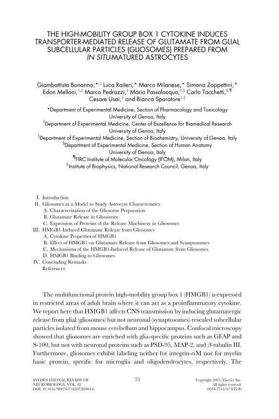
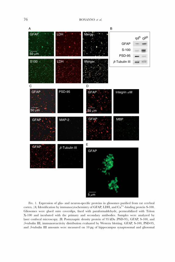
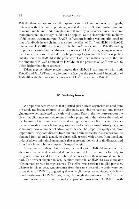
FIG. 1. Expression of glia- and neuron-specific proteins in gliosomes purified from rat cerebral
cortex. (A) Identification by immunocytochemistry of GFAP, LDH, and Ca2þ-binding protein S-100.
Gliosomes were glued onto coverslips, fixed with paraformaldehyde, permeabilized with Triton
X-100 and incubated with the primary and secondary antibodies. Samples were analyzed by
laser confocal microscopy. (B) Postsynaptic density protein of 95 kDa (PSD-95), GFAP, S-100, and
�-tubulin III, immunoreactivity distribution evaluated by Western blotting. GFAP, S-100, PSD-95,
and �-tubulin III amounts were measured on 10 mg of hippocampus synaptosomal and gliosomal
76 BONANNO et al.
HMGB1-INDUCED ASTROCITARY GLUTAMATE RELEASE 77
fraction) and between 10% and 20% Percoll® (synaptosomal fraction) were collect-
ed, washed by centrifugation, and resuspended in physiological medium.
Several studies have taken advantage of the characteristics of the gliosome
preparation to study functional aspects of glial cells. These studies allowed
identification of specific cell distribution, function, and molecular mechanisms
of a number of transmitter and modulator targets, mainly membrane transporters
(see for instance Daniels and Vickroy, 1999; Hirst et al., 1998; Pedrazzi et al., 2006;
Raiteri et al., 2005; Suchak et al., 2003). We characterized in detail gliosomes
purified from adult rat cerebral cortex, pointing out biochemical and morpholog-
ical evidence in support to the concept that our gliosomal fraction is largely
purified from synaptosomes (Stigliani et al., 2006). The confocal microscopy
images shown in Fig. 1A highlight the presence of immunoreactivity for two
proteins selectively expressed in astrocytes, the glial fibrillary acidic protein
(GFAP) and the Ca2þ-binding protein S-100, in purified gliosomes. Gliosomes
were labeled with anti-GFAP or anti S-100 and with anti-lactate dehydrogenase
(LDH) antibodies. Since LDH is an enzyme localized in the cytosol, it can be
assumed that the LDH-positive particles represent resealed gliosomes rather
than membrane debris. These experiments emphasize that the majority (about
90%) of LDH-labeled resealed particles in the gliosomal preparation were positive
for GFAP or for S-100. Also the synaptosomal preparation was extensively stained
by LDH, but it did not show substantial GFAP labeling, indicating low gliosome
contamination (not shown). Western blot experiments substantiated that glio-
somes are enriched with GFAP and S-100, while synaptosomes are enriched
with the neuronal markers postsynaptic density protein of 95 kDa (PSD-95) and
�-tubulin III, which are poorly represented in gliosomes (Fig. 1B). According to
these data, GFAP-positive gliosomes presented only a very modest positiveness for
antibodies raised against the neuronal markers PSD-95, microtubule-associated
protein 2 (MAP-2), or �-tubulin III (Fig. 1C), thus supporting the idea that
gliosomes represent a preparation with low synaptosomal contamination. Of
note, PSD-95, MAP-2, and �-tubulin III extensively marked synaptosomes
under the same experimental conditions (not shown). Moreover, GFAP-expres-
sing gliosomal preparation did not exhibit labeling either for integrin �M or for
myelin basic protein (MBP), two proteins selectively expressed in microglia and
oligodendrocytes, respectively (Fig. 1D). As a control, the antibodies used in the
proteins. (C) Identification by immunocytochemistry of GFAP, PSD-95, microtuble associated protein
type II (MAP-2), and �-tubulin III. Samples were analyzed by laser confocal microscopy. (D) Identifi-
cation by immunocytochemistry of GFAP, integrin �M, or myelin basic protein (MBP). Samples were
analyzed by laser confocal microscopy. (E) High-magnification tridimensional reconstruction of
purified gliosomal particles identified by GFAP immunocytochemistry. Samples were analyzed by
laser confocal microscopy. Modified from Stigliani et al. (2006), with the permission of Blackwell
Publishing Co.

78 BONANNO et al.
latter confocal microscopy experiments revealed a single band of the appropriate
molecular weight in Western blot experiments, using proteins extracted from
microglia primary cell cultures and from white matter of 19-day-old rats (not
shown). Finally, Fig. 1E shows a tridimensional reconstruction at higher-
magnification of GFAP-positive gliosomal particles: these particles appear to be
around 0.6-mm-diameter spheroidal organelles with a propensity to cluster in
physiological medium.
B. GLUTAMATE RELEASE IN GLIOSOMES
Functional experiments conducted in rat cerebral cortex showed that purified
gliosomes are able to take up and release glutamate when exposed to diVerentreleasing stimuli such as ionomycin or ATP. The release of [3H]D-Asp or endoge-
nous glutamate was studied taking advantage of the uniqueness of a superfusion
technique that we have used for several years, mainly with synaptosomes (Raiteri
and Raiteri, 2000). The system consists of several (up to 24) parallel superfusion
chambers thermostated at 37�C in which very thin layers (mostly monolayers) of
synaptosomes, plated on microporous filters, are up–down superfused in condi-
tions in which any released compound is immediately removed by the superfusion
fluid. Such a rapid removal prevents indirect eVects: in particular, the changes of
glutamate release observed following exposure to various agents are essentially
due to direct actions on excitatory amino acid releasing particles with minimal or
no involvement of neighboring elements. The fast taking away of released gluta-
mate does not allow (1) its reuptake and, therefore, its exchange with cytosolic
excitatory amino acid transporter substrates; and (2) its feedback on presynaptic
targets like release-regulating receptors. If substrates just released are virtually
absent from the particle biophase, release by Ca2þ-dependent exocytosis or
by Ca2þ-independent transporter-mediated release can be monitored under
appropriate conditions.
Ionomycin, a Ca2þ-selective ionophore capable to mediate Ca2þ influx with-
out voltage-sensitive Ca2þ channel activation and previously shown to induce
transmitter exocytosis from nerve terminals (Sanchez-Prieto et al., 1987; Verhage
et al., 1991), produced a substantial stimulus-evoked release of [3H]D-Asp or
endogenous glutamate (Stigliani et al., 2006). The release induced by ionomycin
was entirely dependent on the presence of external Ca2þ and significantly
decreased by bafilomycin-A1, a vesicle membrane V-type ATPase inhibitor
(Bowman et al., 1988; Floor et al., 1990), which is expected to prevent the
accumulation of the amino acid into vesicles (Moriyama and Futai, 1990;
Roseth et al., 1995).Under our experimental conditions, low concentrations of
ionomycin appeared to release even higher amounts of glutamate from gliosomes
than from synaptosomes. Conversely, synaptosomes were superior glutamate
HMGB1-INDUCED ASTROCITARY GLUTAMATE RELEASE 79
releasers when higher concentrations of the Ca2þ ionophore were applied. The
dependence on Ca2þ and the sensitivity to bafilomycin-A1 of the ionomycin-
evoked release of glutamate from gliosomes suggest that the stimulus-induced
release is exocytotic in nature. In line, experiments performed with the fluorescent
dye acridine orange (Zoccarato et al., 1999) showed that gliosomes were able to
accumulate the dye into acidic cytoplasmic organelles and that the application of
ionomycin induced fusion of these organelles with the plasma membrane that was
almost totally external Ca2þ dependent (Stigliani et al., 2006).
Gliosomes were able to release glutamate also when exposed to ATP in
superfusion. The ATP-induced glutamate release depended on the concentration
of the nucleotide used and was abolished by the selective P2 receptor antagonist
pyridoxal phosphate-6-azophenyl-20,40-disulfonic acid. The eVect of ATP was
only minimally aVected in the absence of external Ca2þ, but it was significantlydiminished by preloading gliosomes with 1,2-bis(2-aminophenoxy)ethane-N,N,N 0,N 0-tetraacetylmethylester (BAPTA-AM), a cytosolic Ca2þ chelator, suggesting the
involvement of Ca2þ released from intra-gliosomal stores, consequent to P2
receptor activation (Stigliani et al., 2006).
C. EXPRESSION OF PROTEINS OF THE RELEASE MACHINERY IN GLIOSOMES
Taken together, the release and fluorescence experiments described in
the preceding paragraph are consistent with the idea that gliosomes can release
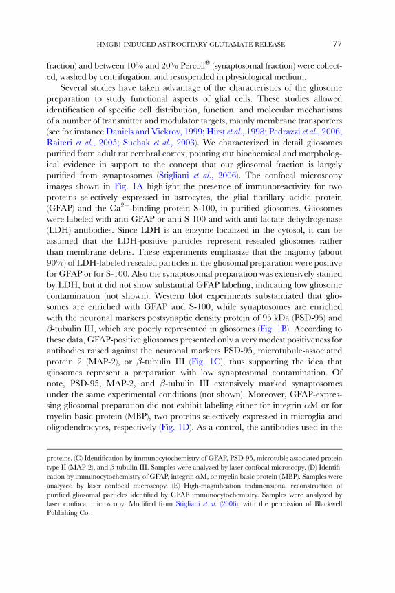
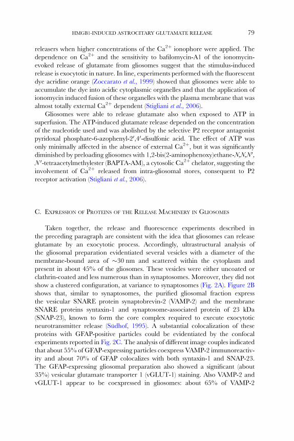
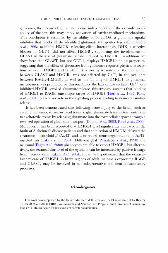
glutamate by an exocytotic process. Accordingly, ultrastructural analysis of
the gliosomal preparation evidentiated several vesicles with a diameter of the
membrane-bound area of �30 nm and scattered within the cytoplasm and
present in about 45% of the gliosomes. These vesicles were either uncoated or
clathrin-coated and less numerous than in synaptosomes. Moreover, they did not
show a clustered configuration, at variance to synaptosomes (Fig. 2A). Figure 2B
shows that, similar to synaptosomes, the purified gliosomal fraction express
the vesicular SNARE protein synaptobrevin-2 (VAMP-2) and the membrane
SNARE proteins syntaxin-1 and synaptosome-associated protein of 23 kDa
(SNAP-23), known to form the core complex required to execute exocytotic
neurotransmitter release (Sudhof, 1995). A substantial colocalization of these
proteins with GFAP-positive particles could be evidentiated by the confocal
experiments reported in Fig. 2C. The analysis of diVerent image couples indicated
that about 55% of GFAP-expressing particles coexpress VAMP-2 immunoreactiv-
ity and about 70% of GFAP colocalizes with both syntaxin-1 and SNAP-23.
The GFAP-expressing gliosomal preparation also showed a significant (about
35%) vesicular glutamate transporter 1 (vGLUT-1) staining. Also VAMP-2 and
vGLUT-1 appear to be coexpressed in gliosomes: about 65% of VAMP-2
Gliosomes
Synaptosomes
A
AstroGlio
GLAST
VAMP-2
GLT-1
Syntaxin-1
E
GFAP vGLUT-1
Merge
Merge
VAMP-2 vGLUT- 1
50 mm
D
SNAP-23
VAMP-2
Syntaxin-1
GlioSyn
B
GFAP VAMP-2 Merge
GFAP Syntaxin-1
SNAP-23GFAP Merge
Merge
50 mm
C
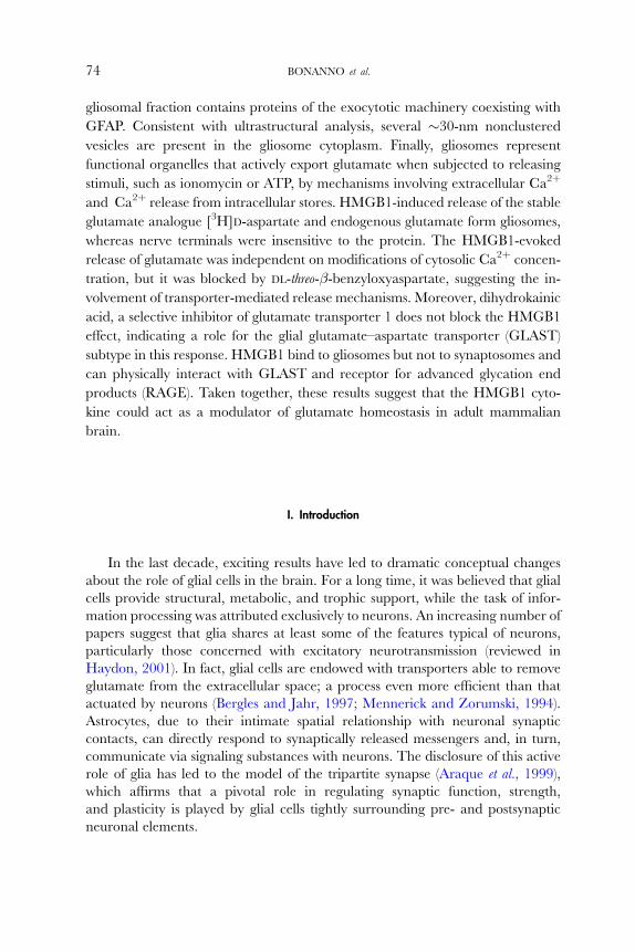
FIG. 2. Presence of cytosol vesicles and expression of proteins of the release machinery in the
gliosomal fraction purified from rat cerebral cortex. (A) Electron micrographs of gliosome (upper
80 BONANNO et al.
HMGB1-INDUCED ASTROCITARY GLUTAMATE RELEASE 81
colocalizes with the vesicular glutamate transporter. Conversely, almost the
totality of vGLUT-1 coexpresses with GFAP or VAMP-2.
Interestingly, expression of the SNARE complex proteins VAMP-2 and
sintaxin-1 and of the glial-specific glutamate transporters of type 1 (GLT-1) and
GLAST were much more enriched in gliosomes than in astrocytes in culture, as
outlined by the Western blot experiments reported in Fig. 2E. It is reasonable to
assume that during brain tissue homogenization gliosomes are formed by a
process similar to that originating synaptosomes, that is, they are ‘‘pinched oV ’’particles coming from glia cell arborizations. It has been proposed that astrocytes
possess dedicated regions at the processes surrounding the synapses by which they
sense the neuronal messengers for a point-to-point neuron to astrocyte communi-
cation. Astrocytes have been suggested to release transmitters from these
specialized areas (Araque et al., 1999; Carmignoto, 2000; Grosche et al., 1999).
Accordingly, a number of evidences indicate that the vesicular release sites of
astrocytes might be situated at the processes rather that at the cell bodies
(reviewed by Montana et al., 2006). It could be proposed that the gliosomal
preparation is enriched with these specific areas, where the release machinery
of the glial cell should be concentrated.
III. HMGB1-Induced Glutamate Release from Gliosomes
A. CYTOKINE PROPERTIES OF HMGB1
Cytokines are a class of small proteins acting via cell surface G-protein–
coupled receptors that mediate diverse metabolic and immunological responses
in other cells (Nathan, 1987). They were first identified as inflammatory mediators
panels) and synaptosome (lower panels) fractions. Purified gliosomes and synaptosomes were fixed,
dehydrated, and embedded in LX112. Ultrathin sections were stained with uranyl acetate and lead
citrate, and analyzed with a FEI CM10 electron microscope. (B) Synaptobrevin 2 (VAMP-2), syntaxin-1,
or synaptosome-associated protein of 23 kDa (SNAP-23) immunoreactivity distribution evaluated by
Western blotting. VAMP-2, syntaxin-1, and SNAP-23 amounts were measured on 10 mg of hippocampus
synaptosomal and gliosomal proteins. (C) Immunocytochemical identification of GFAP, VAMP-2, syn-
taxin-1, and SNAP-23 immunoreactivity in gliosomes. Gliosomes were glued onto coverslips, fixed with
paraformaldehyde, permeabilized with Triton X-100, and incubated with the primary and secondary
antibodies. Samples were analyzed by laser confocal microscopy. (D) Immunocytochemical identification
of GFAP, VAMP-2, and the vesicular glutamate transporter 1 (vGLUT-1) in gliosomes. Samples were
analyzed by laser confocal microscopy. (E) VAMP-2, syntaxin-1, glutamate–aspartate transporter
(GLAST), or glutamate transporter 1 (GLT-1) immunoreactivity distribution evaluated by Western
blotting. VAMP-2, syntaxin-1, GLAST, and GLT-1 amounts were measured on 10 mg of proteins of
cultured astrocytes or gliosomes. Modified from Stigliani et al. (2006), with the permission of Blackwell
Publishing Co.
−1
0
1
2
3
4
5
0.1
[3H
] D-A
sp r
elea
se(p
erce
ntag
e of
ove
rflo
w)
[3H
] D-A
sp r
elea
se(p
erce
ntag
e of
ove
rflo
w)
1 100.01
SynaptosomesGliosomes
0
1
2
3
4
5
3 nM HMGB1Ca2+-free100-mM BAPTA-AM10-mM DL-TBOA50-mM DHK
**
Box A Box B Acidictail
Am
yloi
doge
nic
(12−
27)
Pro
infla
mm
ator
yac
tivity
(89
−108
)
“ME
L C
ell”
bioa
ctiv
epe
ptid
e (1
30−1
39)
RA
GE
-bin
ding
mot
if (1
51−1
83)
Hep
arin
-bin
ding
mot
if (
6−9)
9 85 88 162 216
End
ogen
ous
glut
amat
e re
leas
e(n
mol
/mg
of p
rote
in)
0
1
2
3
4
3 nM HMGB1Ca2+-free10-mM DL-TBOA
**
B
D
C
A
+ +− +− −− −− −
+ +− −− +− −− −
+ +− −− −− +− −
+ +− −− −− −− +
0.3−−
1−−
3−−
3+−
3−+
Helix 1 Helix 2 Helix 3 Helix 4 Helix 5 Helix 6
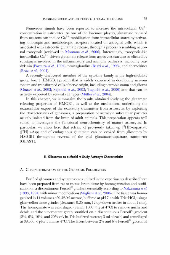
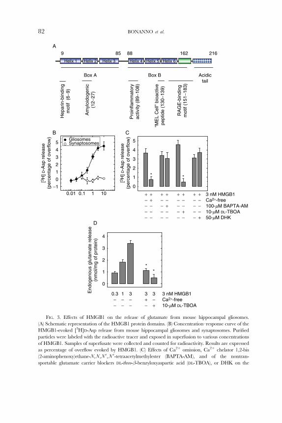
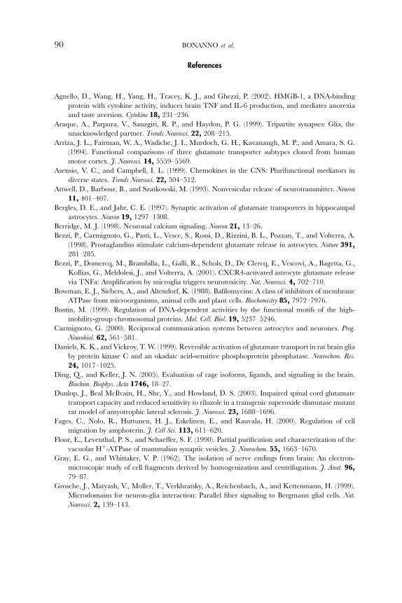
FIG. 3. EVects of HMGB1 on the release of glutamate from mouse hippocampal gliosomes.
(A) Schematic representation of the HMGB1 protein domains. (B) Concentration–response curve of the
HMGB1-evoked [3H]D-Asp release from mouse hippocampal gliosomes and synaptosomes. Purified
particles were labeled with the radioactive tracer and exposed in superfusion to various concentrations
of HMGB1. Samples of superfusate were collected and counted for radioactivity. Results are expressed
as percentage of overflow evoked by HMGB1. (C) EVects of Ca2þ omission, Ca2þ chelator 1,2-bis
(2-aminophenoxy)ethane-N,N,N 0,N 0-tetraacetylmethylester (BAPTA-AM), and of the nontran-
sportable glutamate carrier blockers DL-threo-�-benzyloxyaspartic acid (DL-TBOA), or DHK on the
82 BONANNO et al.
HMGB1-INDUCED ASTROCITARY GLUTAMATE RELEASE 83
of leukoc yte chemota xis and have been subseq uently show n to possess a much
larger repertoire (Rossi and Zlotnik, 2000 ). Various chemok ines and their recep-
tors are widely expressed in the bra in, in some cases from early em bryonic stages
through out adult hood (A sensio and Campb ell, 1999 ). For this re ason, it has been
suggest ed that they m ay ha ve a role in neur odegene ration disease states (A sensio
and Campbel l, 1999; Miller and Me ucci, 1999 ) in ad dition to their infl ammat ory
and immun ological roles.
A disco very by Wang et al. (19 99) reveal ed that HMGB1 is a cytokine.
HMGB1 has been disco vered more than 30 year s ago as a nuc lear-bind ing
protein that is widely express ed and extrem ely conserved in mammal s.
HMGB1 is an arch itectu ral prot ein: it can bend DNA to promote nuc leoprotein
intera ctions and facili tate all sort s of DNA transact ions. The protein (25 kDa)
is structured in three doma ins: two basic HMG-b ox doma ins (box A and box B)
and a long acid ic C-termin al tail ( Bustin, 1999; Tho mas and Traver s, 2001)
(Fig. 3A).
HMGB1 can be actively released from a variety of cells, inclu ding m acro-
phage, pituicy tes, human prima ry periphera l blood m ononuclear ce lls, and
murin e erythroleu kemia cells (Mu ller et al., 2004). Once re leased, HMGB1 can
bind to cell surface re ceptors, such as the re ceptor for advanced glycation end
prod ucts (RAGE), Toll-like rece ptor 2, and Tol l-like receptor 4, to transduc e
signal s that elic it cellular re sponses inclu ding feve r, epith elia barri er dys function ,
chemota ctic cell movemen t, re lease o f proinflam matory cytokin es (e.g., tumor
necros is factor and interleuk in-1� ), and acu te inflamma tion (O’Connor et al.,
2003; Wang et al ., 2004 ). Pr oinflamma tory e Vec ts of H MGB1, ident ified in speci fic
structures of the perip heral and the centra l nervous system , have support ed the
concep t that this protein m ay operat e thro ugh direct activa tion o f signali ng
cascad es, comm only utilized by classic proinfla mmatory cyt okines, or indirectly
by inducing the rele ase of these cytokines, also in the brain (Agnel lo et al. , 20 02;
O’Conn or et al. , 2003; Rong et al. , 2004 ). A lthough HMGB1 is widely expressed in
the dev eloping centr al nervous system , the ad ult bra in maintain s high levels of
HMGB1 only in spe cific areas (Guazzi et al., 2003 ).
We report here tha t extracel lular HM GB1 selec tively m odulates the re lease of
glutam ate in the centra l nervous sys tem by m onitoring the release of excitat ory
amino acid in purified synaptosomes (Gray and Whittaker, 1962) and gliosomes
(Nakamura et al., 1993; Stigliani et al., 2006) prepared from mouse hippocampus
and cerebellum.
HMGB1-evoked [3H]D-Asp release from gliosomes. (D). HMGB1-evoked release of endogenous gluta-
mate from gliosomes and eVects of Ca2þ omission or DL-TBOA. Glutamate was measured by high
performance liquid chromatography separation and fluorimetric detection. Modified from Pedrazzi et al.
(2006), with the permission of Blackwell Publishing Co.
84 BONANNO et al.
B. EFFECT OF HMGB1 ON GLUTAMATE RELEASE FROM GLIOSOMES
AND SYNAPTOSOMES
The level of purification of gliosomal- and synaptosomal-enriched fractions here
isolated from adult mouse cerebellum and hippocampus was studied by confocal
microscopy and Western blotting, as described above in rat cerebral cortex. Studies
aimed to detect the proportion of particles expressing the glial-specificmarkerGFAP
or the neuronal markersMAP-2 and �-tubulin III indicated that nomore than 15%
GFAP labeling was present in the synaptosomal fraction and, conversely, that a
maximum of 20% MAP-2 or �-tubulin III labeling was present in gliosomal
preparations, both in hippocampus and cerebellum (Pedrazzi et al., 2006).
To directly compare the ability of HMGB1 to modulate glutamate release,
synaptosomes and gliosomes were prepared from adult mouse hippocampus or
cerebellum. The preparations were labeled with the stable glutamate analogue
[3H]D-Asp and exposed to HMGB1 in superfusion. HMGB1 stimulated the release
of [3H]D-Asp in a concentration-dependent manner from hippocampal (Fig. 3B) or
cerebellar (not shown) gliosomes. The two concentration–response curves appeared
similar: the maximal potentiation of release was obtained around 3-nM HMGB1
and the EC50 amounted to 0.57 � 0.043 nM and to 0.32 � 0.041 nM in the case
of hippocampal and cerebellar gliosomes, respectively. By contrast, no significant
increase of [3H]D-Asp eZux was observed in synaptosomes under the same experi-
mental conditions. These findings indicate that nanomolar concentrations of ex-
tracellular HMGB1 are suYcient to elicit glutamate release from gliosomes and
that this activity of HMGB1 is specifically directed to glial and not to neuronal
preparations from the same regions of adult mouse brain.
C. MECHANISMS OF THE HMGB1-INDUCED RELEASE OF GLUTAMATE
FROM GLIOSOMES
It is well known that neurotransmitter release may be due to Ca2þ-dependentor Ca2þ-independent mechanisms (Attwell et al., 1993; Berridge, 1998; Levi and
Raiteri, 1993). To determine whether the eZux [3H]D-Asp promoted by HMGB1
involved vesicular exocytosis, which is known to require the entry of Ca2þ from
the external milieu or mobilization of Ca2þ from internal stores to the cytosol
(Berridge, 1998), the eVect of HMGB1 was studied either by omitting Ca2þ from
the extracellular medium or by using gliosomes pretreated with BAPTA-AM.
As shown in Fig. 3C, omission of external Ca2þ largely prevented the HMGB1
eVect, suggesting the occurrence of exocytotic release. Surprisingly, in the presenceof external Ca2þ, the HMGB1-evoked [3H]D-Asp release was not significantly

HMGB1-INDUCED ASTROCITARY GLUTAMATE RELEASE 85
modified in gliosomes pretreated with BAPTA-AM, thus opposing the exocytotic
origin of the process. The lack of eVect observed with BAPTA-AM does not look
as to be due to the incapability of the Ca2þ chelator to remove cytosolic Ca2þ in
the presence of 1.2-mM external Ca2þ. In fact, in another set of experiments,
exposure of hippocampal gliosomes to ATP resulted in a stimulation of the
radioactive tracer release; an eVect reduced to about 60% by BAPTA-AM in
the presence of external Ca2þ. The discrepancy between external Ca2þ depen-
dency and BAPTA-AM independency of the HMGB1-evoked [3H]D-Asp release
prompted us to check the eVect of the nontransportable blocker of glutamate
uptake DL-threo-�-benzyloxyaspartate (DL-TBOA) on the amino acid release eli-
cited by HMGB1. As shown in Fig. 3C, DL-TBOA completely abolished the
eZux of [3H]D-Asp promoted by HMGB1, suggesting that this response is due to
a mechanism mediated by reversal of glutamate transporters (Attwell et al., 1993;
Levi and Raiteri, 1993). Interestingly, dihydrokainic acid (DHK), a selective
inhibitor of GLT-1 glutamate transporter (Arriza et al., 1994) did not significantly
modify the HMGB1-induced [3H]D-Asp overflow.
Due to occurrence of such an ambiguous scenario, these data were confirmed
in a set of experiments carried out to measure endogenous glutamate release in
hippocampus and the eVect of HMGB1. As shown in Fig. 3D, HMGB1 also
stimulated the spontaneous release of endogenous glutamate from hippocampal
gliosomes. The enhancement due the cytokine was strongly reduced (about 70%)
in the absence of external Ca2þ and abolished in the presence of the glutamate
transport blocker DL-TBOA, as in the case of the radioactive tracer.
In sum, the results with [3H]D-Asp and endogenous glutamate suggest that the
release from hippocampal gliosomes exposed to exogenous HMGB1 involves a
carrier-mediated process and concurrently is dependent on external Ca2þ. How-
ever, since carrier-mediated release of neurotransmitters does not depend on
external Ca2þ (Attwell et al., 1993; Levi and Raiteri, 1993), the origin of this
discrepancy has been further explored.
D. HMGB1 BINDING TO GLIOSOMES
To determine whether the lack of HMGB1-induced release of glutamate,
observed in the absence of external Ca2þ, was a consequence of a real Ca2þ
requirement for the amino acid export or depended on altered gliosomes–
HMGB1 interaction in this condition, we measured the binding of [125I]
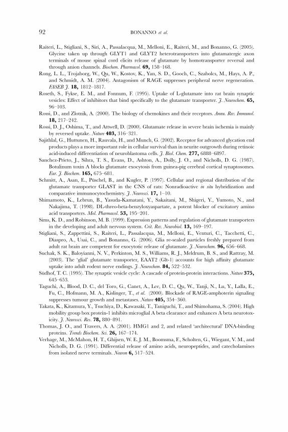
HMGB1 to gliosomes in the absence or in the presence of external Ca2þ. Asshown in Fig. 4A, the labeled protein bound to gliosomal hippocampal particles
and the binding with these particles was significantly reduced when external Ca2þ
was omitted. These results suggest that the interaction of exogenous HMGB1

GLAST
GLT1
IgG heavy chain
IgG heavy chain
IgG light chain
Ip HMGB1
Ip HMGB1
Ip GLASTHMGB1
Gliosomes
+ Ca2+ − Ca2+
Ip Sur Ip SurC
RAGE
0
20
40
60
80
100
Synaptosomes Gliosomes
Gliosomes
HMGB1-bound RAGE
0
20
40
60
80
100
*
+ Ca2+ − Ca2+
ED
HM
GB
1-sp
ecifi
c bi
ndin
g(p
mol
/mg
prot
ein)
0.0
0.5
1.0
1.5
2.0
2.5
3.0
1.2-mM C
a2+
12-mM C
a2+
Ca2+ -fr
ee medium
*
*
1.2-mM C
a2+
GliosomesSynaptosomes
Ca2+ -fr
ee medium
GLAST GLT1
020406080
100
*
Synaptosomes Gliosomes
02040
8060
100
*
A B
1 2 3 4
FIG. 4. Binding properties of HMGB1 to mouse hippocampal gliosomes. (A) Binding of [125I]-
HMGB1 to gliosomes and synaptosomes. The specific binding of the radiolabeled protein to intact
gliosomes or synaptosomes was evaluated in the absence or the presence of 100-fold molar excess of
unlabeled HMGB1 in a standard medium containing 1.2-mM CaCl2 or 12-mM CaCl2 or in a Ca2þ-free medium. (B) Glutamate–aspartate transporter (GLAST) or glutamate transporter 1 (GLT-1) dis-
tribution in synaptosomes and gliosomes evaluated by Western blotting. GLASTand GLT-1 amounts
were measured on 10 mg proteins. (C) HMGB1-binding properties to GLAST or GLT-1 evaluated
86 BONANNO et al.

HMGB1-INDUCED ASTROCITARY GLUTAMATE RELEASE 87
with the glios ome surf ace is prom oted by Ca 2þ, even at micro molar concen tra-
tions . In contra st, synaptoso mes, purifi ed from the same bra in area s, bou nd very
low amounts of the cytokine both in the prese nce or in the absen ce of Ca 2þ
(Fig. 4A), suggestin g that Ca 2þ- activated bindin g sit es for HMGB1 are prefe ren-
tially conta ined in glial particles , in compar ison with nerv e endings.
Only two glutam ate transporter s have been re ported to be expressed in glial
cells, nam ely, GLAST an d GLT-1 (Schmitt et al ., 1997; Sims and Rob inson,
1999 ). As shown in Fig. 4B , co mparison of the amounts of GLA ST and GLT-1,
detect ed by immun oblotting in glios ome and synaptoso me prepar ations, con-
firmed that these transporter s are exclusiv ely (GLA ST) or prefe rentially (GLT-1)
represe nted in the gliosome fraction. The quanti fication of i mmunorea ctive
signal s, obtained with diV erent prepar ations, revea led a 4.5- an d 1.8-fold higheramou nt of GLA ST and GLT-1 in glios omes tha n in syna ptosome s, respe ctively.
We next sought to determin e whether glutama te transporte rs could directl y
intera ct w ith HMGB1 and if Ca 2þ influenced these protein –protein bindings .Figur e 4C sh ows that the GLAST co immunopreci pitated w ith HMGB1 and that
the amounts of GLAST recovered in the HM GB1 coimmun oprecipitat ed m ate-
rial in the presence (lane 1) or the ab sence (lane 3) of Ca 2þ were not sig nificantlydi Verent. By contra st, GLT-1 was detectab le only in the pool of prot eins not
coimm unoprecip itated wi th HMGB1 (lanes 2 and 4). We also examin ed whether
HMGB1 could be coimmun oprecipitat ed using an anti-GLAS T ant ibody. Also in
this condit ion, coimmun oprecip itation of HMGB1 an d GLAST could be
detect ed (Fig. 4C bottom, lanes 1 and 3), co nfirming the physic al interac tion
between the two prot eins. These find ings indicate tha t HMGB1 can associate
with GLAS T, but not with GLT- 1, and tha t this protein–prot ein intera ction is not
a Vected by Ca 2þ .To clarify wheth er the Ca 2þ-promot ed binding of extra cellular H MGB1 to
the gliosom e membra ne co uld be due to the possible par ticipation o f other
HMGB1-b inding protein s, the involvemen t of RAGE was test ed. A report
showe d tha t brain conta ins sever al splice var iants of RAGE, having transmem -
brane or cyt oplasmic localiza tion (Din g and Keller, 200 5). We evaluate d, at first,
the amount of membrane -bound RAGE in glios ome and syna ptosome prepara -
tions . As sh own in Fig. 4D , glios ome membra nes show ed highe r levels of
Western blotting. HMGB1 was added to detergent soluble membrane extracts and immunoprecipita-
tion of HMGB1 (IP HMGB1) or GLAST (IP GLAST) was carried out. (D) Receptor for advanced
glycosilation end product (RAGE) distribution in synaptosomes and gliosomes evaluated by Western
blotting. RAGE amount was measured on 10-mg proteins. (E) HMGB1-binding properties to RAGE
evaluated by Western blotting. RAGE bound to Sepharose®-HMGB1 was measured in the presence
or in the absence of 1.2-mM CaCl2. Modified from Pedrazzi et al. (2006), with the permission of
Blackwell Publishing Co.
88 BONANNO et al.
RAGE than synaptosomes: the quantification of immunoreactive signals,
obtained with diVerent preparations, revealed a 2.7- to 3.0-fold higher amount
of membrane-bound RAGE in gliosomes than in synaptosomes. Since the coim-
munoprecipitation strategy could not be applied, as the electrophoretic mobility
of full-length transmembrane RAGE in Western blotting was superimposed to
that of antibody heavy chain, to measure the eVect of Ca2þ on HMGB1–RAGE
interaction, HMGB1 was bound to Sepharose® beads and its RAGE-binding
properties measured in the absence or presence of Ca2þ using detergent-soluble
membrane fractions extracted from hippocampal gliosomes. RAGE was prefer-
entially bound by HMGB1 in the presence of Ca2þ than in the absence of the ion:
the amount of RAGE retained by HMGB1 in the presence of Ca2þ was 3.5- to
4-fold higher than in its absence.
Taken together these results suggest that HMGB1 can interact with both
RAGE and GLAST on the gliosome surface but the preferential interaction of
HMGB1 with gliosomes in the presence of Ca2þ is driven by RAGE.
IV. Concluding Remarks
We reported here evidence that purified glial derived organelles isolated from
the adult rat brain, referred as to gliosomes, are able to take up and release
glutamate when subjected to a variety of stimuli. Data in the literature support the
view that gliosomes may represent a viable preparation that allows the study of
mechanisms of transmitter release and its regulation in adult astrocytes. Besides
the obvious diVerences between gliosomes and intact cultured astrocytes, glio-
somes may have a number of advantages: they can be prepared rapidly and, most
importantly, originate directly from mature brain astrocytes. Gliosomes can be
obtained from animals acutely or chronically treated with drugs, from knockout
or knockdown animals, from animals that represent models of brain diseases, and
from fresh human brain samples of surgical origin.
In keeping with these observations, the results with HMGB1 underline that
gliosomes are a vital in vitro glial preparation, able to respond selectively to
exogenous stimuli and to reveal subtle diVerences from their neuronal counter-
part. The present chapter, in fact, identifies extracellular HMGB1 as a stimulator
of glutamate release from gliosomes. This eVect was restricted to glial particles,
whereas in this respect, synaptosomes from the same areas of the brain were not
susceptible to HMGB1, suggesting that only gliosomes are equipped with func-
tional mediators of HMGB1 signaling. Although the presence of Ca2þ in the
external medium is required in order to promote association of HMGB1 with
HMGB1-INDUCED ASTROCITARY GLUTAMATE RELEASE 89
gliosomes, the release of glutamate occurs independently of the cytosolic avail-
ability of the ion; this may imply activation of carrier-mediated mechanisms.
This conclusion is sustained by the ability of DL-TBOA, a glutamate uptake
inhibitor that blocks all the identified glutamate transporter types (Shimamoto
et al., 1998), to inhibit HMGB1 releasing eVect. Interestingly, DHK, a selective
blocker of GLT-1, did not aVect HMGB1, supporting the involvement of
GLAST in the rise of glutamate release induced by HMGB1. In addition, we
show here that GLAST, but not GLT-1, displays HMGB1-binding properties,
suggesting that the eZux of glutamate from gliosomes requires physical associa-
tion between HMGB1 and GLAST. It is worthy to note that the interaction
between GLAST and HMGB1 was not aVected by Ca2þ, in contrast, that
between RAGE–HMGB1, as well as the binding of HMGB1 to gliosomal
membranes, was promoted by this ion. Since the lack of extracellular Ca2þ also
inhibited HMGB1-evoked glutamate release, this strongly suggests that binding
of HMGB1 to RAGE, one major target of HMGB1 (Hori et al., 1995; Rong
et al., 2004), plays a key role in the signaling process leading to neurotransmitter
release.
It has been demonstrated that following acute injury to the brain, such as
cerebral ischemia, stroke, or head trauma, glial glutamate transporters contribute
to excitotoxic events by releasing glutamate into the extracellular space through a
reversed operation of glutamate transport (Dunlop et al., 2003; Rossi et al., 2000).
Moreover, it has been reported that HMGB1 level significantly increased in the
brain of Alzheimer’s disease patients and that coinjection of HMGB1 delayed the
clearance of amyloid-� (A�42) and accelerated neurodegeneration in A�42-injected rats (Takata et al., 2004). DiVerent glial (Passalacqua et al., 1998) and
neuronal (Fages et al., 2000) phenotypes are able to export HMGB1, but alterna-
tively, the extracellular level of the cytokine can be increased by passive leakage
from necrotic cells (Takata et al., 2004). It can be hypothesized that the extracel-
lular release of HMGB1, in brain regions of adult mammals expressing RAGE
and GLAST, may be involved in neurodegenerative and neuroinflammatory
processes.
Acknowledgments
This work was supported by the Italian Ministero dell’Istruzione, dell’Universita e della Ricerca
(PRIN 2002 and 2004), FIRB (Post-Genoma and Neuroscience Projects), and University of Genoa. We
thank Ms. Maura Agate for her excellent secretarial assistance.
90 BONANNO et al.
References
Agnello, D., Wang, H., Yang, H., Tracey, K. J., and Ghezzi, P. (2002). HMGB-1, a DNA-binding
protein with cytokine activity, induces brain TNF and IL-6 production, and mediates anorexia
and taste aversion. Cytokine 18, 231–236.
Araque, A., Parpura, V., Sanzgiri, R. P., and Haydon, P. G. (1999). Tripartite synapses: Glia, the
unacknowledged partner. Trends Neurosci. 22, 208–215.
Arriza, J. L., Fairman, W. A., Wadiche, J. I., Murdoch, G. H., Kavanaugh, M. P., and Amara, S. G.
(1994). Functional comparisons of three glutamate transporter subtypes cloned from human
motor cortex. J. Neurosci. 14, 5559–5569.
Asensio, V. C., and Campbell, I. L. (1999). Chemokines in the CNS: Plurifunctional mediators in
diverse states. Trends Neurosci. 22, 504–512.
Attwell, D., Barbour, B., and Szatkowski, M. (1993). Nonvesicular release of neurotransmitter. Neuron
11, 401–407.
Bergles, D. E., and Jahr, C. E. (1997). Synaptic activation of glutamate transporters in hippocampal
astrocytes. Neuron 19, 1297–1308.
Berridge, M. J. (1998). Neuronal calcium signaling. Neuron 21, 13–26.
Bezzi, P., Carmignoto, G., Pasti, L., Vesce, S., Rossi, D., Rizzini, B. L., Pozzan, T., and Volterra, A.
(1998). Prostaglandins stimulate calcium-dependent glutamate release in astrocytes. Nature 391,
281–285.
Bezzi, P., Domercq, M., Brambilla, L., Galli, R., Schols, D., De Clercq, E., Vescovi, A., Bagetta, G.,
Kollias, G., Meldolesi, J., and Volterra, A. (2001). CXCR4-activated astrocyte glutamate release
via TNFa: Amplification by microglia triggers neurotoxicity. Nat. Neurosci. 4, 702–710.
Bowman, E. J., Siebers, A., and Altendorf, K. (1988). Bafilomycins: A class of inhibitors of membrane
ATPase from microorganisms, animal cells and plant cells. Biochemistry 85, 7972–7976.
Bustin, M. (1999). Regulation of DNA-dependent activities by the functional motifs of the high-
mobility-group chromosomal proteins. Mol. Cell. Biol. 19, 5237–5246.
Carmignoto, G. (2000). Reciprocal communication systems between astrocytes and neurones. Prog.
Neurobiol. 62, 561–581.
Daniels, K. K., and Vickroy, T. W. (1999). Reversible activation of glutamate transport in rat brain glia
by protein kinase C and an okadaic acid-sensitive phosphoprotein phosphatase. Neurochem. Res.
24, 1017–1025.
Ding, Q., and Keller, J. N. (2005). Evaluation of rage isoforms, ligands, and signaling in the brain.
Biochim. Biophys. Acta 1746, 18–27.
Dunlop, J., Beal McIlvain, H., She, Y., and Howland, D. S. (2003). Impaired spinal cord glutamate
transport capacity and reduced sensitivity to riluzole in a transgenic superoxide dismutase mutant
rat model of amyotrophic lateral sclerosis. J. Neurosci. 23, 1688–1696.
Fages, C., Nolo, R., Huttunen, H. J., Eskelinen, E., and Rauvala, H. (2000). Regulation of cell
migration by amphoterin. J. Cell Sci. 113, 611–620.
Floor, E., Leventhal, P. S., and SchaeVer, S. F. (1990). Partial purification and characterization of the
vacuolar Hþ-ATPase of mammalian synaptic vesicles. J. Neurochem. 55, 1663–1670.
Gray, E. G., and Whittaker, V. P. (1962). The isolation of nerve endings from brain: An electron-
microscopic study of cell fragments derived by homogenization and centrifugation. J. Anat. 96,
79–87.
Grosche, J., Matyash, V., Moller, T., Verkhratsky, A., Reichenbach, A., and Kettenmann, H. (1999).
Microdomains for neuron-glia interaction: Parallel fiber signaling to Bergmann glial cells. Nat.
Neurosci. 2, 139–143.

HMGB1-INDUCED ASTROCITARY GLUTAMATE RELEASE 91
Guazzi, S., Strangio, A., Franzi, A. T., and Bianchi, M. E. (2003). HMGB1, an architectural
chromatin protein and extracellular signaling factor, has a spatially and temporally restricted
expression pattern in mouse brain. Gene Expr. Patterns 3, 29–33.
Haydon, P. G. (2001). Glia: Listening and talking to the synapse. Nat. Rev. Neurosci. 2, 185–193.
Hirst, W. D., Price, G. W., Rattray, M., and Wilkin, G. P. (1998). Serotonin transporters in adult rat
brain astrocytes revealed by [3H]5-HTuptake into glial plasmalemmal vesicles. Neurochem. Int. 33,
11–22.
Hori, O., Brett, J., Slattery, T., Cao, R., Zhang, J., Chen, J. X., Nagashima, M., Lundh, E. R.,
Vijay, S., Nitecki, D., Morser, J., Stern, D., et al. (1995). The receptor for advanced glycation end
products (RAGE) is a cellular binding site for amphoterin. Mediation of neurite outgrowth and
co-expression of RAGE and amphoterin in the developing nervous system. J. Biol. Chem. 270,
25752–25761.
Levi, G., and Raiteri, M. (1993). Carrier-mediated release of neurotransmitters. Trends Neurosci. 16,
415–419.
Mennerick, S., and Zorumski, C. F. (1994). Glial contributions to excitatory neurotransmission in
cultured hippocampal cells. Nature 368, 59–62.
Miller, R. J., and Meucci, O. (1999). AIDS and the brain: Is there a chemokine connection? Trends
Neurosci. 22, 471–479.
Montana, V., Malarkey, E. B., Verderio, C., Matteoli, M., and Parpura, V. (2006). Vesicular transmit-
ter release from astrocytes. Glia 54, 700–715.
Moriyama, Y., and Futai, M. (1990). H(þ)-ATPase, a primary pump for accumulation of neurotrans-
mitters, is a major constituent of brain synaptic vesicles. Biochem. Biophys. Res. Commun. 173,
443–448.
Muller, S., Ronfani, L., and Bianchi, M. E. (2004). Regulated expression and subcellular localization of
HMGB1, a chromatin protein with a cytokine function. J. Intern. Med. 255, 332–343.
Nakamura, Y., Iga, K., Shibata, T., Shudo, M., and Kataoka, K. (1993). Glial plasmalemmal vesicles:
A subcellular fraction from rat hippocampal homogenate distinct from synaptosomes. Glia 9,
48–56.
Nakamura, Y., Kubo, H., and Kataoka, K. (1994). Uptake of transmitter amino acids by glial
plasmalemmal vesicles from diVerent regions of rat central nervous system. Neurochem. Res. 19,
1145–1150.
Nathan, C. F. (1987). Secretory products of macrophages. J. Clin. Invest. 79, 319–326.
O’Connor, K. A., Hansen, M. K., Rachal Pugh, C., Deak, M. M., Biedenkapp, J. C., Milligan, E. D.,
Johnson, J. D., Wang, H., Maier, S. F., Tracey, K. J., and Watkins, L. R. (2003). Further
characterization of high mobility group box 1 (HMGB1) as a proinflammatory cytokine: Central
nervous system eVects. Cytokine 24, 254–265.Parpura, V., Basarasky, T. A., Liu, F., Jeftinija, K., Jeftinija, S., and Haydon, P. G. (1994). Glutamate-
mediated astrocyte-neuron signalling. Nature 369, 744–747.
Passalacqua, M., Patrone, M., Picotti, G. B., Del Rio, M., Sparatore, B., Melloni, E., and
Pontremoli, S. (1998). Stimulated astrocytes release high-mobility group 1 protein, an inducer
of LAN-5 neuroblastoma cell diVerentiation. Neuroscience 82, 1021–1028.
Pedrazzi, M., Raiteri, L., Bonanno, G., Patrone, M., Ledda, S., Passalacqua, M., Milanese, M.,
Melloni, E., Raiteri, M., Pontremoli, S., and Sparatore, B. (2006). Stimulation of excitatory amino
acid release from adult mouse brain glia subcellular particles by high mobility group box 1 protein.
J. Neurochem. 99, 827–838.
Raiteri, L., and Raiteri, M. (2000). Synaptosomes still viable after 25 years of superfusion. Neurochem.
Res. 25, 1265–1274.
92 BONANNO et al.
Raiteri, L., Stigliani, S., Siri, A., Passalacqua, M., Melloni, E., Raiteri, M., and Bonanno, G. (2005).
Glycine taken up through GLYT1 and GLYT2 heterotransporters into glutamatergic axon
terminals of mouse spinal cord elicits release of glutamate by homotransporter reversal and
through anion channels. Biochem. Pharmacol. 69, 158–168.
Rong, L. L., Trojaborg, W., Qu, W., Kostov, K., Yan, S. D., Gooch, C., Szabolcs, M., Hays, A. P.,
and Schmidt, A. M. (2004). Antagonism of RAGE suppresses peripheral nerve regeneration.
FASEB J. 18, 1812–1817.
Roseth, S., Fykse, E. M., and Fonnum, F. (1995). Uptake of L-glutamate into rat brain synaptic
vesicles: EVect of inhibitors that bind specifically to the glutamate transporter. J. Neurochem. 65,
96–103.
Rossi, D., and Zlotnik, A. (2000). The biology of chemokines and their receptors. Annu. Rev. Immunol.
18, 217–242.
Rossi, D. J., Oshima, T., and Attwell, D. (2000). Glutamate release in severe brain ischemia is mainly
by reversed uptake. Nature 403, 316–321.
Sajithlal, G., Huttunen, H., Rauvala, H., and Munch, G. (2002). Receptor for advanced glycation end
products plays a more important role in cellular survival than in neurite outgrowth during retinoic
acid-induced diVerentiation of neuroblastoma cells. J. Biol. Chem. 277, 6888–6897.
Sanchez-Prieto, J., Sihra, T. S., Evans, D., Ashton, A., Dolly, J. O., and Nicholls, D. G. (1987).
Botulinum toxin A blocks glutamate exocytosis from guinea-pig cerebral cortical synaptosomes.
Eur. J. Biochem. 165, 675–681.
Schmitt, A., Asan, E., Puschel, B., and Kugler, P. (1997). Cellular and regional distribution of the
glutamate transporter GLAST in the CNS of rats: Nonradioactive in situ hybridization and
comparative immunocytochemistry. J. Neurosci. 17, 1–10.
Shimamoto, K., Lebrun, B., Yasuda-Kamatani, Y., Sakaitani, M., Shigeri, Y., Yumoto, N., and
Nakajima, T. (1998). DL-threo-beta-benzyloxyaspartate, a potent blocker of excitatory amino
acid transporters. Mol. Pharmacol. 53, 195–201.
Sims, K. D., and Robinson, M. B. (1999). Expression patterns and regulation of glutamate transporters
in the developing and adult nervous system. Crit. Rev. Neurobiol. 13, 169–197.
Stigliani, S., Zappettini, S., Raiteri, L., Passalacqua, M., Melloni, E., Venturi, C., Tacchetti, C.,
Diaspro, A., Usai, C., and Bonanno, G. (2006). Glia re-sealed particles freshly prepared from
adult rat brain are competent for exocytotic release of glutamate. J. Neurochem. 96, 656–668.
Suchak, S. K., Baloyianni, N. V., Perkinton, M. S., Williams, R. J., Meldrum, B. S., and Rattray, M.
(2003). The ‘glial’ glutamate transporter, EAAT2 (Glt-1) accounts for high aYnity glutamate
uptake into adult rodent nerve endings. J. Neurochem. 84, 522–532.
Sudhof, T. C. (1995). The synaptic vesicle cycle: A cascade of protein-protein interactions. Nature 375,
645–653.
Taguchi, A., Blood, D. C., del Toro, G., Canet, A., Lee, D. C., Qu, W., Tanji, N., Lu, Y., Lalla, E.,
Fu, C., Hofmann, M. A., Kislinger, T., et al. (2000). Blockade of RAGE-amphoterin signaling
suppresses tumour growth and metastases. Nature 405, 354–360.
Takata, K., Kitamura, Y., Tsuchiya, D., Kawasaki, T., Taniguchi, T., and Shimohama, S. (2004). High
mobility group box protein-1 inhibits microglial A beta clearance and enhances A beta neurotox-
icity. J. Neurosci. Res. 78, 880–891.
Thomas, J. O., and Travers, A. A. (2001). HMG1 and 2, and related ‘architectural’ DNA-binding
proteins. Trends Biochem. Sci. 26, 167–174.
Verhage, M., McMahon, H. T., Ghijsen, W. E. J. M., Boomsma, F., Scholten, G., Wiegant, V. M., and
Nicholls, D. G. (1991). DiVerential release of amino acids, neuropeptides, and catecholamines
from isolated nerve terminals. Neuron 6, 517–524.
HMGB1-INDUCED ASTROCITARY GLUTAMATE RELEASE 93
Wang, H., Vishnubhakat, J. M., Bloom, O., Zhang, M., Ombrellino, M., Sama, A., and Tracey, K. J.
(1999). Proinflammatory cytokines (tumor necrosis factor and interleukin 1) stimulate release of
high mobility group protein-1 by pituicytes. Surgery 126, 389–392.
Wang, H., Yang, H., and Tracey, K. J. (2004). Extracellular role of HMGB1 in inflammation and
sepsis. J. Intern. Med. 255, 320–331.
Zoccarato, F., Cavallini, L., and Alexandre, A. (1999). The pH-sensitive dye acridine orange as a tool
to monitor exocytosis/endocytosis in synaptosomes. J. Neurochem. 72, 625–633.
Copyright © 2022 FDOKUMEN