Response variability in Attention-Deficit/Hyperactivity Disorder: a neuronal and glial energetics...
25
BioMed Central Page 1 of 25 (page number not for citation purposes) Behavioral and Brain Functions Open Access Hypothesis Response variability in Attention-Deficit/Hyperactivity Disorder: a neuronal and glial energetics hypothesis Vivienne A Russell* 1 , Robert D Oades 2 , Rosemary Tannock 3 , Peter R Killeen 4 , Judith G Auerbach 5 , Espen B Johansen 6 and Terje Sagvolden 6 Address: 1 Department of Human Biology, Faculty of Health Sciences, University of Cape Town, Anzio Road, Observatory 7925, South Africa, 2 University Clinic for Child and Adolescent Psychiatry, Virchowstr. 174, 45147 Essen, Germany, 3 Research Institute of The Hospital for Sick Children, University of Toronto, Canada, 4 Department of Psychology, Arizona State University, Tempe, AZ 85287-1104, USA, 5 Department of Behavioural Sciences, Ben-Gurion University, Beer Sheva, 84105, Israel and 6 Department of Physiology, University of Oslo, N-0317 Oslo, Norway Email: Vivienne A Russell* - [email protected]; Robert D Oades - [email protected]; Rosemary Tannock - [email protected]; Peter R Killeen - [email protected]; Judith G Auerbach - [email protected]; Espen B Johansen - [email protected]; Terje Sagvolden - [email protected] * Corresponding author 1. Abstract Background: Current concepts of Attention-Deficit/Hyperactivity Disorder (ADHD) emphasize the role of higher-order cognitive functions and reinforcement processes attributed to structural and biochemical anomalies in cortical and limbic neural networks innervated by the monoamines, dopamine, noradrenaline and serotonin. However, these explanations do not account for the ubiquitous findings in ADHD of intra-individual performance variability, particularly on tasks that require continual responses to rapid, externally-paced stimuli. Nor do they consider attention as a temporal process dependent upon a continuous energy supply for efficient and consistent function. A consideration of this feature of intra-individual response variability, which is not unique to ADHD but is also found in other disorders, leads to a new perspective on the causes and potential remedies of specific aspects of ADHD. The hypothesis: We propose that in ADHD, astrocyte function is insufficient, particularly in terms of its formation and supply of lactate. This insufficiency has implications both for performance and development: H1) In rapidly firing neurons there is deficient ATP production, slow restoration of ionic gradients across neuronal membranes and delayed neuronal firing; H2) In oligodendrocytes insufficient lactate supply impairs fatty acid synthesis and myelination of axons during development. These effects occur over vastly different time scales: those due to deficient ATP (H1) occur over milliseconds, whereas those due to deficient myelination (H2) occur over months and years. Collectively the neural outcomes of impaired astrocytic release of lactate manifest behaviourally as inefficient and inconsistent performance (variable response times across the lifespan, especially during activities that require sustained speeded responses and complex information processing). Testing the hypothesis: Multi-level and multi-method approaches are required. These include: 1) Use of dynamic strategies to evaluate cognitive performance under conditions that vary in duration, complexity, speed, and reinforcement; 2) Use of sensitive neuroimaging techniques such as diffusion tensor imaging, magnetic resonance spectroscopy, electroencephalography or magnetoencephalopathy to quantify developmental changes in myelination in ADHD as a potential basis for the delayed maturation of brain function and coordination, and 3) Investigation of the prevalence of genetic markers for factors that regulate energy metabolism (lactate, Published: 23 August 2006 Behavioral and Brain Functions 2006, 2:30 doi:10.1186/1744-9081-2-30 Received: 08 February 2006 Accepted: 23 August 2006 This article is available from: http://www.behavioralandbrainfunctions.com/content/2/1/30 © 2006 Russell et al; licensee BioMed Central Ltd. This is an Open Access article distributed under the terms of the Creative Commons Attribution License (http://creativecommons.org/licenses/by/2.0 ), which permits unrestricted use, distribution, and reproduction in any medium, provided the original work is properly cited.
Transcript of Response variability in Attention-Deficit/Hyperactivity Disorder: a neuronal and glial energetics...

BioMed CentralBehavioral and Brain Functions
ss
Open AcceHypothesisResponse variability in Attention-Deficit/Hyperactivity Disorder: a neuronal and glial energetics hypothesisVivienne A Russell*1, Robert D Oades2, Rosemary Tannock3, Peter R Killeen4, Judith G Auerbach5, Espen B Johansen6 and Terje Sagvolden6Address: 1Department of Human Biology, Faculty of Health Sciences, University of Cape Town, Anzio Road, Observatory 7925, South Africa, 2University Clinic for Child and Adolescent Psychiatry, Virchowstr. 174, 45147 Essen, Germany, 3Research Institute of The Hospital for Sick Children, University of Toronto, Canada, 4Department of Psychology, Arizona State University, Tempe, AZ 85287-1104, USA, 5Department of Behavioural Sciences, Ben-Gurion University, Beer Sheva, 84105, Israel and 6Department of Physiology, University of Oslo, N-0317 Oslo, Norway
Email: Vivienne A Russell* - [email protected]; Robert D Oades - [email protected]; Rosemary Tannock - [email protected]; Peter R Killeen - [email protected]; Judith G Auerbach - [email protected]; Espen B Johansen - [email protected]; Terje Sagvolden - [email protected]
* Corresponding author
1. AbstractBackground: Current concepts of Attention-Deficit/Hyperactivity Disorder (ADHD) emphasize the role ofhigher-order cognitive functions and reinforcement processes attributed to structural and biochemical anomaliesin cortical and limbic neural networks innervated by the monoamines, dopamine, noradrenaline and serotonin.However, these explanations do not account for the ubiquitous findings in ADHD of intra-individual performancevariability, particularly on tasks that require continual responses to rapid, externally-paced stimuli. Nor do theyconsider attention as a temporal process dependent upon a continuous energy supply for efficient and consistentfunction. A consideration of this feature of intra-individual response variability, which is not unique to ADHD butis also found in other disorders, leads to a new perspective on the causes and potential remedies of specificaspects of ADHD.
The hypothesis: We propose that in ADHD, astrocyte function is insufficient, particularly in terms of itsformation and supply of lactate. This insufficiency has implications both for performance and development: H1)In rapidly firing neurons there is deficient ATP production, slow restoration of ionic gradients across neuronalmembranes and delayed neuronal firing; H2) In oligodendrocytes insufficient lactate supply impairs fatty acidsynthesis and myelination of axons during development. These effects occur over vastly different time scales:those due to deficient ATP (H1) occur over milliseconds, whereas those due to deficient myelination (H2) occurover months and years. Collectively the neural outcomes of impaired astrocytic release of lactate manifestbehaviourally as inefficient and inconsistent performance (variable response times across the lifespan, especiallyduring activities that require sustained speeded responses and complex information processing).
Testing the hypothesis: Multi-level and multi-method approaches are required. These include: 1) Use ofdynamic strategies to evaluate cognitive performance under conditions that vary in duration, complexity, speed,and reinforcement; 2) Use of sensitive neuroimaging techniques such as diffusion tensor imaging, magneticresonance spectroscopy, electroencephalography or magnetoencephalopathy to quantify developmental changesin myelination in ADHD as a potential basis for the delayed maturation of brain function and coordination, and3) Investigation of the prevalence of genetic markers for factors that regulate energy metabolism (lactate,
Published: 23 August 2006
Behavioral and Brain Functions 2006, 2:30 doi:10.1186/1744-9081-2-30
Received: 08 February 2006Accepted: 23 August 2006
This article is available from: http://www.behavioralandbrainfunctions.com/content/2/1/30
© 2006 Russell et al; licensee BioMed Central Ltd. This is an Open Access article distributed under the terms of the Creative Commons Attribution License (http://creativecommons.org/licenses/by/2.0), which permits unrestricted use, distribution, and reproduction in any medium, provided the original work is properly cited.
Page 1 of 25(page number not for citation purposes)

Behavioral and Brain Functions 2006, 2:30 http://www.behavioralandbrainfunctions.com/content/2/1/30
glutamate, glucose transporters, glycogen synthase, glycogen phosphorylase, glycolytic enzymes), release ofglutamate from synaptic terminals and glutamate-stimulated lactate production (SNAP25, glutamate receptors,adenosine receptors, neurexins, intracellular Ca2+), as well as astrocyte function (α1, α2 and β-adrenoceptors,dopamine D1 receptors) and myelin synthesis (lactate transporter, Lingo-1, Quaking homolog, leukemia inhibitoryfactor, and Transferrin).
Implications of the hypothesis: The hypothesis extends existing theories of ADHD by proposing aphysiological basis for specific aspects of the ADHD phenotype – namely frequent, transient and impairingfluctuations in functioning, particularly during performance of speeded, effortful tasks. The immediate effects ofdeficient ATP production and slow restoration of ionic gradients across membranes of rapidly firing neurons haveimplications for daily functioning: For individuals with ADHD, performance efficacy would be enhanced ifrepetitive and lengthy effortful tasks were segmented to reduce concurrent demands for speed and accuracy ofresponse (introduction of breaks into lengthy/effortful activities such as examinations, motorway driving,assembly-line production). Also, variations in task or modality and the use of self- rather than system-pacedschedules would be helpful. This would enable energetic demands to be distributed to alternate neural resources,and energy reserves to be re-established. Longer-term effects may manifest as reduction in regional brain volumessince brain areas with the highest energy demand will be most affected by a restricted energy supply and may bereduced in size. Novel forms of therapeutic agent and delivery system could be based on factors that regulateenergy production and myelin synthesis. Since the phenomena and our proposed basis for it are not unique toADHD but also manifests in other disorders, the implications of our hypotheses may be relevant to understandingand remediating these other conditions as well.
2. BackgroundAttention-Deficit/Hyperactivity Disorder (ADHD) is ahighly heritable and heterogeneous condition with awidespread prevalence [1]. It often persists into adult-hood with deleterious effects on educational, social, andoccupational outcomes [2,3]. ADHD is defined by persist-ing, developmentally inappropriate, cross-situational,impairing levels of inattention, impulsiveness, and hyper-activity [4]. Our hypothesis focuses on a common observ-able feature of ADHD, marked moment-to-momentfluctuation in task performance [5-9]. Yet this ubiquitousphenomenon has been viewed as uninteresting randomnoise and ignored in the ADHD research field almostentirely until recently, when it was proposed as an aetio-logically important characteristic requiring systematicanalysis [10]. Behavioural and performance fluctuationsare displayed over multiple time scales, but our primaryinterest here are with those that occur over seconds, ratherthan hours or days.
The genesis of this type of intra-individual performancevariability (variability during continual responding toexternally-paced stimuli) remains unknown. We proposethat it arises from inefficient and inconsistent neuronaltransmission of information, due to a deficient energysupply – lactate production – by the major non-neuronalcomponent of the central nervous system (CNS), theastrocyte [11-13]. Astrocytes play a critical role in provid-ing energy via lactate to rapidly firing neurons. Astrocytescan also provide lactate to oligodendrocytes, which isused as a substrate for myelin synthesis by oligodendro-
cytes [14], and thus enables rapid neurotransmission. Thepresence of receptors for the major brain neurotransmit-ters on astrocytes adds to the robust evidence for theirdirect involvement in neurotransmission [15-18]. Giventhat performance variability is not unique to ADHD butrather a common and unifying feature of several disor-ders, such as Traumatic Brain Injury, Schizophrenia, Nar-colepsy, Phenylketonuria (PKU), as well as ADHD, we donot propose such variance as a specific marker of ADHD.Rather we argue that intra-individual response variabilitymay be an important index of the efficiency of neural sig-nalling which, in turn, is dependent on neurobiologicalregulation of brain function (e.g. factors that regulate theenergy supply to neurons). Also, we suggest that it mayaccount for a substantial proportion of the variance inperformance of executive function tasks such that poortask performance may not reflect impaired executive func-tion per se, but rather an admixture of poor neurobiologi-cal regulation of the external physiological environmentof rapidly firing neurons as well as slowed processingspeed arising from inadequately myelinated neurons, par-ticularly those involved in working memory [19].
First we summarize the evidence for the transient fluctua-tions in task performance of individuals with ADHD, andthe inadequacy of current explanations to account for theshort time scales involved. Our hypothesis, introducedinitially by Todd and Botteron [13], is then elaboratedwith evidence for important elements of the theory.Finally we outline strategies for testing the hypothesis anddiscuss its theoretical and clinical significance.
Page 2 of 25(page number not for citation purposes)

Behavioral and Brain Functions 2006, 2:30 http://www.behavioralandbrainfunctions.com/content/2/1/30
2.1 Transient fluctuations in task performance2.1.1 Intra-individual behavioural variabilityWe propose that increased intra-individual behaviouralvariability in continual responses to rapid, externally-paced stimuli are related to other factors than those caus-ing increased variability in free-operant, non-externallypaced tasks [8,9,20]. The former is timed by objectiveclock time, the latter by behavioural events not necessarilyhighly correlated with objective time. We define intra-individual variability as short-term fluctuations in the per-formance of an individual over a time-scale of seconds. Itis differentiated from systematic and longer-lasting time-related changes in task performance due to practice, learn-ing, developmental growth, or to remission or progres-sion of a clinical condition or disease. This intra-individual variability will be the cause of inter-task varia-bility. While not central in ADHD research, intra-individ-ual response variability has been used as a primarydependent variable in research in normal ageing and inneurological populations [21-27]. Evidence that individu-als with mild dementia exhibit greater intra-individualvariability than both healthy adults and those with arthri-tis suggests that it reflects neurological compromise ratherthan deterioration of general health [22,28]. Moreover,findings from studies incorporating structural or func-tional human brain mapping methods indicate that intra-individual variability is not simply a sequela of generalbrain dysfunction, but is likely related to the functioningof neural circuits that engage the prefrontal cortex, partic-ularly the dorsolateral areas [25,29,30]. Behavioural find-ings indicate that intra-individual response variability is astrong predictor of success [30], suggesting that poor per-formance on control tasks like Go-No-Go, inhibition ver-sions of continuous performance tests (CPT), and stop-signal task may reflect problems in response variabilityrather than poor inhibitory control per se.
Prior to summarizing the empirical research on intra-indi-vidual variability in ADHD, a comment is warranted onthe various approaches to its measurement. The mostcommon method of characterizing variability in responseor reaction times (both denoted here by RT) consists of asingle-point estimate of the standard deviation (SD)around the mean reaction time (MRT) for each individual(ISD). This method has the advantage of simplicity, butbecause it aggregates response indices across time inter-vals, it conflates effects due to practice, learning, overallspeed of responding, and randomness, with possible sys-tematic periodic fluctuations in response times. Mostimportantly, response variability is usually strongly corre-lated with MRT [31]; computation of an intra-individualcoefficient of variation (ICV = ISD/MRT) controls to someextent for the individual's overall speed of response. Twoother measures capitalize to some extent on informationthat may be derived from serial analysis of response, but
do not permit examination of potential periodicitieswithin the moment-to-moment fluctuations in perform-ance. Consecutive variability (CONV = √(∑(RTi - RTi-1)2/(n - 1)), where I = trial number, n = number of trials, √ =square root) provides a measure of local unpredictability(trial-to-trial variability) as well as controlling overallMRT. Autocorrelations (correlations between consecutiveor subsequent observations) measure the extent of linearassociation between one response and subsequentresponding, thereby providing insights into the consist-ency and predictability of behaviour [8,25]. Anotherapproach has been to fit trial-by-trial RT data to the ex-Gaussian distribution, which provides parameters thatmay be related to more theoretically based distributions,such as the Wald [32]. This method permits determina-tion of whether the increase in intra-individual RT varia-bility in ADHD is a general phenomenon occurring acrossthe whole RT distribution, or reflects a specific variability-producing process, such as attentional lapses, restricted tothe tail at the slow end of the distribution [27]. The ex-Gaussian analysis delivers two measures of variability:one from the fast, Gaussian portion of the distribution,and one from the slow exponential tail [31]. In normaldevelopment and ageing, age-related change in intra-indi-vidual variability throughout childhood affects the distri-bution as a whole, whereas in adulthood, differences invariability appears to be due to factors influencing prima-rily the slow end of the RT distribution, such as atten-tional lapses [22,33].
The fast Fourier transform, which measures the power ofperiodic changes at different temporal frequencies, per-mits an assessment of periodicity over time [34]. Any peri-odically recurring patterns of responding within the RTseries are manifest as peaks of power at particular frequen-cies.
2.1.2 Intra-individual variability in ADHDSignificant and reliable differences in the speed and varia-bility of responses have been documented betweenADHD and comparison groups across a wide variety ofneuropsychological tasks. Increased variability is seen intasks requiring continual responses to rapid, externally-paced stimuli as well as in "free-operant" tasks withoutsuch a requirement. Recent studies of South African chil-dren from 7 ethnic groups and Norwegian children, usinga computerized game-like free-operant task [8,9], showedsignificantly lower predictability of responding in ADHDthan in non-ADHD groups. Predictability of responselocation and timing were measured in terms of varianceexplained by autocorrelations. Interestingly, in the free-operant task without externally-paced stimuli, responselocation – but not response timing – was a sensitivebehavioural measure. In tasks requiring continualresponses to rapid, externally-paced stimuli, however,
Page 3 of 25(page number not for citation purposes)

Behavioral and Brain Functions 2006, 2:30 http://www.behavioralandbrainfunctions.com/content/2/1/30
children with ADHD display greater intra-individual vari-ability in response times and are often found to respondmore slowly and less accurately than their typically devel-oping peers [6]. These performance differences have beenfound on the stop-signal task [35-38], a variety of CPT[39-41], time perception, time reproduction and motortiming tests [42-45], and visuomotor preparation tests[46]. Notably, a recent psychometric analysis of severalmeasures of intra-individual variability (ISD, ICV, CONV)as well as measures of central tendency and shape of thereaction time distribution derived from children's per-formance of four different neuropsychological tasks (stopsignal task, CPT, Go-No-Go, n-back working memorytask) yield two important findings [47]. First, measures ofintra-individual variability best discriminated betweenthe ADHD and control groups; second, intra-individualvariability appeared to be a unitary construct in ADHD(individuals with high variability on one task tended tohave high variability on other tasks).
Poorer performance on neuropsychological tasks inADHD is typically interpreted as evidence of executivefunction deficits, an interpretation that is based on slowerand less accurate responses averaged across time. Accom-panying group differences in intra-individual responsevariability are typically ignored – or, worse, become a nui-sance parameter that keeps the inferential statistics fromachieving significance. For example, the interpretation"poor inhibitory control" is commonly based on slowerRTs to the stop signal or more commission errors in theCPT. Yet, it is often intra-individual variability in RT thatcorrelates with the behavioural symptoms of ADHD, notthe mean of the criterion variable [38,40,48,49].Response variability correlates more strongly and reliablywith ratings of ADHD symptoms compared to commis-sion errors or other outcome measures on the CPT in alarge epidemiological sample [40] and compared withstop signal reaction time (i.e. index of inhibitory control)in a large twin sample [38] or average response speed in acommunity sample of typically developing children [48].
Application of the ex-Gaussian model to RT data indicatesthat the greater response time variability in individualswith ADHD is most evident in the slow tail [50-52]: Thereappears to be some variability-producing process, such asmomentary attentional lapses, that affects the slow RTs[50]. Recent evidence from Fourier analysis found thatchildren with ADHD showed increased variability at allparts of the RT spectrum [53], consistent with Williams etal[54] who showed that intra-individual response varia-bility in the fast tail of the distribution is distinct from thatfound in the slow tail. However, there is significantly(50%) more intra-individual variability in reaction timeof individuals with ADHD at a modal frequency of
around 0.05 Hz, indicative of lapses of attention 2 to 4times per minute [53].
Variability is increased at all parts of the distribution [53]which is consistent with Williams et al. [54] who showedthat that intra-individual response variability in the fasttail of the distribution is distinct from that found in theslow tail.
Intra-individual variability in response time has been pro-posed as a possible endophenotype with the potential toindex genetic vulnerability to ADHD [10,55]. Further sup-port for this proposition comes from two moleculargenetic studies that report associations between the 10-repeat risk allele within the dopamine transporter gene(DAT1) and response variability in ADHD [56,57]. Thedopamine transporter is the main site of action of methyl-phenidate, which reduces intra-individual response varia-bility in ADHD [35,53,58].
2.1.2.1 A theoretical modelThe above models provide statistics descriptive of thedata. In order to better constrain our hypotheses by data,we also develop a model of the cascade of energeticresources that we posit in H1. That supply chain has manysimilarities to hydraulic flow through n reservoirs [59],with the decay into the last nth state being analogous tothe repletion of glutamate in the vesicles. Our hypothesisH1 predicts that the decreased efficiency in energy trans-port and glutamate restaging, which we posit to be charac-teristic of ADHD, will cause slower repletion, and entaillarger values for the time constants that measure the meanoccupancy times at each energetic stage. To simplify themodel in light of available data, we assume equal time-constants for each stage. This model predicts a gamma dis-tribution of the time required for the energy level at thelast stage to reach the threshold necessary to support aresponse.
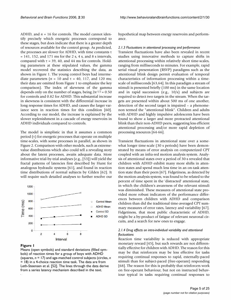
The model has two parameters: the number of candidatestages (n), and their average time constants (τ). Thegamma resembles the familiar ex-Gaussian distribution,and provides a good description of reaction-time data.Leth-Steensen, Elbaz and Douglas [52] compared the per-formance of 17 ADHD boys off medication, with a meanage of 11 years, with 18 age-matched control boys and ten7 year-old boys, on a four-choice reaction-time task. Fourwarning circles were presented on a screen for 2, 4, or 8 sfore periods, followed by a change in the colour of one toyellow. The boys were to press a key corresponding to thechanged stimulus. The authors fit ex-Gaussian densities tothe observed distributions, and also reported the mean(μ) and variance (σ2) of the RT distributions. From those,the parameters of the gamma model may be deduced: n =μ2/σ2 and τ = σ2/μ. This analysis imputed n = 6 stages for
Page 4 of 25(page number not for citation purposes)
Behavioral and Brain Functions 2006, 2:30 http://www.behavioralandbrainfunctions.com/content/2/1/30
ADHD, and n = 16 for controls. The model cannot iden-tify precisely which energetic processes correspond tothese stages, but does indicate that there is a greater depthof resources available for the control group. As predicted,the processes are slower for ADHD, with time constants τ= 141, 152, and 171 ms for the 2 s, 4 s, and 8 s intervals,compared with τ = 39, 40, and 44 ms for controls. Hold-ing parameters at these stipulated values, the gammamodel recovered the statistics describing the data, asshown in Figure 1. The young control boys had interme-diate parameters (n = 10 and τ = 40, 117, and 120 ms;their data are omitted from Figure 1 to emphasize the keycomparison). The index of skewness of the gammadepends only on the number of stages, being 2n-1/2 = 0.50for controls and 0.82 for ADHD. This substantial increasein skewness is consistent with the differential increase inlong response times for ADHD, and causes the larger var-iance seen in reaction times for this condition [60].According to our model, the increase is explained by theslower replenishment in a cascade of energy reservoirs inADHD individuals compared to controls.
The model is simplistic in that it assumes a commonperiod (τ) for energetic processes that operate on multipletime-scales, with some processes in parallel, as shown inFigure 2. Comparison with other models, such as extreme-value distributions which also could tell a revealing storyabout the latent processes, awaits adequate data. Moreinformative trial-by-trial analyses (e.g., [53]) will yield thefractal patterns of latencies first described by Hurst foranalogous hydraulic systems [61], and found in reactiontime distributions of normal subjects by Gilden [62]. Itwill require such detailed analyses to further resolve our
hypothetical map between energy reservoirs and perform-ance.
2.1.3 Fluctuations in attentional processing and performanceTransient fluctuations have also been revealed in recentstudies using innovative methods to capture shifts inattentional processing within relatively short time-scales,ranging from milliseconds to minutes. For example, rapidserial visual presentation (RSVP) paradigms such as theattentional blink design permit evaluation of temporalcharacteristics of information processing within a time-scale of milliseconds [63,64]. In this paradigm a stream ofstimuli is presented briefly (100 ms) in the same locationand in rapid succession (e.g., 10/s) and subjects arerequired to detect two targets in the stream. When the tar-gets are presented within about 500 ms of one another,detection of the second target is impaired – a phenome-non termed the "attentional blink". Children and adultswith ADHD and highly impulsive adolescents have beenfound to show a larger and more protracted attentionalblink than their non-ADHD peers, suggesting less efficientattentional processing and/or more rapid depletion ofprocessing resources [64-66].
Transient fluctuations in attentional state over a some-what longer time-scale (30 s periods) have been demon-strated by means of error analysis on computerized CPTcoupled with an infra-red motion analysis system. Analy-sis of attentional states over a period of 30-s revealed thatchildren with ADHD exhibit many more shifts in atten-tion states and spend much less time in an on-task atten-tion state than their peers [67]. Fidgetiness, as detected bythe motion-analysis system, was found to be related to thepercent of time spent in the 'distracted' attentional state,in which the children's awareness of the relevant stimuliwas diminished. These measures of attentional state pro-vided more robust indicators of the performance differ-ences between children with ADHD and comparisonchildren than did the traditional time-averaged CPT sum-mary measures of error rates, latency and variability [67].Fidgetiness, that most public characteristic of ADHD,might be a by-product of fatigue of relevant neuronal cir-cuits, and a search for new ones to engage.
2.1.4 Drug effects on intra-individual variability and attentional fluctuationsReaction time variability is reduced with appropriatemonetary reward [45], but such rewards are not differen-tially effective for children with ADHD. The reason for thismay be that reinforcers may be less effective for tasksrequiring continual responses to rapid, externally-pacedstimuli than for subject-paced (free-operant) responding[68]. The reason for this is probably that reinforcers workon free-operant behaviour, but not on instructed behav-iour typical in tasks requiring continual responses to
Means (open symbols) and standard deviations (filled sym-bols) of reaction times for a group of boys with ADHD (squares, n = 17) and age-matched control subjects (circles, n = 18) in a 4-choice reaction time taskFigure 1Means (open symbols) and standard deviations (filled sym-bols) of reaction times for a group of boys with ADHD (squares, n = 17) and age-matched control subjects (circles, n = 18) in a 4-choice reaction time task. The data are from Leth-Steensen et al. [52]. The lines through the data derive from a series latency mechanism described in the text.
Page 5 of 25(page number not for citation purposes)

Behavioral and Brain Functions 2006, 2:30 http://www.behavioralandbrainfunctions.com/content/2/1/30
Page 6 of 25(page number not for citation purposes)
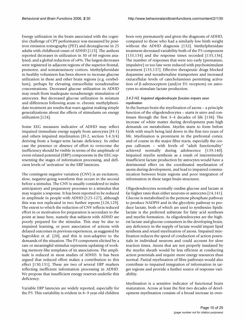
A scheme illustrating a glutamatergic neuron (left) a glial cell (astrocyte) and a small blood vessel (right) and the major compo-nents contributing to hypotheses 1 and 2 (H1 and H2)Figure 2A scheme illustrating a glutamatergic neuron (left) a glial cell (astrocyte) and a small blood vessel (right) and the major compo-nents contributing to hypotheses 1 and 2 (H1 and H2). Neural activity triggers release of the neurotransmitter glutamate that is taken up into the astrocyte (via GLAST and GLT-1 transporters), and stimulates the breakdown of glycogen, the uptake of glucose, and glycolysis, to produce lactate. Rapid neuronal firing is sustained by the energy provided by the astrocyte-neuron lactate shuttle. Energy demands are high during rapid (burst) and maintained rates of neuronal firing. H1: At times of increased neuronal demand, deficient lactate results in decreased neuronal conversion of lactate to acetyl CoA, decreased ATP forma-tion, deficient ATPase function, delayed restoration of ion gradients, elevated extracellular K+, deficient Na+-dependent trans-port of glutamate into astrocytes that is required to drive glycolysis and lactate release by the astrocytes. The result is that situationally appropriate firing rates are achieved only episodically. Methylphenidate treatment results in an increase of the extracellular levels of the catecholamines, NA (and DA) that stimulate glycolysis and release of lactate from the astrocytes. This is followed by glycogen replenishment, thereby correcting the energy deficiency, and restoring appropriate firing rates. H2: A deficient supply of lactate for oligodendrocytes in the developing nervous system slows and reduces the synthesis of fatty acids required for the synthesis of myelin. Poorly myelinated axons would transmit action potentials more slowly, accounting for inefficient integration (coherence) between brain regions and for slow reaction times. A number of neurotrans-mitter receptors present on astrocytes are not illustrated (e.g. muscarinic, α2, DA D3, D4, D5 and receptors for several neu-ropeptides).

Behavioral and Brain Functions 2006, 2:30 http://www.behavioralandbrainfunctions.com/content/2/1/30
rapid, externally-paced stimuli (cf., [68]). Methylpheni-date medication improves accuracy, speeds reaction time,and reduces reaction time variability in individuals withADHD [35,58]. Moreover, methylphenidate has beenshown to improve time on-task, with concomitantdecreases in the number of shifts in attention as the taskprogressed [67]. Such treatment effects suggest that theincreased availability of extra-neuronal catecholaminesarising from the blockade of their uptake by methylpheni-date could lead to increased activity of noradrenaline at β-adrenoceptors and possibly also dopamine acting at D1receptors found on astrocytes [15,69]. A key fact is thatstimulation of α1- and β-adrenoceptors on astrocytesenhances glycolysis and lactate production [69].Dopamine acting on D1 receptors may have similareffects, but these are not well documented.
2.1.5 Existing explanations and their limitationsAt present, various tasks used to measure different aspectsof executive function usually have moderately heavy cog-nitive demands and share requirements for continuousspeeded responding to computer- or experimenter-pro-duced stimuli, rather than being self-paced. Experimenter-paced measures of working memory are substantially bet-ter predictors of reading letter span, continuous operationspan, literacy and mathematics scores than are more tradi-tional, subject-paced measures [70]. We believe they pro-vide better diagnostics of ADHD because, like stress testsfor cardiac function, they most directly challenge the sub-jects' short-term reserves of energy, and that is a majorresource compromised in ADHD.
Slowing of MRT and increasing RT variability over timehas been postulated to reflect mental fatigue, 'resourcedepletion', motivational deficits, fluctuations in executivecontrol, or underlying neurobiological disturbances (e.g.,motor timing deficits), which are caused by the need forcontinuous speeded responding to high-demand tasks[6,20,71-75]. Most of these models cannot account for themoment-to-moment fluctuations in response accuracycharacteristic of ADHD.
The dynamic developmental theory of ADHD [8,9,20]explains intra-individual behavioural variability as defi-cient acquisition of stimulus control of long chains ofbehaviour. This deficiency is rooted in reduced efficacy ofreinforcers ("rewards") combined with poorer extinction("unlearning") of inefficient behaviour. The dynamicdevelopmental theory addresses free-operant behaviourwithout time constraints. However, neither the dynamicdevelopmental theory of ADHD, nor other theories andexplanations account for the intra-individual perform-ance variability of ADHD on tasks that require continualresponses to rapid, externally-paced stimuli. The presentpaper addresses this aspect by postulating that reaction
time variability and other manifestations of transient fluc-tuations in performance, which arise in the context of theneed for continuous rapid neuronal firing, reflect theeffects of transient depletion of neuronal energy on theefficiency of information processing.
3. The hypothesisIn this section we highlight the strengths and limitationsof Todd and Botteron's Energy-Deficiency Model (section3.1), present a brief background on astrocytes and theirrole in the development and function of the CNS (section3.2), formulate the hypotheses (H1 and H2: section 3.3),and provide a detailed summary of evidence supportingthe hypotheses (section 3.4).
3.1 Energy-deficiency modelAccording to Todd and Botteron [13], the genesis of thebehavioural symptoms of ADHD is linked directly toimpairments of the astrocyte-neuron lactate/energy-shut-tle that is based on the astrocytic uptake of glucose fromblood capillaries, its utilization (conversion to lactate)and storage as glycogen [76]. Since acute amphetaminetreatment of young adults has been reported to stimulateglucose uptake in the frontal lobes [77], Todd and Bot-teron hypothesized that reduced catecholaminergic input(in ADHD) leads to a decrease in astrocyte-mediated neu-ronal energy metabolism and impaired frontal function.
Todd and Botteron's model links catecholaminergic activ-ity to the regulation of neuronal energy metabolism. Butit does not make explicit how a decreased neuronal energysupply mediated by hypofunctioning catecholaminesmight alter either cognitive performance based on frontallobe function or account for ADHD symptomatology ingeneral. In contrast, our hypothesis addresses a specificaspect of the clinical presentation, – namely, moment-to-moment fluctuations in task performance that are oftenalso manifest in general behaviour – and we refine andextend the biochemical bases that underlie this hypo-thesis.
3.2 Astrocyte and oligodendrocyte function in the central nervous system (CNS)The CNS comprises two main types of cells: neurons thatare directly involved in information processing, and glialcells (astrocytes, oligodendrocytes and microglia) whichplay a major role in the development and mature functionof the CNS. Astrocytes are responsible for maintaining theenvironment of cells in the CNS: they provide nutrientsand modulate the release and uptake of glutamate, elec-trolytes (Na+/K+) and other by-products of neural activity[78]. In addition, astrocytes play an important role in neu-ral signalling. They have receptors for neurotransmitterssuch as glutamate, GABA, acetylcholine and the monoam-ines as well as for other neuromodulators including neu-
Page 7 of 25(page number not for citation purposes)

Behavioral and Brain Functions 2006, 2:30 http://www.behavioralandbrainfunctions.com/content/2/1/30
ropeptides, cytokines and steroid hormones [15-18,79-84]. They can modulate the excitability of neurons viaoscillations of Ca2+ levels initiated by stimulation ofmetabotropic glutamate receptors [15,85-90].
Oligodendrocytes are responsible for forming myelin, amembranous sheath that is wound spirally around axons,providing electrical insulation that leads to a more than10-fold increase in the speed of signal transmission occur-ring in unmyelinated axons [11].
3.3 Statement of the hypothesisLactate production by astrocytes is insufficient duringbrief periods of increased demand in ADHD. This hypo-thesis has two consequences (Figure 2). H1) The inabilityof astrocytes to provide an adequate supply of lactate torapidly firing neurons results in a localized and transientdeficiency in ATP production, impaired restoration ofionic gradients across neuronal membranes and slowedneuronal firing. This leads to inconsistent performance ofdemanding cognitive tasks; H2) Insufficient provision oflactate for oligodendrocyte function in the longer termgives rise to deficient fatty acid synthesis and delayed orreduced myelination of axons. In turn this leads to lessefficient transmission and longer reaction times. Thehypothesis refers to two effects over very different timescales: The effects on rapidly firing neurons occur overmilliseconds (H1), whereas the effects of reduced lactatesupply for oligodendrocytes when demands are high dur-ing development takes place over months and years (H2).Collectively the neural outcomes of astrocyte dysfunctionwould manifest behaviourally as inefficient and/or incon-sistent performance (i.e. slow and/or variable responsetimes across the lifespan, particularly during tasks thatrequire continuous speeded responses and complex infor-mation processing).
3.4 The hypothesis – discussion3.4.1 H1: Impaired astrocyte function limits energy (lactate) supply to rapidly firing neuronsThe ionic composition of the cell cytoplasm is very differ-ent from extracellular ion concentrations. In the neuronthe ionic gradients across the membrane constitute a storeof potential energy that can drive the influx of Na+ andefflux of K+ ions to generate an action potential, and theinflux of Ca2+ ions to trigger neurotransmitter release andto generate Ca2+ waves in and between both neurons andglial cells. To maintain repeated firing over an extendedtime, neurons require energy to restore the trans-mem-brane gradients of Na+, K+ and Ca2+ ions. Neurons gener-ate the necessary energy in the form of ATP; this drives themembrane-associated Na+/K+ATPase to pump Na+ out ofthe cell and K+ back into the cell. ATP is also required byCa2+ATPase to pump Ca2+ out of the cell or into intracel-lular stores (Figure 2).
Neuronal activity is tightly coupled to glucose utilization[91]. Neural activity triggers oxidative metabolism (toproduce ATP) within neurons followed by breakdown ofenergy stores (glycogen in astrocytes) and uptake of glu-cose from blood capillaries into astrocytes to produce lac-tate [91]. Rapid neuronal firing, however, is sustained bythe astrocyte-neuron lactate shuttle [92]. Lactate is theessential energy source for rapidly firing neurons. It is amore efficient fuel than glucose because it is metabolizedto form ATP more rapidly and, unlike glucose, does notrequire ATP for its metabolism [76]. It is imperative forneurons to make use of the most efficient energy supplieswhen rapid neural processing is required and demands forenergy are high, and the brain has evolved ways to do so[93]. The neurotransmitter glutamate (released by themajority of excitatory neurons in the CNS) stimulates gly-colysis (glucose utilization and lactate production) inastrocytes and release of lactate into the extracellular fluid[91,94]. An inadequate supply of lactate during periods ofrapid neuronal firing, when local energy demand is high,briefly impairs neuronal function, particularly during thelatter part of a train of neural impulses and shortly there-after, causing an extended refractory period. Thisincreased latency to new information is measured in theattentional blink paradigm (section 2.1.3). Compromisedenergy supply, as hypothesized for ADHD, should impairresponse inhibition or alternation at these times.
Periods of rapid neuronal firing will be followed by slowunsynchronized firing exerting less demand on energyresources, allowing replenishment of energy reserves andrestoration of function. Energy reserves will depend onthe prior history of neural and astrocyte activity. Brief peri-ods of energy insufficiency followed by periods of normalsupply are proposed to account for the variability ofbehavioural response seen in ADHD when performingcomplex tasks that require speed and accuracy.
3.4.1.1 Impaired maintenance of ion gradients across the neuronal membraneDecreased availability of lactate to produce energy in theform of ATP would impair the function of membrane-associated Na+/K+ATPase and Ca2+ATPase pumps, result-ing in elevated extracellular K+ and decreased Ca2+ andNa+ concentrations. Consistent with this hypothesis,ADHD children were reported to have decreased urinaryexcretion of Ca2+, Na+ and phosphate, but not K+ ions[95]. Failure to maintain homeostasis of inorganic ionswill alter electrochemical gradients across neuronal andglial cell membranes. As the resting membrane potentialof neurons is dominated by the K+ concentration gradientacross the membrane, failure to reduce elevated extracel-lular K+ concentrations following neural activity will altermembrane potentials, cause depolarization to last longerthan required, and thus impair neuronal function. Raised
Page 8 of 25(page number not for citation purposes)

Behavioral and Brain Functions 2006, 2:30 http://www.behavioralandbrainfunctions.com/content/2/1/30
K+/reduced Na+ gradients are likely to have widespreadeffects in different parts of the brain, promoting for exam-ple monoamine release (e.g. dopamine [96]) and alteringthe conformation of transporters, thus affecting monoam-ine uptake (e.g. dopamine [97] 5-HT [98]).
3.4.1.2 Impaired uptake and removal of extracellular glutamateAstrocytes normally maintain low extracellular levels ofglutamate and K+ [78]. Glutamate transport into astro-cytes is driven by the influx of Na+ along its electrochemi-cal gradient. But the low membrane potential resultingfrom the astrocytes being less able to maintain low extra-cellular K+ concentrations in the ADHD condition willdiminish the electromotive drive for Na+ influx andthereby hamper removal of glutamate from the extracellu-lar fluid [93,99,100]. Failure to maintain low extracellularglutamate levels will impair glutamate neurotransmitterfunction, neuroplasticity, learning and memory, andcould lead to excitotoxicity and cell death, reflected asreduced CNS gray matter. (Small reductions in grey matterare reported for subjects with ADHD [101-103]).
3.4.1.3 Impaired neuromodulator regulation of lactate formationSeveral neurotransmitters can potentially modulate lac-tate production in astrocytes, with the majority of evi-dence supporting a role for noradrenaline acting on α1-,α2- and β-adrenoceptors [88,104-106]. This is importantin view of widely accepted explanations of ADHD symp-toms in terms of catecholamine function [107], psychos-timulant effects on glucose utilization (section 3.1) andefficacy as medication [108]. Glial receptors for dopamine(D1–5) [15,88] and serotonin (5-HT2) [109] have alsobeen reported but their effects on lactate production inastrocytes have not been well documented.
Extracellular lactate decreases immediately after neuronalactivation, but rises again after a short delay [110]. Nor-mally within milliseconds of glial β- or α2-adrenoceptoractivation noradrenaline induces the breakdown of glyco-gen to glucose (glycogenolysis) to supply the needed lac-tate [69,105,111,112]. This is followed by a phase ofglycogen re-synthesis in the astrocytes that can last severalhours [112]. Failure to adequately replenish glycogenstores in the astrocytes would reduce the availability ofenergy substrates required for subsequent or sustainedneuronal activity. Therapeutic agents (e.g. methylpheni-date, amphetamine, atomoxetine, desipramine,modafinil) block the noradrenaline transporter andincrease extracellular concentrations of noradrenalinewhich stimulates glycolysis and lactate production inastrocytes. Although evidence strongly supports a role fornoradrenaline, both dopamine and serotonin are knownto modulate cAMP levels in astrocytes and could thereforealso play a role [15,109].
Glucocorticoid hormones also modulate the supply ofenergy [113,114]. Glucocorticoids inhibit glucose trans-port into neurons and astrocytes and inhibit glycogen syn-thesis stimulated by noradrenergic activity [115]. Thus,glial activity may be down-regulated by increased hypoth-alamo-pituitary-adrenal activity in situations perceived asstressful (e.g. expressed emotion in the family of ADHDchildren or the challenge provided by cognitive tasks pre-sented to the children). High levels of glucocorticoids act-ing on astrocytes would impair glycogen replenishment,deplete energy reserves, and reduce lactate transport.Behaviourally, this will give rise to fatigue and predisposeto psychological problems, as has been proposed for otherdisorders such as Parkinsonism and major depression[116].
Cytokines, small proteins that support communicationbetween cells of the immune system, can be produced byand influence the function of astrocytes (e.g. TNF-alpha,IL-1, IL-6 [117]). Reduced stimulation of β-adrenoceptorsleads not only to decreased glycogenolysis but also toimpaired production of several growth factors (e.g. nervegrowth factor (NGF), basic fibroblast growth factor (basicFGF), transforming growth factor-beta1 (TGF-beta1)along with increased production of nitric oxide and thepro-inflammatory cytokines [118,119]. Cytokine expo-sure can lead to an overproduction of nitric oxide and itsmetabolites that diffuse out and damage mitochondriaand the energy supply in nearby cells (including neurons[120]). TNF-alpha and IL-1 can fundamentally perturb theenergy metabolism of astrocytes promoting the uptake ofglucose without either storing it as glycogen or releasinglactate [121]. This disruption can therefore not onlyimpair short-term demands for energy, but also the long-term requirements for development (see hypothesis H2,section 3.4.3), that in the worst case can lead to apoptosisof the oligodendrocyte [118]. Other cytokines (e.g. thecalcium-binding S100B) also regulate energy metabolism,promote neuronal survival and regeneration [122].Dopamine also stimulates release of growth factors(including NGF, glial-derived nerve growth factor, GDNFand brain-derived nerve growth factor, BDNF) and there-fore plays a crucial role in development of the brain andmaturation of the nervous system [84].
3.4.2 Preliminary evidence: impaired energy supply during rapid neuronal firing in ADHDThis section first considers the limited direct neuroimag-ing evidence of altered energy utilization in the CNS ofpeople with ADHD that could potentially derive fromdeficient astrocyte function [116]. We then discuss elec-troencephalographic (EEG) recordings indicating ineffi-cient communication within and between neuron clustersthat could be attributed to impaired energy supplies.
Page 9 of 25(page number not for citation purposes)
Behavioral and Brain Functions 2006, 2:30 http://www.behavioralandbrainfunctions.com/content/2/1/30
Energy utilization in the brain associated with the cogni-tive challenge of CPT performance was measured by posi-tron emission tomography (PET) and deoxyglucose in 25adults with childhood onset of ADHD [123]. The authorsreported decreases in utilization in 30 of 60 regions ana-lysed, and a global reduction of >8%. The largest decreaseswere registered in adjacent regions of the superior frontal,premotor, and somatosensory cortices. Methylphenidatein healthy volunteers has been shown to increase glucoseutilization in these and other brain regions (e.g. cerebel-lum), perhaps by elevating extracellular noradrenalineconcentrations. Decreased glucose utilization in ADHDmay result from inadequate noradrenergic stimulation ofastrocytes. But decreased glucose utilization in striatumand differences following acute vs. chronic methylpheni-date treatment are results that warn against making simplegeneralizations about the effects of stimulants on energyutilization [124].
Some EEG measures indicative of ADHD may reflectimpaired immediate energy supply from astrocytes (H-1)and others impaired myelination (H-2, section 3.4.3/4)deriving from a longer-term lactate deficiency. In eithercase the presence or absence of effort to overcome theinefficiency should be visible in terms of the amplitude ofevent-related potential (ERP) components in the EEG rep-resenting the stages of information processing, and defi-cient levels of 'activation' in the ERP latencies.
The contingent negative variation (CNV) is an excitatory,slow, negative-going waveform that occurs in the secondbefore a stimulus. The CNV is usually considered to indexanticipatory and preparatory processes to a stimulus thatmay require a response. It has been reported to be reducedin amplitude in people with ADHD [125-127], althoughthis was not replicated in two further reports [128,129].The extent to which the reduction of CNV reflects reducedeffort in or motivation for preparation is secondary to thepoint at issue here, namely that subjects with ADHD arepoorly prepared for the stimulus. This may be due toimpaired learning, or poor association of actions withdelayed outcomes in previous experiences, as suggested bySagvolden et al. [20], and this is non-adaptive to thedemands of the situation. The P3 component elicited by arare or meaningful stimulus represents updating of work-ing memory-like templates of its associations. The ampli-tude is reduced in most studies of ADHD. It has beenargued that reduced effort makes a contribution to thiseffect [130,131]. These are two of several ERP markersreflecting inefficient information processing in ADHD.We propose that insufficient energy reserves underlie thisdeficiency.
Variable ERP latencies are widely reported, especially forthe P3. This variability is evident in 8–9 year-old children
born very prematurely and given the diagnosis of ADHD,compared to those who had a similarly low birth weightwithout the ADHD diagnosis [132]. Methylphenidatetreatment decreased variability both of the P3 component[133,134] and the response times recorded [135,136].The number of responses that were too early (premature,impulsive) or too late were reduced with psychostimulanttreatment [135,137]. Effective therapeutic drugs blockeddopamine and noradrenaline transporters and increasedextracellular levels of catecholamines permitting activa-tion of β-adrenoceptors (and/or D1 receptors) on astro-cytes to stimulate lactate production.
3.4.3 H2. Impaired oligodendrocyte function impairs axon myelinationIn the human brain the myelination of axons – a principlefunction of the oligodendrocytes – starts in utero and con-tinues through the first 3–4 decades of life [138]. Theincrease of white matter during development puts highdemands on metabolism. Myelin starts to form beforebirth with much being laid down in the first two years oflife. Myelination is prominent in the prefrontal cortex,and of course in the major fibre tracts, especially the cor-pus callosum – with levels of "adult functionality"achieved normally during adolescence [139,140].Impaired myelin synthesis as a result of intermittentlyinsufficient lactate production by astrocytes would have adetrimental effect on the coordinated myelination ofaxons during development, and lead to impaired commu-nication between brain regions and poor integration ofinformation in these target brain structures.
Oligodendrocytes normally oxidise glucose and lactate atfar higher rates than either neurons or astrocytes [14,141].Glucose is metabolised in the pentose phosphate pathwayto produce NADPH and in the glycolytic pathway to pro-duce lactate, both of which are used to synthesize lipids:lactate is the preferred substrate for fatty acid synthesisand myelin formation. As oligodendrocytes are the high-est lactate and glucose consumers in the developing brain,any deficiency in the supply of lactate would impair lipidsynthesis and retard myelination of axons. Impaired mye-lination reduces the speed of conduction of action poten-tials in individual neurons and could account for slowreaction times. Axons that are not properly insulated bythe myelin sheath would be less efficient at conductingaction potentials and require more energy resources thannormal. Partial myelination of fibre pathways would alsocontribute to impaired integration of information in tar-get regions and provide a further source of response vari-ability.
Myelination is a sensitive indicator of functional brainmaturation. Across at least the first two decades of devel-opment myelination consists of a broad increase in over-
Page 10 of 25(page number not for citation purposes)

Behavioral and Brain Functions 2006, 2:30 http://www.behavioralandbrainfunctions.com/content/2/1/30
all white matter density as well as a more region-specificprogression, proceeding from posterior to more anteriorregions, [142-145]. In magnetic resonance spectroscopy(MRS) and diffusion tensor imaging (DTI) studies, matu-ration of relatively restricted regions of white matter hasbeen found to correlate with the development of cognitivefunctions such as working memory capacity (left frontalregions), reading ability (left temporal lobe) and evenwith IQ (bilateral association areas [144,146]. In a studyof 100 children with evidence of delayed developmentbut otherwise normal MR-scans, Pujol and colleagues[143] were able to show a reduction of myelination (inpart asymmetric) equivalent to normally developing chil-dren who were some 3 years younger. Also, at the otherend of the lifespan, white matter damage due to axonalloss that occurs in normal ageing has been found to corre-late with working memory performance, even after con-trolling for age [19]. This suggests that working memoryperformance may be particularly dependent on complexnetworks, which in turn depend upon white matter con-nections. These studies conclude that there is a positiverelationship between the density and organization ofmyelinated fibres and the efficiency (maturity) of cogni-tive function [144].
3.4.4 Preliminary evidence of altered myelination in ADHD3.4.4.1 NeuroimagingEvidence for impaired and/or delayed myelinationderives from MR measures of white matter density and theintegrity of myelinated neuronal pathways indexed byDTI and MRS measures of metabolites (e.g. N-acetyl-aspartate, NAA). Support for the functional impairment ofthese pathways comes from EEG recordings (section3.4.4.2).
Seven of the 8 anatomical MRI studies report decreasedtotal white matter volumes in children and adolescentswith ADHD [102,103,147-150]. In the largest MRI studythat scanned over 150 boys with ADHD on at least twoseparate occasions, the reduction in white matter volumewas substantial (a 10% reduction) compared to thosewho had been treated with stimulant medication or hadno diagnosis [103]. Indeed, one study linked smallerwhite matter volumes with slower processing speed, asindexed by the speed of colour-naming [150]. Theseresults implicate a contribution of delayed myelination toADHD cognition.
DTI provides a measure (fractional anisotropy, FA) of thecoherence and integrity of the biochemical microstructureof myelinated pathways. An initial study comparing agroup of 18 children with ADHD with 15 matched con-trols recorded a reduced FA in the right cerebral peduncle/anterior limb of the internal capsule (right neostriatumand premotor cortex), and in the left middle cerebellar
peduncle (left cerebellum and parieto-occipital region).Ashtari and colleagues report that the lower the cerebellarFA, the more severe were the ratings of symptoms of inat-tention [151]. These first findings point to a link betweenwhite matter anomalies and the symptomatology ofADHD.
Such findings are extended by 1H-MRS measures of neu-ronal metabolism in the brains of subjects with ADHD.With this method creatine, choline, taurine, inositol,NAA, glutamate and lactate can be detected and meas-ured. Unfortunately there have been scant attempts toreport on the last two. As yet creatine, choline (and theirphospho-derivatives) – regarded as indicators of lipidmetabolism and membrane integrity – have also not beenstudied in ADHD. The easiest metabolite to measure isNAA. NAA is found in neurons, not in glia, and is regardedas a marker of neuronal density, function, viability andperhaps functional connectivity [152]. It is synthesised bythe enzyme NAA transferase in neuronal mitochondriafrom acetyl coenzyme A (acetyl Co-A) and aspartate, andused by oligodendrocytes to produce acetyl groups for thesynthesis of myelin lipids [153,154]. During developmentNAA levels increase (as choline levels fall) and reflectincreased synthesis (or decreased utilization) in the for-mation of myelin [155,156]. High NAA concentrationscorrelate with increased ADP [157,158] as acetyl CoA lev-els required for ATP synthesis are depleted. Decreases ofNAA levels reflecting neuronal dysfunction are associatedwith neuronal loss in certain parts of the brain in manymajor psychiatric illnesses [159].
Increased NAA levels are reported for the right frontal lobe[160] and fibres entering/leaving left frontal regions (cen-trum semi-ovale [161]) of ADHD subjects compared tohealthy and autistic comparison groups. But group differ-ences were not found in 2 studies of the right frontal lobe[162,163] and decreases were reported in small studies forthe left frontal [164] and right lenticular regions [165].However Yeo et al. [162] noted that the smaller right fron-tal volume for their 17 ADHD children correlated withNAA and choline measures, and that NAA levels in turnrelated to performance on a sustained attention task. Else-where, intriguingly, raised frontal glutamate concentra-tions were noted, particularly on the right [160,163]. As acautionary note, it should be pointed out that animalwork has shown that methylphenidate treatment can leadto increased cortical levels of NAA [166]. Further, theresults of these 6 ADHD studies must be regarded as pre-liminary as they each represent very small subject groups,and the range of brain regions they could sample was veryrestricted. Nonetheless it should be noted that all thestudies recorded changes, and unusually some subjectsshow raised metabolite levels (NAA, choline, glutamate).These results point to changed patterns and rates of mye-
Page 11 of 25(page number not for citation purposes)

Behavioral and Brain Functions 2006, 2:30 http://www.behavioralandbrainfunctions.com/content/2/1/30
lin synthesis (and breakdown) that may reflect intermit-tently insufficient supplies of lactate. The result is anoverall decrease of white matter for a given age-class, asdescribed above.
3.4.4.2 NeurophysiologyWe propose that the functional consequences of theimpaired/delayed developmental laying down of themyelin sheath can be seen in three types of EEG measure:evoked potential (EP) latencies, the topographic distribu-tion of the power spectrum in the quantitative EEG, andin the coherence of the EEG waveforms between brainregions.
EPs representing sensory information ascending in theauditory nerve [167] and the brain stem appear at longerthan normal latencies (e.g., components III and V).Indeed, the transmission times from components I to IIIand I to V are reported to be increased in subjects withADHD [168]. The latency of the steady state visual EP inthe frontal cortex of ADHD children is markedly delayed[169]. Indeed, in their report of a delayed velocity indexfor EPs in ADHD, Ucles et al [170] proposed not only thatabnormal myelination in the cortico-spinal path could beresponsible, but that the result could be indicative of amuch more widespread problem.
A large proportion of patients with ADHD (or narcolepsy,see 3.5.1 below) demonstrate an increased ratio of relativetheta to alpha or beta power in the EEG, especially overanterior brain regions [171,172]. One explanation of thedominant lower firing frequencies could lie with thereduced lactate availability required to sustain rapidly fir-ing neurons. There is usually a marked normalization ofthis balance between oscillation frequencies after methyl-phenidate treatment [173]. In the unmedicated samplethere is a positive correlation between P2, N2 and P3 ERPlatencies, widely reported to be delayed [137] andincreased theta power [174,175]. A plausible reason forthis shift in balance between oscillation frequencies lies ina decreased representation of the faster frequencies owingto deficient neuronal energy supply (H1) and/or reducedmyelination in brain stem reticular sources active in gen-erating some of these rhythms [170] which is consistentwith our hypothesis H2.
A more direct measure of the coupling of activity betweenbrain regions can be estimated by EEG coherence of wave-form between recording sites. Coherence can be conceptu-alised as the correlation in the time domain between 2signals in a given frequency band. EEG coherence nor-mally develops systematically with age in a non-linearfashion. There is evidence of development in longer-rangeinter-hemispheric coherences which are not apparent inboys with ADHD. Furthermore, boys with ADHD show
elevated slow-wave coherences and reduced fast-wavecoherences between hemispheres, although within hemi-spheres the coherence in the theta band is reduced, espe-cially over frontal regions [176-178]. This is most easilyexplained by unusual if not delayed development of thelarge white matter tracts connecting brain regions. Atshort distances between signals the increased coherence atslow wave frequencies in children with ADHD is viewedas consistent with a delay in the pruning back of over-pro-duced synapses and local connections [179]. As would beexpected such long-term alterations remain unaffected byshort-term methylphenidate treatment [180].
3.5 Supportive evidence from related disordersWe propose that a disturbed lactate shuttle in ADHDaccounts for brief transient impairments in rapid neuro-nal firing and delayed myelination: Together they result invariable responses. These impairments may account forsimilar phenomena in other neurodevelopmental disor-ders. We do not regard our hypothesis as specific toADHD – for example, individuals with schizophreniahave been found to respond more slowly and variably onattentional and cognitive tasks [181,182] – nor do we sug-gest that it occurs in all psychiatric disorders. In the fol-lowing sections we select two disorders, PKU andnarcolepsy, to illustrate the presence of phenomena thatappear to model the situation that pertains to ADHD.
3.5.1 Parallel pathological conditions: Phenylketonuria (PKU)PKU results from high concentrations of phenylalanine(Phe), that arise from an inability to convert it into tyro-sine, and which inhibit transport across the blood brainbarrier of neutral amino acids such as tyrosine and tryp-tophan necessary for the synthesis of the three principlemonoamine transmitters. If children are placed on Phe-free diet early enough, microcephaly, mental retardationand motor problems can be avoided, although sub-clini-cal symptoms may remain, particularly in the cognitivedomain [183,184]. Children with PKU (off-diet) are ratedby parents and teachers as more distractible, hyperactive,and impulsive than healthy controls [185], with manysymptoms similar to ADHD (e.g. restlessness, fidgeting,concentration difficulty, short attention span, low frustra-tion tolerance [186]). Realmuto et al. [187] noted that 9/13 of their subjects either manifested or had a history ofcomorbidity with ADHD. Prenatal exposure was associ-ated with a higher likelihood of expressing hyperactive/impulsive symptoms and postnatal exposure was associ-ated with a higher likelihood of expressing inattentivesymptoms [188]. Indeed, a considerable proportion ofpeople with treated PKU take psychostimulant medica-tion for their attentional problems (e.g. 26% of a sampleof 38 school-aged children with PKU [189]).
Page 12 of 25(page number not for citation purposes)
Behavioral and Brain Functions 2006, 2:30 http://www.behavioralandbrainfunctions.com/content/2/1/30
Cognitive impairments of individuals with PKU like thosewith ADHD include variable reaction times, sustainedattention, working memory, executive function, planning,learning, tests of colour-naming, arithmetic, verbal abili-ties and academic performance [187,190-199]. Inter-hemispheric interactions, as measured by slowed transfertimes [200], and a lack of the across-hemisphere advan-tage in performing a name-identity task [183], are affectedin comparison to neurologically intact children. Neuro-physiological studies report slow latencies for several earlyEPs [201], the later P3 component and slower more vari-able motor reaction times [194,195,202]. In the EEG slow(theta) rhythms are characteristic of individuals withPKU. Indeed their high theta/alpha ratio (recallingADHD) was sensitive to treatment reducing the levels ofPhe [203,204].
Functionally, these features reflect delayed myelination,low monoamine levels, and impaired energy availability.Impaired myelination is the primary neuropathologicalfeature of treated or untreated people with PKU: A delay istypical of the younger ages and diffuse demyelination isreported at older ages [205-208]. The finding of low HVAlevels (dopamine metabolite) in the CSF [209] confirmsthe second feature, low dopamine levels. Importantly,changes of dopamine in the rodent model go hand inhand with the depletion and restoration of Phe levels[210]. Noradrenaline and serotonin would also beexpected to be affected. The third feature was addressed in11 young adults with PKU using phosphate MRS. Pietz etal. [204] describe changes of cerebral energy metabolismthat could underlie reduced transmission speed, myelina-tion and catecholamine availability [205]. Among 11measures taken at baseline, only ADP was significantlyelevated, and inorganic phosphate decreased. Phe loadingthen decreased phosphocreatine and ATP levels while fur-ther increasing ADP. This is consistent with Phe inhibitionof pyruvate kinase and the concurrent conversion of ADPto ATP [211]. Impaired pyruvate synthesis would reducelactate production and the ability of astrocytes to meet theenergy requirements of sustained rapid neural firing and ashortage of lactate would also impair the ability of oli-godendrocytes to synthesize myelin.
3.5.2 Parallel pathological conditions: narcolepsyNarcolepsy is a neurological disorder characterized byexcessive daytime sleepiness [212]. Some of the neuropsy-chological characteristics of narcolepsy are strikingly sim-ilar to those seen in ADHD and children with ADHD havean increased tendency to daytime sleepiness [213]. Indi-viduals with narcolepsy have slower reaction times andmore within-task variability of performance than controlsubjects on a variety of attentional tasks ranging fromthose sensitive to arousal and sustained attention, to theexecutive control of attention [214]. Recent studies report
narcolepsy-related deficits in attentional and executivefunction which place high demands on inhibition andtask management, but not on simple tasks of memory andattention [214,215]. The pattern of findings was thoughtto be indicative of a depletion of available cognitiveprocessing resources because of the need for continuousallocation of resources to monitoring.
Like ADHD, hypoarousal (sleepiness) is induced in situa-tions of low stimulation, such as reading or boring repet-itive tasks [112,216-218]. Similarly, children with ADHDor narcolepsy appear to use motor overactivity and fidget-iness to counteract their drowsiness [218-220]. It is inter-esting to speculate how a deficiency in energy supplymight contribute to the other symptoms of ADHD, suchas hyperactivity. Depression of sensory modules couldcertainly lead to a suboptimal level of stimulation, induc-ing sensation-seeking behaviour, with motor, vocal, andother activities displacing the contextually "appropriate"behaviours, which through local energy deficiency can nolonger provide the necessary arousal. Derangements ofcalcium-dependent protein phosphatase and kinase activ-ity impair working memory [221]. These and other impli-cations of the energetics hypothesis require furtherconsideration, lying beyond the scope of this paper.Lastly, as in ADHD analyses, EEG rhythms show the ratioof theta to alpha or beta power in narcolepsy to be higherthan normal [172]. Narcolepsy has been attributed todepletion of the transmitter hypocretin, a hypothalamicneuropeptide that regulates energy metabolism anddopamine activity in certain brain regions [222-224].Modafinil, the drug normally used to treat narcolepsy, iseffective in treating ADHD and can inhibit dopamine re-uptake [220,225-228]. It is also an α1-adrenoceptor ago-nist and can therefore facilitate β-adrenoceptor-stimu-lated lactate production in astrocytes [229,230]. Thus, themechanism of treatment could be the same in both disor-ders. This highlights the similarities between the disordersthat could reflect common underlying disturbances.
4. Testing the hypothesesThe fundamental tenet of our hypotheses (H-1 and H-2)– to be tested – is that a deficient energy supply to rapidlyfiring neurons (the lactate shuttle) underlies moment-to-moment fluctuations in response speed and accuracy(astrocyte mechanisms) and, in the long-term, episodes oflactate deficiency during development delays axon myeli-nation (oligodendrocyte function). These two links needto be demonstrated (section 4.1).
The consequences of these disturbances are proposed tolie with a) decreased neural activity when sustained rapidfiring is required (neurophysiological level), b) delayedand variable cohesion between the components of theneural circuitry responsible for integration of information
Page 13 of 25(page number not for citation purposes)
Behavioral and Brain Functions 2006, 2:30 http://www.behavioralandbrainfunctions.com/content/2/1/30
and selection/organization of appropriate response (psy-chophysiology), and c) the poorly coordinated and intra-individual variability of performance typical of individu-als with ADHD. More evidence is required (section 4.2).
4.1 Is there an energy deficiency?There are several points in the energy cycle where dysfunc-tion could occur. The present hypothesis focuses on onespecific aspect: The provision of adequate amounts of themore efficient metabolic fuel, lactate (rather than glu-cose), to rapidly firing neurons (e.g. glutamate trans-porter, mitochondria, monoaminergic regulation ofastrocyte function: section 4.1.1). Identification of envi-ronmental and/or genetic origins of the lactate deficiencywould contribute to an understanding of intra-individualvariability in performance of high energy-demandingtasks (section 4.1.2). Factors that impair myelination maycontribute to slow responsiveness and are also reviewed(section 4.1.2).
4.1.1. The performance of energetic compartmentsThere is a need for more precise measures of the dynamicswithin and between the major compartments of energyproduction, storage and utilization through studies usinglabelling, stimulation and inhibition of the various con-stituents in animal models of ADHD and tissue culture(e.g. delineation of the quantitative relationship betweenneuronal firing rate and lactate utilization, intra/extracel-lular glutamate flow, mitochondrial function (ATP/ADPratio and oxygen consumption), effects of lactate restric-tion on ionic gradients, function of regulatory factors (e.g.neurotransmitter/neuropeptide receptors, particularlynoradrenergic α1-, α2- and β-adrenoceptors), effect ofimpaired lactate production on myelin synthesis (andaccumulation of NAA), influence of NAA availability onacetyl-group incorporation into myelin). Regional differ-ences in measures of effects of transient energy deficiencyare important to note, since the hypothesis does not pre-dict that all parts of the brain will be affected equally.Greater effects (e.g. reduced size) should be observed inthose brain areas that contain or form part of rapidly fir-ing neural circuits that transiently deplete local energyreserves and hinder synapse formation.
Measures of some of these effects are becoming techni-cally feasible for human studies, in vivo, with MRS. Forexample, changes in glutamate production (tricarboxylicacid cycle), lactate synthesis (glycolysis) and glutaminesynthesis have been demonstrated in neurologicalpatients using labelled carbon (13C) MRS [231,232]. Fur-ther data on the energy metabolites ATP, ADP, inorganicphosphate, phosphocreatine and creatine obtained frommore conventional proton and phosphate MRS studieswould also be useful (cf. section 3.5.1). A second exampleis based on the changes of intracellular calcium oscilla-
tions and extracellular calcium waves generated by neuro-transmitter stimulated glia. These calcium signalsstimulate glutamate release, modulate neuronal excitabil-ity and carbohydrate metabolism [86,89]. Changes in cal-cium concentration can be monitored by new contrastsubstances being developed for MRI investigations. Thesetechniques, and recent applications of 2-photon opticalcalcium imaging in animals, could also address the role ofdopamine D1 and D2 receptors [233] as well as metabo-tropic glutamate receptors and their modulation by α1-adrenoceptors in the stimulation of calcium responses inastrocytes [85,234,235]. A third example is the need forcross-sectional if not longitudinal MRS data on myo-inositol. This is suggested to be a marker of the integrity ofglia and glial transport mechanisms, but the signal hasproved difficult to separate from that for the largeamounts of glycine present that also resonate at 3.6 ppm[236]. Whereas increased levels of myo-inositol areclaimed to reflect gliosis that would not be expected inADHD, decreased levels, as reported for major depressivedisorder, may reflect glial loss or altered glial metabolism[237]. Indeed a functional decrease may apply to ADHD.A preliminary report on 15 subjects with ADHD found anincreased glutamate/myo-inositol ratio [238], that wouldbe consistent with either increased extracellular glutamateconcentrations or an inadequate supply of myo-inositol.
4.1.2 Origins of a putative energy and lipid deficiencyThe principle claim of our hypothesis, that lactate produc-tion and availability is impaired, may be difficult to meas-ure directly in human subjects, but may be best testedwith a pharmacological challenge in animal models ofADHD such as the spontaneously hypertensive rat (SHR)[239,240] and poor (and impulsive) performers on the 5-choice serial reaction time task that show low 2-deoxyglu-cose uptake (index of brain glucose uptake) in the cingu-late and ventrolateral orbital cortices during performanceof the visuospatial task [241]. Lactate production shouldbe recorded at baseline, during performance of the task,and after methylphenidate, atomoxetine or venlafaxintreatment (transport inhibitors of the three monoam-ines). The effect of pretreatment with monoamine recep-tor antagonists could also be investigated. The linkbetween performance, energy availability and at leastsome of these treatments should be established. Othercognitive tests that make use of dynamic strategies(change in contingency) can be used to evaluate possiblecorrelation between changes in lactate production/utiliza-tion and performance speed/accuracy.
Glutamate availability for uptake into astrocytes and stim-ulation of lactate production requires special attention.Impaired release would reduce astrocytic lactate synthesis.The impairment could lie with SNAP-25, a protein impor-tant for the release of the transmitter. There is some evi-
Page 14 of 25(page number not for citation purposes)
Behavioral and Brain Functions 2006, 2:30 http://www.behavioralandbrainfunctions.com/content/2/1/30
dence for an association between polymorphisms of theSNAP-25 gene and ADHD [242,243]. A potential relation-ship to energy production should be investigated. Lactatesynthesis is dependent on glutamate uptake into astro-cytes. Problems with glutamate transport could give rise todeficient lactate synthesis. Metabotropic glutamate recep-tor function and the glutamate/aspartate transporter(GLAST) have been suggested to play a role the develop-ment of fatigue [78,244]. To test this hypothesis, gluta-mate metabolism could be measured in astrocytes ofanimal models of ADHD using an experimental setup thatsimulates the role of neurons (glutamate producers andglutamine consumers) by the addition of glutaminase tothe culture medium [245]. A steady supply of glutamatecan be imposed at the expense of glutamine, and the stressintensity manipulated by changing the glutaminase con-centration [246]. The extracellular concentration of gluta-mate will provide information on the efficiency of fluxthrough the glutamate transporter and glutamine syn-thetase system in restoring the extracellular concentrationof glutamate to a low level [247]. This will provide a meas-ure of the glutamate drive of lactate formation.
More quantification of developmental changes in myeli-nation in ADHD, using sensitive neuroimaging tech-niques such as DTI, MRS, EEG or MEG would beinformative. More simply, for indications of impairedwhite matter generation blood samples should be takenfrom healthy children and those with ADHD to investi-gate lipid availability (serum fatty acid levels and thenature of red blood corpuscle membranes). This approachhas been useful in showing changes in patients with schiz-ophrenia and depression (e.g. reduced polyunsaturatedfatty acids like arachidonic and docosahexaenoic acid[246,247], decreased linoleic acid [248], less high-densitylipoprotein levels [249]). An early report on hyperactivechildren suggested there were slightly decreased levels ofpoly-unsaturated, but increased levels of saturated fattyacids [250]. Chen et al. [251] found decreased nervonicacid, linoleic acid, arachidonic acid, and docosahexaenoicacid in red blood cell membranes of children with ADHDwhile plasma gamma-linolenic acid and red blood celloleic acid were increased. Irmisch noted a potential rela-tionship with stress experienced by their subjects [250]. Asglucocorticoids modulate membrane stability and theadrenal hormone dehydroepiandrosterone influenceslipid synthesis, both steroids should be monitored in therecommended study [252] (section 3.4.1.3). The availa-bility of these fatty acids (particularly of the omega series),as well as marked prostaglandin synthesis (e.g. cyclooxy-genase) around puberty are crucial determinants of syn-apse modification, pruning and maturation processes[253] that may be delayed in ADHD. These studies wouldusefully supplement further delineation of the fibre bun-
dles that are [254] or in ADHD are not showing normaldevelopmental increases of anisotropy studied with DTI.
Of the numerous potential genetic sources of the modifi-cation of energy availability, we would select those thatregulate lactate metabolism (lactate, glutamate and glu-cose transporters, glycogen synthase, glycogen phosphor-ylase, glycolytic enzymes), factors that regulate glutamaterelease from synaptic terminals and hence glutamate-stimulated lactate production (e.g. SNAP25, vesicle trans-porter, metabotropic glutamate receptors, adenosine A1receptors, neurexins, factors that regulate intracellularCa2+), and astrocyte function (e.g. noradrenergic α1, α2and β-adrenoceptors, dopamine D1 receptors). Mito-chondrial function should also be considered, as impair-ment of gene transcription can occur through theaccumulation of mitochondrial DNA mutations [255].One potential candidate is Ant1, a mitochondrial ATP/ADP exchanger that facilitates efflux of ATP out of themitochondria and glutamate uptake into astrocytes [256].Metabotropic glutamate receptors are of special interest asstimulation can produce marked Ca2+ oscillations. Itsexpression represents a glial sensor of the extracellularglutamate concentration that can be used to acutely regu-late excitatory transmission [257].
Likewise, there are many genetic influences on lipid syn-thesis and myelin formation of potential relevance to ourhypothesis on oligodendrocyte function in ADHD. Weselect but one for attention. Lingo-1 is expressed in oli-godendrocytes and is critical for CNS myelin formation.Treatment of neuron and oligodendrocyte cell culturewith soluble Lingo-1 (a transmembrane protein bindingto the Nogo-66 receptor/p75 signaling complex) led tohighly developed and differentiated axons [258,259]. Inanimal studies it appears that its up-regulation may be acharacteristic of activity-induced neural plasticityresponses [260]. Thus, its impaired expression could be afeature of several developmental disorders. We would alsoadvocate the initiation of microarray studies of geneexpression in samples taken from subjects with ADHD.This large scale profiling technique applied to patientswith schizophrenia has already shown a surprisingnumber of genes related to energy metabolism, oli-godendrocyte function and myelin formation that areassociated with psychopathology [261].
4.1 Neurophysiological and behavioural consequences of an energy deficiencyNeuroimaging techniques reflecting glucose or oxygenutilization would be well-suited to examine the effect ofcognitive challenges on glycogen, glucose or lactate utili-zation (e.g. PET with deoxyglucose, near-infra-red spec-troscopy of oxygenated/deoxygenated haemoglobin[262,263]). To what extent do task manipulations (event-
Page 15 of 25(page number not for citation purposes)

Behavioral and Brain Functions 2006, 2:30 http://www.behavioralandbrainfunctions.com/content/2/1/30
rate, delayed reinforcement and difficulty) elicit differen-tial responsiveness according to these measures in within-subject designs with symptomatic and asymptomaticindividuals?
Similar task manipulations should be used during EEGmonitoring of sustained neuronal firing and connectivity(EP latencies, theta/beta ratios and the coherence betweensites of signal processing). Do people with ADHD showan early onset of fatigue or feel that they exert undue effortto overcome the deficiencies across a period of task per-formance? Skin conductance records would provide anindependent reference for changes in the level of activa-tion. However, analyses should carefully consider issuesthat could mask clear-cut associations such as the differenttypes of oscillation pattern and the sub-diagnosis oftenencountered in an ADHD population [264,265]. Baselinemeasures should then be compared with the effects ofmedication and carbohydrate loading.
One test of energy deficiency would be based on the lac-tate and glucose dependency of the neurophysiologicalspike activity underlying LTP [266], the basis of short-term working memories often poorly expressed in ADHD(section 2.1). Animal work has shown that LTP, engen-dered in models of ischemic conditions, is amplified bydopamine D1 action on the AMPA (but not the NMDA)component of LTP [267]. Would ADHD subjects with ademonstrable lactate shuttle impairment show improvedworking memory following psychostimulant induceddopamine D1 stimulation of AMPA function?
A behavioural approach could also be applied. Underhigh cognitive demand (e.g. the incompatible conditionsof asynchronous flanker tasks) energy requirements arehigh and stored resources limited. Mental effort andenergy mobilization may be measured via blood glucoselevels and cardiovascular function [268]. Even though themeasures are relatively simple, they are yet likely to beassociated with the variability of performance over timeand be capable of modification by agents that modifymonoamine transporter function. Support for this predic-tion derives from a report on two cases of spino-cerebellarataxia. The authors describe a strong association betweenPET measures of decreased glucose utilization, SPECTmeasures of reduced dopamine transporter binding in theputamen and poor performance on tests of executive func-tion [269].
5 Implications of the hypothesis5.1 Implications for basic researchAlthough it is not possible to state which brain areas willbe affected by restricted energy (lactate) supply, thosewith the highest energy demands are more likely to beaffected and may be reduced in size [93]. In support of
this implication of the hypothesis, numerous studies havefound reduced brain volume in people with ADHD, par-ticularly prefrontal cortex, cerebellum, corpus callosum,and basal ganglia, areas of the brain engaged in informa-tion processing and planning/selecting the appropriatebehavioural response [102,103,147,270].
The hypothesis H1 implies that people with ADHD maydevelop strategies to overcome their inability to fully uti-lize the more efficient lactate shuttle during periods ofhigh energy demand, i.e. extended high frequency neuro-nal firing. In order to avoid catastrophic depletion oflocalized energy reserves, a strategy may have evolved dur-ing development to avoid prolonged high-frequency fir-ing of neural circuits by switching to other neural circuitsthat produce behaviours that may help to complete thetask. When required to focus and concentrate on perform-ing a specific task, their behaviour is automaticallyswitched to other circuits. The advantage of this strategymay be that they explore the environment more activelyand appear to learn more quickly initially, but they fail tolearn new rules when the contingency changes because oftheir inability to stay on task. This may cause the individ-ual with ADHD to appear easily distracted, because of rap-idly switching from one task to another to avoid highlylocalized depletion of energy reserves. Evidence to sup-port this suggestion is provided by clinical studies. In bothNorwegian and South African populations of childrenwith ADHD, Aase, Meyer and Sagvolden [8,9,271] dem-onstrated increased intra-individual variability of spatiallocation of motor responses, independent of accuracy ofperformance and time on task (greater spatial variabilitybut not greater temporal variability because there were noexternal time demands placed on participants), suggestingthat sensory-motor reflexes were not as efficient as in con-trols, possibly because of impaired energy supply to rap-idly firing neurons in cortico-striatal and/or cortico-cerebellar circuits. Impaired maintenance of high neuro-nal firing rates due to suboptimal ATP production wouldimpair learning at all levels of neural circuits involved inmemory formation, including prefrontal cortex, striatum,cerebellum, and parietal cortex. Variability of spatial loca-tion would engage different neural circuits and may reflecta strategy developed to overcome the problem of insuffi-cient lactate reserves to meet demands of prolonged rapidneuronal firing. Variability persists into adulthood[272,273]. The apparent remission of symptoms in manyadults may be due to their elaboration of a set of redun-dant processing elements, and the neural procedures fortheir seamless engagement, which provide alternativetask-relevant procedures lacking in children.
The hypothesis H1 implies that myelinated and unmyeli-nated axons will be affected differently. Non-myelinatedaxons are less efficient than myelinated axons and we pre-
Page 16 of 25(page number not for citation purposes)

Behavioral and Brain Functions 2006, 2:30 http://www.behavioralandbrainfunctions.com/content/2/1/30
dict that they may be more vulnerable to the lactate defi-ciency. Interestingly this would target non-myelinatednigrostriatal, mesocortical and mesolimbic dopamineprojections, among others, which would affect the devel-opment of their target structures including striatum, pre-frontal cortex and hippocampus. Compromiseddopamine neuronal firing could account for decreaseddopamine function in target areas as proposed bySagvolden et al [20] in the dynamic developmental theoryof ADHD. Drugs used to treat ADHD, such as methylphe-nidate, block the dopamine transporter and can therebyenhance the dopamine signal without requiring increasedfiring of dopamine neurons.
A further critical implication of the hypothesis H1 is thattherapeutic drugs should directly or indirectly stimulatelactate production by astrocytes.
5.2 Implications for theories of ADHDThe current hypothesis extends existing theories of ADHDby proposing a physiological basis for specific aspects ofthe ADHD phenotype – namely frequent, transient andimpairing fluctuations in functioning, particularly duringperformance of speeded, effortful tasks. We briefly pointto the interfaces and overlaps.
One proposal attributes many features of ADHD to prob-lems with the regulation of 'state' that affects the alloca-tion of 'energy' and 'effort' [274,275]. Based on the"cognitive-energetic" model of Sanders [276] they definestate regulation as the allocation of extra effort to defendperformance in the presence of stressors such as high pres-entation rates of stimuli. Whereas CNS activation nor-mally increases with event-rates, long inter-event intervalsengender a sub-optimal hypoactivation in people withADHD, who then are unable to recruit the necessary effortto adjust appropriately to the demands of the situation.Activation can be viewed as a state of readiness in selec-tively targeting possible outcomes of behaviour: The mon-itoring of input, endogenous interactions and feedbackare involved. Effort is viewed as a process necessary forcoupling and re-coupling input-information, neuronalmodules and activational processes [277]. Our hypothesisis broadly consistent with this 'state-regulation' theory,but elaborates details of its potential physiological basis,and that in turn is specifically directed to account forintra-individual variability of performance on sustaineddemand. The distinction with our formulation lies with a)our emphasis on coping with high energy demands ratherthan a state of hypoactivation at low event-rates, and b)our prediction that the consequences of impaired lactateshuttle function lie with reduced rapid neuronal firingwhen that is required, and the delayed development ofmyelination. We do not predict a generalized 'state of acti-
vation' as described by Sergeant and van der Meere[274,275].
Barkley (1997) considers symptoms of ADHD to arisefrom the disruption of neurocognitive control. Like Bar-kley we have selected top-down, executive processes forspecial but not exclusive consideration. We emphasiseintra-individual variability in the control of the mainte-nance or switching of set appropriate to adaptive plan-ning, and behavioural organization towards reasonablegoals. Barkley, however, views these brain-behaviour rela-tions to be mediated fully by inhibitory processes. Ourhypothesis is not so restrictive, and thereby questions thecentrality of inhibitory processes to ADHD. What Barkleysees as inhibition we see as a failure of activation, forwhich we provide a specific mechanism.
Our hypothesis overlaps with the explanations offered bySonuga-Barke [278] and Sagvolden et al. [20]. Sonuga-Barke's dual pathway theory invokes a role for the mesocor-tical dopamine system in modulating (deficient) dorsalfronto-striatal glutamatergic mediation of some executivefunctions. It also envisages a role for the mesolimbicdopamine system in the anomalously functioning rewardand motivation-influencing circuits of the more ventralfrontal-accumbens glutamatergic system [107]. The mes-olimbic role is often associated with the preference ofADHD subjects for immediate rather than delayedrewards. This relates to the dynamic developmental the-ory of Sagvolden et al., which concentrates on the registra-tion of reinforcement and related motivationalconsequences, in particular, a mesolimbic impairment ofthe "efficiency with which the contingency betweenpresent action and future rewards is signalled". In ADHDresearch there is widespread agreement that there is areduction in the control by future rewards of currentbehaviour, and an increase in the extent to which they arediscounted (i.e., a steeper delay of reward gradient).Sagvolden et al. also extend their theory to impaired func-tion of the ascending nigrostriatal and mesocorticaldopamine pathways.
Both theories are driven by interpretations of the relativesuccess of the psychostimulants in terms of dopaminefunction as a neurotransmitter, and the need to explainresponsivity to reinforcement in ADHD in a wider moti-vational context. To a degree the explanations are success-ful. But we would shift emphasis in our interpretation.The first shift is clearly from a neurotransmitter interpre-tation of psychostimulant function to the additional roleof glial stimulation. The second shift concerns the empha-sis on the role of delay periods. Our hypothesis viewsthese as requiring the operation of attentional and work-ing memory components of executive function. Typicallysuch delays require consistent firing of prefrontal neurons
Page 17 of 25(page number not for citation purposes)

Behavioral and Brain Functions 2006, 2:30 http://www.behavioralandbrainfunctions.com/content/2/1/30
with high demands on energy resources [279]]. To adegree our emphasis on impaired function under highdemands is consistent with Sonuga-Barke's description ofimpaired efficiency of mesocortical dopamine control offronto-striatal circuits, and Sagvolden's description of thedifficulty for children with ADHD to comply with (thedemanding) shallow reinforcement gradients. Accordingto our hypothesis, the reinforcement contingency is not somuch a causal factor in ADHD, as it is an occasion inwhich the mechanisms we have outlined are likely to beoperative. People with ADHD will tend to go "off-task"independent of the reinforcement lost because the effortto remain "on-task" requires energy resources that are nolonger available to them.
5.3 Implications for ADHD in the clinicImpaired performance is expressed mainly in tasks of hightemporal and/or cognitive demand and will be less appar-ent in self-paced tasks of low complexity. Proposals formodifying current treatment methods can be made at thebehavioural and the biochemical level. Segmenting tasksto reduce concurrent demands for speed and accuracy ofresponding would be helpful in situations that place highand prolonged demands on neural activity (function).One possibility is to introduce frequent variation into thenature of the task or its modality in order to allow for a re-distribution of the energetic demands to alternate neuralprocessing circuitry. A second possibility is to introducefrequent breaks into ongoing, lengthy and demandingactivities such as school exams, assembly lines and motor-way driving. Both strategies would permit energeticdemands to be distributed to alternate resources and there-establishment of depleted energy reserves. A thirdmethod is the institution of self-paced rather than system-paced scheduling of the required activity. Each strategywould reduce concurrent demands for both the speed andaccuracy of performance during continuous and complexinformation processing tasks. Also, if speed and accuracyare required in a complex task, a third possibility is toreduce the cognitive demands by segmenting the task intosmaller and simpler steps, with breaks between steps.
Investigations of potential therapeutic agents that targetthe energetic system rather than neurotransmitter func-tion may yield additional improvements in treatmentbeyond the positive modulation of the energy balancenoted for monoaminergic drugs in current use. One aimcould be to stimulate creatine kinase function, and thus toincrease the availability of phosphocreatine, and the re-synthesis of ATP. Increasing creatine levels would have theadditional advantage of being neuroprotective [280].While there is clearly a need to restore carbohydrate andenergy reserves and to ensure an adequate supply ofomega fatty acids and other elements essential for myelinsynthesis, it is equivocal whether dietary supplementation
is capable of delivering the requisites to where they areneeded. In the longer term it may prove more feasible tomodify the expression of genes demonstrated to be essen-tial in the regulation of astrocyte function, lactate produc-tion, glutamate transport and myelin synthesis [261].Several novel forms of therapeutic agent and delivery sys-tem based on the components of the energy cycle and thesynthesis of myelin could be tested.
6 AbbreviationsAcetyl CoA acetyl coenzyme A
ADHD attention-deficit hyperactivity disorder
ADP/ATP adenosine di-/tri-phosphate
AMPA alpha-amino-3-hydroxy-5-methyl-4-isoxazolepro-pionic acid
Ca2+ calcium ions
CNS central nervous system
CNV contingent negative variation
CPT continuous performance test
DTI diffusion tensor imaging
EEG electroencephalogram
EP evoked potential
ERP event-related potential
FA fractional anisotropy
GLAST glutamate/aspartate transporter
GLT-1 glutamate transporter
Glu glutamate
1H-MRS proton magnetic resonance spectroscopy
ISD individual standard deviation
K+ potassium ions
MRI magnetic resonance imaging
MRS magnetic resonance spectroscopy
MRT mean reaction time
Page 18 of 25(page number not for citation purposes)

Behavioral and Brain Functions 2006, 2:30 http://www.behavioralandbrainfunctions.com/content/2/1/30
Na+ sodium ions
NAA N-acetyl-aspartate
NMDA N-methyl-d-aspartate
Phe phenylalanine
PKU Phenylketonuria
RT Reaction time, or response time
SD standard deviation
7 Competing interests and authors' contributionsThe authors have no competing interests and are listed inapproximate order of individual contribution to the man-uscript. The first four authors were the major contributors,VA Russell, RD Oades, R Tannock, and PR Killeen fol-lowed by JG Auerbach. All authors contributed to discus-sions and helped to draft the manuscript.
AcknowledgementsThis article is part of the international and interdisciplinary project "ADHD: From genes to therapy" (Project leader: Terje Sagvolden) at the Centre for Advanced Study at the Norwegian Academy of Science and Letters in Oslo, Norway (2004–2005), in which all the authors were participants. The Uni-versity of Cape Town and MRC are acknowledged for their support.
References1. Biederman J, Faraone SV: Attention-deficit hyperactivity disor-
der. Lancet 2005, 366:237-248.2. Kessler RC, Adler L, Ames M, Barkley RA, Birnbaum H, Greenberg P,
Johnston JA, Spencer T, Ustun TB: The prevalence and effects ofadult attention deficit/hyperactivity disorder on work per-formance in a nationally representative sample of workers. JOccup Environ Med 2005, 47:565-572.
3. Biederman J, Faraone SV, Spencer TJ, Mick E, Monuteaux MC, AleardiM: Functional impairments in adults with self-reports of diag-nosed ADHD: A controlled study of 1001 adults in the com-munity. J Clin Psychiatry 2006, 67:524-540.
4. Association AP: Diagnostic and statistical manual of mental disorders:DSM-IV-TR Washington DC, Author; 2000.
5. Houghton S, Douglas G, West J, Whiting K, Wall M, Langsford S,Powell L, Carroll A: Differential patterns of executive functionin children with ADHD according to gender and subtype. JChild Neurol 1999, 14:801-805.
6. Douglas VI: Cognitive control processes in Attention-Deficit/Hyperractivity Disorder. In Handbook of Disruptive Behavior Disor-ders Edited by: Quay HC and Hogan AE. New York, Plenum;1999:105-138.
7. Porrino LJ, Rapoport JL, Behar D, Sceery W, Ismond DR, Bunney WE:A naturalistic assessment of the motor activity of hyperac-tive boys. I. Comparison with normal controls. Arch Gen Psy-chiatry 1983, 40:681-687.
8. Aase H, Sagvolden T: Moment-to-moment dynamics of ADHDbehaviour. Behav Brain Funct 2005, 1:12.
9. Aase H, Meyer A, Sagvolden T: Moment-to-moment dynamics ofADHD behaviour in South African children. Behav Brain Funct2006, 2:11.
10. Castellanos FX, Tannock R: Neuroscience of attention-deficit/hyperactivity disorder: the search for endophenotypes. NatRev Neurosci 2002, 3:617-628.
11. Jessen KR: Glial cells. Int J Biochem Cell Biol 2004, 36:1861-1867.
12. Ostrow LW, Sachs F: Mechanosensation and endothelin inastrocytes--hypothetical roles in CNS pathophysiology. BrainRes Brain Res Rev 2005, 48:488-508.
13. Todd RD, Botteron KN: Is attention-deficit/hyperactivity disor-der an energy deficiency syndrome? Biol Psychiatry 2001,50:151-158.
14. Sanchez-Abarca LI, Tabernero A, Medina JM: Oligodendrocytesuse lactate as a source of energy and as a precursor of lipids.Glia 2001, 36:321-329.
15. Miyazaki I, Asanuma M, az-Corrales FJ, Miyoshi K, Ogawa N: Directevidence for expression of dopamine receptors in astrocytesfrom basal ganglia. Brain Res 2004, 1029:120-123.
16. Moldrich RX, Aprico K, Diwakarla S, O'Shea RD, Beart PM: Astro-cyte mGlu(2/3)-mediated cAMP potentiation is calcium sen-sitive: studies in murine neuronal and astrocyte cultures.Neuropharmacology 2002, 43:189-203.
17. Grimaldi M, Florio T, Schettini G: Somatostatin inhibits inter-leukin 6 release from rat cortical type I astrocytes via theinhibition of adenylyl cyclase. Biochem Biophys Res Commun 1997,235:242-248.
18. Hirst WD, Cheung NY, Rattray M, Price GW, Wilkin GP: Culturedastrocytes express messenger RNA for multiple serotoninreceptor subtypes, without functional coupling of 5-HT1receptor subtypes to adenylyl cyclase. Brain Res Mol Brain Res1998, 61:90-99.
19. Charlton RA, Barrick TR, McIntyre DJ, Shen Y, O'Sullivan M, HoweFA, Clark CA, Morris RG, Markus HS: White matter damage ondiffusion tensor imaging correlates with age-related cogni-tive decline. Neurology 2006, 66:217-222.
20. Sagvolden T, Johansen EB, Aase H, Russell VA: A dynamic develop-mental theory of Attention-Deficit/Hyperactivity Disorder(ADHD) predominantly hyperactive/impulsive and com-bined subtypes. Behav Brain Sci 2005, 28:397-419.
21. Fozard JL, Vercryssen M, Reynolds SL, Hancock PA, Quilter RE: Agedifferences and changes in reaction time: the Baltimore Lon-gitudinal Study of Aging. J Gerontol 1994, 49:179-189.
22. Hultsch DF, MacDonald SW, Dixon RA: Variability in reactiontime performance of younger and older adults. J Gerontol B Psy-chol Sci Soc Sci 2002, 57:101-115.
23. Burton CL, Strauss E, Hultsch DF, Moll A, Hunter MA: Intraindivid-ual variability as a marker of neurological dysfunction: acomparison of Alzheimer's disease and Parkinson's disease.J Clin Exp Neuropsychol 2006, 28:67-83.
24. Stuss DT, Stethem LL, Picton TW, Leech EE, Pelchat G: Traumaticbrain injury, aging and reaction time. Can J Neurol Sci 1989,16:161-167.
25. Stuss DT, Murphy KJ, Binns MA, Alexander MP: Staying on the job:the frontal lobes control individual performance variability.Brain 2003, 126:2363-2380.
26. Bunce D, MacDonald SW, Hultsch DF: Inconsistency in serialchoice decision and motor reaction times dissociate inyounger and older adults. Brain Cogn 2004, 56:320-327.
27. West R, Murphy KJ, Armilio ML, Craik FI, Stuss DT: Lapses ofintention and performance variability reveal age-relatedincreases in fluctuations of executive control. Brain Cogn 2002,49:402-419.
28. Strauss E, Slick DJ, Levy-Bencheton J, Hunter M, MacDonald SW,Hultsch DF: Intraindividual variability as an indicator of malin-gering in head injury. Arch Clin Neuropsychol 2002, 17:423-444.
29. Weissman DH, Roberts KC, Visscher KM, Woldorff MG: The neu-ral bases of momentary lapses in attention. Nat Neurosci 2006,9:971-978.
30. Bellgrove MA, Hester R, Garavan H: The functional neuroana-tomical correlates of response variability: evidence from aresponse inhibition task. Neuropsychologia 2004, 42:1910-1916.
31. Salthouse TA: Attentional blocks are not responsible for age-related slowing. J Gerontol 1993, 48:263-270.
32. Gottlob LR: Location cuing and response time distributions invisual attention. Percept Psychophys 2004, 66:1293-1302.
33. Williams BR, Hultsch DF, Strauss EH, Hunter MA, Tannock R: Incon-sistency in reaction time across the life span. Neuropsychology2005, 19:88-96.
34. Houtveen JH, Molenaar PC: Comparison between the Fourierand Wavelet methods of spectral analysis applied to station-ary and nonstationary heart period data. Psychophysiol 2001,38:729-735.
Page 19 of 25(page number not for citation purposes)
http://www.ncbi.nlm.nih.gov/entrez/query.fcgi?cmd=Retrieve&db=PubMed&dopt=Abstract&list_uids=6847335
http://www.ncbi.nlm.nih.gov/entrez/query.fcgi?cmd=Retrieve&db=PubMed&dopt=Abstract&list_uids=6847335
http://www.ncbi.nlm.nih.gov/entrez/query.fcgi?cmd=Retrieve&db=PubMed&dopt=Abstract&list_uids=6847335
http://www.ncbi.nlm.nih.gov/entrez/query.fcgi?cmd=Retrieve&db=PubMed&dopt=Abstract&list_uids=9196070
http://www.ncbi.nlm.nih.gov/entrez/query.fcgi?cmd=Retrieve&db=PubMed&dopt=Abstract&list_uids=9196070
http://www.ncbi.nlm.nih.gov/entrez/query.fcgi?cmd=Retrieve&db=PubMed&dopt=Abstract&list_uids=9196070
http://www.ncbi.nlm.nih.gov/entrez/query.fcgi?cmd=Retrieve&db=PubMed&dopt=Abstract&list_uids=9795156
http://www.ncbi.nlm.nih.gov/entrez/query.fcgi?cmd=Retrieve&db=PubMed&dopt=Abstract&list_uids=9795156
http://www.ncbi.nlm.nih.gov/entrez/query.fcgi?cmd=Retrieve&db=PubMed&dopt=Abstract&list_uids=9795156
http://www.ncbi.nlm.nih.gov/entrez/query.fcgi?cmd=Retrieve&db=PubMed&dopt=Abstract&list_uids=2731082

Behavioral and Brain Functions 2006, 2:30 http://www.behavioralandbrainfunctions.com/content/2/1/30
35. Bedard AC, Ickowicz A, Logan GD, Hogg-Johnson S, Schachar R, Tan-nock R: Selective inhibition in children with attention-deficithyperactivity disorder off and on stimulant medication. JAbnorm Child Psychol 2003, 31:315-327.
36. Purvis KL, Tannock R: Phonological processing, not inhibitorycontrol, differentiates ADHD and reading disability. J AmAcad Child Adolesc Psychiatry 2000, 39:485-494.
37. Scheres A, Oosterlaan J, Sergeant JA: Response execution andinhibition in children with AD/HD and other disruptive disor-ders: the role of behavioural activation. J Child Psychol Psychiatry2001, 42:347-357.
38. Kuntsi J, Oosterlaan J, Stevenson J: Psychological mechanisms inhyperactivity: I. Response inhibition deficit, working mem-ory impairment, delay aversion, or something else? J Child Psy-chol Psychiatry 2001, 42:199-210.
39. Conners CK, Epstein JN, Angold A, Klaric J: Continuous perform-ance test performance in a normative epidemiological sam-ple. J Abnorm Child Psychol 2003, 31:555-562.
40. Epstein JN, Erkanli A, Conners CK, Klaric J, Costello JE, Angold A:Relations between Continuous Performance Test perform-ance measures and ADHD behaviors. J Abnorm Child Psychol2003, 31:543-554.
41. Mahone EM, Pillion JP, Hoffman J, Hiemenz JR, Denckla MB: Con-struct validity of the auditory continuous performance testfor preschoolers. Dev Neuropsychol 2005, 27:11-33.
42. Mullins C, Bellgrove MA, Gill M, Robertson IH: Variability in timereproduction: difference in ADHD combined and inattentivesubtypes. J Am Acad Child Adolesc Psychiatry 2005, 44:169-176.
43. Rubia K, Taylor A, Taylor E, Sergeant JA: Synchronization, antici-pation, and consistency in motor timing of children withdimensionally defined attention deficit hyperactivity behav-iour. Percept Mot Skills 1999, 89:1237-1258.
44. Toplak ME, Rucklidge JJ, Hetherington R, John SC, Tannock R: Timeperception deficits in attention-deficit/ hyperactivity disor-der and comorbid reading difficulties in child and adolescentsamples. J Child Psychol Psychiatry 2003, 44:888-903.
45. van Meel CS, Oosterlaan J, Heslenfeld DJ, Sergeant JA: Motivationaleffects on motor timing in attention-deficit/hyperactivity dis-order. J Am Acad Child Adolesc Psychiatry 2005, 44:451-460.
46. Hurks PP, Hendriksen JG, Vles JS, Kalff AC, Feron FJ, Kroes M, vanZeben TM, Steyaert J, Jolles J: Verbal fluency over time as ameasure of automatic and controlled processing in childrenwith ADHD. Brain Cogn 2004, 55:535-544.
47. Klein C, Wendling K, Huettner P, Ruder H, Peper M: Intra-SubjectVariability in Attention-Deficit Hyperactivity Disorder(ADHD). Biol Psychiatry 2006.
48. Kuntsi J, Andreou P, Ma J, Borger NA, van der Meere JJ: Testingassumptions for endophenotype studies in ADHD: reliabilityand validity of tasks in a general population sample. BMC Psy-chiatry 2005, 5:40.
49. Verte S, Geurts HM, Roeyers H, Oosterlaan J, Sergeant JA: The rela-tionship of working memory, inhibition, and response varia-bility in child psychopathology. J Neurosci Methods 2006.
50. Hervey AS, Epstein JN, Curry JF, Tonev S, Eugene AL, Keith CC, Hin-shaw SP, Swanson JM, Hechtman L: Reaction time distributionanalysis of neuropsychological performance in an ADHDsample. Child Neuropsychol 2006, 12:125-140.
51. Hervey AS, Epstein JN, Curry JF: Neuropsychology of adults withattention-deficit/hyperactivity disorder: a meta-analyticreview. Neuropsychology 2004, 18:485-503.
52. Leth-Steensen C, Elbaz ZK, Douglas VI: Mean response times, var-iability, and skew in the responding of ADHD children: aresponse time distributional approach. Acta Psychol (Amst)2000, 104:167-190.
53. Castellanos FX, Sonuga-Barke EJ, Scheres A, Di MA, Hyde C, WaltersJR: Varieties of attention-deficit/hyperactivity disorder-related intra-individual variability. Biol Psychiatry 2005,57:1416-1423.
54. Williams BR, Strauss EH, Hultsch DF, Hunter MA, Tannock R: Reac-tion time performance in adolescents with Attention Deficit/Hyperactivity Disorder: Evidence of inconsistency in the fastand slow portions of the RT distribution. J Clin Exp Neuropsy-chology 2006 in press.
55. Kuntsi J, Stevenson J: Psychological mechanisms in hyperactiv-ity: II. The role of genetic factors. J Child Psychol Psychiatry 2001,42:211-219.
56. Bellgrove MA, Hawi Z, Kirley A, Fitzgerald M, Gill M, Robertson IH:Association between dopamine transporter (DAT1) geno-type, left-sided inattention, and an enhanced response tomethylphenidate in attention-deficit hyperactivity disorder.Neuropsychopharmacology 2005, 30:2290-2297.
57. Loo SK, Specter E, Smolen A, Hopfer C, Teale PD, Reite ML: Func-tional effects of the DAT1 polymorphism on EEG measuresin ADHD. J Am Acad Child Adolesc Psychiatry 2003, 42:986-993.
58. Tannock R, Schachar R, Logan G: Methylphenidate and cognitiveflexibility: dissociated dose effects in hyperactive children. JAbnorm Child Psychol 1995, 23:235-266.
59. AT BR: Elements of the theory of Markov processes and their applicationsNew York: McGraw-Hil; 1960.
60. Sergeant J, van der Meere JJ: Additive factor method applied topsychopathology with special reference to childhood hyper-activity. Acta Psychol (Amst) 1990, 74:277-295.
61. Feder J: Fractals Plenum, New York; 1988. 62. Gilden DL: Cognitive emissions of 1/f noise. Psychol Rev 2001,
108:33-56.63. Shapiro KL, Raymond JE, Arnell KM: Attention to visual pattern
information produces the attentional blink in rapid serial vis-ual presentation. J Exp Psychol Hum Percept Perform 1994,20:357-371.
64. Shapiro K, Schmitz F, Martens S, Hommel B, Schnitzler A: Resourcesharing in the attentional blink. Neuroreport 2006, 17:163-166.
65. Armstrong IT, Munoz DP: Attentional blink in adults with atten-tion-deficit hyperactivity disorder. Influence of eye move-ments. Exp Brain Res 2003, 152:243-250.
66. Li CS, Lin WH, Chang HL, Hung YW: A psychophysical measureof attention deficit in children with attention-deficit/hyper-activity disorder. J Abnorm Psychol 2004, 113:228-236.
67. Teicher MH, Lowen SB, Polcari A, Foley M, McGreenery CE: Novelstrategy for the analysis of CPT data provides new insightinto the effects of methylphenidate on attentional states inchildren with ADHD. J Child Adolesc Psychopharmacol 2004,14:219-232.
68. Catania AC, Matthews BA, Shimoff E: Instructed versus shapedhuman verbal behavior: Interactions with nonverbalresponding. J Exp Anal Behav 1982, 38:233-248.
69. Magistretti PJ, Sorg O, Yu N, Martin JL, Pellerin L: Neurotransmit-ters regulate energy metabolism in astrocytes: implicationsfor the metabolic trafficking between neural cells. Dev Neuro-sci 1993, 15:306-312.
70. Lepine R, Barrouillet P, Camos V: What makes working memoryspans so predictive of high-level cognition? Psychon Bull Rev2005, 12:165-170.
71. Barkley RA: Behavioral inhibition, sustained attention, andexecutive functions: constructing a unifying theory ofADHD. Psychol Bull 1997, 121:65-94.
72. Sergeant J: The cognitive-energetic model: an empiricalapproach to attention-deficit hyperactivity disorder. NeurosciBiobehav Rev 2000, 24:7-12.
73. Sergeant JA, Geurts H, Huijbregts S, Scheres A, Oosterlaan J: Thetop and the bottom of ADHD: a neuropsychological perspec-tive. Neurosci Biobehav Rev 2003, 27:583-592.
74. Sonuga-Barke EJ: The dual pathway model of AD/HD: an elab-oration of neuro-developmental characteristics. NeurosciBiobehav Rev 2003, 27:593-604.
75. West J, Houghton S, Douglas G, Whiting K: Response inhibition,memory amd attention in boys with attention-deficit/hyper-activity disorder. Educational Psychology 2002, 22:533-551.
76. Magistretti PJ, Pellerin L: Cellular mechanisms of brain energymetabolism and their relevance to functional brain imaging.Philos Trans R Soc Lond B Biol Sci 1999, 354:1155-1163.
77. Ernst M, Zametkin AJ, Matochik J, Schmidt M, Jons PH, Liebenauer LL,Hardy KK, Cohen RM: Intravenous dextroamphetamine andbrain glucose metabolism. Neuropsychopharmacology 1997,17:391-401.
78. Ronnback L, Hansson E: On the potential role of glutamatetransport in mental fatigue. J Neuroinflammation 2004, 1:22.
79. Dunn SL, Young EA, Hall MD, McNulty S: Activation of astrocyteintracellular signaling pathways by interleukin-1 in rat pri-mary striatal cultures. Glia 2002, 37:31-42.
80. Ho A, Gore AC, Weickert CS, Blum M: Glutamate regulation ofGDNF gene expression in the striatum and primary striatalastrocytes. Neuroreport 1995, 6:1454-1458.
Page 20 of 25(page number not for citation purposes)
http://www.ncbi.nlm.nih.gov/entrez/query.fcgi?cmd=Retrieve&db=PubMed&dopt=Abstract&list_uids=7642836
http://www.ncbi.nlm.nih.gov/entrez/query.fcgi?cmd=Retrieve&db=PubMed&dopt=Abstract&list_uids=7642836
http://www.ncbi.nlm.nih.gov/entrez/query.fcgi?cmd=Retrieve&db=PubMed&dopt=Abstract&list_uids=2251931
http://www.ncbi.nlm.nih.gov/entrez/query.fcgi?cmd=Retrieve&db=PubMed&dopt=Abstract&list_uids=2251931
http://www.ncbi.nlm.nih.gov/entrez/query.fcgi?cmd=Retrieve&db=PubMed&dopt=Abstract&list_uids=2251931
http://www.ncbi.nlm.nih.gov/entrez/query.fcgi?cmd=Retrieve&db=PubMed&dopt=Abstract&list_uids=8189198
http://www.ncbi.nlm.nih.gov/entrez/query.fcgi?cmd=Retrieve&db=PubMed&dopt=Abstract&list_uids=8189198
http://www.ncbi.nlm.nih.gov/entrez/query.fcgi?cmd=Retrieve&db=PubMed&dopt=Abstract&list_uids=8189198
http://www.ncbi.nlm.nih.gov/entrez/query.fcgi?cmd=Retrieve&db=PubMed&dopt=Abstract&list_uids=7805583
http://www.ncbi.nlm.nih.gov/entrez/query.fcgi?cmd=Retrieve&db=PubMed&dopt=Abstract&list_uids=7805583
http://www.ncbi.nlm.nih.gov/entrez/query.fcgi?cmd=Retrieve&db=PubMed&dopt=Abstract&list_uids=7805583
http://www.ncbi.nlm.nih.gov/entrez/query.fcgi?cmd=Retrieve&db=PubMed&dopt=Abstract&list_uids=9000892
http://www.ncbi.nlm.nih.gov/entrez/query.fcgi?cmd=Retrieve&db=PubMed&dopt=Abstract&list_uids=9000892
http://www.ncbi.nlm.nih.gov/entrez/query.fcgi?cmd=Retrieve&db=PubMed&dopt=Abstract&list_uids=9000892
http://www.ncbi.nlm.nih.gov/entrez/query.fcgi?cmd=Retrieve&db=PubMed&dopt=Abstract&list_uids=9397427
http://www.ncbi.nlm.nih.gov/entrez/query.fcgi?cmd=Retrieve&db=PubMed&dopt=Abstract&list_uids=9397427
http://www.ncbi.nlm.nih.gov/entrez/query.fcgi?cmd=Retrieve&db=PubMed&dopt=Abstract&list_uids=7488747

Behavioral and Brain Functions 2006, 2:30 http://www.behavioralandbrainfunctions.com/content/2/1/30
81. Kinor N, Geffen R, Golomb E, Zinman T, Yadid G: Dopamineincreases glial cell line-derived neurotrophic factor in humanfetal astrocytes. Glia 2001, 33:143-150.
82. Shimizu M, Nishida A, Zensho H, Miyata M, Yamawaki S: Down-reg-ulation of 5-hydroxytryptamine7 receptors by dexametha-sone in rat frontocortical astrocytes. J Neurochem 1997,68:2604-2609.
83. Tomozawa Y, Inoue T, Satoh M: Expression of type I interleukin-1 receptor mRNA and its regulation in cultured astrocytes.Neurosci Lett 1995, 195:57-60.
84. Ohta K, Kuno S, Mizuta I, Fujinami A, Matsui H, Ohta M: Effects ofdopamine agonists bromocriptine, pergolide, cabergoline,and SKF-38393 on GDNF, NGF, and BDNF synthesis in cul-tured mouse astrocytes. Life Sci 2003, 73:617-626.
85. Biber K, Laurie DJ, Berthele A, Sommer B, Tolle TR, Gebicke-HarterPJ, van CD, Boddeke HW: Expression and signaling of group Imetabotropic glutamate receptors in astrocytes and micro-glia. J Neurochem 1999, 72:1671-1680.
86. Cooper MS: Intercellular signaling in neuronal-glial networks.Biosystems 1995, 34:65-85.
87. Glowinski J, Marin P, Tence M, Stella N, Giaume C, Premont J: Glialreceptors and their intervention in astrocyto-astrocytic andastrocyto-neuronal interactions. Glia 1994, 11:201-208.
88. Hertz L, Schousboe I, Hertz L, Schousboe A: Receptor expressionin primary cultures of neurons or astrocytes. Prog Neuropsy-chopharmacol Biol Psychiatry 1984, 8:521-527.
89. Parpura V, Haydon PG: Physiological astrocytic calcium levelsstimulate glutamate release to modulate adjacent neurons.Proc Natl Acad Sci U S A 2000, 97:8629-8634.
90. Parpura V, Basarsky TA, Liu F, Jeftinija K, Jeftinija S, Haydon PG:Glutamate-mediated astrocyte-neuron signalling. Nature1994, 369:744-747.
91. Pellerin L, Magistretti PJ: Ampakine CX546 bolsters energeticresponse of astrocytes: a novel target for cognitive-enhanc-ing drugs acting as alpha-amino-3-hydroxy-5-methyl-4-isoxa-zolepropionic acid (AMPA) receptor modulators. JNeurochem 2005, 92:668-677.
92. Kasischke KA, Vishwasrao HD, Fisher PJ, Zipfel WR, Webb WW:Neural activity triggers neuronal oxidative metabolism fol-lowed by astrocytic glycolysis. Science 2004, 305:99-103.
93. Attwell D, Gibb A: Neuroenergetics and the kinetic design ofexcitatory synapses. Nat Rev Neurosci 2005, 6:841-849.
94. Gladden LB: Lactate metabolism: a new paradigm for thethird millennium. J Physiol 2004, 558:5-30.
95. Oades RD, Daniels R, Rascher W: Plasma neuropeptide-Y levels,monoamine metabolism, electrolyte excretion and drinkingbehavior in children with attention-deficit hyperactivity dis-order. Psychiatry Res 1998, 80:177-186.
96. Adell A, Artigas F: The somatodendritic release of dopamine inthe ventral tegmental area and its regulation by afferenttransmitter systems. Neurosci Biobehav Rev 2004, 28:415-431.
97. Zhen J, Chen N, Reith ME: Differences in interactions with thedopamine transporter as revealed by diminishment of Na(+)gradient and membrane potential: dopamine versus othersubstrates. Neuropharmacology 2005, 49:769-779.
98. Hilber B, Scholze P, Dorostkar MM, Sandtner W, Holy M, Boehm S,Singer EA, Sitte HH: Serotonin-transporter mediated efflux: apharmacological analysis of amphetamines and non-amphet-amines. Neuropharmacology 2005, 49:811-819.
99. Pellerin L: How astrocytes feed hungry neurons. Mol Neurobiol2005, 32:59-72.
100. Hansson E, Ronnback L: Altered neuronal-glial signaling inglutamatergic transmission as a unifying mechanism inchronic pain and mental fatigue. Neurochem Res 2004,29:989-996.
101. Carmona S, Vilarroya O, Bielsa A, Tremols V, Soliva JC, Rovira M,Tomas J, Raheb C, Gispert JD, Batlle S, Bulbena A: Global andregional gray matter reductions in ADHD: a voxel-basedmorphometric study. Neurosci Lett 2005, 389:88-93.
102. Durston S, Hulshoff Pol HE, Schnack HG, Buitelaar JK, Steenhuis MP,Minderaa RB, Kahn RS, van EH: Magnetic resonance imaging ofboys with attention-deficit/hyperactivity disorder and theirunaffected siblings. J Am Acad Child Adolesc Psychiatry 2004,43:332-340.
103. Castellanos FX, Lee PP, Sharp W, Jeffries NO, Greenstein DK, ClasenLS, Blumenthal JD, James RS, Ebens CL, Walter JM, Zijdenbos A, Evans
AC, Giedd JN, Rapoport JL: Developmental trajectories of brainvolume abnormalities in children and adolescents withattention-deficit/hyperactivity disorder. JAMA 2002,288:1740-1748.
104. Aoki C, Venkatesan C, Go CG, Forman R, Kurose H: Cellular andsubcellular sites for noradrenergic action in the monkey dor-solateral prefrontal cortex as revealed by the immunocyto-chemical localization of noradrenergic receptors and axons.Cereb Cortex 1998, 8:269-277.
105. Subbarao KV, Hertz L: Effect of adrenergic agonists on glycog-enolysis in primary cultures of astrocytes. Brain Res 1990,536:220-226.
106. Sorg O, Magistretti PJ: Characterization of the glycogenolysiselicited by vasoactive intestinal peptide, noradrenaline andadenosine in primary cultures of mouse cerebral corticalastrocytes. Brain Res 1991, 563:227-233.
107. Oades RD: Function and dysfunction of monoamine interac-tions in children and adolescents with AD/HD. In Neurotrans-mitter interactions and cognitive function Edited by: Levin ED, Butcher LLand Decker M. Basel, Birkhauser Verlag; 2006.
108. Swanson JM, Sergeant JA, Taylor E, Sonuga-Barke EJS, Jensen PS,Cantwell DP: Attention-deficit hyperactivity disorder andhyperkinetic disorder. Lancet 1998, 351:429-433.
109. Hosli L, Hosli E: Receptors for dopamine and serotonin onastrocytes of cultured rat central nervous system. J Physiol(Paris) 1987, 82:191-195.
110. Aubert A, Costalat R, Magistretti PJ, Pellerin L: Brain lactate kinet-ics: Modeling evidence for neuronal lactate uptake upon acti-vation. Proc Natl Acad Sci U S A 2005, 102:16448-16453.
111. Fillenz M, Lowry JP, Boutelle MG, Fray AE: The role of astrocytesand noradrenaline in neuronal glucose metabolism. Acta Phys-iol Scand 1999, 167:275-284.
112. Sorg O, Magistretti PJ: Vasoactive intestinal peptide andnoradrenaline exert long-term control on glycogen levels inastrocytes: blockade by protein synthesis inhibition. J Neurosci1992, 12:4923-4931.
113. Virgin CEJ, Ha TP, Packan DR, Tombaugh GC, Yang SH, Horner HC,Sapolsky RM: Glucocorticoids inhibit glucose transport andglutamate uptake in hippocampal astrocytes: implicationsfor glucocorticoid neurotoxicity. J Neurochem 1991,57:1422-1428.
114. Tombaugh GC, Sapolsky RM: Corticosterone accelerateshypoxia- and cyanide-induced ATP loss in cultured hippoc-ampal astrocytes. Brain Res 1992, 588:154-158.
115. Allaman I, Pellerin L, Magistretti PJ: Glucocorticoids modulateneurotransmitter-induced glycogen metabolism in culturedcortical astrocytes. J Neurochem 2004, 88:900-908.
116. Cotter DR, Pariante CM, Everall IP: Glial cell abnormalities inmajor psychiatric disorders: the evidence and implications.Brain Res Bull 2001, 55:585-595.
117. Gabryel B, Trzeciak HI: Role of astrocytes in pathogenesis ofischemic brain injury. Neurotox Res 2001, 3:205-221.
118. De KJ, Zeinstra E, Wilczak N: Astrocytic beta2-adrenergicreceptors and multiple sclerosis. Neurobiol Dis 2004,15:331-339.
119. Culmsee C, Stumm RK, Schafer MK, Weihe E, Krieglstein J: Clen-buterol induces growth factor mRNA, activates astrocytes,and protects rat brain tissue against ischemic damage. Eur JPharmacol 1999, 379:33-45.
120. Heales SJ, Bolanos JP, Stewart VC, Brookes PS, Land JM, Clark JB:Nitric oxide, mitochondria and neurological disease. BiochimBiophys Acta 1999, 1410:215-228.
121. Yu N, iejewski-Lenoir D, Bloom FE, Magistretti PJ: Tumor necrosisfactor-alpha and interleukin-1 alpha enhance glucose utiliza-tion by astrocytes: involvement of phospholipase A2. MolPharmacol 1995, 48:550-558.
122. Rothermundt M, Peters M, Prehn JH, Arolt V: S100B in brain dam-age and neurodegeneration. Microsc Res Tech 2003, 60:614-632.
123. Zametkin A, Nordahl T, Gross M, King C, Semple W, Rumsey J, Ham-burger S, Cohen R: Cerebral glucose metabolism in adults withhyperactivity of childhood onset. The New England Journal ofMedicine 1990, 323:1361-1366.
124. Volkow ND, Wang GJ, Fowler JS, Hitzemann R, Gatley J, Ding YS,Wong C, Pappas N: Differences in regional brain metabolicresponses between single and repeated doses of methylphe-nidate. Psychiatry Res 1998, 83:29-36.
Page 21 of 25(page number not for citation purposes)
http://www.ncbi.nlm.nih.gov/entrez/query.fcgi?cmd=Retrieve&db=PubMed&dopt=Abstract&list_uids=9166758
http://www.ncbi.nlm.nih.gov/entrez/query.fcgi?cmd=Retrieve&db=PubMed&dopt=Abstract&list_uids=9166758
http://www.ncbi.nlm.nih.gov/entrez/query.fcgi?cmd=Retrieve&db=PubMed&dopt=Abstract&list_uids=9166758
http://www.ncbi.nlm.nih.gov/entrez/query.fcgi?cmd=Retrieve&db=PubMed&dopt=Abstract&list_uids=7478255
http://www.ncbi.nlm.nih.gov/entrez/query.fcgi?cmd=Retrieve&db=PubMed&dopt=Abstract&list_uids=7478255
http://www.ncbi.nlm.nih.gov/entrez/query.fcgi?cmd=Retrieve&db=PubMed&dopt=Abstract&list_uids=7727707
http://www.ncbi.nlm.nih.gov/entrez/query.fcgi?cmd=Retrieve&db=PubMed&dopt=Abstract&list_uids=7927648
http://www.ncbi.nlm.nih.gov/entrez/query.fcgi?cmd=Retrieve&db=PubMed&dopt=Abstract&list_uids=7927648
http://www.ncbi.nlm.nih.gov/entrez/query.fcgi?cmd=Retrieve&db=PubMed&dopt=Abstract&list_uids=7927648
http://www.ncbi.nlm.nih.gov/entrez/query.fcgi?cmd=Retrieve&db=PubMed&dopt=Abstract&list_uids=6099585
http://www.ncbi.nlm.nih.gov/entrez/query.fcgi?cmd=Retrieve&db=PubMed&dopt=Abstract&list_uids=6099585
http://www.ncbi.nlm.nih.gov/entrez/query.fcgi?cmd=Retrieve&db=PubMed&dopt=Abstract&list_uids=7911978
http://www.ncbi.nlm.nih.gov/entrez/query.fcgi?cmd=Retrieve&db=PubMed&dopt=Abstract&list_uids=7911978
http://www.ncbi.nlm.nih.gov/entrez/query.fcgi?cmd=Retrieve&db=PubMed&dopt=Abstract&list_uids=9754697
http://www.ncbi.nlm.nih.gov/entrez/query.fcgi?cmd=Retrieve&db=PubMed&dopt=Abstract&list_uids=9754697
http://www.ncbi.nlm.nih.gov/entrez/query.fcgi?cmd=Retrieve&db=PubMed&dopt=Abstract&list_uids=9754697
http://www.ncbi.nlm.nih.gov/entrez/query.fcgi?cmd=Retrieve&db=PubMed&dopt=Abstract&list_uids=9617922
http://www.ncbi.nlm.nih.gov/entrez/query.fcgi?cmd=Retrieve&db=PubMed&dopt=Abstract&list_uids=9617922
http://www.ncbi.nlm.nih.gov/entrez/query.fcgi?cmd=Retrieve&db=PubMed&dopt=Abstract&list_uids=2085749
http://www.ncbi.nlm.nih.gov/entrez/query.fcgi?cmd=Retrieve&db=PubMed&dopt=Abstract&list_uids=2085749
http://www.ncbi.nlm.nih.gov/entrez/query.fcgi?cmd=Retrieve&db=PubMed&dopt=Abstract&list_uids=1664773
http://www.ncbi.nlm.nih.gov/entrez/query.fcgi?cmd=Retrieve&db=PubMed&dopt=Abstract&list_uids=1664773
http://www.ncbi.nlm.nih.gov/entrez/query.fcgi?cmd=Retrieve&db=PubMed&dopt=Abstract&list_uids=1664773
http://www.ncbi.nlm.nih.gov/entrez/query.fcgi?cmd=Retrieve&db=PubMed&dopt=Abstract&list_uids=9482319
http://www.ncbi.nlm.nih.gov/entrez/query.fcgi?cmd=Retrieve&db=PubMed&dopt=Abstract&list_uids=9482319
http://www.ncbi.nlm.nih.gov/entrez/query.fcgi?cmd=Retrieve&db=PubMed&dopt=Abstract&list_uids=3503927
http://www.ncbi.nlm.nih.gov/entrez/query.fcgi?cmd=Retrieve&db=PubMed&dopt=Abstract&list_uids=3503927
http://www.ncbi.nlm.nih.gov/entrez/query.fcgi?cmd=Retrieve&db=PubMed&dopt=Abstract&list_uids=1334506
http://www.ncbi.nlm.nih.gov/entrez/query.fcgi?cmd=Retrieve&db=PubMed&dopt=Abstract&list_uids=1334506
http://www.ncbi.nlm.nih.gov/entrez/query.fcgi?cmd=Retrieve&db=PubMed&dopt=Abstract&list_uids=1334506
http://www.ncbi.nlm.nih.gov/entrez/query.fcgi?cmd=Retrieve&db=PubMed&dopt=Abstract&list_uids=1680166
http://www.ncbi.nlm.nih.gov/entrez/query.fcgi?cmd=Retrieve&db=PubMed&dopt=Abstract&list_uids=1680166
http://www.ncbi.nlm.nih.gov/entrez/query.fcgi?cmd=Retrieve&db=PubMed&dopt=Abstract&list_uids=1680166
http://www.ncbi.nlm.nih.gov/entrez/query.fcgi?cmd=Retrieve&db=PubMed&dopt=Abstract&list_uids=1356586
http://www.ncbi.nlm.nih.gov/entrez/query.fcgi?cmd=Retrieve&db=PubMed&dopt=Abstract&list_uids=1356586
http://www.ncbi.nlm.nih.gov/entrez/query.fcgi?cmd=Retrieve&db=PubMed&dopt=Abstract&list_uids=1356586
http://www.ncbi.nlm.nih.gov/entrez/query.fcgi?cmd=Retrieve&db=PubMed&dopt=Abstract&list_uids=7565637
http://www.ncbi.nlm.nih.gov/entrez/query.fcgi?cmd=Retrieve&db=PubMed&dopt=Abstract&list_uids=7565637
http://www.ncbi.nlm.nih.gov/entrez/query.fcgi?cmd=Retrieve&db=PubMed&dopt=Abstract&list_uids=7565637
http://www.ncbi.nlm.nih.gov/entrez/query.fcgi?cmd=Retrieve&db=PubMed&dopt=Abstract&list_uids=2233902
http://www.ncbi.nlm.nih.gov/entrez/query.fcgi?cmd=Retrieve&db=PubMed&dopt=Abstract&list_uids=2233902
http://www.ncbi.nlm.nih.gov/entrez/query.fcgi?cmd=Retrieve&db=PubMed&dopt=Abstract&list_uids=9754703

Behavioral and Brain Functions 2006, 2:30 http://www.behavioralandbrainfunctions.com/content/2/1/30
125. van Leeuwen TH, Steinhausen HC, Overtoom CCE, Pascual-MarquiRD, van't Klooster B, Rothenberger A, Sergeant JA, Brandeis D: TheContinuous Performance Test revisited with neuroelectricmapping: impaired orienting in children with attention defi-cits. Behav Brain Res 1998, 94:97-110.
126. Hennighausen K, Schulte-Korne G, Warnke A, Remschmidt H: [Con-tingent negative variation (CNV) in children with hyperki-netic syndrome--an experimental study using theContinuous Performance Test (CPT)]. Z Kinder JugendpsychiatrPsychother 2000, 28:239-246.
127. Perchet C, Revol O, Fourneret P, Mauguiere F, Garcia-Larrea L:Attention shifts and anticipatory mechanisms in hyperactivechildren: an ERP study using the Posner paradigm. Biol Psychi-atry 2001, 50:44-57.
128. Strandburg RJ, Marsh JT, Brown WS, Asarnow RF, Higa J, Harper R,Guthrie D: Continuous-processing--related event-relatedpotentials in children with attention deficit hyperactivity dis-order. Biol Psychiatry 1996, 40:964-980.
129. Yordanova J, Dumais-Huber C, Rothenberger A: Coexistence oftics and hyperactivity in children: no additive at the psycho-physiological level. Int J Psychophysiol 1996, 21:121-133.
130. Kok A: On the utility of P3 amplitude as a measure ofprocessing capacity. Psychophysiol 2001, 38:557-577.
131. Polich J, Kok A: Cognitive and biological determinants of P300:an integrative review. Biol Psychol 1995, 41:103-146.
132. Potgieter S, Vervisch J, Lagae L: Event related potentials duringattention tasks in VLBW children with and without attentiondeficit disorder. Clin Neurophysiol 2003, 114:1841-1849.
133. Klorman R, Brumaghim JT, Fitzpatrick PA, Borgstedt AD: Methyl-phenidate speeds evaluation processes of attention deficitdisorder adolescents during a continuous performance test.J Abnorm Child Psychol 1991, 19:263-283.
134. Lazzaro I, Anderson J, Gordon E, Clarke S, Leong J, Meares R: Singletrial variability within the P300 (250-500 ms) processing win-dow in adolescents with attention deficit hyperactivity disor-der. Psychiatry Res 1997, 73:91-101.
135. Smithee JA, Klorman R, Brumaghim JT, Borgstedt AD: Methylphe-nidate does not modify the impact of response frequency orstimulus sequence on performance and event-related poten-tials of children with attention deficit hyperactivity disorder.J Abnorm Child Psychol 1998, 26:233-245.
136. Sunohara GA, Malone MA, Rovet J, Humphries T, Roberts W, TaylorMJ: Effect of methylphenidate on attention in children withattention deficit hyperactivity disorder (ADHD): ERP evi-dence. Neuropsychopharmacology 1999, 21:218-228.
137. Karayanidis F, Robaey P, Bourassa M, De KD, Geoffroy G, Pelletier G:ERP differences in visual attention processing betweenattention-deficit hyperactivity disorder and control boys inthe absence of performance differences. Psychophysiol 2000,37:319-333.
138. Bartzokis G, Beckson M, Lu PH, Nuechterlein KH, Edwards N, MintzJ: Age-related changes in frontal and temporal lobe volumesin men: a magnetic resonance imaging study. Arch Gen Psychi-atry 2001, 58:461-465.
139. Durston S, Hulshoff Pol HE, Casey BJ, Giedd JN, Buitelaar JK, van EH:Anatomical MRI of the developing human brain: what havewe learned? J Am Acad Child Adolesc Psychiatry 2001, 40:1012-1020.
140. Reiss AL, Abrams MT, Singer HS, Ross JL, Denckla MB: Brain devel-opment, gender and IQ in children. A volumetric imagingstudy. Brain 1996, 119 ( Pt 5):1763-1774.
141. Medina JM, Tabernero A: Lactate utilization by brain cells andits role in CNS development. J Neurosci Res 2005, 79:2-10.
142. Paus T, Collins DL, Evans AC, Leonard G, Pike B, Zijdenbos A: Mat-uration of white matter in the human brain: a review of mag-netic resonance studies. Brain Res Bull 2001, 54:255-266.
143. Pujol J, Lopez-Sala A, Sebastian-Galles N, Deus J, Cardoner N, Sori-ano-Mas C, Moreno A, Sans A: Delayed myelination in childrenwith developmental delay detected by volumetric MRI. Neu-roimage 2004, 22:897-903.
144. Schmithorst VJ, Wilke M, Dardzinski BJ, Holland SK: Cognitivefunctions correlate with white matter architecture in a nor-mal pediatric population: a diffusion tensor MRI study. HumBrain Mapp 2005, 26:139-147.
145. Barnea-Goraly N, Menon V, Eckert M, Tamm L, Bammer R, Karchem-skiy A, Dant CC, Reiss AL: White matter development during
childhood and adolescence: a cross-sectional diffusion tensorimaging study. Cereb Cortex 2005, 15:1848-1854.
146. Nagy Z, Westerberg H, Klingberg T: Maturation of white matteris associated with the development of cognitive functionsduring childhood. J Cogn Neurosci 2004, 16:1227-1233.
147. Filipek PA, Semrud-Clikeman M, Steingard RJ, Renshaw PF, KennedyDN, Biederman J: Volumetric MRI analysis comparing subjectshaving attention-deficit hyperactivity disorder with normalcontrols. Neurology 1997, 48:589-601.
148. Mostofsky SH, Cooper KL, Kates WR, Denckla MB, Kaufmann WE:Smaller prefrontal and premotor volumes in boys withattention-deficit/hyperactivity disorder. Biol Psychiatry 2002,52:785-794.
149. Overmeyer S, Bullmore ET, Suckling J, Simmons A, Williams SC, San-tosh PJ, Taylor E: Distributed grey and white matter deficits inhyperkinetic disorder: MRI evidence for anatomical abnor-mality in an attentional network. Psychol Med 2001,31:1425-1435.
150. Semrud-Clikeman M, Steingard RJ, Filipek P, Biederman J, Bekken K,Renshaw PF: Using MRI to examine brain-behavior relation-ships in males with attention deficit disorder with hyperac-tivity. J Am Acad Child Adolesc Psychiatry 2000, 39:477-484.
151. Ashtari M, Kumra S, Bhaskar SL, Clarke T, Thaden E, Cervellione KL,Rhinewine J, Kane JM, Adesman A, Milanaik R, Maytal J, Diamond A,Szeszko P, Ardekani BA: Attention-deficit/hyperactivity disor-der: a preliminary diffusion tensor imaging study. Biol Psychia-try 2005, 57:448-455.
152. Kegeles LS, Humaran TJ, Mann JJ: In vivo neurochemistry of thebrain in schizophrenia as revealed by magnetic resonancespectroscopy. Biol Psychiatry 1998, 44:382-398.
153. Demougeot C, Garnier P, Mossiat C, Bertrand N, Giroud M, Beley A,Marie C: N-Acetylaspartate, a marker of both cellular dys-function and neuronal loss: its relevance to studies of acutebrain injury. J Neurochem 2001, 77:408-415.
154. Chakraborty G, Mekala P, Yahya D, Wu G, Ledeen RW: Intraneu-ronal N-acetylaspartate supplies acetyl groups for myelinlipid synthesis: evidence for myelin-associated aspartoacy-lase. J Neurochem 2001, 78:736-745.
155. Toft PB, Christiansen P, Pryds O, Lou HC, Henriksen O: T1, T2, andconcentrations of brain metabolites in neonates and adoles-cents estimated with H-1 MR spectroscopy. J Magn Reson Imag-ing 1994, 4:1-5.
156. Kato T, Nishina M, Matsushita K, Hori E, Mito T, Takashima S: Neu-ronal maturation and N-acetyl-L-aspartic acid developmentin human fetal and child brains. Brain Dev 1997, 19:131-133.
157. Clark JB: N-acetyl aspartate: a marker for neuronal loss ormitochondrial dysfunction. Dev Neurosci 1998, 20:271-276.
158. Pan JW, Takahashi K: Interdependence of N-acetyl aspartateand high-energy phosphates in healthy human brain. Ann Neu-rol 2005, 57:92-97.
159. Steen RG, Hamer RM, Lieberman JA: Measurement of brainmetabolites by 1H magnetic resonance spectroscopy inpatients with schizophrenia: a systematic review and meta-analysis. Neuropsychopharmacology 2005, 30:1949-1962.
160. Courvoisie H, Hooper SR, Fine C, Kwock L, Castillo M: Neuromet-abolic functioning and neuropsychological correlates in chil-dren with ADHD-H: preliminary findings. J Neuropsychiatry ClinNeurosci 2004, 16:63-69.
161. Fayed N, Modrego PJ: Comparative study of cerebral whitematter in autism and attention-deficit/hyperactivity disor-der by means of magnetic resonance spectroscopy. AcadRadiol 2005, 12:566-569.
162. Yeo RA, Hill DE, Campbell RA, Vigil J, Petropoulos H, Hart B, ZamoraL, Brooks WM: Proton magnetic resonance spectroscopyinvestigation of the right frontal lobe in children with atten-tion-deficit/hyperactivity disorder. J Am Acad Child Adolesc Psychi-atry 2003, 42:303-310.
163. MacMaster FP, Carrey N, Sparkes S, Kusumakar V: Proton spec-troscopy in medication-free pediatric attention-deficit/hyperactivity disorder. Biol Psychiatry 2003, 53:184-187.
164. Hesslinger B, Thiel T, Tebartz van EL, Hennig J, Ebert D: Attention-deficit disorder in adults with or without hyperactivity:where is the difference? A study in humans using short echo(1)H-magnetic resonance spectroscopy. Neurosci Lett 2001,304:117-119.
Page 22 of 25(page number not for citation purposes)
http://www.ncbi.nlm.nih.gov/entrez/query.fcgi?cmd=Retrieve&db=PubMed&dopt=Abstract&list_uids=9708843
http://www.ncbi.nlm.nih.gov/entrez/query.fcgi?cmd=Retrieve&db=PubMed&dopt=Abstract&list_uids=9708843
http://www.ncbi.nlm.nih.gov/entrez/query.fcgi?cmd=Retrieve&db=PubMed&dopt=Abstract&list_uids=9708843
http://www.ncbi.nlm.nih.gov/entrez/query.fcgi?cmd=Retrieve&db=PubMed&dopt=Abstract&list_uids=8915555
http://www.ncbi.nlm.nih.gov/entrez/query.fcgi?cmd=Retrieve&db=PubMed&dopt=Abstract&list_uids=8915555
http://www.ncbi.nlm.nih.gov/entrez/query.fcgi?cmd=Retrieve&db=PubMed&dopt=Abstract&list_uids=8915555
http://www.ncbi.nlm.nih.gov/entrez/query.fcgi?cmd=Retrieve&db=PubMed&dopt=Abstract&list_uids=8792201
http://www.ncbi.nlm.nih.gov/entrez/query.fcgi?cmd=Retrieve&db=PubMed&dopt=Abstract&list_uids=8792201
http://www.ncbi.nlm.nih.gov/entrez/query.fcgi?cmd=Retrieve&db=PubMed&dopt=Abstract&list_uids=8792201
http://www.ncbi.nlm.nih.gov/entrez/query.fcgi?cmd=Retrieve&db=PubMed&dopt=Abstract&list_uids=8534788
http://www.ncbi.nlm.nih.gov/entrez/query.fcgi?cmd=Retrieve&db=PubMed&dopt=Abstract&list_uids=8534788
http://www.ncbi.nlm.nih.gov/entrez/query.fcgi?cmd=Retrieve&db=PubMed&dopt=Abstract&list_uids=1865045
http://www.ncbi.nlm.nih.gov/entrez/query.fcgi?cmd=Retrieve&db=PubMed&dopt=Abstract&list_uids=1865045
http://www.ncbi.nlm.nih.gov/entrez/query.fcgi?cmd=Retrieve&db=PubMed&dopt=Abstract&list_uids=9463842
http://www.ncbi.nlm.nih.gov/entrez/query.fcgi?cmd=Retrieve&db=PubMed&dopt=Abstract&list_uids=9463842
http://www.ncbi.nlm.nih.gov/entrez/query.fcgi?cmd=Retrieve&db=PubMed&dopt=Abstract&list_uids=9463842
http://www.ncbi.nlm.nih.gov/entrez/query.fcgi?cmd=Retrieve&db=PubMed&dopt=Abstract&list_uids=9700516
http://www.ncbi.nlm.nih.gov/entrez/query.fcgi?cmd=Retrieve&db=PubMed&dopt=Abstract&list_uids=9700516
http://www.ncbi.nlm.nih.gov/entrez/query.fcgi?cmd=Retrieve&db=PubMed&dopt=Abstract&list_uids=8931596
http://www.ncbi.nlm.nih.gov/entrez/query.fcgi?cmd=Retrieve&db=PubMed&dopt=Abstract&list_uids=8931596
http://www.ncbi.nlm.nih.gov/entrez/query.fcgi?cmd=Retrieve&db=PubMed&dopt=Abstract&list_uids=8931596
http://www.ncbi.nlm.nih.gov/entrez/query.fcgi?cmd=Retrieve&db=PubMed&dopt=Abstract&list_uids=9065532
http://www.ncbi.nlm.nih.gov/entrez/query.fcgi?cmd=Retrieve&db=PubMed&dopt=Abstract&list_uids=9065532
http://www.ncbi.nlm.nih.gov/entrez/query.fcgi?cmd=Retrieve&db=PubMed&dopt=Abstract&list_uids=9065532
http://www.ncbi.nlm.nih.gov/entrez/query.fcgi?cmd=Retrieve&db=PubMed&dopt=Abstract&list_uids=9777167
http://www.ncbi.nlm.nih.gov/entrez/query.fcgi?cmd=Retrieve&db=PubMed&dopt=Abstract&list_uids=9777167
http://www.ncbi.nlm.nih.gov/entrez/query.fcgi?cmd=Retrieve&db=PubMed&dopt=Abstract&list_uids=9777167
http://www.ncbi.nlm.nih.gov/entrez/query.fcgi?cmd=Retrieve&db=PubMed&dopt=Abstract&list_uids=7908545
http://www.ncbi.nlm.nih.gov/entrez/query.fcgi?cmd=Retrieve&db=PubMed&dopt=Abstract&list_uids=7908545
http://www.ncbi.nlm.nih.gov/entrez/query.fcgi?cmd=Retrieve&db=PubMed&dopt=Abstract&list_uids=7908545
http://www.ncbi.nlm.nih.gov/entrez/query.fcgi?cmd=Retrieve&db=PubMed&dopt=Abstract&list_uids=9105660
http://www.ncbi.nlm.nih.gov/entrez/query.fcgi?cmd=Retrieve&db=PubMed&dopt=Abstract&list_uids=9105660
http://www.ncbi.nlm.nih.gov/entrez/query.fcgi?cmd=Retrieve&db=PubMed&dopt=Abstract&list_uids=9105660
http://www.ncbi.nlm.nih.gov/entrez/query.fcgi?cmd=Retrieve&db=PubMed&dopt=Abstract&list_uids=9778562

Behavioral and Brain Functions 2006, 2:30 http://www.behavioralandbrainfunctions.com/content/2/1/30
165. Sun L, Jin Z, Zang YF, Zeng YW, Liu G, Li Y, Seidman LJ, Faraone SV,Wang YF: Differences between attention-deficit disorder withand without hyperactivity: a 1H-magnetic resonance spec-troscopy study. Brain Dev 2005, 27:340-344.
166. Stoller BE, Garber HJ, Tishler TA, Oldendorf WH: Methylpheni-date increases rat cerebral cortex levels of N-acetyl-asparticacid and N-acetyl-aspartyl-glutamic acid. Biol Psychiatry 1994,36:633-636.
167. Sohmer H, Student M: Auditory nerve and brain-stem evokedresponses in normal, autistic, minimal brain dysfunction andpsychomotor retarded children. Electroencephalogr Clin Neuro-physiol 1978, 44:380-388.
168. Lahat E, Avital E, Barr J, Berkovitch M, Arlazoroff A, Aladjem M:BAEP studies in children with attention deficit disorder. DevMed Child Neurol 1995, 37:119-123.
169. Silberstein RB, Farrow M, Levy F, Pipingas A, Hay DA, Jarman FC:Functional brain electrical activity mapping in boys withattention- deficit/hyperactivity disorder. Arch Gen Psychiatry1998, 55:1105-1112.
170. Ucles P, Lorente S, Rosa F: Neurophysiological methods testingthe psychoneural basis of attention deficit hyperactivity dis-order. Childs Nerv Syst 1996, 12:215-217.
171. Clarke AR, Barry RJ, McCarthy R, Selikowitz M, Brown CR: EEG evi-dence for a new conceptualisation of attention deficit hyper-activity disorder. Clin Neurophysiol 2002, 113:1036-1044.
172. Saletu MT, Anderer P, Saletu-Zyhlarz GM, Mandl M, Arnold O,Nosiska D, Zeitlhofer J, Saletu B: EEG-mapping differencesbetween narcolepsy patients and controls and subsequentdouble-blind, placebo-controlled studies with modafinil. EurArch Psychiatry Clin Neurosci 2005, 255:20-32.
173. Clarke AR, Barry RJ, Bond D, McCarthy R, Selikowitz M: Effects ofstimulant medications on the EEG of children with atten-tion-deficit/hyperactivity disorder. Psychopharmacology (Berl)2002, 164:277-284.
174. Barry RJ, Johnstone SJ, Clarke AR: A review of electrophysiologyin attention-deficit/hyperactivity disorder: II. Event-relatedpotentials. Clin Neurophysiol 2003, 114:184-198.
175. Lazzaro I, Gordon E, Whitmont S, Meares R, Clarke S: The modu-lation of late component event related potentials by pre-stimulus EEG theta activity in ADHD. Int J Neurosci 2001,107:247-264.
176. Barry RJ, Clarke AR, McCarthy R, Selikowitz M, Johnstone SJ: EEGcoherence adjusted for inter-electrode distance in childrenwith attention-deficit/hyperactivity disorder. Int J Psychophysiol2005, 58:12-20.
177. Barry RJ, Clarke AR, McCarthy R, Selikowitz M, Johnstone SJ, Hsu CI,Bond D, Wallace MJ, Magee CA: Age and gender effects in EEGcoherence: II. Boys with attention deficit/hyperactivity disor-der. Clin Neurophysiol 2005, 116:977-984.
178. Chabot RJ, Serfontein G: Quantitative electroencephalographicprofiles of children with attention deficit disorder. Biol Psychi-atry 1996, 40:951-963.
179. Thatcher RW, Krause PJ, Hrybyk M: Cortico-cortical associationsand EEG coherence: a two-compartmental model. Electroen-cephalogr Clin Neurophysiol 1986, 64:123-143.
180. Clarke AR, Barry RJ, McCarthy R, Selikowitz M, Johnstone SJ, AbbottI, Croft RJ, Magee CA, Hsu CI, Lawrence CA: Effects of methyl-phenidate on EEG coherence in attention-deficit/hyperactiv-ity disorder. Int J Psychophysiol 2005, 58:4-11.
181. Maier W, Franke P, Kopp B, Hardt J, Hain C, Rist F: Reaction timeparadigms in subjects at risk for schizophrenia. Schizophr Res1994, 13:35-43.
182. Vinogradov S, Poole JH, Willis-Shore J, Ober BA, Shenaut GK:Slower and more variable reaction times in schizophrenia:what do they signify? Schizophr Res 1998, 32:183-190.
183. Banich MT, Passarotti AM, White DA, Nortz MJ, Steiner RD: Inter-hemispheric interaction during childhood: II. Children withearly-treated phenylketonuria. Dev Neuropsychol 2000,18:53-71.
184. Stemerdink BA, van der Meere JJ, van der Molen MW, Kalverboer AF,Hendrikx MM, Huisman J, van der Schot LW, Slijper FM, van SpronsenFJ, Verkerk PH: Information processing in patients with earlyand continuously-treated phenylketonuria. Eur J Pediatr 1995,154:739-746.
185. Kalverboer AF, van der Schot LW, Hendrikx MM, Huisman J, SlijperFM, Stemerdink BA: Social behaviour and task orientation inearly-treated PKU. Acta Paediatr Suppl 1994, 407:104-105.
186. Sullivan JE, Chang P: Review: emotional and behavioral func-tioning in phenylketonuria. J Pediatr Psychol 1999, 24:281-299.
187. Realmuto GM, Garfinkel BD, Tuchman M, Tsai MY, Chang PN, FischRO, Shapiro S: Psychiatric diagnosis and behavioral character-istics of phenylketonuric children. J Nerv Ment Dis 1986,174:536-540.
188. Antshel KM, Waisbren SE: Developmental timing of exposureto elevated levels of phenylalanine is associated with ADHDsymptom expression. J Abnorm Child Psychol 2003, 31:565-574.
189. Arnold GL, Vladutiu CJ, Orlowski CC, Blakely EM, DeLuca J: Preva-lence of stimulant use for attentional dysfunction in childrenwith phenylketonuria. J Inherit Metab Dis 2004, 27:137-143.
190. Antshel KM, Waisbren SE: Timing is everything: executive func-tions in children exposed to elevated levels of phenylalanine.Neuropsychology 2003, 17:458-468.
191. Burgard P, Rey F, Rupp A, Abadie V, Rey J: Neuropsychologic func-tions of early treated patients with phenylketonuria, on andoff diet: results of a cross-national and cross-sectional study.Pediatr Res 1997, 41:368-374.
192. Channon S, German E, Cassina C, Lee P: Executive functioning,memory, and learning in phenylketonuria. Neuropsychology2004, 18:613-620.
193. Diamond A, Prevor MB, Callender G, Druin DP: Prefrontal cortexcognitive deficits in children treated early and continuouslyfor PKU. Monogr Soc Res Child Dev 1997, 62:i-208.
194. Huijbregts SC, de Sonneville LM, Licht R, van Spronsen FJ, VerkerkPH, Sergeant JA: Sustained attention and inhibition of cognitiveinterference in treated phenylketonuria: associations withconcurrent and lifetime phenylalanine concentrations. Neu-ropsychologia 2002, 40:7-15.
195. Huijbregts SC, de Sonneville LM, van Spronsen FJ, Berends IE, Licht R,Verkerk PH, Sergeant JA: Motor function under lower andhigher controlled processing demands in early and continu-ously treated phenylketonuria. Neuropsychology 2003,17:369-379.
196. Leuzzi V, Pansini M, Sechi E, Chiarotti F, Carducci C, Levi G,Antonozzi I: Executive function impairment in early-treatedPKU subjects with normal mental development. J InheritMetab Dis 2004, 27:115-125.
197. Lou HC, Lykkelund C, Gerdes AM, Udesen H, Bruhn P: Increasedvigilance and dopamine synthesis by large doses of tyrosineor phenylalanine restriction in phenylketonuria. Acta PaediatrScand 1987, 76:560-565.
198. Schmidt E, Rupp A, Burgard P, Pietz J, Weglage J, de SL: Sustainedattention in adult phenylketonuria: the influence of the con-current phenylalanine-blood-level. J Clin Exp Neuropsychol 1994,16:681-688.
199. Ullrich K, Weglage J, Oberwittler C, Pietsch M, Funders B, von EH,Colombo JP: Effect of L-dopa on visual evoked potentials andneuropsychological tests in adult phenylketonuria patients.Eur J Pediatr 1996, 155 Suppl 1:S74-S77.
200. Gourovitch ML, Craft S, Dowton SB, Ambrose P, Sparta S: Inter-hemispheric transfer in children with early-treated phe-nylketonuria. J Clin Exp Neuropsychol 1994, 16:393-404.
201. Korinthenberg R, Ullrich K, Fullenkemper F: Evoked potentialsand electroencephalography in adolescents with phenylke-tonuria. Neuropediatrics 1988, 19:175-178.
202. Wiersema JR, van der Meere JJ, Roeyers H: State regulation andresponse inhibition in children with ADHD and children withearly- and continuously treated phenylketonuria. J InheritMetab Dis 2006, in press:.
203. Epstein CM, Trotter JF, Averbook A, Freeman S, Kutner MH, Elsas LJ:EEG mean frequencies are sensitive indices of phenylalanineeffects on normal brain. Electroencephalogr Clin Neurophysiol 1989,72:133-139.
204. Pietz J, Rupp A, Ebinger F, Rating D, Mayatepek E, Boesch C, Kreis R:Cerebral energy metabolism in phenylketonuria: findings byquantitative In vivo 31P MR spectroscopy. Pediatr Res 2003,53:654-662.
205. Surtees R, Blau N: The neurochemistry of phenylketonuria. EurJ Pediatr 2000, 159 Suppl 2:S109-S113.
206. Pietz J: Neurological aspects of adult phenylketonuria. CurrOpin Neurol 1998, 11:679-688.
Page 23 of 25(page number not for citation purposes)
http://www.ncbi.nlm.nih.gov/entrez/query.fcgi?cmd=Retrieve&db=PubMed&dopt=Abstract&list_uids=7833432
http://www.ncbi.nlm.nih.gov/entrez/query.fcgi?cmd=Retrieve&db=PubMed&dopt=Abstract&list_uids=7833432
http://www.ncbi.nlm.nih.gov/entrez/query.fcgi?cmd=Retrieve&db=PubMed&dopt=Abstract&list_uids=7833432
http://www.ncbi.nlm.nih.gov/entrez/query.fcgi?cmd=Retrieve&db=PubMed&dopt=Abstract&list_uids=7851667
http://www.ncbi.nlm.nih.gov/entrez/query.fcgi?cmd=Retrieve&db=PubMed&dopt=Abstract&list_uids=7851667
http://www.ncbi.nlm.nih.gov/entrez/query.fcgi?cmd=Retrieve&db=PubMed&dopt=Abstract&list_uids=9862554
http://www.ncbi.nlm.nih.gov/entrez/query.fcgi?cmd=Retrieve&db=PubMed&dopt=Abstract&list_uids=9862554
http://www.ncbi.nlm.nih.gov/entrez/query.fcgi?cmd=Retrieve&db=PubMed&dopt=Abstract&list_uids=9862554
http://www.ncbi.nlm.nih.gov/entrez/query.fcgi?cmd=Retrieve&db=PubMed&dopt=Abstract&list_uids=8739408
http://www.ncbi.nlm.nih.gov/entrez/query.fcgi?cmd=Retrieve&db=PubMed&dopt=Abstract&list_uids=8739408
http://www.ncbi.nlm.nih.gov/entrez/query.fcgi?cmd=Retrieve&db=PubMed&dopt=Abstract&list_uids=8739408
http://www.ncbi.nlm.nih.gov/entrez/query.fcgi?cmd=Retrieve&db=PubMed&dopt=Abstract&list_uids=8915554
http://www.ncbi.nlm.nih.gov/entrez/query.fcgi?cmd=Retrieve&db=PubMed&dopt=Abstract&list_uids=8915554
http://www.ncbi.nlm.nih.gov/entrez/query.fcgi?cmd=Retrieve&db=PubMed&dopt=Abstract&list_uids=2424729
http://www.ncbi.nlm.nih.gov/entrez/query.fcgi?cmd=Retrieve&db=PubMed&dopt=Abstract&list_uids=2424729
http://www.ncbi.nlm.nih.gov/entrez/query.fcgi?cmd=Retrieve&db=PubMed&dopt=Abstract&list_uids=7947414
http://www.ncbi.nlm.nih.gov/entrez/query.fcgi?cmd=Retrieve&db=PubMed&dopt=Abstract&list_uids=7947414
http://www.ncbi.nlm.nih.gov/entrez/query.fcgi?cmd=Retrieve&db=PubMed&dopt=Abstract&list_uids=9720123
http://www.ncbi.nlm.nih.gov/entrez/query.fcgi?cmd=Retrieve&db=PubMed&dopt=Abstract&list_uids=9720123
http://www.ncbi.nlm.nih.gov/entrez/query.fcgi?cmd=Retrieve&db=PubMed&dopt=Abstract&list_uids=9720123
http://www.ncbi.nlm.nih.gov/entrez/query.fcgi?cmd=Retrieve&db=PubMed&dopt=Abstract&list_uids=8582426
http://www.ncbi.nlm.nih.gov/entrez/query.fcgi?cmd=Retrieve&db=PubMed&dopt=Abstract&list_uids=8582426
http://www.ncbi.nlm.nih.gov/entrez/query.fcgi?cmd=Retrieve&db=PubMed&dopt=Abstract&list_uids=7766941
http://www.ncbi.nlm.nih.gov/entrez/query.fcgi?cmd=Retrieve&db=PubMed&dopt=Abstract&list_uids=7766941
http://www.ncbi.nlm.nih.gov/entrez/query.fcgi?cmd=Retrieve&db=PubMed&dopt=Abstract&list_uids=3746280
http://www.ncbi.nlm.nih.gov/entrez/query.fcgi?cmd=Retrieve&db=PubMed&dopt=Abstract&list_uids=3746280
http://www.ncbi.nlm.nih.gov/entrez/query.fcgi?cmd=Retrieve&db=PubMed&dopt=Abstract&list_uids=9078537
http://www.ncbi.nlm.nih.gov/entrez/query.fcgi?cmd=Retrieve&db=PubMed&dopt=Abstract&list_uids=9078537
http://www.ncbi.nlm.nih.gov/entrez/query.fcgi?cmd=Retrieve&db=PubMed&dopt=Abstract&list_uids=9421921
http://www.ncbi.nlm.nih.gov/entrez/query.fcgi?cmd=Retrieve&db=PubMed&dopt=Abstract&list_uids=9421921
http://www.ncbi.nlm.nih.gov/entrez/query.fcgi?cmd=Retrieve&db=PubMed&dopt=Abstract&list_uids=9421921
http://www.ncbi.nlm.nih.gov/entrez/query.fcgi?cmd=Retrieve&db=PubMed&dopt=Abstract&list_uids=2442957
http://www.ncbi.nlm.nih.gov/entrez/query.fcgi?cmd=Retrieve&db=PubMed&dopt=Abstract&list_uids=2442957
http://www.ncbi.nlm.nih.gov/entrez/query.fcgi?cmd=Retrieve&db=PubMed&dopt=Abstract&list_uids=2442957
http://www.ncbi.nlm.nih.gov/entrez/query.fcgi?cmd=Retrieve&db=PubMed&dopt=Abstract&list_uids=7836491
http://www.ncbi.nlm.nih.gov/entrez/query.fcgi?cmd=Retrieve&db=PubMed&dopt=Abstract&list_uids=7836491
http://www.ncbi.nlm.nih.gov/entrez/query.fcgi?cmd=Retrieve&db=PubMed&dopt=Abstract&list_uids=7836491
http://www.ncbi.nlm.nih.gov/entrez/query.fcgi?cmd=Retrieve&db=PubMed&dopt=Abstract&list_uids=8828615
http://www.ncbi.nlm.nih.gov/entrez/query.fcgi?cmd=Retrieve&db=PubMed&dopt=Abstract&list_uids=8828615
http://www.ncbi.nlm.nih.gov/entrez/query.fcgi?cmd=Retrieve&db=PubMed&dopt=Abstract&list_uids=7929707
http://www.ncbi.nlm.nih.gov/entrez/query.fcgi?cmd=Retrieve&db=PubMed&dopt=Abstract&list_uids=7929707
http://www.ncbi.nlm.nih.gov/entrez/query.fcgi?cmd=Retrieve&db=PubMed&dopt=Abstract&list_uids=7929707
http://www.ncbi.nlm.nih.gov/entrez/query.fcgi?cmd=Retrieve&db=PubMed&dopt=Abstract&list_uids=3264602
http://www.ncbi.nlm.nih.gov/entrez/query.fcgi?cmd=Retrieve&db=PubMed&dopt=Abstract&list_uids=3264602
http://www.ncbi.nlm.nih.gov/entrez/query.fcgi?cmd=Retrieve&db=PubMed&dopt=Abstract&list_uids=3264602
http://www.ncbi.nlm.nih.gov/entrez/query.fcgi?cmd=Retrieve&db=PubMed&dopt=Abstract&list_uids=2464484
http://www.ncbi.nlm.nih.gov/entrez/query.fcgi?cmd=Retrieve&db=PubMed&dopt=Abstract&list_uids=2464484

Behavioral and Brain Functions 2006, 2:30 http://www.behavioralandbrainfunctions.com/content/2/1/30
207. Malamud N: Neuropathology of phenylketonuria. J NeuropatholExp Neurol 1966, 25:254-268.
208. Shah SN, Peterson NA, McKean CM: Cerebral lipid metabolismin experimental hyperphenylalaninaemia: incorporation of14C-labelled glucose into total lipids. J Neurochem 1970,17:279-284.
209. Burlina AB, Bonafe L, Ferrari V, Suppiej A, Zacchello F, Burlina AP:Measurement of neurotransmitter metabolites in the cere-brospinal fluid of phenylketonuric patients under dietarytreatment. J Inherit Metab Dis 2000, 23:313-316.
210. Joseph B, Dyer CA: Relationship between myelin productionand dopamine synthesis in the PKU mouse brain. J Neurochem2003, 86:615-626.
211. Feksa LR, Cornelio AR, Dutra-Filho CS, de Souza Wyse AT, WajnerM, Wannmacher CM: Characterization of the inhibition ofpyruvate kinase caused by phenylalanine and phenylpyruvatein rat brain cortex. Brain Res 2003, 968:199-205.
212. Hood BM, Harbord MG: Paediatric narcolepsy: complexities ofdiagnosis. J Paediatr Child Health 2002, 38:618-621.
213. Golan N, Shahar E, Ravid S, Pillar G: Sleep disorders and daytimesleepiness in children with attention-deficit/hyperactive dis-order. Sleep 2004, 27:261-266.
214. Rieger M, Mayer G, Gauggel S: Attention deficits in patients withnarcolepsy. Sleep 2003, 26:36-43.
215. Naumann A, Bellebaum C, Daum I: Cognitive deficits in nar-colepsy. J Sleep Res 2006, 15:329-338.
216. Volk S, Schulz H, Yassouridis A, Wilde-Frenz J, Simon O: The influ-ence of two behavioral regimens on the distribution of sleepand wakefulness in narcoleptic patients. Sleep 1990,13:136-142.
217. Weinberg WA, Brumback RA: Primary disorder of vigilance: anovel explanation of inattentiveness, daydreaming, bore-dom, restlessness, and sleepiness. J Pediatr 1990, 116:720-725.
218. Weinberg WA, Harper CR: Vigilance and its disorders. NeurolClin 1993, 11:59-78.
219. Palm L, Persson E, Bjerre I, Elmqvist D, Blennow G: Sleep andwakefulness in preadolescent children with deficits in atten-tion, motor control and perception. Acta Paediatr 1992,81:618-624.
220. Wise MS: Childhood narcolepsy. Neurology 1998, 50:S37-S42.221. Runyan JD, Moore AN, Dash PK: A role for prefrontal calcium-
sensitive protein phosphatase and kinase activities in work-ing memory. Learn Mem 2005, 12:103-110.
222. Nishino S, Ripley B, Overeem S, Lammers GJ, Mignot E: Hypocretin(orexin) deficiency in human narcolepsy. Lancet 2000,355:39-40.
223. Thannickal TC, Moore RY, Nienhuis R, Ramanathan L, Gulyani S,Aldrich M, Cornford M, Siegel JM: Reduced number of hypocre-tin neurons in human narcolepsy. Neuron 2000, 27:469-474.
224. Peyron C, Faraco J, Rogers W, Ripley B, Overeem S, Charnay Y,Nevsimalova S, Aldrich M, Reynolds D, Albin R, Li R, Hungs M,Pedrazzoli M, Padigaru M, Kucherlapati M, Fan J, Maki R, Lammers GJ,Bouras C, Kucherlapati R, Nishino S, Mignot E: A mutation in acase of early onset narcolepsy and a generalized absence ofhypocretin peptides in human narcoleptic brains. Nat Med2000, 6:991-997.
225. Rugino TA, Samsock TC: Modafinil in children with attention-deficit hyperactivity disorder. Pediatr Neurol 2003, 29:136-142.
226. Swanson JM: Role of executive function in ADHD. J Clin Psychia-try 2003, 64 Suppl 14:35-39.
227. Turner DC, Clark L, Dowson J, Robbins TW, Sahakian BJ: Modafinilimproves cognition and response inhibition in adult atten-tion-deficit/hyperactivity disorder. Biol Psychiatry 2004,55:1031-1040.
228. Wisor JP, Eriksson KS: Dopaminergic-adrenergic interactionsin the wake promoting mechanism of modafinil. Neuroscience2005, 132:1027-1034.
229. Hermant JF, Rambert FA, Duteil J: Awakening properties ofmodafinil: effect on nocturnal activity in monkeys (Macacamulatta) after acute and repeated administration. Psychophar-macology (Berl) 1991, 103:28-32.
230. Duteil J, Rambert FA, Pessonnier J, Hermant JF, Gombert R, AssousE: Central alpha 1-adrenergic stimulation in relation to thebehaviour stimulating effect of modafinil; studies with exper-imental animals. Eur J Pharmacol 1990, 180:49-58.
231. Otsuki T, Kanamatsu T, Tsukada Y, Goto Y, Okamoto K, WatanabeH: Carbon 13-labeled magnetic resonance spectroscopyobservation of cerebral glucose metabolism: metabolism inMELAS: case report. Arch Neurol 2005, 62:485-487.
232. Otsuki T, Nakama H, Kanamatsu T, Tsukada Y: Glutamate metab-olism in epilepsy: 13C-magnetic resonance spectroscopyobservation in the human brain. Neuroreport 2005,16:2057-2060.
233. Reuss B, Lorenzen A, Unsicker K: Dopamine and epinephrine,but not serotonin, downregulate dopamine sensitivity in cul-tured cortical and striatal astroglial cells. Receptors Channels2001, 7:441-451.
234. Kerr JN, Greenberg D, Helmchen F: Imaging input and output ofneocortical networks in vivo. Proc Natl Acad Sci U S A 2005,102:14063-14068.
235. Muyderman H, Angehagen M, Sandberg M, Bjorklund U, Olsson T,Hansson E, Nilsson M: Alpha 1-adrenergic modulation ofmetabotropic glutamate receptor-induced calcium oscilla-tions and glutamate release in astrocytes. J Biol Chem 2001,276:46504-46514.
236. Cecil KM, Jones BV: Magnetic resonance spectroscopy of thepediatric brain. Top Magn Reson Imaging 2001, 12:435-452.
237. Coupland NJ, Ogilvie CJ, Hegadoren KM, Seres P, Hanstock CC,Allen PS: Decreased prefrontal Myo-inositol in major depres-sive disorder. Biol Psychiatry 2005, 57:1526-1534.
238. Moore CM, Biederman J, Wozniak J, Mick E, Aleardi M, Wardrop M,Dougherty M, Harpold T, Hammerness P, Randall E, Renshaw PF: Dif-ferences in brain chemistry in children and adolescents withattention deficit hyperactivity disorder with and withoutcomorbid bipolar disorder: a proton magnetic resonancespectroscopy study. Am J Psychiatry 2006, 163:316-318.
239. Sagvolden T: Behavioral validation of the spontaneouslyhypertensive rat (SHR) as an animal model of attention-def-icit/hyperactivity disorder (AD/HD). Neurosci Biobehav Rev2000, 24:31-39.
240. Russell VA, Sagvolden T, Johansen EB: Animal models of atten-tion-deficit hyperactivity disorder. Behav Brain Funct 2005, 1:9.
241. Barbelivien A, Ruotsalainen S, Sirviö J: Metabolic alterations in theprefrontal and cingulate cortices are related to behavioraldeficits in a rodent model of attention-deficit hyperactivitydisorder. Cereb Cortex 2001, 11:1056-1063.
242. Faraone SV: Genetics of adult attention-deficit/hyperactivitydisorder. Psychiatr Clin North Am 2004, 27:303-321.
243. Thapar A, O'Donovan M, Owen MJ: The genetics of attentiondeficit hyperactivity disorder. Hum Mol Genet 2005, 14 SpecNo. 2:R275-R282.
244. Vermeiren C, Najimi M, Vanhoutte N, Tilleux S, de HI, Maloteaux JM,Hermans E: Acute up-regulation of glutamate uptake medi-ated by mGluR5a in reactive astrocytes. J Neurochem 2005,94:405-416.
245. Fonseca LL, Monteiro MA, Alves PM, Carrondo MJ, Santos H: Cul-tures of rat astrocytes challenged with a steady supply ofglutamate: new model to study flux distribution in the gluta-mate-glutamine cycle. Glia 2005, 51:286-296.
246. Arvindakshan M, Sitasawad S, Debsikdar V, Ghate M, Evans D, Hor-robin DF, Bennett C, Ranjekar PK, Mahadik SP: Essential polyun-saturated fatty acid and lipid peroxide levels in never-medicated and medicated schizophrenia patients. Biol Psychi-atry 2003, 53:56-64.
247. Assies J, Lieverse R, Vreken P, Wanders RJ, Dingemans PM, LinszenDH: Significantly reduced docosahexaenoic and docosapen-taenoic acid concentrations in erythrocyte membranes fromschizophrenic patients compared with a carefully matchedcontrol group. Biol Psychiatry 2001, 49:510-522.
248. Vaddadi KS, Gilleard CJ, Soosai E, Polonowita AK, Gibson RA, Bur-rows GD: Schizophrenia, tardive dyskinesia and essentialfatty acids. Schizophr Res 1996, 20:287-294.
249. Huang TL, Chen JF: Serum lipid profiles and schizophrenia:effects of conventional or atypical antipsychotic drugs in Tai-wan. Schizophr Res 2005, 80:55-59.
250. Irmisch G, Wiechert P, Hassler F, Langemann I: Fatty acid patternsof serum lipids and the hypermotoric syndrome. Neuro-sciences 1992, 18:77-82.
251. Chen JR, Hsu SF, Hsu CD, Hwang LH, Yang SC: Dietary patternsand blood fatty acid composition in children with attention-
Page 24 of 25(page number not for citation purposes)
http://www.ncbi.nlm.nih.gov/entrez/query.fcgi?cmd=Retrieve&db=PubMed&dopt=Abstract&list_uids=5949175
http://www.ncbi.nlm.nih.gov/entrez/query.fcgi?cmd=Retrieve&db=PubMed&dopt=Abstract&list_uids=5531260
http://www.ncbi.nlm.nih.gov/entrez/query.fcgi?cmd=Retrieve&db=PubMed&dopt=Abstract&list_uids=5531260
http://www.ncbi.nlm.nih.gov/entrez/query.fcgi?cmd=Retrieve&db=PubMed&dopt=Abstract&list_uids=5531260
http://www.ncbi.nlm.nih.gov/entrez/query.fcgi?cmd=Retrieve&db=PubMed&dopt=Abstract&list_uids=2330472
http://www.ncbi.nlm.nih.gov/entrez/query.fcgi?cmd=Retrieve&db=PubMed&dopt=Abstract&list_uids=2330472
http://www.ncbi.nlm.nih.gov/entrez/query.fcgi?cmd=Retrieve&db=PubMed&dopt=Abstract&list_uids=2330472
http://www.ncbi.nlm.nih.gov/entrez/query.fcgi?cmd=Retrieve&db=PubMed&dopt=Abstract&list_uids=2329420
http://www.ncbi.nlm.nih.gov/entrez/query.fcgi?cmd=Retrieve&db=PubMed&dopt=Abstract&list_uids=2329420
http://www.ncbi.nlm.nih.gov/entrez/query.fcgi?cmd=Retrieve&db=PubMed&dopt=Abstract&list_uids=2329420
http://www.ncbi.nlm.nih.gov/entrez/query.fcgi?cmd=Retrieve&db=PubMed&dopt=Abstract&list_uids=8441374
http://www.ncbi.nlm.nih.gov/entrez/query.fcgi?cmd=Retrieve&db=PubMed&dopt=Abstract&list_uids=1392387
http://www.ncbi.nlm.nih.gov/entrez/query.fcgi?cmd=Retrieve&db=PubMed&dopt=Abstract&list_uids=1392387
http://www.ncbi.nlm.nih.gov/entrez/query.fcgi?cmd=Retrieve&db=PubMed&dopt=Abstract&list_uids=1392387
http://www.ncbi.nlm.nih.gov/entrez/query.fcgi?cmd=Retrieve&db=PubMed&dopt=Abstract&list_uids=9484422
http://www.ncbi.nlm.nih.gov/entrez/query.fcgi?cmd=Retrieve&db=PubMed&dopt=Abstract&list_uids=1672457
http://www.ncbi.nlm.nih.gov/entrez/query.fcgi?cmd=Retrieve&db=PubMed&dopt=Abstract&list_uids=1672457
http://www.ncbi.nlm.nih.gov/entrez/query.fcgi?cmd=Retrieve&db=PubMed&dopt=Abstract&list_uids=1672457
http://www.ncbi.nlm.nih.gov/entrez/query.fcgi?cmd=Retrieve&db=PubMed&dopt=Abstract&list_uids=1973114
http://www.ncbi.nlm.nih.gov/entrez/query.fcgi?cmd=Retrieve&db=PubMed&dopt=Abstract&list_uids=1973114
http://www.ncbi.nlm.nih.gov/entrez/query.fcgi?cmd=Retrieve&db=PubMed&dopt=Abstract&list_uids=1973114
http://www.ncbi.nlm.nih.gov/entrez/query.fcgi?cmd=Retrieve&db=PubMed&dopt=Abstract&list_uids=8827855

Behavioral and Brain Functions 2006, 2:30 http://www.behavioralandbrainfunctions.com/content/2/1/30
Publish with BioMed Central and every scientist can read your work free of charge
"BioMed Central will be the most significant development for disseminating the results of biomedical research in our lifetime."
Sir Paul Nurse, Cancer Research UK
Your research papers will be:
available free of charge to the entire biomedical community
peer reviewed and published immediately upon acceptance
cited in PubMed and archived on PubMed Central
yours — you keep the copyright
Submit your manuscript here:http://www.biomedcentral.com/info/publishing_adv.asp
BioMedcentral
deficit hyperactivity disorder in Taiwan. J Nutr Biochem 2004,15:467-472.
252. Loria RM, Padgett DA, Huynh PN: Regulation of the immuneresponse by dehydroepiandrosterone and its metabolites. JEndocrinol 1996, 150 Suppl:S209-S220.
253. Horrobin DF: The membrane phospholipid hypothesis as abiochemical basis for the neurodevelopmental concept ofschizophrenia. Schizophr Res 1998, 30:193-208.
254. Snook L, Paulson LA, Roy D, Phillips L, Beaulieu C: Diffusion tensorimaging of neurodevelopment in children and young adults.Neuroimage 2005, 26:1164-1173.
255. Beal MF: Mitochondria take center stage in aging and neuro-degeneration. Ann Neurol 2005, 58:495-505.
256. Buck CR, Jurynec MJ, Gupta DK, Law AK, Bilger J, Wallace DC, McK-eon RJ: Increased adenine nucleotide translocator 1 in reac-tive astrocytes facilitates glutamate transport. Exp Neurol2003, 181:149-158.
257. Kim CH, Braud S, Isaac JT, Roche KW: Protein kinase C phospho-rylation of the metabotropic glutamate receptor mGluR5 onSerine 839 regulates Ca2+ oscillations. J Biol Chem 2005,280:25409-25415.
258. Mi S, Miller RH, Lee X, Scott ML, Shulag-Morskaya S, Shao Z, ChangJ, Thill G, Levesque M, Zhang M, Hession C, Sah D, Trapp B, He Z,Jung V, McCoy JM, Pepinsky RB: LINGO-1 negatively regulatesmyelination by oligodendrocytes. Nat Neurosci 2005, 8:745-751.
259. Mi S, Lee X, Shao Z, Thill G, Ji B, Relton J, Levesque M, Allaire N, Per-rin S, Sands B, Crowell T, Cate RL, McCoy JM, Pepinsky RB: LINGO-1 is a component of the Nogo-66 receptor/p75 signalingcomplex. Nat Neurosci 2004, 7:221-228.
260. Trifunovski A, Josephson A, Ringman A, Brene S, Spenger C, Olson L:Neuronal activity-induced regulation of Lingo-1. Neuroreport2004, 15:2397-2400.
261. Katsel P, Davis KL, Haroutunian V: Variations in myelin and oli-godendrocyte-related gene expression across multiple brainregions in schizophrenia: a gene ontology study. Schizophr Res2005, 79:157-173.
262. Tsujimoto S, Yamamoto T, Kawaguchi H, Koizumi H, Sawaguchi T:Prefrontal cortical activation associated with working mem-ory in adults and preschool children: an event-related opticaltopography study. Cereb Cortex 2004, 14:703-712.
263. Ehlis AC, Herrmann MJ, Wagener A, Fallgatter AJ: Multi-channelnear-infrared spectroscopy detects specific inferior-frontalactivation during incongruent Stroop trials. Biol Psychol 2005,69:315-331.
264. Barry RJ, Clarke AR, Johnstone SJ: A review of electrophysiologyin attention-deficit/hyperactivity disorder: I. Qualitative andquantitative electroencephalography. Clin Neurophysiol 2003,114:171-183.
265. Barry RJ, Clarke AR, Johnstone SJ, Oades RD: Electrophysiology inattention-deficit/hyperactivity disorder. Int J Psychophysiol 2005,58:1-3.
266. Sakurai T, Yang B, Takata T, Yokono K: [Exogenous lactate sus-tains synaptic activity and neuronal viability, but fails toinduce long-term potentiation (LTP)]. Nippon Ronen IgakkaiZasshi 2000, 37:962-965.
267. Saulle E, Centonze D, Martin AB, Moratalla R, Bernardi G, CalabresiP: Endogenous dopamine amplifies ischemic long-termpotentiation via D1 receptors. Stroke 2002, 33:2978-2984.
268. Fairclough SH, Houston K: A metabolic measure of mentaleffort. Biol Psychol 2004, 66:177-190.
269. Minnerop M, Joe A, Lutz M, Bauer P, Urbach H, Helmstaedter C,Reinhardt M, Klockgether T, Wullner U: Putamen dopaminetransporter and glucose metabolism are reduced in SCA17.Ann Neurol 2005, 58:490-491.
270. Castellanos FX, Giedd JN, Marsh WL, Hamburger SD, Vaituzis AC,Dickstein DP, Sarfatti SE, Vauss YC, Snell JW, Lange N, Kaysen D,Krain AL, Ritchie GF, Rajapakse JC, Rapoport JL: Quantitativebrain magnetic resonance imaging in attention-deficithyperactivity disorder. Arch Gen Psychiatry 1996, 53:607-616.
271. Aase H, Sagvolden T: Infrequent, but not frequent, reinforcersproduce more variable responding and deficient sustainedattention in young children with attention-deficit/hyperac-tivity disorder (ADHD). J Child Psychol Psychiatry 2006,47:457-471.
272. Tinius TP: The Integrated Visual and Auditory ContinuousPerformance Test as a neuropsychological measure. Arch ClinNeuropsychol 2003, 18:439-454.
273. Boonstra AM, Oosterlaan J, Sergeant JA, Buitelaar JK: Executivefunctioning in adult ADHD: a meta-analytic review. PsycholMed 2005, 35:1097-1108.
274. Sergeant J, Oosterlaan J, van der Meere J: Information processingand energetic factors in attention-deficit/hyperactivity disor-der. In Handbook of disruptive behavior disorders Edited by: Quay HCand Hogan AE. New York, Plenum Press; 1999:75-104.
275. van der Meere JJ: State regulation and ADHD. In Attention DeficitHyperactivity Disorder: from genes to animal models to patients Edited by:D G and D M. New York, Humana Press; 2005:413-433.
276. Sanders AF: Towards a model of stress and human perform-ance. Acta Psychol (Amst) 1983, 53:61-97.
277. Pribram KH, McGuinness D: Attention and para-attentionalprocessing. Event-related brain potentials as tests of amodel. Ann N Y Acad Sci 1992, 658:65-92.
278. Sonuga-Barke EJ: Causal models of attention-deficit/hyperac-tivity disorder: from common simple deficits to multipledevelopmental pathways. Biol Psychiatry 2005, 57:1231-1238.
279. Funahashi S, Chafee MV, Goldman-Rakic PS: Prefrontal neuronalactivity in rhesus monkeys performing a delayed anti-sac-cade task. Nature 1993, 365:753-756.
280. Andres RH, Huber AW, Schlattner U, Perez-Bouza A, Krebs SH,Seiler RW, Wallimann T, Widmer HR: Effects of creatine treat-ment on the survival of dopaminergic neurons in culturedfetal ventral mesencephalic tissue. Neuroscience 2005,133:701-713.
Page 25 of 25(page number not for citation purposes)
http://www.ncbi.nlm.nih.gov/entrez/query.fcgi?cmd=Retrieve&db=PubMed&dopt=Abstract&list_uids=8943803
http://www.ncbi.nlm.nih.gov/entrez/query.fcgi?cmd=Retrieve&db=PubMed&dopt=Abstract&list_uids=8943803
http://www.ncbi.nlm.nih.gov/entrez/query.fcgi?cmd=Retrieve&db=PubMed&dopt=Abstract&list_uids=9589514
http://www.ncbi.nlm.nih.gov/entrez/query.fcgi?cmd=Retrieve&db=PubMed&dopt=Abstract&list_uids=9589514
http://www.ncbi.nlm.nih.gov/entrez/query.fcgi?cmd=Retrieve&db=PubMed&dopt=Abstract&list_uids=9589514
http://www.ncbi.nlm.nih.gov/entrez/query.fcgi?cmd=Retrieve&db=PubMed&dopt=Abstract&list_uids=8660127
http://www.ncbi.nlm.nih.gov/entrez/query.fcgi?cmd=Retrieve&db=PubMed&dopt=Abstract&list_uids=8660127
http://www.ncbi.nlm.nih.gov/entrez/query.fcgi?cmd=Retrieve&db=PubMed&dopt=Abstract&list_uids=8660127
http://www.ncbi.nlm.nih.gov/entrez/query.fcgi?cmd=Retrieve&db=PubMed&dopt=Abstract&list_uids=6869047
http://www.ncbi.nlm.nih.gov/entrez/query.fcgi?cmd=Retrieve&db=PubMed&dopt=Abstract&list_uids=6869047
http://www.ncbi.nlm.nih.gov/entrez/query.fcgi?cmd=Retrieve&db=PubMed&dopt=Abstract&list_uids=1497264
http://www.ncbi.nlm.nih.gov/entrez/query.fcgi?cmd=Retrieve&db=PubMed&dopt=Abstract&list_uids=1497264
http://www.ncbi.nlm.nih.gov/entrez/query.fcgi?cmd=Retrieve&db=PubMed&dopt=Abstract&list_uids=1497264
http://www.ncbi.nlm.nih.gov/entrez/query.fcgi?cmd=Retrieve&db=PubMed&dopt=Abstract&list_uids=8413653
http://www.ncbi.nlm.nih.gov/entrez/query.fcgi?cmd=Retrieve&db=PubMed&dopt=Abstract&list_uids=8413653