CHALLENGES IN ENZYME MECHANISM AND ENERGETICS
55
CHALLENGES IN ENZYME MECHANISM AND ENERGETICS Daniel A. Kraut, Kate S. Carroll, and Daniel Herschlag Department of Biochemistry, B400 Beckman Center, 279 Campus Drive, Stanford University, Stanford, California 94305-5307; email: [email protected]; [email protected]; [email protected] Key Words catalysis, thermodynamics, cooperativity, protein engineering, evolution, site-directed mutagenesis, dynamics f Abstract Since the discovery of enzymes as biological catalysts, study of their enormous catalytic power and exquisite specificity has been central to biochemistry. Nevertheless, there is no universally accepted comprehensive description. Rather, numerous proposals have been presented over the past half century. The difficulty in developing a comprehensive description for the catalytic power of enzymes derives from the highly cooperative nature of their energetics, which renders impossible a simple division of mechanistic features and an absolute partitioning of catalytic contributions into independent and energetically additive components. Site-directed mutagenesis has emerged as an enormously powerful approach to probe enzymatic catalysis, illuminating many basic features of enzyme function and behavior. The emphasis of site-directed mutagenesis on the role of individual residues has also, inadvertently, limited experimental and conceptual attention to the fundamentally cooperative nature of enzyme function and energetics. The first part of this review highlights the structural and functional interconnectivity central to enzymatic catal- ysis. In the second part we ask: What are the features of enzymes that distinguish them from simple chemical catalysts? The answers are presented in conceptual models that, while simplified, help illustrate the vast amount known about how enzymes achieve catalysis. In the last section, we highlight the molecular and energetic questions that remain for future investigation and describe experimental approaches that will be necessary to answer these questions. The promise of advancing and integrating cutting edge conceptual, experimental, and computational tools brings mechanistic enzymology to a new era, one poised for novel fundamental insights into biological catalysis. CONTENTS INTRODUCTION ........................................ 518 THE COMPLEX ENERGETICS OF ENZYMATIC CATALYSIS .......... 519 Assigning Specific Energetic Contributions to Catalysis: the Limits of Energetic Additivity ........................................... 520 AR REVIEWS IN ADVANCE 10.1146/annurev.biochem.72.121801.161617 (Some corrections may occur before final publication online and in print) Annu. Rev. Biochem. 2003. 72:517–571 DOI: 10.1146/annurev.biochem.72.121801.161617 Copyright © 2003 by Annual Reviews. All rights reserved 517 0066-4154/03/0707-0517$14.00 First published online as a Review in Advance on April 10, 2003
Transcript of CHALLENGES IN ENZYME MECHANISM AND ENERGETICS

CHALLENGES IN ENZYME MECHANISM AND
ENERGETICS
Daniel A. Kraut, Kate S. Carroll, and Daniel HerschlagDepartment of Biochemistry, B400 Beckman Center, 279 Campus Drive, StanfordUniversity, Stanford, California 94305-5307; email: [email protected];[email protected]; [email protected]
Key Words catalysis, thermodynamics, cooperativity, protein engineering,evolution, site-directed mutagenesis, dynamics
f Abstract Since the discovery of enzymes as biological catalysts, study of theirenormous catalytic power and exquisite specificity has been central to biochemistry.Nevertheless, there is no universally accepted comprehensive description. Rather,numerous proposals have been presented over the past half century. The difficulty indeveloping a comprehensive description for the catalytic power of enzymes derivesfrom the highly cooperative nature of their energetics, which renders impossible asimple division of mechanistic features and an absolute partitioning of catalyticcontributions into independent and energetically additive components. Site-directedmutagenesis has emerged as an enormously powerful approach to probe enzymaticcatalysis, illuminating many basic features of enzyme function and behavior. Theemphasis of site-directed mutagenesis on the role of individual residues has also,inadvertently, limited experimental and conceptual attention to the fundamentallycooperative nature of enzyme function and energetics. The first part of this reviewhighlights the structural and functional interconnectivity central to enzymatic catal-ysis. In the second part we ask: What are the features of enzymes that distinguishthem from simple chemical catalysts? The answers are presented in conceptualmodels that, while simplified, help illustrate the vast amount known about howenzymes achieve catalysis. In the last section, we highlight the molecular andenergetic questions that remain for future investigation and describe experimentalapproaches that will be necessary to answer these questions. The promise ofadvancing and integrating cutting edge conceptual, experimental, and computationaltools brings mechanistic enzymology to a new era, one poised for novel fundamentalinsights into biological catalysis.
CONTENTS
INTRODUCTION. . . . . . . . . . . . . . . . . . . . . . . . . . . . . . . . . . . . . . . . 518THE COMPLEX ENERGETICS OF ENZYMATIC CATALYSIS. . . . . . . . . . 519
Assigning Specific Energetic Contributions to Catalysis: the Limits of EnergeticAdditivity . . . . . . . . . . . . . . . . . . . . . . . . . . . . . . . . . . . . . . . . . . . 520
AR REVIEWS IN ADVANCE 10.1146/annurev.biochem.72.121801.161617(Some corrections may occur before final publication online and in print)
Annu. Rev. Biochem. 2003. 72:517–571DOI: 10.1146/annurev.biochem.72.121801.161617
Copyright © 2003 by Annual Reviews. All rights reserved
5170066-4154/03/0707-0517$14.00
First published online as a Review in Advance on April 10, 2003

Can Quantitative Energetic Contributions Be Assigned to Specific CatalyticStrategies?. . . . . . . . . . . . . . . . . . . . . . . . . . . . . . . . . . . . . . . . . . 527
Assigning the Signature of a Residue: Dissecting Binding and Catalytic Contri-butions . . . . . . . . . . . . . . . . . . . . . . . . . . . . . . . . . . . . . . . . . . . . 529
Summary. . . . . . . . . . . . . . . . . . . . . . . . . . . . . . . . . . . . . . . . . . . . 535THE DISTINGUISHING PROPERTIES OF ENZYMES: COMPARISON TOSMALL MOLECULE CHEMICAL CATALYSTS . . . . . . . . . . . . . . . . . . . 536A Hypothetical Enzyme and Reaction for Comparison. . . . . . . . . . . . . . . . 536Model I. “Catalytic” Residues. . . . . . . . . . . . . . . . . . . . . . . . . . . . . . . 537Model II. Positioned “Catalytic” Residues. . . . . . . . . . . . . . . . . . . . . . . . 537Model III. Positioned “Binding” and “Catalytic” Residues. . . . . . . . . . . . . . 538Model IV: Tuning Interactions and Binding Energy. . . . . . . . . . . . . . . . . . 541Summary. . . . . . . . . . . . . . . . . . . . . . . . . . . . . . . . . . . . . . . . . . . . 544
MEETING THE CHALLENGES OF UNDERSTANDING ENZYME MECHA-NISM: A MODERN PERSPECTIVE. . . . . . . . . . . . . . . . . . . . . . . . . . . 545Twenty-First Century Technology For Enzymology. . . . . . . . . . . . . . . . . . 545Questions at the Frontier of Enzymology. . . . . . . . . . . . . . . . . . . . . . . . 549
DISTINGUISHING BETWEEN THE TRANSITION STATE AND GROUNDSTATE TO PROVIDE CATALYSIS . . . . . . . . . . . . . . . . . . . . . . . . . . . 557
WHAT IS THE DYNAMIC BEHAVIOR OF ENZYMES, AND WHAT ARE THEENERGETIC AND FUNCTIONAL CONSEQUENCES OF THESE MOTIONS?. 562
PERSPECTIVE. . . . . . . . . . . . . . . . . . . . . . . . . . . . . . . . . . . . . . . . . 565
INTRODUCTION
Much of the focus of biochemical investigations throughout the last half of thetwentieth century was on the mechanism by which enzymes achieve theirenormous rate enhancements and exquisite specificity. Following the identifica-tion of proteins as the primary catalysts in biology by Sumner in 1926 (1),progress unraveling the chemical pathways underlying enzyme action was rapidand extensive. Enzymatic cofactors and coenzymes were identified, their chem-ical properties uncovered, and by a combination of nonenzymatic and enzymaticstudies, their roles in facilitating distinct classes of reactions were elucidated(2–6). Although fascinating mysteries remain concerning the chemical mecha-nism of numerous enzymes, especially those involving oxidation-reduction andfree radical chemistry, a reasonably sophisticated student confronted with anunfamiliar enzymatic transformation can, in most cases, identify what coenzymesor cofactors are likely to be involved, determine whether energy input such asATP hydrolysis is utilized, and write a likely chemical reaction mechanism.
But enzymes are considerably better catalysts than isolated cofactors, generalacids and bases, and other simple, small molecule catalysts. Enzymatic rateenhancements of 101021023, relative to the uncatalyzed transformations inaqueous solution, are common, as is exquisite specificity (7–10). And enzymesaccomplish these enormous rate accelerations using amino acid side chains andcofactors that have limited intrinsic reactivity, relative to catalysts employed in
AR REVIEWS IN ADVANCE 10.1146/annurev.biochem.72.121801.161617
518 KRAUT y CARROLL y HERSCHLAG

organic synthesis. Beyond determination of the chemical mechanisms by whichthese side chains and cofactors operate, much attention has been paid to theenergetic properties of enzymes that lead to this enhanced catalysis and to waysto describe these features (2, 3, 11–42). In this case, however, the central lessonsare less clear from a casual inspection of the literature.
Why does the origin of enzymatic catalysis remain unsettled? Part of theanswer is that enzymes use multiple mechanisms for catalysis. For example,some active sites take advantage of charge accumulation in the transition state togive strengthened electrostatic interactions, whereas others take advantage ofcharge dispersal and stabilize the transition state relative to the ground statewithin a relatively nonpolar pocket (43–45); some use general acids and bases,and others use metal ions. Furthermore, each enzyme uses a combination ofstrategies to achieve its prodigious catalysis (46–53).
But appreciation of the multiplicity of catalytic strategies is not sufficient tounderstand the difficulty in comprehending and describing enzymatic catalysis. Itis necessary to recognize and appreciate the complexity of enzyme energetics.Catalytic mechanisms and contributions cannot be separated and summed toprovide a quantitative accounting of catalysis. This is not a limitation of ourexperimental abilities, but rather, energetic nonadditivity is a fundamental prop-erty of enzymes.
Site-directed mutagenesis has emerged as a powerful tool to probe individualamino acids within an enzyme. The ability to change a specific amino acid andthereby modulate catalysis has been invaluable in determining which groups aredirectly involved in a reaction. Further, site-directed mutagenesis has allowed theconsequences from a wide array of side chain substitutions to be assessed and hasbeen instrumental, in conjunction with other techniques, in unraveling energetic,functional, structural, and dynamic properties of the protein matrix. Nevertheless,site-directed mutagenesis focuses attention on individual residues, which tempts us toignore the interconnectivity and nonadditivity inherent to enzymatic energetics.
First, we describe why a quantitative breakdown of catalysis into independent andenergetically additive factors is not possible and how this complicates the standardscientific reductionist tendency to understand via a divide and conquer approach. Wethen describe a series of conceptual models that address the question: What are thefeatures of enzymes that distinguish them from simple chemical catalysts? Finally,we formulate questions and describe experimental approaches that will be key inbringing us to the next level of understanding of enzyme catalysis.
THE COMPLEX ENERGETICS OF ENZYMATICCATALYSIS
As scientists, we search for underlying patterns in Nature. This leads to thereductionist pursuit to find simple principles and commonalities that providesatisfying explanations for complex and seemingly disparate behaviors. Follow-
AR REVIEWS IN ADVANCE 10.1146/annurev.biochem.72.121801.161617
519ENZYME MECHANISM AND ENERGETICS

ing a reductionist path, one might want to interrogate each enzymatic residue,especially those in the active site, by site-directed mutagenesis to quantitativelydetermine its contribution to binding and to catalysis. One might also want toidentify catalytic strategies and determine how much of the rate enhancementarises from general base catalysis, general acid catalysis, electrostatic interac-tions with a substrate group that has an increased charge in the transition state,or other mechanisms.
Unfortunately, the fully reductionist approaches outlined above for enzymaticcatalysis are incomplete and even misleading. By understanding theseapproaches and their flaws, we can appropriately evaluate specific experimentaldata and conclusions, develop a more general description of enzymatic catalysis,and, most importantly, define approaches that will substantially advance ourappreciation for how enzymes are able to achieve their enormous rate enhance-ments and exquisite specificity. In this section, the limits of reductionism appliedto enzyme catalysis are described and the interconnectivity of enzyme energeticsis highlighted.
Assigning Specific Energetic Contributions to Catalysis: theLimits of Energetic Additivity
It is commonly stated, following a site-directed mutagenesis experiment, that aparticular residue or hydrogen bond contributes a certain amount of free energyto binding or to catalysis (or stability in the case of protein folding). There aretwo problems with such statements. The first is that the energetic value is derivedfrom DDG, notDG (Scheme 1). The reaction of a mutant enzyme is compared tothat of the wild type: Each is characterized by a free energy of activation (DG‡),which represents the free energy difference between each ground state andtransition state; thus, the difference between the mutant and wild-type reactionsis a four way comparison—a difference of differences, or aDDG value. As fourdifferent states are being compared, a singleDG value that represents thecontribution of one residue to catalysis in the wild-type enzyme cannot beextracted. Nor is it possible to devise some other scheme to do this—all such
Scheme 1.
AR REVIEWS IN ADVANCE 10.1146/annurev.biochem.72.121801.161617
520 KRAUT y CARROLL y HERSCHLAG

values used to assess the contribution of a residue rely on some comparison state,whether explicitly stated or not, and are thus inherently relative; the relativenature of thermodynamic values is introduced in general terms in physicalchemistry texts (54). The second problem deals with energetic nonadditivity,discussed below.
If it were possible to quantitatively assign an energetic value that describes thecontribution from one residue, then one ought to be able to do this for eachresidue and, ultimately, sum the energetic contributions to obtain a quantitativedescription of catalysis. Stated another way, implicit in assignments of specificenergetic values is an assumption that the groups involved are independent of oneanother; this renders their energetic effects additive. Energetic additivity has beenobserved in many site-directed mutagenesis experiments probing more than onemutation simultaneously (55). There is, however, no fundamental expectation ofenergetic additivity in chemical systems; additivity holds as an approximationonly in special cases in which local factors dominate (55–59). Below basicexperimental and conceptual examples are reviewed to illustrate the limitationsof energetic additivity. Recognizing the energetic and functional interconnect-edness of chemical systems is a key step in developing a deeper understandingof enzyme catalysis.
The most common example of energetic additivity, taught in introductorycollege chemistry courses, pertains to enthalpies of formation of organic mole-cules. In the 1950s and 1960s, Benson and colleagues derived group additivityprinciples, which have proven remarkably powerful for predicting heats offormation (DHf) for organic molecules (60, 61). These rules work well becauselocal factors dominate bond enthalpies and are hardly perturbed by the remainderof the molecule. The classic exception to simple group additivity rules forDHf
is benzene, which is;30 kcal mol21 more stable than predicted based on addingtogether the single and double bond energies of “cyclohexatriene” (62). Considerthe thought experiment of Figure 1. Building benzene from hexane one bond ata time, a six-membered ring can first be formed to give cyclohexane, and then thedouble bonds can be added. (Figure 1, patha); the final double bond (DB3)contributes this extra;30 kcal mol21 of energy. However, if instead the threedouble bonds are first added to give hexatriene, and then the ring is closed withthe addition of a single bond (SB6), it is this final single bond that provides theextra energy of;30 kcal mol21 (Figure 1, pathb). The final bond formedappears to be extraordinarily stable, although it is a different bond in each path.This paradox arises because the enthalpy is not a local property of the new bondalone, but rather a property of the system, and this distributed property is notintroduced until the system is fully formed—until the last bond is in place.Aromaticity and resonance stabilization provide ad hoc explanations for theunexpected stability of benzene and conjugated compounds, i.e., the observednonadditivity. We now know that this extra stability arises from electrondelocalization throughout the benzene ring or conjugated system. The properties,
AR REVIEWS IN ADVANCE 10.1146/annurev.biochem.72.121801.161617
521ENZYME MECHANISM AND ENERGETICS

Figure 1 The bonds in benzene do not make independent, additive contributions tothe molecule’s stability. In pathwaya, benzene is constructed from hexane by firstforming a sixth carbon-carbon single bond (SB6) to close the ring (with concomitantbreakage of two C-H bonds and formation of H2 gas; this occurs in each step but isomitted for clarity), followed by formation of three carbon-carbon double bonds(DB1, DB2, DB3). Although the first two double bonds cost approximately the sameamount of energy, the formation of the final double bond (DB3) is more favorable by;30 kcal mol-1 (-5.0 versus 27.6 and 26.4 kcal mol-1). In pathwayb, the double bondsare first added to hexane, followed by the single bond closure of the hexatriene ring.Now the three double bonds are all of about the same energy (29.7, 24.2, and 26.1kcal mol-1) while the formation of the single bond is more favorable by;30 kcalmol21 (SB6 is 10.2 and -20.9 kcal mol-1 in pathwaya andb respectively) (62a). The30 kcal mol-1 of resonance energy present in benzene can be expressed in a singlebond or a double bond, depending on how the molecule is constructed, whichindicates that the bond energies depend on one another.
AR REVIEWS IN ADVANCE 10.1146/annurev.biochem.72.121801.161617
522 KRAUT y CARROLL y HERSCHLAG

and thus energetics, of benzene are not simply the sum of nearly independentlocal bonding interactions.
An analogous thought experiment conducted on an enzyme demonstrates thatnonlocal factors are also critical for enzyme function, so enzymes cannot beconsidered additive systems. We start with a wild-type enzyme that catalyzes aketose isomerization (similar to the classic enzyme triosephosphate isomerase)and contains as “catalytic residues” a base to remove a proton (glutamate) and ahydrogen bonding group that stabilizes the negative charge that develops on acarbonyl oxygen atom (histidine) (Figure 2). When we replace these “catalyticresidues” with alanine, the enzyme loses all catalytic activity, as all of the otherresidues are considered binding or structural residues in this model. For thepurposes of illustration, imagine that we continue replacing residues until theresult is an unstructured poly-alanine of the same length as the starting enzyme.We now add back the wild-type residues one at a time (Figure 3). Three paths areconsidered. If we first add back the residues required for structure, then the“binding residues,” and only at the end add back the “catalytic residues,” theaddition of the last residues will cause a large increase in catalytic activity, asexpected for “catalytic residues” (Figure 3, pathwaya). However, if afterrestoring the structural residues the “catalytic residues” are added next, there willbe little to no catalytic activity. Without binding interactions to hold the substrate
Figure 2 A hypothetical ketose isomerase enzyme. The first reaction step is shown,in which glutamate (D) is used as a general base to remove the proton alpha to thesubstrate’s carbonyl group and histidine (H) donates a hydrogen bond to stabilizedeveloping negative charge on the carbonyl oxygen atom. The enzyme loses essen-tially all catalytic activity if the His and Glu residues (shown by the magenta H andD) are both mutated to alanine leading to their designation as “catalytic residues.”“Binding residues” (i.e., other residues contacting the substrate) are depicted as bluelines, and the remaining structural residues are depicted by the black outline.
AR REVIEWS IN ADVANCE 10.1146/annurev.biochem.72.121801.161617
523ENZYME MECHANISM AND ENERGETICS

Figure 3 The interdependence of so-called catalytic, binding and structural resi-dues. The enzyme from Figure 2 has been mutated to polyalanine, and three differentpathways for conversion back to the functional enzyme are explored (a, b, andc). Thepathway taken determines which residues appear to be important for catalysis,demonstrating that the functions of the individual residues are interdependent. The“catalytic” histidine and glutamate are shown in magenta (either as alanine (A) beforemutation or as H and D after mutation). “Binding residues” are shown as bluealanines that are converted to blue lines upon mutagenesis to their wild-type identity,and upon formation of a binding site, the substrate is shown bound. “Structuralresidues” are shown either as black alanines or by the black outline. (Becauseenzymes typically have.100 residues, not all residues are depicted.)
AR REVIEWS IN ADVANCE 10.1146/annurev.biochem.72.121801.161617
524 KRAUT y CARROLL y HERSCHLAG

in place, the “catalytic residues” cannot perform. (The relationship betweenbinding and catalysis is discussed below.) Now addition of the “binding residues”brings the enzyme across the threshold to catalytic activity (pathwayb). Finally,if the “catalytic residues” and the “binding residues” are restored in either order,no catalysis is realized in the poly-alanine background (pathwayc). It is onlyupon addition of sufficient “structural residues” to stabilize the overall fold andposition of the “binding” and “catalytic” residues that function is restored. Thuseach of the structural residues will exhibit the phenotype of a catalytic residuewhen it is the one that tips the balance to allow formation of the active structure.
The thought experiment of Figure 3 demonstrates that binding and catalyticresidues do not act in isolation—they are not independent of the other residues.Rather, all of the residues contribute to binding and catalysis by the definitiontypically applied in simple site-directed mutagenesis experiments, i.e., whichresidue when removed causes a loss in the particular function of interest. Theonly difference in Figure 3 is that the enzyme system is probed more deeply bycarrying out more extensive mutagenesis than is typical (or practical). Theresulting distributive assignment of function is equivalent to stating that theenzyme is a cooperative system, a statement we are perfectly comfortable within other contexts. Thus, independent energetic contributions to catalysis cannotbe assigned on a residue-by-residue basis. Similarly for benzene, the C-H groupscontribute together to the extraordinary stability.
EXPERIMENTAL EXAMPLES AND CONCEPTUAL ANALOGIES TO FURTHER ELUCIDATE
THE LIMITS OF ADDITIVITY The conserved sequences of the self-cleaving RNAsreferred to as the hammerhead and hairpin ribozymes are depicted in Figure 4.Both ribozymes catalyze strand scission to give a 59-hydroxyl and a 29,39-cyclicphosphate, and each self-cleaving RNA has been converted into a multipleturnover ribozyme by separating a catalytic core (outlined letters) from asubstrate strand (63–66). The hammerhead ribozyme was subjected to a system-atic subtractive mutagenesis approach, akin to alanine scanning for proteinenzymes, in which each of the thirteen conserved bases was individually replacedwith a hydrogen atom (to give an abasic residue). Despite the modest catalysis bythis ribozyme of;108 fold, nearly all of these residues gave enormous ratedecreases, typically 1042106 fold (67, 68). These large and widespread effectsare distinct from observations with protein enzymes in which mutation of only afew residues typically give large effects on catalysis (57, 68).
It is of course highly unlikely that each of these ribozyme residues plays adirect role in catalysis; nor do the modifications significantly affect substratebinding (68, 69). What then is the origin of the large and distributed mutationaleffects in the hammerhead core? There is evidence that the resting state of thehammerhead ribozyme is a non-catalytic conformation, so that the core mustassemble with each catalytic event (70, 71); this is akin to the situation with thehypothetical enzyme when the “structural residues” added last gave energeticsignatures of “catalytic residues” (Figure 3, pathc). Indeed, one form of the
AR REVIEWS IN ADVANCE 10.1146/annurev.biochem.72.121801.161617
525ENZYME MECHANISM AND ENERGETICS

hairpin ribozyme behaves similarly to the hammerhead, with energetic signaturesfrom mutation of many core residues, whereas addition of a remote structuralelement to aid proper folding removes the large effects of all but one of theseconserved residues (64, 72, 73). The remaining susceptible residue presumablyplays a more direct role in the chemical process. We emphasize that the discoveryof a less mutationally sensitive form of the hairpin ribozyme does not mean thatthe other residues are unimportant—their importance is merely masked inexperiments when residues are mutated individually.
Consider, by analogy, the two houses shown in Figure 5. The one on the leftis a minimal unit, and the one on the right is well-built, with many reinforcingbeams. The primitive house, lacking structural redundancy, represents the ham-merhead ribozyme. In this primitive house removal of any board, i.e., any
Figure 4 The secondary structure of a hammerhead (top) and a hairpin (bottom)ribozyme with bound substrates. The conserved catalytic core residues are shown inoutline, and the cleavage site in the oligonucleotide substrate is shown with an arrow(63–66).
AR REVIEWS IN ADVANCE 10.1146/annurev.biochem.72.121801.161617
526 KRAUT y CARROLL y HERSCHLAG
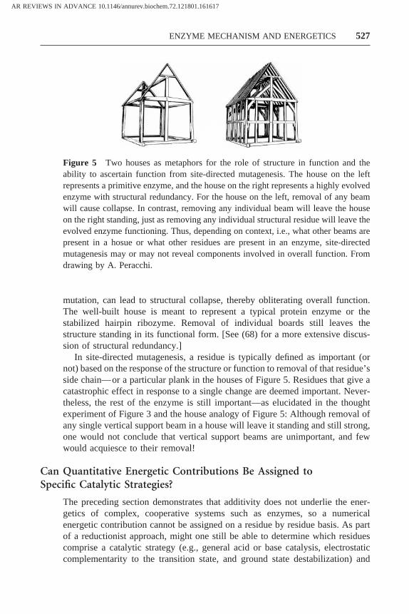
mutation, can lead to structural collapse, thereby obliterating overall function.The well-built house is meant to represent a typical protein enzyme or thestabilized hairpin ribozyme. Removal of individual boards still leaves thestructure standing in its functional form. [See (68) for a more extensive discus-sion of structural redundancy.]
In site-directed mutagenesis, a residue is typically defined as important (ornot) based on the response of the structure or function to removal of that residue’sside chain—or a particular plank in the houses of Figure 5. Residues that give acatastrophic effect in response to a single change are deemed important. Never-theless, the rest of the enzyme is still important—as elucidated in the thoughtexperiment of Figure 3 and the house analogy of Figure 5: Although removal ofany single vertical support beam in a house will leave it standing and still strong,one would not conclude that vertical support beams are unimportant, and fewwould acquiesce to their removal!
Can Quantitative Energetic Contributions Be Assigned toSpecific Catalytic Strategies?
The preceding section demonstrates that additivity does not underlie the ener-getics of complex, cooperative systems such as enzymes, so a numericalenergetic contribution cannot be assigned on a residue by residue basis. As partof a reductionist approach, might one still be able to determine which residuescomprise a catalytic strategy (e.g., general acid or base catalysis, electrostaticcomplementarity to the transition state, and ground state destabilization) and
Figure 5 Two houses as metaphors for the role of structure in function and theability to ascertain function from site-directed mutagenesis. The house on the leftrepresents a primitive enzyme, and the house on the right represents a highly evolvedenzyme with structural redundancy. For the house on the left, removal of any beamwill cause collapse. In contrast, removing any individual beam will leave the houseon the right standing, just as removing any individual structural residue will leave theevolved enzyme functioning. Thus, depending on context, i.e., what other beams arepresent in a hosue or what other residues are present in an enzyme, site-directedmutagenesis may or may not reveal components involved in overall function. Fromdrawing by A. Peracchi.
AR REVIEWS IN ADVANCE 10.1146/annurev.biochem.72.121801.161617
527ENZYME MECHANISM AND ENERGETICS

thereby quantitate the energetic contribution from this strategy? For numerousenzymes, it is known, for example, which residues donate or abstract a proton ingeneral acid or base catalysis. There are nevertheless limitations in our ability toassign catalytic function to specific residues, and our ability to assign energeticcontributions to specific catalytic strategies is even more limited. These limita-tions are illustrated in the following examples.
Experiments with a PI-specific phospholipase C are instructive with respect tofunctional interconnections between residues (74). Tsai and coworkers examinedthe catalytic histidines (H32, the general base, and H82, the general acid) as wellas two aspartate residues, D274 and D33, thought to hydrogen bond with thehistidines. Mutation of either “catalytic” histidine to alanine lead to a ratedecrease of 105, which lends support to the idea that the histidines are thecatalytic residues. However, mutating D274 to alanine caused a 104 fold drop inrate, and the D33A mutant had a 103 fold drop in rate. These data indicate thatmutation of the residues adjacent to the general acid and general base abrogategeneral acid and base catalysis, even though these are not the residues directlyinvolved in proton donation to or removal from the substrate. The catalysisassociated with proton removal or donation is not just a function of the residuethat accepts or donates the proton; it is also connected to properties of thesurrounding enzyme environment, i.e., the residues around the proton donor andacceptor that determine positioning, electrostatic potentials, and solvation.Although a catalytic value to acid/base catalysis or to a specific residue cannot beassigned from the above experiments, they, along with many other experiments,demonstrate the interconnectedness of the active site and the power of site-directed mutagenesis in uncovering these connections.1,2
Because multiple interactions influence a given catalytic strategy, it is impos-sible to separate the contribution of a given residue in the strategy. Furthermore,enzymes use multiple sets of catalytic strategies, and these strategies are alsointerconnected, preventing assignment of stabilization energies to a specificstrategy. This is illustrated by a hypothetical serine esterase. The active sitecontains a general base to remove the proton from the attacking serine residue,an oxyanion hole that stabilizes the development of negative charge on theincipient oxyanion, and several groups that bind and position the attacking serinewith respect to the ester carbon. It would be desirable to determine the amount
1In some cases there may be significant amounts of shared covalent character in hydrogenbonds, and additional proton rearrangements may accompany proton abstraction from ordonation to substrates. These possibilities further illustrate the limitations in discreteassignment of catalytic function. Elucidation of the nature of bonding in these situationsas the functional, structural, and energetic origins and consequences of the bondingrepresents an exciting challenge (75–77).2Other elegant studies have combined site-directed mutagenesis with isotope effects andvariation in the identity of the leaving group to reveal roles of residues in general acidcatalysis (78–81).
AR REVIEWS IN ADVANCE 10.1146/annurev.biochem.72.121801.161617
528 KRAUT y CARROLL y HERSCHLAG

of catalysis provided by each strategy, but a problem arises. The hydrogen bonddonors in the oxyanion hole help position the ester carbon with respect to theserine nucleophile in addition to stabilizing charge buildup on the transition stateoxyanion. Similarly, the residue that acts as a general base, because of itsplacement in the active site, helps position the incipient oxyanion with respect tothe residues that make up the oxyanion hole, aiding this catalytic function inaddition to directly facilitating proton removal. Thus, the catalytic strategies areinterconnected: Mutating a group involved in one type of catalysis can adverselyaffect another catalytic strategy as well. The energetic contributions of eachcatalytic strategy are not cleanly separable.
In summary, while it is often possible to assign direct chemical participationin catalysis to a particular residue, the residue’s capability to act depends on itsneighbors and surroundings. Thus, responsibility for a catalytic strategy cannotbe assigned to a single residue. Furthermore, the catalytic strategies that anenzyme uses to facilitate reaction are not independent of one another. Thisfunctional and energetic interdependency prevents a quantitative dissection ofenzymatic catalysis into types of stabilization.
Assigning the Signature of a Residue: Dissecting Bindingand Catalytic Contributions
The above examples demonstrate the limitations in assigning energetic contri-butions to individual residues and to individual catalytic strategies. One mightalso like to know which reaction step or steps a particular residue facilitates:Does the residue contribute to the binding or chemical step, or both? Such apartitioning of function into neat categories would be highly appealing from areductionist standpoint, distinguishing the residues (or functionalities) responsi-ble for getting the substrate localized to the active site from those that actuallycarry out the chemical transformation. Indeed, in the literature this sort ofassignment often follows site-directed mutagenesis experiments; residues that,when mutated, give increases in KM are typically ascribed roles in binding, andresidues that give decreases in kcat are typically ascribed roles in the chemicalstep. (We assume for simplicity that KM 5 Kd and kcat 5 kchemical step.)
Albery & Knowles, following their classic determination of the triose phos-phate isomerase (TIM) kinetic mechanism in the late 1970s, formalized thisclassification and suggested potential evolutionary ramifications (Figure 6) (31,32). They noted that the addition of a residue in the course of evolution couldstabilize the ground state and transition state equally (Figure 6a, uniformbinding) or could stabilize the transition state without affecting the ground state(Figure 6b, specific transition state stabilization); a residue could also give amixed effect, stabilizing both the ground and transition states but providing morestabilization to the transition state (Figure 6c, differential binding).
As noted above, these categories correspond to what are commonly consid-ered binding and catalytic residues. This mechanistic distinction however, was atodds with the perspective articulated by Jencks. In 1973 Jencks stated “on closer
AR REVIEWS IN ADVANCE 10.1146/annurev.biochem.72.121801.161617
529ENZYME MECHANISM AND ENERGETICS

examination. . . the classical separation of considerations of enzymatic catalysisinto the specific binding of substrates and chemical catalysis breaks downcompletely (15).”
Although site-directed mutagenesis had not yet been applied to enzymes,experiments with substrate analogs had clearly established the interconnection ofbinding and catalysis. For example, addition of amino acid residues remote fromthe site of chemical transformation for elastase substrates increased the rate oftransformation of the already bound substrate (kcat); similarly, the binding oftransition state analogs but not substrates was enhanced (82, 83). The most basicpoint emphasized by Jencks was that binding interactions (through the intrinsicbinding energy provided) can facilitate the chemical transformation (16). Thebinding interactions can help by positioning substrates with respect to oneanother, by positioning substrates with respect to functional groups on theenzyme, and by enforcing electrostatically or sterically destabilizing ground stateinteractions that are relieved as the substrate undergoes electronic and geometricrearrangement in the transition state (16).
DIRECT DEMONSTRATION OF THE USE OF BINDING INTERACTIONS FOR CATALYSIS
More recent experiments have directly demonstrated this interconnectionbetween binding interactions and the chemical transformation of bound sub-
Figure 6 Free energy-reaction profiles demonstrating potential catalytic effects ofmutants, as described by Albery & Knowles (31, 32). The profile for a primitive enzyme isshown in black, and possible results from introduction of potentially advantageous muta-tions are shown in magenta. In typical site-directed mutagenesis experiments, the effectswould be in the opposite direction, starting with the magenta profile and going to the blackone. (A) Uniform binding. All enzyme-bound species are stabilized equally by the mutation,accelerating a reaction with subsaturating concentrations of substrate, but not with saturat-ing substrate (see also Figure 9). (B) Specific transition state stabilization. The mutationcauses stabilization of the transition state without stabilization of the ground state. Thistransition state interaction leads directly to enhanced reaction of the bound substrate, and italso increases the rate of reaction of unbound substrate. (C) Differential binding. There isa continuum of possible energetic effects between the extremes shown ina andb in whichmutations can stabilize both transition state and the ground state but provide greaterstabilization to the transition state.
AR REVIEWS IN ADVANCE 10.1146/annurev.biochem.72.121801.161617
530 KRAUT y CARROLL y HERSCHLAG

strates (84). The RNA enzyme derived from theTetrahymena thermophilagroupI intron catalyzes a reaction analogous to the first step in intron self-splicing(Equation 1). The catalytic mechanism of this RNA enzyme has been studiedextensively, using presteady state kinetics to establish a complete kinetic andthermodynamic framework for individual reaction steps and a plethora ofsubstrate analogs with single functional group substitutions to probe interactionswith the RNA enzyme and their energetic consequences (85–88).
CCCUCUPAAAAA 1 GOH 3 CCCUCUOH 1 GpAAAAA 1.
(S) (P)
Binding of an oligonucleotide 59-splice site analog (S) occurs in two distinctsteps (Figure 7a). In the first step, S forms a duplex with a complementarysequence of the RNA enzyme to form the open complex. In the second step, thisduplex docks into the core of the enzyme and makes tertiary interactions; Figure7b shows the functional groups of the duplex involved in these binding interac-tions, which include several 29-hydroxyl groups, and the exocyclic amino groupof G22, which forms a GzU wobble pair to specify the cleavage site. The twobinding states are key to the analysis described below, because the boundoligonucleotide substrate is either positioned for reaction (closed complex) orlocalized to the enzyme only by base pairing and thus bound but not positionedfor reaction (open complex).
Figure 7 Substrate binding to theTetrahymenagroup I RNA enzyme. (a) Bindingoccurs in two steps. First the oligonucleotide substrate (S) binds to the internal guidesequence (IGS) of the ribozyme via base-pairing to form the open complex (Kd
IGS).The resulting helix then docks into the active site of the enzyme to form the closedcomplex (Kdock). (b) Schematic diagram showing functional groups in the substratehelix that make tertiary interactions with the active site. Only the groups varied in theworks referred to in the text are shown, including the -3 (three residues 59 of thesubstrate’s cleavage site) 29-hydroxyl group and the exocyclic amino group of G22on the enzyme strand (84). The energetic cost (in kcal mol-1) of replacement of eachgroup by a hydrogen atom is also shown (84 and references therein).
AR REVIEWS IN ADVANCE 10.1146/annurev.biochem.72.121801.161617
531ENZYME MECHANISM AND ENERGETICS

In the course of probing the roles of individual functional groups (andattempting to assign discrete roles in binding and catalysis), it was discoveredthat addition of a single functional group to the substrate, the 29-hydroxyl groupof U(-3), could give either uniform binding or specific transition state stabiliza-tion, depending on the context, i.e., depending on the other groups and interac-tions present (Figure 8a). [U(-3) refers to the substrate position three residues 59of the cleavage site (Figure 7b).] With the wild-type enzyme, addition of this29-hydroxyl gave uniform binding, as witnessed by a decreased dissociationconstant without any change in the rate of the chemical step (Figure 8a, wildtype). In a mutant with the 29-hydroxyl and exocyclic amino group of G22 absentto give deoxyinosine (dI22), addition of the 29-hydroxyl at U(-3) gave the sametotal energetic contribution of 1 kcal mol21 as the wild-type, but the energy wasexpressed only in increasing the rate of the chemical step, thereby giving specifictransition state stabilization (Figure 8a, dI22). A group remote from the site ofchemical transformation giving uniform binding in the context of the wild-typeenzyme, and thus typically thought of as providing binding interactions, caninstead specifically stabilize the transition state in a mutant context.
The explanation for these results provides insight into the interconnectionbetween binding and catalysis. Recall the two binding states of S, open andclosed (Figure 7a). In the otherwise wild-type context, the duplex with S formsenough tertiary interactions such that it docks and remains in the closed complexwhether or not the 29-hydroxyl at U(-3) is present (Figure 8b). Thus, evenwithout the U(-3) 29-hydroxyl group, the substrate is positioned for reaction. Thedocked substrate makes the same remote interactions in the transition state as inthe ground state, so addition of the U(-3) 29-hydroxyl group gives the same 1 kcalmol21 stabilization to both states and uniform binding is observed. In the mutantcontext, removal of two additional tertiary contacts from the docked complex hastipped the energetic balance so that now the open or unpositioned complex is thestable ground state conformation (Figure 8c). Addition of the 29-hydroxyl atU(-3) has no effect on binding—it is not sufficient to tip the balance back to theclosed complex where an interaction with the enzyme core can be made.Nevertheless, the reaction must occur through the docked complex. Thus, the
™™™™™™™™™™™™™™™™™™™™™™™™™™™™™™™™™™™™™™™™™™™™™™™™™™™™™™™™™™™™™™™™™™™™™™™™3Figure 8 The same functional group in different enzyme contexts can provide uniformbinding or specific transition state stabilization. Free energy profiles (a) show that addingthe same 29-hydroxyl group (going from red to black) at the -3 position of the oligonucle-otide substrate (Figure 7) of theTetrahymenaRNA enzyme results in uniform binding inthe wild-type enzyme and specific transition state stabilization in the dI22 mutant (missingthe 29-hydroxyl group and the exocyclic amino group at position 22). Models of thesignificantly populated bound states of the substrate for the wild type (b) and mutant dI22(c) that explain the respective uniform binding and specific transition state stabilizationphenotypes upon addition of the 29-hydroxyl group at position -3 (shown in red) (84).
AR REVIEWS IN ADVANCE 10.1146/annurev.biochem.72.121801.161617
532 KRAUT y CARROLL y HERSCHLAG

AR REVIEWS IN ADVANCE 10.1146/annurev.biochem.72.121801.161617
533ENZYME MECHANISM AND ENERGETICS

U(-3) 29-hydroxyl interaction is made in the transition state, thereby providingspecific transition state stabilization.
Indeed it was shown that each of the functional groups contributingbinding energy to docking can contribute to binding or catalysis; this resultsin a uniform binding or a specific transition state stabilization phenotypedependent only on the total binding energy available from the other func-tional groups present. If few are present, the duplex is undocked and specifictransition state stabilization is observed; if sufficient groups are alreadypresent to favor the docked complex, then uniform binding is observed.Functional groups that make binding interactions remote from the site ofchemical transformation can contribute specific transition state stabilizationinstead of uniform binding.
The fundamental contribution of binding interactions to catalysis are maskedby the presence of other binding and positioning interactions. Only when asufficient number of these are removed (and an energetic threshold is crossedsuch that the unpositioned state is more stable than the positioned state) is theunderlying contribution to catalysis with bound substrate revealed; what appearsto be a uniform binding interaction is shown to provide transition state stabili-zation in a different context.
As Albery & Knowles noted, a uniform binding contribution such as that seenin the wild-type ribozyme does not provide catalysis for the enzyme/substratecomplex, because the barrier for the chemical step is not lowered (Figure 9) (31).Nevertheless, uniform binding contributions can increase reaction from freeenzyme and substrate (kcat/KM), and a “binding” residue that gives a uniformbinding phenotype when mutated in the context of a modern day enzyme couldhave been selected early in evolution on the basis of a contribution to transitionstate stabilization via substrate positioning. The later selection for residues thatprovide additional binding interactions would mask this early and continuing rolein catalysis.
These possible changes in phenotype over the course of evolution reveala basic limitation of site-directed mutagenesis. Removal of one or tworesidues may be insufficient to unmask the catalytic contributions of “bind-ing” residues, and removal of more residues almost invariably leads torearrangement of bound complexes to a variety of non-productive andpartially productive complexes that obscure straightforward energetic inter-pretation [e.g., (89, 90)].
In summary, residues involved in positioning, as in the example above, playcritical roles in catalysis. Their importance, however, is not readily uncovered bysite-directed mutagenesis. The Tetrahymena RNA enzyme has properties, suchas simple two-state binding in a positioned or an unpositioned complex, thatprovided an opportunity to directly demonstrate the inextricable link betweenbinding and catalysis, a link that is at the heart of enzymatic catalysis as furtherelaborated in the following section.
AR REVIEWS IN ADVANCE 10.1146/annurev.biochem.72.121801.161617
534 KRAUT y CARROLL y HERSCHLAG

Summary
We have considered three ways to subdivide enzyme function: assigning quan-titative energetic contributions to individual residues; assigning quantitativeenergetic contributions to the catalytic strategies used by the enzyme; andassigning energetic contributions to residues in binding versus chemical reactionsteps. Each is not possible.
Correspondingly, limitations of site-directed mutagenesis have been revealed.This approach, while providing many important insights, cannot provide a uniqueenergetic signature for a residue or a catalytic strategy. Site-directed mutagenesiscan reveal the importance of an active site residue for catalysis; for example,replacement of a Glu residue acting as a general base catalyst by Ala will greatlycompromise catalysis. However, roles of “binding” residues in catalysis can bemasked by the presence of multiple positioning interactions, and structuralrearrangements upon removal of the interacting groups can obscure or amplifytheir underlying contributions.
In all cases the context, i.e., the properties of the surrounding residues, mattersquantitatively and/or qualitatively and prevents a unique breakdown of enzyme
Figure 9 Free energy reaction profiles demonstrating the requirement for preferentialtransition state stabilization relative to ground state stabilization for catalysis. For simplic-ity, assume that the chemical step is rate-limiting, in this figure and throughout the review.(a) The uncatalyzed reaction of S, with an activation barrier ofDGa
‡ and a rate constant ofka. (b) Enzyme 1 (E1) stabilizes the ground state (GS) and transition state (TS) equally,leaving the reaction barrierDGb
‡ equal to the reaction barrierDGa‡ for the uncatalyzed
reaction; this enzyme is not a catalyst askb 5 ka. (c) Enzyme 2 (E2) stabilizes the transitionstate more than it stabilizes the transition state such thatDGc
‡ , DGa‡ so that the rate constant
kc is larger thanka. Thus, this enzyme is a catalyst.
AR REVIEWS IN ADVANCE 10.1146/annurev.biochem.72.121801.161617
535ENZYME MECHANISM AND ENERGETICS

function into independent, energetically additive components. This complexityarises from the cooperative nature of enzyme structure and function. Asdescribed in the final section of this review, multiple comparisons of theenergetics and physical properties of wild-type and mutant enzymes with cognateand modified substrates are essential tools in revealing the behavior and prop-erties of these cooperative systems.
THE DISTINGUISHING PROPERTIES OF ENZYMES:COMPARISON TO SMALL MOLECULE CHEMICALCATALYSTS
The task of understanding enzyme catalysis is rendered more difficult by theabsence of a single answer attainable by quantitative dissection into individualcontributions. In the absence of an absolute answer, understanding must besought through relativistic analysis. Multiple comparisons and multiple types ofcomparisons are needed to provide different perspectives and insights. In thissection, we define a general comparison with the question: What distinguishesenzymes from simple, small molecule chemical catalysts? To address thisquestion, we consider a series of model enzymes that successively build infeatures, that are associated with enzymatic catalysis, and that ask for eachmodel: Can it account for the known properties of enzyme catalysis? The modelshighlight catalytic properties of enzymes that are understood and also illuminateareas that remain to be elucidated. This sets the stage for the final section of thisreview, where the questions, comparisons, and approaches that will be requiredto further our understanding of enzymatic catalysis are considered.
A Hypothetical Enzyme and Reaction for Comparison
The enzyme models are used to recreate the hypothetical enzyme shownschematically in Figure 10. This enzyme catalyzes an enolization reaction,analogous to triose phosphate isomerase, ketosteriod isomerase, and many otherwell-studied enzymes (6, 47, 91). Structural work on this hypothetical enzymeidentified a His residue at the active site acting as a general base to deprotonatethe methylene group and a Gln residue hydrogen bonding to and therebystabilizing the incipient anionic enolate oxygen atom. In addition, site-directedmutagenesis of these so-called “catalytic” residues to Ala gave dramatic reduc-tions in catalysis, 106-fold for His and 104-fold for Gln upon side chain removal.As the overall rate enhancement observed by this enzyme is 1010, it is temptingto conclude that all of the catalytic power of this enzyme has been identified.There is a basic flaw with this logic, however. The catalysis does not come solelyfrom what are referred to as the “catalytic groups.” This point was emphasizedin the previous section, and its fundamental relationship to enzymatic catalysis ishighlighted in the models below.
AR REVIEWS IN ADVANCE 10.1146/annurev.biochem.72.121801.161617
536 KRAUT y CARROLL y HERSCHLAG

Model I. “Catalytic” Residues
Taking literally the notion that His and Gln are the catalytic residues of thehypothetical enzyme (Figure 10), it follows that adding both histidine andglutamine to solution should provide catalysis that matches that of the enzyme(Figure 11). Of course this is not the case. The “catalytic” residues only allowenormous rate enhancements when placed within the context of the foldedenzyme, as shown also by the Ala mutagenesis example above (Figure 3). Thisrealization leads to a second model.
Model II. Positioned “Catalytic” Residues
Clearly the “catalytic” groups of enzymes need to be positioned with respect toone another. This is accomplished in Model II (Figure 12). The second modelenzyme has a hypothetical framework with covalent interconnections that giveprecise positioning of the His and Gln residues without the large size necessaryto form tertiary structure in a protein. This allows us to separately address thepotential special effects of the protein framework later and thus to conceptuallydistinguish these effects. Imagine that advances in synthetic chemistry allowsconstruction of a nano-scaffold that provides precise positioning of the “catalyt-ic” residues in this model.
How does the Model II enzyme rate as a catalyst? Although the His and Glnresidues are properly positioned, there is nothing to position the substrate with
Figure 10 Hypothetical enzyme used as a starting point to develop models explain-ing enzymatic activity. The enzyme catalyzes an enolization reaction using histidineas a general base to remove the proton alpha to the carbonyl group and glutamine asa hydrogen bond donor to stabilize developing negative charge on the carbonyloxygen atom. Removal of these two residues leads to complete loss of catalysis.
AR REVIEWS IN ADVANCE 10.1146/annurev.biochem.72.121801.161617
537ENZYME MECHANISM AND ENERGETICS

respect to these catalytic groups. While reaction may be very efficient when thesubstrate is appropriately positioned within the active site, such positioningis highly improbable for the Model II enzyme. This problem is corrected inModel III.
Model III. Positioned “Binding” and “Catalytic” Residues
Model III maintains the His and Gln positioning from Figure 12 but introducesbinding interactions with the groups that flank the enolization site (Figure 13).Two successive versions of Model III are shown. In the first (Figure 13a), thesebinding interactions are sufficient to localize the substrate to the active site underthe hypothetical physiological conditions. However, even though the substrate isin the active site most of the time, it is not ordinarily positioned for reaction, asrepresented by its misalignment and mobility in the ground state (Figure 13a).This model illustrates that optimal catalysis requires positioning beyond the lossof translational entropy, as has been described previously in several differentways [e.g., (22, 92, 93–95)].
The situation of Model IIIa is precisely analogous to the Tetrahymena RNAenzyme example above: In a mutant context the substrate was localized to theenzyme in the open complex but not positioned for reaction (Figure 8). As for the
Figure 11 Model I: “catalytic residues” in solution. Histidine and glutamine (onlytheir side chains are shown) are added to a solution containing the substrate.
AR REVIEWS IN ADVANCE 10.1146/annurev.biochem.72.121801.161617
538 KRAUT y CARROLL y HERSCHLAG

RNA enzyme, the solution is to add more binding interactions (i.e., interactionsremote from the site of chemical transformation) to provide the necessaryfixation of the substrate within the active site (Figure 13b). These additionalbinding interactions, illustrated by the green lines in Model IIIb, position thebound substrate correctly for reaction and thereby speed the chemical transfor-mation of the bound substrate.3
In Model III the groups that contribute to binding and reaction of the boundcomplex are interconnected energetically and functionally. The “binding” resi-dues contribute to catalysis, and the so-called “catalytic” Gln helps in positioningby providing an additional attachment point for the substrate. This functional
3Binding interactions and binding energy in actual enzymes can be added not only byintroducing residues that directly contact the substrate but also by adding more distantresidues that help position the directly interacting residues for binding and catalysis. Thislater indirect mechanism was elegantly demonstrated by sequence and structural changesof a catalytic antibody through its maturation (96, 97).
Figure 12 Model II: positioned “catalytic residues.” The histidine and glutamineresidues are positioned on a rigid low molecular weight framework such that they arecorrectly oriented for reaction. However, the model cannot position the substraterelative to the so-called catalytic groups.
AR REVIEWS IN ADVANCE 10.1146/annurev.biochem.72.121801.161617
539ENZYME MECHANISM AND ENERGETICS

AR REVIEWS IN ADVANCE 10.1146/annurev.biochem.72.121801.161617
540 KRAUT y CARROLL y HERSCHLAG

interconnectedness would not be revealed by single site-directed mutants, but itis nevertheless at the heart of enzyme function.
Model IV: Tuning Interactions and Binding Energy
Would the last model perform catalysis as efficiently as an actual enzyme? Thereare two likely inadequacies, both related to the strength of binding interactions,addressed and corrected below. (Additional features lacking in these models mayor may not prove to be important for catalysis; these features at the frontier ofscientific understanding are introduced and discussed in the last section of thisreview.)
Enzymes need to catalyze reactions relative to reactions in aqueous solution,and preferential stabilization of the transition state over the ground state isrequired to accomplish this (Figure 9). In solution, reactions for amide and esterhydrolysis and proton abstractiona to carbonyl groups (as in Figure 14) occurwith oxyanion-like transition states. There is likely to be hydrogen bond donationfrom at least two water molecules to the oxyanion; presumably a penalty inelectrostatic interaction energy would be paid, relative to aqueous solution, ifonly a single hydrogen bond were donated by the enzyme. Indeed, in active sitescarbonyl oxygen atoms that develop oxyanion character typically accept twohydrogen bonds from the enzyme (98, 99). Therefore a second hydrogen bonddonor to the oxyanion is added in Model IVa (Figure 15a).4 Related issues ofelectrostatic stabilization within the active site are treated in greater depth in thefinal section.
The second inadequacy derives, paradoxically, from binding interactionsbeing too strong. Speaking anthropomorphically, an enzyme wants to maximizebinding interactions with its substrates to maximize the energy potentially
4In the case of ketosteroid isomerase, mutation of the catalytic base (Asp38) and ahydrogen bonding group (Tyr14) seemed to account for all of the catalytic enhancementof the enzyme, but it was later found that another group (Asp99) also donated a hydrogenbond to the enolate oxygen atom and that mutation of this residue was detrimental tocatalysis (100, 101).
4™™™™™™™™™™™™™™™™™™™™™™™™™™™™™™™™™™™™™™™™™™™™™™™™™™™™™™™™™™™™™™™™™™™™™™™™Figure 13 Model III: substrate positioning and catalysis. (a) This framework provides alimited number of interactions with nonreactive portions of the substrate. These interactionsare sufficient to localize the substrate to the active site, but considerable motion remains(depicted by motion lines). Thus, the substrate will be properly aligned for catalysis only afraction of the time. (b) Groups making additional substrate interactions are introduced.This allows the substrate to be bound in a conformation that is aligned for reaction, with theproton that will be removed positioned with respect to the histidine lone pair and thecarbonyl oxygen atom positioned with respect to the active site glutamine (compare toFigure 8).
AR REVIEWS IN ADVANCE 10.1146/annurev.biochem.72.121801.161617
541ENZYME MECHANISM AND ENERGETICS

available for catalysis and specificity (16, 29). In Model IIIb binding interactionswere strengthened to position the substrate in a reactive conformation. Superfi-cially one would expect increased precision in the alignment of active siteresidues to provide precise complementarity for the desired substrate and anability to provide maximal preferential stabilization of the transition state relativeto the ground state, thereby increasing specificity and catalysis. However, thehighly precise positioning of the enzyme models creates problems, paradoxicallydue to their limited flexibility.
Maximal binding interactions will require, in general, interactions of theenzyme on all sides of the substrate. Enzyme flexibility is then needed to allowsubstrate ingress and product egress. Indeed, enzymatic flap closures and hingemotions to accomplish this are extraordinarily common (102–107). A joint istherefore introduced in the otherwise rigid enzyme to give open and closed statesthat allow both ready access to the binding pocket and recognition of all elementsof the substrate (Figure 15b, Model IVb).
The precise positioning of binding groups in the new model enzyme nowseems optimal to position the substrate for reaction in the closed conformation.Nevertheless, problems derived from binding interactions persist for both cata-lytic turnover and specificity. Precise positioning can cause binding to be toostrong, because the modest structural rearrangements that mitigate against verystrong binding in real situations are eliminated in the more rigid model enzyme.With very strong binding, product dissociation is slowed and the maximalturnover rate can be lowered [e.g., (85, 108, 109)]. Indeed, it has been suggested
Figure 14 Multiple hydrogen bonds to anionic oxygen atoms. A solution enoliza-tion reaction comparable to that used to develop the enzyme-like models (Figure 10).Hydrogen bonds to the carbonyl oxygen atom in the ground state get stronger in thetransition state (as depicted by the larger dots).
AR REVIEWS IN ADVANCE 10.1146/annurev.biochem.72.121801.161617
542 KRAUT y CARROLL y HERSCHLAG

Figure 15 Model IV: tuning interactions and binding energy. (a) A second hydrogenbonding interaction is added to the negative charge forming on the carbonyl oxygen atom.(b) A hinge point is added (arrow); this allows the enzyme to open so that substrate andproduct can enter and exit the active site while maintaining the specific interactions madeon all sides of the substrate. (c) The general base is changed to aspartate, which has a fullnegative charge. Burial of this negative charge in the ground state, upon substrate binding,is destabilizing relative to a neutral group or to interactions in solution; this is representedby the purple lines. This interaction preferentially weakens ground state binding, whichallows for rapid turnover and high specificity.
AR REVIEWS IN ADVANCE 10.1146/annurev.biochem.72.121801.161617
543ENZYME MECHANISM AND ENERGETICS

that limitations of antibodies as catalysts may arise from a too-rigid structuraltemplate that leads to slow product dissociation (110; see also 111). Too-strongbinding also decreases specificity, because even substrates that only make asubset of the binding interactions will react faster than they dissociate, and bothdesirable and undesirable substrates can therefore react at the rate of diffusionalbinding (104, 112). To remedy these problems, motion is introduced into thebinding residues in Model IVb, thereby weakening binding sufficiently to allowundesirable substrates to dissociate in preference to reacting (104, 113).
Another way to weaken binding interactions without compromising stabili-zation of the transition state is to replace the His general base with a negativelycharged Asp residue (Figure 15c, Model IVc). Ground state binding is weakenedbecause the negatively charged Asp is positioned adjacent to a proton of thehydrophobic substrate methylene group by the combination of the model’sstructural scaffold and its binding interactions. In the transition state, thisdestabilizing interaction is relieved as proton transfer from the methylene groupneutralizes the Asp residue.5 Ground-state destabilization has been proposed innumerous enzyme systems [e.g., (114–116)] and can contribute to an enzyme’sability to provide preferential stabilization of a reaction’s transition state relativeto its ground state (Figure 9) [see (16) for a more detailed discussion of therelevant energetics].
Summary
A progression of enzyme models has been presented to help identify andunderstand the features of enzyme catalysts that are special, or distinct, relativeto simple chemical catalysts. Most fundamental is the energetic and functionalinterconnection of binding and catalysis. This property of enzymes intimatelylinks specificity and catalysis, because only the correct substrates make theinteractions that lead to efficient catalysis. The inextricable linkage betweenbinding and catalysis was noted early on by Polanyi, Pauling, and Jencks (16, 27,28, 117) and has been highlighted by many subsequent enzymologists [e.g., (13,29, 35, 93)].
Returning to the question of whether our most advanced enzyme modelswould rival natural enzymes in specificity and catalysis, the answer is not known.Instead, a different question is posed below: What features of actual enzymes arenot accounted for by the simplified enzyme models? This comparison helpsidentify and frame central questions that remain about enzymatic catalysis andthe properties and behavior of enzymes.
5Assessment of ground state destabilization requires comparison to some standard con-dition (e.g., aqueous solution or gas phase), and one can imagine making differentcomparisons in the same system, some of which give ground state destabilization andothers of which do not. Further, whether or not ground state destabilization occurs in anyspecific case will depend upon what other interactions are made with the Asp residue andupon the environment surrounding the active site.
AR REVIEWS IN ADVANCE 10.1146/annurev.biochem.72.121801.161617
544 KRAUT y CARROLL y HERSCHLAG

MEETING THE CHALLENGES OF UNDERSTANDINGENZYME MECHANISM: A MODERN PERSPECTIVE
The face of mechanistic enzymology has changed dramatically over the past twodecades. Researchers have gone from speculation and indirect assays for residuesinvolved in the chemical catalysis to direct tests via site-directed mutagenesis[e.g., (33, 118, 119)]. Mechanistic schemes have progressed from cartoonrepresentations of enzymes to atomic resolution supported by X-ray crystallog-raphy. Presteady state kinetics has supplanted steady state kinetics for in-depthenzymological investigations, allowing reactions to be defined in terms ofmicroscopic steps. These advances leave us poised to attain an understanding ofenzyme mechanism at unprecedented depth.
There are, nevertheless, critical limitations of even these contemporaryapproaches. Site-directed mutagenesis allows a residue to be removed and effectson function the be probed, but this response is read out in energetic terms (fromrate and equilibrium effects), which must then be given molecular interpretations.The structures of wild-type and mutant enzymes can be obtained, but these arestatic pictures that do not capture the catalytic process.
How can we do this better? A bridge between structure and energetics mustbe created through both an understanding of dynamics and an understanding ofthe physical properties of enzymes that goes beyond a description of thedifferences between mutants and the wild-type (Scheme 1). In this section,questions at the boundaries of the understanding of enzyme mechanism andenergetics are raised. These questions are framed both by the intellectual historyof mechanistic enzymology and also by the enzyme-like models developed in thelast section (Figures 11, 12, 13, 15). The primary differences between actualenzymes and these enzyme-like models lie in what surrounds the groups directlyinteracting with substrates and the rigidity or flexibility of the interconnectionsbetween these groups. Thus, these comparisons may be particularly adept atrevealing the functional and behavioral consequences from the enzyme’s molec-ular architecture beyond the active site.
In addition to formulating the right questions to provide insights into thefunction and behavior of enzymes, a deep understanding of enzyme mechanismand energetics will require developing existing and new experimental andcomputational tools, and then bringing these tools to bear on these and otherquestions. Below, we first describe tools that are sufficiently developed torecognize their power in this pursuit and then turn to the pressing questions inmechanistic enzymology that will require our attention, imagination, and inven-tiveness in the coming years.
Twenty-First Century Technology For Enzymology
X-RAY CRYSTALLOGRAPHY As noted above, high resolution X-ray crystallo-graphic structures of enzymes, often with substrate or transition state analogs
AR REVIEWS IN ADVANCE 10.1146/annurev.biochem.72.121801.161617
545ENZYME MECHANISM AND ENERGETICS

bound, are now common. More structures of individual enzymes that are underintense mechanistic scrutiny are needed: structures of one enzyme with sub-strates, products, and a variety of analogs bound. No transition state analog isperfect, so rearrangements of the enzyme structure often occur to accommodatethese imperfections.6 Thus, structures with multiple analogs should be sought tohelp unravel the types of rearrangements likely to occur within the enzyme [e.g.,(120)]. And these structures are needed at ultrahigh resolution, resolution higherthan 1 Å [e.g., (121, 122)]. Because many of the interactions of interest arehydrogen bonds and most enzymatic reactions involve transfer of one or moreprotons, the inability to directly observe the hydrogen’s electron density in lowerresolution structures limits mechanistic interpretation. At 0.9 Å resolution, abouthalf of the hydrogens have assignable electron density (G.A. Petsko, personalcommunication); also, neutron diffraction, while technically challenging, allowsobservation of the hydrogen atoms (123). Another value of ultrahigh resolutionX-ray structures is a reduction of the error in measuring interatomic distances, asprecise distances can be of great value in understanding hydrogen bondinginteractions and the positioning of groups for bond formation (G.A. Petsko,personal communication; 76, 77, 124).
ENZYME DYNAMICS AND NMR Energy cannot be discerned even from thehighest resolution structure. One fundamental reason for this is that structureis not static, and free energy is a function of enthalpy and entropy, i.e., thenumber of substates accessible and the energy of each of these states. It isclear that enzymes are highly dynamic entities, held together by noncovalentinteractions that are weak compared to covalent bonds. Thus, to understandthe basic functioning of enzymes in terms of energetics and the conforma-tional states utilized in function, the dynamic behavior of enzymes must beinterrogated. Currently the most powerful approach to study dynamics isNMR, which provides (in principle) the ability to probe motions on time-scales ranging from picosecond to millisecond and greater for each residue(125–129). Nevertheless, bringing such extensive data together into dynamicmolecular models is a formidable challenge (see Computation of EnzymeBehavior and Function below).
SITE-DIRECTED MUTAGENESIS WITH UNNATURAL AMINO ACIDS Limitations ofsite-directed mutagenesis have been highlighted throughout this review. Site-directed mutagenesis cannot provide a reductionist dissection and understandingof enzymatic catalysis (as is sometimes claimed or implied), and the functionalimportance of certain enzymatic features are masked from discovery by site-
6Rearrangements of noncovalent connections are typically easier than rearrangement ofcovalent bonds. Thus rearrangements of the packing and positioning within the enzyme ismore likely than substantial rearrangement of the covalent bonds of imperfect transitionstate analogs.
AR REVIEWS IN ADVANCE 10.1146/annurev.biochem.72.121801.161617
546 KRAUT y CARROLL y HERSCHLAG

directed mutagenesis. Nevertheless, site-directed mutagenesis has proven enor-mously powerful in identifying residues with direct roles in catalysis. Further,assessing the functional, energetic, and structural response to a vast number ofmutations has provided vital information about the behavior of the proteinscaffold [e.g., (130–132)].
But despite the usefulness of generalities from site-directed mutagenesis, thescale of the changes introduced often renders interpretation difficult. For exam-ple, a Glu residue thought to act as a general base is often replaced by Ala. In thismutant the ability of the group to accept a proton is not altered; rather, it isobliterated. Further, residues around the active site can rearrange in response toremoval of the two hydrogen-bond-accepting oxygens of Glu (Scheme 2a). Evenif an apparently more conservative change is made of Glu to Gln, two (or three)hydrogen bond acceptors are lost, and two hydrogen bond donors are added;changes that are also likely to lead to substantial destabilization and localrearrangement. If the carboxylate functionality is instead maintained by mutatingto Asp, the situation is no better. Now a conformational rearrangement orintervening solvent is required to allow the Asp to act as a general base. Indeed,there is ample evidence for active site rearrangement in response to mutation, andremaining active site functional groups have even been known to rearrange totake the place of the removed group (133).
What we would like to be able to do is to alter, in an incisive and systematicfashion, the properties of a particular enzymatic functional group. Instead ofremoving the general base, its electronic properties could be varied withoutvarying [or with minimal variation in (134)] its size and geometry (Scheme 2b).The response to this variation can be compared to that observed in relatedsolution reactions. The behavior of this series of analogs can also be comparedwith different substrates and with enzyme variants at other positions. Thesecomparisons can help reveal the interplay of general base catalysis with othercatalytic and structural features of the enzyme and substrate. This approachmirrors that of physical organic chemistry, which has uncovered much about
Scheme 2.
AR REVIEWS IN ADVANCE 10.1146/annurev.biochem.72.121801.161617
547ENZYME MECHANISM AND ENERGETICS

solution reactions through careful and systematic variation of reactants andconditions (62, 135).
Accomplishing these goals will require ready access to series of unnaturalamino acids (and even nonamino acids incorporated into the peptide backbone).And large quantities of protein will be needed so that structural and spectroscopicwork can be carried out alongside functional assays. Fortunately, solid phasesynthesis and intein ligation methods are emerging that promise to provide readyaccess to the large amounts of unnaturally substituted proteins that will berequired for this next level of mechanistic analysis [(136–139); see also (140)].
VIBRATIONAL PROBES: SPECTROSCOPY AND ISOTOPE EFFECTS Changes in NMRchemical shifts for residues extending well beyond the site of mutation suggestthat mutagenesis changes interactions throughout the protein, even in cases inwhich no structural perturbation is evident from crystallography [e.g., (141)].And even the most conservative unnatural amino acid substitutions still leave onecomparing properties of a wild-type and mutant enzyme (Scheme 1).
Vibrational probes have the special quality of allowing direct measurement ofbonding properties without recourse to mutation. Because bond vibrations arehighly sensitive to the molecular surroundings, information is presented about theenvironment felt within the enzyme. This may allow one of the most vexingquestions in enzymology to be addressed: What is the solvation behavior in anenzyme active site? This question is raised below.
PRESTEADY STATE KINETICS AND SINGLE MOLECULE MEASUREMENTS In the pastdecades, presteady state methods, which allow the examination of individualreaction steps, have continued to improve in terms of equipment availability,background literature, lower sample requirements, and data fitting approaches(142, 143). Most recently single molecule fluorescence approaches have begun toemerge in enzymology (144–147). Single molecule approaches provide a poten-tial window to intermediate and transient states that do not accumulate and allowdissection of population averages into the behavior of the individual constituents.
COMPUTATION OF ENZYME BEHAVIOR AND FUNCTION Enzymes are complexcooperative systems. Measured rate and equilibrium constants and even thermo-dynamic parameters (DH, DS,DCp) provide miniscule information relative to thiscomplexity. Although NMR relaxation has the potential to assay the dynamics ofevery residue, it does not provide information about the correlation of thesemotions. Computation will be essential to integrate information from multipleexperimental approaches and to provide a thorough and detailed characterizationof enzyme behavior and function. Computation may also provide suggestions fornew experiments and predictions of their outcomes. Indeed, computation can inprinciple probe the response of enzymes to systematic controlled variations withhigher precision than even the unnatural amino acid approach outlined above.
AR REVIEWS IN ADVANCE 10.1146/annurev.biochem.72.121801.161617
548 KRAUT y CARROLL y HERSCHLAG

The charge or size of any group can be varied in silico and the effects assessed[e.g., (148)].
It is encouraging that there has been a considerable increase in the number ofinvestigators applying computational approaches to problems in enzymologyover the past few years. Nevertheless, enormous limitations remain: Simulationscannot be carried out on the timescales of enzymatic reactions; the potentialsused are limited, typically lacking group polarizability; only part of the proteincan be treated quantum mechanically; and not all configurations can be sampled.Computations typically start from structural coordinates determined crystallo-graphically. But several ensemble kinetic studies and more recent single mole-cule studies have suggested that the activity of individual enzyme molecules canvary on the millisecond and longer timescales (149–153); i.e., slow conforma-tional transitions may have profound effects on catalysis. The resulting uncer-tainties about the catalytic competence of a given structure, if widespread, willlimit current computational approaches, but these uncertanties may also provideopportunities to more incisively test computational models and, subsequently,predict functional behaviors.
Computational power will certainly continue to grow in the coming years astechnical challenges are met. But this alone will not render computation apowerful tool in enzymology. Theoreticians and experimentalists will need tojoin forces to develop an extensive interface that provides experimental con-straints for computational development and experimental tests of detailed pre-dictions from computational models. This is a critical challenge for bothcommunities; without this partnership, computation will remain ungrounded byexperiment, and experimental observations will not be placed in the mostadvantageous context of comprehensive and predictive models.
Questions at the Frontier of Enzymology
As described in the first section of this review, there is no absolute or universalbreakdown of energy and function into component parts. Instead, comparisonsare needed to understand enzymes and their catalysis. Most basically, theenzyme-catalyzed is compared to the uncatalyzed reaction in solution. But evenhere numerous comparisons are possible, and different comparisons can lead todifferent insights. The enzymatic reaction can be compared to reaction in the gasphase, to general acid, to general base, or to metal ion catalyzed reactions inaqueous or other solutions, for example. And for each comparison a standardstate must be defined: What concentrations will be used to compare reactions thathave different molecularities (154)?
There has been considerable discussion in the enzymological literature aboutthe choice of standard state. One idea is to use physiological concentrations ofsubstrates. Physiological concentrations are often near enzymes’ KM values, anobservation that can be rationalized in terms of an evolutionary driving force toincrease catalytic throughput. Throughput can be increased by increasing KM
while maintaining kcat/KM at a constant value; this frees enzyme that would
AR REVIEWS IN ADVANCE 10.1146/annurev.biochem.72.121801.161617
549ENZYME MECHANISM AND ENERGETICS

otherwise be sequestered in enzyme/substrate, intermediate and product com-plexes and thus inaccessible for reaction with new substrate. Once kcat/KM is nearthe diffusional limit and KM is near the physiological substrate concentration,there is little further advantage from increased KM or kcat – values; most of theenzyme is free to react with substrate, and this reaction occurs at a rate near thediffusional limit (155).
But a set of comparisons that provides insight into the evolution of enzymefunction may not be the most powerful tool to derive mechanistic and energeticinsights. Indeed, the same interaction can help, not affect, or hinder catalysis,depending on the standard state used. Consider a residue that when added to anevolving enzyme contributes uniform binding (Figure 6a). With subsaturatingsubstrate, this residue speeds the enzymatic reaction by increasing the probabilitythat the substrate will be bound at the enzyme’s active site. With saturatingsubstrate, there is no effect for the enzyme because the ground and transitionstates are stabilized to the same extent; this leaves an identical barrier toovercome. For an enzyme (say more evolutionarily advanced) that already bindssubstrates (and products) strongly, addition of this same residue can hindercatalysis by slowing rate-limiting product release. Combining these comparisonsprovides a richer understanding of the enzyme than any individual comparison,but the comparisons must be defined explicitly. Often disagreements in theliterature arise because two groups are asking different questions and makingdifferent comparisons, and therefore getting different answers. Spelling outmodels, assumptions, and comparisons as explicitly as possible fosters commu-nication of ideas and sharing of perspectives within and beyond the enzymologi-cal community.
Comparing our small model enzymes with real enzymes, we start with twobasic questions stemming from their differences: Why are most enzymes pro-teins, and why are enzymes so big? Three sections of questions follow; eachdeals with different aspects of the consequences of the large size and protein-aceous nature of enzymes. Throughout, comparisons highlight the limitationsimposed by and opportunities created by the molecular properties of enzymes.
WHY ARE MOST ENZYMES PROTEINS? Modern-day biological catalysts arose fromaccidents of evolution, tempered by the capabilities and limitations of theirmolecular constituents. Currently the vast majority of biological catalysts areproteins, but several RNA enzymes remain from what many believe to have beenan RNA world early in evolution (156–158).
Why did the RNAs, like the dinosaurs, lose their preeminence? The simplestexplanation is that RNA side chains lack the functional groups that tend to bedirectly involved in chemical transformations within protein enzyme active sites,such as the carboxylate group of Asp and Glu, the amine cation of Lys, theimidazole of His, and the sulfhydryl of Cys (159). This then begs the question:Why did these side chains evolve for proteins but not RNA? There are manypossible (and not mutually exclusive) answers. Perhaps the metabolic machinery
AR REVIEWS IN ADVANCE 10.1146/annurev.biochem.72.121801.161617
550 KRAUT y CARROLL y HERSCHLAG

could not readily explore and adapt to the synthesis of nucleic acid side chainswith a wide array of functional groups, or perhaps RNA was constrainedevolutionarily by simultaneous requirements to act as an information store and afunctional macromolecule. A newly emerging macromolecule such as a peptidemight have been freer to evolve, akin to gene duplication freeing one copy of arequired gene to explore sequence space and new functions. There may be anadditional constraint for RNA that is more fundamental to its molecular charac-ter: a limited ability to pack tightly, exclude water, and thereby manipulate theactive site environment (see 159).
No single experiment or approach could explain why proteins are the domi-nant biological catalyst, given the complexity of this question and its intimateconnection with evolution. But an ability to assess the different classes ofmacromolecules as catalysts, deconvolved from their evolutionary history, wouldspeak to their catalytic potential. Early mechanistic work explored the catalyticpotential of lipid micelles and carbohydrate templates (160, 161), and developedand developing in vitro selection methods will provide more powerfulapproaches to assess catalytic potential. In vitro selection of RNA (and DNA)enzymes, the emerging ability to carry out in vitro selections with proteins, andfuture tools to allow encoded selection of unnatural polymers will allow mech-anistic comparisons of enzymes that carry out the same molecular transformationbut are made from different building blocks (159, 162–164). Protein-like sidechains and functionality can be introduced into RNA, and conversely, the numberand type of protein side chains can be limited (165, 166). It will also beinteresting to explore whether protein or other templates can effectively use themore reactive functionalities exploited in organic and inorganic synthesis. Ingeneral, these approaches should allow isolation of catalytic consequences fromdifferent aspects of the structure of each polymer family.
WHY ARE ENZYMES SO BIG? Only a small fraction of the residues of an enzymedirectly participate in the chemical transformation or contact the substrates. It isgenerally recognized that the remaining residues are involved in forming theoverall structure necessary to position the residues that make these directinteractions [e.g., (167)]. The enzyme-like models of the last section (Figures 11,12, 13, 15) use covalent bonds to arrange the groups directly contacting thesubstrates, whereas noncovalent forces accomplish this positioning for proteins.Noncovalent forces are weaker than covalent bonds, and the hydrophobic effectrequires gathering a quorum of nonpolar groups. Thus, a polypeptide of substan-tial length is required to allow folding to a functional structure, whereas themodels with covalent architecture can be quite small.
It would be wonderful to separate the catalytic consequences from the largesize of protein enzymes by creating low molecular weight catalysts akin to themodels of the previous section. Interestingly, several smaller structured peptides,especially those acting as neurotoxins, contain multiple disulfide bridges (168).Could disulfide fortification be used to obtain smaller polypeptide catalysts? Not
AR REVIEWS IN ADVANCE 10.1146/annurev.biochem.72.121801.161617
551ENZYME MECHANISM AND ENERGETICS

all attempts to engineer disulfides into preexisting protein templates haveincreased stability, and many have been destabilizing [e.g., (169–171)].Advances in computational prediction of structure and stability will be requiredto successfully design such a potential catalyst.
Even if much smaller polypeptides could be designed to rival enzymes incatalytic efficiency, it does not necessarily follow that Nature would have doneso. In some cases, the larger size is used to recognize larger substrates or to carryout complex reactions (like the passage of DNA strands by topoisomerase). Butthere are likely additional causes of this variation (see 159). It is possible that theevolutionary driving force to conserve energy by making smaller proteins is notstrong or not strong enough relative to the difficulty of this task through naturalselection. From a functional standpoint, enzymes of course do more than catalyzereactions; many bind other proteins and non-substrate ligands as part of controlmechanisms and the integration of cellular signals and responses.
CONSEQUENCES OF THE PROTEINACEOUS NATURE OF ENZYMES The low molec-ular weight enzyme-like models (Figures 11, 12, 13, 15) were designed to havethe same groups as actual enzymes directly interacting with the substrates anddirectly participating in the chemical transformation. The difference lies in whatsurrounds these interacting groups and the rigidity or flexibility of the intercon-nections between these groups. These molecular properties impose limitations onwhat enzymes can do and also create opportunities. For example, the less rigidbut closely packed protein scaffold allows residues directly interacting withsubstrates to be energetically and structurally interconnected with the residuesthat appear to constitute the enzyme’s framework for the binding and catalyticresidues. This linkage in turn allows communication of binding and chemicalevents at the active site to other sites on the enzyme. The coupling that results isthe raw material for allostery, signaling, and complex coupled reactions such asthe proton pumping and ATP synthesis carried out by the F1/F0 ATPase. In thediscussions that follow, we emphasize the consequences for basic catalytic eventsrather than these more complex conformational events.
How does the solvation environment differ between enzymes and solution? Themost obvious difference between the reaction at an enzyme active site versus ourlow molecular weight model is that there is a lot more “stuff” that surrounds theinteracting residues in the protein enzyme. The proteinaceous material excludessolvent, thereby introducing both difficulties and opportunities for the proteinenzyme.
Difficulties arise because most transition states have greater charge separationthan ground states. Imagine that there is only grease (hydrophobic groups) in thearea surrounding the substrate. Even with direct polar and ionic interactions at theactive site, the enzyme-bound transition state may be less stable than that inaqueous solution because of the large energetic cost of full desolvation of theinner and outer water layers; this unfavorable effect could then cancel other
AR REVIEWS IN ADVANCE 10.1146/annurev.biochem.72.121801.161617
552 KRAUT y CARROLL y HERSCHLAG

catalytic contributions, thereby rendering the enzyme ineffectual (172; K.S.Carroll & D. Herschlag, unpublished calculations). But enzymes do have dipoles,minimally in each backbone unit, and dipolar and charged side chains can beintroduced that are not in direct contact with the substrate. The structural taskwould then be to align these more remote groups to stabilize the transition statevia longer range electrostatic interactions in a manner somewhat akin to solvation(Scheme 3b, S‡). Indeed, several examples of helix dipoles oriented to makefavorable transition state interactions have been noted (173–176). It is possiblethat enzymes transform the potential liability of solvent exclusion into an asset.This could be accomplished by having more numerous oriented dipoles thanwater, by prepaying the cost to orient the stabilizing dipoles during folding of theenzyme, and by increasing the electrostatic discrimination between transition andground states, as described below.
It remains to be determined, experimentally and computationally, to whatextent such alignments exist and what the energetic and catalytic consequencesof such alignment are. Computational methods are improving in this area butcannot yet be considered reliable or predictive, and thus these methods must bebolstered by recourse to experiment. Vibrational spectroscopy provides a pow-
Scheme 3.
AR REVIEWS IN ADVANCE 10.1146/annurev.biochem.72.121801.161617
553ENZYME MECHANISM AND ENERGETICS

erful opportunity to assess the electrostatic environment at different places withinan enzyme, because bond vibrations are highly sensitive to the environment(177–182).
Any interaction that can discriminate between the ground state and transitionstate can be beneficial to catalysis. The solvent-excluding protein matrix may beable to maximize the difference in electrostatic energy between the ground andtransition states. In bulk solution, for a given change in charge (Dq1), theenergetic response in the presence of second charge (q2) is determined by thedielectric constant (e) of the medium (Equation 2). The concept of a dielectricconstant loses meaning on the molecular scale,7 but the response of the localenvironment to changes in charge distribution must still be considered. This isbecause electrostatic changes occur along a reaction coordinate, in going from aground state to a transition state. Our model catalyst, by virtue of its small size,does not prevent water from closely approaching the bound reactants. Thus,water’s strong dipole, small size, and ability to readily rearrange to stabilizeeither the ground state or the transition state can mitigate against large electro-static discrimination between these states. In contrast, the larger size of proteinenzymes and their tight packing could increase the electrostatic discrimination byexcluding water and by placing the enzyme dipoles and charges within astructural matrix that minimizes their ability to rearrange in response to changesin the charge distribution at the active site (Scheme 3ab) (36, 37, 185–187).
DE }Dq1q2
«r2.
We are aware of two experiments that provide initial tests of whether proteinenzymes do indeed provide environments that allow greater electrostatic dis-crimination. One utilized a series of ligands with varying hydrogen bondaccepting ability and compared the change in binding affinity observed on theenzyme with changes in hydrogen bonding equilibria in solution (188, 189); theother used unnatural amino acids, a series of fluorotyrosines, and determinedfolding stability (190).8
7The response to a change in charge within a molecular environment can vary dependingon which individual site is substituted and can even vary for different substitutions at thesame site. In addition, actual experiments typically involve more than a simple change incharge, e.g., pKa measurements have been used to assess the electrostatic environment ofan enzyme, but the observed pKa value will be affected by the ability to rearrange toaccommodate the change in size and change in hydrogen bond donors and acceptorspresent as a result of protonation. In other words, the observed response is a function ofthe local surroundings and their properties, not simply some global property of the protein.Thus, a dielectric constant or even a single effective dielectric constant cannot be definedfor a protein interior (178, 183, 184).8It is important to recognize that these hydrogen bonding experiments give results in termsof affinities or stabilities, not individual bond strengths, and so rely critically on an ability
AR REVIEWS IN ADVANCE 10.1146/annurev.biochem.72.121801.161617
554 KRAUT y CARROLL y HERSCHLAG

One of these experiments suggested hydrogen bonding energetics similar towhat is observed in aqueous solution, whereas the other suggested a substantiallygreater sensitivity in the enzyme environment (185). Much additional work willbe required to determine whether different enzyme sites give different behaviorsand to more completely characterize these interactions. For example, it will be ofgreat value to combine these energetic analyses with controls to monitor possiblestructural effects and vibrational spectroscopy to provide complementary infor-mation about the hydrogen bonds and their environment.
Can enzymes distort substrate conformations to facilitate reaction? As notedabove, catalysis relies on preferential stabilization of a transition state relative tothe corresponding ground state (Figure 9). Enzymes can pick out certain substrateconformers, and this can favor the desired reaction and also disfavor undesirableside reactions. For example, enzyme-bound Schiff base conjugates to pyridoxyalphosphate position the hydrogen destined for removal perpendicular to thepyridoxal ring, favoring removal of that proton and disfavoring removal of otherprotons (191, 192); triose phosphate isomerase binds its substrate in such a wayas to disfavor elimination of the phosphate from the enediolate intermediate(193–195).
Enzymes can select conformers from solution, but can enzymes actuallydistort substrates to the desired conformer, akin to the older idea that enzymesmight act like the ancient torture device referred to as the rack, which stretchedvictims to uncomfortable and painful lengths (39)? Do enzymes cause analogousdiscomfort and pain for chemical bonds? On the one hand, the enzyme structureis enforced by noncovalent interactions, which tend to be weaker than thecovalent bonds of substrates; thus distortions of these covalent bonds might bedifficult to accomplish at an enzyme active site. Steel bars can distort rubberbands, but rubber bands are unlikely to bend steel! It is easier to imagine ourmodel enzyme, with its rigid template enforced by a network of covalent bonds,distorting substrates. On the other hand, the cooperative formation and tightpacking of protein native structure might enhance rigidity along the coordinateneeded for bond distortion, and the enzyme’s electrostatic environment mightalso promote distortion. The ability to distort substrates and the extent of thisdistortion will be a function of the sensitivity of a particular substrate motion toapplied physical and electrostatic forces and the magnitude of these forces in theactive site.
X-ray structures have suggested that glycosidases can distort sugar confor-mations toward the half chair, the conformation adopted in the oxycarbonium-like transition state (196–198). Distortions of bound substrates of serine
to vary the property of a functional group without removing it and with minimalgeometrical side effects; traditional site-directed mutagenesis cannot effectively addressthis question.
AR REVIEWS IN ADVANCE 10.1146/annurev.biochem.72.121801.161617
555ENZYME MECHANISM AND ENERGETICS

proteases and other enzymes have been detected by changes in vibrational states,vibrational spectroscopy, and isotope effects on binding [e.g., (199, 200)].
How can these distortions be understood? Most substrate distortions may arisesimply from placing substrates in an environment designed to stabilize a differentspecies, the transition state, rather than a design by the enzyme to physicallydistort the ground state (178). This suggestion is in keeping with the idea thatenzymes evolved active sites to complement transition states and not groundstates as discussed previously (27, 28, 117).
Knowledge of the physical environment of enzyme active sites and how thisenvironment can affect bound ligands and transition states is in its early stages(see below). Higher resolution structures with bound ligands will help addressthis question and so will additional vibrational spectroscopy and binding isotopeeffect data. These complementary approaches will be most powerful carried outwith extensive series of substrates, substrate and transition state analogs, andsubtly and systematically modified enzymes supplemented with kinetic andthermodynamic analysis.
Do enzymes change the nature of transition states from those observed insolution? This question parallels that concerning ground state distortions, buthere the focus is on the transition state. In strict energetic terms, the least amountof transition state stabilization is required if the enzyme stabilizes the most stablesolution transition state. However it may be easier for an enzyme to provide morestabilization along an alternative reaction path or with a modified transition state.
Modest changes in transition state structures have been surmised for glycosyltransfer and hydrolase enzymes based on extensive isotope effect analysis and oncalculated vibrational modes for series of potential transition state geometries;nevertheless, all of the transition states obtained from these analyses retainconsiderable oxycarbonium ion character as observed in solution (201). Therehave been many suggestions of altered transition states in enzymatic reactions,most prevalently for phosphoryl transfer reactions. These suggestions havetypically been based on inspection of structures with bound substrate, substrateanalogs, or transition state analogs, whereas functional data that can assessaspects of transition state structure—isotope effects and linear free energyrelationships—do not support these proposals [e.g., (202–204)]. In general,transition state analogs cannot be assumed to faithfully mimic transition states,and resting ground-state structures do not neccesarily represent reactive confor-mations. Further, the extent of transition state distortion will be a function of therelative energies of the different potential transition state structures, the elec-tronic and geometrical properties of these potential transition states, and theforces at the enzyme active site, precisely analogous to the situation for groundstate distortions. Experimentally the challenge is greater for transition statedistortions, because transition states can only be probed indirectly. Character-ization of enzymatic (and solution) transition states will remain a major chal-
AR REVIEWS IN ADVANCE 10.1146/annurev.biochem.72.121801.161617
556 KRAUT y CARROLL y HERSCHLAG

lenge, one that will require a synthesis of energetic, structural, spectroscopic, andcomputational approaches.
DISTINGUISHING BETWEEN THE TRANSITION STATEAND GROUND STATE TO PROVIDE CATALYSIS
As demonstrated schematically in Figure 9, catalysis requires preferential stabi-lization of a transition state relative to a ground state. The term “transition statestabilization” is often used in the literature to refer to specific electrostaticinteractions that are strengthened in the transition state. However, any effect thatleads to greater catalysis gives, by definition, transition state stabilization; i.e.,stabilization of the transition state relative to the ground state is not a mechanismof catalysis but rather an energetic truism that follows from transition state theory(16, 21, 29). The imprecision in the usage of this term has led to unnecessaryconfusion and disagreement in the literature. Transition state stabilization doesnot provide an explanation for catalysis. Instead, it specifies the rate advantagerelative to a comparison reaction, and thus it encompasses all possible catalyticfactors, which range from general acid and base catalysis to direct electrostaticinteractions, to indirect effects from the protein matrix that affect electrostaticinteraction energies (185), and to changes in the number of accessible states.
Preferential stabilization of the transition state is a remarkably difficult task.Most of a substrate such as ATP or NADH is unchanged in going to the transitionstate, and the enzyme must focus its attention on the region(s) of bond makingand breaking. The differences between ground and transition states can bedivided, at least conceptually, into three areas: electrostatic changes, geometricalchanges, and changes in the freedom of motion of substrates. Each is dealt within the following questions. The rigidity versus flexibility of the active site andsurrounding protein is key in all cases for determining the capabilities andlimitations of enzymes to take advantage of each of these differences. The issueof enzyme dynamics is addressed in a separate question at the end of this section,because this basic property of enzymes has relevance for nearly all of the otherquestions posed and arguably represents the largest challenge to deepening ourunderstanding of enzyme function and energetics.
How do enzymes discriminate between the electrostatic properties of transitionand ground states, and how well do they do this? This topic has been coveredin previous sections of this review, so here only a brief recap is provided. Mostgenerally, charge build-up in transition states is stabilized by well-positionedhydrogen bonds and metal ions and by the presence of general acid and baseresidues to donate and accept protons. Indeed, the majority of biological reac-tions involve some shuttling of protons; for in-depth discussions of this topic, see(2, 205, 206). Hydrogen bond donors and acceptors are also common, and anexample was emphasized in the models of Figures 11, 12, 13, and 15. Of course,
AR REVIEWS IN ADVANCE 10.1146/annurev.biochem.72.121801.161617
557ENZYME MECHANISM AND ENERGETICS

there are also hydrogen bonds to and from water for the corresponding solutionreactions. Enzymes can provide catalysis via these interactions by (a) usingstronger hydrogen bond donors and acceptors than water (185, 207, 208); (b)prepositioning the donors and acceptors to avoid a reorganization penalty fromsolution; and (c) creating an enzyme environment that allows a greater change inelectrostatic stabilization energy than in solution (Equation 2; Scheme 3; Figure15c). For reactions such as decarboxylations in which charge is more delocalizedin the transition state than in the ground state, the active site may provideelectrostatic interactions that are weaker or more repulsive than in solution (44).Nevertheless, some electrostatic interactions between the enzyme and the sub-strate will typically be required to position substrates for reaction (209).
Can enzymes discriminate between ground and transition states on the basis ofgeometrical differences? This question is highly related to those above con-cerning the ability of an enzyme to distort or change ground state and transitionstates. To underscore this, the same reaction classes, glycosidases and proteases,are used. Geometrical changes typically occur in the process of bond formationand breaking, such as planar ground states going to tetrahedral-like transitionstates (e.g., sp2 carbons going to sp3).
It has long been suggested that glycosidases can bind preferentially tocompounds in the half chair geometry, the geometry adopted in the oxycarbo-nium ion-like transition state that has sp2 character at C1 (198) (Scheme 4a).However, the compounds used also have electrostatic differences. The carbonylgroup, introduced in the transition state analog of Scheme 4b to give an sp2 centerat C1 carbon and to favor the half chair conformation, also introduces a dipole.We cannot distinguish whether this analog binds strongly to the enzyme becauseof the partial positive charge (d1) at C1 (which can interact with a neighboringactive site Asp residue), because the half chair geometry fits better sterically intothe active site, or because of a combination of these factors.
Similarly, tetrahedral transition state analogs of proteases often bind strongly.But the charge density on the oxygen atom of these analogs is greater than on thecarbonyl oxygen atom of substrates so that stronger electrostatic interactions with
Scheme 4.
AR REVIEWS IN ADVANCE 10.1146/annurev.biochem.72.121801.161617
558 KRAUT y CARROLL y HERSCHLAG

the protease’s oxyanion hole could be responsible for the observed affinityincrease instead of geometrical factors.
The motion of atoms at the reaction center, such as the carbonyl oxygen atomin a peptide bond or the non-bridging phosphoryl oxygen atoms in a phosphateester, will be on the scale of;0.1–0.2 Å, very small changes (see also 210, 211).On the other hand, the distances between groups forming new single bonds willchange on the order of an angstrom. Alternatively or in addition, the geometricalchanges at the reaction center might be propagated to regions of the substrate thatare more distal to give larger changes, somewhat akin to allosteric changes inenzyme structure. Such a lever arm effect would require limited conformationalflexibility in the substrate, which could be intrinsic to the substrate or could beimposed by interactions with the enzyme.
The length scale that enzymes can sense is not known, and it will be necessaryto understand much more about the flexibility of proteins to address this questionat its most general level. For example, it has been suggested that enzymesprecisely position the nucleophile and leaving group in phosphoryl and glycosyltransfer reactions, such that the predominant reaction coordinate motion is that ofthe phosphorus atom of the transferred phosphoryl group or the C19 carbon of thetransferred sugar as it migrates from the leaving group to the nucleophile (W.W.Cleland, personal communication; 124, 212, 213).
Testing possibilities for geometrical discrimination will require developmentof substrate and amino acid series that allow electrostatic and geometrical effectsto be distinguished. Indeed, the desired variations to test for geometrical versuselectrostatic discrimination can, in principle, best be carried out computationally,where features can be varied individually and systematically. This underscoresthe need to use experiment to guide the development of more powerful compu-tational approaches.
How well can enzymes position substrates and catalytic groups for reaction?The role of positioning of substrates and catalytic groups with respect to oneanother has received an enormous amount of attention over the years, and mayhave been described with more different terms than any other physical phenom-ena [e.g., (35, 40, 42, 95, 214–217)]. Here the intellectual history of this subjectis briefly reviewed, with the idea of laying out basic concepts and emphasizingthe important challenges that remain. Additional perspectives can be found in anumber of treatments in the literature [e.g., (218–223)].
Early on it was thought that the largest rate enhancement obtainable frompositioning was;55 M (2, 19). The units of molarity come from comparing aunimolecular process with a bimolecular process (Equation 3), and the enhance-ment is often referred to as an effective concentration or effective molarity (154).The value 55 M is the concentration of pure water and represents an approximatelimit for physically attainable concentrations. However, comparisons of intra-
AR REVIEWS IN ADVANCE 10.1146/annurev.biochem.72.121801.161617
559ENZYME MECHANISM AND ENERGETICS

and inter-molecular nonenzymatic reactions gave effective concentrations farexceeding 55 M. For example, rate enhancements corresponding to effectiveconcentrations of up to;108 M for intramolecular formation of anhydrides wereobserved by Bruice & Pandit, and many other examples with large effectiveconcentrations were also observed (224, 225). Koshland recognized that 55 Mshould not be a limit because even if two substrates are next to one another, theycan be situated in a nonreactive orientation (42). He then surmised that enzymesand model systems could do better by orienting or steering the orbitals thatneeded to react. This insight allowed the limit of 55 M to be surpassedconceptually and remains the most intuitive teaching tool. While the quantitativetheory of orbital steering that grew out of this insight is inconsistent withdeterminations of orientational restrictions in actual chemical bonds (94, 226,227), the underlying insight is correct and instructive.
A 1BO¡k2(M21s21)
A-B A 1 BO¡k1(s21)
A-B 3.
k1
k2
5s2 1
M 2 1s2 1 5 M
Page & Jencks considered this problem from a thermodynamic perspective(94). They recognized that the process of bond formation is accompanied by anentropic transformation. Basically, two molecules that together have six degreesof translational entropy and six degrees of rotational entropy are converted intoone molecule with three degrees of translational entropy, three degrees ofrotational entropy, and six extra degrees of vibrational entropy. Because there istypically much greater entropy in translational and (free) rotational modes thanin vibrational modes, there can be a large loss in entropy upon formation of a newbond (or in the transition state for new bond formation, as this has partialcovalent bonds). If an enzyme positions substrates such that this entropy hasalready been lost on binding, the entropy loss can result in a rate enhancement.
Entropic values for this process can be estimated reasonably well in the gasphase. Page & Jencks then used simplifying assumptions to convert estimated gasphase values to solution. The value obtained of;108-1010 M represents anestimated maximum rate enhancement for a bimolecular reaction from suchpositioning effects.
In more recent computational work, Bruice revisited a series of compoundsthat gave very large effective concentrations for intramolecular anhydride for-mation (228). The results suggest that as the effective concentration increases,the enthalpic rather than the entropic barrier to anhydride formation decreases.Apparently, cyclization of the unconstrained parent compounds results in theformation of unfavorable eclipsed geometry of methylene groups; constrainingthe starting material in ring structures allows this strain energy to be paid by the
AR REVIEWS IN ADVANCE 10.1146/annurev.biochem.72.121801.161617
560 KRAUT y CARROLL y HERSCHLAG

introduced covalent bonds. The results are consistent with the treatment of Page& Jencks, because the rate enhancements observed in this series are larger thanpredicted from simply freezing the rotational entropy of the molecule (94) andthus imply that an additional factor is involved in the rate enhancement. Althoughthe intramolecular series investigated by Bruice and colleagues does not havedirect biological relevance, as there are a limited number of biological cyclizationreactions and thus few requirements for directly analogous eclipsed geometries,the result makes the general and important point that constraining molecules canovercome entropic and enthalpic barriers to reaction.
Effective molarities on the order of 1042105 M and larger have been obtainedfor enzyme ligands (229–232). These observations and the well-ordered bindingsites and well-positioned ligands observed in X-ray crystallography and NMRdynamics are consistent with an ability of enzymes to effectively minimizesubstrate entropy and position groups for reaction [e.g., (127, 129, 233)]. Buteffective molarities, as noted above, can contain nonentropic contributions, anddisorder and relaxation values from crystallography and NMR cannot be reliablytransformed into quantitative measures of entropy.
The obvious solution would then seem to be direct measurements of entropiesof binding and activation. Unfortunately, such measures are generally uninfor-mative. The discussion above has focused on the configurational entropy of thereactants in the ground and transition states. However, thermodynamic parame-ters such as entropy changes are properties of the entire system. Indeed, most ofthe degrees of freedom are associated with solvent molecules, and many solventmolecules are displaced when a substrate binds to an enzyme. And even if onecould focus only on the enzyme-ligand complex, most of these vibrational modespredominantly involve the enzyme. Thus, there is a disconnect between what wethink of conceptually as entropy (related to the number of accessible configura-tions of the substrates) and the actual thermodynamic observable.
In principle, with sufficiently accurate potentials and advanced computationalskills, one could model the system and fully describe its thermodynamics. Buteven if this were possible, it is not clear what would be gained conceptually. Theentropy change is one number, and it represents an enormously complex sum ofterms. Although these brute force approaches would certainly be useful, there isa more immediate need of conceptual advances to solve this central but vexingproblem. Is it possible to understand configurational entropy and/or a positioningterm that describes how preorganization of the various enzymatic componentsprovide a rate advantage? Can the mixing of thermodynamic terms in enthalpy/entropy compensation that occurs in practice be meaningfully deconvoluted?Careful comparative work focusing on revealing thermodynamic trends mayprovide the best route to such advances. For example, obtainingDDS andDDHvalues for a series of enzymes and ligands varied systematically at a particularlocation, in conjunction with structural and dynamic data, may lead to newenergetic insights [e.g., (234)].
AR REVIEWS IN ADVANCE 10.1146/annurev.biochem.72.121801.161617
561ENZYME MECHANISM AND ENERGETICS

WHAT IS THE DYNAMIC BEHAVIOR OF ENZYMES,AND WHAT ARE THE ENERGETIC AND FUNCTIONALCONSEQUENCES OF THESE MOTIONS?
As noted above in the section “Consequences of the Proteinaceous Nature ofEnzymes,” the noncovalent forces holding proteins together result in dynamicbehavior, and this behavior can be important for functions such as productrelease, complex multi-step reactions, allostery, and signal transduction. Here wediscuss the role of dynamics in the catalytic event and the importance ofunderstanding dynamics to understand this event. Two extreme views of theimportance of dynamics can be considered. Both extremes can be ruled out butare instructive.
The first view is that dynamics are essentially inconsequential to catalysis—that we need only consider the average or most stable enzyme structure toaccount for catalysis. This model is depicted schematically in Figure 16a; thereaction rate or probability of reaction is a shallow function of enzyme motion,which drops off only for extreme conformational fluctuations. In-depth studies ofisotope effects for enzymes that react via hydrogen tunneling events pioneered byKlinman and coworkers have provided an unexpected window into dynamics(235–238). Although the details of these systems are beyond the scope of thisreview, several features of these reactions appear only to be explained by modelsin which the majority of reaction occurs from an enzyme state that represents aconformational excursion from the most stable structure (represented schemati-cally in Figure 16b). An oversimplified but conceptually useful view of thismodel comes from recognizing that tunneling events are extremely sensitive to
Figure 16 Potential interplay between enzymatic conformational states and reac-tivity. The possible conformations of an enzyme are represented by the black energywell (DGconf); the probability of reacting from a given conformation is shown by theblue line, which is accentuated by the red under the curve. The product of these twoprobabilities in which 95% of the reaction will occur is shown with the hatch marks.Two possibilities are shown. (a) Reaction occurs from the most stable enzyme state,and fluctuations are not important for reactivity. (b) Most reaction occurs fromenzyme conformations away from the most stable states.
AR REVIEWS IN ADVANCE 10.1146/annurev.biochem.72.121801.161617
562 KRAUT y CARROLL y HERSCHLAG

barrier widths. It is therefore easy to imagine rare conformational excursions thatbring the hydrogen donor and acceptor close to one another providing such alarge increase in reaction probability that most of the reaction occurs throughthese rare states. More generally, dynamics, or conformational excursions, canlead to distance changes between reacting groups and to rearrangements ofenzyme dipoles that facilitate reaction.
The opposite extreme view to the illustration in Figure 16a maintains that theoverall dynamics of the enzyme are exquisitely tuned to facilitate the reaction.There are at least two types of arguments against this extreme view. The firstcomes from recognition that the barrier crossing for breaking of a covalent bondoccurs on the subpicosecond timescale. Most motions and rearrangements of theprotein matrix, because of its large size, are expected to occur on much slowertimescales. Thus models invoking overall enzyme vibrational modes that directlycorrelate with and guide the bond breaking process have been strongly dis-counted (12, 149). Even motions of water molecules in solution reactions do notappear to be directly coupling to or in resonance with subpicosecond bondmaking and breaking events (239). Second, if the overall vibrations of theenzyme were exquisitely tuned, one might expect modifications that alter thesevibrational modes to have major catalytic consequences. This experiment hasbeen done many times, although not with this question in mind. Many enzymeshave been extensively mutated in their cores for structural and folding studieswith little effect on activity (see references in 68).9
In summary, fully choreographed enzyme dynamics that mirror bond forma-tion and cleavage events appear to be highly unlikely, but the opposite extreme,reaction probabilities that are insensitive to dynamic fluctuations (Figure 16a), is,at a minimum, not universal. The questions then arise: Is reaction throughconformational excursions a special property of enzymes with large tunnelingcontributions or is it a more general phenomenon? Are the motions that lead toreaction favored on the enzyme’s energy landscape, having been tuned throughevolution, or are these dynamics random events that are inherent properties of theprotein scaffold? For example, it is easier to imagine evolutionarily choreo-graphed motions for enzymes that recognize substrates with high specificity, asopposed to enzymes that work more broadly on classes of substrates.
We do not yet have the tools to describe or dissect the dynamic behavior ofenzymes. As noted above, NMR can provide dynamic information on a varietyof timescales, but the trajectories and correlations of motions are not revealed.
9Similar results have been obtained from solvent isotope effect measurements. The largenumber of solvent isotope experiments carried out provide no indication of larger thanexpected rate effects that could arise if the exchange of deuterium for protium in backboneamide bonds throughout the enzyme disrupted vibrational coupling, and primary isotopeeffect experiments with unique substitutions at nonexchangeable sites provide no indica-tion of smaller than expected rate effects that could be attributed to a large reduced massfrom coupled motions that involve portions of the protein (13).
AR REVIEWS IN ADVANCE 10.1146/annurev.biochem.72.121801.161617
563ENZYME MECHANISM AND ENERGETICS

Computational approaches for these problems are in their infancy and willcertainly be required. But even if the dynamics of an enzyme could be completelydescribed, how would these motions be understood and related to function? Ifcomputational power were sufficiently strong, it might be possible to predictwhat motions are integral to catalysis and what substitutions would disrupt thosemotions. But as always, functional disruptions must be interpreted with extremecare: Residues that are candidates for important dynamic processes may also (orinstead) contribute to catalysis in other ways.
Presumably it will still be necessary to use comparisons in an effort to identifyand, if possible, isolate features of the enzyme dynamics linked to function. Butwhat comparisons are likely to be fruitful? One idea is to compare the dynamicsand catalysis of a series of homologous enzymes that catalyze the same reactionbut are derived from organisms that grow at different temperatures and to carryout these comparisons as a function of temperature [e.g., (240–242)]. Anadditional step would be to carry out in vivo selections using this series ofenzymes to replace the wild-type enzyme in organisms that grow over a range oftemperatures and then to analyze the dynamics and function of the resultantenzymes, again as a function of temperature.
In summary, protein dynamics are clearly integral to some biological mech-anisms. One often sees claims that a specific set of dynamic behaviors probed ina particular protein are then important to that protein’s function. But consider thefollowing analogies. A weight lifter is straining under a heavy weight, with herarms and even her entire body shaking; the shaking is a consequence of the strainon her muscles, not the causative feature allowing her to lift the weight! Amagician waves a wand and would have the audience believe that thesemovements are the cause of the astounding acts he performs; these dynamics arenot causative and, indeed, are meant to distract and mislead the audience.Although we assume no analogous intent on the part of enzymes, the samedanger nevertheless lurks. There are many different timescales, directions,distances, and degrees of coupling of enzyme motions; it would be astounding ifall of these dynamic events were finely choreographed by evolution to accom-plish function. There are simply too many degrees of freedom to expect any buta miniscule subset of motions to be directly linked to function. But, these fewmotions may be critical to a complete description of catalysis.
Finally, understanding enzyme dynamics is critical for understanding catalysisof individual reaction steps, because a complete energetic description of anenzyme/substrate system requires an accounting of the number of accessiblestates as well as the energy (or enthalpy) of those states. Although we often referto the transition state for a reaction and use two-dimensional reaction coordinatesand transition state theory in discussing nonenzymatic and enzymatic reactions,a single transition state structure, a single reaction coordinate, and an exclusivelyclassical view of the transition state will be inadequate to fully describe andunderstand enzymatic reactions, their mechanisms, and their energetics (12, 236,
AR REVIEWS IN ADVANCE 10.1146/annurev.biochem.72.121801.161617
564 KRAUT y CARROLL y HERSCHLAG

237, 243, 244). These represent daunting, yet fascinating challenges in the questto unravel the secrets of enzyme action.
PERSPECTIVE
The enormous rate accelerations and exquisite specificity of enzymes have capturedthe imaginations of generations of scientists. These features, rate acceleration andspecificity, are inextricably linked, and in this linkage lies a key to enzyme function.Over the past decades, the level of atomic detail, kinetic description, and energeticinsight obtained has revolutionized our understanding, and clarified our view, of themost basic behavior and function of biological macromolecules.
Enzymology now lies poised at the interstices of chemistry, physics, biology,and evolution. From this new perspective and with tools of unprecedented poweravailable, we have the capacity to ask still more probing questions and to applynovel and more powerful approaches. But even with this enormous power at ourhands, we cannot expect breakthroughs to come easily. The systems are complexand irreducible to independent component parts; new experimental and compu-tational tools are needed; and conceptual insights will be required to phrasequestions and to parlay these questions into understanding. We look forward tothese enormous challenges in enzymology and to the next era of mechanisticunderstanding that lies ahead.
ACKNOWLEDGMENTS
We would like to thank John Brauman, Mo Cleland, Pehr Harbury, JudithKlinman, Geeta Narlikar, Greg Petsko, Shu-ou Shan, Vern Schramm, PeterTonge, Dick Wolfenden, and members of the Herschlag lab for stimulatingdiscussions and/or comments on the manuscript and Alessio Peracchi for art-work. DH would like to thank the many enzymologists, in addition to those listedabove, who have shared their perspectives and ideas over the years, in conver-sation and in print, and Peter and Patricia Walter for acting as surrogate hostswhile portions of this review were written. In most cases references representexamples rather than inclusive lists; we apologize for the numerous omissions ofimportant citations required because of space constraints. This work was sup-ported by grants from the National Institutes of Health and the Packard Foun-dation. DAK is an HHMI predoctoral fellow.
The Annual Review of Biochemistryis online at http://biochem.annualreviews.org
LITERATURE CITED
1. Sumner JB. 1926.J. Biol. Chem.69:435–41
2. Jencks WP. 1969.Catalysis in Chemistry
and Enzymology. New York: McGraw-Hill. 644 pp.
3. Bruice TC, Benkovic SJ. 1966.Bioor-
AR REVIEWS IN ADVANCE 10.1146/annurev.biochem.72.121801.161617
565ENZYME MECHANISM AND ENERGETICS

ganic Mechanisms, Vols. 1, 2. NewYork: Benjamin. 362 pp. 419 pp.
4. Dolphin D, Poulson R, Avramovic O,eds. 1970.Coenzymes and Cofactors,Vols. 1, 2, 3. New York: Wiley
5. Walsh C. 1979.Enzymatic ReactionMechanism. San Francisco: Freeman.978 pp.
6. Silverman RB. 2002. The OrganicChemistry of Enzyme-Catalyzed Reac-tions. San Diego: Academic. 717 pp.
7. Radzicka A, Wolfenden R. 1995.Sci-ence267:90–93
8. Radzicka A, Wolfenden R. 1996.J. Am.Chem. Soc.118:6105–9
9. Snider MJ, Wolfenden R. 2000.J. Am.Chem. Soc.122:11507–8
10. Wolfenden R, Ridgeway C, Young G.1998.J. Am. Chem. Soc.120:833–34
11. Bruice TC, Benkovic SJ. 2000.Biochem-istry 39:6267–74
12. Cannon WR, Singleton SF, Benkovic SJ.1996.Nat. Struct. Biol.3:821–33
13. Cannon WR, Benkovic SJ. 1998.J. Biol.Chem.273:26257–60
14. Dewar MJ. 1986.Enzyme36:8–2015. Jencks WP. 1973.PAABS Rev.2:235–4316. Jencks WP. 1975.Adv. Enzymol. Relat.
Areas Mol. Biol.43:219–41017. Kirsch JF. 1973.Annu. Rev. Biochem.
42:205–3418. Koshland DE Jr. 1958.Proc. Natl. Acad.
Sci. USA44:98–10419. Koshland DE Jr. 1962.J. Theor. Biol.
2:75–8620. Levit M. 1974. InPeptides, Polypeptides
and Proteins, ed. ER Blout, FA Bowery,M Goodman, N Lotan, pp. 99–113. NewYork: Wiley
21. Lienhard GE. 1973.Science180:149–5422. Menger FM. 1992.Biochemistry 31:
5368–7323. Murphy DJ. 1995.Biochemistry 34:
4507–1024. Northrop DB. 1999. Adv. Enzymol.
Relat. Areas Mol. Biol.73:25–5525. Page MI. 1979. Int. J. Biochem.
10:471–76
26. Page MI. 1987. InEnzyme Mechanisms,ed. MI Page, A Williams, pp. 1–13. Lon-don: R. Soc. Chem.
27. Pauling L. 1946.Chem. Eng. News24:1375–77
28. Pauling L. 1948.Am. Sci.36:51–5829. Wolfenden R. 1976.Annu. Rev. Biophys.
Bioeng.5:271–30630. Schowen RL. 1978. InTransition States
of Biochemical Processes, pp. 77–114.New York: Plenum
31. Albery WJ, Knowles JR. 1976.Biochem-istry 15:5631–40
32. Albery WJ, Knowles JR. 1977.Angew.Chem. Int. Ed.16:285–93
33. Fersht AR. 1999.Structure and Mecha-nism in Protein Science. New York:Freeman
34. Kraut J. 1988.Science242:533–4035. Mildvan AS. 1974.Annu. Rev. Biochem.
43:357–9936. Warshel A. 1978.Proc. Natl. Acad. Sci.
USA75:5250–5437. Warshel A. 1998.J. Biol. Chem.273:
27035–3838. Blow D. 2000.Struct. Fold. Des.8(R):
77–8139. Lumry R. 1959.Enzymes1:15740. Westheimer FH. 1962.Adv. Enzymol.
24:441–8241. Mader MM, Bartlett PA. 1997.Chem.
Rev.97:1281–30242. Storm DR, Koshland DE Jr. 1970.Proc.
Natl. Acad. Sci. USA66:445–5243. Crosby J, Stone R, Lienhard GE. 1970.
J. Am. Chem. Soc.92:2891–90044. Alston TA, Abeles RH. 1987.Biochem-
istry 26:4082–8545. Lightstone FC, Zheng YJ, Maulitz AH,
Bruice TC. 1997.Proc. Natl. Acad. Sci.USA94:8417–20
46. Whitty A, Fierke CA, Jencks WP. 1995.Biochemistry34:11678–89
47. Knowles JR. 1991.Nature350:121–2448. Fersht AR. 1986.Trends Biochem. Sci.
11:321–2549. Davis JP, Zhou MM, Van Etten RL.
1994.J. Biol. Chem.269:8734–40
AR REVIEWS IN ADVANCE 10.1146/annurev.biochem.72.121801.161617
566 KRAUT y CARROLL y HERSCHLAG

50. Admiraal SJ, Schneider B, Meyer P,Janin J, Veron M, et al. 1999.Biochem-istry 38:4701–11
51. Ray WJ, Long JW. 1976.Biochemistry15:3993–4006
52. Carter P, Wells JA. 1990.Proteins7:335–42
53. Rye CS, Withers SG. 2000.Curr. Opin.Chem. Biol.4:573–80
54. Levine IN. 2002.Physical Chemistry.New York: McGraw-Hill. 986 pp.
55. Wells JA. 1990. Biochemistry 29:8509–17
56. Mildvan AS, Weber DJ, Kuliopulos A.1992. Arch. Biochem. Biophys.294:327–40
57. Plapp BV. 1995.Methods Enzymol.249:91–119
57a. Dill KA. 1997. J. Biol. Chem. 272:701–04
58. LiCata VJ, Ackers GK. 1995.Biochem-istry 34:3133–39
59. Mark AE, van Gunsteren WF. 1994.J.Mol. Biol. 240:167–76
60. Benson SW, Cruickshank FR, GoldenDM, Haugen GR, O’Neal HE, et al.1969.Chem. Rev.69:279–324
61. Benson SW, Buss JH. 1958.J. Chem.Phys.29:546–72
62. Carey FA, Sundberg RJ. 1990.AdvancedOrganic Chemistry: Structure andMechanisms. New York: Plenum
62a. Natl. Instit. Stand. Technol. 2001.NISTStandard Reference Database Number69. http://webbook.nist.gov/chemistry/
63. Buzayan JM, Hampel A, Bruening G.1986.Nucleic Acids Res.14:9729–43
64. Berzal-Herranz A, Joseph S, ChowriraBM, Butcher SE, Burke JM. 1993.EMBO J.12:2567–73
65. Symons RH. 1992.Annu. Rev. Biochem.61:641–71
66. Hertel KJ, Pardi A, Uhlenbeck OC, Koi-zumi M, Ohtsuka E, et al. 1992.NucleicAcids Res.20:3252
67. Peracchi A, Beigelman L, Usman N,Herschlag D. 1996.Proc. Natl. Acad.Sci. USA93:11522–27
68. Peracchi A, Karpeisky A, Maloney L,Beigelman L, Herschlag D. 1998.Bio-chemistry37:14765–75
69. Peracchi A, Matulic-Adamic J, Wang S,Beigelman L, Herschlag D. 1998.RNA4:1332–46
70. McKay DB. 1996.RNA2:395–40371. Wang S, Karbstein K, Peracchi A, Beig-
elman L, Herschlag D. 1999.Biochemis-try 38:14363–78
72. Esteban JA, Walter NG, Kotzorek G,Heckman JE, Burke JM. 1998.Proc.Natl. Acad. Sci. USA95:6091–96
73. Walter NG, Burke JM, Millar DP. 1999.Nat. Struct. Biol.6:544–49
74. Hondal RJ, Zhao Z, Kravchuk AV, LiaoH, Riddle SR, et al. 1998.Biochemistry37:4568–80
75. Gerlt JA, Kreevoy MM, Cleland WW,Frey PA. 1997.Chem. Biol.4:259–67
76. Cleland WW, Frey PA, Gerlt JA. 1998.J. Biol. Chem.273:25529–32
77. Harris TK, Mildvan AS. 1999.Proteins35:275–82
78. Thompson JE, Raines RT. 1994.J. Am.Chem. Soc.116:5467–68
79. Hengge AC, Sowa GA, Wu L, ZhangZY. 1995.Biochemistry34:13982–87
80. McCain DF, Catrina IE, Hengge AC,Zhang ZY. 2002.J. Biol. Chem.277:11190–200
81. MacLeod AM, Tull D, Rupitz K, WarrenRA, Withers SG. 1996.Biochemistry35:13165–72
82. Thompson RC, Blout ER. 1973.Bio-chemistry12:57–65
83. Thompson RC, Blout ER. 1973.Bio-chemistry12:66–71
84. Narlikar GJ, Herschlag D. 1998.Bio-chemistry37:9902–11
85. Herschlag D, Cech TR. 1990.Biochem-istry 29:10159–71
86. Cech TR, Herschlag D, Piccirilli JA,Pyle AM. 1992. J. Biol. Chem.267:17479–82
87. Cech TR, Herschlag D. 1996. InNucleicAcids and Molecular Biology, ed. F Eck-
AR REVIEWS IN ADVANCE 10.1146/annurev.biochem.72.121801.161617
567ENZYME MECHANISM AND ENERGETICS

stein, DMJ Lilley, pp. 1–17. Berlin:Springer-Verlag
88. Karbstein K, Carroll KS, Herschlag D.2002.Biochemistry41:11171–83
89. Huang Z, Wagner CR, Benkovic SJ.1994.Biochemistry33:11576–85
90. Wagner CR, Huang Z, Singleton SF,Benkovic SJ. 1995.Biochemistry 34:15671–80
91. Ha NC, Choi G, Choi KY, Oh BH. 2001.Curr. Opin. Struct. Biol.11:674–78
92. Koshland DE Jr, Neet KE. 1968.Annu.Rev. Biochem.37:359–410
93. Bruice TC. 1976.Annu. Rev. Biochem.45:331–73
94. Page MI, Jencks WP. 1971.Proc. Natl.Acad. Sci. USA68:1678–83
95. Bruice TC, Lightstone FC. 1999.Acc.Chem. Res.32:127–36
96. Boder ET, Midelfort KS, Wittrup KD.2000. Proc. Natl. Acad. Sci. USA97:10701–5
97. Wedemayer GJ, Patten PA, Wang LH,Schultz PG, Stevens RC. 1997.Science276:1665–69
98. Menard R, Storer AC. 1992.Biol. Chem.Hoppe-Seyler373:393–400
99. Holden HM, Benning MM, Haller T,Gerlt JA. 2001. Acc. Chem. Res.34:145–57
100. Kuliopulos A, Talalay P, Mildvan AS.1990.Biochemistry29:10271–80
101. Wu ZR, Ebrahimian S, Zawrotny ME,Thornburg LD, Perez-Alvarado GC, etal. 1997.Science276:415–18
102. Citri N. 1973. Adv. Enzymol. Relat.Areas Mol. Biol.37:397–648
103. Anderson CM, Zucker FH, Steitz TA.1979.Science204:375–80
104. Herschlag D. 1988.Bioorg. Chem.16:62–96
105. Kempner ES. 1993. FEBS Lett.326:4–10
106. Gerstein M, Lesk AM, Chothia C. 1994.Biochemistry33:6739–49
107. Gerstein M, Krebs W. 1998.NucleicAcids Res.26:4280–90
108. Young B, Herschlag D, Cech TR. 1991.Cell 67:1007–19
109. Gallagher DT, Mayhew M, Holden MJ,Howard A, Kim KJ, Vilker VL. 2001.Proteins44:304–11
110. Benkovic SJ, Adams JA, Borders CL Jr,Janda KD, Lerner RA. 1990.Science250:1135–39
111. Hilvert D. 2000.Annu. Rev. Biochem.69:751–93
112. Herschlag D. 1991.Proc. Natl. Acad.Sci. USA88:6921–25
113. Alber T. 1981.Structural origins of thecatalytic power of triose phosphateisomerase. PhD thesis. MIT, Cambridge
114. Makinen MW. 1998.Spectrochim. ActaA 54(A):2269–81
115. Kelemen BR, Schultz LW, Sweeney RY,Raines RT. 2000. Biochemistry39:14487–94
116. Narlikar GJ, Gopalakrishnan V, McCon-nell TS, Usman N, Herschlag D. 1995.Proc. Natl. Acad. Sci. USA92:3668–72
117. Polanyi M. 1921. Z. Elektrochem.27:142–50
118. Wells JA, Estell DA. 1988.Trends Bio-chem. Sci.13:291–97
119. Fersht AR, Winter G. 1992.Trends Bio-chem. Sci.17:292–94
120. Kicksa GA, Tyler PC, Evans GB, Fur-neaux RH, Shi W, et al. 2002.Biochem-istry. In press
121. Teeter MM, Roe SM, Heo NH. 1993.J.Mol. Biol. 230:292–311
122. Kuhn P, Knapp M, Soltis SM, GanshawG, Thoene M, Bott R. 1998.Biochemis-try 37:13446–52
123. Niimura N. 1999.Curr. Opin. Struct.Biol. 9:602–8
124. Schramm VL, Shi W. 2001.Curr. Opin.Struct. Biol.11:657–65
125. Palmer AG III. 1993.Curr. Opin. Bio-technol.4:385–91
126. Wand AJ. 2001.Nat. Struct. Biol.8:926–31
127. Ishima R, Torchia DA. 2000.Nat. Struct.Biol. 7:740–43
AR REVIEWS IN ADVANCE 10.1146/annurev.biochem.72.121801.161617
568 KRAUT y CARROLL y HERSCHLAG

128. Kay LE. 1998. Biochem. Cell Biol.76:145–52
129. Stone MJ. 2001.Acc. Chem. Res.34:379–88
130. Matthews BW. 1996. FASEB J.10:35–41
131. Chen J, Stites WE. 2001.Biochemistry40:15280–89
132. Silverman JA, Balakrishnan R, HarburyPB. 2001.Proc. Natl. Acad. Sci. USA98:3092–97
133. Peracchi A. 2001.Trends Biochem. Sci.26:497–503
134. Xiao G, Parsons JF, Tesh K, ArmstrongRN, Gilliland GL. 1998.J. Mol. Biol.281:323–39
135. Lowry TH, Richardson KS. 1997.Mech-anism and Theory in Organic Chemistry.Baltimore: Addison-Wesley. 1090 pp.
136. Miranda LP, Alewood PF. 2000.Biopolymers55:217–26
137. Kochendoerfer GG, Kent SB. 1999.Curr. Opin. Chem. Biol.3:665–71
138. Dawson PE, Kent SB. 2000.Annu. Rev.Biochem.69:923–60
139. Blaschke UK, Silberstein J, Muir TW.2000.Methods Enzymol.328:478–96
140. Wang L, Schultz PG. 2002.Chem. Com-mun.1:1–11
141. Spudich G, Lorenz S, Marqusee S. 2002.Protein Sci.11:522–28
142. Johnson KA. 1995.Methods Enzymol.249:38–61
143. Fierke CA, Hammes GG. 1995.MethodsEnzymol.249:3–37
144. Ha T. 2001.Methods25:78–86145. Kagawa Y. 1999.Adv. Biophys.36:1–25146. Xie XS, Lu HP. 1999.J. Biol. Chem.
274:15967–70147. Zhuang XW, Kim H, Pereira MJB, Bab-
cock HP, Walter NG, Chu S. 2002.Sci-ence296:1473–76
148. Karplus M, Evanseck JD, Joseph D,Bash PA, Field MJ. 1992.Faraday Dis-cuss. 93:239–48
149. Hammes GG. 2002. Biochemistry41:8221–28
150. Dunn SM, Batchelor JG, King RW.1978.Biochemistry17:2356–64
151. Lu HP, Xun LY, Xie XS. 1998.Science282:1877–82
152. Beebe JA, Fierke CA. 1994.Biochemis-try 33:10294–304
153. Matthews CR. 1993.Annu. Rev. Bio-chem.62:653–83
154. Jencks WP. 1981.Proc. Natl. Acad. Sci.USA78:4046–50
155. Fersht AR. 1974.Proc. R. Soc. LondonSer. B187:397–407
156. Joyce GF. 2002.Nature418:214–21157. Cech TR. 1993.Gene135:33–36158. Orgel LE. 1998.Trends Biochem. Sci.
23:491–95159. Narlikar GJ, Herschlag D. 1997.Annu.
Rev. Biochem.66:19–59160. Kunitake T, Shinkai S. 1980.Adv. Phys.
Org. Chem.17:435–87161. Breslow R, Dong SD. 1998.Chem. Rev.
98:1997–2012162. Wilson DS, Szostak JW. 1999.Annu.
Rev. Biochem.68:611–47163. Roberts RW, Ja WW. 1999.Curr. Opin.
Struct. Biol.9:521–29164. Gartner ZJ, Kanan MW, Liu DR. 2002.
J. Am. Chem. Soc.124:10304–6165. Vaish NK, Fraley AW, Szostak JW,
McLaughlin LW. 2000. Nucleic AcidsRes.28:3316–22
166. Roth A, Breaker RR. 1998.Proc. Natl.Acad. Sci. USA95:6027–31
167. Shoichet BK, Baase WA, Kuroki R,Matthews BW. 1995.Proc. Natl. Acad.Sci. USA92:452–56
168. Dutton JL, Craik DJ. 2001.Curr. Med.Chem.8:327–44
169. Matsumura M, Becktel WJ, Levitt M,Matthews BW. 1989.Proc. Natl. Acad.Sci. USA86:6562–66
170. Mitchinson C, Wells JA. 1989.Biochem-istry 28:4807–15
171. Burton RE, Hunt JA, Fierke CA, OasTG. 2000.Protein Sci.9:776–85
172. Warshel A, Aqvist J, Creighton S. 1989.Proc. Natl. Acad. Sci. USA86:5820–24
AR REVIEWS IN ADVANCE 10.1146/annurev.biochem.72.121801.161617
569ENZYME MECHANISM AND ENERGETICS

173. Lockhart DJ, Kim PS. 1992.Science257:947–51
174. Doran JD, Carey PR. 1996.Biochemistry35:12495–502
175. Davies C, Heath RJ, White SW, RockCO. 2000.Struct. Fold. Des.8:185–95
176. Sitkoff D, Lockhart DJ, Sharp KA,Honig B. 1994.Biophys. J.67:2251–60
177. Bublitz GU, Boxer SG. 1997.Annu. Rev.Phys. Chem.48:213–42
178. Cheng H, Nikolic-Hughes I, Wang JH,Deng H, O’Brien PJ, et al. 2002.J. Am.Chem. Soc.124:11295–306
179. Tonge PJ, Carey PR. 1992.Biochemistry31:9122–25
180. Tonge PJ, Carey PR. 1995.Acc. Chem.Res.28:8–13
181. Gilson HS, Honig BH, Croteau A, Zar-rilli G, Nakanishi K. 1988.Biophys. J.53:261–69
182. Callender R, Deng H. 1994.Annu. Rev.Biophys. Biomol. Struct.23:215–45
183. Schutz CN, Warshel A. 2001.Proteins44:400–17
184. Sham YY, Muegge I, Warshel A. 1998.Biophys. J.74:1744–53
185. Shan S, Herschlag D. 1999.MethodsEnzymol.308:246–76
186. Honig B, Nicholls A. 1995.Science268:1144–49
187. Chong LT, Dempster SE, Hendsch ZS,Lee LP, Tidor B. 1998.Protein Sci.7:206–10
188. Petrounia IP, Blotny G, Pollack RM.2000.Biochemistry39:110–16
189. Petrounia IP, Pollack RM. 1998.Bio-chemistry37:700–5
190. Thorson JS, Chapman E, Schultz PG.1995.J. Am. Chem. Soc.117:9361–62
191. Palcic MM, Floss HG. 1986. InVitaminB6 Pyridoxal Phosphate, ed. D Dolphin,R Poulson, O Avramovic, pp. 25–68.New York: Wiley
192. Kirsch JF, Eichele G, Ford GC, VincentMG, Jansonius JN, et al. 1984.J. Mol.Biol. 174:497–525
193. Alber T, Banner DW, Bloomer AC,Petsko GA, Phillips D, et al. 1981.Phi-
los. Trans. R. Soc. London Ser. B293:159–71
194. Banner DW, Bloomer AC, Petsko GA,Phillips DC, Pogson CI, et al. 1975.Nature255:609–14
195. Pompliano DL, Peyman A, Knowles JR.1990.Biochemistry29:3186–94
196. Kuroki R, Weaver LH, Matthews BW.1993.Science262:2030–33
197. Sidhu G, Withers SG, Nguyen NT,McIntosh LP, Ziser L, Brayer GD. 1999.Biochemistry38:5346–54
198. Zechel DL, Withers SG. 1999.Acc.Chem. Res.33:11–18
199. LaReau RD, Wan W, Anderson VE.1989.Biochemistry28:3619–24
200. Tonge PJ, Carey PR. 1990.Biochemistry29:10723–27
201. Schramm VL. 1998.Annu. Rev. Bio-chem.67:693–720
202. O’Brien PJ, Herschlag D. 1999.J. Am.Chem. Soc.121:11022–23
203. O’Brien PJ, Herschlag D. 2002.Bio-chemistry41:3207–25
204. Hengge AC. 1998. InComprehensiveBiological Catalysis, ed. ML Sinnott, pp.517–42. London: Academic
205. Jencks WP. 1972.J. Am. Chem. Soc.94:4731–32
206. Jencks WP. 1976.Acc. Chem. Res.9:425–32
207. Hine J. 1972.J. Am. Chem. Soc.94:5766–71
208. Stahl N, Jencks WP. 1986.J. Am. Chem.Soc.108:4196–205
209. Maegley KA, Admiraal SJ, Herschlag D.1996. Proc. Natl. Acad. Sci. USA93:8160–66
210. Zhang M, Zhou M, Van Etten RL, Stauf-facher CV. 1997.Biochemistry36:15–23
211. Alhambra C, Wu L, Zhang ZY, Gao J.1998.J. Am. Chem. Soc.120:3858–66
212. Cleland WW, Hengge AC. 1995.FASEBJ. 9:1585–94
213. Fedorov A, Shi W, Kicska G, Fedorov E,Tyler PC, et al. 2001.Biochemistry40:853–60
214. Bruice TC. 1970. InThe Enzymes, ed.
AR REVIEWS IN ADVANCE 10.1146/annurev.biochem.72.121801.161617
570 KRAUT y CARROLL y HERSCHLAG

PD Boyer, pp. 217–79. New York: Aca-demic
215. Winstein S, Lindegren CR, Marshall H,Ingraham LL. 1953.J. Am. Chem. Soc.75:147–55
216. Page MI, Jencks WP. 1987.Gazz. Chim.Ital. 117:455–60
217. Milstien S, Cohen LA. 1970.Proc. Natl.Acad. Sci. USA67:1143–47
218. DeLise C, Crothers DM. 1973.Biopoly-mers12:1689–704
219. Murphy KP, Xie D, Thompson KS,Amzel LM, Freire E. 1994.Proteins18:63–67
220. Finkelstein AV, Janin J. 1989.ProteinEng.3:1–3
221. Tidor B, Karplus M. 1994.J. Mol. Biol.238:405–14
222. Mammen M, Shakhnovich EI, DeutchJM, Whitesides GM. 1998.J. Org.Chem.63:3821–30
223. Dunitz JD. 1995.Chem. Biol.2:709–12224. Bruice TC, Pandit UK. 1960.Proc. Natl.
Acad. Sci. USA46:402–4225. Kirby AJ. 1980.Adv. Phys. Org. Chem.
17:183–278226. Jencks WP, Page MI. 1974.Biochem.
Biophys. Res. Commun.57:887–92227. Bruice TC, Brown A, Harris DO. 1971.
Proc. Natl. Acad. Sci. USA68:658–61228. Lightstone FC, Bruice TC. 1996.J. Am.
Chem. Soc.118:2595–605
229. Miller BG, Snider MJ, Short SA,Wolfenden R. 2000. Biochemistry39:8113–18
230. Carlow D, Wolfenden R. 1998.Bio-chemistry37:11873–78
231. Nakamura CE, Abeles RH. 1985.Bio-chemistry24:1364–76
232. Bode W. 1979.J. Mol. Biol.127:357–74233. Sanders CR II, Tian GC, Tsai MD. 1989.
Biochemistry28:9028–43234. Jen-Jacobson L, Engler LE, Jacobson
LA. 2000. Struct. Fold. Des.8:1015–23235. Klinman JP. 1989.Trends Biochem. Sci.
14:368–73236. Sutcliffe MJ, Scrutton NS. 2002.Eur.
J. Biochem.269:3096–102237. Knapp MJ, Klinman JP. 2002.Eur.
J. Biochem.269:3113–21238. Kohen A, Klinman JP. 1999.Chem. Biol.
6(R):191–98239. Gertner BJ, Whitnell RM, Wilson KR,
Hynes JT. 1991.J. Am. Chem. Soc.113:74–87
240. Kohen A, Cannio R, Bartolucci S, Klin-man JP. 1999.Nature399:496–99
241. Tsai S, Klinman JP. 2001.Biochemistry40:2303–11
242. Kohen A, Klinman JP. 2001.J. Am.Chem. Soc.122:10738–39
243. Kramers HA. 1940.Physica7:284–304244. Borgis DC, Lee S, Hynes JT. 1989.
Chem. Phys. Lett.162:19–26
AR REVIEWS IN ADVANCE 10.1146/annurev.biochem.72.121801.161617
571ENZYME MECHANISM AND ENERGETICS