Peroxide-Dependent MGL Sulfenylation Regulates 2-AG-Mediated Endocannabinoid Signaling in Brain...
11
Article Peroxide-Dependent MGL Sulfenylation Regulates 2-AG-Mediated Endocannabinoid Signaling in Brain Neurons Graphical Abstract Highlights d The second messenger hydrogen peroxide inhibits monoacylglycerol lipase (MGL) d Hydrogen peroxide sulfenylates cysteines C201 and C208 in MGL d MGL sulfenylation elevates 2-AG-mediated endocannabinoid signaling in neurons d MGL sulfenylation may serve a presynaptic control point for endocannabinoid signaling Authors Emmanuel Y. Dotsey, Kwang-Mook Jung, ..., Marco Mor, Daniele Piomelli Correspondence [email protected] In Brief Hydrogen peroxide serves as a second messenger through reversible oxidation of protein cysteine residues. Dotsey et al. report that oxidation of MGL may regulate 2-AG-mediated endocannabinoid signaling in brain. Dotsey et al., 2015, Chemistry & Biology 22, 619–628 May 21, 2015 ª2015 Elsevier Ltd All rights reserved http://dx.doi.org/10.1016/j.chembiol.2015.04.013
Transcript of Peroxide-Dependent MGL Sulfenylation Regulates 2-AG-Mediated Endocannabinoid Signaling in Brain...

Article
Peroxide-Dependent MGL Sulfenylation Regulates
2-AG-Mediated Endocannabinoid Signaling in BrainNeuronsGraphical Abstract
Highlights
d The second messenger hydrogen peroxide inhibits
monoacylglycerol lipase (MGL)
d Hydrogen peroxide sulfenylates cysteines C201 and C208 in
MGL
d MGL sulfenylation elevates 2-AG-mediated endocannabinoid
signaling in neurons
d MGL sulfenylation may serve a presynaptic control point for
endocannabinoid signaling
Dotsey et al., 2015, Chemistry & Biology 22, 619–628May 21, 2015 ª2015 Elsevier Ltd All rights reservedhttp://dx.doi.org/10.1016/j.chembiol.2015.04.013
Authors
Emmanuel Y. Dotsey,
Kwang-Mook Jung, ..., Marco Mor,
Daniele Piomelli
In Brief
Hydrogen peroxide serves as a second
messenger through reversible oxidation
of protein cysteine residues. Dotsey et al.
report that oxidation of MGLmay regulate
2-AG-mediated endocannabinoid
signaling in brain.

Chemistry & Biology
Article
Peroxide-Dependent MGL Sulfenylation Regulates2-AG-Mediated Endocannabinoid Signalingin Brain NeuronsEmmanuel Y. Dotsey,1 Kwang-Mook Jung,2 Abdul Basit,3 Don Wei,2 Jennifer Daglian,2 Federica Vacondio,4
Andrea Armirotti,3 Marco Mor,4 and Daniele Piomelli1,2,3,5,*1Department of Pharmacology, University of California, Irvine, Irvine, CA 92697, USA2Department of Anatomy and Neurobiology, University of California, 3101 Gillespie NRF, Irvine, CA 92697-1275, USA3Drug Discovery and Development, Istituto Italiano di Tecnologia, 16163 Genoa, Italy4Dipartimento Farmaceutico, University of Parma, Parma 43100, Italy5Department of Biological Chemistry, University of California, Irvine, Irvine, CA 92697, USA
*Correspondence: [email protected]://dx.doi.org/10.1016/j.chembiol.2015.04.013
SUMMARY
The second messenger hydrogen peroxide trans-duces changes in the cellular redox state by revers-ibly oxidizing protein cysteine residues to sulfenicacid. This signaling event regulates many cellularprocesses but has never been shown to occur inthe brain. Here, we report that hydrogen peroxideheightens endocannabinoid signaling in brain neu-rons through sulfenylation of cysteines C201 andC208 in monoacylglycerol lipase (MGL), a serinehydrolase that deactivates the endocannabinoid2-arachidonoyl-sn-glycerol (2-AG) in nerve terminals.The results suggest that MGL sulfenylation may pro-vide a presynaptic control point for 2-AG-mediatedendocannabinoid signaling.
INTRODUCTION
Activation of metabotropic glutamate receptor 5 (mGluR5) in
dendritic spines stimulates release of the lipid-derived endocan-
nabinoid transmitter, 2-arachidonoyl-sn-glycerol (2-AG), which
crosses the synapse in a retrograde direction to engage CB1
cannabinoid receptors on axon terminals and suppress gluta-
mate release (Castillo et al., 2012). This negative feedback
mechanism is thought to modulate excitatory synaptic trans-
mission throughout the CNS (Katona and Freund, 2008). Along
with mGluR5, another checkpoint for endocannabinoid signaling
may be provided by monoacylglycerol lipase (MGL), a presyn-
aptic serine hydrolase that cleaves 2-AG into arachidonic acid
and glycerol, terminating its biological actions (Dinh et al.,
2002; Gulyas et al., 2004). Genetic and pharmacological inter-
ventions that either enhance (Jung et al., 2012) or interrupt
(Hohmann et al., 2005; Long et al., 2009) MGL activity are
known to affect the pool of bioactive 2-AG but whether post-
translational MGL modifications influence 2-AG signaling re-
mains undetermined.
The catalytic site of MGL is partially covered by a flexible cap
domain that controls the entry of substrates into the active site of
Chemistry & Biology 22,
the enzyme (Labar et al., 2010; Bertrand et al., 2010; Schalk-Hihi
et al., 2011) (Figure 1A). This domain harbors two cysteine resi-
dues (C201 and C208) that mediate the interaction of MGL
with thiol-reacting inhibitors such as benzisothiazolinones (King
et al., 2009) and N-acyl-substituted maleimides (Saario et al.,
2005). A third cysteine (C242) located within the active site, in
close proximity to the catalytic nucleophile S122, directly influ-
ences catalysis (Labar et al., 2010; Saario et al., 2005). These
residues might be targeted by the second messenger, hydrogen
peroxide (H2O2), whichmodifies protein function (Patel and Rice,
2012) through reversible oxidation of the thiol groups of cysteine
(-SH) to sulfenic acid (-SOH) (Dickinson and Chang, 2011; Lo
Conte and Carroll, 2013). Because ionotropic glutamate recep-
tors stimulate H2O2 production (Patel and Rice, 2012) and
H2O2 influences the activities of several functionally important
neuronal proteins (Rice, 2011), we hypothesized that peroxide-
dependent sulfenylation of the regulatory cysteines, C201 and
C208, might inhibit MGL and, by doing so, heighten 2-AG-medi-
ated endocannabinoid signaling. If confirmed experimentally,
this mechanism would provide a presynaptic control point for
2-AG-mediated endocannabinoid signaling at central excitatory
synapses.
RESULTS AND DISCUSSION
H2O2 Inhibits MGLThe flexible cap domain of MGL, which contains the regulatory
cysteines C201 and C208, controls the entry of substrates into
the active site of the enzyme (Labar et al., 2010; Bertrand
et al., 2010; Schalk-Hihi et al., 2011) (Figure 1A). We hypothe-
sized that peroxide-dependent sulfenylation of these two resi-
dues might inhibit MGL and thus heighten 2-AG-mediated
signaling. As a first test of this idea, we exposed recombinant
rat MGL to concentrations of H2O2 that are known to reversibly
affect protein function without causing oxidative damage (Rice,
2011). H2O2 inhibited MGL with a median inhibitory concentra-
tion (IC50) of 8.2 ± 1.3 mM (Figure 1B). The inhibition was rapid
(Figure 1C), displayed non-competitive Michaelis-Menten ki-
netics (Figure 1D), and was fully reversible upon enzyme dilution
(Figure 1E). By contrast, even at very high concentrations, H2O2
did not affect the activity of recombinant diacylglycerol lipase-a
619–628, May 21, 2015 ª2015 Elsevier Ltd All rights reserved 619

A B
C D
E F
Figure 1. H2O2 Inhibits MGL
(A) Structure of human MGL showing cysteine
residues C201 and C208 (on the cap domain, red)
and C242 (close to catalytic nucleophile S122).
(B) Effects of H2O2 on wild-typeMGL (filled circles),
MGL mutants C201A (filled diamonds), C208A
(filled triangles) or C201/208A (empty squares), and
DGL-a (filled squares).
(C) Time course of MGL inhibition by H2O2 (10 mM,
shaded squares; 100 mM, filled squares).
(D) Kinetics of MGL inhibition by H2O2 (vehicle,
open circles).
(E) Rapid dilution of MGL incubated with vehicle
(circles), H2O2 (filled squares, 10 mM final), or irre-
versible inhibitor methylarachidonylfluorophospho-
nate (MAFP, inverted triangles, 23 nM final).
(F) Effects of H2O2 on wild-type MGL (filled circles)
and MGL C242 mutants C242A (inverted triangles)
and C242S (triangles).
Error bars represent the SEM.
(DGL-a) (Figure 1B), the serine hydrolase that catalyzes mGluR5-
operated 2-AG formation in spines (Castillo et al., 2012; Katona
and Freund, 2008).
Role of Regulatory Cysteines C201 and C208We used site-directed mutagenesis to determine whether the
three regulatory cysteine residues present in MGL (C201,
C208, and C242) contribute to peroxide-dependent enzyme in-
hibition. Replacing either C201 or C208 with alanine caused a
rightward shift in the potency of H2O2 (Figure 1B; Table S1).
Importantly, an even more pronounced shift was observed
when both C201 and C208 were mutated (Figure 1B; Table
S1), which is suggestive of a cooperative interaction between
the two residues. By contrast, replacing C242 with alanine or
serine had no effect on H2O2-induced inhibition of MGL activity
(Figure 1F). The inhibitory actions of H2O2 were also apparent
when the oxidant was applied on intact mouse Neuro-2a cells.
In these experiments, we used H2O2 at concentrations of
30–300 mM, in consideration of the fact that only 1%–10% of
externally applied H2O2 crosses the cell membrane (Rice,
2011). In native Neuro-2a cells, H2O2 inhibited MGL activity
in a concentration-dependent manner (Figure S1A), and this
effect was paralleled by an increase in intracellular 2-AG con-
620 Chemistry & Biology 22, 619–628, May 21, 2015 ª2015 Elsevier Ltd All rights reserved
tent (Figure S1B). In Neuro-2a cells that
overexpressed the C201/C208 double-
mutant form of MGL, H2O2 only weakly
inhibited MGL activity (Figure S1C) and
did not increase 2-AG levels (Fig-
ure S1D). Together, these findings indi-
cate that H2O2 inhibits MGL at concen-
trations similar to those demonstrated
to be active on other biologically relevant
targets, such as protein tyrosine phos-
phatase 1B (Denu and Tanner, 1998)
and endothelial growth factor receptors
(Paulsen et al., 2011). Inhibition occurs
through an allosteric mechanism that
involves the redox-sensitive cysteines
C201 and C208, which respond cooperatively to H2O2-induced
oxidation.
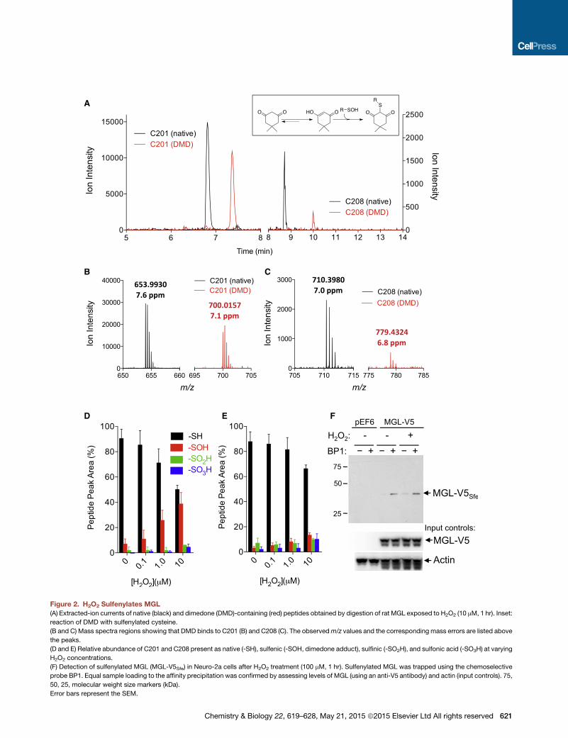
H2O2 Causes C201 and C208 SulfenylationTo determine whether MGL is a substrate for cysteine sulfenyla-
tion, we incubated the purified recombinant enzyme with H2O2
in the presence of dimedone (a chemoselective probe that reacts
with short-lived sulfenyl groups in cysteines to form stable thio-
ethers; Figure 2A, inset; Pan and Carroll, 2014) and then digested
the protein with trypsin. Liquid chromatography/mass spectrom-
etry (LC/MS) analyses of the tryptic digest showed that dimedone
was exclusively bound to the peptides containing C201 (SEV
DLYNSDPLICHAGVK) (Figure 2A and B) and C208 (VCFGIQLL
NAVSR) (Figures 2A and 2C). No other dimedone-bound peptides
were observed (Figure S2). Further high-resolution LC/MS studies
revealed that C201 was mainly present as thiol or sulfenic acid,
although minor quantities (1%–7%) of sulfinic (-SO2H) and sul-
fonic (-SO3H) acids were also detected (Figure 2D). C208 was
less sensitive than C201 to oxidation and yielded approximately
equal amounts of sulfenic, sulfinic, and sulfonic acids (Figure 2E).
Tandem MS analyses of the two dimedone-labeled tryptic frag-
ments of MGL allowed us unequivocally to assign C201 and
C208 as the sites of sulfenylation induced by H2O2 (Figures 3A
A
B C
D E F
Figure 2. H2O2 Sulfenylates MGL
(A) Extracted-ion currents of native (black) and dimedone (DMD)-containing (red) peptides obtained by digestion of rat MGL exposed to H2O2 (10 mM, 1 hr). Inset:
reaction of DMD with sulfenylated cysteine.
(B and C) Mass spectra regions showing that DMD binds to C201 (B) and C208 (C). The observedm/z values and the corresponding mass errors are listed above
the peaks.
(D and E) Relative abundance of C201 and C208 present as native (-SH), sulfenic (-SOH, dimedone adduct), sulfinic (-SO2H), and sulfonic acid (-SO3H) at varying
H2O2 concentrations.
(F) Detection of sulfenylated MGL (MGL-V5Sfe) in Neuro-2a cells after H2O2 treatment (100 mM, 1 hr). Sulfenylated MGL was trapped using the chemoselective
probe BP1. Equal sample loading to the affinity precipitation was confirmed by assessing levels of MGL (using an anti-V5 antibody) and actin (input controls). 75,
50, 25, molecular weight size markers (kDa).
Error bars represent the SEM.
Chemistry & Biology 22, 619–628, May 21, 2015 ª2015 Elsevier Ltd All rights reserved 621

A
B
Figure 3. Tandem Mass Analyses of Cysteine-Containing Tryptic Fragments of MGL Treated with H2O2
(A) Tandem mass spectra of peptide SEVDLYNSDPLICHAGVK, showing that sulfenylation (detected as DMD adduct) occurred on C201.
(B) Tandem mass spectra of peptide VCFGIQLLNAVSR, showing that sulfenylation (detected as DMD adduct) occurred on C208.
and 3B). Lastly, using biotin-1,3-cyclopentanedione (BP1), a
biotin-containing dimedone derivative that can be used for affinity
precipitation (Qian et al., 2011), we were able to document the
presence of sulfenylated MGL in intact Neuro-2a cells overex-
pressing the enzyme and exposed to H2O2 (100 mM) (Figure 2F).
While we found no evidence of sulfenyl amide modification of
C201 and C208, it is possible that this unstable chemical species
was also transiently generated (Salmeen et al., 2003). Formation
of a sulfenyl amide at C208 would explain why this residue is
less prone to sulfenylation than is C201. Despite this unanswered
622 Chemistry & Biology 22, 619–628, May 21, 2015 ª2015 Elsevier
question, the results outlined above clearly identify MGL residues
C201 and C208 as targets for peroxide-dependent sulfenylation.
MGL Sulfenylation Interrupts 2-AG Degradation inNeuronsTo determine whether cysteine sulfenylation influences 2-AG
deactivation in intact cells, we incubated primary cultures of rat
cortical neurons with H2O2 (30–300 mM) and measured MGL ac-
tivity in intact cells using a dedicated in situ assay. H2O2 inhibited
MGL in a concentration-dependent manner (Figure 4A) and this
Ltd All rights reserved

A B C
D
E
F
Figure 4. Peroxide-Dependent Inhibition of MGL Activity and 2-AG Accumulation in Brain Neurons(A–D) Effects of H2O2 (filled bars) or vehicle (open bars) on (A) MGL activity, (B) 2-AG levels, and (C) 1-stearoyl-2-arachidonoylglycerol (SAG), arachidonic acid
(AA), and anandamide (AEA) in rat cortical neurons in primary cultures. (D) Detection of sulfenylated MGL (MGLSfe) in primary cortical neurons in cultures after
H2O2 treatment (300 mM, 1 hr). Sulfenylated MGL was trapped using the chemoselective probe BP1. Equal sample loading to the affinity precipitation was
confirmed by measuring MGL (anti-MGL antibody) and actin (input controls). 50, 25, molecular weight size markers (kDa).
(E) Ion current of the BP1-adduct of MGL peptide bearing C201 (590.53m/z, z = 4) extracted from the control incubation (black trace) and incubation of neurons
with H2O2 (red trace).
(F) Ion current of the BP1-adduct of MGL peptide bearing C208 (606.30m/z, z = 3) extracted from the control incubation (black trace) and incubation of neurons
with H2O2 (red trace). In both (E) and (F), the high-resolutionmass spectra reported in the inset matches the expected charge state andm/z value. ***P < 0.001 and
*P < 0.05 compared with vehicle, two-tailed Student’s t test.
Error bars represent the SEM.
Chemistry & Biology 22, 619–628, May 21, 2015 ª2015 Elsevier Ltd All rights reserved 623

A B C
Figure 5. GSH Depletion Inhibits MGL Activity and Causes 2-AG Accumulation in Brain Neurons
(A–C) Effects of BSO (filled bars) or vehicle (open bars) on (A) GSH levels, (B) MGL activity, and (C) 2-AG levels in neuronal cultures. ***P < 0.001 and *P < 0.05
compared with vehicle, two-tailed Student’s t test.
Error bars represent the SEM.
effect was paralleled by an increase in intracellular 2-AG content
(Figure 4B). By contrast, we observed no change in the levels
of other lipids that are biogenetically or functionally related to
2-AG, including 1-steroyl-2-arachidonoyl-sn-glycerol (a 2-AG
precursor), arachidonic acid (a product of 2-AG hydrolysis),
or anandamide (an endocannabinoid neurotransmitter that is
not hydrolyzed by MGL) (Figure 4C). The presence of sul-
fenylated MGL in H2O2-treated neurons was evaluated using
affinity precipitation combined with western blot and LC/MS
analyses. We incubated primary neurons in cultures with vehicle
or H2O2 (300 mM), exposed them to the biotin-linked probe,
BP1, and then concentrated sulfenyl-containing proteins by
affinity precipitation using streptavidin-agarose beads. Blots
probed with a selective anti-MGL antibody confirmed the
presence of BP1-bound sulfenylated MGL in neurons exposed
to H2O2, but not control neurons (Figure 4D). In a separate
experiment, we subjected the affinity precipitates to shotgun
LC-MS/MS proteomics. MGL peptides bearing BP1 bound to
C201 (Figure 4E) or C208 (Figure 4F) were detected in precipitates
of intact neurons treated with H2O2. Despite a low signal-to-noise
ratio, due to the naturally low abundance of MGL, signals
matching the predicted accurate mass values and charge states
were detected in neurons treated with H2O2 (red traces), whereas
no such signal was detected in control cells (black traces).
Tandem MS data of the same peptides, matching the expected
primary sequence, are reported in Figure S3. Additional evi-
dence that the redox status can affect MGL activity was
obtained by blocking glutathione (GSH) biosynthesis with the
GSH synthase inhibitor, L-buthionine sulfoximine (BSO) (Griffith
and Meister, 1979). Exposing cortical neurons to BSO depleted
intracellular GSH stores (Figure 5A), lowered MGL activity (Fig-
ure 5B), and increased 2-AG content in a concentration-depen-
dent (Figure 5C) and time-dependent manner (Figure S4).
Confirming the generality of this response, experiments with
cultures of mouse Neuro-2a cells yielded similar results
(Figure S5).
624 Chemistry & Biology 22, 619–628, May 21, 2015 ª2015 Elsevier
MGL Sulfenylation Enhances 2-AG-Mediated SignalingThe data reported above suggest that MGL sulfenylation im-
pairs 2-AG degradation, but does it also enhance 2-AG
signaling? To address this question, we exploited the fact
that CB1 receptor activation protects neurons from oxidative
damage (Nagayama et al., 1999; Panikashvili et al., 2001).
We incubated Neuro-2a cells with a high concentration of
H2O2 (300 mM) and assessed cell damage using three com-
plementary methods: lactate dehydrogenase (LDH) release
into the medium, 3-(4,5-dimethylthiazol-2-yl)-2,5-diphenylte-
trazolium bromide (MTT) reduction, and caspase-3 activity.
In addition to native Neuro-2a cells, we assessed the effects
of H2O2 on Neuro-2a cells overexpressing MGL (Figure 6) or
the 2-AG-synthesizing enzyme DGL-a (Figure 7). As expected,
MGL overexpression decreased cellular 2-AG levels, which
were partially restored by treatment with the MGL inhibitor
JZL184 (1 mM) (Figure 6A). Along with this effect, MGL overex-
pression enhanced H2O2-induced toxicity (Figures 6B–6D),
which was (1) heightened by the CB1 antagonist rimonabant
(but not by the CB2 antagonist AM630) and (2) tempered by
MGL blockade with JZL184 (Figures 6B–6D). Conversely,
DGL-a overexpression increased 2-AG levels in Neuro-2a cells
(Figure 7A), and this response was accompanied by a sub-
stantial decrease in H2O2-induced toxicity (Figures 7B–7D).
The cytotoxic effect of H2O2 was reinstated by treatment
with the CB1 antagonist rimonabant (but not by the CB2
antagonist AM630) (Figures 7B–7D), suggesting that it was
mediated by 2-AG-dependent activation of CB1 receptors.
Consistent with this view, in primary neuronal cultures, H2O2
toxicity was enhanced by CB1, but not CB2, receptor blockade
(Figure S6). Treatment with JZL184 alone did not affect cyto-
toxicity in Neuro-2a cells (LDH release %: vehicle control,
3.46 ± 0.59; JZL184, 1.73 ± 2.60, n = 3). These observations
indicate that peroxide-dependent MGL deactivation accrues
the cellular pool of 2-AG that is involved in signaling at CB1
receptors.
Ltd All rights reserved

A B
C D
Figure 6. Reduction of 2-AG Levels Attenu-
ates Peroxide-Dependent CB1 Signaling
(A) 2-AG levels in Neuro-2a cells overexpressing
MGL.
(B–D) Effects of H2O2, alone or combined with CB1
antagonist rimonabant, CB2 antagonist AM630, or
MGL inhibitor JZL184 (each at 1 mM), on wild-type
(open bars) or MGL-overexpressing Neuro-2a
cells: (B) lactate dehydrogenase release, (C) MTT
reduction, and (D) caspase-3 activity. ***P < 0.001,
**P < 0.05, and *P < 0.05 compared withMock, and
###P < 0.001 compared with MGL, two-tailed
Student’s t test.
Error bars represent the SEM.
SIGNIFICANCE
Current models conceptualize 2-AG-dependent endocanna-
binoid transmission at excitatory synapses as being pri-
marily, if not exclusively, driven by postsynaptic mGluR5
activation and consequent 2-AG production (Castillo et al.,
2012). Our findings expand this view by identifying sulfenyla-
tion-dependent MGL inhibition as a presynaptic mechanism
throughwhich the local redox state regulates 2-AG-mediated
signaling. Physiopathological signals that stimulate H2O2
production might enhance the ability of 2-AG to modulate
synaptic activity by temporarily interrupting 2-AG deactiva-
tion. The results also raise two questions. The first pertains
to the sources of H2O2 involved inMGL regulation. Identifying
such sources will require additional work but likely candi-
dates include mitochondria respiration (Sies, 2014) and iono-
tropic glutamate receptor-operated activation of NADPH
oxidase (Brennan et al., 2009; Paulsen et al., 2011). The sec-
ond question is connected to the first and concerns the
context in which the redox control of MGL activity might be
operational. Dysfunctional redox states such as brain
ischemia are associated with rises in the levels of H2O2 and
other reactive oxygen species (Armogida et al., 2012). In
those conditions, MGL sulfenylation might act as an intrinsic
neuroprotective mechanism by potentiating 2-AG signaling
at CB1 receptors. Nevertheless, localized foci of heightened
H2O2 production (Mishina et al., 2011) might be sufficient to
deactivate MGL even under physiological conditions, partic-
ularly at synapses that experience high-frequency synaptic
activity and use glutamate as a neurotransmitter (Patel and
Chemistry & Biology 22, 619–628, May 21, 2015
Rice, 2012). In that context, MGL sulfe-
nylation may strengthen endocannabi-
noid-mediated retrograde transmission
by lowering the presynaptic degrada-
tion of 2-AG generated in postsynaptic
spines. Activation of mitochondrial
CB1 receptors (Benard et al., 2012)
might stimulate respiration and reactive
oxygen species production, further
enhancing this negative feedback
loop. Finally, allostericMGLmodulators
that exploit this regulatory process,
such as benzisothiazolinone deriva-
tives (King et al., 2009), might find
therapeutic application in stroke, neurodegeneration, and
chronic neuropathic pain.
EXPERIMENTAL PROCEDURES
Materials
2-AG, 2-arachidonoyl-sn-glycerol-d8 (2-AG-d8), arachidonic acid-d8 (AA-d8),
4-nitrophenyl-4-(dibenzo[d][1,3]dioxol-5-yl(hydroxy)methyl)piperidine-1-car-
boxylate (JZL184), and [6-iodo-2-methyl-1-[2-(4-morpholinyl)ethyl]-1H-indol-
3-yl](4-methoxyphenyl)-methanone (AM630) were purchased from Cayman
Chemical. 1,3-diheptadecanoyl-sn-glycerol (DHDG), 1(3)-heptadecenoyl-sn-
glycerol (1-HG), 1(3)-oleoyl-sn-glycerol, heptadecanoic acid, and heptadece-
noic acid were from Nu-Chek Prep. BSO, DMSO, 2-oleoyl-sn-glycerol (2-OG),
H2O2, and BSA were from Sigma-Aldrich. N-(piperidin-1-yl)-5-(4-chloro-
phenyl)-1-(2,4-dichlorophenyl)-4-methyl-1H-pyrazole-3-carboximide hydro-
chloride (rimonabant) was from RTI International. Chromatography solvents
were from Honeywell Burdick & Jackson. Porcine trypsin (proteomic grade)
was from Sigma-Aldrich.
Enzyme Assays
In vitro MGL activity was measured as described by King et al. (2007). Briefly,
we transiently transfected HeLa cells with plasmid DNA encoding recombinant
rat MGL using Superfect reagent (Qiagen). We harvested cells in ice-cold Tris-
HCl (50 mM [pH 8.0]) containing 0.32 M sucrose. We prepared homogenates
by sonicating cells for 1 min on ice followed by three freeze-thawing cycles.
Homogenates were incubated with various agents for 10 min at 37�C in assay
buffer (50 mM Tris-HCl [ pH 8.0] containing 0.5 mg/ml fatty acid-free BSA). The
enzyme substrate 2-OG (10 mM), which we use in preference to 2-AG to in-
crease the signal-to-noise ratio in the assay, was added to the mixture and
incubated for 10 additional min at 37�C. Reactions were stopped by
adding chloroform/methanol (2:1, vol/vol), containing heptadecanoic acid
(5 nmol/sample) as internal standard. After centrifugation at 2,000 3 g at
4�C for 10 min, the organic layers were collected and dried under N2. The lipid
extracts were suspended in chloroform/methanol (1:3, vol/vol) and analyzed
ª2015 Elsevier Ltd All rights reserved 625

A B
C D
Figure 7. Elevation of 2-AG Levels Strengthens Peroxide-Dependent
CB1 Signaling
(A) 2-AG levels in Neuro-2a cells overexpressing DGL-a.
(B–D) Effects of H2O2, alone or combined with rimonabant, AM630, or JZL184,
on wild-type or DGL-a-overexpressing Neuro-2a cells: (B) LDH release,
(C) MTT reduction, and (D) caspase-3 activity. ***P < 0.001, **P < 0.05, and
*P < 0.05 compared with Mock, two-tailed Student’s t test.
Error bars represent the SEM.
by LC-MS. DGL activity was measured in vitro as described by Jung et al.
(2007). Rapid dilution assays were performed as described by King et al.
(2009) and Copeland (2005).
MGL Activity Assay In Situ
MGL activity in primary cortical neurons was determined as described by
Marrs et al. (2010) with minor modifications. Neurons were prepared using
pregnant Wistar rats at embryonic day 18–20 (Stella and Piomelli, 2001). After
10 days in culture, cells were treated with the indicated reagents or vehicle for
30 min in B-27-supplemented Neurobasal medium (Life Technologies). Cells
were rinsed once with medium and incubated with substrate 1-HG (1 mM) for
10 min. After rinsing twice with ice-cold PBS, cells were harvested and lipids
were extracted as described above, using heptadecanoic acid as internal
standard. Non-specific hydrolysis of 1-HG was measured in the presence of
excess 1(3)-oleoyl-sn-glycerol (100 mM), and baseline value was subtracted
from each data point. Neuro-2a cells were partially differentiated by overnight
serum deprivation and cultured in complete DMEM medium supplemented
with 0.15% fatty acid-free BSA.
Lipid Quantitation
The levels of 2-AG, anandamide, arachidonic acid, and 1-stearoyl-2-arachido-
noyl-sn-glycerol (SAG) were determined by LC-MS or LC-MS/MS, as
described by Jung et al. (2007).
Protein and Western Blot Analyses
Bicinchoninic acid (BCA) protein assay and western blot analyses were per-
formed as described by Jung et al. (2007) using monoclonal anti-V5 antibody
(1:5,000, Life Technologies), polyclonal anti-MGL antibody (1:1,000) (Dinh
et al., 2002), and anti-actin monoclonal antibody (1:1,000, Calbiochem).
626 Chemistry & Biology 22, 619–628, May 21, 2015 ª2015 Elsevier
Site-Directed Mutagenesis
Mutagenesis studies were performed using a QuikChange II XL Site-Directed
Mutagenesis Kit (Strategene) following manufacturer’s instructions. We used
rat MGL-pEF6/V5-His plasmid DNA as a template (King et al., 2009) and veri-
fied all plasmids by DNA sequencing.
MGL Purification
Recombinant rat MGL was expressed in E. coli and purified as described by
King et al. (2007) with minor modification.
Sulfenic Acid Trapping In Vitro
A sample (200 ml) of MGL solution (40 mM) was transferred in a NanoSep 3K
microcentrifuge tube (Pall) and buffer-exchanged (403) versus HEPES
(50 mM [pH 7.4]), 300 mM NaCl 0.1% Triton X-100. The buffer was degassed
with N2 for 2 hr prior to use. The sample was split into four 50-ml aliquots and
incubated with dimedone at a final 20-mM concentration. H2O2 was added to
final 0.1-, 1-, and 10-mMconcentrations. Incubation without H2O2 was used as
negative control. Only open-air oxidation was allowed for this control. The
sampleswere incubated at 37�C for 1 hr with shaking. Reactions were stopped
adding 1 ml of cold acetone, and tubes were centrifuged at 14,000 rpm for
10 min. Acetone was removed and protein pellets were dried under N2. The
precipitate was further washed with 1 ml of methanol and then was dissolved
in 50 ml of ammonium bicarbonate (50 mM [pH 8]). Trypsin (1 ml of a 1 mg/ml so-
lution) was added to each tube and samples were incubated at 37�Covernight.
Samples were centrifuged at 10,0003 g for 10 min and diluted with 0.1% for-
mic acid in water containing 1% acetonitrile for LC-MS analysis.
LC-MS/MS Analyses of Peptides
Tryptic peptideswere separatedusing anAcquityUPLCBEHC18 (13100mm,
1.7 mm) column coupled to a Synapt G2 qTOF mass spectrometer (Waters).
The following gradient conditions were used: A, 0.1% formic acid in water;
B, 0.1% formic acid in acetonitrile. After 1 min at 97% A, a linear gradient
was applied to 45% A in 12 min, then to 0% A in the next 3 min, followed by
2 min at 0% A, and reconditioning to 97% A in 0.1 min. Total run time was
19 min. Injection volume was set at 5 ml. MS and MS/MS analyses were per-
formed in positive electrospray ionization mode. The capillary voltages was
set at 3 kV. The cone voltage was set at 30 V. The source temperature was
120�C. Desolvation gas and cone gas (N2) flow were 600 and 20 l/hr, respec-
tively. Desolvation temperature was 400�C. Data were acquired in MSe mode
(mass spectra acquisition with alternate high and low collision energy) with
MS/MS fragmentation performed in the trap region. Low-energy scans were
acquired at fixed 4 eV potential, and high-energy scans were acquired with
an energy ramp from 25 to 35 eV. Scan rate was set to 0.4 s per spectrum.
Scan range was set to 50 to 1,600 m/z. Leucine enkephalin (2 ng/ml) was
infused as lock mass for spectra recalibration. LC-MS/MS data were analyzed
using Proteinlynx software to map MGL peptides, calculate MGL sequence
coverage, and quantify naive and oxidized peptides. Additional de novo
sequencing of dimedone adducts at cysteine 201 and 208 was manually per-
formed using Biolynx software to verify the peptide sequence and assign the
modified residue.
Sulfenic Acid Trapping in Intact Cells
Neuro-2a cells were transfected with plasmids encodingMGL-V5 or the empty
vector (pEF6) and, 2 days after transfection, were incubated with H2O2 for 1 hr.
Rat cortical neurons were treated with H2O2 for 1 hr after 8 days in vitro.
Following treatment, cells were scraped in lysis buffer containing 50 mM
Tris-HCl (pH 7.4), 150 mM NaCl, 2 mM EDTA, 1% Triton X-100, and a mixture
of protease inhibitors (Roche Diagnostics) in the presence or absence of 1 mM
BP1 (KeraFAST) (Qian et al., 2011). We incubated themixtures on ice for 30min
with gentlemixing every 5min. After centrifuging at 1,0003 g for 10min at 4�C,the supernatants were mixed with 10 ml of streptavidin-agarose (Pierce) at a
protein concentration of 0.5 mg/ml, and incubated at 4�C overnight. The affin-
ity precipitates were collected by brief centrifugation and washed six times
with lysis buffer without BP1. For western blot analyses, proteins in the precip-
itates were eluted by adding 20 ml of Laemmli sample buffer and incubating at
74�C for 20 min. For LC-MS/MS analyses, washed beads were stored in dry
ice. Prior to LC-MS/MS, the beads were allowed to reach room temperature
and were suspended in 25 ml of 2% SDS, 6 M urea, and 2 M thiourea in
Ltd All rights reserved

PBS. Samples were kept for 15min at room temperature and then incubated at
96�C for 15 min to cleave the biotin-streptavidin bond (Rybak et al., 2004, with
slight modifications). Samples were allowed to reach room temperature and
were centrifuged for 10 min at 4,000 3 g. Supernatants were collected and
the beads were further washed with 25 ml of water and incubated again at
96�C for 15 min. After centrifugation (10 min, 4,000 3 g) the supernatants
were pooled and the beads discarded. The resulting 50 ml of supernatant
was added with 2 ml of 100 mM DTT and incubated at 50�C for 30 min. Iodoa-
cetamide (100 mM, 4 ml) was added and the samples were incubated at room
temperature in the dark for 30min. After reduction and alkylation, released pro-
teins were precipitated by adding 1 ml of ice-cold acetone and then centri-
fuged for 10 min at 4,000 3 g at 4�C. The supernatant was discarded and
the pellets were dried under N2. Samples were suspended in 40 ml of Rapigest
(Waters) previously dissolved in 100 mM ammonium bicarbonate (pH 8), at a
final concentration of 1 mg/ml. Trypsin was then added (1 ml, dissolved at
0.5 mg/ml in 0.1% formic acid), and the samples were incubated overnight
at 37�C. The following day, samples were added with 1 ml of trifluoroacetic
acid, incubated at 37�C for 1 hr, and centrifuged to remove hydrolyzed Rapi-
gest (10 min at 4,0003 g). Supernatants were collected (30 ml) and added with
10 ml of formic acid 0.1% in water/acetonitrile (97:3). An aliquot (5 ml) was
loaded on a nano-ultra-high performance liquid chromatography system
equipped with a T3 C18 reversed-phase column (75 mm3 250 mm). Peptides
were eluted with a linear gradient of acetonitrile in water (both containing 0.1%
formic acid) from 3% to 50% in 120 min. Flow rate was set to 350 nl per min.
Eluted peptides were analyzed in positive ion mode by high-resolutionMS/MS
on a Synapt G2 qTOF mass spectrometer (Waters). Tandem mass spectra
were acquired in data-dependent acquisitionmode, by selecting charge states
ranging from 2 to 5 in the 300–1,200m/z range. Collision energy was automat-
ically set according to the precursor ion charge state and m/z value. MS and
MS/MS spectra were recalibrated using glu-fibrinopeptide and leucine
enkephalin, respectively (both continuously infused in the source as lock
mass). Proteomics data were analyzed with both PLGS 3.2 and Biolynx soft-
ware (Waters) to determine the presence and the primary sequence of the
BP1 adducts of MGL peptides bearing C201 and C208.
Cell Viability Assays
Cell viability was assessed using the MTT assay, following a standard protocol
(Sigma-Aldrich). Briefly, cells were seeded in 96-well plates at a concentration
of 5 3 103 cells/well. After 24 hr, they were transfected with MGL or DGL-a
plasmids (0.5 mg DNA/well) and cultured for 24 hr. They were treated with
agents for 30 min, followed by a treatment with H2 O2 (300 mM) for 24 hr.
MTT was added and plates were further incubated for 4 hr. Absorbance
was measured using a SpectraMax M5 microplate reader (Molecular Devices)
at 570 nm with reference at 650 nm. Cell viability was calculated and
expressed as percentage of control cells. Cell death was quantified by
measuring LDH release using a Cytotoxicity Detection KitPlus (Roche Diagnos-
tics) and caspase-3 activation using a fluorescent assay kit (Cayman Chemi-
cal). Total GSH levels in cells were determined using an assay kit (Cayman
Chemical).
Statistical Analyses
All results are expressed asmean ± SEM. Non-linear regression analyses were
performed using Prism version 5.0 (GraphPad Software). Statistical signifi-
cance was assessed by two-tailed Student’s t test or one-way ANOVA with
Dunnett’s post test.
SUPPLEMENTAL INFORMATION
Supplemental Information includes six figures and one table and can be found
with this article online at http://dx.doi.org/10.1016/j.chembiol.2015.04.013.
AUTHOR CONTRIBUTIONS
E.Y.D. carried out MGL inhibition and cell viability experiments with help from
D.W. and J.D. A.B., K.-M.J., and A.A. performed MGL sulfenylation studies.
E.Y.D, K.-M.J., A.A., F.V., M.M., and D.P. analyzed the data. M.M. and D.P. de-
signed the project. D.P. coordinated the project and wrote the manuscript with
help from E.Y.D, K.-M.J., A.A., and M.M.
Chemistry & Biology 22,
ACKNOWLEDGMENTS
This work was supported by grants DA012413 and DA031387 from NIDA (to
D.P.). The contribution of the Agilent Technologies/University of California,
Irvine Analytical Discovery Facility, is gratefully acknowledged.
Received: January 7, 2015
Revised: March 27, 2015
Accepted: April 15, 2015
Published: May 21, 2015
REFERENCES
Armogida, M., Nistico, R., and Mercuri, N.B. (2012). Therapeutic potential of
targeting hydrogen peroxide metabolism in the treatment of brain ischaemia.
Br. J. Pharmacol. 166, 1211–1224.
Benard, G., Massa, F., Puente, N., Lourenco, J., Bellocchio, L., Soria-Gomez,
E., Matias, I., Delamarre, A., Metna-Laurent, M., Cannich, A., et al. (2012).
Mitochondrial CB1 receptors regulate neuronal energy metabolism. Nat.
Neurosci. 15, 558–564.
Bertrand, T., Auge, F., Houtmann, J., Rak, A., Vallee, F., Mikol, V., Berne, P.F.,
Michot, N., Cheuret, D., Hoornaert, C., andMathieu, M. (2010). Structural basis
for human monoglyceride lipase inhibition. J. Mol. Biol. 396, 663–673.
Brennan, A.M., Suh, S.W., Won, S.J., Narasimhan, P., Kauppinen, T.M., Lee,
H., Edling, Y., Chan, P.H., and Swanson, R.A. (2009). NADPH oxidase is the
primary source of superoxide induced by NMDA receptor activation. Nat.
Neurosci. 12, 857–863.
Castillo, P.E., Younts, T.J., Chavez, A.E., and Hashimotodani, Y. (2012).
Endocannabinoid signaling and synaptic function. Neuron 76, 70–81.
Copeland, R.A. (2005). Evaluation of enzyme inhibitors in drug discovery. A
guide for medicinal chemists and pharmacologists. Methods Biochem. Anal.
46, 1–265.
Denu, J.M., and Tanner, K.G. (1998). Specific and reversible inactivation of
protein tyrosine phosphatases by hydrogen peroxide: evidence for a sulfenic
acid intermediate and implications for redox regulation. Biochemistry 37,
5633–5642.
Dickinson, B.C., and Chang, C.J. (2011). Chemistry and biology of reactive ox-
ygen species in signaling or stress responses. Nat. Chem. Biol. 7, 504–511.
Dinh, T.P., Carpenter, D., Leslie, F.M., Freund, T.F., Katona, I., Sensi, S.L.,
Kathuria, S., and Piomelli, D. (2002). Brain monoglyceride lipase participating
in endocannabinoid inactivation. Proc. Natl. Acad. Sci. USA 99, 10819–10824.
Griffith, O.W., and Meister, A. (1979). Potent and specific inhibition of gluta-
thione synthesis by buthionine sulfoximine (S-n-butyl homocysteine sulfoxi-
mine). J. Biol. Chem. 254, 7558–7560.
Gulyas, A.I., Cravatt, B.F., Bracey, M.H., Dinh, T.P., Piomelli, D., Boscia, F.,
and Freund, T.F. (2004). Segregation of two endocannabinoid-hydrolyzing
enzymes into pre- and postsynaptic compartments in the rat hippocampus,
cerebellum and amygdala. Eur. J. Neurosci. 20, 441–458.
Hohmann, A.G., Suplita, R.L., Bolton, N.M., Neely, M.H., Fegley, D., Mangieri,
R., Krey, J.F., Walker, J.M., Holmes, P.V., Crystal, J.D., et al. (2005). An endo-
cannabinoid mechanism for stress-induced analgesia. Nature 435, 1108–
1112.
Jung, K.M., Astarita, G., Zhu, C., Wallace, M., Mackie, K., and Piomelli, D.
(2007). A key role for diacylglycerol lipase-alpha in metabotropic glutamate
receptor-dependent endocannabinoid mobilization. Mol. Pharmacol. 72,
612–621.
Jung, K.M., Clapper, J.R., Fu, J., D’Agostino, G., Guijarro, A., Thongkham, D.,
Avanesian, A., Astarita, G., DiPatrizio, N.V., Frontini, A., et al. (2012). 2-arach-
idonoylglycerol signaling in forebrain regulates systemic energy metabolism.
Cell Metab. 15, 299–310.
Katona, I., and Freund, T.F. (2008). Endocannabinoid signaling as a synaptic
circuit breaker in neurological disease. Nat. Med. 14, 923–930.
King, A.R., Duranti, A., Tontini, A., Rivara, S., Rosengarth, A., Clapper, J.R.,
Astarita, G., Geaga, J.A., Luecke, H., Mor, M., et al. (2007). URB602 inhibits
619–628, May 21, 2015 ª2015 Elsevier Ltd All rights reserved 627

monoacylglycerol lipase and selectively blocks 2-arachidonoylglycerol degra-
dation in intact brain slices. Chem. Biol. 14, 1357–1365.
King, A.R., Dotsey, E.Y., Lodola, A., Jung, K.M., Ghomian, A., Qiu, Y., Fu, J.,
Mor, M., and Piomelli, D. (2009). Discovery of potent and reversible monoacyl-
glycerol lipase inhibitors. Chem. Biol. 16, 1045–1052.
Labar, G., Bauvois, C., Borel, F., Ferrer, J.L., Wouters, J., and Lambert, D.M.
(2010). Crystal structure of the human monoacylglycerol lipase, a key actor
in endocannabinoid signaling. Chembiochem 11, 218–227.
Lo Conte, M., and Carroll, K.S. (2013). The redox biochemistry of protein sul-
fenylation and sulfinylation. J. Biol. Chem. 288, 26480–26488.
Long, J.Z., Li, W., Booker, L., Burston, J.J., Kinsey, S.G., Schlosburg, J.E.,
Pavon, F.J., Serrano, A.M., Selley, D.E., Parsons, L.H., et al. (2009).
Selective blockade of 2-arachidonoylglycerol hydrolysis produces cannabi-
noid behavioral effects. Nat. Chem. Biol. 5, 37–44.
Marrs, W.R., Blankman, J.L., Horne, E.A., Thomazeau, A., Lin, Y.H., Coy, J.,
Bodor, A.L., Muccioli, G.G., Hu, S.S., Woodruff, G., et al. (2010). The serine hy-
drolase ABHD6 controls the accumulation and efficacy of 2-AG at cannabinoid
receptors. Nat. Neurosci. 13, 951–957.
Mishina, N.M., Tyurin-Kuzmin, P.A., Markvicheva, K.N., Vorotnikov, A.V.,
Tkachuk, V.A., Laketa, V., Schultz, C., Lukyanov, S., and Belousov, V.V.
(2011). Does cellular hydrogen peroxide diffuse or act locally? Antioxid.
Redox Signal. 14, 1–7.
Nagayama, T., Sinor, A.D., Simon, R.P., Chen, J., Graham, S.H., Jin, K., and
Greenberg, D.A. (1999). Cannabinoids and neuroprotection in global and focal
cerebral ischemia and in neuronal cultures. J. Neurosci. 19, 2987–2995.
Pan, J., and Carroll, K.S. (2014). Chemical biology approaches to study protein
cysteine sulfenylation. Biopolymers 101, 165–172.
Panikashvili, D., Simeonidou, C., Ben-Shabat, S., Hanus, L., Breuer, A.,
Mechoulam, R., and Shohami, E. (2001). An endogenous cannabinoid (2-AG)
is neuroprotective after brain injury. Nature 413, 527–531.
628 Chemistry & Biology 22, 619–628, May 21, 2015 ª2015 Elsevier
Patel, J.C., and Rice, M.E. (2012). Classification of H2O2 as a neuromodulator
that regulates striatal dopamine release on a subsecond time scale. ACS
Chem. Neurosci. 3, 991–1001.
Paulsen, C.E., Truong, T.H., Garcia, F.J., Homann, A., Gupta, V., Leonard,
S.E., and Carroll, K.S. (2011). Peroxide-dependent sulfenylation of the EGFR
catalytic site enhances kinase activity. Nat. Chem. Biol. 8, 57–64.
Qian, J., Klomsiri, C., Wright, M.W., King, S.B., Tsang, A.W., Poole, L.B., and
Furdui, C.M. (2011). Simple synthesis of 1,3-cyclopentanedione derived
probes for labeling sulfenic acid proteins. Chem. Commun. (Camb.) 47,
9203–9205.
Rice, M.E. (2011). H2O2: a dynamic neuromodulator. Neuroscientist 17,
389–406.
Rybak, J.N., Scheurer, S.B., Neri, D., and Elia, G. (2004). Purification of bio-
tinylated proteins on streptavidin resin: a protocol for quantitative elution.
Proteomics 4, 2296–2299.
Saario, S.M., Salo, O.M., Nevalainen, T., Poso, A., Laitinen, J.T., Jarvinen, T.,
and Niemi, R. (2005). Characterization of the sulfhydryl-sensitive site in the
enzyme responsible for hydrolysis of 2-arachidonoyl-glycerol in rat cerebellar
membranes. Chem. Biol. 12, 649–656.
Salmeen, A., Andersen, J.N., Myers, M.P., Meng, T.C., Hinks, J.A., Tonks,
N.K., and Barford, D. (2003). Redox regulation of protein tyrosine phosphatase
1B involves a sulphenyl-amide intermediate. Nature 423, 769–773.
Schalk-Hihi, C., Schubert, C., Alexander, R., Bayoumy, S., Clemente, J.C.,
Deckman, I., DesJarlais, R.L., Dzordzorme, K.C., Flores, C.M., Grasberger,
B., et al. (2011). Crystal structure of a soluble form of human monoglyceride
lipase in complex with an inhibitor at 1.35 A resolution. Protein Sci. 20,
670–683.
Sies, H. (2014). Role of metabolic H2O2 generation: redox signaling and oxida-
tive stress. J. Biol. Chem. 289, 8735–8741.
Stella, N., and Piomelli, D. (2001). Receptor-dependent formation of endoge-
nous cannabinoids in cortical neurons. Eur. J. Pharmacol. 425, 189–196.
Ltd All rights reserved