Delivery and Quantification of Hydrogen Peroxide Generated ...
10
IOP Publishing Journal Title Journal XX (XXXX) XXXXXX https://doi.org/XXXX/XXXX xxxx-xxxx/xx/xxxxxx 1 © xxxx IOP Publishing Ltd Delivery and Quantification of Hydrogen Peroxide Generated via Cold Atmospheric Pressure Plasma through Biological Material H. J. Hathaway 1* , B. L. Patenall 2 , N. T. Thet 2 , A. C. Sedgwick 2 , G. T. Williams 2 , A. T. A. Jenkins 2 , S. L. Allinson 3 and R. D. Short 1 1 Department of Chemistry, Lancaster University, Lancaster, UK 2 Department of Chemistry, University of Bath, Bath, UK 3 Division of Biomedical and Life Sciences, Lancaster University, Lancaster, UK *E-mail: [email protected] Received xxxxxx Accepted for publication xxxxxx Published xxxxxx Abstract The ability of plasma-generated hydrogen peroxide (H2O2) to traverse bacterial biofilms and the subsequent fate of the generated H2O2 has been investigated. An in vitro model, comprising a nanoporous membrane impregnated with artificial wound fluid and biofilms of varying maturity was treated with a helium-driven, cold atmospheric pressure plasma jet. The concentration of H2O2 generated below the biofilms was quantified. The results showed that the plasma-generated H2O2 interacted significantly with the biofilm, thus exhibiting a reduction in concentration across the underlying nanoporous membrane. Biofilm maturity exhibited a significant effect on the penetration depth of H2O2, suggesting that well established, multilayer biofilms are likely to offer a shielding effect with respect to cells located in the lower layers of the biofilm, thus rendering them less susceptible to plasma disinfection. This may prove clinically significant in the plasma treatment of chronic, deep tissue infections such as diabetic and venous leg ulcers. Our results are discussed in the context of plasma-biofilm interactions, with respect to the fate of the longer lived reactive species generated by cold atmospheric pressure plasma, such as H2O2. Keywords: cold atmospheric pressure plasma, hydrogen peroxide, bacterial biofilm 1. Introduction The ability to treat antibiotic resistant microorganisms has become paramount in the fight against bacterial infection owing to their ever-increasing prevalence. The apparent stagnation in antimicrobial drug development has fuelled urgent initiatives from a number of prominent healthcare organisations tailored towards treating and ultimately preventing such infections [1-3]. The extent of the current crisis is exemplified according to a predicted annual death toll of 10 million by 2050 arising solely from drug resistant infections (surpassing that of cancer) [4]. In addition to antimicrobial resistance (both de novo and pre-existing), the ability of bacteria to form biofilms, (dense, multilayer communities of bacterial cells protected by an extracellular polymeric substance (EPS)), further encumbers effective disinfection owing to changes in the physiological state of the cells contained within. Notoriously difficult to treat, it is estimated that biofilms are associated with 65% of all bacterial infections, rising to 85% when considering chronic infections [5]. Biofilms are able to form on a multitude of surfaces; both biotic and abiotic, thus providing a significant reservoir of Page 1 of 10 AUTHOR SUBMITTED MANUSCRIPT - JPhysD-120495.R2 1 2 3 4 5 6 7 8 9 10 11 12 13 14 15 16 17 18 19 20 21 22 23 24 25 26 27 28 29 30 31 32 33 34 35 36 37 38 39 40 41 42 43 44 45 46 47 48 49 50 51 52 53 54 55 56 57 58 59 60
-
Upload
khangminh22 -
Category
Documents
-
view
0 -
download
0
Transcript of Delivery and Quantification of Hydrogen Peroxide Generated ...

IOP Publishing Journal Title
Journal XX (XXXX) XXXXXX https://doi.org/XXXX/XXXX
xxxx-xxxx/xx/xxxxxx 1 © xxxx IOP Publishing Ltd
Delivery and Quantification of Hydrogen Peroxide
Generated via Cold Atmospheric Pressure Plasma
through Biological Material
H. J. Hathaway1*, B. L. Patenall2, N. T. Thet2, A. C. Sedgwick2, G. T. Williams2, A. T.
A. Jenkins2, S. L. Allinson3 and R. D. Short1
1 Department of Chemistry, Lancaster University, Lancaster, UK 2 Department of Chemistry, University of Bath, Bath, UK 3 Division of Biomedical and Life Sciences, Lancaster University, Lancaster, UK
*E-mail: [email protected]
Received xxxxxx
Accepted for publication xxxxxx
Published xxxxxx
Abstract
The ability of plasma-generated hydrogen peroxide (H2O2) to traverse bacterial biofilms and the subsequent fate of the
generated H2O2 has been investigated. An in vitro model, comprising a nanoporous membrane impregnated with artificial
wound fluid and biofilms of varying maturity was treated with a helium-driven, cold atmospheric pressure plasma jet. The
concentration of H2O2 generated below the biofilms was quantified. The results showed that the plasma-generated H2O2
interacted significantly with the biofilm, thus exhibiting a reduction in concentration across the underlying nanoporous
membrane. Biofilm maturity exhibited a significant effect on the penetration depth of H2O2, suggesting that well established,
multilayer biofilms are likely to offer a shielding effect with respect to cells located in the lower layers of the biofilm, thus
rendering them less susceptible to plasma disinfection. This may prove clinically significant in the plasma treatment of chronic,
deep tissue infections such as diabetic and venous leg ulcers. Our results are discussed in the context of plasma-biofilm
interactions, with respect to the fate of the longer lived reactive species generated by cold atmospheric pressure plasma, such
as H2O2.
Keywords: cold atmospheric pressure plasma, hydrogen peroxide, bacterial biofilm
1. Introduction
The ability to treat antibiotic resistant microorganisms has
become paramount in the fight against bacterial infection
owing to their ever-increasing prevalence. The apparent
stagnation in antimicrobial drug development has fuelled
urgent initiatives from a number of prominent healthcare
organisations tailored towards treating and ultimately
preventing such infections [1-3]. The extent of the current
crisis is exemplified according to a predicted annual death toll
of 10 million by 2050 arising solely from drug resistant
infections (surpassing that of cancer) [4]. In addition to
antimicrobial resistance (both de novo and pre-existing), the
ability of bacteria to form biofilms, (dense, multilayer
communities of bacterial cells protected by an extracellular
polymeric substance (EPS)), further encumbers effective
disinfection owing to changes in the physiological state of the
cells contained within. Notoriously difficult to treat, it is
estimated that biofilms are associated with 65% of all bacterial
infections, rising to 85% when considering chronic infections
[5]. Biofilms are able to form on a multitude of surfaces; both
biotic and abiotic, thus providing a significant reservoir of
Page 1 of 10 AUTHOR SUBMITTED MANUSCRIPT - JPhysD-120495.R2
123456789101112131415161718192021222324252627282930313233343536373839404142434445464748495051525354555657585960

Journal XX (XXXX) XXXXXX Author et al
2
pathogenic microorganisms responsible for numerous human
infections. Biofilms are reported to be involved in half of all
hospital-acquired infections, which the World Health
Organisation estimates to affect 4.5 million people per annum
in Europe, highlighting the significant challenge currently
facing the global healthcare community [6, 7].
Wound infections, specifically chronic wound infections have
a reported prevalence of 6% in the UK and account for at least
5.5% of the total NHS expenditure [8]. In the USA such non-
healing wounds carry an annual cost of $25 billion and it has
been estimated that 1-2% of the population in developed
countries will develop a chronic wound in their lifetime [9].
Wounds (venous, pressure, arterial and diabetic) are the
leading cause of amputation, the latter being responsible for
70% of all lower limb amputations which occur globally every
30 seconds as a direct result of non-healing diabetic ulcers [10,
11]. The protective nature of the EPS along with the induction
of various quorum sensing pathways and the subsequent
alteration in gene expression, has rendered biofilms largely
recalcitrant to common antimicrobial agents. Hence, the need
for effective treatment strategies has become imperative in
reducing both the social and economic burden of wound
infection [12].
Cold atmospheric pressure plasma (CAP) has been proven to
deliver a potent antimicrobial cocktail of reactive species able
to successfully decontaminate numerous clinically relevant
single- and mixed-species biofilms, including those associated
with significant drug resistance; termed the ‘ESKAPE’
pathogens [13-15]. The composition and delivery of reactive
species generated during CAP therapy, such as reactive
oxygen and nitrogen species (RONS) may be controlled
according to the plasma parameters employed, providing an
attractive therapeutic treatment option currently undergoing
significant investigation within the scientific community [16,
17]. Extensive discussion can be found in the literature
surrounding the effects of the plasma source, the variation in
operating conditions and the current progress and challenges
facing CAP therapy in the control of microbial biofilms and
wound infection [18-22]. The interaction of non-thermal
plasma with biological material has been well documented in
terms of bactericidal effects, phenotypic and genetic
consequences (in both eukaryotic and prokaryotic cells) and
with respect to plasma-activated liquids [23-25]. Studies have
been conducted using tissue surrogates such as gelatin and
agarose, alongside cellular mimics such as phospholipid
vesicles to track plasma delivery of RONS [26-30].
Furthermore, the biofilm penetration depth of CAP-generated
reactive species has been reported as a function of cell death
and via computational modelling [31-33]. However, the direct
quantification of longer-lived species, such as hydrogen
peroxide (H2O2) has yet to be conducted through living
biofilms. This study reports the effects of biofilm composition
on the delivery of plasma-generated H2O2 across the cellular
interface, thus confirming the ability of biologically active
plasma-generated products to traverse bacterial biofilms
according to biofilm density/maturity. The presence of H2O2,
or lack thereof, may provide insight into the fate of the longer-
lived species generated by CAP and the subsequent
implications this may have for the safe and effective
decontamination of chronic wounds.
2. Materials and Methods
2.1 Materials
Tryptic Soy Broth (TSB), Tryptic Soy Agar (TSA), Luria-
Bertani (LB) broth, phosphate buffered saline (PBS) tablets
(pH 7.4), Brain Heart Infusion (BHI) agar, foetal calf serum
(FCS) (HyClone), sodium chloride (NaCl), potassium iodide
(KI), H2O2, peptone, poly (vinyl alcohol) (PVA) (MW 14600–
18600 g mol-1), carboxymethyl cellulose (CMC) and agarose
were all purchased from Sigma-Aldrich, UK. Biofilms were
formed on sterile, nanoporous polycarbonate filter membranes
(Whatman), 19 mm in diameter with an average pore size of
200 nm.
2.2 CAP jet
The plasma jet used consists of a single electrode
configuration with a 150 mm glass capillary tube and a 15 mm
external ring copper electrode operating at a distance of 40
mm from the end of the tube [30]. Helium was used as the
carrier gas at a fixed flow rate of 0.6 standard litres per minute
(SLPM). The jet was operated at an applied voltage of 10 kV
p-p and a frequency of 25 kHz. The distance from the end of
the capillary tube to the surface of the substrate was 5 mm.
The jet used predominantly produces H2O2 under the
described operation parameters [28]. There is evidence that
the initial interaction of the plasma with ambient air in the gas
phase is the primary production site for H2O2 [34].
2.3 Quantification of H2O2
Potassium iodide (KI) was used to quantify H2O2
concentration in solution via the generation of a standard
curve. 1M KI in deionised (DI) water was added to varying
concentrations of H2O2 at a ratio of 1:1 and incubated at 25 C
for 30 minutes. Absorbance measurements were taken at 410
nm using a microplate reader (Spectrostar Omega, BMG
Labtech) and used to produce a calibration curve
(Supplementary Information Figure 1) [35]. For quantification
of plasma-generated H2O2 in PBS, 350 L of PBS was added
to each well in a 96 well microtiter plate and treated for 0.5 -
5 minutes. 100 L of the treated solution was then removed,
added to 1M KI (1:1 v/v) and incubated at 25 C for 30
minutes. Absorbance measurements were taken as before and
the concentration calculated using the calibration curve.
Page 2 of 10AUTHOR SUBMITTED MANUSCRIPT - JPhysD-120495.R2
123456789101112131415161718192021222324252627282930313233343536373839404142434445464748495051525354555657585960

Journal XX (XXXX) XXXXXX Author et al
3
2.4 Bacterial strains and growth conditions
Methicillin-resistant Staphylococcus aureus (MRSA) 252 and
Pseudomonas aeruginosa (P. aeruginosa) PA01 were sourced
from a bacterial strain collection belonging to the Biophysical
Research Group housed at the University of Bath, UK. Single
colonies were cultured from freezer stocks (stored at -80 C)
on TSA or LB agar for MRSA 252 and PA01, respectively.
Overnight cultures were grown from single colonies in TSB
or LB liquid media for MRSA 252 and PA01, respectively.
Cultures were incubated at 37C with agitation (200 rpm) for
18 hours to achieve a cell density of 109 colony forming units
(CFU) / ml.
2.5 Bacterial biofilm formation
Overnight cultures of MRSA 252 and PA01 were centrifuged
at 10,000 x g for 10 minutes, washed and resuspended in fresh
PBS to an optical density of ~ 0.2 (~ 106 CFU/ml). White
sterile polycarbonate filter membranes positioned on BHI agar
were inoculated with 20 L artificial wound fluid (FCS mixed
in equal volume with 0.85% NaCl and 0.1% peptone) in order
to better model the wound environment and components
supporting bacterial growth and adhesion. 30 L of bacterial
subculture was added to the conditioned membranes.
Membranes were UV sterilised for 10 minutes prior to
bacterial inoculation. Plates were incubated at 32C for 8, 12
and 24 hours [36].
2.6 Quantification of viable bacterial cells
Following incubation, biofilms were removed from the plates
and transferred into 5 mL of PBS. The solutions were vortexed
at 3000 rpm for 1 minute and sonicated for 15 minutes. This
process was repeated twice to ensure complete detachment of
the bacterial cells from the membrane. Serial dilutions were
carried out in PBS and plated on TSA or LB agar with
subsequent incubation at 37C for 24 hours to enumerate the
number of viable bacteria.
2.7 CAP treatment of bacterial biofilms
Bacterial biofilms grown for 8, 12 and 24 hours were placed
atop 350 L of PBS in a 96 well microtiter plate (full
experimental set-up is shown in Figure 2 Supplementary
Information). Biofilms were plasma treated for 5 minutes,
with manual movement of the jet around the circumference of
the well similarly as in vivo wound treatments. Following
plasma treatment, 100 L of PBS was removed from each well
and the concentration of H2O2 determined as before via
correlation to the standard curve. In order to assess any time-
dependent generation of H2O2 as a result of plasma treatment,
additional 8 hour biofilms were subject to plasma treatment
followed by a further 4 hour incubation in situ at 32 C prior
to H2O2 quantification. Separate experiments were undertaken
to quantify the number of viable bacterial cells (post plasma
treatment) as previously described. All experiments were
conducted in triplicate for each bacterial species and each time
point.
2.8 Temperature measurement of plasma jet
The temperature of the CAP jet and biofilm onto which the jet
was directed was measured using a Xenics® GOBI-640-GigE
thermal imaging camera. Plasma temperature was assessed
when treating a PA01 biofilm grown for 24 hours and placed
atop 350 L PBS in 96 well microtiter plate.
2.9 Topical application of H2O2
The effect of H2O2 on bacterial survival was investigated via
topical application of H2O2 to 8, 12 and 24 hour biofilms of
both MRSA 252 and PA01. 100 L of 550 M H2O2 (the same
concentration of H2O2 produced in solution by the plasma jet
in 5 minutes) was applied to the surface of the biofilms. The
concentration of H2O2 below the biofilms was quantified after
5 minutes, and after 4 hours incubation at 32 C as previously
described.
2.10 Hydrogel formulation
PVA/ CMC hydrogels were prepared by dissolving PVA (5%
w/v) in DI water and heating to 97 °C with constant stirring to
facilitate dissolution. The solution was supplemented with
0.5% w/v CMC, cast to a thickness of 1 mm and stored for 18
hours at -20 °C to promote cryogenic gelation. Discs of gel
were cut to a diameter of ~1 cm before being placed atop 350
L PBS in a 96 well plate and subject to CAP treatment.
Agarose hydrogels were prepared by dissolving agarose (1.5%
w/v) in DI water and heating to 90 °C. The solution was
cooled, supplemented with 0.5 M KI and cast to a thickness of
1 cm. Biofilms were placed atop discs of gel (~17 mm in
diameter) before treatment with the CAP jet.
3. Results
3.1 CAP generation of H2O2 in PBS
As previously reported, an increase in CAP exposure time
results in a greater production of H2O2 [28]. The CAP jet used
and the time dependent generation of H2O2 in PBS is shown in
Figure 1. The KI calibration curve used to calculate
concentration is shown in Figure 1 Supplementary
Information.
Page 3 of 10 AUTHOR SUBMITTED MANUSCRIPT - JPhysD-120495.R2
123456789101112131415161718192021222324252627282930313233343536373839404142434445464748495051525354555657585960

Journal XX (XXXX) XXXXXX Author et al
4
Figure 1. (A) CAP jet used in this study. (B) Generation of
H2O2 in PBS as a function of plasma treatment time. Means
and standard deviations from three independent replicates are
presented.
3.2 Temperature measurement of plasma jet
Measurement of the temperature of the plasma jet in contact
with a P. aeruginosa biofilm reached a maximum temperature
of 34 ˚C (Figure 2).
Figure 2. Temperature of plasma jet (top colour spot) showing
a temperature gradient within the capillary tube and biofilm
temperature at point of plasma contact (5 mm gap distance,
image captured after 5 minutes, scale bar corresponds to
temperature in ˚C).
3.3 CAP treatment of bacterial biofilms
The effect of biofilm maturity on the transmission of plasma-
generated H2O2 was first evaluated by calculating bacterial
cell density of untreated biofilms at various time points,
corresponding to biofilm incubation time. For comparative
purposes, biofilms of Gram-positive MRSA 252 and Gram-
negative P. aeruginosa PA01 were chosen as representative
isolates commonly associated with chronic wound infection
[37]. Further experiments were conducted evaluating the
effect of CAP treatment on cell density as a function of biofilm
maturity as shown in Figure 3. A scanning electron
micrograph of a 24 hour biofilm is shown in the
Supplementary Information (Figure 3) in order to highlight the
density of the bacterial matrix.
A
B
Page 4 of 10AUTHOR SUBMITTED MANUSCRIPT - JPhysD-120495.R2
123456789101112131415161718192021222324252627282930313233343536373839404142434445464748495051525354555657585960

Journal XX (XXXX) XXXXXX Author et al
5
Figure 3. Effect of 5 minutes CAP treatment on cell density
(CFU/ ml) of P. aeruginosa PA01 (A) and MRSA 252 (B)
biofilms at varying maturities relative to untreated control
biofilms. Means and standard deviations from three biological
replicates are presented.
Owing to the high bacterial cell densities in the biofilms used
in this study, it is not unexpected that the application of CAP
fails to reduce the density of the mature biofilms. Plasma
treatment of 8 hour biofilms reduces the cell density but does
not necessarily provide a clinically significant reduction in
biomass with viable cell count remaining arguably high post
treatment. Previous studies have focused on the recovery of
biofilms post CAP treatement as a function of biofilm maturity
[38]. Additional studies have been reported in which CAP
therapy is able to significantly reduce or even eliminate
bacterial biofilms (albeit often when treating biofilms with a
lower bioburden), however this study concerns the fate of
CAP-generated RONS as opposed to their biological effect
[39-40]. Therefore high density biofilms indicative of chronic
infection were chosen as the cellular interface over which to
monitor H2O2 formation.
Prior to establishing the concentration of H2O2 able to traverse
the cellular interface, it was necessary to evaluate transmission
across the polycarbonate membrane on which the biofilms
were grown. Plasma treatment through the nanoporous
membrane for 5 minutes resulted in a 33% reduction in H2O2
concentration in the PBS below the membrane (relative to
direct treatment of PBS), as shown in Figure 4. This was taken
into account in subsequent experiments by the introduction of
a transmission factor (TF) of 0.67 to account for the
interference of the membrane.
Figure 4. Effect of the polycarbonate membrane (a function
of the experimental protocol) on the generation of H2O2 in
PBS after 5 minutes, showing a significant reduction in H2O2
concentration. Means and standard deviations from three
independent replicates are presented and have been analysed
using an unpaired t test: ** p = 0.0029.
Considering the TF, the presence of AWF on the membranes
(included as a pre-treatment prior to bacterial inoculation) was
also investigated. Membranes were pre-treated with a
conditioning layer of AWF and incubated at 37 °C for 8, 12 or
24 hours (in-keeping with subsequent biofilm growth
conditions) to establish any interference from the conditioning
layer over time. The concentration of H2O2 generated in the
PBS below the membrane impregnated with AWF was
comparable to the membrane only (data shown in Figure 4),
regardless of incubation time as summarised in Table 1.
CF
U/m
l
8 H
our
Bio
f ilm
12 H
our
Bio
f ilm
24 H
our
Bio
f ilm
1 0 0
1 0 5
1 0 1 0
1 0 1 5
P A 0 1 (U n tre a te d )
P A 0 1 (T re a te d )
CF
U/m
l
8 H
our
Bio
f ilm
12 H
our
Bio
f ilm
24 H
our
Bio
f ilm
1 0 0
1 0 5
1 0 1 0
1 0 1 5
M R S A 2 5 2 (U n tre a te d )
M R S A 2 5 2 (T re a te d )
A
B
Page 5 of 10 AUTHOR SUBMITTED MANUSCRIPT - JPhysD-120495.R2
123456789101112131415161718192021222324252627282930313233343536373839404142434445464748495051525354555657585960

Journal XX (XXXX) XXXXXX Author et al
6
Results illustrate no statistical significance with respect to
H2O2 concentration as a result of the inclusion of AWF or the
incubation period. This suggests little measurable interference
of the AWF on the transmission of H2O2 across the interface.
Table 1. Concentration of H2O2 generated across
polycarbonate membranes inoculated with AWF following
incubation for various time periods (taking into account the
TF). Means and standard deviations from three biological
replicates are presented and have been analysed using a one-
way ANOVA with multiple comparisons.
Incubation Time/ hours [H2O2]/ TF / µM
8 374.9 ± 40.4
12 520.5 ± 13.1
24 400.2 ± 96.8
However, Figure 5 shows the presence of bacterial biofilms
(of all maturities) to have a significant effect on the
transmission of H2O2 relative to the membrane + AWF only.
Biofilms grown for 8 hours, corresponding to approximately
109 CFU/ml, reduced the concentration of H2O2 by
approximately half, whereas biofilms approaching 1010
CFU/ml almost entirely prevented the detection of H2O2. The
results show a significant difference in H2O2 concentration
when comparing 8 hour biofilms of both MRSA and P.
aeruginosa to the corresponding 12 and 24 hour biofilms,
which may be as a direct result of the observed difference in
bacterial cell death (seen in Figure 3) when comparing 8 hour
biofilms to more mature biofilms. There is no significant
difference observed between the 12 and 24 hour biofilms of
either MRSA or P. aeruginosa; likewise, there is no difference
in H2O2 concentration between the bacterial species at each
time point.
Figure 5. Effect of biofilm composition on the generation of
H2O2 in PBS below the interface as a function of biofilm
maturity. Means and standard deviations from three biological
replicates are presented and have been analysed using a two-
way ANOVA with multiple comparisons: **** p <0.0001.
In order to investigate time-dependent formation and/or
diffusion of plasma-generated H2O2, PVA/ CMC hydrogels
were used as a model system prior to commencing biofilm
analysis. The gels were placed atop wells containing PBS and
treated with the CAP jet for 5 minutes. The concentration of
H2O2 in the PBS was then determined immediately (0 hours
incubation) and following incubation at 32 °C (skin
temperature) for 1 - 8 hours (Figure 5) [41]. In this case the
incubation period facilitated the formation of H2O2 in a time
dependent manner over the course of 8 hours, reaching a
maximum concentration within ~ 4 hours.
Figure 6. Effect of incubation time on H2O2 concentration in
PBS post CAP treatment of PVA/CMC hydrogels. Means and
standard deviations from three independent replicates are
presented and have been analysed using a one-way ANOVA
with multiple comparisons: **** p < 0.0001, *** p < 0.001,
** p < 0.0001. Statistical significance is reported with respect
to the concentration of H2O2 detected immediately post
exposure (0 hours).
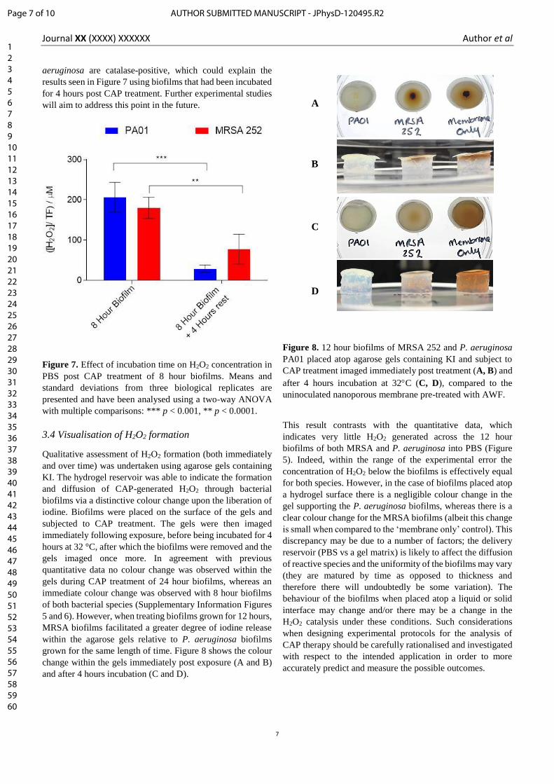
However, this trend was not seen when subjecting 8 hour
biofilms to the same incubation conditions. Conversely, the
concentration of H2O2 was lower for both MRSA and P.
aeruginosa post incubation (Figure 7). Previous studies have
indicated the role of catalase in protecting biofilms from the
effects of hydrogen peroxide by catalysing its decomposition
into oxygen and water [42-43]. Both S. aureus and P.
Page 6 of 10AUTHOR SUBMITTED MANUSCRIPT - JPhysD-120495.R2
123456789101112131415161718192021222324252627282930313233343536373839404142434445464748495051525354555657585960
Journal XX (XXXX) XXXXXX Author et al
7
aeruginosa are catalase-positive, which could explain the
results seen in Figure 7 using biofilms that had been incubated
for 4 hours post CAP treatment. Further experimental studies
will aim to address this point in the future.
Figure 7. Effect of incubation time on H2O2 concentration in
PBS post CAP treatment of 8 hour biofilms. Means and
standard deviations from three biological replicates are
presented and have been analysed using a two-way ANOVA
with multiple comparisons: *** p < 0.001, ** p < 0.0001.
3.4 Visualisation of H2O2 formation
Qualitative assessment of H2O2 formation (both immediately
and over time) was undertaken using agarose gels containing
KI. The hydrogel reservoir was able to indicate the formation
and diffusion of CAP-generated H2O2 through bacterial
biofilms via a distinctive colour change upon the liberation of
iodine. Biofilms were placed on the surface of the gels and
subjected to CAP treatment. The gels were then imaged
immediately following exposure, before being incubated for 4
hours at 32 °C, after which the biofilms were removed and the
gels imaged once more. In agreement with previous
quantitative data no colour change was observed within the
gels during CAP treatment of 24 hour biofilms, whereas an
immediate colour change was observed with 8 hour biofilms
of both bacterial species (Supplementary Information Figures
5 and 6). However, when treating biofilms grown for 12 hours,
MRSA biofilms facilitated a greater degree of iodine release
within the agarose gels relative to P. aeruginosa biofilms
grown for the same length of time. Figure 8 shows the colour
change within the gels immediately post exposure (A and B)
and after 4 hours incubation (C and D).
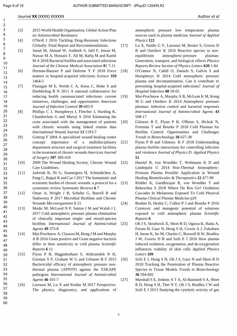
Figure 8. 12 hour biofilms of MRSA 252 and P. aeruginosa
PA01 placed atop agarose gels containing KI and subject to
CAP treatment imaged immediately post treatment (A, B) and
after 4 hours incubation at 32C (C, D), compared to the
uninoculated nanoporous membrane pre-treated with AWF.
This result contrasts with the quantitative data, which
indicates very little H2O2 generated across the 12 hour
biofilms of both MRSA and P. aeruginosa into PBS (Figure
5). Indeed, within the range of the experimental error the
concentration of H2O2 below the biofilms is effectively equal
for both species. However, in the case of biofilms placed atop
a hydrogel surface there is a negligible colour change in the
gel supporting the P. aeruginosa biofilms, whereas there is a
clear colour change for the MRSA biofilms (albeit this change
is small when compared to the ‘membrane only’ control). This
discrepancy may be due to a number of factors; the delivery
reservoir (PBS vs a gel matrix) is likely to affect the diffusion
of reactive species and the uniformity of the biofilms may vary
(they are matured by time as opposed to thickness and
therefore there will undoubtedly be some variation). The
behaviour of the biofilms when placed atop a liquid or solid
interface may change and/or there may be a change in the
H2O2 catalysis under these conditions. Such considerations
when designing experimental protocols for the analysis of
CAP therapy should be carefully rationalised and investigated
with respect to the intended application in order to more
accurately predict and measure the possible outcomes.
A
B
C
D
Page 7 of 10 AUTHOR SUBMITTED MANUSCRIPT - JPhysD-120495.R2
123456789101112131415161718192021222324252627282930313233343536373839404142434445464748495051525354555657585960

Journal XX (XXXX) XXXXXX Author et al
8
3.5 Topical application of H2O2
Owing to the multitude of different processes occurring during
CAP treatment which go beyond the direct operating
parameters (to include gas mixing with atmospheric gases and
mixing at the solution interface) it is challenging to provide a
representative control for the analysis of plasma-generated
species across biological interfaces [44]. Consequently, we
have focused solely on H2O2 transmission (as this is the major
long-lived species generated by the jet described). H2O2 at the
same concentration as generated by the CAP jet in 5 minutes
was topically applied to P. aeruginosa and MRSA biofilms
determine equivalency between plasma-assisted penetration
and topical therapy. Whilst it is difficult to compare the results
directly, topically applied H2O2 was unable to traverse
biofilms of all maturities indicated by the absence of any
quantifiable concentration of H2O2 in the PBS reservoir below.
This was confirmed immediately after the 5 minute exposure
time and following a subsequent 4 hours incubation at 32 C.
This result suggests that plasma-generated H2O2 (and possibly
other CAP-generated RONS), are better able to penetrate
bacterial biofilms than topical application of the same
chemical species. Again, this is likely a result of the additional
processes occurring, including continuous gas flow as a
driving force into the biofilms, localised heating and/or
synergistic effects from other plasma-generated RONS.
4. Discussion and Conclusions
The bioburden of the mature biofilms used in this study is
arguably much higher than seen in some other studies.
However, healthy skin is reportedly permanently colonised
with 103 – 104 microorganisms per cm2 skin (rising to 106 in
moist areas), as part of a healthy skin microflora [45].
Therefore, dense biofilms with high microbial cell counts
were used to represent the exponential proliferation of
opportunistic pathogens associated with untreated chronic
infection. In this study the use of CAP is intended as a
therapeutic treatment as opposed to a preventative measure
(whereby treatment may commence at lower cell densities
than those often considered indicative of progressive
infection).
In contrast to other studies, this work describes the effect of
bacterial biofilms on the fate of plasma-generated products
rather than any direct cellular effects of CAP therapy on the
biofilms - an important consideration in the future clinical
application of cold plasma. This investigation highlights the
ability of plasma-generated reactive species to traverse live
biofilms, remaining active despite interaction with the
bacterial species present. This is observed up to cell densities
of 109 CFU/ml, above which the concentration of longer lived
species is significantly reduced when evaluated across the
interface of the biofilm. Determining the exact mechanism of
cellular interaction with plasma-generated H2O2 is ongoing.
Owing to the complexity in determining the exact pathway of
plasma-generated H2O2 with respect to both the gas phase and
the liquid interface, there are a number of considerations to
take into account such as direct interaction of biofilm
components (including catalase) with plasma-generated H2O2,
alongside the transport and any subsequent reactions with
additional plasma-generated RONS [46-47]. Nonetheless, the
ability of high density bacterial biofilms to influence the
transmission of such species may have implications for the
application of CAP therapy in the treatment of chronic
infection, whereby high bacterial loads may be readily
encountered. In the context of deep-seated infections, the
ability to deliver RONS directly into the infected tissues may
vary with different device designs or may in fact be a
ubiquitous challenge associated with CAP therapy. This
should be established with the intention of improving RONS
delivery and penetration in order to successfully apply this
technology to highly contaminated wounds. Alongside
ensuring the efficacy of CAP therapy via adequate penetration
and delivery of plasma-generated reactive species, the effects
of such species on other cellular entities cannot be
underestimated. Ultimately, for the succesful therapeutic
treatement of infection, a balance must be established between
efficacious elimination of pathogenic organisms and
implementaion of adequate controls for the protection of the
surrounding host tissue.
Further research will focus on establishing the effect of
catalase on the lifetime and bioavaliability of plasma-
generated H2O2, the effect of CAP-induced bacterial cell death
on the transmission of H2O2 and any protective effects offered
by bacterial biofilms with respect to surrounding host tissues.
Additional research will also aim to investigate the formation
and transmission of other reactive species such as O2∙-, ∙OH,
∙HO2, to determine additional transportation mechanisms of
plasma-generated RONS with respect to the material interface
(biotic or abiotic).
Acknowledgements
The authors would like to thank the EPSRC for grant
EP/R003556/1. B Patenall would like to thank James Tudor
and Mr and Mrs Watson for additional funding. G Williams
would also like to thank Public Health England.
References
[1] 2017 Innovative Medicines Initiative, ND4BB New
Drugs for Bad Bugs
[2] 2015 CDC National Action Plan for the Combating
Antibiotic-Resistant Bacteria
Page 8 of 10AUTHOR SUBMITTED MANUSCRIPT - JPhysD-120495.R2
123456789101112131415161718192021222324252627282930313233343536373839404142434445464748495051525354555657585960
Journal XX (XXXX) XXXXXX Author et al
9
[3] 2015 World Health Organisation, Global Action Plan
on Antimicrobial Resistance
[4] O'Neill J 2016 Tackling Drug-Resistant Infections
Globally: Final Report and Reccommendations.
[5] Jamal M, Ahmad W, Andleeb S, Jalil F, Imran M,
Nawaz M A, Hussain T, Ali M, Rafiq M and Kamil
M A 2018 Bacterial biofilm and associated infections
Journal of the Chinese Medical Association 81 7-11
[6] Herman-Bausier P and Dufrene Y F 2018 Force
matters in hospital-acquired infections Science 359
1464-5
[7] Flanagan M E, Welsh C A, Kiess C, Hoke S and
Doebbeling B N 2011 A national collaborative for
reducing health careassociated infections: current
initiatives, challenges, and opportunities American
Journal of Infection Control 39 685-9
[8] Phillips C J, Humphreys I, Fletcher J, Harding K,
Chamberlain G and Macey S 2016 Estimating the
costs associated with the management of patients
with chronic wounds using linked routine data
International Wound Journal 13 1193-7
[9] Gottrup F 2004 A specialized wound-healing center
concept: importance of a multidisciplinary
department structure and surgical treatment facilities
in the treatment of chronic wounds American Journal
of Surgery 187 38S-43S
[10] 2009 The Wound Healing Society, Chronic Wound
Care Guidelines.
[11] Jarbrink K, Ni G, Sonnergren H, Schmidtchen A,
Pang C, Bajpai R and Car J 2017 The humanistic and
economic burden of chronic wounds: a protocol for a
systematic review Systematic Reviews 6 7
[12] Omar A, Wright J B, Schultz G, Burrell R and
Nadworny P 2017 Microbial Biofilms and Chronic
Wounds Microorganisms 5 15
[13] Modic M, McLeod N P, Sutton J M and Walsh J L
2017 Cold atmospheric pressure plasma elimination
of clinically important single- and mixed-species
biofilms International Journal of Antimicrobial
Agents 49 375-8
[14] Mai-Prochnow A, Clauson M, Hong J M and Murphy
A B 2016 Gram positive and Gram negative bacteria
differ in their sensitivity to cold plasma Scientific
Reports 6 11
[15] Flynn P B, Higginbotham S, Alshraiedeh N H,
Gorman S P, Graham W G and Gilmore B F 2015
Bactericidal efficacy of atmospheric pressure non-
thermal plasma (APNTP) against the ESKAPE
pathogens International Journal of Antimicrobial
Agents 46 101-7
[16] Laroussi M, Lu X and Keidar M 2017 Perspective:
The physics, diagnostics, and applications of
atmospheric pressure low temperature plasma
sources used in plasma medicine Journal of Applied
Physics 122
[17] Lu X, Naidis G V, Laroussi M, Reuter S, Graves D
B and Ostrikov K 2016 Reactive species in non-
equilibrium atmospheric-pressure plasmas:
Generation, transport, and biological effects Physics
Reports-Review Section of Physics Letters 630 1-84
[18] O'Connor N, Cahill O, Daniels S, Galvin S and
Humphreys H 2014 Cold atmospheric pressure
plasma and decontamination. Can it contribute to
preventing hospital-acquired infections? Journal of
Hospital Infection 88 59-65
[19] Mai-Prochnow A, Murphy A B, McLean K M, Kong
M G and Ostrikov K 2014 Atmospheric pressure
plasmas: Infection control and bacterial responses
International Journal of Antimicrobial Agents 43
508-17
[20] Gilmore B F, Flynn P B, O'Brien S, Hickok N,
Freeman T and Bourke P 2018 Cold Plasmas for
Biofilm Control: Opportunities and Challenges
Trends in Biotechnology 36 627-38
[21] Flynn P B and Gilmore. B F 2018 Understanding
plasma biofilm interactions for controlling infection
and virulence Journal of Physics D: Applied Physics
51
[22] Haertel B, von Woedtke T, Weltmann K D and
Lindequist U 2014 Non-Thermal Atmospheric-
Pressure Plasma Possible Application in Wound
Healing Biomolecules & Therapeutics 22 477-90
[23] Rödder K, Gandhirajan R, von Woedtke T and
Bekeschus S 2018 Where Do Ros Go? Oxidation
Cascades In Melanoma Exposed To Cold Physical
Plasma Clinical Plasma Medicine p29
[24] Boehm D, Heslin C, Cullen P J and Bourke P 2016
Cytotoxic and mutagenic potential of solutions
exposed to cold atmospheric plasma Scientific
Reports 6
[25] Oh J S, Strudwick X, Short R D, Ogawa K, Hatta A,
Furuta H, Gaur N, Hong S H, Cowin A J, Fukuhara
H, Inoue K, Ito M, Charles C, Boswell R W, Bradley
J W, Graves D B and Szili E J 2016 How plasma
induced oxidation, oxygenation, and de-oxygenation
influences viability of skin cells Applied Physics
Letters 109
[26] Szili E J, Hong S H, Oh J S, Gaur N and Short R D
2018 Tracking the Penetration of Plasma Reactive
Species in Tissue Models Trends in Biotechnology
36 594-602
[27] Marshall S E, Jenkins A T A, Al-Bataineh S A, Short
R D, Hong S H, Thet N T, Oh J S, Bradley J W and
Szili E J 2013 Studying the cytolytic activity of gas
Page 9 of 10 AUTHOR SUBMITTED MANUSCRIPT - JPhysD-120495.R2
123456789101112131415161718192021222324252627282930313233343536373839404142434445464748495051525354555657585960

Journal XX (XXXX) XXXXXX Author et al
10
plasma with self-signalling phospholipid vesicles
dispersed within a gelatin matrix Journal of Physics
D-Applied Physics 46
[28] Oh J S, Szili E J, Gaur N, Hong S H, Furuta H, Kurita
H, Mizuno A, Hatta A and Short R D 2016 How to
assess the plasma delivery of RONS into tissue fluid
and tissue Journal of Physics D-Applied Physics 49
[29] Szili E J, Bradley J W and Short R D 2014 A 'tissue
model' to study the plasma delivery of reactive
oxygen species Journal of Physics D-Applied
Physics 47
[30] Szili E J, Gaur N, Hong S H, Kurita H, Oh J S, Ito M,
Mizuno A, Hatta A, Cowin A J, Graves D B and
Short R D 2017 The assessment of cold atmospheric
plasma treatment of DNA in synthetic models of
tissue fluid, tissue and cells Journal of Physics D-
Applied Physics 50
[31] Chen C, Liu D X, Liu Z C, Yang A J, Chen H L,
Shama G and Kong M G 2014 A Model of Plasma-
Biofilm and Plasma-Tissue Interactions at Ambient
Pressure Plasma Chemistry and Plasma Processing
34 403-41
[32] Xiong Z, Du T, Lu X, Cao Y and Pan Y 2011 How
deep can plasma penetrate into a biofilm? Applied
Physics Letters 98
[33] Pei X, Lu X, Liu J, Liu D, Yang Y, Ostrikov K, Chu
P K and Pan Y 2012 Inactivation of a 25.5μm
Enterococcus faecalis biofilm by a room-
temperature, battery-operated, handheld air plasma
jet Journal of Physics D: Applied Physics 45
[34] Gorbanev Y, O'Connell D and Chechik V 2016 Non‐
Thermal Plasma in Contact with Water: The Origin
of Species Chemistry: A European Journal 22 3496.
[35] Junglee S, Urban L, Sallanon H and Lopez-Lauri F
2014 Optimized Assay for Hydrogen Peroxide
Determination in Plant Tissue Using Potassium
Iodide American Journal of Analytical Chemistry 05
730
[36] Thet N T, Wallace L, Wibaux A, Boote N and
Jenkins A T A 2018 Development of a mixed-species
biofilm model and its virulence implications in
device related infections Journal of Biomedical
Materials Research Part B Applied Biomaterials 107
129-37
[37] Bowler P G, Duerden B I and Armstrong D G 2001
Wound microbiology and associated approaches to
wound management Clinical Microbiology Reviews
14 244
[38] Patenall B L, Hathaway, H., Sedgwick, A.C., Thet,
N.T., Williams, G.T., Young, A.E., Allinson, S.L.,
Short, R.D. and Jenkins, A.T.A. 2018 Limiting
Pseudomonas aeruginosa Biofilm Formation Using
Cold Atmospheric Pressure Plasma Plasma Medicine
8 269-277
[39] Ziuzina D, Boehm D, Patil S, Cullen P J and Bourke
P 2015 Cold Plasma Inactivation of Bacterial
Biofilms and Reduction of Quorum Sensing
Regulated Virulence Factors PLoS ONE 10
[40] Alkawareek M Y, Algwari Q T, Laverty G, Gorman
S P, Graham W G, O'Connell D and Gilmore B F
2012 Eradication of Pseudomonas aeruginosa
Biofilms by Atmospheric Pressure Non-Thermal
Plasma PLOS ONE 7
[41] Fierheller M and Sibbald G 2010 A Clinical
Investigation into the Relationship between
Increased Periwound Skin Temperature and Local
Wound Infection in Patients with Chronic Leg Ulcers
Advances in Skin & Wound Care 23 369-78
[42] Stewart S P, Roe F, Rayner J, Elkins J G,
Lewandowski Z, Ochsner U A and Hassett D J 2000
Effect of Catalase on Hydrogen Peroxide Penetration
into Pseudomonas aeruginosa Biofilms Applied and
Environmental Microbiology 66 836 – 838
[43] Ochieng’ Olwal C, Oyieng’ Ang’ienda P and Otieno
Ochiel D 2019 Alternative sigma factor B (σB) and
catalase enzyme contribute to Staphylococcus
epidermidis biofilm’s tolerance against physico-
chemical disinfection Scientific Reports 9
[44] Girard F, Peret M, Dumont et al., 2018 Correlations
between gaseous and liquid phase chemistries
induced by cold atmosppheric plasmas in a
physiological buffer, Physical Chemistry Chemical
Physics, 20, 9198-9210
[45] Greene J N 1996 The microbiology of colonization,
including techniques for assessing and measuring
colonization Infection Control and Hospital
Epidemiology 17 114-8
[46] Bruggeman P and Schram D C 2010 On OH
production in water containing atmospheric pressure
plasmas Plasma Sources Science and Technology 19
[47] Liu J, He B, Chen Q, Li J, Xiong Q, Yue G, Zhang
X, Yang S, Liu H and Liu QH 2016 Direct synthesis
of hydrogen peroxide from plasma-water interactions
Scientific Reports 5
Page 10 of 10AUTHOR SUBMITTED MANUSCRIPT - JPhysD-120495.R2
123456789101112131415161718192021222324252627282930313233343536373839404142434445464748495051525354555657585960