Multidetector-Row Computed Tomography in the Evaluation of Transjugular Intrahepatic Portosystemic...
30
Središnja medicinska knjižnica Ajduk, M., Pavić, L., Bulimbašić, S., Šarlija, M., Pavić, P., Patrlj, L., Brkljačić, B. (2009) Multidetector-Row Computed Tomography in Evaluation of Atherosclerotic Carotid Plaques Complicated with Intraplaque Hemorrhage. Annals of Vascular Surgery, 23 (2). pp. 186- 193 http://www.elsevier.com/locate/issn/08905096 http://www.sciencedirect.com/science/journal/08905096 http://dx.doi.org/10.1016/j.avsg.2008.05.008 http://medlib.mef.hr/600 University of Zagreb Medical School Repository http://medlib.mef.hr/
-
Upload
independent -
Category
Documents
-
view
6 -
download
0
Transcript of Multidetector-Row Computed Tomography in the Evaluation of Transjugular Intrahepatic Portosystemic...

Središnja medicinska knjižnica
Ajduk, M., Pavić, L., Bulimbašić, S., Šarlija, M., Pavić, P., Patrlj, L.,
Brkljačić, B. (2009) Multidetector-Row Computed Tomography in
Evaluation of Atherosclerotic Carotid Plaques Complicated with
Intraplaque Hemorrhage. Annals of Vascular Surgery, 23 (2). pp. 186-
193
http://www.elsevier.com/locate/issn/08905096 http://www.sciencedirect.com/science/journal/08905096 http://dx.doi.org/10.1016/j.avsg.2008.05.008 http://medlib.mef.hr/600
University of Zagreb Medical School Repository
http://medlib.mef.hr/

Multidetector-Row Computed Tomography in Evaluation
of Atherosclerotic Carotid Plaques Complicated with
Intraplaque Hemorrhage
Authors: Marko Ajduk, Ladislav Pavić1, Stela Bulimbašić2, Mirko Šarlija,
Predrag Pavić, Leonardo Patrlj, Boris Brkljačić1
Department of Vascular Surgery, University Hospital Dubrava, Zagreb, Croatia
1Department of Radiology, University Hospital Dubrava, Zagreb, Croatia
2Depatment of Pathology, University Hospital Dubrava, Zagreb, Croatia
Corresponding author: Marko Ajduk, MD, Department of Vascular Surgery,
University Hospital Dubrava, Av. Gojka Šuška 6, 10000 Zagreb, Croatia
E-mail: [email protected], Tel.+38591201956
1
Abstract
Objective: To determine sensitivity and specificity of multidetector-row
computed tomography in detecting atherosclerotic carotid plaques complicated
with intraplaque hemorrhage.
Material: Carotid plaques from 31 patients operated for carotid artery stenosis.
Methods: Results of preoperative multidetector-row computed tomography
analysis of carotid plaques were compared with results of histological analysis
of the same plaque areas. Carotid endarterectomy was performed within one
week of multidetector-row computed tomography. American Heart Association
classification of atherosclerotic plaques was applied for histological
classification.
Results: Median tissue density of carotid plaques complicated with intraplaque
hemorrhage was 22 Hounsfield units. Median tissue density of noncalcified
segments of uncomplicated plaques was 59 Hounsfield units (p=0.0062). The
highest tissue density observed for complicated plaques was 31 Hounsfield
units. Multidetector-row computed tomography detected plaques complicated
with hemorrhage with sensitivity of 100% and specificity of 64.7%, with tissue
density of 31 Hounsfield units as a threshold value.
Conclusion: Multidetector-row computed tomography showed high level of
sensitivity and moderate level of specificity in detecting atherosclerotic carotid
plaques complicated with hemorrhage.
2
Key words: carotid plaque; hemorrhage; multidetector-row computed
tomography
Introduction
Large randomized controlled trials demonstrated benefit of carotid
endarterectomy (CEA) in symptomatic patients with high degree carotid artery
stenosis.1,2 Asymptomatic patients benefit less from CEA with absolute risk
reduction of only about 1% annually, during the five years follow-up. 3,4
Therefore, large number of asymptomatic patients must be operated to prevent
small number of neurological events. The total number of operated
asymptomatic patients may be lowered, if subgroups of asymptomatic patients
that benefit most from carotid endarterectomy could be identified. Several
studies showed higher incidence of neurological incidents in patients with so-
called soft plaques (plaques predominantly consisting of lipids, tissue debris
and hemorrhage).5-10 Ultrasound analysis of carotid plaques demonstrated that
hypoechoic plaques represent an independent risk factor for stroke incidence in
adults aged 65 years or older.11 Takaya et al. followed asymptomatic patients
for 38 months and showed that patients with intraplaque hemorrhage on initial
MRI had 5.2 times higher incidence of cerebrovascular events.12 The American
Heart Association (AHA) classification of atherosclerotic plaques defines eight
types of plaques, according to histological content (Table I).13,14 Atherosclerotic
carotid plaques complicated with intraplaque hemorrhage (AHA type VIb) are
considered as unstable and are associated with a higher incidence of
3

cerebrovascular events.12,15-17 Computed tomography (CT) angiography
demonstrated high accuracy in diagnosing carotid artery stenosis.18-22
Additional feature of computed tomography is its ability to measure tissue
density (expressed as a number of Hounsfield units [HU]). Thus, it can provide
some information about the type of analyzed tissue. Atherosclerotic carotid
plaques with lower tissue density on multidetector-row CT (MDCT) are
associated with lower incidence of cerebrovascular events.6,7 While single slice
computed tomography showed conflicting results in determining carotid plaque
composition, MDCT showed good correlation of findings with histological
analysis of coronary plaques.23-27 Histological analysis of coronary plaques
showed that remodeling of atherosclerotic plaque changes its histological
content. Therefore, the period between imaging and histological analysis should
be as short as possible.28 We compared results of MDCT and histological
analysis and calculated sensitivity and specificity of MDCT in detection of AHA
type VIb atherosclerotic carotid plaques (plaques complicated with intraplaque
hemorrhage, most often containing mixture of lipids, hemorrhage and necrotic
debris). Carotid endarterectomy was performed within one week of MDCT.
Material and methods
Carotid plaques from 31 consecutive patients operated for carotid artery stenosis
were included in this prospective study. There were 21 male and 10 female
4

patients, age between 51 and 87, median 70 years. There were 6 symptomatic
and 25 asymptomatic patients (Table II). Patients who experienced cerebral
insult, transient ischemic attack or amaurosis fugax on the side of affected
carotid artery within six months of MDCT were considered symptomatic.
Indications for carotid endarterectomy were symptomatic patients with carotid
artery stenosis >60% and asymptomatic patients with stenosis >70%. All
patients had the same imaging evaluation: color duplex-doppler first, and
MDCT in patients with carotid stenosis >60% on doppler.
Endarterectomy was performed within one week of MDCT evaluation. Approval
from the institutional ethical committee was obtained.
Two experienced radiologists performed doppler examination, using Logiq 9
scanner, with 7-9 and 9-14 MHz probes (GE-Healthcare, Milwaukee, Wi,
U.S.A.).
MDCT analysis
The Siemens (Erlangen, Germany) Somatom Sensation 16-row MDCT scanner
was used. One radiologist evaluated collected data on Siemens Leonardo
Syngo2004A workstation. The standardized optimized contrast-enhanced
protocol was used with intermediate reconstruction: 120 kVp, 120 mAs,
collimation 16x0.75 mm, pitch 1, slice thickness 0.75 mm. Iopamidol was used
as contrast medium (370 mg iodine/ml, 4 ml/s, 70 mm3, 325 psi). Transversal
multi-planar reconstructions, orthogonal to vessel long axis in both coronal and
5

sagittal planes, were used for plaque analysis. Three measurements of tissue
density were performed on visually least dense area of plaque at level of
maximal stenosis. Measurements were performed on 2 mm2 circle area and the
smallest value was recorded (Figures 1 and 2). Calcifications are obvious on
MDCT and were not further analyzed. Distance between carotid bifurcation and
level of maximal stenosis was recorded, in order to help pathologist in finding
corresponding level for histological analysis. Percentage of stenosis was
calculated applying North American Symptomatic Carotid Endarterectomy Trial
(NASCET) criteria.14
Surgical technique
Patients underwent carotid endarterectomy under locoregional or general
anesthesia, with selective use of intraluminal shunt in the former group of
patients and in all patients in the latter group. Four patients were operated under
general anesthesia. Two of them had concomitant cardiac surgery, one had
history of epileptic seizures, and one patient with contralateral occlusion had an
explicit wish for general anesthesia. Care was taken to preserve the
morphologic integrity of plaques as much as possible. There were no verified
perioperative insults. The patient with contralateral carotid occlusion had
transient postoperative weakness of the contralateral hand without CT evidence
of ischemic brain lesion.
6

Histological analysis
Immediately after carotid endarterectomy, plaques were formalin-fixed (10%
buffered formaldehyde) and sent for histological analysis. One pathologist,
blinded for MDCT plaque density, performed histological analysis. If
calcifications were extensive, plaques were first decalcified using 20% nitric
acid. That procedure eliminates calcifications while preserving remaining
histological content. Samples were sliced in serial manner, starting from
bifurcation, followed by sections 2 mm apart toward internal carotid artery.
Performed serial sections technique assured precise measurement of distance
between the bifurcation and the level of maximal stenosis.
Plaques sections were embedded in paraffin and cut at 4 μm thin slices, using
standard process. Slices were stained with hematoxylin and with Mallory
trichrome if necessary (Figures 3 and 4). One pathologist examined all plaques
and classified them according to the American Heart Association classification
of atherosclerotic plaques. The radiologist that performed MDCT analysis was
involved in histological analysis, to make sure that same plaque areas were
analysed on MDCT and histology.
Data analysis
Difference of median tissue density between AHA plaques type VIb and other
plaque types was calculated using Mann-Whitney U-test. Value of p<0.05 were
7

considered statistically significant. To determine cut off value of tissue density,
ROC analysis was used.
Results
There were 14 (45%) AHA VIb plaques and 17 (55%) other AHA types (V, VII
and VIII). Median MDCT tissue density (TD) of type VIb plaques was 22 HU
(range, -17 to 31), and median tissue density of non-calcified segments of non-
complicated plaques was 59 HU (range, -6 to 150), (p=0.0062, Mann-Whitney
U-test), (Figure 5). ROC analysis showed 100 % sensitivity and 64.7%
specificity of MDCT in detecting plaques complicated with intraplaque
hemorrhage, with tissue density of 31 HU as a threshold value (i.e., no plaque
with MDCT tissue density over 31 HU was complicated with intraplaque
hemorrhage), (Figure 6). Four of six plaques from symptomatic patients were
AHA type VI b and 10 of 25 plaques from asymptomatic patients were AHA
type VIb.
Discussion
This study showed that MDCT could detect atherosclerotic carotid plaque
complicated with hemorrhage with 100% sensitivity, with tissue density of 31
HU as a threshold value. Previous studies showed inconclusive results
8

regarding the accuracy of single slice computed tomography in analysing
plaque composition.23,24 De Weert et al. showed good correlation between in
vivo MDCT findings and histological findings, however in their analysis of 15
carotid plaques, the period between MDCT evaluation and endarterectomy was
up to three months.29 During that period, remodeling of plaques could change
their histological appearance. Histological analysis of coronary plaques
performed within one week after infarction showed morphologic features of
instability, while plaques taken later were histologicalally similar to those in
patients with stable angina.28 To minimize inaccuracy resulting from this fact,
all patients in this study were operated within one week of MDCT analysis.
To our knowledge, among studies comparing in vivo MDCT and histological
analysis of carotid plaques and providing the period between MDCT and
endarterectomy of less than one week, this study enrolled the largest number of
patients.
Our aim was to identify plaques with intraplaque hemorrhage (AHA type VIb)
using MDCT. Carotid plaques are most often heterogeneous and small areas of
plaque often have mixed histological content. Lipids, hemorrhage and necrotic
debris ("soft tissue”) have the lowest TD on MDCT and other tissue
components (fibrosis or calcifications) increase TD. This can influence MDCT
results by giving higher TD values even in predominantly soft areas of plaque
due to the partial volume effect. Similar effect is produced by contrast medium
in vessel lumen and calcified portions of the plaque. To minimize influence of
9
this effect, we performed three measurements per slice on chosen plaque area
(visually least dense area) and recorded only the smallest value, since no plaque
component has lower TD than lipids, hemorrhage or necrotic debris.
With 2 mm2 area on which tissue density was measured and 0.75 mm slice
thickness, 1.5 mm3 of tissue volume was measured by each measurement. Even
plaques with such a small MDCT detectable amount of soft tissue (which is
most often combination of lipids, hemorrhage and necrotic debris) should
probably be regarded as potentially vulnerable, since it is impossible to tell by
MDCT whether hemorrhage within plaque is expanding or reducing due to
plaque remodeling. To provide the same plaque level for MDCT and
histological analysis we measured the distance from bifurcation to level of
maximal stenosis on MDCT and that value was used by the pathologist to find
corresponding level of specimen for histological analysis. It might have
happened that a minor degree of longitudinal plaque shrinkage occured during
the histological processing, but it is unlikely to be significant because of the low
overall water content of plaques. However, serial slicing and embedding of the
whole plaque (including the planes below and above MDCT measured level of
maximal stenosis), and possibility of additional slices from deeper levels of
paraffin embedded material, assure that the level of the narrowest lumen
(maximal stenosis) had been chosen for the analysis.
Current clinical practice in the treatment of asymptomatic patients with carotid
artery stenosis differs among different countries and even among different
10

institutions within the same country. 30-43 In general, asymptomatic patients with
carotid artery stenosis are treated more conservatively in Europe than in the
United States. Asymptomatic patients with carotid artery stenosis comprise 11-
52% and 37-92% of all patients operated for carotid artery stenosis in Europe
and the United States, respectively.30-43 Several authors have indicated that
operative treatment of asymptomatic patients with carotid artery stenosis should
be considered only for medically stable patients with ≥80% stenosis with life
expectancy of at least 5 years, and only if a <3% perioperative complication rate
can be achieved.44,45 Asymptomatic patients with complicated plaque and <80%
carotid artery stenosis, who would not be treated if the above mentioned
recommendations are applied, could benefit from a diagnostic method that is
able to detect some features of carotid plaque associated with increased risk of a
cerebrovascular event. Asymptomatic patients with carotid artery stenosis of
<80% and uncomplicated plaque probably require only the best medical
therapy.44-47 The decision which diagnostic method to use and when to treat
asymptomatic patients is affected not only by the results of large trials, but also
by diagnostic resources available, medical system funds, and the possibility of
treating patients with carotid artery stenosis with low morbidity and mortality at
a particular institution. MDCT increases the cost of diagnostic evaluation for
each patient if compared to duplex analysis alone, and exposes patient to
radiation. However, MDCT is noninvasive and accurate in diagnosing carotid
artery stenosis.18-22 Furthermore, MDCT showed very good interobserver
11

agreement in the evaluation of degree of carotid artery stenosis and can also
provide information about the type of analyzed tissue and the presence of
intracranial arterial stenosis. 18-22,29,48,49 Studies dealing with doppler
examination of carotid artery stenosis showed marked interobserver
variabilities.50-52 Several studies based on measurement of grey scale median of
carotid plaques showed conflicting results regarding the correlation of findings
with histological content, while studies based on visual evaluation of ultrasound
findings showed high variability of intra- and interobserver agreement.53 In our
view, duplex and MDCT are complementary studies. We perform duplex
examination first, followed by MDCT, if >60% carotid artery stenosis is found
on duplex. We believe that higher cost of diagnostic evaluation that includes
MDCT could be in part compensated by the potential reduction of the number of
operated asymptomatic patients.
Conclusion
Multidetector-row computed tomography showed very high level of sensitivity
and moderate level of specificity in detecting hemorrhage within atherosclerotic
carotid plaque. Plaques with TD over 31 HU on MDCT were not complicated
with intraplaque hemorrhage. Technical advancements of CT equipment may
probably increase the specificity of the method.
12

References:
1 North American Symptomatic Carotid Endarterectomy Trial Collaborators:
Beneficial effect of carotid endarterectomy in symptomatic patients with high-
grade carotid stenosis. N Engl J Med 325:445, 1991.
2 European Carotid Surgery Trialist's Collaborative Group: Medical Research
Council European Carotid Surgery Trial: Interim results for symptomatic
patients with severe (70-99%) or with mild (0-29%) carotid stenosis. Lancet
337:1235, 1991.
3 Executive Committee for the Asymptomatic Carotid Atherosclerosis Study.
Endarterectomy for Asymptomatic Carotid Artery Stenosis. JAMA, Volume
273(18).May 10, 1995.1421-1428.
4 Halliday A, Mansfield A, Marro J, et al. Asymptomatic Carotid Surgery Trial
(ACST) Collaborative Group. Prevention of disabling and fatal strokes by
successful carotidendarterectomy in patients without recent neurologicalal
symptoms: randomised controlled trial. Lancet 2004; 363: 1491–502
5 Grønholdt MLM, Nordestgaard BG, Schroeder TV, et al. Ultrasonic
Echolucent Carotid Plaques Predict Future Strokes. Circulation 2001;104;68-73
6 Serfaty JM, Nonent M, Nighoghossian N, et al. for the CARMEDAS Study
Group. Plaque density on CT, a potential marker of ischemic stroke. Neurology
2006;66:118-120
13

7 Nandalur KR, Baskur E, Hagspiel KD, et al. Calcified carotid atherosclerotic
plaque is assosiated less with ischemic symptoms than is noncalcified plaque on
MDCT. AJR 2005;184:295-298
8 European Carotid Plaque Study Group: Carotid Artery Plaque Composition –
Relationship to Clinical Presentation and Ultrasoundm B-mode Imaging. Eur J
Vasc Endovasc Surg 10, 23-30 (1995).
9 Carra G, Visona A, Bonanome A, et al. Carotid plaque morphology and
cerebrovascular events. Int Angiol. 2003 Sep;22(3):284-9.
10 Mathiesen EB, Bonaa KH, Joakimsen O. Echolucent plaques are associated
with high risk of ischemic cerebrovascular events in carotid stenosis: the
Tromso Study. Circulation. 2001;103:2171–2175.
11 Polak JF, Shemanski L, O’Leary DH, et al. Hypoechoic plaque at us of the
carotid artery: An independent risk factor for incident stroke in adults aged 65
years or older. Cardiovascular Health Study. Radiology. 1998;208:649–654.
12 Takaya N, Yuan C, Chu B, et al. Association Between Carotid Plaque
Characteristics and Subsequent Ischemic Cerebrovascular Events: A Prospective
Assessment With MRI--Initial Results. Stroke 2006;37;818-823.
13 Stary HC, Chandler AB, Dinsmore R, et al. A Definition of Advanced Types
of Atherosclerotic Lesions and a Histologicalal Classification of
Atherosclerosis: A Report From the Comittee on Vascular Lesions of the
Council an Arteriosclerosis, American Heart Association. Circulation,
Volume92(5).September 1, 1995.1355-1374
14

14 Herbert C. Stary. Natural History and Histologicalal Classification of
Atherosclerotic Lesions : An Update. Arterioscler Thromb Vasc Biol.
2000;20:1177-1178.
15 Rao DS, Goldin JG, Fishbein MC. Determinants of plaque instability in
atherosclerotic vascular disease.Cardiovasc Pathol. 2005 Nov-Dec;14(6):285-93.
16 Mofidi R, Crotty TB, McCarthy P, et al. Association between plaque
instability, angiogenesis and symptomatic carotid occlusive disease. Br J Surg
88:945–950, 2001.
17 Imparato AM, Riles TS, Mintzer R, et al. The importance of hemorrhage in
the relationship between gross morphologic characteristics and cerebral
symptoms in 376 carotid artery plaques. Ann Surg. 1983;197:195–203.
18 Josephson SA, Bryant SO, Mak HK, et al. Evaluation of carotid stenosis
using CT angiography in the initial evaluation of stroke and TIA. Neurology
2004;63:457-460.
19 Chen CJ, Lee TH, Hsu HL, et al. Multi-Slice CT Angiography in Diagnosing
Total Versus Near Occlusions of the Internal Carotid Artery: Comparison With
Catheter Angiography. Stroke 2004;35;83-85.
20 Koelemay MJW, Nederkoorn PJ, Reitsma JB, et al. Systematic Review of
Computed Tomographic Angiography for Assessment of Carotid Artery
Disease. Stroke 2004;35;2306-2312.
21 Moll R, Dinkel HP. Value of the CT angiography in the diagnosis of
common carotid artery bifurcation disease: CT angiography versus digital
15

subtraction angiography and color flow Doppler. European Journal of Radiology
39 (2001) 155–162
22 Ibarra-de Grassa B, Romero-Vidal FJ, Munoz-Martinez V. Usefulness of
arteriography with multislice spiral computed tomography in the diagnosis of
preocclusive stenosis of the cervical internal carotid artery. Rev Neurol. 2003
Oct 1-15;37(7):632-6.
23 Walker LJ, Ismail A, McMeekin W, et al. Computed Tomography
Angiography for the Evaluation of Carotid Atherosclerotic Plaque: Correlation
With Histopathology of Endarterectomy Specimens. Stroke 2002;33;977-981.
24 Estes JM, Quist WC, Lo Gerfo FW, et al. Noninvasive characterization of
plaque morphology using helical computed tomography. J Cardiovasc Surg
(Torino) 1998;39:527–534.
25 Oliver TB, Lammie GA, Wright AR, et al. Atherosclerotic plaque at the
carotid bifurcation: CT angiographic appearance with histopathologic
correlation. AJNR Am J Neuroradiol 1999;20:897–901.
26 Schroeder S, Kuettner A, Leitritz M, et al. Reliability of Differentiating
Human Coronary Plaque Morphology Using Contrast-Enhanced Multislice
Spiral Computed Tomography: A Comparison With Histology. Journal of
Computer Assisted Tomography. 28(4):449-454, July/August 2004.
27 Schroeder S, Kopp AF, Baumbach A, et al. Noninvasive Detection and
Evaluation of Atherosclerotic Coronary Plaques With Multislice Computed
Tomography. J Am Coll Cardiol Vol. 37, No. 5. April 2001:1430-5.
16

28 Depre C, Wijns W, Robert AM, et al. Pathology of Unstable Plaque:
Correlation With the Clinical Severity of Acute Coronary Syndromes. J Am Coll
Cardiol Vol. 30, No 3. September 1997:694-702.
29 De Weert TT, Ouhlous M, Meijering E, et al. In Vivo Characterization and
Quantification of Atherosclerotic Carotid Plaque Components With
Multidetector Computed Tomography and Histopathological Correlation.
Arterioscler Thromb Vasc Biol. 2006;26:2366-2372.)
30 McPhee JT, Hill JS, Ciocca RG, Messina LM, Eslami MH. Carotid
endarterectomy was performed with lower stroke and death rates than carotid
artery stenting in the United States in 2003 and 2004. J Vasc Surg. 2007
Dec;46(6):1112-1118.
31 Kragsterman B, Björck M, Lindbäck J, Bergqvist D, Pärsson H, on behalf of
the Swedish Vascular Registry (Swedvasc). Long-Term Survival After Carotid
Endarterectomy for Asymptomatic Stenosis Stroke. 2006;37:2886-2891.)
32 Šoša T, Ajduk M, Erdelez L, Škopljanac A. Carotid Surgery 2004. State of
the Art, Prognosis and Perspective. Acta Clin Croatica 43:suppl1(2004);106-
117.
33 Mayo, Sara W. MD; Eldrup-Jorgensen, Jens MD; Lucas, F. L. PhD;
Wennberg, David E. MD; Bredenberg, Carl E. MD. Carotid endarterectomy
after NASCET and ACAS: A statewide study. Journal of Vascular Surgery.
27(6):1017-1023, June 1998.
17

34 Long GW, Nuthakki V, Bove PG, Brown OW, Shanley CJ, Bendick PJ,
Rimar S, Kitzmiller J, Zelenock GB. Contemporary outcomes for carotid
endarterectomy at a large community-based academic health center. Ann Vasc
Surg. 2007 May;21(3):321-7.
35 LaMuraglia GM, Brewster DC, Moncure AC, Dorer DJ, Stoner MC, Trehan
SK, Drummond EC, Abbott WM, Cambria RP. Carotid endarterectomy at the
millennium: what interventional therapy must match. Ann Surg. 2004
Sep;240(3):535-44.
36 Mehta RH, Zahn R, Hochadel M, Ischinger T, Jung J, Hauptmann KE, Mark
B, Zeymer U, Schramm A, Senges J. Comparison of in-hospital outcomes of
patients with versus without previous carotid endarterectomy undergoing carotid
stenting (from the German ALKK CAS Registry). Am J Cardiol. 2007 May
1;99(9):1288-93.
37 Smurawska LT, Bowyer B, Rowed D, Maggisano R, Oh P, Norris JW.
Changing practice and costs of carotid endarterectomy in Toronto, Canada.
Stroke. 1998 Oct;29(10):2014-7.
38 Karp HR, Flanders WD, Shipp CC, Taylor B, Martin D. Carotid
endarterectomy among Medicare beneficiaries: a statewide evaluation of
appropriateness and outcome. Stroke. 1998 Jan;29(1):46-52.
39 Setacci C, Chisci E, de Donato G, Setacci F, Galzerano G. Carotid Artery
Stenting in a Single Center: Are Six Years of Experience Enough to Achieve the
Standard of Care? Eur J Vasc Endovasc Surg 34, 655e662 (2007)
18

40 H. Rodgers*1,3, S. E. Oliver2, R. Dobson3 and R. G. Thomson3, on behalf
of the Northern Regional Carotid Endarterectomy Audit Group†. A Regional
Collaborative Audit of the Practice and Outcome of Carotid Endarterectomy in
the United Kingdom. Eur J Vasc Endovasc Surg 19, 362–369 (2000)
41 Wong JH, Lubkey TB, Suarez-Almazor ME, Findlay JM. Improving
the appropriateness of carotid endarterectomy results of a prospective city-wide
audit. Stroke 1999; 30: 12–15.
42 Cebul RD, Snow RJ, Pine R, Hertzer NR, Norris DG. Indications, outcomes
and provider volumes for carotid endarterectomy. JAMA 1998; 279: 1282–
1287.
43 Melissano G, Castellano R, Mazzitelli S, Zoppei G, Chiesa R . Safe and
Cost-effective Approach to Carotid Surgery. Eur J Vasc Endovasc Surg 14, 164-
169 (1997)
44 Mayo Dodick, David W. MD; Meissner, Irene MD; Meyer, Fredric B. MD;
Cloft, Harry J. MD, PhD Evaluation and Management of Asymptomatic Carotid
Artery Stenosis. Mayo Clinic Proceedings. 79(7):937-944, July 2004
45 Rockman, Caron B. MD; Riles, Thomas S. MD; Lamparello, Patrick J. MD;
Giangola, Gary MD; Adelman, Mark A. MD; Stone, David BA; Guareschi,
Claudio MD; Goldstein, Jonathan BA; Landis, Ronnie RN Natural history and
management of the asymptomatic, moderately stenotic internal carotid artery.
Journal of Vascular Surgery. 25(3):423-431, March 1997.
19

46 European Carotid Surgery Trialists' Collaborative Group. Risk of stroke in
the distribution of an asymptomatic carotid artery. Lancet 1995;345:209-12.
47 CASANOVA Study Group. Carotid surgery versus medical therapy in
asymptomatic carotid stenosis. Stroke 1991;22:1229-35.
48 Saba L, Mallarini G. MDCTA of carotid plaque degree of stenosis:
evaluation of interobserver agreement. AJR Am J Roentgenol. 2008
Jan;190(1):W41-6.
49 Schuknecht B. High-concentration contrast media (HCCM) in CT
angiography of the carotid system: impact on therapeutic decision making.
Neuroradiology. 2007 Jul;49 Suppl 1:S15-26
50 Jahromi AS, Cinà CS, Liu Y, Clase CM. Sensitivity and specificity of color
duplex ultrasound measurement in the estimation of internal carotid artery
stenosis: a systematic review and meta-analysis. J Vasc Surg. 2005
Jun;41(6):962-72.
51 Mikkonen RH, Kreula JM, Virkkunen PJ. Reproducibility of Doppler
ultrasound measurements. Acta Radiol. 1996 Jul;37(4):545-50.
52 Henry-Feugeas M, Alkilic-Genauzeau I, Aymé N, Schouman-Claeys E.
Variability of ultrasonography velocity assessment of the carotid arteries. J
Radiol. 2000 Apr;81(4):445-9.
53 Sztajzel R. Ultrasonographic assessment of the morphological characteristics
of the carotid plaque. SWISS MED WKLY 2005;135:635–643
20
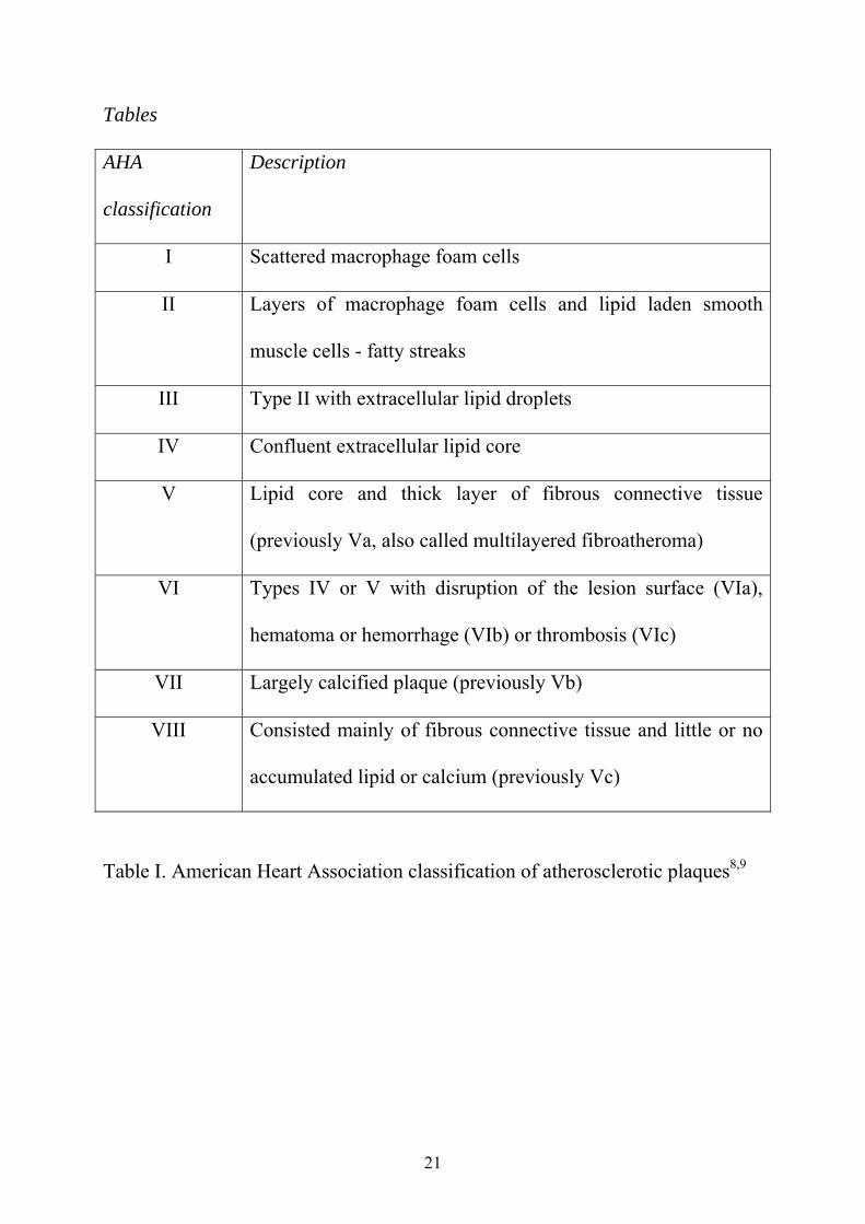
Tables
AHA
classification
Description
I Scattered macrophage foam cells
II Layers of macrophage foam cells and lipid laden smooth
muscle cells - fatty streaks
III Type II with extracellular lipid droplets
IV Confluent extracellular lipid core
V Lipid core and thick layer of fibrous connective tissue
(previously Va, also called multilayered fibroatheroma)
VI Types IV or V with disruption of the lesion surface (VIa),
hematoma or hemorrhage (VIb) or thrombosis (VIc)
VII Largely calcified plaque (previously Vb)
VIII Consisted mainly of fibrous connective tissue and little or no
accumulated lipid or calcium (previously Vc)
Table I. American Heart Association classification of atherosclerotic plaques8,9
21

Patient No
Age (years)
Gender Symptoms Stenosis %
(MDCT)
Tissue density (HU)
AHA plaque type
Stenosis %
(duplex) 1 67 M No 70 59 VII 65 2 74 M No 95 -11,6 VIb 90 3 65 M No 95 -17,6 VIb 95 4 56 M No 95 62,6 V 95 5 87 M No 90 -23,6 V 90 6 77 M No 90 63 V 90 7 70 M No 90 62,8 V 90 8 77 M No 90 22,2 VIb 90 9 82 F Yes 80 31,2 VIb 85
10 80 M No 70 18,2 VIb 70 11 62 M Yes 80 22,4 V 75 12 81 M Yes 90 28,5 VIb 90 13 68 M No 80 62,8 V 70 14 77 M No 95 25,2 VIb 75 15 68 F No 70 60,7 V 65 16 79 M No 90 24,1 VIb 70 17 61 M No 80 14,7 VIb 65 18 68 M No 80 21,7 VIb 70 19 77 M No 80 17,7 V 80 20 72 M Yes 90 23,3 VIb 80 21 65 M No 80 42,1 V 95 22 64 M No 95 26,8 VIb 95 23 61 F No 90 6,7 V 80 24 76 F Yes 80 131 VII 70 25 59 F Yes 95 -3 VIb 80 26 76 F No 90 150 V 80 27 67 F No 90 -4,0 VIb 90 28 64 F No 70 28,6 V 75 29 51 F No 80 35,8 VIII 70 30 80 F No 90 11,6 V 95 31 72 M No 70 59,8 V 75
Plaque type
Median Range
VIb 22 -17 to 31
Summary Median 70 Range 51-87
M 21 F 10
Yes 6 No 25
Mean 84.8±8.6
Other 59 -6 to 150
VIb 14 Other 17
Mean 80.8±10.3
Table II. Patients’ characteristics, MDCT, histological and duplex findings. HU-
Hounsfield units, MDCT- multidetector-row computed tomography, AHA-
American Heart Association
22

LEGENDS:
Figure 1. MDCT. Plaque with the least measured tissue density of -3 Hounsfield
units (HU). ICA-internal carotid artery, MDCT- multidetector-row computed
tomography.
23

Figure 2. MDCT. Plaque with the least measured tissue density of 59,8
Hounsfield units (HU). ICA-internal carotid artery, MDCT- multidetector-row
computed tomography.
24

Figure 3. Hemorrhage within plaque (plaque type VIb). Same plaque as on
Figure 1. H&E, original magnification x40. P- plaque, L-lumen.
25

Figure 4. Multilayered fibroatheroma (plaque type V). Same plaque as on Figure
2. H&E, original magnification x40. P- plaque, L-lumen.
26

Figure 5. Box-whisker plots of tissue density of plaques without hemorrhage and
plaques with hemorrhage. HU-Hounsfield units.
27

Figure 6. ROC analysis. Cut off value of 31.2 Hounsfield units (HU), sensitivity
100%, specificity 64.71%, area under curve 0.79, p=0.0004.
28

29
Table I. American Heart Association classification of atherosclerotic plaques.8,9
Table II. Patients’ characteristics, MDCT, histological and duplex findings. HU-
Hounsfield units, MDCT- multidetector-row computed tomography, AHA-
American Heart Association.