Intrahepatic growth and maturation of Gnathostoma turgidum in the natural definitive opossum host,...
6
Intrahepatic growth and maturation of Gnathostoma turgidum in the natural definitive opossum host, Didelphis virginiana Sylvia Páz Díaz-Camacho a , Francisco Delgado-Vargas a , Kaethe Willms b , María del Carmen de la Cruz-Otero a , José Guadalupe Rendón-Maldonado a , Lilia Robert b , Silvia Antuna c , Yukifumi Nawa a,d, ⁎ a Facultad de Ciencias Químico Biológicas, Universidad Autónoma de Sinaloa, Culiacán, Sinaloa, México b Departamento de Microbiología y Parasitología, Facultad de Medicina, Universidad Nacional Autónoma de México, México D.F., México c Departamento de Biología Celular y Tisular, Facultad de Medicina, Universidad Nacional Autónoma de México, México D.F., México d Division of Parasitology, Department of Infectious Diseases, Faculty of Medicine, University of Miyazaki, Miyazaki, Japan abstract article info Article history: Received 14 March 2010 Received in revised form 17 April 2010 Accepted 21 April 2010 Available online 4 May 2010 Keywords: Gnathostoma turgidum Advanced 3rd stage larvae Opossum Didelphis virginiana Gnathostoma turgidum is a gastric nematode parasite of opossums found in the Americas. We recently found that G. turgidum juveniles appear in the liver of the opossums where they become mature adults and almost synchronously move to the stomach during certain months of the year, suggesting the importance of the liver for the growth and maturation of this species in the final hosts. In this study we attempted to detect G. turgidum larvae in the liver of opossums, Didelphis virginiana that are the natural final hosts. The results show that tiny (b 3 mm in length) third stage larvae (L3) appeared in the liver of opossums around November and December. Also in the liver, we found large L3 of up to about 10 mm in length together with juveniles and mature adults from February to March. In spite of their length, large L3 have 4 rows of hooklets, and their gonads remained undeveloped. Morphological features of the small and large L3 of G. turgidum are described including scanning electron microscope images. The seasonal switching of the several growth stages of G. turgidum from small L3 to adult worms in the liver and eventual migration to the stomach in opossums suggests the unique feature of G. turgidum utilizing the liver as the maturation site. © 2010 Elsevier Ireland Ltd. All rights reserved. 1. Introduction Gnathostomosis is a food-borne zoonosis caused by vigorous migration of the advanced 3rd stage larvae (AL3) of the genus Gnathostoma in the human body. Infection occurs by ingesting raw or insufficiently cooked fresh fish meat contaminated with Gnathostoma AL3. The disease is endemic where people have the custom of consuming raw or under-cooked fish dishes. Thailand and Japan have been known as the most famous endemic areas of this disease, although patients have been found in many other Asian countries [1–4]. In Mexico, the first human gnathostomosis case was found in 1970 [5] and the country has been recently found to be heavily endemic for this disease [6–10]. Three Gnathostoma species, Gnathostoma binucleatum, Gnathostoma turgidum, and the newly identified Gnathostoma lamothei Bertoni-Ruiz et al. 2005, are recognized as native species in Mexico [11]. Among them, only G. binucleatum has been proven to be a pathogen for humans [12,13]. Recently we found a highly endemic area of G. turgidum in common opossums, Didelphis virginiana [14,15] in the southern part of Sinaloa State, Mexico, where G. binucleatum is also highly endemic and an acute outbreak of human gnathostomiasis due to G. binucleatum infection was recorded [9]. In spite of the co-existence of two Gnathostoma species in this small area, we could find only G. binucleatum AL3 in an array of intermediate/paratenic hosts such as estuarine fish, ichthyophagous birds, amphibians and reptiles [16,17]. In our 10 year survey in this area since 2001, we have not yet found G. turgidum larvae in any of those intermediate/paratenic hosts, suggesting that G. binucleatum and G. turgidum have separate natural lifecycles in the same geographic area. Moreover, we found that G. turgidum larvae develop to juveniles and fully mature adults in the liver of the natural final host opossum before they appear in the final tissue, the gastric wall [14,15]. This intrahepatic maturation of G. turgidum appears to be unique from the maturation process of other Gnathostoma species. Moreover, recently extremely small AL3 of G. turgidum were found in natural and experimental intermedi- ate/paratenic hosts [18], and two large G. turgidum larvae were found accidentally in the liver of a four-eyed opossum, which were proposed as the 4th stage larvae of G. turgidum [19]. In the present study, we describe morphological features of small and large size G. turgidum larvae obtained from the liver of common opossums, D. virginiana. The significance of the seasonal appearance of those Parasitology International 59 (2010) 338–343 ⁎ Corresponding author. Department of Helminthology, Faculty of Tropical Medicine, Mahidol University, 420/6 Ratchawithi Rd., Bangkok 10400, Thailand. Tel./fax: +66 2643 5600. E-mail address: [email protected] (Y. Nawa). 1383-5769/$ – see front matter © 2010 Elsevier Ireland Ltd. All rights reserved. doi:10.1016/j.parint.2010.04.004 Contents lists available at ScienceDirect Parasitology International journal homepage: www.elsevier.com/locate/parint
-
Upload
independent -
Category
Documents
-
view
3 -
download
0
Transcript of Intrahepatic growth and maturation of Gnathostoma turgidum in the natural definitive opossum host,...
Parasitology International 59 (2010) 338–343
Contents lists available at ScienceDirect
Parasitology International
j ourna l homepage: www.e lsev ie r.com/ locate /par in t
Intrahepatic growth and maturation of Gnathostoma turgidum in the naturaldefinitive opossum host, Didelphis virginiana
Sylvia Páz Díaz-Camacho a, Francisco Delgado-Vargas a, Kaethe Willms b,María del Carmen de la Cruz-Otero a, José Guadalupe Rendón-Maldonado a, Lilia Robert b,Silvia Antuna c, Yukifumi Nawa a,d,⁎a Facultad de Ciencias Químico Biológicas, Universidad Autónoma de Sinaloa, Culiacán, Sinaloa, Méxicob Departamento de Microbiología y Parasitología, Facultad de Medicina, Universidad Nacional Autónoma de México, México D.F., Méxicoc Departamento de Biología Celular y Tisular, Facultad de Medicina, Universidad Nacional Autónoma de México, México D.F., Méxicod Division of Parasitology, Department of Infectious Diseases, Faculty of Medicine, University of Miyazaki, Miyazaki, Japan
⁎ Corresponding author. Department of HelminthologMahidol University, 420/6 Ratchawithi Rd., Bangkok 12643 5600.
E-mail address: [email protected] (
1383-5769/$ – see front matter © 2010 Elsevier Irelanddoi:10.1016/j.parint.2010.04.004
a b s t r a c t
a r t i c l e i n f oArticle history:Received 14 March 2010Received in revised form 17 April 2010Accepted 21 April 2010Available online 4 May 2010
Keywords:Gnathostoma turgidumAdvanced 3rd stage larvaeOpossumDidelphis virginiana
Gnathostoma turgidum is a gastric nematode parasite of opossums found in the Americas. We recently foundthat G. turgidum juveniles appear in the liver of the opossums where they become mature adults and almostsynchronously move to the stomach during certain months of the year, suggesting the importance of theliver for the growth and maturation of this species in the final hosts. In this study we attempted to detect G.turgidum larvae in the liver of opossums, Didelphis virginiana that are the natural final hosts. The results showthat tiny (b3 mm in length) third stage larvae (L3) appeared in the liver of opossums around November andDecember. Also in the liver, we found large L3 of up to about 10 mm in length together with juveniles andmature adults from February to March. In spite of their length, large L3 have 4 rows of hooklets, and theirgonads remained undeveloped. Morphological features of the small and large L3 of G. turgidum are describedincluding scanning electron microscope images. The seasonal switching of the several growth stages of G.turgidum from small L3 to adult worms in the liver and eventual migration to the stomach in opossumssuggests the unique feature of G. turgidum utilizing the liver as the maturation site.
y, Faculty of Tropical Medicine,0400, Thailand. Tel./fax: +66
Y. Nawa).
Ltd. All rights reserved.
© 2010 Elsevier Ireland Ltd. All rights reserved.
1. Introduction
Gnathostomosis is a food-borne zoonosis caused by vigorousmigration of the advanced 3rd stage larvae (AL3) of the genusGnathostoma in the human body. Infection occurs by ingesting raw orinsufficiently cooked fresh fish meat contaminated with GnathostomaAL3. The disease is endemic where people have the custom ofconsuming raw or under-cooked fish dishes. Thailand and Japan havebeen known as the most famous endemic areas of this disease,although patients have been found in many other Asian countries[1–4]. In Mexico, the first human gnathostomosis case was found in1970 [5] and the country has been recently found to be heavilyendemic for this disease [6–10]. Three Gnathostoma species,Gnathostoma binucleatum, Gnathostoma turgidum, and the newlyidentified Gnathostoma lamothei Bertoni-Ruiz et al. 2005, arerecognized as native species in Mexico [11]. Among them, only G.binucleatum has been proven to be a pathogen for humans [12,13].Recently we found a highly endemic area of G. turgidum in common
opossums, Didelphis virginiana [14,15] in the southern part ofSinaloa State, Mexico, where G. binucleatum is also highly endemicand an acute outbreak of human gnathostomiasis due to G.binucleatum infection was recorded [9]. In spite of the co-existenceof two Gnathostoma species in this small area, we could find only G.binucleatum AL3 in an array of intermediate/paratenic hosts such asestuarine fish, ichthyophagous birds, amphibians and reptiles[16,17]. In our 10 year survey in this area since 2001, we have notyet found G. turgidum larvae in any of those intermediate/paratenichosts, suggesting that G. binucleatum and G. turgidum have separatenatural lifecycles in the same geographic area. Moreover, we foundthat G. turgidum larvae develop to juveniles and fully mature adultsin the liver of the natural final host opossum before they appear inthe final tissue, the gastric wall [14,15]. This intrahepatic maturationof G. turgidum appears to be unique from the maturation process ofother Gnathostoma species. Moreover, recently extremely small AL3of G. turgidum were found in natural and experimental intermedi-ate/paratenic hosts [18], and two large G. turgidum larvae werefound accidentally in the liver of a four-eyed opossum, which wereproposed as the 4th stage larvae of G. turgidum [19]. In the presentstudy, we describe morphological features of small and large size G.turgidum larvae obtained from the liver of common opossums, D.virginiana. The significance of the seasonal appearance of those
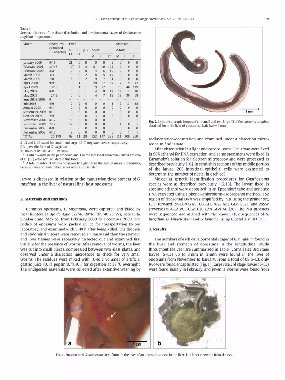
Table 1Seasonal changes of the tissue distribution and developmental stages of Gnathostomaturgidum in opossums.
Month Opossumsexamined(+ve/total)
Liver Stomach
S–L3
L–L3
JUV Adults Adults
M F T* M F T
January 2009 9/10 21 0 9 0 0 2 0 0 0February 2008 37/47 0b 9 3 81 49 152 0 0 0February 2009 2/2 0 0 8 4 6 10 0 0 0March 2008 3/3 0 0 2 8 5 17 0 0 0March 2009 7/8 3 0 2 19 7 31 0 0 0April 2008 9/9a 0 1 1 20 37 57 7 5 12April 2009 13/15 0 1 1 9 27 36 73 46 119May 2008 6/8 0 0 1 4 9 17 17 13 30May 2009 12/13 0 0 1 4 7 12 38 30 68June 2008/2009 0 – – – – – – – – –
July 2008 4/6 0 0 0 0 0 1 15 13 28August 2008 0/1 0 0 0 0 0 0 0 0 0September 2008 0/1 0 0 0 0 0 0 0 0 0October 2009 2/9 0 0 0 3 0 3 0 0 0November 2008 8/12 18 0 0 0 0 0 0 1 1November 2009 7/12 17 0 0 0 0 0 1 0 1December 2008 0/9 0 0 0 0 0 0 0 0 0December 2009 6/13 9 0 0 0 0 0 0 1 1TOTAL 125/178 68 11 28 152 147 338 151 109 260
S–L3 and L–L3 stand for small- and large–L3 G. turgidum larvae, respectively.JUV: juvenile form of G. turgidum.M: male, F: female, and T = total.
a 3 adult worms in the peritoneum and 1 in the intestinal subserosa (Diaz-Camachoet al. [17) were not included in this table.
b A total number of worms occasionally higher than the sum of males and females,because those of unidentified sexes were also included.
Fig. 2. Light microscopic images of one small and two large L3 of Gnathostoma turgidumobtained from the liver of opossums. Scale bar=1 mm.
339S.P. Díaz-Camacho et al. / Parasitology International 59 (2010) 338–343
larvae is discussed in relation to the maturation/development of G.turgidum in the liver of natural final host opossums.
2. Materials and methods
Common opossums, D. virginiana, were captured and killed bylocal hunters at Ojo de Agua (22°45′28″N, 105°40′25″W), Tecualilla,Sinaloa State, Mexico, from February 2008 to December 2009. Thebodies of opossums were packed in ice for transportation to ourlaboratory, and examined within 48 h after being killed. The thoracicand abdominal viscera were removed en masse and then the stomachand liver tissues were separately dissected out and examined firstvisually for the presence of worms. After removal of worms, the liverwas cut into small pieces, compressed between two glass plates, andobserved under a dissection microscope to check for very smallworms. The residues were mixed with 10-fold volumes of artificialgastric juice (0.1% pepsin/0.7%HCl) for digestion at 37 °C overnight.The undigested materials were collected after extensive washing by
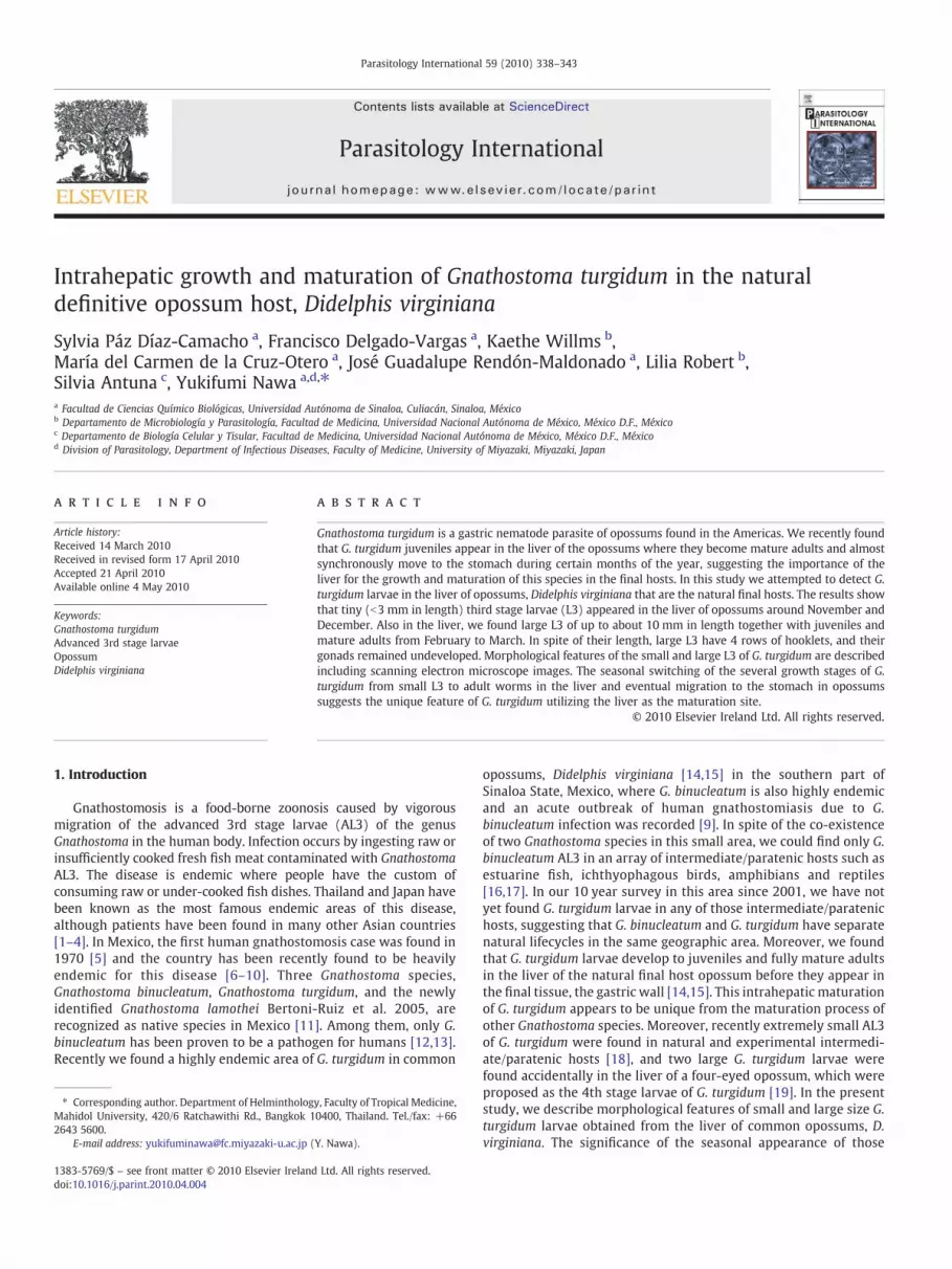
Fig. 1. Encapsulated Gnathostoma larva found in the liver of an op
sedimentation/decantation and examined under a dissection micro-scope to find larvae.
After observation in a lightmicroscope, some live larvaewere fixedin 90% ethanol for DNA extraction, and some specimens were fixed inKarnovsky's solution for electron microscopy and were processed asdescribed previously [16]. In semi-thin sections of the middle portionof the larvae, 200 intestinal epithelial cells were examined todetermine the number of nuclei in each cell.
Molecular genetic identification procedures for Gnathostomaspecies were as described previously [12,13]. The larvae fixed inabsolute ethanol were deposited in an Eppendorf tube and genomicDNA extracted using a phenol–chloroform–isopropanol method. ITS2region of ribosomal DNA was amplified by PCR using the primer set,LC1 (forward) 5′-CGA GTA TCG ATG AAG AAC GCA GC-3′ and 28SW(reverse) 5′-GCA ACC CGA CTC CAA GGA AC [20]. The PCR productswere sequenced and aligned with the known ITS2 sequences of G.turgidum, G. binucleatum and G. lamothei using Clustal X v1.83 [21].
3. Results
The numbers of each developmental stages of G. turgidum found inthe liver and stomach of opossums in the longitudinal studythroughout the year are summarized in Table 1. Small size 3rd stagelarvae (S–L3) up to 3 mm in length were found in the liver ofopossums from November to January. From a total of 68 S–L3, onlytwowere found encapsulated (Fig. 1). Large size 3rd stage larvae (L–L3)were found mainly in February, and juvenile worms were found from
ossum. a: cyst in the liver. b: a larva emerging from the cyst.

Table 2Body length of various developmental stages of Gnathostoma turgidum in opossums.
Time ofthe year
Liver Stomach
Larvae (mm) Juveniles (cm) Adults (cm) Adults (cm)
S–L3 L–L3 M F M F
Nov–Dec 2.75±0.74 (28)Feb–Mar 9.19±1.30 (9) 3.28±0.61 (6) 3.48±0.57 (10) 3.47 1.01 (11)Apr 3.55±0.84 (25) 3.92±0.79 (58) 4.74±0.74 (77) 5.40±0.89 (54)July 5.51±0.82 (11) 6.78±1.00 (12)
S–L3 and L–L3 stand for small- and large-L3 G. turgidum larvae, respectively.JUV: juvenile form of G. turgidum.M: male and F: female.The figures in bracket are the total number of worms examined.
340 S.P. Díaz-Camacho et al. / Parasitology International 59 (2010) 338–343
February to May. Young adult worms began to appear in the liver ofopossums as early as January and were continuously found in the liveruntil April, when the transition of worms from the liver to the stomachbegan. Fully mature adult worms, of which females can lay eggs, wereseen in the liver around April but the majority of them moved to thestomach during May and July, and had almost completely disappearedby August. From August to October (a rainy season in the study area)only few residual adult wormswere seen either in the liver or stomach.
As shown in Fig. 2, S–L3 were morphologically similar to theadvanced 3rd stage larvae (AL3) of other Gnathostoma species, withthe body length of 1.5–4 mm. In contrast, G. turgidum L–L3 were large,measuring about 7–10 mm in length and 0.3–0.4 mm in width.Chronological changes of the body length along with the develop-mental stages of G. turgidum are summarized in Table 2.
By scanning electron microscopic (SEM) observations (Fig. 3), thehead bulb of both S–L3 and L–L3 was clearly distinguished from therest of the body by the bulb shape and was covered with four rows ofsmall single-pointed hooklets with an irregular polygonal or oblong
Fig. 3. SEM images of the anterior half of Gnathostoma turgidum L3 and a juvenile worm. Thworm. Panels (d), (e) and (f) illustrate the cuticular spines of the corresponding stages. W
base (Fig. 3a, b). While body and head bulb sizes increased from S–L3to L–L3, their hooklet size did not change much, so that the hookletson the head bulb of L–L3 were scanty with wide spaces between them(Fig. 3b). The headbulb of the juvenile worm has 9 rows of well-developed hooklets (Fig. 3c). On the anterior 1/5 to 1/3 of the body ofS–L3 and L–L3, single-pointed tiny spines were arranged along thetransverse striations (Fig. 3d, e), which were difficult to see by lowpower light microscopy. In contrast, the anterior half of the juvenileworms were, like the mature adults [14], densely covered withirregularly arranged multi-dentated cuticular spines (Fig. 3f).Throughout their development, the caudal end of the body of G.turgidum had no cuticular spines, though they had well-developedcircular rings (Fig. 4a, b). The posterior 2/3 to 1/2 of the body of G.turgidum L–L3 had an apparently smooth surface with extremely finestriations (Fig. 4a). The caudal end of juvenile worms showedremarkable wrinkles, but no spines (Fig. 4b). A wide terminal openingwas observed on the ventral surface (Fig. 4a, b). Semi-thin sections ofthe mid-body (Fig. 5) revealed that the intestinal epithelial cells are
e cephalic bulb and the anterior portion of (a) small-L3, (b) large-L3, and (c) juvenilehite arrows in (a) and (d) indicate the excretory pore.
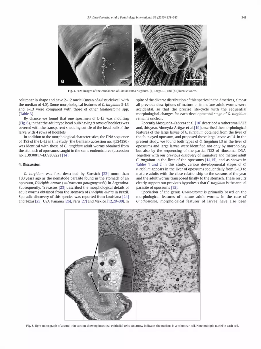
Fig. 4. SEM images of the caudal end of Gnathostoma turgidum. (a) Large-L3, and (b) juvenile worm.
341S.P. Díaz-Camacho et al. / Parasitology International 59 (2010) 338–343
columnar in shape and have 2–12 nuclei (mean of 4.8 nuclei/cell withthe median of 4.0). Some morphological features of G. turgidum S–L3and L–L3 were compared with those of other Gnathostoma spp.(Table 3).
By chance we found that one specimen of L–L3 was moulting(Fig. 6), in that the adult type head bulb having 9 rows of hooklets wascovered with the transparent shedding cuticle of the head bulb of thelarva with 4 rows of hooklets.
In addition to themorphological characteristics, the DNA sequenceof ITS2 of the L–L3 in this study (the GenBank accession no. FJ524380)was identical with those of G. turgidum adult worms obtained fromthe stomach of opossums caught in the same endemic area (accessionno. EU930817–EU930822) [14].
4. Discussion
G. turgidum was first described by Stossich [22] more than100 years ago as the nematode parasite found in the stomach of anopossum, Didelphis azarae (=Dracaena paraguayensis) in Argentina.Subsequently, Travassos [23] described the morphological details ofadult worms obtained from the stomach of Didelphis aurita in Brazil.Sporadic discovery of this species was reported from Louisiana [24]and Texas [25], USA, Panama [26], Peru [27] andMexico [12,28–30]. In
Fig. 5. Light micrograph of a semi-thin section showing intestinal epithelial cells. An
spite of the diverse distribution of this species in the Americas, almostall previous descriptions of mature or immature adult worms wereaccidental, so that the precise life-cycle with the sequentialmorphological changes for each developmental stage of G. turgidumremains unclear.
RecentlyMosqueda-Cabrera et al. [18] described a rather small AL3and, this year, Almeyda-Artigas et al. [19] described themorphologicalfeatures of the large larvae of G. turgidum obtained from the liver ofthe four-eyed opossum, and proposed those large larvae as L4. In thepresent study, we found both types of G. turgidum L3 in the liver ofopossums and large larvae were identified not only by morphologybut also by the sequencing of the partial ITS2 of ribosomal DNA.Together with our previous discovery of immature and mature adultG. turgidum in the liver of the opossums [14,15], and as shown inTables 1 and 2 in this study, various developmental stages of G.turgidum appears in the liver of opossums sequentially from S–L3 tomature adults with the close relationship to the seasons of the yearand the adult worms transposed finally to the stomach. These resultsclearly support our previous hypothesis that G. turgidum is the annualparasite of opossums [15].
Speciation of the genus Gnathostoma is primarily based on themorphological features of mature adult worms. In the case ofGnathostoma, morphological features of larvae have also been
arrow indicates the nucleus in a columnar cell. Note multiple nuclei in each cell.
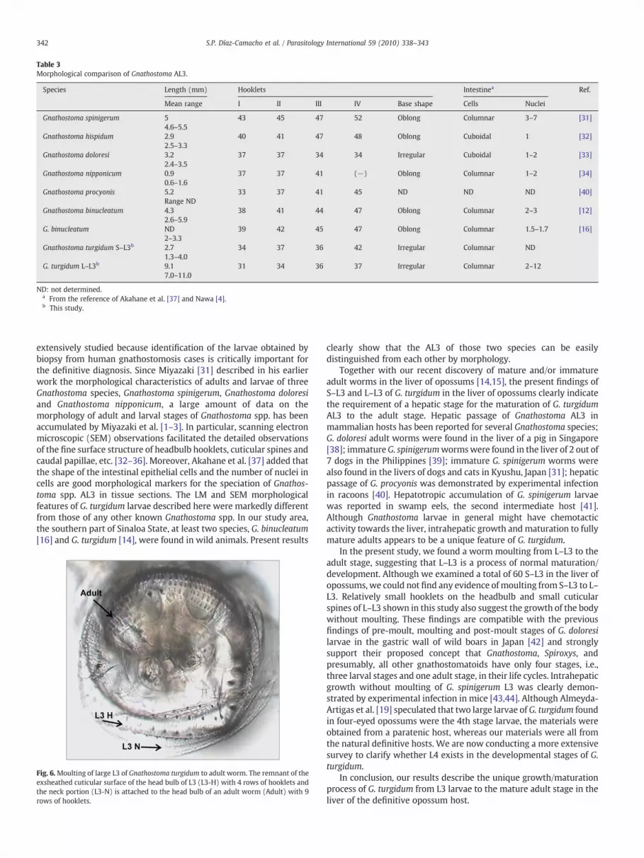
Table 3Morphological comparison of Gnathostoma AL3.
Species Length (mm) Hooklets Intestinea Ref.
Mean range I II III IV Base shape Cells Nuclei
Gnathostoma spinigerum 54.6–5.5
43 45 47 52 Oblong Columnar 3–7 [31]
Gnathostoma hispidum 2.92.5–3.3
40 41 47 48 Oblong Cuboidal 1 [32]
Gnathostoma doloresi 3.22.4–3.5
37 37 34 34 Irregular Cuboidal 1–2 [33]
Gnathostoma nipponicum 0.90.6–1.6
37 37 41 (−) Oblong Columnar 1–2 [34]
Gnathostoma procyonis 5.2Range ND
33 37 41 45 ND ND ND [40]
Gnathostoma binucleatum 4.32.6–5.9
38 41 44 47 Oblong Columnar 2–3 [12]
G. binucleatum ND2–3.3
39 42 45 47 Oblong Columnar 1.5–1.7 [16]
Gnathostoma turgidum S–L3b 2.71.3–4.0
34 37 36 42 Irregular Columnar ND
G. turgidum L–L3b 9.17.0–11.0
31 34 36 37 Irregular Columnar 2–12
ND: not determined.a From the reference of Akahane et al. [37] and Nawa [4].b This study.
342 S.P. Díaz-Camacho et al. / Parasitology International 59 (2010) 338–343
extensively studied because identification of the larvae obtained bybiopsy from human gnathostomosis cases is critically important forthe definitive diagnosis. Since Miyazaki [31] described in his earlierwork the morphological characteristics of adults and larvae of threeGnathostoma species, Gnathostoma spinigerum, Gnathostoma doloresiand Gnathostoma nipponicum, a large amount of data on themorphology of adult and larval stages of Gnathostoma spp. has beenaccumulated by Miyazaki et al. [1–3]. In particular, scanning electronmicroscopic (SEM) observations facilitated the detailed observationsof the fine surface structure of headbulb hooklets, cuticular spines andcaudal papillae, etc. [32–36]. Moreover, Akahane et al. [37] added thatthe shape of the intestinal epithelial cells and the number of nuclei incells are good morphological markers for the speciation of Gnathos-toma spp. AL3 in tissue sections. The LM and SEM morphologicalfeatures of G. turgidum larvae described here were markedly differentfrom those of any other known Gnathostoma spp. In our study area,the southern part of Sinaloa State, at least two species, G. binucleatum[16] and G. turgidum [14], were found in wild animals. Present results
Fig. 6.Moulting of large L3 of Gnathostoma turgidum to adult worm. The remnant of theexsheathed cuticular surface of the head bulb of L3 (L3-H) with 4 rows of hooklets andthe neck portion (L3-N) is attached to the head bulb of an adult worm (Adult) with 9rows of hooklets.
clearly show that the AL3 of those two species can be easilydistinguished from each other by morphology.
Together with our recent discovery of mature and/or immatureadult worms in the liver of opossums [14,15], the present findings ofS–L3 and L–L3 of G. turgidum in the liver of opossums clearly indicatethe requirement of a hepatic stage for the maturation of G. turgidumAL3 to the adult stage. Hepatic passage of Gnathostoma AL3 inmammalian hosts has been reported for several Gnathostoma species;G. doloresi adult worms were found in the liver of a pig in Singapore[38]; immatureG. spinigerumwormswere found in the liver of 2 out of7 dogs in the Philippines [39]; immature G. spinigerum worms werealso found in the livers of dogs and cats in Kyushu, Japan [31]; hepaticpassage of G. procyonis was demonstrated by experimental infectionin racoons [40]. Hepatotropic accumulation of G. spinigerum larvaewas reported in swamp eels, the second intermediate host [41].Although Gnathostoma larvae in general might have chemotacticactivity towards the liver, intrahepatic growth and maturation to fullymature adults appears to be a unique feature of G. turgidum.
In the present study, we found a worm moulting from L–L3 to theadult stage, suggesting that L–L3 is a process of normal maturation/development. Although we examined a total of 60 S–L3 in the liver ofopossums, we could not find any evidence of moulting from S–L3 to L–L3. Relatively small hooklets on the headbulb and small cuticularspines of L–L3 shown in this study also suggest the growth of the bodywithout moulting. These findings are compatible with the previousfindings of pre-moult, moulting and post-moult stages of G. doloresilarvae in the gastric wall of wild boars in Japan [42] and stronglysupport their proposed concept that Gnathostoma, Spiroxys, andpresumably, all other gnathostomatoids have only four stages, i.e.,three larval stages and one adult stage, in their life cycles. Intrahepaticgrowth without moulting of G. spinigerum L3 was clearly demon-strated by experimental infection in mice [43,44]. Although Almeyda-Artigas et al. [19] speculated that two large larvae of G. turgidum foundin four-eyed opossums were the 4th stage larvae, the materials wereobtained from a paratenic host, whereas our materials were all fromthe natural definitive hosts. We are now conducting a more extensivesurvey to clarify whether L4 exists in the developmental stages of G.turgidum.
In conclusion, our results describe the unique growth/maturationprocess of G. turgidum from L3 larvae to the mature adult stage in theliver of the definitive opossum host.

343S.P. Díaz-Camacho et al. / Parasitology International 59 (2010) 338–343
Acknowledgements
This work is supported in part by the following grants: Programade Fomento y Apoyo a Proyectos de Investigación no. PI-PROFAPI-06070 and Consejo Nacional de Ciencia y Tecnología no. P48161-M.The authors thank the excellent technical assistance of Magda LuzZazueta Ramos, Josefina Sicairos Félix, Samuel Campista León, ÁngelBojórquez Contreras, Roberto Guzmán Loreto and Oscar FranciscoMartínez Contreras in the Facultad de Ciencias Químico Biológicas,Universidad Autónoma de Sinaloa, Culiacán, México. Special thanks goto Armando Zepeda Rodríguez, Depto. de Biología Celular y Tisular,Facultad de Medicina, Universidad Nacional Autónoma de México, forassistance in scanning electron microscopy.
References
[1] Miyazaki I. On the genus Gnathostoma and human gnathostomiasis, with specialreference to Japan. Exp Parasitol 1960;9:338–70.
[2] Miyazaki I. An illustrated book of helminthic zoonoses. Japan, Tokyo: InternationalMedical Foundation; 1991. p. 369–409.
[3] Daengsvang S. A monograph on the genus Gnathostoma and Gnathostomiasis inThailand. Tokyo: SEAMIC/IMFJ; 1980. pp. 1–87.
[4] Nawa Y. Historical review and current status of gnathostomiasis in Asia. SoutheastAsian J Trop Med Public Health 1991;22:217–9 Suppl.
[5] Peláez D, Pérez-Reyes R. Gnatostomiasis humana en América. Rev LatinoamMicrobiol 1970;12:83–91.
[6] Martínez-Cruz JM, Bravo-Zamudio R, Aranda-Patraca A, Martínez- Marañón R. LaGnathostomiasis en México. Salud Pública Méx 1989;31:541–9.
[7] Ogata K, Nawa Y, Akahane H, Díaz-Camacho SP, Lamothe-Argumedo R, Cruz-ReyesA. Short report: gnathostomiasis in Mexico. Am J Trop Med Hyg 1998;58:316–8.
[8] Lamothe-Argumedo R. La gnathostomiasis en México: un problema de saludpública. An Inst Biol Univ Nac Aut Méx, Ser Zool 2003;74:99–103 in Spanish.
[9] Díaz-Camacho SP, Willms K, de la Cruz-Otero Ma del C, Zazueta-Ramos ML,Bayliss-Gaxiola S, Castro-Velásquez R, Osuna-Ramírez I, Bojorquez-Contreras A,Torres-Montoya EH, Sánchez-Gonzales S. Acute outbreak of gnathostomiasis in afishing community in Sinaloa, Mexico. Parasitol Int 2003;5:133–40.
[10] Waikagul J, Díaz-Camacho SP. Gnathostomosis. In: Murrell KD, Fried B, editors.Food-borne parasitic zoonoses: fish and plant-borne parasites. World ClassParasitesNY: Springer Science; 2007. p. 235–61.
[11] Bertoni-Ruíz F, García-Prieto L, Osorio-Sarabia D, León-Règagnon V. A new speciesof Gnathostoma (Nematoda: Gnathostomatidae) in Procyon lotor hernandezii fromMexico. J Parasitol 2005;91:1143–9.
[12] Almeyda-Artigas RJ, Bargues MD, Mas-Coma S. ITS-2 rDNA sequencing ofGnathostoma species (Nematoda) and elucidation of the species causinggnathostomiasis in the Americas. J Parasitol 2000;86:537–44.
[13] León-Règagnon V, García-Prieto L, Osorio-Sarabia D, Martinez-Salazar E, Oce-guera-Figueroa A, Lamothe-Argumedo R, et al. Molecular systematics ofGnathostoma spp. Bull Cent Res Inst Fukuoka Univ Series E: InterdisciplinarySciences 2003;1:237–47.
[14] Díaz-Camacho SP, Willms K, Rendón-Maldonado JG, de la Cruz Otero Ma del C,Delgado-Vargas F, Robert L, et al. Discovery of an endemic area of Gnathostomaturgidum infection among opossums, Didelphis virginiana, in Mexico. J Parasitol2009;95:617–22.
[15] Nawa Y, de la Cruz Otero Ma del C, Zazueta-Ramos ML, Bojórquez- Contreras A,Sicairos-Félix J, Campista-León S, et al. Is Gnathostoma turgidum an annual parasiteof opossums? Drastic seasonal changes of infection in Didelphis virginiana inMexico. J Parasitol 2009;95:908–12.
[16] Díaz -Camacho SP, Willms K, Zazueta-Ramos ML, de la Cruz Otero Ma del C, NawaY, Akahane H. Morphology of Gnathostoma spp. isolated from natural hosts inSinaloa, Mexico. Parasitol Res 2002;88:639–45.
[17] Díaz-Camacho SP, de la Cruz Otero Ma del C, Zazueta-Ramos ML, Angel Bojórquez-Contreras A, Sicarios-Félix J, Campista-León S, et al. Identification of estuarine fishDormitator latifrons as an intermediate host and Eleotris picta as a paratenic hostfor Gnathostoma binucleatum in Sinaloa, Mexico. Parasitol Res 2008;103:1421–5.
[18] Mosqueda Cabrera MÁ, Sánchez Miranda E, Carranza Calderón L, Ortiz Nájera HE.Finding advanced third-stage larvae of Gnathostoma turgidum Stossich, 1902 inMexico from natural and experimental host and contributions to the life cycledescription. Parasitol Res 2009;104:1219–25.
[19] Almeyda-Artigas RJ, Mosqueda-Cabrera MÁ, Sánchez-Núñez E. Precocity ofGnathostoma turgidum in naturally infected four-eyed opossum Philander opossumpallidus from Temascal, Oaxaca, Mexico. Parasitol Res 2010;106:439–43.
[20] Ando K, Tsunemori M, Akahane H, Tesana S, Hasegawa H, Chinzei Y. Comparativestudy on DNA sequences of ribosomal DNA and cytochrome c oxidase subunit 1 ofmitochondrial DNA among five species of gnathostomes. J Helminthol 2006;80:7–13.
[21] Thompson JD, Gibson TJ, Plewniak F, Jeanmougin F, Higgins DG. The ClustalXwindows interface: flexible strategies for multiple sequence alignment aided byquality analysis tools. Nucleic Acids Res 1997;24:4876–82.
[22] Stossich M. Sopra aleuni nematode della collezione helmintologica del Prof. Dott.Corrado Paroua. Boll Mus Zool Anat Comp Univ Genova 1902;116:1–16.
[23] Travassos L. Contribuicóes para o conhecimento da fauna helmintologicaBrasileira. XVIII. Sobre as especies brasileiras do genero Gnathostoma Owen,1836. Sci Med 1925;3(8):508–17.
[24] Dikmans G. A new nematode worm Viannaia bursobscura from the opossum, witha note on the other parasites of the opossum. Proc US Nat'l Museum 1931;79:1–4Art. 31.
[25] Chandler AC. Notes on the helminth parasites of the opossum (Didelphisvirginiana) in Southeast, Texas with descriptions of four new species. Proc USNat'l Museum 1932;81:1–15 Art 16.
[26] Caballero y Caballero E. Estudios helmintológicos de la región oncocercosa deMéxico y de la República de Guatemala. Nematodo. An Esc Nal Cienc Biol Méx1958;9:61–76 in Spanish with English summary.
[27] Foster AO. Some helminths of the woolly opossum in Panama. Trans AmerMicroscopic Soc 1939;58:185–98.
[28] Miyazaki I, Kifune T, Habe S. Rep Fukuoka Univ Sc Exp to Peru 1976. Occ Publ 1976(1).
[29] Lamothe-Argumedo R, Akahane H, Osario-Sarabia D, García-Prieto L. Hallazgo deGnathostoma turgidum en Didelphys virginiana de Temascal, Oaxaca, Mexico. AnInst Biol Univ Nac Auton Mex Ser Zool 1998;69:225–9 in Spanish.
[30] Monet-Mendoza A, Osorio-Sarabia O, García-Prieto L. Helminths of the Virginiaopossum Didelphys virginiana (Mammalia: Didelphidae) in Mexico. J Parasitol2005;91:213–9.
[31] Miyazaki I. Studies on Gnathostoma occurring in Japan (Nematoda: Gnathosto-midae). II. Life history of Gnathostoma andmorphological comparison on its larvalforms. Kyushu Mem Med Sci 1954;2:122–43.
[32] Kondo K, Akao N, Takakura Y, Ohnishi Y, Konishi Y, Yoshimura H. Scanningelectron microscopy (SEM) of larvae and adult worms of Gnathostoma hispidum.Jpn J Parasitol 1984;33:577–86 in Japanese.
[33] Koga M, Ishii Y. Surface morphology of the advanced third-stage laravae ofGnathostoma doloresi: an electron microscopic study. Jpn J Parasitol 1987;36:231–5.
[34] Ando H, Tanaka H, Taniguchi Y, Shimizu M, Kondo K. Two human cases ofgnathostomiasis and discovery of a second intermediate host of Gnathostomanipponicum in Japan. J Parasitol 1988;74:626–7.
[35] Koga M, Akahane H, Lamothe-Argumedo R, Osorio-Sarabia D, Garcia-Prieto L,Martínez-Cruz JM, et al. Surface ultrastructure of larval Gnathostoma cf.binucleatum from Mexico. Comp Parasitol 2000;67:244–9.
[36] Bertoni-Ruíz F, García-Prieto L, León-Règagnon V, Osorio-Sarabia D, Mendoza-Garfias B. Estudio comparativo de algunas especies Americanas del géneroGnathostoma (Nematoda: Gnathostomatidae) mediante microscopía electrónicade barrido. Abst the 8th IntAmer Congr Electron Microscopy (IASEM2005),Habana, Cuba, Sept. 2005; 2005. p. 25–30.
[37] Akahane H, Sano M, Mako T. Morphological difference in cross sections ofadvanced third-stage larvae of Gnathostoma spinigerum, G. hispidum, and G.doloresi. Jpn J Parasitol 1986;35:465–7.
[38] Sandosham AA. Malaysian parasites XV. Seven new worms from miscellaneoushosts. Study Inst Med Res Malaya 1953;26:212–26.
[39] Africa CM, Refuerzo PG, Garcia EY. Observation on the life cycle of Gnathostomaspinigerum. Philippine J Sci 1936;59:513–23.
[40] Ash LR. Development of Gnathostoma procyonis Chandler 1942 in the first andsecond intermediate host. J Parasitol 1962;48:298–305.
[41] Nuamtanong S, Waikagul J, Anantaphruti MT. Gnathostome infection in swampeels, Fluta alba, in central Thailand. Southeast Asian J Trop Med Public Health1998;29:144–7.
[42] Imai J, Hasegawa H. Molting of Gnathostoma doloresi (Nematoda: Gnathostoma-toidea) in the definitive host. J Parasitol 2001;87:14–8.
[43] Anantaphruti M, Waikagul J, Nithi-Uthai S, Pubampen S, Rojekittikhun W.Detection of humoral immune response to Gnathostoma spinigerum in mice.Southeast Asian J Trop Med Publ Health 1986;17:172–6.
[44] Rojekittikhun W, Pubampen S. Morphological variation and abnormality ofcephalic hooklets of Gnathostoma spinigerum hepatic stage larvae from laboratoryinfected mice. Southeast Asian J Trop Med Publ Health 1998;29:118–22.