The marsupial CD8 gene locus: Molecular cloning and expression analysis of the alpha and beta...
14
Research paper The marsupial CD8 gene locus: Molecular cloning and expression analysis of the alpha and beta sequences in the gray short-tailed opossum (Monodelphis domestica) and the tammar wallaby (Macropus eugenii) Louise. G. Duncan a, *, Sham. V. Nair a , Elizabeth. M. Deane b a Department of Biological Sciences, Division of Environmental and Life Sciences, Macquarie University, NSW 2109, Australia b The Chancelry, The Australian National University, Canberra, ACT 0200, Australia 1. Introduction In mammalian adaptive cell-mediated host defence, the CD8 receptor is a key immune molecule. It is expressed on MHC class I restricted cytotoxic T cells whose principal function is the cytolysis of virus-infected or tumour cells. The CD8 receptor associates closely with the T cell receptor in the recognition of the MHC–peptide complex displayed by the target cell. This interaction initiates signal transduction events that activate the T cell and ultimately result in the destruction of the infected cell (reviewed in Abbas and Lichtman, 2003). Cell-mediated cytotoxicity is itself achieved through exocytosis of the cytotoxic T cell’s cytoplasmic granules which are composed of a pore- forming protein, perforin and enzymes including gran- zymes that induce the apoptotic death of the target cell (Stenger et al., 1999). In eutherian mammals, the CD8 gene locus is composed of two distinct, though closely linked genes, namely CD8a and CD8b (Gorman et al., 1988). These genes map to human chromosome 2 and mouse chromosome 6, within the same overall linkage group as the Igk gene locus Veterinary Immunology and Immunopathology 129 (2009) 14–27 ARTICLE INFO Article history: Received 28 July 2008 Received in revised form 28 November 2008 Accepted 1 December 2008 Keywords: CD8 cDNA cloning Cell-mediated immunity Cytotoxic T lymphocytes Gene structure Marsupial Opossum Tammar ABSTRACT In eutherian mammals, CD8 is a key receptor of cytotoxic T cells and plays a pivotal role in the recognition and elimination of infected host cells by cell-mediated cytotoxicity. Here, we report the molecular cloning and expression analysis of CD8a and CD8b cDNAs in two marsupial species, the gray short-tailed opossum and the tammar wallaby. The opossum and tammar CD8 sequences share a high degree of amino acid identity of 63% (CD8a) and 57% (CD8b) to each other as well as 36–45% (CD8a) and 38–41% (CD8b) with their eutherian counterparts. In addition, many of the signature features of eutherian CD8a and CD8b are preserved in both marsupials including the two invariant cysteines that form the intra-chain disulphide bond in the extracellular IgSfV domain and the two hinge region cysteines involved in dimerisation between the two subunits. The p56 lck binding motif in the cytoplasmic tail of the CD8a subunit is also conserved. Interestingly, the opossum CD8a and the tammar CD8b sequences have a truncated cytoplasmic tail. RT-PCR analysis of CD8a and CD8b transcripts in the tissues of the adult opossum and tammar showed broad tissue expression with a high level of expression observed in the lymphoid tissues of both marsupials. Furthermore, RT-PCR analysis of CD8a and CD8b transcripts in the immune tissues of tammar young over the first 120 days of pouch life revealed a pattern of expression analogous to the maturation of the lymphoid tissues. This is the first report confirming the presence of CD8 in the tissues of a marsupial and will provide the tools to further analyse T cell subsets in this unique group of mammals. ß 2008 Elsevier B.V. All rights reserved. * Corresponding author. Tel.: +61 2 9850 9259; fax: +61 2 9850 9671. E-mail address: [email protected] (Louise. G. Duncan). Contents lists available at ScienceDirect Veterinary Immunology and Immunopathology journal homepage: www.elsevier.com/locate/vetimm 0165-2427/$ – see front matter ß 2008 Elsevier B.V. All rights reserved. doi:10.1016/j.vetimm.2008.12.003
Transcript of The marsupial CD8 gene locus: Molecular cloning and expression analysis of the alpha and beta...

Research paper
Veterinary Immunology and Immunopathology 129 (2009) 14–27
Contents lists available at ScienceDirect
Veterinary Immunology and Immunopathology
journal homepage: www.e lsev ier .com/ locate /vet imm
The marsupial CD8 gene locus: Molecular cloning and expression analysisof the alpha and beta sequences in the gray short-tailed opossum(Monodelphis domestica) and the tammar wallaby (Macropus eugenii)
Louise. G. Duncan a,*, Sham. V. Nair a, Elizabeth. M. Deane b
a Department of Biological Sciences, Division of Environmental and Life Sciences, Macquarie University, NSW 2109, Australiab The Chancelry, The Australian National University, Canberra, ACT 0200, Australia
A R T I C L E I N F O
Article history:
Received 28 July 2008
Received in revised form 28 November 2008
Accepted 1 December 2008
Keywords:
CD8
cDNA cloning
Cell-mediated immunity
Cytotoxic T lymphocytes
Gene structure
Marsupial
Opossum
Tammar
A B S T R A C T
In eutherian mammals, CD8 is a key receptor of cytotoxic T cells and plays a pivotal role in
the recognition and elimination of infected host cells by cell-mediated cytotoxicity. Here,
we report the molecular cloning and expression analysis of CD8a and CD8b cDNAs in two
marsupial species, the gray short-tailed opossum and the tammar wallaby. The opossum
and tammar CD8 sequences share a high degree of amino acid identity of 63% (CD8a) and
57% (CD8b) to each other as well as 36–45% (CD8a) and 38–41% (CD8b) with their
eutherian counterparts. In addition, many of the signature features of eutherian CD8a and
CD8b are preserved in both marsupials including the two invariant cysteines that form the
intra-chain disulphide bond in the extracellular IgSfV domain and the two hinge region
cysteines involved in dimerisation between the two subunits. The p56lck binding motif in
the cytoplasmic tail of the CD8a subunit is also conserved. Interestingly, the opossum
CD8a and the tammar CD8b sequences have a truncated cytoplasmic tail. RT-PCR analysis
of CD8a and CD8b transcripts in the tissues of the adult opossum and tammar showed
broad tissue expression with a high level of expression observed in the lymphoid tissues of
both marsupials. Furthermore, RT-PCR analysis of CD8a and CD8b transcripts in the
immune tissues of tammar young over the first 120 days of pouch life revealed a pattern of
expression analogous to the maturation of the lymphoid tissues. This is the first report
confirming the presence of CD8 in the tissues of a marsupial and will provide the tools to
further analyse T cell subsets in this unique group of mammals.
� 2008 Elsevier B.V. All rights reserved.
1. Introduction
In mammalian adaptive cell-mediated host defence, theCD8 receptor is a key immune molecule. It is expressed onMHC class I restricted cytotoxic T cells whose principalfunction is the cytolysis of virus-infected or tumour cells.The CD8 receptor associates closely with the T cell receptorin the recognition of the MHC–peptide complex displayedby the target cell. This interaction initiates signal
* Corresponding author. Tel.: +61 2 9850 9259; fax: +61 2 9850 9671.
E-mail address: [email protected] (Louise. G. Duncan).
0165-2427/$ – see front matter � 2008 Elsevier B.V. All rights reserved.
doi:10.1016/j.vetimm.2008.12.003
transduction events that activate the T cell and ultimatelyresult in the destruction of the infected cell (reviewed inAbbas and Lichtman, 2003). Cell-mediated cytotoxicity isitself achieved through exocytosis of the cytotoxic T cell’scytoplasmic granules which are composed of a pore-forming protein, perforin and enzymes including gran-zymes that induce the apoptotic death of the target cell(Stenger et al., 1999).
In eutherian mammals, the CD8 gene locus is composedof two distinct, though closely linked genes, namely CD8aand CD8b (Gorman et al., 1988). These genes map tohuman chromosome 2 and mouse chromosome 6, withinthe same overall linkage group as the Igk gene locus

L.G. Duncan et al. / Veterinary Immunology and Immunopathology 129 (2009) 14–27 15
(Gibson et al., 1978; Sukhatme et al., 1985). The geneproduct, the CD8 receptor, is expressed on thymocytes andperipheral T lymphocytes as a disulphide-linked hetero-dimer comprising of one alpha and one beta chain (Shiueet al., 1993). However, a homodimer of two alpha chains isexpressed on intraepithelial lymphocytes (IELs) and someNK cell populations (Guy-Grand et al., 1991; Moebius et al.,1991). Even though the heterodimeric form is recognizedas a more efficient coreceptor (Wong et al., 2003), theprecise role of the beta chain is poorly understood andmany of the functions of the CD8 molecule have beenattributed to the alpha chain (Zamoyska, 1994). It is thischain that has been shown to bind to MHC class I molecules(reviewed in Gao and Jakobsen, 2000) and contains thedocking site for the protein tyrosine kinase, p56lck
(Veillette et al., 1988), which is known to initiate signaltransduction events.
Marsupials are a unique group of mammals. At birth,the marsupial young are highly altricial, they have nofunctional lymphoid tissue and the maturation of theirimmune system occurs postnatally (Old and Deane, 2000).This, coupled with their evolutionary divergence fromtheir eutherian relatives approximately 130 million yearsago (Janke et al., 1994), makes marsupials ideal models fordevelopmental and comparative studies. However, despitethe potential for research in this area, the immuneresponse in marsupials is still poorly understood andhas, in part, been hampered by the paucity of antibodiesthat have reactivity to marsupial cell population markers(Hemsley et al., 1995; Jones et al., 1993). To date, suchcapability is restricted to the lymphocyte surface markersCD3, CD5 and CD79. These reagents are fundamental forstudying the development of the immune tissues in theneonatal marsupial and of increasing importance incharacterizing pathogenesis of infectious diseases afflict-ing marsupial populations (Kreiss et al., 2008).
Recently, the whole genomes of two model marsupialspecies, the gray short-tailed opossum (Monodelphis
domestica) (Mikkelsen et al., 2007) and the tammarwallaby (Macropus eugenii) (Renfree, 2007), have beensequenced. The opossum’s genome was the first marsupialgenome sequence to be released into the public domainand is available on the Ensembl database. This data hasallowed the in silico identification of predicted gene modelsfor key immune molecules including cytokines, CD4, CD8a(Wong et al., 2006), chemokines, defensins, cathelicidinsand NK cell receptors (Belov et al., 2007). Whilst large-scale bioinformatic analyses such as these provide valu-able insight into the opossum immunome, particularly forgenes that previously proved difficult to isolate usingstandard molecular techniques, the fine-scaled data ongene structure (exon/intron boundaries as well as the 50
and 30 ends of genes) has not been obtained. To this end,recent studies have used the opossum genome sequencedata not only to confirm predicted gene models throughthe isolation and expression analysis of immune genes inthe opossum itself (Belov et al., 2006) but also to furthercharacterize these same genes in other distantly relatedmarsupials (Baker and Miller, 2007; Duncan et al., 2007;Parra et al., 2007). This molecular characterization iscritical for developing marsupial-specific reagents to
enable the marsupial immune system to be as wellcharacterized as their eutherian relatives.
We previously reported (Duncan et al., 2007) themolecular cloning and expression analysis of CD4sequences in the tammar wallaby and the gray short-tailed opossum. The tammar and opossum are distantlyrelated and last shared a common ancestor approximately60–70 million years ago (Kirsch et al., 1997; Nilsson et al.,2004). Both marsupials are well established modelmarsupial species and have been the focus of a significantamount of fundamental research. In this study we presentthe molecular cloning and expression analysis of the otherT cell subset marker CD8, encompassing the CD8a andCD8b sequences, in these two model marsupials.
2. Materials and methods
2.1. Annotation of the opossum CD8a and CD8b genes
The M. domestica whole genome sequence data releasedfrom the Broad Institute was accessed through the Ensembldatabase (www.ensembl.org/Monodelphis_domestica/index.html). The MonDom version 2.0 (MonDom2.0)Ensembl assembly was analysed by BLASTN search (usingdefault search parameters) using the human CD8a andCD8b cDNA sequences (GenBank accession nos. M12828and Y00805, respectively) as query. The putative opossumCD8a and CD8b genes identified on the Ensembl databasewere further analysed using gene prediction softwareincluding Genscan (http://genes.mit.edu/GENSCAN.html)and Genewise2 (www.ebi.ac.uk/Wise2/) using the defaultparameters. The resultant Genscan and Genewise modelswere compared to the Ensembl gene predictions andmanually curated to construct the final annotation of theopossum CD8a and CD8b genes. These sequences were usedto design primers for PCR analysis and cloning of theopossum and tammar CD8a and CD8b cDNA sequences.
2.2. Animal tissues and cDNA preparation
Opossum tissues from a 4-month-old male animal weresupplied by the Department of Pharmacology, Universityof Melbourne, Victoria, Australia. Adult tammar wallabytissues were collected opportunistically from animalshoused at the Macquarie University Fauna Park (NorthRyde, NSW, Australia) and CSIRO, Sustainable Ecosystems(Canberra, Australia) that had been euthanized as part ofother approved experimental work. Total RNA was isolatedfrom opossum and tammar spleen tissue using TRI reagent(Molecular Research Centre, OH, USA) following themanufacturer’s instructions. For RT-PCR, cDNA wassynthesised using SuperScript II reverse transcriptionsystem (Invitrogen, CA, USA) and RACE-ready first-strandcDNA was synthesised using the SMART RACE cDNAamplification kit (BD Biosciences Clontech, CA, USA)according to the manufacturer’s protocol.
2.3. Cloning of opossum CD8a and CD8b cDNA
The full-length opossum CD8a and CD8b cDNAsequences were isolated by RACE PCR using the universal
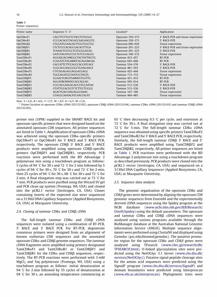
Table 1
Primer sequences.
Primer name Sequence 50–30 Locationa Application
OpCD8aF1 CACCTTCTTGCTCTACCTGTCGGG Opossum 350–373 30 RACE-PCR and tissue expression
OpCD8aR1 CCCGACAGGTAGAGCAAGAAGGTG Opossum 350–373 50 RACE-PCR
OpCD8aR2 CTGGATGGAAGAGTTGGTGGCTGC Opossum 606–629 Tissue expression
OpCD8bF1 CTCTCCCCACAGCGACAGTTTGA Opossum 201–223 30 RACE-PCR and tissue expression
OpCD8bR1 TCAAACTGTCGCTGTGGGGAGAG Opossum 201–223 50 RACE-PCR
OpCD8bR2 GATGTAGATGCCGCTGTCCGAGGG Opossum 349–372 Tissue expression
TamCD8aF1 AGGASGACSARGGCTACTAYTWCTG Tammar 433–457 RT-PCR
TamCD8aR1 CCAGATGTAGAKRTCACAGGMGAA Tammar 645–668 RT-PCR
TamCD8aF2 CACCATTCTTCCAGCCACCATCAGC Tammar 554–578 30 RACE-PCR
TamCD8aR2 CGGCACGAAGGGGCTGAAGAACA Tammar 481–503 50 RACE-PCR
TamCD8aF3 CTTCAGAGAGGAGGACGAGG Tammar 425–444 Tissue expression
TamCD8aR3 TGGCAGATGGTAATGGTAGTG Tammar 713–733 Tissue expression
TamCD8bF1 GGAACYCRGSTSARWGTGGTTG Tammar 451–472 RT-PCR
TamCD8bR1 AGGAYRCMWSCCACCAGCAG Tammar 595–614 RT-PCR
TamCD8bF2 CCCCAGGAAGAGACCGTGCAATAC Tammar 513–536 30 RACE-PCR
TamCD8bR2 GTATTGCACGGTCTCTTCCTGGGG Tammar 513–536 50 RACE-PCR
TamCD8bF3 AGACTGACCAAGAGGCAAAG Tammar 167–186 Tissue expression
TamCD8bR3 GTAGGCAAAACATCAACCACAT Tammar 464–485 Tissue expression
Note: S = G/C, R = A/G, Y = C/T, W = A/T, K = G/T, M = C/A.a Primer location in opossum CD8a cDNA (EU152102), opossum CD8b cDNA (EU152104), tammar CD8a cDNA (EU152103) and tammar CD8b cDNA
(EU152105).
L.G. Duncan et al. / Veterinary Immunology and Immunopathology 129 (2009) 14–2716
primer mix (UPM) supplied in the SMART RACE kit andopossum-specific primers that were designed based on theannotated opossum CD8 sequences. All primer sequencesare listed in Table 1. Amplification of opossum CD8a cDNAwas achieved using the opossum CD8a-specific primersOpCD8aF1 or OpCD8aR1 for 30 RACE and 50 RACE PCR,respectively. The opossum CD8b 30 RACE and 50 RACEproducts were amplified using opossum CD8b-specificprimers OpCD8bF1 and OpCD8bR1, respectively. PCRreactions were performed with the BD Advantage 2polymerase mix using a touchdown program as follows:5 cycles of 94 8C for 30 s and 72 8C for 2 min followed by 5cycles of 94 8C for 30 s, 70 8C for 30 s and 72 8C for 2 min,then 25 cycles of 94 8C for 30 s, 68 8C for 30 s and 72 8C for2 min. A final elongation step was carried out at 72 8C for7 min. PCR products were purified using the Wizard SV geland PCR clean-up system (Promega, WI, USA) and clonedinto the pCR2.1 vector (Invitrogen, CA, USA). Clonescontaining inserts of the expected size were sequencedon a 3130xl DNA Capillary Sequencer (Applied Biosystems,CA, USA) at Macquarie University.
2.4. Cloning of tammar CD8a and CD8b cDNA
The full-length tammar CD8a and CD8b cDNAsequences were isolated using a combination of RT-PCR,50 RACE and 30 RACE PCR. For RT-PCR, degenerateconsensus primers were designed from an alignment ofknown eutherian CD8 sequences and the annotatedopossum CD8a and CD8b genome sequences. The tammarcDNA fragments were amplified using primers designatedTamCD8aF1 and TamCD8aR1 or TamCD8bF1 andTamCD8bR1 for the CD8a and CD8b sequences, respec-tively. The RT-PCR reactions were performed with 3 mMMgCl2 and Taq polymerase (Promega, WI, USA) using atouchdown program as follows: initial denaturation at94 8C for 2 min followed by 35 cycles of denaturation at94 8C for 30 s, an annealing temperature commencing at
65 8C then decreasing 0.5 8C per cycle, and extension at72 8C for 30 s. A final elongation step was carried out at72 8C for 5 min. The full-length tammar CD8a cDNAsequence was obtained using specific primers TamCD8aF2and TamCD8aR2 for 30 RACE and 50 RACE PCR, respectively.Similarly, the full-length tammar CD8b 30 RACE and 50
RACE products were amplified using TamCD8bF2 andTamCD8bR2, respectively. All primer sequences are listedin Table 1. PCR reactions were performed with the BDAdvantage 2 polymerase mix using a touchdown programas described previously. PCR products were cloned into thepCR2.1 vector (Invitrogen, CA, USA) and sequenced on a3130xl DNA Capillary Sequencer (Applied Biosystems, CA,USA) at Macquarie University.
2.5. Sequence data analysis
The genomic organisation of the opossum CD8a andCD8b genes were determined by aligning the opossum CD8
genomic sequences from Ensembl and the experimentallyderived cDNA sequences using the Spidey program at theNCBI database (www.ncbi.nlm.nih.gov/IEB/Research/Ostell/Spidey) using the default parameters. The opossumand tammar CD8a and CD8b cDNA sequences wereanalysed using various programs available through theBioManager database at the Australian National GenomicInformation Service (ANGIS). Multiple sequence align-ments were performed using ClustalW and displayed usingGenedoc (psc.edu/biomed/genedoc). The putative promo-ter region for the opossum CD8a and CD8b genes wereanalysed using TFsearch (www.cbrc.jp/research/db/TFSEARCH.html). O-linked glycosylation sites were pre-dicted using the NetOGlyc 3.1 Server (www.cbs.dtu.dk/services/NetOGlyc/). Putative signal peptide cleavage sitesfor the amino acid sequences were predicted using theSignalP program (www.cbs.dtu.dk/services/SignalP) anddomain boundaries were predicted using Interproscan(www.ebi.ac.uk/interproscan). Phylogenetic trees were

L.G. Duncan et al. / Veterinary Immunology and Immunopathology 129 (2009) 14–27 17
drawn from the amino acid sequence alignment using thePHYLIP group of programs (Felsenstein, 1989), accessed viaBioManager. The phylogenetic tree was displayed usingthe Treeview program (Page, 1996) and the bootstrapmethod was used to indicate confidence values for treenodes based on 100 replicates.
2.6. mRNA expression profile of CD8a and CD8b transcripts
in adult and pouch young tissues
Total RNAs and cDNAs were prepared from spleen, liver,kidney, lung, heart, brain and gut from the adult opossumand adult tammar and also included blood for the tammar.Total RNAs and cDNAs from the cervical thymus, spleen,gut, lung, blood and liver of tammar pouch young samples(10 days, 21 days, 60 days and 120 days) were prepared.Age of the pouch young was estimated based on headlength measurements (Poole et al., 1991). Tissues fromthree individuals were analysed for each age group (adultand pouch young) except for opossum where only samplesfrom an individual animal was available. For analysingexpression in opossum tissues, the primer sequences wereOpCD8aF1 and OpCD8aR2 or OpCD8bF1 and OpCD8bR2for CD8a and CD8b, respectively. The expression oftammar CD8a and CD8b was analysed using the primersequences TamCD8aF3 and TamCD8aR3 or TamCD8bF3and TamCD8bR3, respectively. The primer sequences arelisted in Table 1. Expression of the house-keeping geneG3PDH was documented as an internal control. The RT-PCRreactions were performed using 2.5 mM MgCl2 and Flexi-Taq polymerase (Promega, WI, USA) under the followingconditions: initial denaturation at 94 8C for 2 min followedby 35 cycles of denaturation at 94 8C for 30 s, annealing for30 s at 65, 60 and 55 8C for opossum CD8a and CD8b,tammar CD8a and CD8b, and G3PDH, respectively, andelongation at 72 8C for 30 s. A final elongation step wascarried out at 72 8C for 5 min.
Fig. 1. Comparison of the CD8 gene locus in opossum and human. Not drawn to s
non-coding exon) and numbered in roman numerals whilst introns are designate
and intron as well as the lengths between the genes are indicated. The structura
areas) is shown for human only. L, leader; IgSfV, extracellular IgSfV domain; T
3. Results
3.1. Genomic organisation of the opossum CD8 gene locus
BLASTN analysis of the Ensembl MonDom2.0 assemblyusing the human CD8a and CD8b cDNA sequences asquery identified putative opossum CD8 homologs onscaffold 63. For the opossum CD8a genomic sequence,two Ensembl gene models were predicted, with eachdisplaying variations in the location of the exon–intronboundaries. Similarly, in the opossum CD8b genomicsequence, four Ensembl gene models were predicted. Toascertain the correct gene organisation of the opossumCD8a and CD8b genes, the genomic sequences wereextracted from the Ensembl database and further analysedusing Genscan and Genewise. These results were amalga-mated with the Ensembl gene predictions to form the finalannotation of the opossum CD8 genes. The annotatedsequences were manually curated to ensure they con-formed to the gt/ag rule and the characteristic intron splicepattern exhibited by known CD8 genes. These finalsequences were used to isolate the full-length opossumand tammar wallaby CD8a and CD8b cDNA sequences.
The opossum CD8 gene locus, shown in Fig. 1, has beenlocalised on chromosome 1 in the most recent Ensemblassembly, MonDom5.0. The CD8a and CD8b genes aresituated 48.8 kb apart in the same transcription orienta-tion, arranged head-to-tail with the beta gene locatedupstream of the alpha gene. This gene arrangementshowed conserved synteny with the human CD8 genelocus on chromosome 2. The genomic organisation of theopossum CD8a and CD8b genes were assembled from analignment of the Ensembl genomic sequences and theexperimentally derived cDNA sequences (refer to Figs. 2Aand 4A for details). The opossum CD8a gene is organisedinto six exons and five introns, as is its human homolog(Nakayama et al., 1989). However, in contrast to the
cale. Exons are boxed (closed box denotes coding exon; open box denotes
d by a solid line. The length (in bp unless specified otherwise) of each exon
l arrangement of the protein domains in relation to coding exons (shaded
M, transmembrane; Cyt, cytoplasmic tail.

Fig. 2. The nucleotide sequence (numbered at the top) and deduced amino acid sequence (one-letter abbreviation code, numbered below in bold) for CD8ain the opossum (A) and the tammar wallaby (B). For the opossum sequence, the nucleotide sequence is numbered relative to the potential transcription start
site at position +1 (designated by a filled arrow) which coincides with the start of the cDNA sequence isolated by RACE PCR. Putative factor-binding sites in
the promoter region are shown in bold and named above the sequence. The location of the introns in the opossum sequence are shown in italics (gt/ag sites
in italics and underlined; intron number in roman numerals and length of intron in brackets). For both sequences, the predicted mature polypeptide begins
at position +1 indicated with a filled arrowhead. Extracellular cysteine residues are circled. Potential N-linked and O-linked glycosylation sites are
underlined and the polyadenylation signal is double underlined.
L.G. Duncan et al. / Veterinary Immunology and Immunopathology 129 (2009) 14–2718

L.G. Duncan et al. / Veterinary Immunology and Immunopathology 129 (2009) 14–27 19
human CD8a gene, which is 6.3 kb in length, the opossumgene spans 23.9 kb. The difference in the sizes of thesegenes is due to the large intronic region (intron 5) which is18.3 kb in opossum, compared to only 2.6 kb in humans.Although the length of intron 5 in the opossum was notconfirmed in this study, this region requires furtherinvestigation as transcriptional regulatory elements(including an enhancer) are present in intron 5 of thehuman CD8a gene (Hanke et al., 1995). The opossum CD8bgene spans 20.8 kb and is organised into six exons and fiveintrons, revealing significant similarities to its humanhomolog (19.6 kb). Analysis of the opossum CD8a andCD8b coding sequence revealed agreement with the intronsplice patterns of other CD8a and CD8b genes (Parnes,1989). Thus, introns 1–4 contain the common type I (alsoknown as phase I) splice motif and intron 5 contains a typeII (phase II) motif in both genes.
The putative promoter region for opossum CD8a(Fig. 2A) and opossum CD8b (Fig. 4A) was localised to thefirst exon of each gene and contained an Initiator (Inr)sequence (Smale and Kadonaga, 2003) and transcriptionstart site at the adenine at position +1. Analysis of theputative opossum promoter regions revealed that, liketheir human CD8a (Gao and Kavathas, 1993; Nakayamaet al., 1989; Norment et al., 1989) and CD8b (DiSanto et al.,1993; Nakayama et al., 1992) homologs, the promotersequences for both opossum genes lack TATA and CAATmotifs and are GC-rich. In addition, potential binding sitesfor known regulatory binding proteins were identified inthe putative opossum promoter sequences. These includethe consensus recognition sequences for Sp1 (an activatorof basal level of transcription), CREB (a cyclic AMPresponse element binding protein), MAZ (a zinc fingertranscription factor reported to have DNA bendingcapabilities) and c-Myb (a molecule important for T-celldevelopment). It is interesting to note that recognitionsites for CREB, MAZ and c-Myb binding proteins were alsoidentified in the putative opossum CD4 promoter region(Duncan et al., 2007).
3.2. Characterisation of marsupial CD8a cDNA sequences
The full-length opossum and tammar wallaby CD8acDNA sequences were isolated using the annotatedopossum genomic CD8a sequence. The CD8a cDNAsequences, shown for opossum (GenBank accession no.EU152102) in Fig. 2A and tammar (GenBank accessionno. EU152103) in Fig. 2B, comprised 987 and 944nucleotides, respectively. Both marsupial sequenceshave an identical polyadenylation signal (TATAAA)upstream of the polyA tail. The open-reading frame(ORF) translated into a predicted protein of 237 aminoacid residues in the opossum and 241 residues in thetammar, initiated from the first 50-proximal ATG codon(Kozak, 1983) in both marsupial sequences. The matureprotein resulting from signal sequence cleavage waspredicted to be 214 and 216 residues in the opossum andtammar, respectively.
Analysis of the extracellular region of the opossum andtammar CD8a sequences revealed five cysteine residues.The two cysteines capable of forming an intra-chain
disulphide bond, thereby stabilizing the Ig fold in the IgSfVdomain (human C-22, C-94), were conserved. The internalcysteine residue (human C-33), found in most mammalsbut absent in avian and teleost CD8a sequences, was alsoconserved in marsupials. In addition, the cysteines in thehinge region which are capable of dimer formation (humanC-143, C-160) were present in both marsupial CD8asequences. The hinge region also contains multiple O-linked glycosylation sites that allow the human CD8amolecule to exhibit an extended structure (Classon et al.,1992). There were seven predicted O-linked glycosylationsites in both marsupial CD8a sequences. The number of N-linked glycosylation motifs (NX[T/S], where X is anyresidue except proline), for the CD8a sequence invertebrates varies considerably. Although no such motifshave been located in the human (Littman et al., 1985),chicken (Tregaskes et al., 1995) and many fish species(Hansen and Strassburger, 2000; Moore et al., 2005), themouse CD8a (Nakauchi et al., 1985) has three predicted N-linked glycosylation sites. In marsupials, the tammarsequence had no N-linked glycosylation sites, whilst theopossum had three predicted sites, all of which differed intheir location relative to those described in the mouse.
Alignment of the marsupial CD8a amino acidsequences with other known vertebrate sequences isshown in Fig. 3, and illustrates the level of sequenceidentity with opossum CD8a. It should be noted that thetammar CD8a sequence had similar identity to that shownfor the opossum. Overall, the opossum CD8a amino acidsequence shared 63% identity with tammar CD8a whilstidentity with other vertebrate CD8a sequences was muchlower, ranging from 42% (human) to 32% (chicken). For thestructural domains, the transmembrane region revealedthe highest identity of 76% between the marsupial CD8asequences and 56% identity with both human and chickenCD8a. The opossum CD8a cytoplasmic tail was truncatedby five residues (Fig. 3). Also evident in the cytoplasmic tailwas the p56lck docking site, a cysteine motif CXC (Shawet al., 1990; Turner et al., 1990) shown here as highlyconserved across mammals and chicken. Interestingly, aBLASTN analysis of the Ensembl MonDom5.0 assemblyusing the human p56lck sequence as query, identified theopossum p56lck sequence (ENSMODG00000017464) onchromosome 4 (data not shown). This opossum p56lck
sequence was found to contain the consensus sequencemotif CXXC known in mammals to be important for theassociation with CD8a.
3.3. Characterisation of marsupial CD8b cDNA sequences
The full-length opossum and tammar wallaby CD8bcDNA sequences were isolated using the annotatedopossum CD8b sequence. The CD8b cDNA sequences,shown for opossum (GenBank accession no. EU152104) inFig. 4A and tammar (GenBank accession no. EU152105) inFig. 4B, comprised 1399 and 1492 nucleotides, respec-tively. Both marsupial sequences had an identical poly-adenylation signal (AATAAA) upstream of the polyA tail.Translation of the ORF initiated from the first and second50-proximal ATG codon (Kozak, 1983) in tammar andopossum, respectively resulted in a predicted protein of

Fig. 3. Alignment of the amino acid sequence of opossum CD8a with other vertebrates (including tammar) showing the level of sequence identity (Id%) with
opossum CD8a. The alignment was generated using ClustalW (BioManager) and identical residues (>60%) are shaded. Dashes indicate gaps introduced into
the sequence to optimise the alignment. The leader sequence, IgSfV domain, hinge segment, transmembrane region and cytoplasmic tail boundaries
indicated above the sequences were predicted using Interproscan. The extracellular cysteines are indicated with an asterisk and the p56lck binding motif is
underlined. GenBank accession numbers are as follows: human M12828, mouse M12825, cat AB000485, cow X59416, guinea pig AY303773 and chicken
Z22726.
L.G. Duncan et al. / Veterinary Immunology and Immunopathology 129 (2009) 14–2720
214 amino acid residues in the opossum and 207 residuesin the tammar. Signal sequence cleavage was predicted togenerate a mature protein of 195 and 188 residues in theopossum and tammar, respectively.
Analysis of the extracellular region of opossum andtammar CD8b revealed four conserved cysteine residues.The two cysteines capable of forming an intra-chaindisulphide bond in the IgSfV domain (human C-20, C-95)were conserved, as were the two cysteines in the hingeregion (human C-134, C-147) capable of intermoleculardimer formation. Interestingly, the opossum CD8bsequence contained an extra cysteine in the IgSfV domain(C-62 in opossum) in a similar region to an additionalcysteine reported in the homologous sequence from the cat(Pecoraro et al., 1996). Similar to CD8a, the hinge region ofhuman CD8b also contains multiple potential O-linkedglycosylation sites. There were eight predicted sites in theopossum and five in the tammar. For N-linked glycosyla-tion sites, most eutherian CD8b sequences have one N-linked glycosylation site that falls into one of two locations(mouse N-13, human N-81) in the IgSfV domain (Nakauchiet al., 1987; Norment and Littman, 1988). There were no
sites in tammar and one in opossum that was uniquecompared to the common sites seen in eutherians.
Alignment of the marsupial CD8b amino acidsequences with other known vertebrate sequences isshown in Fig. 5, and illustrates the level of sequenceidentity with opossum CD8b. It should be noted that thetammar CD8b sequence had similar identity to that shownfor the opossum. Overall, the opossum CD8b amino acidsequence shared 57% identity with tammar CD8b, whilstidentity with other vertebrate CD8b sequences was muchlower ranging from 39% (human) to 27% (chicken). For thestructural domains, the transmembrane region revealedthe highest identity of 71% between the marsupial CD8bsequences and 47–33% identity with human and chickenCD8b, respectively. The tammar CD8b cytoplasmic tailwas truncated by four residues (Fig. 5).
3.4. Phylogeny of vertebrate CD8a and CD8b sequences
Phylogenetic analysis of the vertebrate CD8 amino acidsequences is shown in Fig. 6. The CD8a and CD8bsequences formed two distinct clusters, an alpha cluster

Fig. 4. The nucleotide sequence (numbered at the top) and deduced amino acid sequence (one-letter abbreviation code, numbered below in bold) for CD8b
in the opossum (A) and the tammar wallaby (B). For the opossum sequence, the nucleotide sequence is numbered relative to the potential transcription start
site at position +1 (designated by a filled arrow) and the start of the cDNA sequence isolated by RACE PCR is denoted with a filled square. Putative factor-
binding sites in the promoter region are shown in bold and named above the sequence. The location of introns in the opossum sequence are shown in italics
(gt/ag sites in italics and underlined; intron number in roman numerals and length of intron in brackets). The predicted mature polypeptide begins at
position +1 indicated with a filled arrowhead. Extracellular cysteine residues are circled. Potential N-linked and O-linked glycosylation sites are underlined
and the polyadenylation signal is double underlined.
L.G. Duncan et al. / Veterinary Immunology and Immunopathology 129 (2009) 14–27 21

Fig. 5. Alignment of the amino acid sequence of opossum CD8b with other vertebrates (including tammar) showing the level of sequence identity (Id%) with
opossum CD8b. The alignment was generated using ClustalW (BioManager) and identical residues (>60%) are shaded. Dashes indicate gaps introduced into
the sequence to optimise the alignment. The leader sequence, IgSfV domain, hinge segment, transmembrane region and cytoplasmic tail boundaries
indicated above the sequences were predicted using Interproscan. The extracellular cysteines are indicated with an asterisk. GenBank accession numbers
are as follows: human Y00805, mouse M16799, cat AB000484, guinea pig AY303774, chicken Z26484 and axolotl AF242416.
L.G. Duncan et al. / Veterinary Immunology and Immunopathology 129 (2009) 14–2722
and a beta cluster. Within each cluster, the tammar andopossum sequences formed their own monophyletic clade,situated as a sister group to the eutherians.
3.5. Tissue-specific expression of marsupial CD8a and CD8btranscripts
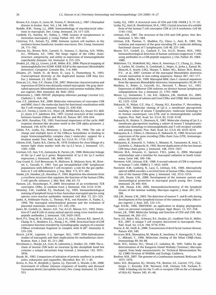
The expression profile of CD8a and CD8b transcripts inadult tissues of the tammar and opossum (Fig. 7) reveal ahigh level of expression in the immune-associated tissues,including the spleen, lung, gut as well as the blood(tammar only). Transcripts were also detected in the liver,kidney and heart, but not in the brain of either marsupial.Furthermore, RT-PCR analysis of CD8 transcripts in thetissues of tammar pouch young at various ages is shown inFig. 8. CD8a and CD8b transcripts were detected in thecervical thymus, lung, blood and liver from day 10postpartum. However, transcripts were not detected inthe spleen and gut until day 21 postpartum. G3PDH wasused to ensure integrity of the RNA/cDNA and expressionwas observed in all samples assayed for CD8.
4. Discussion
The advent of whole genome sequence data provides avaluable tool for the isolation of homologous genes indivergent species that previously proved difficult usingstandard molecular techniques. The gray short-tailedopossum was the first marsupial to be the focus of awhole genome sequencing project and this data isavailable on the Ensembl database. A recent report byWong et al. (2006) identified probable gene models for CD4and CD8a in the opossum genome using a bioinformaticapproach. However, these sequences were not confirmedby cDNA or genomic cloning and, like the Ensemblannotations, are predictions only. In our studies, we havecloned the full-length cDNA sequences for CD4 (Duncanet al., 2007) and CD8a in the opossum. This is a necessarystep given that gene prediction software frequently predictexon/intron boundaries incorrectly and often fail tosuccessfully identify the 50 and 30 ends of genes (as seenin the opossum CD4 and CD8a sequences predicted byWong et al., 2006 and the Ensembl annotations). This work
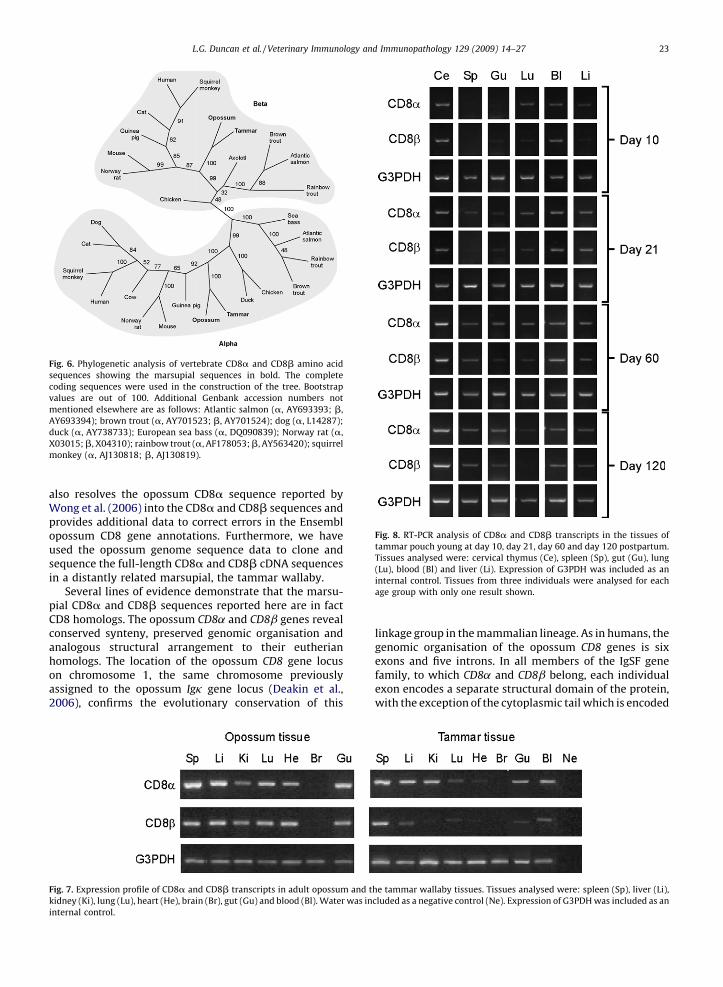
Fig. 6. Phylogenetic analysis of vertebrate CD8a and CD8b amino acid
sequences showing the marsupial sequences in bold. The complete
coding sequences were used in the construction of the tree. Bootstrap
values are out of 100. Additional Genbank accession numbers not
mentioned elsewhere are as follows: Atlantic salmon (a, AY693393; b,
AY693394); brown trout (a, AY701523; b, AY701524); dog (a, L14287);
duck (a, AY738733); European sea bass (a, DQ090839); Norway rat (a,
X03015; b, X04310); rainbow trout (a, AF178053; b, AY563420); squirrel
monkey (a, AJ130818; b, AJ130819).
Fig. 8. RT-PCR analysis of CD8a and CD8b transcripts in the tissues of
tammar pouch young at day 10, day 21, day 60 and day 120 postpartum.
Tissues analysed were: cervical thymus (Ce), spleen (Sp), gut (Gu), lung
(Lu), blood (Bl) and liver (Li). Expression of G3PDH was included as an
internal control. Tissues from three individuals were analysed for each
age group with only one result shown.
L.G. Duncan et al. / Veterinary Immunology and Immunopathology 129 (2009) 14–27 23
also resolves the opossum CD8a sequence reported byWong et al. (2006) into the CD8a and CD8b sequences andprovides additional data to correct errors in the Ensemblopossum CD8 gene annotations. Furthermore, we haveused the opossum genome sequence data to clone andsequence the full-length CD8a and CD8b cDNA sequencesin a distantly related marsupial, the tammar wallaby.
Several lines of evidence demonstrate that the marsu-pial CD8a and CD8b sequences reported here are in factCD8 homologs. The opossum CD8a and CD8b genes revealconserved synteny, preserved genomic organisation andanalogous structural arrangement to their eutherianhomologs. The location of the opossum CD8 gene locuson chromosome 1, the same chromosome previouslyassigned to the opossum Igk gene locus (Deakin et al.,2006), confirms the evolutionary conservation of this
Fig. 7. Expression profile of CD8a and CD8b transcripts in adult opossum and t
kidney (Ki), lung (Lu), heart (He), brain (Br), gut (Gu) and blood (Bl). Water was in
internal control.
linkage group in the mammalian lineage. As in humans, thegenomic organisation of the opossum CD8 genes is sixexons and five introns. In all members of the IgSF genefamily, to which CD8a and CD8b belong, each individualexon encodes a separate structural domain of the protein,with the exception of the cytoplasmic tail which is encoded
he tammar wallaby tissues. Tissues analysed were: spleen (Sp), liver (Li),
cluded as a negative control (Ne). Expression of G3PDH was included as an

L.G. Duncan et al. / Veterinary Immunology and Immunopathology 129 (2009) 14–2724
by two exons. Conservation of this feature in the opossumCD8a and CD8b genes result in the same distinctivestructural domain arrangement exhibited by their euther-ian homologs. Thus, they have a single extracellular IgSfVdomain, a hinge segment, a transmembrane region and acytoplasmic tail (Littman, 1987).
The evolutionary relationship of the marsupial CD8subunits to their eutherian counterparts is seen in thephylogenetic tree where the opossum and tammarsequences form a separate monophyletic clade as a sistergroup to the eutherians within the distinct alpha and betaclusters of vertebrate CD8. The relatively high sequenceconservation between the two marsupials for each CD8subunit at the amino acid level (CD8a, 63%; CD8b, 57%),mirrors that found between other distantly relatedmammals with a similar evolutionary divergence suchas human and mouse. Nonetheless, the CD8 subunits arepoorly conserved across species showing identities com-pared with marsupials as low as 42% (CD8a) and 39%(CD8b) for eutherians, 32% (CD8a) and 27% (CD8b) foravians, as well as 21% (CD8a) and 20% (CD8b) for teleosts.That said, despite this low sequence conservation, manystructural and functional features of the CD8a and theCD8b subunit are evolutionarily conserved across verte-brates. Present in both subunits are the two invariantcysteine residues that form a disulphide bond in the IgSfVdomain as well as the two cysteines involved in polypep-tide dimerisation and the multiple potential O-linkedglycosylation sites in the hinge region. Also, the extra-cellular domain of the CD8a subunit acts as a ligand for theMHC class I molecule and the cytoplasmic tail of CD8acontains a docking site for the kinase, p56lck (Leahy, 1995).
The crystal structure of the MHC class I molecule incomplex with the two subunits of CD8aa, designatedCD8a1 and CD8a2, has been resolved in the human (Gaoet al., 1997) and the mouse (Kern et al., 1998). Bothsubunits of the CD8a homodimer interact with the MHCa3 domain, whilst the CD8a1 subunit makes additionalcontacts with the MHC a2 domain. Briefly, the interactionprimarily involves the exposed negatively charged loop(residues 223–229) in the MHC a3 domain (Salter et al.,1990) that is clamped between the CDR-like loops of thetwo CD8 subunits (Gao et al., 1997). In addition, the MHCa2 domain residues Q115, D122, E128 (Sun et al., 1995)interact with the A and B strands of CD8 (Giblin et al.,1994). For marsupials, the exposed loop in the MHC a3domain and the a2 domain residues are highly conservedin opossum (Miska and Miller, 1999) and tammar (Siddleet al., 2006). Furthermore, specific residues within theCDR-like loops, in particular the CDR3 loop and the A/Bstrands in both marsupial CD8a sequences are conserved.
The other important molecule that associates with theCD8a subunit is the kinase p56lck. The docking site for p56lck
on the CD8a molecule has been shown in eutherians to bemediated by cysteine motifs within both the cytoplasmictail of the CD8a subunit and the N-terminal domain of p56lck
(Shaw et al., 1990; Turner et al., 1990). The cysteine motif,CXC, in the CD8a subunit is highly conserved in marsupialsas in eutherians and chicken. Furthermore, the opossump56lck sequence located on the Ensembl database was foundto contain the consensus sequence motif CXXC in the N-
terminal domain as seen in eutherians. Conservation ofspecific residues and motifs within the CD8a subunit as wellas the MHC and p56lck molecules in these marsupialssuggests that similar interactions to those described ineutherians are plausible in marsupials.
We previously reported (Duncan et al., 2007) anintriguing feature of the opossum and tammar CD4sequences wherein their cytoplasmic tails were truncatedby seven residues when compared to eutherian sequences.Similarly, we show here that the opossum CD8a and thetammar CD8b cytoplasmic tails are also truncated. As withmarsupial CD4, the truncation in opossum CD8a isunlikely to affect the signaling pathway involving p56lck
as the docking site is located N-terminal to the truncation.Interestingly, if the stop codon in the opossum CD8asequence (position-215) and the tammar CD8b sequence(position-189) is ignored, then the succeeding basesequence would, upon translation, yield a protein productremarkably similar to that observed in the tammar CD8aand opossum CD8b sequences, respectively. Althoughvarious cytoplasmic tail lengths arising from alternativesplicing variants have been reported in mouse CD8a(Zamoyska et al., 1985) and human CD8b (Norment andLittman, 1988), the marsupial CD8 truncations are notsplice variants and have been shown in opossum to beencoded within the genome. However, the functionalsignificance of these cytoplasmic tail truncations in themarsupials is unknown.
An interesting observation that is evident from theanalysis of the CD8a and the CD8b homologs acrossvertebrates is the apparent structural similarities of thesetwo subunits to each other. However, despite thisstructural homology, the CD8a and CD8b subunits arenot closely related to each other showing identities at theamino acid level as low as 16% (human), 15% (tammar) and13% (opossum). In mammals, the CD8b gene is the larger ofthe two genes with the increased length of this geneattributed to longer introns. Conversely, for the translatedproduct, it is the CD8a protein that is the larger of the twomolecules with the difference observed mainly in theincreased length of the hinge and cytoplasmic regions. Theextracellular domain in both subunits resembles an IgVdomain composed of two beta sheets of five and fourstrands connected by an array of loops analogous to the IgCDRs (Leahy et al., 1992). In mammals including theopossum and tammar, the CD8b sequence contains thetwo invariant cysteine residues on strand B and strand Fthat form the canonical disulphide bridge in IgV domains.However, the CD8a IgV domain sequence in mammalscontains three cysteine residues (C-22, C-33 and C-94 inhuman). In human and mouse, disulphide bond formationhas been shown for the canonical disulphide bridgebetween C-22 on strand B and C-94 on strand F (Kernet al., 1998; Leahy et al., 1992) but also as an unusualdisulphide bridge between C-22 on strand B and C-33 onstrand C (Classon et al., 1992; Kirszbaum et al., 1989).Although the structural adjustment that is required toachieve these two alternate disulphide linkages is minor,these disulphide bond isoforms may exhibit slightlydifferent properties in these animals (Kirszbaum et al.,1989; Leahy, 1995).

L.G. Duncan et al. / Veterinary Immunology and Immunopathology 129 (2009) 14–27 25
The tissue-specific expression of CD8a and CD8btranscripts in the tammar pouch young tissues wasanalysed by RT-PCR and accords well with the gradualmaturation of the lymphoid-associated tissues over thefirst 120 days of pouch life (Basden et al., 1997). Thedetection of CD8 transcripts in the cervical thymus andblood from day 10 postpartum corresponds with the onsetof maturity of the cervical thymus by day 21 and the firstappearance of lymphocytes in the blood of the tammar asearly as day 5 (Basden et al., 1996, 1997). CD8 expressionwas not detected in the spleen and gut at 10 dayspostpartum but was first observed from day 21 post-partum. This expression is consistent with the maturationof the spleen and gut by day 60 and day 120 postpartum,respectively (Basden et al., 1996, 1997). It is interesting tocompare the CD8 tissue-specific expression pattern to anearlier report showing the distribution of T cells in thetissues of tammar pouch young by immunohistochemicalanalysis using an anti-human CD3 antibody (Old andDeane, 2003). In this study, CD3+ cells were observed in thecervical thymus and gut at 12 days postpartum, the spleenat 21 days postpartum but were not detected in the liver orlung (CD3+ cells were observed in the blood vessels of thelung) at any age. The expression of CD8 transcripts in thecervical thymus and gut at an earlier and later age,respectively than reported for CD3, together with theexpression of CD8 transcripts in the liver and lung whenCD3 was not detected in these tissues, raises the questionof whether these CD8 transcripts are expressed on the Tcell surface at the ages specified or if the results reportedhere were affected by the high level of CD8 expression inthe blood vessels. The development of marsupial-specificreagents and immunohistochemical investigation isrequired to determine whether CD8 is present in thetissue beds themselves.
Previous analyses of the distribution of CD8a in thetissues of marsupials including the tammar wallaby(Cisternas and Armati, 2000; Old and Deane, 2002) usingcommercial eutherian antibodies failed to show anyreactivity in marsupials. Using the sequence data pre-sented in this study, it is possible to explain this lack ofreactivity. The anti-human CD8a antibody (Mason et al.,1992), recognizes the C-terminus of the cytoplasmicregion, and the anti-rat CD8a antibody MRC OX-8 (Classonet al., 1992) recognizes a 24 amino acid epitope in thehinge region. Our data reveal that low sequence common-ality of 38% and 20%, respectively to their correspondingepitope in the tammar wallaby is the basis for this poorreactivity. It should be noted that the majority ofcommercial anti-human CD8a antibodies are designedto recognize the IgSfV domain which is the least conserveddomain. As a result, these antibodies are highly specific forthe target species for which they were designed. This lackof cross-reactivity was demonstrated for squirrel monkeyusing anti-human antibodies specific for the IgSfV domain(Ureta-Vidal et al., 1999) that failed to show any reactivityeven though they share 81% identity in their IgSfV domainsequences. These issues highlight the need for marsupial-specific antibodies. These tools are fundamental, not onlyfor obtaining baseline information on normal lymphoidtissues in healthy animals, but also for characterizing
pathogenesis of infectious diseases afflicting marsupialsincluding Chlamydial infection in koalas (Brown et al.,1987), Mycobacterial and Toxoplasma infection reportedin a broad range of marsupial hosts (Buddle and Young,2000; Canfield et al., 1990), and the devil facial-tumourdisease (DFTD) recently reported in Tasmanian devilpopulations (Kreiss et al., 2008; Pearse and Swift, 2006).
In summary, T cell function in the marsupial immunesystem has not been extensively studied due, in part, to alack of molecular data and species cross-reactive anti-bodies. The work reported in this paper was conducted toaddress this deficit. Here, the genome sequence data of anAmerican marsupial, the gray short-tailed opossum, notonly provided the basis for isolating the CD8a and CD8btranscripts in this species but also facilitated the isolationof these transcripts in an Australian marsupial, the tammarwallaby. These results complement those describedpreviously for the marsupial CD4 (Duncan et al., 2007).Taken together, this work provides fundamental compara-tive data on T cell subsets showing the relationship withinthe marsupials as well as with their eutherian counter-parts. The identification of CD4 and CD8 in marsupials is acritical first step towards understanding the host’s defencemechanisms against pathogens (bacterial, viral, parasitic)and tumours and will provide the tools for futureimmunopathological studies. Furthermore, the character-isation of CD8 transcripts in the tissues of tammar pouchyoung provides valuable insight into the development ofthe immune tissues and the onset of immunologicalcompetence in neonatal marsupials.
Acknowledgements
The authors would like to thank Norman Saunders andKate Dziegielewska (Department of Pharmacology, Uni-versity of Melbourne, Vic) for supplying the opossumtissue and Lyn Hinds (CSIRO, Division of Entomology,Canberra) for supplying the tammar wallaby tissue. Wealso thank the Macquarie University Fauna Park staff fortheir assistance in animal handling. This work wassupported by a Macquarie University Postgraduate award.
References
Abbas, A.K., Lichtman, A.H., 2003. Cellular and Molecular Immunology,5th edition. Saunders, Philadelphia.
Baker, M.L., Miller, R.D., 2007. Evolution of mammalian CD1: marsupialCD1 is not orthologous to the eutherian isoforms and is a pseudogenein the opossum Monodelphis domestica. Immunology 121, 113–121.
Basden, K., Cooper, D.W., Deane, E.M., 1996. Development of the blood-forming tissues of the Tammar Wallaby Macropus eugenii. Reprod.Fertil. Dev. 8, 989–994.
Basden, K., Cooper, D.W., Deane, E.M., 1997. Development of the lymphoidtissues of the tammar wallaby Macropus eugenii. Reprod. Fertil. Dev. 9,243–254.
Belov, K., Deakin, J.E., Papenfuss, A.T., Baker, M.L., Melman, S.D., Siddle,H.V., Gouin, N., Goode, D.L., Sargeant, T.J., Robinson, M.D., Wakefield,M.J., Mahony, S., Cross, J.G.R., Benos, P.V., Samollow, P.B., Speed, T.P.,Marshall Graves, J.A., Miller, R.D., 2006. Reconstructing an ancestralmammalian immune supercomplex from a marsupial major histo-compatibility complex. PLoS Biol. 4, e46.
Belov, K., Sanderson, C.E., Deakin, J.E., Wong, E.S.W., Assange, D., McColl,K.A., Gout, A., de Bono, B., Barrow, A.D., Speed, T.P., Trowsdale, J.,Papenfuss, A.T., 2007. Characterization of the opossum immunegenome provides insights into the evolution of the mammalianimmune system. Genome Res. 17, 982–991.
L.G. Duncan et al. / Veterinary Immunology and Immunopathology 129 (2009) 14–2726
Brown, A.S., Girjes, A., Lavin, M., Timms, P., Woolcock, J., 1987. Chlamydialdisease in koalas. Aust. Vet. J. 64, 346–350.
Buddle, B.M., Young, L.J., 2000. Immunobiology of mycobacterial infec-tions in marsupials. Dev. Comp. Immunol. 24, 517–529.
Canfield, P.J., Hartley, W., Dubey, J., 1990. Lesions of toxoplasmosis inAustralian marsupials. J. Comp. Pathol. 103, 159–167.
Cisternas, P.A., Armati, P.J., 2000. Immune system cell markers in thenorthern brown bandicoot, Isoodon macrourus. Dev. Comp. Immunol.24, 771–782.
Classon, B.J., Brown, M.H., Garnett, D., Somoza, C., Barclay, A.N., Willis,A.C., Williams, A.F., 1992. The hinge region of the CD8a chain:structure, antigenicity, and utility in expression of immunoglobulinsuperfamily domains. Int. Immunol. 4, 215–225.
Deakin, J.E., Olp, J.J., Graves, J.A.M., Miller, R.D., 2006. Physical mapping ofimmunoglobulin loci IGH@, IGK@, IGL@ in the opossum (Monodelphisdomestica). Cytogenet. Genome Res. 114, 94H.
DiSanto, J.P., Smith, D., de Bruin, D., Lacy, E., Flomenberg, N., 1993.Transcriptional diversity at the duplicated human CD8 beta loci.Eur. J. Immunol. 23, 320–326.
Duncan, L.G., Nair, S.V., Deane, E.M., 2007. Molecular characterisation andexpression of CD4 in two distantly related marsupials: the gray short-tailed opossum (Monodelphis domestica) and tammar wallaby (Macro-pus eugenii). Mol. Immunol. 44, 3641–3652.
Felsenstein, J., 1989. PHYLIP: phylogeny inference package (version 3.2).Cladistics 5, 164–166.
Gao, G.F., Jakobsen, B.K., 2000. Molecular interactions of coreceptor CD8and MHC class I: the molecular basis for functional coordination withthe T-cell receptor. Immunol. Today 21, 630–636.
Gao, G.F., Tormo, J., Gerth, U.C., Wyer, J.R., McMichael, A.J., Stuart, D.I., Bell,J.I., Jones, E.Y., Jakobsen, B.K., 1997. Crystal structure of the complexbetween human CD8aa and HLA-A2. Nature 387, 630–634.
Gao, M.H., Kavathas, P.B., 1993. Functional importance of the cyclic AMPresponse element-like decamer motif in the CD8 alpha promoter. J.Immunol. 150, 4376–4385.
Giblin, P.A., Leahy, D.J., Mennone, J., Kavathas, P.B., 1994. The role ofcharge and multiple faces of the CD8a/a homodimer in binding tomajor histocompatibility complex class I molecules: support for abivalent model. Proc. Natl. Acad. Sci. U.S.A. 91, 1716–1720.
Gibson, D.M., Taylor, B.A., Cherry, M., 1978. Evidence for close linkage of amouse light chain marker with the Ly-2,3 locus. J. Immunol. 121,1585–1590.
Gorman, S.D., Sun, Y.H., Zamoyska, R., Parnes, J.R., 1988. Molecular linkageof the Ly-3 and Ly-2 genes. Requirement of Ly-2 for Ly-3 surfaceexpression. J. Immunol. 140, 3646–3653.
Guy-Grand, D., Cerf-Bensussan, N., Malissen, B., Malassis-Seris, M., Briot-tet, C., Vassalli, P., 1991. Two gut intraepithelial CD8+ lymphocytepopulations with different T cell receptors: a role for the gut epithe-lium in T cell differentiation. J. Exp. Med. 173, 471–481.
Hanke, J.H., Hambor, J.E., Kavathas, P., 1995. Repetitive Alu elements forma cruciform structure that regulates the function of the human CD8aT cell specific enhancer. J. Mol. Biol. 246, 63–73.
Hansen, J.D., Strassburger, P., 2000. Description of an ectothermic TCRcoreceptor, CD8a, in rainbow trout. J. Immunol. 164, 3132–3139.
Hemsley, S.W., Canfield, P.J., Husband, A.J., 1995. Immunohistologicalstaining of lymphoid tissue in four Australian marsupial species usingspecies cross-reactive antibodies. Immunol. Cell. Biol. 73, 321–325.
Janke, A., Feldmaier-Fuchs, G., Thomas, W.K., von Haeseler, A., Paabo, S.,1994. The marsupial mitochondrial genome and the evolution ofplacental mammals. Genetics 137, 243–256.
Jones, M., Cordell, J.L., Beyers, A.D., Tse, A.G.D., Mason, D.Y., 1993. Detec-tion of T and B cells in many animal species using cross-reactive anti-peptide antibodies. J. Immunol. 150, 5429–5435.
Kern, P.S., Teng, M.-K., Smolyar, A., Liu, J.-H., Liu, J., Hussey, R.E., Spoerl, R.,Chang, H.-C., Reinherz, E.L., Wang, J.-H., 1998. Structural basis of CD8coreceptor function revealed by crystallographic analysis of a murineCD8aa ectodomain fragment in complex with H-2Kb. Immunity 9,519–530.
Kirsch, J.A.W., Lapointe, F.-J., Springer, M.S., 1997. DNA-hybridisationstudies of marsupials and their implications for methatherian classi-fication. Aust. J. Zool. 45, 211–280.
Kirszbaum, L., Sharpe, J.A., Goss, N., Lahnstein, J., Walker, I.D., 1989. The a-chain of murine CD8 lacks an invariant Ig-like disulphide bond butcontains a unique intrachain loop instead. J. Immunol. 142, 3931–3936.
Kozak, M., 1983. Comparison of initiation of protein synthesis in prokar-yotes, eukaryotes and organelles. Microbiol. Rev. 47, 1–45.
Kreiss, A., Fox, N., Bergfeld, J., Quinn, S.J., Pyecroft, S., Woods, G.M., 2008.Assessment of cellular immune responses of healthy and diseasedTasmanian devils (Sarcophilus harrisii). Dev. Comp. Immunol. 32, 544–553.
Leahy, D.J., 1995. A structural view of CD4 and CD8. FASEB J. 9, 17–25.Leahy, D.J., Axel, R., Hendrickson, W.A., 1992. Crystal structure of a soluble
form of the human T cell coreceptor CD8 at 2.6 A resolution. Cell 68,1145–1162.
Littman, D.R., 1987. The structure of the CD4 and CD8 genes. Ann. Rev.Immunol. 5, 561–584.
Littman, D.R., Thomas, Y., Maddon, P.J., Chess, L., Axel, R., 1985. Theisolation and sequence of the gene encoding T8: a molecule definingfunctional classes of T lymphocytes. Cell 40, 237–246.
Mason, D.Y., Cordell, J.L., Gaulard, P., Tse, A.G.D., Brown, M.H., 1992.Immunohistological detection of human cytotoxic/suppressor T cellsusing antibodies to a CD8 peptide sequence. J. Clin. Pathol. 45, 1084–1088.
Mikkelsen, T.S., Wakefield, M.J., Aken, B., Amemiya, C.T., Chang, J.L., Duke,S., Garber, M., Gentles, A., Goodstadt, L., Heger, A., Jurka, J., Kamal, M.,Mauceli, E., Searle, S.M.J., Sharpe, T., Baker, M.L., Batzer, M.A., Benos,P.V., et al., 2007. Genome of the marsupial Monodelphis domesticareveals innovation in non-coding sequences. Nature 447, 167–177.
Miska, K.B., Miller, R.D., 1999. Marsupial MHC class I: classical sequencesfrom the opossum Monodelphis domestica. Immunogenetics 50, 89–93.
Moebius, U., Kober, G., Griscelli, A.L., Hercend, T., Meuer, S.C., 1991.Expression of different CD8 isoforms on distinct human lymphocytesubpopulations. Eur. J. Immunol. 21, 1793–1800.
Moore, L.J., Somamoto, T., Lie, K.K., Dijkstra, J.M., Hordvik, D.I., 2005.Characterisation of salmon and trout CD8a and CD8b. Mol. Immunol.42, 1225–1234.
Nakauchi, H., Nolan, G.P., Hsu, C., Huang, H.S., Kavathas, P., Herzenberg,L.A., 1985. Molecular cloning of Lyt-2, a membrane glycoproteinmarking a subset of mouse T lymphocytes: molecular homology toits human counterpart, Leu-2/T8, and to immunoglobulin variableregions. Proc. Natl. Acad. Sci. U.S.A. 82, 5126–5130.
Nakauchi, H., Shinkai, Y., Okumura, K., 1987. Molecular cloning of Lyt-3, amembrane glycoprotein marking a subset of mouse T lymphocytes:molecular homology to immunoglobulin and T-cell receptor variableand joining regions. Proc. Natl. Acad. Sci. U.S.A. 84, 4210–4214.
Nakayama, K.-I., Tokito, S., Okumura, K., Nakauchi, H., 1989. Structure andexpression of the gene encoding CD8a chain (Leu-2/T8). Immunoge-netics 30, 393–397.
Nakayama, K., Kawachi, Y., Tokito, S., Minami, N., Yamamoto, R., Imai, T.,Gachelin, G., Nakauchi, H., 1992. Recent duplication of the two humanCD8 beta-chain genes. J. Immunol. 148, 1919–1927.
Nilsson, M.A., Arnason, U., Spencer, P.B.S., Janke, A., 2004. Marsupialrelationships and a timeline for marsupial radiation in South Gond-wana. Gene 340, 189–196.
Norment, A.M., Littman, D.R., 1988. A second subunit of CD8 is expressedin human T cells. EMBO J. 7, 3433–3439.
Norment, A.M., Lonberg, N., Lacy, E., Littman, D.R., 1989. Alternativelyspliced mRNA encodes a secreted form of human CD8a, characteriza-tion of the human CD8a gene. J. Immunol. 142, 3312–3319.
Old, J.M., Deane, E.M., 2000. Development of the immune system andimmunological protection in marsupial pouch young. Dev. Comp.Immunol. 24, 445–454.
Old, J.M., Deane, E.M., 2002. Immunohistochemistry of the lymphoidtissues of the tammar wallaby, Macropus eugenii. J. Anat. 201, 257–266.
Old, J.M., Deane, E.M., 2003. The detection of mature T- and B-cells duringdevelopment of the lymphoid tissues of the tammar wallaby (Macro-pus eugenii). J. Anat. 203, 123–131.
Page, R.D.M., 1996. TREEVIEW: an application to display phylogenetictrees on personal computers. Comput. Appl. Biosci. 12, 357–358.
Parnes, J.R., 1989. Molecular biology and function of CD4 and CD8. Adv.Immunol. 44, 265–311.
Parra, Z.E., Baker, M.L., Schwarz, R.S., Deakin, J.E., Lindblad-Toh, K., Miller,R.D., 2007. A unique T cell receptor discovered in marsupials. Proc.Natl. Acad. Sci. U.S.A. 104, 9776–9781.
Pearse, A.-M., Swift, K., 2006. Transmission of devil facial-tumour disease.Nature 439, 549.
Pecoraro, M.R., Shimojima, M., Maeda, K., Inoshima, Y., Kawaguchi, Y., Kai,C., Mikami, T., 1996. Molecular cloning of the feline CD8b-chain.Immunology 89, 84–88.
Poole, W.E., Simms, N.G., Wood, J.T., Lubulwa, M., 1991. Tables for agedetermination of the Kangaroo Island Wallaby (Tammar), Macropuseugenii, from body measurements. In: Technical Memorandum No.32, CSIRO Division of Wildlife and Ecology, Canberra.
Renfree, M.B., 2007. The genome of a Gondwanan mammal. BioEssays 29,1073–1076.
Salter, R.D., Benjamin, R.J., Wesley, P.K., Buxton, S.E., Garrett, T.P.J., Clay-berger, C., Krensky, A.M., Norment, A.M., Littman, D.R., Parham, P.,1990. A binding site for the T-cell co-receptor CD8 on the a3 domainof HLA-A2. Nature 345, 41–46.

L.G. Duncan et al. / Veterinary Immunology and Immunopathology 129 (2009) 14–27 27
Shaw, A.S., Chalupny, J., Whitney, J.A., Hammond, C., Amrein, K.E.,Kavathas, P., Sefton, B.M., Rose, J.K., 1990. Short related sequencesin the cytoplasmic domains of CD4 and CD8 mediate binding to theamino-terminal domain of the p56lck tyrosine protein kinase. Mol.Cell. Biol. 10, 1853–1862.
Shiue, L., Gorman, S.D., Parnes, J.R., 1993. A second chain of human CD8 isexpressed on peripheral blood lymphocytes. J. Exp. Med. 168, 1993–2005.
Siddle, H.V., Deakin, J.E., Baker, M.L., Miller, R.D., Belov, K., 2006. Isolationof major histocompatibility complex Class I genes from the tammarwallaby (Macropus eugenii). Immunogenetics 58, 487–493.
Smale, S.T., Kadonaga, J.T., 2003. The RNA polymerase II core promoter.Ann. Rev. Biochem. 72, 449–479.
Stenger, S., Rosat, J.-P., Bloom, B.R., Krensky, A.M., Modlin, R.L., 1999.Granulysin: a lethal weapon of cytolytic T cells. Immunol. Today 20,390–394.
Sukhatme, V.P., Vollmer, A.C., Erikson, J., Isobe, M., Croce, C., Parnes, J.R.,1985. Gene for the human T cell differentiation antigen Leu-2/T8 isclosely linked to the k light chain locus on chromosome 2. J. Exp. Med.161, 429–434.
Sun, J., Leahy, D.J., Kavathas, P.B., 1995. Interaction between CD8 andmajor histocompatibility complex (MHC) class I mediated by multiplecontact surfaces that include the a2 and a3 domains of MHC class I. J.Exp. Med. 182, 1275–1280.
Tregaskes, C.A., Kong, F.-k., Paramithiotis, E., Chen, C.-L.H., Ratcliffe, M.J.H.,Davison, T.F., Young, J.R., 1995. Identification and analysis of the
expression of CD8ab and CD8aa isoforms in chickens reveals amajor TCR-gd CD8ab subset of intestinal intraepithelial lympho-cytes. J. Immunol. 154, 4485–4494.
Turner, J.M., Brodsky, M.H., Irving, B.A., Levin, S.D., Perimutter, R.M.,Littman, D.R., 1990. Interaction of the unique N-terminal region oftyrosine kinase p56lck with cytoplasmic domains of CD4 and CD8 ismediated by cysteine motifs. Cell 60, 755–765.
Ureta-Vidal, A., Garcia, Z., Lemonnier, F.A., Kazanji, M., 1999.Molecular characterization of cDNAs encoding squirrel monkey(Saimiri sciureus) CD8 a and b chains. Immunogenetics 49, 718–721.
Veillette, A., Bookman, M.A., Horak, E.M., Bolen, J.B., 1988. The CD4 andCD8 T cell surface antigens are associated with the internal mem-brane tyrosine-protein kinase p56lck. Cell 55, 301–308.
Wong, E.S.W., Young, L.J., Papenfuss, A.T., Belov, K., 2006. In silico identi-fication of opossum cytokine genes suggests the complexity of themarsupial immune system rivals that of eutherian mammals. Immu-nome Res. 2, 4.
Wong, J.S., Wang, X., Witte, T., Nie, L., Carvou, N., Kern, P., Chang, H.-C.,2003. Stalk region of b-chain enhances the coreceptor function ofCD8. J. Immunol. 171, 867–874.
Zamoyska, R., 1994. The CD8 coreceptor revisited: one chain good, twochains better. Immunity 1, 243–246.
Zamoyska, R., Vollmer, A.C., Sizer, K.C., Liaw, C.W., Parnes, J.R., 1985. TwoLyt-2 polypeptides arise from a single gene by alternative splicingpatterns of mRNA. Cell 43, 153–163.