Acrosome formation during sperm transit through the epididymis in two marsupials, the tammar wallaby...
10
J. Anat. (1999) 194, pp. 223–232, with 8 figures Printed in the United Kingdom 223 Acrosome formation during sperm transit through the epididymis in two marsupials, the tammar wallaby (Macropus eugenii) and the brushtail possum (Trichosurus vulpecula) MINJIE LIN AND JOHN C. RODGER Cooperative Research Centre for Conservation and Management of Marsupials, Department of Biological Sciences, University of Newcastle, NSW, Australia (Accepted 12 November 1998) In certain Australian marsupials including the tammar wallaby (Macropus eugenii) and the brushtail possum (Trichosurus vulpecula), formation of the acrosome is not completed in the testis but during a complex differentiation process as spermatozoa pass through the epididymis. Using transmission and scanning electron microscopy this paper defined the process of acrosome formation in the epididymis, providing temporal and spatial information on the striking reorganisation of the acrosomal membranes and matrix and of the overlying sperm surface involved. On leaving the testis wallaby and possum spermatozoa had elongated ‘ scoop ’-shaped acrosomes projecting from the dorsal surface of the head. During passage down the epididymis, this structure condensed into the compact button-like organelle found on ejaculated spermatozoa. This condensation was achieved by a complex process of infolding and fusion of the lateral projections of the ‘ scoop ’. In the head of the epididymis the rims of the lateral scoop projections became shorter and thickened and folded inwards, to eventually meet midway along the longitudinal axis of the acrosome. As spermatozoa passed through the body of the epididymis the lateral projections fused together. Evidence of this fusion of the immature outer acrosomal membrane is the presence of vesicles within the acrosomal matrix which persist even in ejaculated spermatozoa. When spermatozoa have reached the tail of the epididymis the acrosome condenses into its mature form, as a small button-like structure contained within the depression on the anterior end of the nucleus. During the infolding process, the membranes associated with the immature acrosome are either engulfed into the acrosomal matrix (outer acrosomal membrane), or eliminated from the sperm head as tubular membrane elements (cytoplasmic membrane). Thus the surface and organelles of the testicular sperm head are transient structures in those marsupials with posttesticular acrosome formation and this must be taken into consideration in attempts to dissect the cell and molecular biology of fertilisation. Key words : Sperm maturation ; marsupials. In eutherian mammals, spermatozoa leaving the testis are immotile and infertile. Fertilising capability is achieved by posttesticular maturation processes as spermatozoa pass through the epididymis. The epi- thelial cells of the epididymis, under the control of androgens, create the environment in which sperma- tozoa differentiate functionally to achieve full pro- gressive motility, the ability to bind to the egg coat (zona pellucida) and to undergo fusion with the egg Correspondence to Dr Minjie Lin, Department of Biological Sciences, University of Newcastle, NSW 2308, Australia. Tel : ›61-2-49215707 ; fax: ›61-2-49216899 or 49216923 ; e-mail : biml!cc.newcastle.edu.au. membrane. All are critical aspects of sperm function for successful fertilisation (reviewed in Cooper, 1986 ; Moore, 1990 ; Yanagimachi, 1994). There is a large body of evidence that epididymal maturation is also critical for marsupial spermatozoa to acquire ferti- lizing capability (Setchell, 1970 ; Harding et al. 1979 ; Jones et al. 1987; Rodger, 1991; Jones & Clulow, 1994 ; Temple-Smith, 1994 ; Mate & Rodger, 1996). However, the marsupial pattern of epididymal matu- ration involves far more structural change than is seen in eutherians.
-
Upload
newcastle-au -
Category
Documents
-
view
1 -
download
0
Transcript of Acrosome formation during sperm transit through the epididymis in two marsupials, the tammar wallaby...

J. Anat. (1999) 194, pp. 223–232, with 8 figures Printed in the United Kingdom 223
Acrosome formation during sperm transit through the
epididymis in two marsupials, the tammar wallaby (Macropus
eugenii) and the brushtail possum (Trichosurus vulpecula)
MINJIE LIN AND JOHN C. RODGER
Cooperative Research Centre for Conservation and Management of Marsupials, Department of Biological Sciences,
University of Newcastle, NSW, Australia
(Accepted 12 November 1998)
In certain Australian marsupials including the tammar wallaby (Macropus eugenii) and the brushtail possum
(Trichosurus vulpecula), formation of the acrosome is not completed in the testis but during a complex
differentiation process as spermatozoa pass through the epididymis. Using transmission and scanning
electron microscopy this paper defined the process of acrosome formation in the epididymis, providing
temporal and spatial information on the striking reorganisation of the acrosomal membranes and matrix
and of the overlying sperm surface involved. On leaving the testis wallaby and possum spermatozoa had
elongated ‘scoop’-shaped acrosomes projecting from the dorsal surface of the head. During passage down
the epididymis, this structure condensed into the compact button-like organelle found on ejaculated
spermatozoa. This condensation was achieved by a complex process of infolding and fusion of the lateral
projections of the ‘scoop’. In the head of the epididymis the rims of the lateral scoop projections became
shorter and thickened and folded inwards, to eventually meet midway along the longitudinal axis of the
acrosome. As spermatozoa passed through the body of the epididymis the lateral projections fused together.
Evidence of this fusion of the immature outer acrosomal membrane is the presence of vesicles within the
acrosomal matrix which persist even in ejaculated spermatozoa. When spermatozoa have reached the tail of
the epididymis the acrosome condenses into its mature form, as a small button-like structure contained
within the depression on the anterior end of the nucleus. During the infolding process, the membranes
associated with the immature acrosome are either engulfed into the acrosomal matrix (outer acrosomal
membrane), or eliminated from the sperm head as tubular membrane elements (cytoplasmic membrane).
Thus the surface and organelles of the testicular sperm head are transient structures in those marsupials with
posttesticular acrosome formation and this must be taken into consideration in attempts to dissect the cell
and molecular biology of fertilisation.
Key words : Sperm maturation; marsupials.
In eutherian mammals, spermatozoa leaving the testis
are immotile and infertile. Fertilising capability is
achieved by posttesticular maturation processes as
spermatozoa pass through the epididymis. The epi-
thelial cells of the epididymis, under the control of
androgens, create the environment in which sperma-
tozoa differentiate functionally to achieve full pro-
gressive motility, the ability to bind to the egg coat
(zona pellucida) and to undergo fusion with the egg
Correspondence to Dr Minjie Lin, Department of Biological Sciences, University of Newcastle, NSW 2308, Australia. Tel : 61-2-49215707;
fax: 61-2-49216899 or 49216923; e-mail : biml!cc.newcastle.edu.au.
membrane. All are critical aspects of sperm function
for successful fertilisation (reviewed in Cooper, 1986;
Moore, 1990; Yanagimachi, 1994). There is a large
body of evidence that epididymal maturation is also
critical for marsupial spermatozoa to acquire ferti-
lizing capability (Setchell, 1970; Harding et al. 1979;
Jones et al. 1987; Rodger, 1991; Jones & Clulow,
1994; Temple-Smith, 1994; Mate & Rodger, 1996).
However, the marsupial pattern of epididymal matu-
ration involves far more structural change than is seen
in eutherians.

A major sperm structure formed in the marsupial
epididymis is the midpiece fibre network. This set of
helically wound fibres completely surrounds the
posterior midpiece in the spermatozoa of all mar-
supials studied (Temple-Smith & Bedford, 1967;
Harding et al. 1976, 1979, 1984; Lin et al. 1997). In the
possum, the first ultrastructural evidence of this
structure is found in spermatozoa from the distal
caput epididymis and its formation is completed as
spermatozoa pass through the proximal cauda epi-
didymis (Temple-Smith & Bedford, 1976). In some
groups of Australian marsupials there is an even more
striking posttesticular structural differentiation even
which results in the formation of the compact
acrosome from the very extensive immature structure
present at spermiation (Cummins, 1976; Temple-
Smith & Bedford, 1976; Harding et al. 1983; Mate &
Rodger, 1996; Setiadi et al. 1997). In American
marsupials an equally dramatic posttesticular event is
the pairing of spermatozoa at their acrosomal faces as
they pass down the epididymis (Rodger, 1982;
Temple-Smith, 1994).
The acrosome, an organelle that lies on the sperm
head, plays vital roles in fertilisation. In most
vertebrates the acrosome is fully formed in the testis
and undergoes no morphological change in the
excurrent ducts (e.g. fish, birds and monotremes;
Jones & Lin, 1993), or only very minor modification
(most eutherians and some marsupials ; Harding et al.
1979; Bedford, 1991). However, in the phalangerid
(large possums) and macropodid (wallabies and
kangaroos) marsupial families, acrosome formation is
not completed until spermatozoa have passed at least
halfway through the epididymis (Temple-Smith,
1994). Such complex transformation of the acrosome
is never seen in any other mammals (see review by
Bedford, 1996). Our previous study on the tammar
wallaby and the brushtail possum, the first to examine
testicular sperm using scanning electron microscopy
(SEM), found that when the mature spermatids are
released into the lumen of the seminiferous tubule to
become spermatozoa, their acrosomes are a ‘scoop’-
shaped sheet of folded tissue extending away from the
dorsal surface of the head (Lin et al. 1997). This shape
is completely different to the acrosome of ejaculated
spermatozoa, which is a compact button lodged in a
depression on the dorsal side of the nucleus (Harding
et al. 1976; Cummins, 1976; Setiadi et al. 1997).
Obviously, it is extremely difficult to unequivocally
describe this extraordinary transformation of the
marsupial acrosome by 2-dimensional transmission
electron microscope (TEM) images alone. As a result,
there is no general agreement on the pattern of the
acrosomal formation in the epididymis, even for the
brushtail possum and tammar wallaby, the most
studied species. For example, the acrosome structure
in the head region of the epididymis, has been
described as either a cup-like structure (Harding et al.
1976; Cummins, 1976; Setiadi et al. 1997) or like the
fingers of a glove with many projections of membrane
and matrix over its dorsal surface (Temple-Smith &
Bedford, 1976). The present study thus set out to
characterise in detail the formation of the tammar
wallaby acrosome in the epididymis using both TEM
and SEM to provide temporal and spatial information
on this complex process of differentiation. Special
attention was given to the reorganisation of the cell
membrane over the sperm head and of the acrosomal
membranes because of their critical role in fertilis-
ation. A briefer description of the essentially similar
morphological maturation of the brushtail possum
acrosome during transit of the epididymis is also
included.
Animals
Five adult male tammar wallabies (Macropus eugenii)
were obtained from Kangaroo Island, South Austra-
lia, and maintained in the breeding yard of the
Marsupial Cooperative Research Centre at the Uni-
versity of Newcastle, New South Wales, Australia.
These animals had been previously used as semen
donors and were known to produce ejaculates con-
taining large numbers of highly motile spermatozoa.
Three adult brushtail possums (Trichosurus vulpecula)
were trapped in the Canterbury region of New
Zealand and housed for up to 3 wk in the Animal
House of Landcare Research at Lincoln prior to
collection of tissues. The possums used were part of a
study of spermatogenesis and all had apparently
normal sperm production. The use of protected
animals and animal experimentation were approved
by the appropriate Australian state authorities and by
the Animal Care and Ethics Committees of the
University of Newcastle and Landcare Research
respectively.
Dissection of tissues
The animals were killed with an overdose of sodium
pentobarbitone (30 mg}kg, intravenously) via a lat-
eral tail vein (wallaby) or intracardiac (possum) after
sedation with CO#}O
#(Jolly, 1993). The testis and
epididymis were freed from the scrotum and prepared
for electron microscopic examination. Each epididy-
224 M. Lin and J. C. Rodger

Fig. 1. Diagram of the epididymis of the tammar wallaby showing
the dissected epididymal segments for this study: 1, proximal head;
2, distal head; 3, proximal body; 4, distal body; 5, proximal tail ; 6,
distal tail. T, testis ; V, vas deferens.
Fig. 2. Scanning (a) and transmission (b) electron micrographs showing the testicular sperm of the tammar wallaby. Note the immature
acrosome (A) is in a ‘scoop’ shape and holding a patch of Sertoli cell cytoplasm (S). The acrosome (A) is lodged in the anterior end of dorsal
surface of the nucleus (N) which is perpendicular to the midpiece (M) of the sperm. CD, cytoplasmic droplet.
mis was dissected and divided into segments of (1) the
proximal head, (2) the distal head, (3) the proximal
body, (4) the distal body, (5) the proximal tail and (6)
the distal tail (see Fig. 1). These segments corre-
sponded to the 22 regions of the tammar wallaby
epididymis described by Jones et al. (1984) as (1)
regions 1–4, (2) regions 5–8, (3) regions 9–12, (4)
regions 13–18, (5) regions 19–20 and (6) regions
21–22. The regionalisation of the epididymis of the
brushtail possum was similar to that for the tammar
wallaby.
Preparation for transmission electron microscopy
The dissected tissues were fixed in 2.5% (v}v)
glutaraldehyde and 2% paraformaldehyde in 0.1
cacodylate buffer for 4 h at room temperature or
overnight at 4 °C. The tissues were postfixed in 1%
osmium tetroxide for 1 h. After dehydration through
serial concentrations of acetone, the tissues were
embedded in Spurr’s resin (Agar Scientific, Essex,
UK). Sections (70–100 nm) were cut on an Ultracut E
ultramicrotome (Reichert-Jung, Austria) with a dia-
mond knife (Diatome, Bienne, Switzerland), and
Acrosome formation in marsupials 225
stained with 1% uranyl acetate in 30% (v}v) ethanol
(Watson, 1958) for 5–10 min, followed by lead citrate
(Reynolds, 1963) for 10–20 min. Transmission elec-
tron micrographs (TEM) were taken with a JEOL-
100CX electron microscope (JEOL, Tokyo, Japan)
operating at 80 kV.
Preparation for scanning electronmicroscopy
The tissues for SEM were prepared by using the same
fixative procedures as for TEM, except that the tissues
were fixed overnight at 4 °C. They were then treated
with 1% osmium tetroxide for 4 h. After dehydration
and critical point drying, the tissues were coated with
gold and examined in a JSM 840 scanning electron
microscope (JEOL, Tokyo, Japan) operated at 15 kV.
Tammar wallaby testis
Testicular spermatozoa in the lumen of the sem-
iniferous tubules and rete testis had an elongated
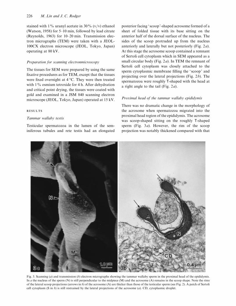
Fig. 3. Scanning (a) and transmission (b) electron micrographs showing the tammar wallaby sperm in the proximal head of the epididymis.
In a the nucleus of the sperm (N) is still perpendicular to the midpiece (M) and the acrosome (A) remains in the scoop shape. Note the rims
of the lateral scoop projections (arrows in b) of the acrosome (A) are thicker than those of the testicular sperm (see Fig. 2). A patch of Sertoli
cell cytoplasm (S in b) is still restrained by the lateral projections of the acrosome (a). CD, cytoplasmic droplet.
posterior facing ‘scoop’-shaped acrosome formed of a
sheet of folded tissue with its base sitting on the
anterior half of the dorsal surface of the nucleus. The
sides of the scoop protruded up from the nucleus
anteriorly and laterally but not posteriorly (Fig. 2a).
At this stage the acrosome scoop contained a remnant
of Sertoli cell cytoplasm which in SEM appeared as a
small circular body (Fig. 2a). In TEM the remnant of
Sertoli cell cytoplasm was closely attached to the
sperm cytoplasmic membrane filling the ‘scoop’ and
projecting over the lateral projections (Fig. 2b). The
spermatozoa were roughly T-shaped with the head at
a right angle to the tail (Fig. 2a).
Proximal head of the tammar wallaby epididymis
There was no dramatic change in the morphology of
the acrosome when spermatozoa migrated into the
proximal head region of the epididymis. The acrosome
was scoop-shaped sitting on the roughly T-shaped
sperm (Fig. 3a). However, the rim of the scoop
projection was notably thickened compared with that
226 M. Lin and J. C. Rodger

Fig. 4. Scanning (a) and transmission (b, c) electron micrographs
showing the tammar wallaby sperm in the distal head of the
epididymis. The lateral scoop projections of the acrosome (A)
become shorter and thicker. The rims of the 2 opposite
projections start to approach to each other at the central region
of the acrosome. The patch of Sertoli cell cytoplasm disappears
from the area enclosed by the acrosome lateral projections.
Note the sperm cytoplasmic membrane (arrow in b) on the
acrosome area is also lifted to the cell surface and no longer
exists between the 2 opposite lateral projections of the acrosome.
b and c show that many tubular membrane elements (T) occur
on the top of the cytoplasmic membrane. CD, cytoplasmic
droplet ; M, midpiece of the sperm; N, sperm nucleus.
Acrosome formation in marsupials 227

Fig. 5. Scanning and transmission electron micrographs showing the tammar wallaby sperm in the proximal (a) and distal body (b) of the
epididymis. The sperm nucleus (N) is oriented to be nearly parallel to the midpiece (M) of the sperm in the proximal epididymis body
(a). In b the acrosome (A) is further condensed and its lateral projections start to fuse along the longitudinal axis of the acrosome when sperm
are in the distal epididymis body. (b) Note the midpiece fibre network (F) is produced when the sperm transit through the epididymis body.
of the testicular spermatozoa (Fig. 3b, cf Fig. 2b). The
remnant of Sertoli cell cytoplasm looked similar in
SEM (Fig. 3a) but appeared reduced in volume into a
circular droplet contained within the scoop projec-
tions in TEM (Fig. 3b). In addition, the Sertoli cell
remnant and cytoplasmic membrane lining the scoop
were no longer in close contact.
Distal head of the tammar wallaby epididymis
The rims of the acrosome lateral projections
were markedly thickened and shortened (Fig. 4b).
The lateral projections were inward folding and
approached each other to meet in the midline of the
acrosome and midway along its length (Fig. 4a). As
the acrosomal projections condensed, the acrosomal
surface area was substantially reduced and the
cytoplasmic membrane which previously was closely
applied to the acrosomal membrane and lined the
inner surface of the scoop was lifted to above the
projections (Fig. 4b). This lifting of the cytoplasmic
membrane was associated with the formation of
tubular membrane elements across the surface of the
acrosome area (Fig. 4c). At this stage the sperm head
remained perpendicular to the midpiece but the Sertoli
cell remnant was no longer evident.
Proximal and distal body of the tammar wallaby
epididymis
Within the proximal body of the epididymis, the
sperm head had rotated to a position so that its long
axis was nearly parallel to the sperm midpiece and the
base of the acrosome was restricted to its final site, a
depression on the anterior third of the dorsal surface
of the nucleus (Fig. 5a). Shortening and thickening of
the acrosome lateral projections were advanced and
the space between the 2 lateral projections was reduced
to a narrow slit along the length of the acrosome (Fig.
5a, b). At this stage tubular membrane elements
overlying cytoplasmic membrane above the acrosome
were no longer present, and the first evidence of the
228 M. Lin and J. C. Rodger

Fig. 6. Scanning and transmission electron micrographs showing the tammar wallaby sperm in the proximal (b) and distal tail (a) of the
epididymis. The multiple-point fusion of the lateral projections can still be seen in the acrosome matrix (see b) of the sperm in the distal tail
of the epididymis. The acrosome (A) condenses to its final compact button shape, lodged into the depression on the distal third of the dorsal
surface of the nucleus (N). M, sperm midpiece.
midpiece fibre network was seen (Fig. 5b). When
spermatozoa passed through the distal body of the
epididymis the lateral projections of the acrosome
fused together to produce a compact unified structure.
Proximal and distal tail of the wallaby epididymis
SEM indicated that the acrosome had condensed into
its mature form as a small compact button-like
structure as found in ejaculated spermatozoa (Fig.
6a). However, the fusion process continued in the
acrosome interior evidenced by a line of fusion
vacuoles within the matrix (Fig. 6b).
Brushtail possum
SEM and TEM demonstrated that acrosome form-
ation in the epididymis of the brushtail possum is very
similar to that described above for the tammar
wallaby. On leaving the testis the acrosome of the
possum was also a scoop-shaped sheet of folding
tissue on the dorsal side of the sperm head (Fig. 7a)
As the possum spermatozoa pass down the epi-
didymis, the acrosome condenses into a compact
button-like structure (Fig. 7b). condensation of the
acrosome occurred by a complex process of infolding
and fusion of the scoop projections, which was
essentially the same as occurred in the tammar wallaby
(Fig. 8a–c).
Using both TEM and SEM, the present study has
been able to visualise the 3-dimensional process of
acrosome formation which occurs as spermatozoa
pass through the epididymis in the tammar wallaby
and brushtail possum. It has been known for some
time that on leaving the testis the acrosomes of both
species are in an immature form but the exact nature
of this structure and the manner by which it was
transformed into the compact button-like mature
acrosome seen in ejaculated spermatozoa was not
Acrosome formation in marsupials 229

Fig. 7. Scanning electron micrographs of the brushtail possum sperm. (a) The acrosome (A) with a scoop shape is located on the anterior
dorsal surface of the nucleus (N). (b) Possum sperm from the distal tail of the epididymis. Note the acrosome (A) is in a compact button-like
shape. M, the sperm midpiece; N, the sperm nucleus.
clear. In our early study of the tammar wallaby (Lin
et al. 1997) we reported, based on SEM, that the
immature acrosome was a backward facing scoop-
shaped structure. The present study confirmed this
finding and showed that the immature possum
acrosome is also a rear-facing scoop. Earlier studies
had interpreted TEM images of the immature possum
acrosome as a cup-like structure (Harding et al. 1976)
or as resembling fingers of a glove (Temple-Smith &
Bedford, 1976). Given these different views of the 3-
dimensional structure of the immature acrosome, it
has been difficult to interpret the large number of
TEM images of the process of maturation produced
since the original reports for the possum (Cummins,
1976; Harding et al. 1976, Temple-Smith & Bedford,
1976). Our recent studies in the tammar wallaby
(Setiadi et al. 1997; Lin et al. 1997), although
providing substantial further TEM data on the
process of acrosome formation, still lacked the clear 3-
dimensional view revealed by the present SEM
observations. In addition, we have confirmed that an
essentially similar process also occurs in the possum.
Posttesticular acrosome formation in both species,
and presumably other marsupials where the phenom-
enon occurs (Harding et al. 1979; Temple-Smith
1994), involves a complex folding and fusing of the
anterior and lateral projections of the scoop-shaped
acrosome which commenced as spermatozoa entered
the distal head of the epididymis. This folding process
was completed by the time spermatozoa reached the
proximal tail region of the epididymis, confirming
earlier observation (possum: Harding et al. 1979;
Temple-Smith, 1994; wallaby: Setiadi et al. 1997). In
the tammar wallaby remnants of the fusion of the
infolded acrosomal membrane persisted within the
acrosomal matrix as a variable number of small
vesicles in many spermatozoa in the distal tail of the
epididymis. Such vesicles or vacuoles have been
reported in ejaculated possum sperm (Sistina et al.
1993). During the infolding process the cystoplasmic
membrane which covered the lateral projections of the
immature acrosome and the underlying outer acro-
somal membrane were completely remodelled. The
cytoplasmic membrane appeared to be eliminated
from the spermatozoa as tubules of budded off
membrane,which is in agreementwith earlier interpret-
ations (Harding et al. 1976; Lin et al. 1997). This
study and our earlier study (Setiadi et al. 1997) clearly
230 M. Lin and J. C. Rodger

Fig. 8. Transmission electron micrographs of brushtail possum sperm in the epididymis body, showing the fusion of acrosome lateral
projections. (a) The acrosome (A) before the fusion and its lateral projections are engulfed a droplet of Sertoli cell cytoplasm (arrow in the
insert) when the sperm is in the proximal epididymis body. (b) Two acrosome lateral projections (A) infolding and approaching each other
(see insert) during passage of the sperm through the proximal epididymis body. (c) Acrosome (A, also in the insert) after its lateral projections
have fused together when the sperm is in the distal epididymis body. The multiple-point fusion of the lateral projections can be seen in the
acrosome matrix. Bars in inserts, 0.5 µm. N, Nucleus.
indicate that the fate of the outer acrosomal mem-
brane which lines the immature acrosome surface
within the scoop is also complex but quite different to
the cytoplasmic membrane. Acrosomal membrane
was never observed to be shed during the maturation
processes but appeared to reduce in surface area with
the contracting matrix to which it closely adhered.
The midline fusion of the acrosome was achieved by
multiple-point fusion of the immature outer acro-
somal membrane and evidence of this fusion event
persisted as vesicles within the acrosomal matrix.
Thus the cytoplasmic membrane over the acrosomal
surface of the mature sperm head, the most likely site
of sperm zona pellucida binding and the site at which
the acrosome reaction is probably initiated (reviewed
in Rodger, 1991; Mate & Rodger 1996), is likely to be
a very different, or highly modified, structure from
that present at spermiation. Similarly the outer
acrosomal membrane which is involved in fusion with
the cytoplasmic membrane at the acrosome reaction is
probably also a different, or highly modified structure,
to that presented at spermiation. Therefore in those
marsupial species where the acrosome is formed after
spermiation, attempts to understand the role of these
structures in fertilisation must focus on the molecular
character of the spermatozoa which have completed
acrosome formation, that is spermatozoa from the vas
deferens or ejaculate, and not on testicular sperm.
This contrasts with the situation in eutherian mam-
mals where it has been the practice to seek critical
sperm antigens and receptors in testicular sperm to
eliminate the complication of later interactions be-
tween the sperm surface and products of the epi-
didymis which may mask functional integral mem-
brane proteins.
This study found that the remnant of Sertoli cell
cytoplasm held in the scoop shape of the acrosome of
testicular spermatozoa (Lin et al. 1997) remains within
the immature acrosome scoop when spermatozoa pass
through the proximal head of the epididymis. In SEM
this remnant appeared as a small droplet sitting within
the scoop. However, TEM cross sections revealed that
the Sertoli cell cytoplasmic remnant fully filled the
space of the acrosome scoop and was firmly attached
to the inner surface of the lateral projections of the
immature scoop. This implied that the remnant may
Acrosome formation in marsupials 231

play a role in supporting the delicate acrosomal
projections which make up the sides of the scoop.
When the acrosomal lateral projections condensed
and started to fuse together in the distal head of the
epididymis, the Sertoli cell cytoplasmic droplet lost
contact with the scoop projections and appeared to be
eliminated from the shrinking scoop. It is also possible
that the Sertoli cell cytoplasmic remnant contributes
materials or regulates the process of acrosome
maturation although presumably the major factor in
the process of posttesticular sperm maturation is the
epididymal environment. Although the morphological
changes described here are unique marsupial phenom-
ena it suggests that these species are excellent models
for studies of the fundamentals of mammalian sperm
maturation and its regulation by the epididymal
environment since such overt and readily visible
morphological indicators occur. In eutherian mam-
mals the equivalent maturation events can only be
monitored indirectly by complex studies of function
such as IVF. In contrast, our work would suggest that
simple light microscopy on fresh or fixed sperm
samples would in many experimental situations be a
definitive indicator of sperm maturation or its dis-
ruption in the wallaby or possum.
We are indebted to Ms Amanda Harman for her
skilled assistance with electron microscopy. We also
wish to thank Dr Karen Mate and Dr David Key for
their helpful comments on the manuscript. This work
was supported by the Australian Government’s
Cooperative Research Centres Program.
BEDFORD JM (1991) The coevolution of mammalian gametes. In
Comparative Overview of Mammalian Fertilization (ed. Dunbar
BS, O’Rand MG), pp. 3–35. New York: Plenum Press.
BEDFORD JM (1996) What marsupial gametes disclose about
gamete function in eutherian mammals. Reproduction, Fertility
and Development 8, 569–580.
COOPER TG (1986) The Epididymis, Sperm Maturation and
Fertilisation. Heidelberg: Springer.
CUMMINS JM (1976) Epididymal maturation of spermatozoa in
the marsupial Trichosurus vulpecula : change in motility and gross
morphology. Australian Journal of Zoology 24, 499–511.
HARDING, HR, CARRICK FN, SHOREY CD (1976) Ultra-
structural changes in spermatozoa of the brush-tailed Possum,
Trichosurus vulpecula (Marsupialia), during epididymal transit.
Part II : the acrosome. Cell and Tissue Research 171, 61–73.
HARDING HR, CARRICK FN, SHOREY CD (1979) Special
features of sperm structure and function in marsupials. In The
Spermatozoon : Maturation, Motility, Surface Properties and
Comparative Aspects (ed. Fawcett DW, Bedford JM), pp.
289–303. Baltimore–Munich: Urban & Schwarzenberg.
HARDING HR, CARRICK FN, SHOREY CD (1983) Acrosome
development during spermiogenesis and epididymal sperm
maturation in Australian marsupials. In The Sperm Cell (ed.
Andre J), pp. 411–444. The Hague: Martinus Nijhoff.
HARDING HR, CARRICK FN, SHOREY CD (1984) Sperm
ultrastructure and development in the honey possum, Tarsipes
rostratus. In Possums and Gliders (ed. Smith AP, Hume ID), pp.
451–456, Sydney: Australian Mammal Society.
JOLLY SE (1993) Carbon dioxide as an anaesthetic agent for use
on the brushtail possum Trichosurus vulpecula in captivity. New
Zealand Journal of Zoology 16, 67–68.
JONES RC, CLULOW J, STONE GM, SETCHELL BP (1987)
The role of the initial segments of the epididymis in sperm
maturation in mammals. In New Horizons in Sperm Cell Research
(ed. Mohri H), pp. 63–71. Tokyo: Japan Scientific Society.
JONES RC, LIN M (1993) Spermatogenesis in birds. In Oxford
Review of Reproductive Biology (ed. Milligan SR), vol. 15, pp.
233–264, Oxford: Oxford University Press.
JONES RC, CLULOW J (1994) Interaction of sperm and the
reproductive ducts of the male tammar wallaby, Macropus
eugenii. Reproduction, Fertility and Development 6, 437–444.
LIN M, HARMAN A, RODGER JC (1997) Spermiogenesis and
spermiation in a marsupial, the tammar wallaby (Macropus
eugenii). Journal of Anatomy 190, 377–395.
MATE KE, RODGER JC (1996) Capacitation and the acrosome
reaction in marsupial spermatozoa. Reproduction, Fertility and
Development 8, 595–603.
MOORE HDM (1990) The epididymis. In Scientific Foundations of
Urology (ed. Chisholm GD, Fair WD), pp. 399–410. Oxford:
Heinemann Medical.
REYNOLDS ES (1963) The use of lead citrate at high pH as an
electron-opaque stain in electron microscopy. Journal of Cell
Biology 17, 208–212.
RODGER JC (1991) Fertilization of marsupials. In A Comparative
Overview of Mammalian Fertilization (ed. Dunbar BS, O’Rand
MG), pp. 117–135. New York: Plenum Press.
SETCHELL BP (1970) Fluid secretion by the testis of Australian
marsupial, Macropus eugenii. Comparative Biochemistry and
Physiology 36, 411–414.
SETIADI D, LIN M, RODGER JC (1997) Posttesticular de-
velopment of spermatozoa of the tammar wallaby (Macropus
eugenii). Journal of Anatomy 190, 275–288.
TEMPLE-SMITH PD (1994) Comparative structure and function
of marsupial spermatozoa. Reproduction, Fertility and Devel-
opment 6, 421–435.
TEMPLE-SMITH PD, BEDFORD JM (1976) The features of
sperm maturation in the epididymis of a marsupial, the
brushtailed possum Trichosurus vulpecula. American Journal of
Anatomy 147, 471–500.
WATSON ML (1958) Staining of tissue sections for electron
microscopy with heavy metals. Journal of Biophysical and
Biochemical Cytology 4, 475–478.
YANAGIMACHI R (1994) Mammalian fertilization. In The
Physiology of Reproduction (ed. Knobil E, Neill JD), pp. 189–317.
New York: Raven Press.
232 M. Lin and J. C. Rodger