Intrahepatic Murine CD8 T-Cell Activation Associates With a Distinct Phenotype Leading to...
16
Intrahepatic murine CD8 T cell activation associates with a distinct phenotype leading to Bim-dependent death Lauren E Holz 1 , Volker Benseler 1,4 , David G Bowen 1 , Philippe Bouillet 2 , Andreas Strasser 2 , Lorraine O'Reilly 2 , William d'Avigdor 1 , Alex G Bishop 3 , Geoffrey W McCaughan 1 , and Patrick Bertolino 1 1 Centenary Institute, AW Morrow Gastroenterology and Liver Centre, Royal Prince Alfred Hospital and Faculty of Medicine, University of Sydney, Camperdown, NSW, 2050, Australia 2 The Walter and Eliza Hall Institute of Medical Research, Parkville, VIC, 3050, Australia 3 Collaborative Transplant Laboratory, Blackburn Building, University of Sydney, NSW, 2006, Australia 4 Department of Surgery, University of Regensburg, Bavaria, 93053, Germany Abstract Background and aims—Chronic infections by hepatotropic viruses such as hepatitis B and C are generally associated with an impaired CD8 T cell immune response that is unable to clear the virus. The liver is increasingly recognised as an alternative site where primary activation of CD8 T cells takes place, a property that might explain its role in inducing tolerance. However, the molecular mechanism by which intra-hepatically activated T cells are tolerized is unknown. Here we investigated the phenotype and fate of naïve CD8 T cells activated by hepatocytes in vivo. Methods—Transgenic mouse models in which the antigen is expressed in lymph nodes and/or in the liver were adoptively transferred with naïve CD8+ T cells specific for the hepatic antigen. Results—Liver-activated CD8 T cells displayed poor effector functions and a unique CD25 low CD54 low phenotype. This phenotype was associated with increased expression of the proapoptotic protein Bim, and caspase-3, demonstrating that these cells are programmed to die following intrahepatic activation. Importantly, we show that T cells deficient for Bim survived following intrahepatic activation. Conclusions—This study identifies Bim for the first time as a critical initiator of T cell death in the liver. Thus, strategies inhibiting the upregulation of this molecule could potentially be used to rescue CD8 T cells, clear the virus and reverse the outcome of viral chronic infections affecting the liver. Introduction In recent years it has emerged that despite being a non-lymphoid organ, the liver displays immunological properties distinct from other solid organs and is associated with the induction of T cell tolerance 1 . This property has been demonstrated in several clinical settings including transplantation 2 , and hepatotropic viral infections, such as those induced by HBV and HCV 3 . Correspondence: Dr Patrick Bertolino; Phone: +61 2 95656186; [email protected]. Financial disclosures: None of the authors have a conflict of interest or financial disclosure to declare NIH Public Access Author Manuscript Gastroenterology. Author manuscript; available in PMC 2010 October 17. Published in final edited form as: Gastroenterology. 2008 September ; 135(3): 989–997. doi:10.1053/j.gastro.2008.05.078. NIH-PA Author Manuscript NIH-PA Author Manuscript NIH-PA Author Manuscript
Transcript of Intrahepatic Murine CD8 T-Cell Activation Associates With a Distinct Phenotype Leading to...
Intrahepatic murine CD8 T cell activation associates with a distinctphenotype leading to Bim-dependent death
Lauren E Holz1, Volker Benseler1,4, David G Bowen1, Philippe Bouillet2, AndreasStrasser2, Lorraine O'Reilly2, William d'Avigdor1, Alex G Bishop3, Geoffrey WMcCaughan1, and Patrick Bertolino11Centenary Institute, AW Morrow Gastroenterology and Liver Centre, Royal Prince Alfred Hospitaland Faculty of Medicine, University of Sydney, Camperdown, NSW, 2050, Australia2The Walter and Eliza Hall Institute of Medical Research, Parkville, VIC, 3050, Australia3Collaborative Transplant Laboratory, Blackburn Building, University of Sydney, NSW, 2006,Australia4Department of Surgery, University of Regensburg, Bavaria, 93053, Germany
AbstractBackground and aims—Chronic infections by hepatotropic viruses such as hepatitis B and C aregenerally associated with an impaired CD8 T cell immune response that is unable to clear the virus.The liver is increasingly recognised as an alternative site where primary activation of CD8 T cellstakes place, a property that might explain its role in inducing tolerance. However, the molecularmechanism by which intra-hepatically activated T cells are tolerized is unknown. Here weinvestigated the phenotype and fate of naïve CD8 T cells activated by hepatocytes in vivo.
Methods—Transgenic mouse models in which the antigen is expressed in lymph nodes and/or inthe liver were adoptively transferred with naïve CD8+ T cells specific for the hepatic antigen.
Results—Liver-activated CD8 T cells displayed poor effector functions and a unique CD25low
CD54low phenotype. This phenotype was associated with increased expression of the proapoptoticprotein Bim, and caspase-3, demonstrating that these cells are programmed to die followingintrahepatic activation. Importantly, we show that T cells deficient for Bim survived followingintrahepatic activation.
Conclusions—This study identifies Bim for the first time as a critical initiator of T cell death inthe liver. Thus, strategies inhibiting the upregulation of this molecule could potentially be used torescue CD8 T cells, clear the virus and reverse the outcome of viral chronic infections affecting theliver.
IntroductionIn recent years it has emerged that despite being a non-lymphoid organ, the liver displaysimmunological properties distinct from other solid organs and is associated with the inductionof T cell tolerance1. This property has been demonstrated in several clinical settings includingtransplantation2, and hepatotropic viral infections, such as those induced by HBV and HCV3.
Correspondence: Dr Patrick Bertolino; Phone: +61 2 95656186; [email protected] disclosures: None of the authors have a conflict of interest or financial disclosure to declare
NIH Public AccessAuthor ManuscriptGastroenterology. Author manuscript; available in PMC 2010 October 17.
Published in final edited form as:Gastroenterology. 2008 September ; 135(3): 989–997. doi:10.1053/j.gastro.2008.05.078.
NIH
-PA Author Manuscript
NIH
-PA Author Manuscript
NIH
-PA Author Manuscript

Many models have been proposed to explain the “liver tolerance effect”, but the molecular andcellular mechanism/s mediating this phenomenon remain unknown. We have demonstratedthat the liver is an alternative site of primary activation for CD8 T cells4. This finding, recentlyconfirmed by other groups5–8, has opened the way to possible new explanations for liver-induced immunological tolerance.
Many liver cells have been shown to act as antigen presenting cells (APCs) for naïve Tcells9. Our study focuses on activation of naïve T cells by hepatocytes, a type of activation thatmight play a major role during the early stages of HCV infection and/or peripheral toleranceto hepatocyte-specific antigens. Three recent pieces of evidence suggest that hepatocytes canactivate naïve T cells: firstly, hepatocytes can act as highly efficient APCs for naïve CD8 Tcells in vitro10. Secondly, in the absence of basal membrane separating hepatocytes and liversinusoidal endothelial cells (LSEC), pseudopods of circulating lymphocytes have been shownto extend through the fenestrations in LSEC and contact hepatocytes11. Finally, naïve T cellreceptor (TCR) transgenic T cells adoptively transferred into transgenic mice expressingantigen exclusively on hepatocytes were activated in the liver, demonstrating that primary Tcell activation occurred in situ12.
Our previous in vitro studies suggested that in contrast to T cells activated in lymph nodes(LN), T cells activated in the liver exhibit poor effector function and are short-lived, thusleading to tolerance rather than effector function12, 13, however, the molecular mechanismsregulating this process in vivo have not been elucidated.
In this study, we used transgenic mouse models in which T cells were activated in the liver byhepatocytes and/or in the LN by professional APCs to investigate the phenotype and fate ofCD8 T cells activated at these two sites, and to identify the mechanism of T cell death followingintra-hepatic activation.
MethodsMice
Des-TCR14, BimDes, Met-Kb15, Alb-Kb16 and 178.317 mice were maintained at the CentenaryInstitute under pathogen free conditions (see Suppl. data 1 for strain details).
Generation of chimeric miceB10.BR mice were irradiated with 9.5Gy and reconstituted with 5×10⁶– 107 bm leukocytesfrom either B10.BR or Met-Kb mice. Chimeric mice were left for 6 weeks before experimentswere performed.
Adoptive transfer experimentsSingle cell suspensions of pooled LN cells were labeled with CFSE (Molecular Probes, Eugene,OR, USA) as previously described12. 2×10⁶ CFSE-labeled DesRAG lymphocytes, or1.5×107 Des-TCR or BimDes lymphocytes were injected into the tail veins of recipient mice.Serum ALT levels were measured at the indicated time points.
Immunofluorescent staining and flow cytometric analysisLiver, spleen, blood and LN leukocytes were prepared as previously described12, 18.Monoclonal antibodies were purchased from Becton Dickinson (Franklin Lakes, NJ, USA) orCell Signaling Technology (Danvers, MA, USA). Antibodies specific for the clonotypictransgenic TCR Desiré were generated in-house. 3C5, which recognizes all isoforms ofBim19 was purchased from Alexis (Lausen, Switzerland). Annexin V, surface and intracellularstaining followed by flow cytometric analysis were carried out as previously described4,20.
Holz et al. Page 2
Gastroenterology. Author manuscript; available in PMC 2010 October 17.
NIH
-PA Author Manuscript
NIH
-PA Author Manuscript
NIH
-PA Author Manuscript

For cytokine assays, lymphocytes were stimulated in vitro for 6 h with C57BL/6 splenocytesand Brefeldin A added after 4 h (Sigma Aldrich, Australia)21 before staining.
CTL assaysIn vitro CTL assays were performed as previously described12. For in vivo assays, recipientmice were injected with 106 DesRAG cells. CTL activity was tested 4 days later by injectingrecipient mice with a 1:1 mix of 178.3 (target expressing H-2Kb) and control B10.BR(H-2Kb−) splenocytes (107 cells total), each previously labelled with 3 μM or 0.5 μM CFSErespectively. The percentages of CFSElow propidium iodide (PI)- cells and CFSEhi PI- thatsurvived in the liver and spleen of recipient animals were detected by FACS analysis 24 h post-splenocyte transfer and % CFSElow/%CFSEhigh ratios were calculated for each liver and spleensamples. These ratios were used to determine percentage of specific killing according to theequation below: % Specific killing = [1-ratio B10.BR/ratio Met or Alb-Kb] × 100.
ResultsMost donor CD8 T cells isolated from the liver and LN at day 2 post-adoptive transfer do notrecirculate and have been activated in situ
To analyze the phenotype of CD8 T cells activated in the liver and compare them to T cellsactivated in LN, we used a well characterized Tg mouse line (Met-Kb mice) in which the allo-major histocompatibility (MHC) class I molecule H-2Kb is expressed in both the LN on bm-derived cells and in the liver on hepatocytes4. Previous studies have shown that in Met-Kb
mice, activation of adoptively transferred naïve Des-TCR CD8 T cells specific for H-2Kb
occurs simultaneously in both the liver and LN4. As the Des-TCR cells recognize the intactH-2Kb molecule, cross-presentation is not possible in this model. To ensure that donor T cellswere naïve, we used Des-TCR Tg mice bred onto a RAG-1−/− background (DesRAG mice)and tracked the recirculation of these cells following activation in either the liver or LNs toidentify a suitable time to phenotype these cells.
To follow the recirculation of DesRAG cells activated in the LN alone we took advantage ofthe leaky expression of the transgene on bm-derived cells in Met-Kb Tg mice4. Bone marrowcells from Met-Kb mice were used to reconstitute lethally irradiated B10.BR mice, which donot express this antigen, in order to generate Met-Kb bm→B10.BR (M-B) chimeras. Thesemice expressed H-2Kb on bm-derived cells in lymphoid tissues but detectable levels of thetransgene were not found on resident hepatic bm-derived cells4. B10.BR bm →B10.BR (B-B)chimeras served as controls. Two months after hematopoietic reconstitution, CFSE-labeledDesRAG cells were transferred into M-B and B-B chimeric mice. Two days after transfer, mostDesRAG cells isolated from the LN of M-B mice were activated, which is consistent withantigen expression at this site, while the donor-derived CD8 T cells remained naïve in B-Bmice (Fig.1A). Conversely, very few activated DesRAG T cells were found in the blood, liversor spleens of M-B mice at this time point. At day 3 however, significant numbers of LN-activated CD8 cells were found in the liver (Suppl. Fig.1A), indicating that the majority of LN-activated CD8 cells did not begin to recirculate until after 2 days of activation.
To follow the recirculation of donor CD8 T cells activated solely in the liver, we used Alb-Kb recipient mice, which only express H-2Kb on hepatocytes12, 16. Activation of DesRAGcells transferred into these mice was restricted to the liver, and observed as early as day 1. Themajority of donor T cells migrate to lymphoid tissues (Fig.1B and Supp. Fig.1B) where theymaintain a naïve phenotype however there is a small but significant antigen-specific retentionof donor T cells in the livers of Alb-Kb mice. Thus over time in Alb-Kb mice all donor T cellsare eventually activated in the liver (Holz et al. manuscript in preparation). Small numbers ofactivated donor cells could be detected in the blood and spleen of Alb-Kb mice at day 2 post-
Holz et al. Page 3
Gastroenterology. Author manuscript; available in PMC 2010 October 17.
NIH
-PA Author Manuscript
NIH
-PA Author Manuscript
NIH
-PA Author Manuscript

transfer but not in the LN, indicating that although hepatocyte-activated T cells did recirculateearly after activation, they were unable to enter LN on day 2 (Fig.1B), a finding consistent withtheir down-regulation of CD62L (Fig.2B).
From these experiments we conclude that most activated T cells found in the liver and LN atday 2 after antigen encounter represented cells genuinely activated in these compartmentsrather than recirculating cells. Thus, we chose this time point to study the phenotype ofhepatocyte- and LN-activated CD8 T cells.
CD25 and CD54 but not other activation markers discriminate hepatocyte-from LN-activatedCD8 T cells
To address the question whether hepatocyte-activated T cells display a distinct phenotype fromthose activated in the LN, CFSE-labeled DesRAG cells were injected into Met-Kb and B10.BRmice and analyzed two days later for `classical activation markers' (CD69, CD44, CD25,CD62L), adhesion molecules (CD54, LFA-1, β7 β1 and αIEL integrins, CD11c) and chemokineand cytokine receptors (CCR5, CD25, CD122, CD132). As assessed by dilution of their CFSEcontent, donor CD8 T cells isolated from the liver and LNs of Met-Kb mice divided at acomparable rate, whereas cells transferred into B10.BR mice did not proliferate and displayeda naïve phenotype (Fig.2A). The majority of markers were either upregulated or downregulatedon donor CD8 cells to a similar extent regardless of the site of activation. Two notabledifferences were CD25, the alpha chain of the interleukin-2 receptor (IL-2R), and CD54 orintercellular adhesion molecule-1 (ICAM-1). Both of these markers were expressed atsignificantly higher levels on those donor T cells activated in the LN of Met-Kb mice thanthose from the liver (Fig.2A). Interestingly, the beta (CD122) and gamma (CD132) chains ofthe IL-2R were not differentially expressed between liver- versus LN-activated CD8 T cells(Fig.2A). The phenotype of donor T cells isolated from the liver of Alb-Kb mice was analogousto that seen in the liver of Met-Kb mice (Figs. 2A and B) suggesting that it is a general phenotypethat characterizes T cell activation by hepatocytes. As expected, in the Alb-Kb model, activationof donor cells did not occur in the LN thus DesRAG T cells displayed a naive phenotype,similar to CD8 T cells isolated from B10.BR control animals (Fig.2B).
A detailed time course study of CD25 and CD54 expression on blood-, liver-, LN- and spleen-isolated cells from as early as 2 h post-transfer up to day 5 into Met-Kb or B10.BR mice revealedthat both LN-and liver-activated DesRAG T cells began to express CD25 and CD54 as soonas 14 h post-transfer and continued to express these marker at this increased level for up to 24h. However, in contrast to T cells activated in the LN, expression of these markers was notmaintained on liver-activated T cells after 24 h (Suppl. Fig.2).
Collectively, these results demonstrate that hepatocytes are capable of activating naïve CD8 Tcells, but imprint a distinct programme of differentiation, leading to unsustained expression ofCD25 and CD54.
Hepatocyte-activated CD8 T cells exhibit impaired effector function in vivoThe hallmark of fully activated CD8 T cells is the ability to exert effector functions, such ascytokine production and cytolytic activity. To assess the ability of hepatocyte- and LN-activated CD8 T cells to kill target cells, we performed an in vivo CTL assay (see details in themethods). Donor T cells activated in Met-Kb mice were effective CTL with an approximately75% killing rate, whereas those activated in the livers of Alb-Kb mice displayed only a 5% orlower killing rate (Fig.3A). As expression of H-2Kb in Alb-Kb and Met-Kb hepatocytes isanalogous resulting in the same level of activation12, and the mice differ only in activation ofdonor T cells in the LNs, we concluded that LN-activated T cells became efficient CTLs,whereas those activated in the liver displayed poor cytotoxic function. Previous in vitro studies
Holz et al. Page 4
Gastroenterology. Author manuscript; available in PMC 2010 October 17.
NIH
-PA Author Manuscript
NIH
-PA Author Manuscript
NIH
-PA Author Manuscript

using the same number of CTL per well have demonstrated that poor effector function observedin Alb-Kb mice was not only due to a difference in CTL frequency but also a consequence ofa CTL-intrinsic defect12 (see also Fig.5A).
The ability of liver- and LN-activated T cells to produce IFN-γ and IL-2 was also assessedfollowing activation in Met-Kb mice. Within 14 h of activation, LN-activated T cells producedapproximately twice the copies of IFN-γ mRNA than liver-activated T cells (Suppl. Fig.3A).Similarly, IL-2 mRNA expression was greater early after activation in those cells activated inthe LN (Suppl. Fig.3B).
This difference was confirmed at the protein level using flow cytometry (Fig.3B and C). Asimilar defect was observed in T cells activated in the livers of Alb-Kb mice (Suppl. Fig.4).
These data suggest that, consistent with their inability to maintain CD25 and CD54 expression,naïve CD8 T cells activated by hepatocytes were defective in producing IL-2 and IFN-γ anddeveloping cytotoxic effector function.
Hepatocyte-activated T cells die by neglect in vivoCo-stimulation and secretion of IL-2 during primary activation has long been shown toinfluence the survival of T cells22. Our previous in vitro findings21 combined with the lowexpression of IL-2 and CD25 on liver-activated CD8 T cells observed in this study are alsoconsistent with the notion that these T cells suffer a defect in survival. To determine whetherhepatocyte-activated CD8 T cells also underwent premature death in vivo, we analyzedexpression of pro- and anti-apoptotic members of the Bcl-2 family by Real Time RT-PCR at14 and 24 h post-transfer. Expression of the anti-apoptotic member Bcl-xL was comparablebetween liver- and LN-activated T cells at both time points (data not shown). However,significant differences were found in the expression of the proapoptotic Bcl-2 family memberBim, such that 24 h after activation liver-activated T cells expressed between 2- to 3-fold moremRNA copies (Suppl. Fig.5). As the balance between anti-and pro-apoptotic Bcl-2 familymembers regulates cell death, these results suggest that liver-activated T cells are more proneto death than those activated in LNs. These observations were confirmed at the protein levelby intracellular staining for Bcl-2, Bcl-xL and Bim followed by flow cytometric analysis (Fig.4A). Bcl-2 and Bcl-xL protein expression strictly correlated with primary T cell activation:there was a strong increase in both of these proteins in donor CD8 T cells activated in the liverand LN of Met-Kb mice but no increase was seen in control, non-activated CD8 cells from theLNs of B10.BR mice. Importantly, there was no difference in expression levels of Bcl-2 andBcl-xL between CD8 T cells activated in the liver versus LN, suggesting that these proteinsdid not discriminate between cells activated at these two sites (Fig.4A). In contrast to Bcl-2and Bcl-xL, Bim expression in donor CD8 cells remained low following activation in Met-Kb LNs but was strongly up-regulated in donor CD8 T cells activated in both Met-Kb and Alb-Kb livers (Fig.4A and data not shown). That hepatocyte-activated T cells were undergoingapoptosis was confirmed by the detection of activated caspase 3 and cell surface annexin V atday 3 post-transfer in liver-activated CD8 cells in both Met-Kb and Alb-Kb mice (Fig.4B and4C). DesRAG T cells in the LN of both lines displayed low levels of Annexin V and activeform of caspase 3. These results provide strong evidence that hepatocyte-activated T cellsexpress high levels of Bim at day 2 and started to die as soon as day 3 in vivo.
Death of hepatocyte-activated T cells is Bim-dependentTo demonstrate more directly the role of Bim in T cell death following intrahepatic activation,we generated Des-TCR mice bred onto a Bim deficient background23 (BimDes). Loss of Bimdid not affect the phenotype and effector function of Des-TCR T cells activated in Met-Kb andAlb-Kb mice. Using both in vitro and in vivo CTL assays (Fig.5A and Suppl. Fig.6B and C)
Holz et al. Page 5
Gastroenterology. Author manuscript; available in PMC 2010 October 17.
NIH
-PA Author Manuscript
NIH
-PA Author Manuscript
NIH
-PA Author Manuscript

and cytokine expression (Fig.5B), T cells isolated from Met-Kb mice displayed good effectorfunction in contrast to activated T cells isolated from Alb-Kb mice, regardless of Bimexpression. Furthermore BimDes T cells activated by hepatocytes were also CD25low
CD54low (Suppl. Fig.6A and data not shown).
To determine whether the survival of liver-activated T cells was Bim-dependent, naïve BimDesand wildtype Des-TCR cells were transferred into Alb-Kb or control B10.BR mice. Consistentwith the role of Bim in protecting T cells from death caused by cytokine deprivation,significantly greater numbers of donor CD8 T cells could be harvested from Alb-Kb recipientmice when donor T cells lacked the Bim gene at late time points (day 15) (Fig.5C). Interestingly,“rescued” hepatocyte-activated BimDes cells did not accumulate in the liver, but rathermigrated to lymphoid tissues following intrahepatic activation (Fig.5C). There was noenhanced survival of BimDes cells in B10.BR mice; thus, the effect seen in Alb-Kb mice wasactivation and antigen-specific. Despite increased numbers of BimDes CD8 cells, Alb-Kb micedid not develop overt hepatitis at day 5, as determined by measurement of ALT levels in serum(Fig.5D). This is consistent with lack of CTL activity detected at the peak of hepatitis (Fig.5A). In contrast, Met-Kb mice injected with Des-TCR or BimDes T cells developed similarhepatitis at day 5 post-transfer, as previously reported (Fig.5D)12. These results suggest thateffector function and survival due to loss of Bim are independent parameters: CTL activity isnot generated or restored in Bim-deficient T cells surviving intrahepatic activation in the Alb-Kb model and is not altered in T cells that have acquired this function following activation inthe LNs of Met-Kb mice.
This may indicate that the BimDes CD8 cells that persist abnormally upon transfer into Alb-Kb mice do not cause immunopathology because they are anergic. To investigate thispossibility, we tested the ability of these lymphocytes to proliferate following in vitrorestimulation. A significant number of BimDes CD8 cells recovered from Alb-Kb mice at day15 divided efficiently when cultured with allogeneic splenocytes but not when cultured withB10.BR splenocytes (Fig.5E), indicating that these cells were not anergic. These resultsdemonstrate that Bim is essential for the premature death of CD8 T cells activated byhepatocyte-derived antigens in the liver and they also indicate that preventing the death of theseT cells on its own does not lead to immunopathology.
DiscussionAlthough primary activation of CD8 T cells in the liver and subsequent tolerance has now beendemonstrated by several independent studies4, 6–8, 24, the molecular mechanisms responsiblefor this process remain unknown. In this study, we focused on T cell activation by hepatocytesin the absence of inflammation, a situation that is relevant to very early stages of HCV infectionand/or peripheral tolerance to hepatocyte-expressed antigens. We provide evidence thatfollowing intrahepatic primary activation CD8 T cells acquire a tolerant phenotype,characterized by poor effector function and early Bim-dependent apoptotic death.
Primary T cell activation in the liver occurs under a very different context compared to theconfined environment of the LN. Despite this difference, T cells activated by hepatocytes orLN-APCs both upregulated several classical `activation markers' in a similar fashion, at leastinitially. Amongst the markers examined, CD25 and CD54 were the only two molecules thatwere differentially expressed between LN- versus hepatocyte-activated T cells. Detailedkinetic analysis suggested that this phenotype was imprinted very early during the activationprocess, as striking differences in CD54 expression were evident at day one, even before theT cells had undergone their first division. It is tempting to speculate that the differences inexpression of CD25 and CD54 between liver- versus LN-activated T cells is caused bydifferences in co-stimulatory signals received by the T cells during primary activation in these
Holz et al. Page 6
Gastroenterology. Author manuscript; available in PMC 2010 October 17.
NIH
-PA Author Manuscript
NIH
-PA Author Manuscript
NIH
-PA Author Manuscript

two locations. CD25 transcription in T cells that have not received B7:CD28 co-stimulationhas been reported to be much lower than that achieved in T cells activated with optimal co-stimulation25.
Unlike professional APCs, in the absence of inflammation hepatocytes do not expresscostimulatory molecules, such as CD80 and CD86 (Supp. Fig.7) and thus cannot provide thefull signal required to produce efficient levels of IL-2 and CD25. As CD25 expression isamplified by IL-2:IL-2R stimulation25, this mechanism might explain why hepatocyte-activated T cells initiated but failed to maintain high expression of CD25. Alternatively, or inaddition, the confined environment of the LNs might allow IL-2 to accumulate and therebyincrease CD25 expression, while the cytokine is rapidly diluted when T cells produce it withinthe liver. The lower expression of IL-2 detected within T cells isolated from the liver comparedto those activated in LNs supports the first model.
Due to their low expression of CD25 and low IL-2 production, it would be expected thathepatocyte-activated T cells are more prone to death. Developmentally programmed and stress-induced apoptotic cell death is controlled by pro- (Bim, Bak, Bax, Puma) and anti-apoptotic(Bcl-2, Bcl-xL) members from the Bcl-2 family of proteins26. In healthy cells Bax and Bak arekept in check by the anti-apoptotic Bcl-2 family members. Apoptotic stimuli, such as cytokinewithdrawal, cause activation of pro-apoptotic proteins (e.g. Bim), induction of the caspasecascade and apoptosis.
We found that the expression of Bcl-2 and Bcl-xL was upregulated following T cell activationand their levels were similar between those cells activated in the liver or LNs. Interestingly,levels of Bim increased in T cells activated in the liver but not in those activated in the LN.The induction of Bim, the expression of Annexin V at the cell surface and the presence ofactive caspase-3 in hepatocyte-activated T cells indicates that these cells are programmed todie, and this is consistent with studies on T cells activated by hepatocytes in vitro10. Des-TCRCD8 T cells lacking Bim accumulated to considerably greater numbers upon injection intoAlb-Kb mice than control Des-TCR CD8 cells. To our knowledge this is the first report showingthat T cells activated by hepatocytes in situ die after 3 days of activation and that Bim is essentialfor this process. Given the defects in IL-2 and IL-2Rα chain production seen in liver-activatedCD8 T cells, it appears likely that Bim is induced as a consequence of insufficient IL-2/IL-2Rsignaling. Consistent with this notion, loss of Bim was shown to render T cells resistant to IL-2deprivation-induced apoptosis23.
In accordance with previous studies27, “rescued” Bim-deficient T cells did not accumulate atthe site of antigen expression, but were rather found to accumulate in lymphoid tissues.However not all liver-activated BimDes cells persisted indefinitely in lymphoid tissues of Alb-Kb mice. At day 30 following transfer, fewer BimDes T cells were recovered from these animals(data not shown), suggesting that other Bim-independent mechanisms regulate T cell death inthe longer term.
Despite their increased survival in vivo and their ability to exert full effector function, BimDescells were unable to cause hepatitis in Alb-Kb mice. These results are consistent with thoseobtained in a model of T cell tolerance to an islet β cell antigen28, providing further evidencethat preventing death of autoreactive T cells alone is not sufficient to induce autoimmunity.
Functional and deletional tolerance following intrahepatic primary T cell activation seems tocontradict recent studies showing that naïve T cells activated in a mouse liver allograft acquiredfull effector function6. The reasons for discrepancies between the two models remain unclear,but might be due to the very different intrahepatic environment in which the T cells wereprimed. In our model, naïve T cells are activated in the liver solely by hepatocytes and in theabsence of inflammation or tissue damage. In contrast, in the allograft transplant model all
Holz et al. Page 7
Gastroenterology. Author manuscript; available in PMC 2010 October 17.
NIH
-PA Author Manuscript
NIH
-PA Author Manuscript
NIH
-PA Author Manuscript
hepatic cells, not only hepatocytes but also Kupffer cells and LSEC, participate in the activationof naïve T cells and notably, this occurs in the context of significant surgery-induced damage/inflammation.
Although tolerogenic mechanisms involved in liver transplantation remain to be addressed,those mediated by hepatocytes in the absence of inflammation are equally important. This typeof antigen presentation might play an important role in peripheral tolerance. It is possible thatsome T cells specific for antigens presented exclusively by hepatocytes escape thymiccensorship and meet their cognate antigen in the liver. We propose that in this context, naïveT cells will undergo limited proliferation without acquisition of effector functions, rapidlyfollowed by Bim-dependent apoptosis.
Our findings may also be relevant to early HBV and HCV infection. Both HBV and HCV arehepatotropic and non-cytopathic, suggesting that it might take several days before cross-presentation of viral antigens is initiated in LNs. During this time, T cell deletion followingintrahepatic activation might create a hole in the repertoire of high affinity virus-specific Tcells thereby compromising subsequent cross-presentation induced immune responses inlymphoid tissues. We have hypothesized13, 29 that this mechanism contributes to the ability ofHBV and HCV to persist in the host and establish chronic infections. The role of the innateimmune response in this process remains unclear and need to be investigated. Recent studiesindicate that HBV induces little expression of pro-inflammatory cytokines during theincubation time, suggesting that it acts as a “stealth virus” that circumnavigates the activationof innate immunity.30 Our model would predict that antigen presentation in the liver underthese conditions would favour tolerance rather than immunity. Interestingly, using an unbiasedapproach of gene expression profiling, a recent study33 has demonstrated that Bim is one ofthe main molecule upregulated on HBV-specific CD8 T cells in patients with chronic comparedto resolved infection. Our work suggests that this pro-apoptotic phenotype is induced byhepatocyte priming and contributes to the failure of viral control in these patients. In contrastto HBV, HCV infection appears to activate innate immunity.30 If this activation is importantto the triggering of effective adaptive immunity, it is tempting to speculate that HCV hasevolved other strategies to take advantage of intrahepatic activation and counteract this earlyimmune defence. Several studies,31, 32 suggest that HCV spreads very quickly. This mightallow the virus to induce tolerance in a short period of time before full activation of the innateimmunity. Other inhibitory mechanisms, such as PD-1 engagement, regulatory T cells andHCV-inhibitory effects on dendritic cells, might operate at a later phase of the infection tomaintain immunological tolerance.29
In conclusion, this study provides the first evidence that T cells activated by hepatocytes acquirea phenotype distinct from those activated in LN, characterized by abnormally low expressionof CD25 and CD54, poor effector function, and high expression of pro-apoptotic proteins, aphenotype consistent with death caused by cytokine deprivation. Identification of Bim as acritical regulator of this death pathway provides a new target for novel strategies to reverse orpromote this process to modulate immune responses in the liver.
Supplementary MaterialRefer to Web version on PubMed Central for supplementary material.
AcknowledgmentsWe thank M Gengos and J Burgess for genotyping our mice, M Mourelle and B Harper for animal husbandry, A Smithand C Brownlee for their assistance with FACS analysis, R Newton for supplying antibodies, and S Flamant for helpwith the analysis of Real Time PCR data.
Holz et al. Page 8
Gastroenterology. Author manuscript; available in PMC 2010 October 17.
NIH
-PA Author Manuscript
NIH
-PA Author Manuscript
NIH
-PA Author Manuscript

Grant support: This work was supported by grants and fellowships from the NHMRC Australia (NHMRC programgrant 358308 and NHMRC Project grant 457043), the Leukemia and Lymphoma Society of America (LLS) and theNIH. LEH is a recipient of a Dora Lush scholarship from the NHMRC. PB is supported by an NHMRC fellowship.
Abbreviations
ALT serum alanine aminotransferase
APC antigen presenting cell
Bcl-2 B cell lymphoma 2
BFA Brefeldin A
bm bone marrow
CFSE CarboxyFluoroscein Succinimidyl Ester
HCV hepatitis C virus
IL-2R Interleukin-2 receptor
LSEC Liver sinusoidal endothelial cell
LN Lymph node
M.F.I Mean fluorescence intensity
PI Propidium iodide
References1. Crispe IN. Hepatic T cells and liver tolerance. Nat Rev Immunol 2003;3:51–62. [PubMed: 12511875]2. Benseler V, McCaughan GW, Schlitt HJ, Bishop GA, Bowen DG, Bertolino P. The liver: a special
case in transplantation tolerance. Semin Liver Dis 2007;27:194–213. [PubMed: 17520518]3. Bowen DG, Walker CM. Adaptive immune responses in acute and chronic hepatitis C virus infection.
Nature 2005;436:946–952. [PubMed: 16107834]4. Bertolino P, Bowen DG, McCaughan GW, Fazekas De St Groth B. Antigen-specific primary activation
of CD8+ T cells within the liver. J Immunol 2001;166:5430–5438. [PubMed: 11313380]5. Morimoto J, Tan X, Teague RM, Ohlen C, Greenberg PD. Induction of Tolerance in CD8+ T Cells to
a Transgenic Autoantigen Expressed in the Liver Does Not Require Cross-Presentation. J Immunol2007;178:6849–6860. [PubMed: 17513733]
6. Klein I, Crispe IN. Complete differentiation of CD8+ T cells activated locally within the transplantedliver. J Exp Med 2006;203:437–447. [PubMed: 16476766]
7. Wuensch SA, Pierce RH, Crispe IN. Local Intrahepatic CD8+ T Cell Activation by a Non-Self- AntigenResults in Full Functional Differentiation. J Immunol 2006;177:1689–1697. [PubMed: 16849478]
8. Derkow K, Loddenkemper C, Mintern J, Kruse N, Klugewitz K, Berg T, Wiedenmann B, Ploegh HL,Schott E. Differential priming of CD8 and CD4 T-cells in animal models of autoimmune hepatitis andcholangitis. Hepatology 2007;46:1155–1165. [PubMed: 17657820]
9. Bertolino P, McCaughan GW, Bowen DG. Role of primary intrahepatic T-cell activation in the `livertolerance effect'. Immunology & Cell Biology 2002;80:84–92. [PubMed: 11869365]
10. Bertolino P, Trescol-Biemont MC, Rabourdin-Combe C. Hepatocytes induce functional activationof naive CD8+ T lymphocytes but fail to promote survival. European J Immunol 1998;28:221–236.[PubMed: 9485202]
11. Warren A, Le Couteur DG, Fraser R, Bowen DG, McCaughan GW, Bertolino P. T lymphocytesinteract with hepatocytes through fenestrations in murine liver sinusoidal endothelial cells.Hepatology 2006;44:1182–1190. [PubMed: 17058232]
12. Bowen DG, Zen M, Holz L, Davis T, McCaughan GW, Bertolino P. The site of primary T cellactivation is a determinant of the balance between intrahepatic tolerance and immunity. J. Clin. Invest2004;114:701–712. [PubMed: 15343389]
Holz et al. Page 9
Gastroenterology. Author manuscript; available in PMC 2010 October 17.
NIH
-PA Author Manuscript
NIH
-PA Author Manuscript
NIH
-PA Author Manuscript

13. Bowen DG, McCaughan GW, Bertolino P. Intrahepatic immunity: a tale of two sites? Trends Immunol2005;26:512–517. [PubMed: 16109501]
14. Hua C, Boyer C, Buferne M, Schmitt-Verhulst AM. Monoclonal antibodies against an H-2Kb-specificcytotoxic T cell clone detect several clone-specific molecules. J Immunol 1986;136:1937–1944.[PubMed: 3485135]
15. Morahan G, Brennan FE, Bhathal PS, Allison J, Cox KO, Miller JFAP. Expression in transgenic miceof class I histocompatibility antigens controlled by the metallothionein promoter. Proc. Natl. Acad.Sci. USA 1989;86:3782–3786. [PubMed: 2657728]
16. Schönrich G, Momburg F, Malissen M, Schmitt-Verhulst AM, Malissen B, Hammerling GJ, ArnoldB. Distinct mechanisms of extrathymic T cell tolerance due to differential expression of self antigen.Int Immunol 1992;4:581–590. [PubMed: 1627495]
17. Heath WR, Karamalis F, Donoghue J, Miller JF. Autoimmunity caused by ignorant CD8+ T cells istransient and depends on avidity. J Immunol 1995;155:2339–2349. [PubMed: 7650369]
18. Bertolino P, Heath WR, Hardy CL, Morahan G, Miller JF. Peripheral deletion of autoreactive CD8+ T cells in transgenic mice expressing H-2Kb in the liver. European J Immunol 1995;25:1932–42.[PubMed: 7621869]
19. O'Connor L, Strasser A, O'Reilly LA, Hausmann G, Adams JM, Cory S, Huang DC. Bim: a novelmember of the Bcl-2 family that promotes apoptosis. Embo J 1998;17:384–395. [PubMed: 9430630]
20. Liston A, Lesage S, Gray DH, O'Reilly LA, Strasser A, Fahrer AM, Boyd RL, Wilson J, Baxter AG,Gallo EM, Crabtree GR, Peng K, Wilson SR, Goodnow CC. Generalized resistance to thymic deletionin the NOD mouse; a polygenic trait characterized by defective induction of Bim. Immunity2004;21:817–830. [PubMed: 15589170]
21. Bertolino P, Trescol-Biemont MC, Thomas J, Fazekas de St Groth B, Pihlgren M, Marvel J,Rabourdin-Combe C. Death by neglect as a deletional mechanism of peripheral tolerance. IntImmunol 1999;11:1225–1238. [PubMed: 10421780]
22. Alves NL, Arosa FA, van Lier RA. Common gamma chain cytokines: Dissidence in the details.Immunol Lett. 2006
23. Bouillet P, Metcalf D, Huang DC, Tarlinton DM, Kay TW, Kontgen F, Adams JM, Strasser A.Proapoptotic Bcl-2 relative Bim required for certain apoptotic responses, leukocyte homeostasis, andto preclude autoimmunity. Science 1999;286:1735–1738. [PubMed: 10576740]
24. Lang KS, Georgiev P, Recher M, Navarini AA, Bergthaler A, Heikenwalder M, Harris NL, Junt T,Odermatt B, Clavien PA, Pircher H, Akira S, Hengartner H, Zinkernagel RM. Immunoprivilegedstatus of the liver is controlled by Toll-like receptor 3 signaling. J Clin Invest 2006;116:2456–2463.[PubMed: 16955143]
25. Diehn M, Alizadeh AA, Rando OJ, Liu CL, Stankunas K, Botstein D, Crabtree GR, Brown PO.Genomic expression programs and the integration of the CD28 costimulatory signal in T cellactivation. Proc Natl Acad Sci U S A 2002;99:11796–11801. [PubMed: 12195013]
26. Strasser A. The role of BH3-only proteins in the immune system. Nat Rev Immunol 2005;5:189–200.[PubMed: 15719025]
27. Pellegrini M, Belz G, Bouillet P, Strasser A. Shutdown of an acute T cell immune response to viralinfection is mediated by the proapoptotic Bcl-2 homology 3-only protein Bim. Proc Natl Acad SciU S A 2003;100:14175–14180. [PubMed: 14623954]
28. Davey GM, Kurts C, Miller JF, Bouillet P, Strasser A, Brooks AG, Carbone FR, Heath WR. Peripheraldeletion of autoreactive CD8 T cells by cross presentation of self-antigen occurs by a Bcl-2-inhibitable pathway mediated by Bim. J Exp Med 2002;196:947–955. [PubMed: 12370256]
29. Bertolino, P.; Bowen, D.; McCaughan, G. Immunological Parameters determining adaptive immuneresponses to hepatitis C virus infection. In: Ed, SNY., editor. The use of IFNs in HCV disease: frombiology to clinical application. 2007.
30. Wieland SF, Chisari FV. Stealth and cunning: hepatitis B and hepatitis C viruses. J Virol2005;79:9369–9380. [PubMed: 16014900]
31. Krawczynski K, Beach MJ, Bradley DW, Kuo G, di Bisceglie AM, Houghton M, Reyes GR, KimJP, Choo QL, Alter MJ. Hepatitis C virus antigen in hepatocytes: immunomorphologic detection andidentification. Gastroenterology 1992;103:622–9. [PubMed: 1378804]
Holz et al. Page 10
Gastroenterology. Author manuscript; available in PMC 2010 October 17.
NIH
-PA Author Manuscript
NIH
-PA Author Manuscript
NIH
-PA Author Manuscript

32. Agnello V, Abel G, Knight GB, Muchmore E. Detection of widespread hepatocyte infection in chronichepatitis C. Hepatology 1998;28:573–584. [PubMed: 9696027]
Holz et al. Page 11
Gastroenterology. Author manuscript; available in PMC 2010 October 17.
NIH
-PA Author Manuscript
NIH
-PA Author Manuscript
NIH
-PA Author Manuscript
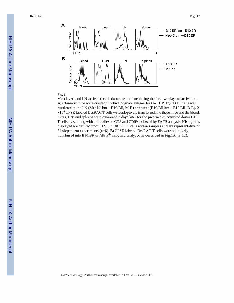
Fig. 1.Most liver- and LN-activated cells do not recirculate during the first two days of activation.A) Chimeric mice were created in which cognate antigen for the TCR Tg CD8 T cells wasrestricted to the LN (Met-Kb bm→B10.BR, M-B) or absent (B10.BR bm→B10.BR, B-B). 2×10⁶ CFSE-labeled DesRAG T cells were adoptively transferred into these mice and the blood,livers, LNs and spleens were examined 2 days later for the presence of activated donor CD8T cells by staining with antibodies to CD8 and CD69 followed by FACS analysis. Histogramsdisplayed are derived from CFSE+CD8+PI− T cells within samples and are representative of2 independent experiments (n=6). B) CFSE-labeled DesRAG T cells were adoptivelytransferred into B10.BR or Alb-Kb mice and analyzed as described in Fig.1A (n=12).
Holz et al. Page 12
Gastroenterology. Author manuscript; available in PMC 2010 October 17.
NIH
-PA Author Manuscript
NIH
-PA Author Manuscript
NIH
-PA Author Manuscript

Fig. 2.Hepatocyte-activated CD8 T cells display a unique CD25low CD54low phenotype. 2 ×10⁶
CFSE-labeled DesRAG T cells were adoptively transferred into B10.BR, Met-Kb or Alb-Kb
mice and analyzed as for Fig.1A. Histograms displayed are derived from CFSE+CD8+PI−cells in all samples.
Holz et al. Page 13
Gastroenterology. Author manuscript; available in PMC 2010 October 17.
NIH
-PA Author Manuscript
NIH
-PA Author Manuscript
NIH
-PA Author Manuscript

Fig. 3.Hepatocyte-activated CD8 T cells exhibit defects in cytotoxicity and cytokine production. A)106 DesRAG T cells were adoptively transferred into Met-Kb, Alb-Kb or B10.BR mice and anin vivo CTL assay was performed at day 4. Histograms (left panel) displayed are gated on CFSE+PI− lymphocytes within samples. Results displayed in the right panel represent the means oftriplicate samples ± S.E.M in 3 independent experiments. B) and C) CFSE-labeled DesRAGT cells were injected into the tail veins of B10.BR or Met-Kb mice. After 2 days, livers andLN were harvested, lymphocytes isolated, stimulated ex vivo, stained and analyzed forintracellular IFN-γ (B) or intracellular IL-2 (C) by flow cytometry. Histograms arerepresentative of 3 individual experiments performed in triplicate and gated on CFSE+CD8+cells in all samples.
Holz et al. Page 14
Gastroenterology. Author manuscript; available in PMC 2010 October 17.
NIH
-PA Author Manuscript
NIH
-PA Author Manuscript
NIH
-PA Author Manuscript

Fig. 4.Hepatocyte-activated CD8 cells express excess Bim and are abnormally prone to apoptosis.CFSE-labeled DesRAG T cells were adoptively transferred into Met-Kb, Alb-Kb or B10.BRmice. Livers and LN were harvested after 2 (A) or 3 days (B and C) and lymphocytes isolated.Cells were stained with anti-CD8 and Desiré antibodies and CFSE+CD8+ Des−TCR+ cellswere gated and analyzed by FACS to detect intracellular Bcl-xL, Bcl-2, Bim (A), the activatedform of caspase-3 (B) or cell surface Annexin V (C). Except for C), histograms displayed arerepresentative of 3 individual experiments performed in triplicate.
Holz et al. Page 15
Gastroenterology. Author manuscript; available in PMC 2010 October 17.
NIH
-PA Author Manuscript
NIH
-PA Author Manuscript
NIH
-PA Author Manuscript

Fig. 5.Loss of Bim promotes accumulation of Des-TCR CD8 T cells activated in the liver but doesnot enhance immunopathology. For all experiments 1.5 ×10⁷ Des-TCR or BimDes T cells wereinjected into the tail veins of B10.BR or Alb-Kb mice. Met-Kb mice were used as positivecontrols in some experiments. (A) Livers of recipient mice were harvested at day 6 and CTLactivity was measured in vitro. (B) Organs were harvested after 2 days and assessed for cytokineexpression as for Figure 3B. (C) Organs were harvested after 15 days, leukocytes isolated,counted and stained with anti-CD8 and Desiré antibodies. The percentages of Des+CD8+PI−cells was determined by FACS and the mean ± S.E.M of total numbers of CD8+Des+ T cellsfrom 2 independent experiments are shown (n=8). (D) Serum ALT levels were measured andplotted against time for Alb-Kb and B10.BR recipient mice. Day 5 results for all recipient linesare shown as histograms. The mean ± S.E.M from 2 independent experiments is shown (n=8).(E) LN and spleens from recipient mice in Fig.5C were harvested at day 15. Lymphocytes wereisolated, CFSE labeled and cultured for 2 days with irradiated C57BL/6 or B10.BR splenocytes.CFSE profiles were analyzed by FACS 48 h later and representative histograms from 2independent experiments are shown.
Holz et al. Page 16
Gastroenterology. Author manuscript; available in PMC 2010 October 17.
NIH
-PA Author Manuscript
NIH
-PA Author Manuscript
NIH
-PA Author Manuscript