Yield and Protein Response of Wheat Cultivars to Polymer-Coated Urea and Urea
Upload
independentCategory
view
2download
0
Urea Amidolyase (DUR1,2) Contributes to Virulence andKidney Pathogenesis of Candida albicansDhammika H. M. L. P. Navarathna1, Michail S. Lionakis2, Martin J. Lizak3, Jeeva Munasinghe3,
Kenneth W. Nickerson4, David D. Roberts1*
1 Laboratory of Pathology, Center for Cancer Research, National Cancer Institute, National Institutes of Health, Bethesda, Maryland, United States of America, 2 Fungal
Pathogenesis Unit, Laboratory of Clinical Infectious Diseases, National Institute of Allergy and Infectious Diseases, National Institutes of Health, Bethesda, Maryland, United
States of America, 3 National Institute of Neurological Disorders and Stroke, National Institutes of Health, Bethesda, Maryland, United States of America, 4 School of
Biological Sciences, University of Nebraska, Lincoln, Nebraska, United States of America
Abstract
The intracellular enzyme urea amidolyase (Dur1,2p) enables C. albicans to utilize urea as a sole nitrogen source. Becausedeletion of the DUR1,2 gene reduces survival of C. albicans co-cultured with a murine macrophage cell line, we investigatedthe role of Dur1,2p in pathogenesis using a mouse model of disseminated candidiasis. A dur1,2D/dur1,2D strain wassignificantly less virulent than the wild-type strain, showing significantly higher survival rate, better renal function, anddecreased and less sustained fungal colonization in kidney and brain. Complementation of the mutant restored virulence.DUR1,2 deletion resulted in a milder host inflammatory reaction. Immunohistochemistry, flow cytometry, and magneticresonance imaging showed decreased phagocytic infiltration into infected kidneys. Systemic cytokine levels of wild-typemice infected with the dur1,2 mutant showed a more balanced systemic pro-inflammatory cytokine response. Host geneexpression and protein analysis in infected kidneys revealed parallel changes in the local immune response. Significantdifferences were observed in the kidney IL-1 inflammatory pathway, IL-15 signaling, MAP kinase signaling, and thealternative complement pathway. We conclude that Dur1,2p is important for kidney colonization during disseminatedcandidiasis and contributes to an unbalanced host inflammatory response and subsequent renal failure. Therefore, thisCandida-specific enzyme may represent a useful drug target to protect the host from kidney damage associated withdisseminated candidiasis.
Citation: Navarathna DHMLP, Lionakis MS, Lizak MJ, Munasinghe J, Nickerson KW, et al. (2012) Urea Amidolyase (DUR1,2) Contributes to Virulence and KidneyPathogenesis of Candida albicans. PLoS ONE 7(10): e48475. doi:10.1371/journal.pone.0048475
Editor: Neeraj Chauhan, New Jersey Medical School, University of Medicine and Dentistry of New Jersey, United States of America
Received July 12, 2012; Accepted September 26, 2012; Published October 29, 2012
This is an open-access article, free of all copyright, and may be freely reproduced, distributed, transmitted, modified, built upon, or otherwise used by anyone forany lawful purpose. The work is made available under the Creative Commons CC0 public domain dedication.
Funding: This work was supported by the Intramural Research Program of the NIH, National Cancer Institute, Center for Cancer Research (DDR), the University ofNebraska Tobacco Settlement Biomedical Research Enhancement Fund (KWN), the John C. and Nettie V. David Memorial Trust Fund (KWN), Ann L. Kelsall and theFarnesol and Candida albicans Research Fund, University of Nebraska Foundation (KWN). The funders had no role in study design, data collection and analysis,decision to publish, or preparation of the manuscript.
Competing Interests: The authors have declared that no competing interests exist.
* E-mail: [email protected]
Introduction
Candida albicans is the most prevalent fungal pathogen of
humans. It can be isolated from ca. 30% of patients in intensive
care units [1], and patients with disseminated candidiasis have
mortality rates of 30–40% [2]. C. albicans is a formidable
opportunistic pathogen in part due to its metabolic and
morphological flexibility, ability to adapt to different locations
within the body, and rapid shifting from commensal colonization
of the gastrointestinal tract to being an invasive pathogen [3,4].
Investigation of fungal virulence factors has identified important
roles for genes that regulate yeast to hypha switching, phenotypic
switching from white to opaque cells associated with mating,
biofilm formation, adhesion towards epithelial cells, and a variety
of extracellular enzymes.
Our laboratories have focused on identifying novel virulence
factors including farnesol [5], heme oxygenase [6], and urea
metabolism via urea amidolyase [7]. Urea is an end product of
human nitrogen metabolism, and thus provides a nitrogen source
that C. albicans can exploit without competing with its host for
nutrients. Urea catabolism is known to contribute to virulence of
bacterial pathogens such as Helicobacter pylori [8] and Proteus mirabilis
[9]. Urea degradation is also exploited by some fungal pathogens.
The metalloenzyme urease that hydrolyses urea is a well known
virulence factor for Cryptococcus neoformans. [10] and enhances
pathogenesis of Coccidioidal infections [11]. Deletion of urease
significantly reduced brain infection and pathology of mice and
significantly increased host survival [12]. Therefore, it is pertinent
to investigate urea catabolism and virulence of C. albicans.
The higher fungi exhibit a dichotomy with regard to urea
utilization. Like all hemiascomycetes (yeasts), C. albicans uses the
energy requiring bifunctional enzyme urea amidolyase (Dur1,2p),
whereas other higher fungi use the nickel-containing urease [13].
The enzyme urea amidolyase, encoded by DUR1,2 (Degradation
of URea), was first characterized in the yeast Candida utilis [14].
This cytoplasmic, biotin dependent enzyme consists of a single
protein chain with domains for both urea carboxylase and
allophanate hydrolase activity. We made a dur1,2D/dur1,2Dmutant (KWN6) and the complemented strain dur1,2D::
DUR1,2/dur1,2::DUR1,2 (KWN8) [15]. The mutant strain
KWN6 was unable to use urea as a nitrogen source and unable
to escape from macrophages [15]. Dur1,2p is a cytoplasmic
PLOS ONE | www.plosone.org 1 October 2012 | Volume 7 | Issue 10 | e48475

enzyme and gains access to urea primarily via the transporter
Dur3, encoded by orf 19.781, which is co-regulated with Dur1,2p
[7]. Examination of the evolutionary origins of urea amidolyase
by comparing 64 fungal genomes and 56 bacterial genomes
revealed that the urea amidolyase genes currently found in fungi
likely are the result of a horizontal gene transfer event ca. 400
mya from the beta- or gamma- proteobacteria [16].
The present paper compares the C. albicans dur1,2D/dur1,2Dmutant (KWN6) and reconstituted strains with their wild type
parent A72 with regard to virulence in mice, fungal burden,
inflammatory cell infiltration into infected kidneys, and triggering
of local and systemic host inflammatory gene expression. Infection
with KWN6 without urea amidolyase led to moderate kidney
colonization, less inflammation in the kidneys, improved kidney
function, and better survival of the host. Identification of urea
amidolyase as a virulence factor for C. albicans further implies that
the distribution of urea within the body influences the organ
tropism of C. albicans. In addition, Dur1,2p expression alters local
inflammatory responses leading to clearance of the respective
organs and local and systemic cytokine responses that regulate host
immunity against C. albicans.
Materials and Methods
Ethics StatementExperimental protocols, housing, and care of mice were
conducted in an AAALAC approved facility according to animal
study protocol LP-022 approved by the National Cancer Institute
Animal Care and Use Committee.
Strains and growth conditionsThe C. albicans strains used for this study are A72 WT strain
(ATCC MYA-2430) KWN6 (dur1,2D/dur1,2D), KWN7 (dur1,2D/
dur,12D::DUR1,2), and KWN8 (dur1,2D::DUR1,2/dur,12D::-
DUR1,2) [15]. For challenge of mice, C. albicans cells were grown
overnight in 50 mL of Yeast Peptone Dextrose (YPD) medium at
30uC with aeration as previously described [17]. Cells were
harvested by centrifugation at 5000 rpm for 10 min, washed once
with 50 mL of sterile, non-pyrogenic normal saline (Quality
Biological Inc. Gaithersburg, MD) and resuspended in 10 mL of
saline before quantifying cell numbers using a Petroff-Hausser
counting chamber. The cell suspensions were adjusted to the final
concentration for parenteral administration using non-pyrogenic
sterile saline.
Mouse inoculation with C. albicansSix to eight week-old (18–20 g) BALB/c female mice (Charles
River Laboratories, Wilmington, MA) were randomly allocated to
groups of five animals. Each group of mice was inoculated
intravenously in the lateral caudal tail vein using a 27 gauge needle
with a volume of 0.1 ml containing 106 C. albicans cells [5,17].
Clinical signs of illness in each mouse were evaluated three times
daily, and mice that displayed severe signs were euthanized
immediately by placing them in a closed chamber filled with CO2
and processed for complete necropsy and collection of tissues for
histopathological examination. To longitudinally monitor effects of
C. albicans DUR1,2 on serum cytokines and chemokines, organ
burden and host immune responses, mice were euthanized
sequentially from 6 h to 240 h post-inoculation (PI). A total of
40 mice were inoculated with A72, 40 were inoculated with
KWN6, and 10 control mice received no fungal challenge. Three
animals from each group were sacrificed at 6, 12, and 24 h and
then every other day until seven day PI for histopathology and
cytokine assays. The 10 control animals, i.e., untreated and
uninfected, were sacrificed, and the organs and sera were
collected. The mean results for these 10 control animals were
used as time zero values. Sera separated from the blood collected
from individual mice were stored at 280uC until analysis.
To reproduce the CFU study, histopathology, IHC, and flow
cytometry, five mice per group were infected with C. albicans and
euthanized at specific time periods for sample collection. Mice of
3 days PI were used For MRI studies to image phagocytic cell
infiltration and conduct histopathology and IHC. Ultra-small
particles of iron oxide (USPIO) were injected 24 h before MRI.
Clinical signs of illness were monitored four times daily whilst
subjected to MRI procedures.
To analyze renal function and validate NanoString gene
expression data using qRT-PCR, two groups of mice were
infected with WT (A72) and KWN6 (dur1,2D/dur1,2D). Non infected
mice were used as negative controls. At 3 days PI the mice were euthanized,
and blood was collected for serum kidney function markers analysis and organs
for RNA extraction.
For analysis of local inflammatory gene expression, two groups
of mice consisting of 5 mice per group were infected with A72
wild-type and KWN6. Mice were euthanized at 3 day PI, and
kidneys were harvested for RNA extraction.
14C-Urea uptakeC. albicans cells were grown overnight in 50 ml of YPD, washed
and resuspended in PBS, and used as the inocula (0.2 OD) for
fresh cultures then grown in GPP (glucose phosphate proline
medium) at 30uC for 3–4 h while shaking at 150 rpm. Cell
numbers were counted and adjusted so that all uptake assays
employed an equal number of cells. Assays were done in 5 ml of
GP (glucose phosphate) buffer in 14 ml BD tubes (BD Biosciences,
NJ) with or without 5 mM sodium azide, using ca. 0.56108 cells
and 1 mCi per 1.15 mg of 14C-urea (American Radiolabeled
Chemicals, St. Louis, MO) per ml (19.2 mM urea). Cells
incubating with 14C-urea were shaken at 150 rpm, and duplicate
0.2 ml samples were collected at 5, 15, 30, 60, and 120 min. A
0.2 ml sample was overlaid on 100 ml of 30% sucrose in 0.4 ml
microfuge tubes without caps and centrifuged for 30 s, repeated
with the second 0.2 ml sample, and the top 0.3 ml was aspirated
off to remove unbound radioactive urea, whereupon 300 ml of
PBS was added, and the cells were centrifuged again before
aspirating all liquids leaving the yeast pellet. The bottom piece of
the microfuge tube was cut off, added to scintillation fluid, mixed
by shaking, and radioactivity was quantified by scintillation
counting.
Organ burden quantificationThree mice from each group were euthanized at day 1, 3, 5 PI
to determine the fungal burden in their kidneys. At the time of
necropsy, kidneys were harvested from each mouse and placed in
sterile Eppendorf tubes. The tissues were kept at 4uC until the next
day, when each kidney was weighed and homogenized in 1.0 ml of
nonpyrogenic sterile saline. Then, 10 fold serial dilutions of 1022,
1024 and 1026 in 0.1 ml of the homogenates were spread on
triplicate plates of Nickerson’s medium, also known as BiGGY
agar, a selective and differential medium for C. albicans [18]. After
48 h of incubation at 30uC, colony number, morphology, and
color were recorded, and numbers of CFU per kidney were
estimated. C. albicans appears as brown to black colonies with no
pigment diffusion and no sheen [18].
Necropsy and HistopathologyImmediately after euthanasia, macroscopic changes were
recorded, and the brain, heart, lungs, liver, spleen, and right
Candida albicans DUR1,2 Enhances Virulence
PLOS ONE | www.plosone.org 2 October 2012 | Volume 7 | Issue 10 | e48475

kidneys were immersed in buffered 10% formalin, processed for
paraffin embedding, sectioned at 5 mm, and stained with
haematoxylin and eosin (H&E). Grocott’s modification of
Gomori’s methenamine-silver (GMS) stain was used for detection
of fungi in situ [19]. Histopathology images from sections of
formalin-fixed and paraffin-embedded tissues stained with Go-
mori’s methenamine-silver or H&E were obtained using a light
microscope (Olympus BX51) fitted with a digital camera (Nikon
DXM1200F) and ScanScope XT digital scanner (Aperio). Images
were processed with Adobe Photoshop and Aperio ImageScope
v11.1.2.760 (Aperio).
ImmunohistochemistrySlides were deparaffinized in xylene (thrice for 10 min) and
rehydrated in graded alcohol (100%, 95%, and 70%). Antigen
retrieval was performed in a jar containing Target Retrieval
Solution (pH 6.10; Dako Corp.) for 20 min, followed by cooling at
room temperature for 20 min, and then washed with PBS twice
for 10 min. Endogenous peroxidase activity was quenched by
incubating with peroxidase block (pretreatment of the tissue
section with hydrogen peroxide prior to incubation of primary
antibody) for 30 min to avoid Endogenous peroxidase activity.
After washing the slides with Wash Buffer Solution (Dako),
nonspecific binding was reduced using Protein Block Serum-Free
(Dako) for 10 min. The slides were incubated with anti iNOS
antibody (1:50, overnight at 4uC, ab15323). Slides were then
incubated with streptavidin-biotin (Dako LSBA+ kit, horseradish
peroxidase). 3,39-Diaminobenzidine (Dako) was used as chromo-
gen for 5 min, and hematoxylin was used for counterstaining.
Negative control slides omitted the primary antibody. iNOS was
located predominantly within the cells. Nuclei were negative. The
intensity of the staining was evaluated using light microscopy and
Adobe Photoshop.
MRI proceduresMice were anesthetized in an induction chamber with a 30%
oxygen/70% nitrogen (oxygen-enhanced air) gas mixture contain-
ing 5% isoflurane. After anesthesia was induced (indicated by loss
of righting reflex, decreased respiratory rate and non-responsive to
toe pinch), isoflurane was reduced to 1.5–2%, and the animals
were maintained via a nose cone.
For MR imaging of the region of interest, the body of the
anesthetized mouse was restrained in a plastic holder with either
vet wrap or tape. Care was taken to allow for chest expansion to
facilitate breathing. A respiratory sensor pillow was placed under
the mouse or attached above to monitor respiratory rate and
pattern. MRI may be performed for up to 3 hours; however a
typical anatomical scan was approximately 1 hour or less.
Macrophage infiltration of the kidneys was observed using ultra
small particles of iron oxide (USPIO). Using a gradient echo
sequence (TR/TE = 200/10 ms, FOV = 50632 mm, matrix
= 2566256, 4 averages) T2*-weighted images of the kidneys were
acquired 3 days post inoculation, 24 hours following i.v admin-
istration of a USPIO contrast agent Molday IONTM (Bio PAL,
MA) 0.1 ml suspended in 10 ml of saline(0.2 mmol of iron per kg
body weight).
Kidney inflammatory cell quantification by flowcytometry
To quantitatively define the inflammatory cell infiltration in the
infected kidneys, the animals were perfused with normal saline and
organs were harvested. Single-cell suspensions were obtained, and
flow cytometry analysis was performed as reported previously [20].
Briefly, to obtain kidney single-cell suspensions, the organs were
finely minced and digested at 37uC in digestion solution
containing 0.4 mg/ml of Liberace CITL and 300 U/ml of grade
II DNAse I for 20 min with intermittent shaking. Digested tissue
was passed through a 70 mm filter, washed, and the remaining red
cells were lysed with ACK lysing buffer for 30 s. Then, the cells
were passed through a 40 mm filter, washed, and suspended in
8 ml of 40% Percoll. The suspension was overlaid on 3 ml of 70%
Percoll and centrifuged at 2,000 rpm for 30 min at RT. The
leukocytes at the interphase were isolated, washed three times in
FACS buffer, suspended in PBS, and passed through a 40 mm
filter. Suspensions of 100 ml were fixed with 100 ml of 2%
paraformaldehyde (USB, Cleveland, Ohio, USA) for leukocyte
quantification using PE-conjugated fluorescent particles (Spher-
otech, Lake Forest, Ill., USA). The cells were stained with a Live/
Dead fluorescent dye (Invitrogen, Carlsbad, Calif., California,
USA) for 10 min (1:500) and then incubated with rat anti-mouse
CD16/32 (2.4G2; BD Biosciences, San Jose, California, USA) for
10 min (1:100) at 4uC to block Fc receptors. Then, cells were
incubated at 4uC for 30 min with the following antibodies: FITC-
conjugated anti-mouse Ly6c (AL-21, BD Biosciences) and CD19
(1D3; eBioscience, San Diego, California, USA); eFluorH 450-
conjugated anti-mouse MHC II (I-A/I-E; M5/114.15.2) and CD3
(17A2); eBioscience,); PE-conjugated anti-mouse Ly6G (1A8) and
NK1.1 (PK136; BD Biosciences), and MHC II (M5/114.15.2;
eBioscience, San Diego, Calif., USA); PE-Cy7-conjugated anti-
mouse CD45 (Ly-5; BD Biosciences), CD8 (53-6.7), and F4/80
(BM8; eBioscience); APC-conjugated anti-mouse CD45 (Ly-5),)
and CD11c (HL3; BD Biosciences), and CD19 (1D3; eBioscience);
APC-Cy7-conjugated anti-mouse CD11b (M1/70) and CD3
(17A2; CD45 (Ly-5; BD Biosciences), and APC-fluor-780-conju-
gated anti-mouse CD4(RM4–5; eBioscience). After three washes
with FACS buffer, cells were fixed with 2% paraformaldehyde.
FACS was performed on an LSRII (BD Biosciences), and data
were analyzed using FlowJo software (version 8.8.4; Treestar,
Ashland, Oreg., USA). Quantification of the leukocyte subsets in
the infected kidneys was performed using PE-conjugated fluores-
cent particles (Spherotech, Lake Forest, Ill., USA) as previously
described [20].
Determination of serum cytokines and kidney proteinmarkers
Murine serum was collected from sacrificed mice at various time
points following infection with wild- type and mutant C. albicans
strains. A Luminex -bead array (Mouse cytokine/Chemokine
LINCOplex Kit, catalog no. 551287, Linco Research, Inc. St
Charles MO) was used for detection of the cytokines IL-1a, IL-4,
IL-6, IL-10, IL12, IL17, and IFN-c, and TNF- a, and G-CSF
according to the manufacturer’s specifications. IL-7, IL-15, MIP-
1a, MIP1b, and MIP-2 levels of kidney extracts and serum were
analysed using the Luminex bead array Milliplex MAP Kit
(catalog no MPXMCYTO-70K, Millipore, Billerica, MA).
NanoString gene expression analysisExpression of specific mRNAs in total kidney RNA was
analyzed using the NanoString methodology as previously
reported [21], and was conducted at the DNA sequencing core
facility of NIH. Briefly, 100 ng of total RNA per kidney were
hybridized to the target specific mouse inflammatory gene Codeset
at 65uC.
The Codeset contained probes against a panel of 179 genes
encoding proteins involved in mouse inflammation and six internal
reference genes and were used to analyze local inflammatory
response in kidneys. The hybridized reactions were loaded onto
Candida albicans DUR1,2 Enhances Virulence
PLOS ONE | www.plosone.org 3 October 2012 | Volume 7 | Issue 10 | e48475

the NanoString Prep station, which removes excess reporter, binds
the reporter to the cartridge surface, and stretches the probes for
scanning. Subsequently, the cartridges were loaded onto the
NanoString Digital Analyzer and scanned.
An Excel-based method described by the manufacturer was
used to perform normalization to six internal controls and basic
statistical analysis on the data. The normalized results are
expressed as the relative mRNA level, and values for kidneys
infected with WT and DUR1,2 deleted strain were averaged and
are presented as mean 6 s.d. Statistical significance was calculated
using Student’s t-test and was set as p,0.05. Genes that were
statistically significant and showed .2.5-fold higher or lower
expression are listed in the Table 1. Using MetaCoreTM pathway
analysis software from Genego [21] up- and down-regulated genes
clusters were analyzed for association with specific signaling
pathways and direct interactions.
RNA Extraction and Gene expression analysis by RT-PCRRNA isolation was done using a standard hot phenol procedure
[22]. Reverse transcription was conducted using 5 mg of total
RNA extracted from each sample using Superscript III reverse
transcriptase according to the manufacturer’s instructions for
oligo-dT priming (Invitrogen). Quantitative PCR was conducted
as previously described [6] using Absolute QPCR SYBR Green
Mix (Thermo Scientific), Opticon I instrument, and Opticon I
software (BioRad). Samples were analyzed by PCR in triplicate
and normalized to internal CDC36 mRNA levels. Melting curve
analysis was performed to assure a single product was produced in
each reaction [6]. The qPCR primers used in this study are
designed using Primer3 software [23] and listed in Table S1, and
all products were 100–150 bases in length.
StatisticsThe probability of survival as a function of time was determined
by the Kaplan-Meier method, and significance was determined by
the log-rank (Mantel-Cox) test and Jehan-Breslow-Wilcoxon test
using GraphPad Prism software. The mixed procedure of the SAS
system [24] was used to analyze serum cytokine expression
patterns among all treatment groups at various time points. Three
randomly selected mice from each group were euthanized at each
time point for longitudinal comparisons. Data were analyzed for
significant differences by comparing means of each triplicate
reading at various time points assuming that the cytokine
expression levels within each group of mice are normally
distributed [25]. Fungal burden and 14C urea absorbance were
analyzed using Student’s t test. qRT-PCR gene expression data
were analyzed by one-way ANOVAs using GraphPad Prism
software.
Results
Validation of the urea amidolyase null mutantThe null strain KWN6 was constructed from A72, a wild type
Candida albicans, as part of our study of arginine and its metabolite
urea in the escape of C. albicans from the mouse macrophage cell
line RAW264.7 [15]. KWN6 could not use urea as the sole source
of nitrogen, could not make germ tubes in response to either
arginine or urea, and could not escape from the mouse
macrophage cells. All of these abilities were restored in the
reconstituted strain KWN8. In contrast, KWN6 grew as well as its
parent on all other complex and defined media tested [15].
Collectively, these observations indicated that Dur1,2p is neces-
sary for urea metabolism in C. albicans [15]. Further confirmation
that urea amidolyase activity had been eliminated from KWN6
Table 1. Inflammatory gene expression in infected mouse kidneys by NanoString analysis.
Gene p value F C gene p value F C Gene p value F C
Nos2* 0.0153 54.2 Il10rb 0.0002 22.6 Rps6ka5 0.0249 25.1
Ccl3* 0.0000 42.1 Keap1 0.0024 22.6 Kng1 0.0034 25.2
Il1rn 0.0205 37.1 Hmgn1 0.0004 22.7 Prkca 0.0019 26.1
Fos* 0.0454 26.6 Nfe2l2 0.0003 22.8 Il7 0.0046 26.5
Maff 0.0000 24.2 Mknk1 0.0004 22.8 Traf2* 0.0225 27.2
Ccl4 0.0050 18.2 Mapkapk5 0.0031 23.0 Mef2a 0.0292 27.6
Il18rap 0.0002 17.9 Map3k7 0.0023 23.0 Plcb1 0.0155 27.6
Il1a 0.0120 15.8 Tlr3 0.0152 23.0 Fxyd2 0.0437 27.7
Tnf 0.0004 14.5 Map2k4 0.0022 23.2 Nfatc3 0.0054 28.0
Il1b 0.0096 14.1 Nr3c1 0.0196 23.4 Ly96 0.0005 28.6
Itgb2 0.0446 12.6 Raf1 0.0100 23.5 Map2k6* 0.0160 29.7
Cebpb 0.0151 9.1 Gnaq 0.0009 23.6 Il15* 0.0260 213.5
Il8rb 0.0413 9.0 Map3k5 0.0048 24.1 C8a* 0.0234 2193.2
Hspb1 0.0341 8.9 C2 0.0007 24.2
Tlr2 0.0008 6.5 Creb1 0.0217 24.3
C3 0.0097 5.8 Lta 0.0035 24.3
Limk1 0.0178 3.9 Mef2c 0.0122 24.3
Cxcr4 0.0095 2.8 Tgfb2 0.0328 24.7
Tgfb1 0.0055 2.7 Stat1 0.0011 24.9
F C – fold change in mRNA expression in kidneys of mice 3 days after infection with A72 wild type versus the dur1,2D strain KWN6. * indicates selected genes validatedwith qRT-PCR (Figure S1).doi:10.1371/journal.pone.0048475.t001
Candida albicans DUR1,2 Enhances Virulence
PLOS ONE | www.plosone.org 4 October 2012 | Volume 7 | Issue 10 | e48475

was provided by assessing 14C-urea uptake by the respective strains
(Fig. 1A). The absence of Dur1,2p in KWN6 reduces but does not
eliminate induction of the major urea transporter Dur3p in the
presence of urea [7]. Despite its relative deficit in urea transport
activity, cellular accumulation of urea via Dur3p or other putative
urea transporters was ca. 20- to 40-fold greater for KWN6 than for
the A72 parent (Fig. 1A). Thus, intracellular urea accumulation is
primarily limited by its catabolism by Dur1,2p. Urea uptake in the
reconstituted KWN8 returned to levels close to those of the wild
type parent, confirming that urea metabolism is restored in this
strain (Fig. 1A). As observed for S. cerevisiae [26], urea transport is
an energy dependent process, and the elevated intracellular urea
accumulation in KWN6 was prevented in the presence of sodium
azide (Fig. 1A). These experiments confirm the physiological
function of urea amidolyase in vitro and show that energy
dependent urea transport remains active in the urea amidolyase
null mutant.
DUR1,2 deletion reduces virulence in miceThe well-established mouse model of disseminated candidiasis
was used to compare virulence among the WT and manipulated
strains. Mice infected with the dur1,2D/dur1,2D strain (KWN6)
had a significantly higher survival rate compared with mice
infected with the WT A72 strain (n = 15, p,0.01, Fig. 1B). The
Gehan-Breslow-Wilcoxon test hazard ratio estimates indicated
3.4-times higher lethality for WT infection than KWN6.
Restoration of one copy (KWN7) or both copies of DUR1,2
(KWN8) restored virulence but not fully to the level of the WT
parent (Fig. 1B, P,0.01). However, by 7–8 weeks the survival of
mice infected with the A72, KWN7 and KWN8 strains did not
differ significantly.
Disruption of DUR1, 2 impairs kidney and braincolonization
Urea is present in serum at 70–150 mg/L [27], at high
concentrations in the kidneys and brain [28,29], and much lower
concentrations in other organs [30]. If C. albicans uses urea as a
nitrogen source in vivo, the virulence defect observed in Fig 1B
could arise from Dur1,2p-dependent colonization of organs
containing high levels of urea. Consistent with this hypothesis,
the fungal burdens for A72 and KWN6 were dramatically
different in the mouse kidneys (Fig. 2A left) and brain (Fig. 2A
right). Analysis of fungal CFU at 3 days PI showed 15-fold
(p,0.01) and 12-fold (p,0.001) lower CFU in kidneys and brains,
respectively, of mice injected with the urea amidolyase null mutant
(Fig. 2A).
CFU values can underestimate organ burden for filamentous
fungi, so we used GMS staining to confirm the role of urea
catabolism in determining the fungal burden (Fig. 2B). Coloniza-
tion was reduced at all time points in mice infected with the urea
amidolyase null mutant KWN6. Differences were observed in both
kidneys and brain (Fig. 2B). At 1 day PI, C. albicans colonies were
scattered throughout the cortex of kidneys infected with wild type
C. albicans, while the KWN6 infected group showed minimal
colonization (Fig. 2B). By day 3 PI, WT infected kidneys showed
extensive fungal invasion (Fig. 2B left), as did the brain tissue
(Fig. 2B right). In both cases the WT C. albicans had a higher fungal
level compared with the mutant infection. Hyphal filaments were
more abundant in kidneys infected with WT, but the mutant did
show limited filamentous growth in the kidney.
Longitudinal GMS staining was used to visualize the clearance
of fungi from the organs by the mouse immune system. Candida
colonization was reduced or eliminated in the brain and the kidney
cortex of KWN6 infected mice after 5 days, whereas the WT
infected mice failed to clear these colonizations (Fig. 2B). As
reported previously [20,31], Candida colonization during patho-
genesis gradually shifted from the cortex to the kidney pelvis
starting around day 3 PI, and by day 7–9 PI the WT C. albicans
formed large fungal balls in the pelvis, whereas the KWN6 strain
showed only mild colonization (data not shown). Beyond day 9 PI,
we did not observe Candida colonies in KWN6 infected mice
kidneys, whereas surviving WT infected mice maintained Candida
colonies at 17 days PI (data not shown). After initial colonization,
C. albicans is quickly cleared from liver, spleen and heart by the
Figure 1. Dur1,2p function in urea metabolism and virulence. (A). Urea uptake by WT (A72), dur1,2D/dur1,2D mutant and reconstituted C.albicans strain dur1,2D::DUR1,2/dur1,2D::DUR1,2. Radioactive uptake into 4256106 cells is presented after exposure to 14C urea for 5 and 15 min inthe absence or presence of sodium azide. Values shown are the average of triplicate experiments 6 SE. Closed bars indicate 5 min uptake, and openbars indicate 15 min uptake of radioactive urea. (B). Effect of DUR1,2 deletion on mouse mortality following intravenous infection. Survival of miceinjected with WT C. albicans A72 (N), the null mutant KWN6 (&), single copy reconstituted KWN7 (.), fully reconstituted KWN8 (m), and anuninfected control group of 5 mice (not shown) was assessed daily. Each infected group contained 15 mice.doi:10.1371/journal.pone.0048475.g001
Candida albicans DUR1,2 Enhances Virulence
PLOS ONE | www.plosone.org 5 October 2012 | Volume 7 | Issue 10 | e48475

mouse immune system, whereas kidney and brain are sites of
chronic colonization [20,49]. Our results suggest that colonization
in kidney and brain cannot be sustained in the absence of
Dur1,2p.
Disruption of DUR1,2 preserves kidney functionMice infected with WT C. albicans had significantly impaired
kidney function compared with either uninfected mice or KWN6
infected mice as determined by their elevated blood urea nitrogen
(BUN) and creatinine levels (Fig. 3A). The kidney damage
indicated by these markers was 10- and 5-fold greater, respec-
tively, in the WT parental strain than the urea amidolyase null
mutant (Fig 3A). WT infected mice at 3 day PI had BUN
= 210650 mg/dL vs KWN6 infected mice which had
2061.2 mg/dL (p,0.002), while the serum creatinine levels of
WT infected mice were 1.960.5 mg/dL vs KWN6 infected mice,
which had 0.460.2 mg/dL (p,0.01).
Dur1,2p regulates the kidney inflammatory responseConsistent with the greater fungal colonization of the kidneys at
day 3 PI (Fig. 2), PAS and H&E stained kidney sections at day 3 PI
showed that disruption of DUR1,2 decreased the inflammatory
response in infected kidneys at 3 days PI for mice infected with
KWN6 (Fig. 3B). No signs of inflammation were observed 6–12 h
PI, but by day 1 PI WT infected kidneys showed scattered foci of
inflammation with PMN and macrophages visible in the kidneys
(data not shown). At this time, only a few localized inflammatory
reactions were observed in mouse kidneys infected with KWN6.
After day 3 PI, kidneys infected with both strains had multiple foci
of inflammation, with C. albicans colonies surrounded by PMN,
macrophages, and central necrosis. However, kidneys infected
with KWN6 consistently showed less fungal colonization and
inflammation than those with WT infection (Fig. 3B, PAS
staining). H&E staining confirmed that less pronounced inflam-
matory responses and tissue necrosis were associated with KWN6
infections (Fig 3B, H&E staining). By day 5 PI, WT infections
Figure 2. Dur1,2p expression promotes kidney and brain colonization. (A). Kidney and brain fungal burdens of mice infected with parentalA72 and mutant KWN6 strains. Gray bars represent mean CFU of WT infected left kidney homogenates determined by CFU on BiGGY agar from sixrepresentative serial dilutions from each kidney, representing three mice per time point infected. Checkered bars represent similar mean values formice administered with KWN6. (B) GMS-stained mouse kidney and brain tissues to determine C. albicans colonization. Representative kidney andbrain sections harvested day 1, 3 and 5 PI from mice infected with WT or KWN6 are shown. Scale bar indicates 100 mm.doi:10.1371/journal.pone.0048475.g002
Candida albicans DUR1,2 Enhances Virulence
PLOS ONE | www.plosone.org 6 October 2012 | Volume 7 | Issue 10 | e48475

Figure 3. Dur1,2p expression impairs kidney function and increases inflammation. A. Kidney function tests of mice infected with A72 (WT)vs KWN6 at day 3 PI. Open bars represent serum levels of BUN and creatinine in non-infected control mice. Checkered bars represent KWN6 infectedmice, and closed bars represent A72 (WT) infected mice. Note the levels of BUN and creatinine are significantly higher in WT infected sera comparedwith KWN6 infected mice. B. PAS and H&E-stained mouse kidney tissues to assess inflammatory responses and tissue necrosis caused by C. albicanscolonization. Representative sections of infected mouse kidneys (group of 3 mice) harvested at day 3 PI are shown. Scale bars indicate magnification.Arrows indicate regions of PMN and C. albicans accumulation in PAS stains and PMN inflammatory foci in H&E stains. Demarcated regions by whitesquares are shown under higher magnifications in the lower panels.doi:10.1371/journal.pone.0048475.g003
Candida albicans DUR1,2 Enhances Virulence
PLOS ONE | www.plosone.org 7 October 2012 | Volume 7 | Issue 10 | e48475

showed granulomatous kidney lesions with many macrophages
and some PMN surrounding the Candida colonies, whereas the
KWN6 infected kidneys showed better resolution of the initial
colonization (data not shown).
For the first day PI, no inflammation was observed in the brain
(data not shown), but by day 3 PI the brains of mice infected with
WT C. albicans showed inflammatory foci containing macrophages,
PMN, and Candida, whereas brains of mice infected with KWN6
showed fewer inflammatory lesions with less macrophages (data
not shown).
Deletion of DUR1,2 reduces neutrophil infiltration intoinfected kidneys
To quantify the local inflammatory response in infected kidneys,
we conducted FACS analysis of infiltrating leukocytes at day 3
(Figs. 4A–C) and 5 PI (data not shown). Examination of
neutrophils, monocytes, macrophages, dendritic cells, CD8+ T
cells, CD4+ T cells, and NK cells for their infiltration into kidneys
showed that at day 3 PI, mice infected with the urea amidolyase
null mutant had significantly lower neutrophil accumulation on
both a percentage basis and actual cell counts (6-fold, p,0.0001,
and 20-fold, p,0.001, respectively) than did kidneys infected with
the WT (Fig 4A and B). In contrast, KWN6 infected kidneys
contained a significantly higher percentage of macrophages
(p,0.05, Fig. 4C left). This is consistent with the previously
observed inability of KWN6 to kill murine macrophages [15].
However, absolute macrophage cell numbers were not signifi-
cantly different (Fig. 4C right). The levels of kidney colonization by
WT C. albicans and KWN6 were consistent with the data in Fig. 2A
in that kidneys infected with KWN6 had 12-fold lower CFU at 3
days PI (p,0.001, data not shown). These findings also confirmed
our histological analysis of fungal invasion by PAS staining and
inflammatory response by H&E staining (Fig. 3B). FACS and CFU
analyses at day 5 PI did not reveal a significant difference in
neutrophil infiltration (data not shown). However, the mutant
strain trended towards lower neutrophil accumulation and fungal
colonization. FACS analysis at day 5 PI showed that kidneys
infected with the mutant strain had a trend toward a higher
percentage (26.98% versus 11.32%) and greater absolute numbers
(0.5916106 versus 0.0996106 cells/kidney) of neutrophils com-
pared to kidneys infected with the wild-type strain (P = 0.13; data
not shown). The day five CFU data may be misleading because
histopathology showed considerably less kidney colonization by
KWN6 at day 5. This discrepancy may be explained by differences
in filamentation, breakage of which during homogenization may
lead to an underestimation of CFU in the WT infected kidneys.
Kidney damage assessed by magnetic resonanceimaging (MRI)
As a third method to confirm the differential inflammatory
responses to these two strains, we conducted an MRI analysis of
kidney function. T2* weighted MRI imaging was performed on
infected mice 3 days PI and 24 h after i.v. loading with ultra-small
particles of iron oxide (USPIO) as a contrast agent to label
phagocytes. Thus, more iron indicates more phagocytes which in
turn indicates greater inflammatory reactions in kidney. The top
panel of Fig. 4D shows representative MRI sections through the
kidneys of infected and non-infected mice, while the lower panels
show GMS and H&E stained sections of the same kidneys after
imaging. Kidneys of mice infected with the WT strain had
widespread phagocyte infiltration of the kidneys as shown by
visible perturbation of the MRI T2* signal (Fig. 4D top center
panel). GMS and H&E staining of sections from the same kidneys
confirmed heavy colonization by C. albicans (GMS) and massive
inflammatory reactions (H&E) accompanied by numerous PMNs
and macrophages. In mice infected with KWN6, the inflammatory
response was greatly reduced and more heterogeneous (Fig 4D
right panels). GMS and H&E staining of sections from KWN6
infected kidneys confirmed a reduced fungal burden and less
inflammation, indicated by white arrows. Thus, by three
independent methods, deletion of DUR1,2 resulted in a reduced
and more localized immune response to the infection.
Deletion of DUR1,2 alters host systemic cytokine andchemokine responses
To further characterize the inflammatory responses in infected
mice, we examined serum levels of 10 cytokines and 4 chemokines
that may participate in immunity against systemic candidiasis in
the mice (Fig. 5). Sera were analyzed pre-infection (day 0) and over
the same time frame used for the histopathology and fungal
burden analyses. Most dramatically, the proinflammatory cytokine
IL-6 was significantly higher in WT infected mice at days 1–5 PI,
showing serum increases of 500- to 1200-fold with p,0.01, 0.05
and 0.05, respectively. The importance of IL-6 to the immune
response to candidiasis has been noted previously [25,32]. Serum
TNFa levels of WT infected mice were also significantly elevated
by day 5 PI (150-fold, p,0.01) as were IL-4, IL 10 and Il-17,
which were elevated 18-, 20-, and 10-fold with p,0.05, 0.05 and
0.001, respectively. Similarly, levels of the chemokines MIP-1aand MIP-1b were elevated ca. 100-fold in WT infected mice at
day 3 PI with p,0.01. In addition, the pro-inflammatory cytokine
IL-1a was elevated significantly in both groups at day 1 PI as was
the chemokine G-CSF at all time points examined (Fig. 5).
Interestingly, by day 5 PI the KWN6 infected mice had
dramatically lower levels of G-CSF (p,0.05) compared with the
WT. In contrast, we did not observe significant changes in serum
levels for IL-7, IL-12, IL-15, IFNc, and MIP-2 (Fig. 5).
Deletion of DUR1,2 alters inflammatory gene expressionin kidneys
Based on the differences in kidney pathology observed during
the course of infection with KWN6 versus WT (Figs. 3B and 4D),
we examined local host inflammatory responses in infected kidneys
using NanoString gene expression profiling for 179 mouse
inflammatory genes. Three kidneys of each type were examined
at 3 days PI, and 51 genes were identified that showed more than
2.5-fold over- or under-expression with p,0.05. Of these, 19 were
over-expressed in WT infected kidneys compared with KWN6
infected kidneys, and 32 were down regulated (Table 1). The fold
change ranged from plus 54-fold for NOS2 to minus 193-fold for
C8a. We validated the magnitude of these changes using qRT-
PCR to quantify changes in mRNA abundance for 9 of the highly
up- and down-regulated genes, indicated by asterisks in Table 1
(Figure S1). Consistent with the observed differences in kidney
inflammatory responses indicated by H&E and PAS staining
(Fig 3), mRNA encoding the proinflammatory marker iNOS
(NOS2) showed the highest over-expression in kidneys infected
with WT versus the DUR1,2 deleted strain. This iNOS increase
was accompanied by a greater than 14-fold over-expression of
mRNAs for the pro-inflammatory mediators IL-1a, IL-1b, and
TNF-a. These changes are consistent with the 26-fold increase in
c-Fos, which is known to mediate IL-1 induction in an oral
epithelial C. albicans infection [33]. A 12-fold over-expression of the
leukocyte-specific integrin b2 is also consistent with the observed
differences in inflammatory cell infiltration. Two members of the
MIP chemokine family that play important roles in the recruit-
Candida albicans DUR1,2 Enhances Virulence
PLOS ONE | www.plosone.org 8 October 2012 | Volume 7 | Issue 10 | e48475

Figure 4. Dur1,2p expression increases inflammatory cell infiltration in infected kidneys. A. Representative FACS plots of kidneyneutrophils at day 3 PI with WT and KWN6. B Neutrophils expressed as percent of CD45+ cells (left panel) and as absolute numbers per kidney (right).C. Macrophages expressed as percent of CD45+ cells (left panel) and as absolute numbers per kidney (right). D. Top panels show transverse MRimages of 8 week old BALB/c mice 24 h post injection of USPIO agent. Images are representative of at least three non-infected mice per group (left
Candida albicans DUR1,2 Enhances Virulence
PLOS ONE | www.plosone.org 9 October 2012 | Volume 7 | Issue 10 | e48475

ment of phagocytes to sites of inflammation [34] were also
markedly increased. CCL3 (MIP1a) and CCL4 (MIP1b) were
increased 42- and 18-fold, respectively, in WT infected kidneys
(Table 1).
In a separate experiment, expression of the same set of genes in
KWN6 and A72 infected kidneys were compared with expression
in non-infected mouse kidneys by qRT-PCR (Table 2). All of the
altered gene expression patterns observed in the first experiment
(Table 1) except for CCL3 were confirmed to be significant
(p,0.05, Table 2).
Comparison of the inflammatory protein response ininfected kidneys
To determine whether the DUR1,2-dependent differences in
local inflammatory gene expression result in altered expression of
the respective proteins, we analyzed six representative proteins in
the kidney extracts (Fig. 6) using the same time points as used for
our initial screening. Consistent with the differences in respective
mRNA levels, at day 3 PI, we observed significantly up-regulated
Ccl3 (MIP1a), Ccl4 (MIP1b), and MIP-2 in kidneys infected with
WT relative to those infected with KWN6 (p,0.0001, 0.01, and
0.0001 respectively, Fig. 6A). Comparative IHC using a Nos2
antibody similarly showed that WT infected kidney lesions (Fig. 6B
left) had more iNOS specific staining than the KWN6 infected
kidneys (Fig. 6B right). In contrast, the decreases in IL-7 and IL-15
panels), Candida infected (middle panels), and infected with KWN6 strain (right panels). Small localized candida colonization by KWN6 is indicated byarrowheads. Massive phagocytic infiltration in kidneys infected with WT is demonstrated by T2* spoiled signal in the kidney cortex and medulla.Lower panels show GMS and H&E stained sections indicating colonization and inflammatory reactions in the respective kidneys imaged in the toppanels.doi:10.1371/journal.pone.0048475.g004
Figure 5. Effects of Dur1,2p expression in C. albicans on host serum cytokines and chemokines. Serum levels of the indicated proteinswere assessed at 1 to 5 days PI for mice infected iv with WT (gray bars) or KWN6 (open bars). Values at time 0 are mean values determined for serafrom 5 control mice, and other data are mean 6 standard deviation for 3 mice at each time point. * = p,0.05; ** = p,0.01; *** = p,0.001.doi:10.1371/journal.pone.0048475.g005
Table 2. Inflammatory gene expression in qRT-PCR.
GeneKWN6 infectedkidneys
A72 infectedkidneys p value F C
Nos2 0.860.4 12.665.5 0.04 16.1
Fos 7.863.8 52.8629.5 0.05 6.8
Ccl3 3.461.0 4.761.8 0.34 1.4
C8a 26.361.5 272.9643.3 0.05 211.5
MAP2k6 24.061.2 29.461.6 0.01 22.3
IL-15 24.061.4 27.860.1 0.01 22.0
Traf2 20.860.1 21.660.3 0.01 21.9
IL-7 21.160.2 20.560.9 0.01 20.4
Gene expression in kidneys of mice infected with KWN6 and A72 WT after 3days is presented as a fold change compared to normal uninfected kidneys.Results are the average of three mice per group with SE.doi:10.1371/journal.pone.0048475.t002
Candida albicans DUR1,2 Enhances Virulence
PLOS ONE | www.plosone.org 10 October 2012 | Volume 7 | Issue 10 | e48475
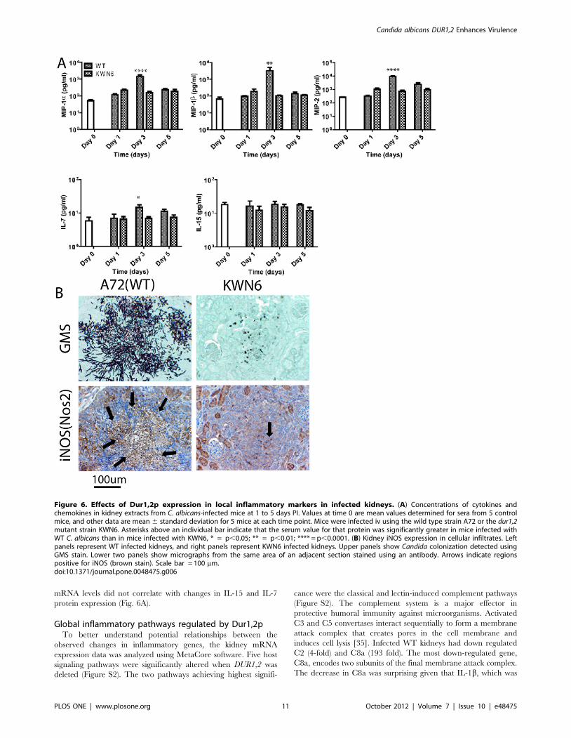
mRNA levels did not correlate with changes in IL-15 and IL-7
protein expression (Fig. 6A).
Global inflammatory pathways regulated by Dur1,2pTo better understand potential relationships between the
observed changes in inflammatory genes, the kidney mRNA
expression data was analyzed using MetaCore software. Five host
signaling pathways were significantly altered when DUR1,2 was
deleted (Figure S2). The two pathways achieving highest signifi-
cance were the classical and lectin-induced complement pathways
(Figure S2). The complement system is a major effector in
protective humoral immunity against microorganisms. Activated
C3 and C5 convertases interact sequentially to form a membrane
attack complex that creates pores in the cell membrane and
induces cell lysis [35]. Infected WT kidneys had down regulated
C2 (4-fold) and C8a (193 fold). The most down-regulated gene,
C8a, encodes two subunits of the final membrane attack complex.
The decrease in C8a was surprising given that IL-1b, which was
Figure 6. Effects of Dur1,2p expression in local inflammatory markers in infected kidneys. (A) Concentrations of cytokines andchemokines in kidney extracts from C. albicans-infected mice at 1 to 5 days PI. Values at time 0 are mean values determined for sera from 5 controlmice, and other data are mean 6 standard deviation for 5 mice at each time point. Mice were infected iv using the wild type strain A72 or the dur1,2mutant strain KWN6. Asterisks above an individual bar indicate that the serum value for that protein was significantly greater in mice infected withWT C. albicans than in mice infected with KWN6, * = p,0.05; ** = p,0.01; **** = p,0.0001. (B) Kidney iNOS expression in cellular infiltrates. Leftpanels represent WT infected kidneys, and right panels represent KWN6 infected kidneys. Upper panels show Candida colonization detected usingGMS stain. Lower two panels show micrographs from the same area of an adjacent section stained using an antibody. Arrows indicate regionspositive for iNOS (brown stain). Scale bar = 100 mm.doi:10.1371/journal.pone.0048475.g006
Candida albicans DUR1,2 Enhances Virulence
PLOS ONE | www.plosone.org 11 October 2012 | Volume 7 | Issue 10 | e48475

elevated in the same kidneys, induces C8a in other organs such as
liver [36]. These urea-dependent changes in complement function
in the kidneys may account for the known ability of WT C. albicans
to limit host humoral immunity [37].
The IL-15 pathway and several downstream genes involved in
this pathway including TRAF2, a signal transducer that associates
with the IL-15 receptor [38], were significantly down regulated in
mouse kidneys infected with WT strain (Table 1,2 and Figure S1).
IL-15 is a pleiotropic cytokine involved in proliferation, differen-
tiation, immune responses, and cell survival [39]. Dysregulation of
IL-15 is emerging as important for the pathogenesis of auto-
immune diseases and host immune responses to cancer [40], and
thus IL-15 may exert a protective role against candidiasis by
regulating T-cell differentiation, phagocytosis, neutrophil stimula-
tion, or monocyte migration.
The IL-1 signaling pathway also achieved significance. IL-1
stimulates a broad spectrum of immune and inflammatory
responses [41]. Kidneys of mice infected with the WT strain up-
regulated IL-1a (15 fold), IL-1b (14 fold), and the interleukin 1
receptor antagonist IL1RN (37-fold) relative to those infected with
the strain lacking DUR1,2. These changes are consistent with the
increased expression of the IL-1-regulated transcription factor
MAFF and inflammation in the WT infection [42]. Additional
elements of the IL-1 pathways including TNFa, TGFb and iNOS
were also significantly upregulated. TNFa and IL-1b are inducers
of Nos2 gene expression [43,44], which is consistent with the
observed changes in iNOS and protein and mRNA expression.
Although the high levels of NO produced by iNOS have potent
anti-microbial activity, Candida may be resistant to this host
defense due to expression of YHB1, which converts NO to
nontoxic nitrite [45].
The direct interaction algorithm identified interactions among 8
of the up-regulated genes that mediate pro-inflammatory respons-
es (Figure S3a). Similarly, 17 of the down regulated genes have
known direct interactions (Figure S3b).
Discussion
Our results show that deletion of DUR1,2 significantly reduces
virulence in a mouse model of disseminated candidiasis and that
virulence is restored when DUR1,2 is reconstituted. The decreased
pathogenicity of KWN6 (dur1,2D/dur1,2D) was evidenced by
decreased colonization of the brain and kidneys, decreased kidney
damage, and a decrease in the extent and type of the inflammatory
response mounted by the infected mice. In particular, cytokine and
chemokine concentrations in serum and kidneys were dramatically
altered. Taken together, these results indicate that DUR1,2 and the
encoded urea amidolyase act as a virulence factor for murine
disseminated candidiasis and potentially for other infections
caused by C. albicans. From a more global perspective, they
suggest an important role of urea availability in determining the
location and severity of C. albicans infections. Notably, the organs
in which C. albicans can establish persistent colonization are those
with the highest urea contents [27,28,29]. Urea in the kidney is
concentrated along the renal corticomedullary axis by the function
of several active urea transporters. In healthy kidneys urea is
further concentrated and sequestered in collecting ducts, but
fungal infection, tissue necrosis, and the host inflammatory
response may cause accumulation of urea [29], which further
enhances virulence.
In addition to providing nitrogen for growth, our data indicate
that urea metabolism by C. albicans has profound effects on the
host inflammatory and immune responses. In addition to its
established role in escape from macrophages, urea metabolism has
both local and systemic effects on the host immune response. The
long blood circulating time and progressive phagocytic uptake of
USPIO particles [46] enabled us to use MRI imaging as a tool to
non-invasively follow phagocyte recruitment and tissue inflamma-
tion in infected mice. These imaging results were validated by
histopathology, immunohistochemistry and expression of inflam-
matory cytokines and the activated phagocytic marker iNOS. The
50-fold increase in iNOS mRNA expression in WT C. albicans
infected kidneys could result from increased neutrophil or M1
macrophage infiltration [47].
Increased survival of mice infected with the dur1,2 mutant may
be explained in part by its inability to persist beyond 7 days PI in
kidneys of infected mice. A number of previous pathogenesis
studies indicated that kidney is the key battleground for survival of
candidiasis [28,48,49,50]. Approximately 90% of C. albicans cells
are cleared from mouse blood within 3 min of tail vein injection
[51]. In addition to kidney, other organs including brain, liver,
lung, and spleen are initially colonized by C. albicans in
immunocompetent mice, but except for kidney and to a lesser
degree brain, all the organs are cleared by 4 days PI [20,49].
Colonization selectively persists in the kidney in both mice and
humans [52].
C. albicans infection is associated with increased levels of pro
inflammatory monocyte derived cytokines such as TNFa, IL-1,
and IL-6 [53] as well as high IL-10, which contribute to the
suppression of immunity against candidiasis [54]. In addition, Th2
responses, indicated by high levels of IL-4, are detrimental to a
host/patient with disseminated candidiasis [55,56]. We recently
reported that the decreased virulence of a C. albicans hmx1 mutant
is associated with alterations in systemic cytokine levels that
indicate a more balanced host immune response [6]. We propose
that the loss of urea degradation that results from deletion of
DUR1,2 has a similar balancing effect on systemic host immunity.
Urea metabolism by C. albicans in the kidney also exacerbates
local host inflammatory gene responses. Several of these
chemokines attract neutrophils, and their persistence causes
collateral damage to host tissue that may lead to tissue necrosis
and impaired kidney function. The improved kidney function and
reduced Candida colonization and inflammatory reactions associ-
ated with KWN6 infection suggest that the role of urea
metabolism in colonization and inflammation in the kidney
involves the control of local inflammatory reactions, particularly
neutrophils at early time points. For, example, MIP2 and IL-7
were up-regulated locally in WT infected kidneys compared with
the KWN6 infected kidneys but were not altered systemically. This
more balanced immune response may prevent the chronic stage of
colonization of the renal medulla and pelvis [6,31], contributing to
the higher survival in mice infected with the DUR1,2 mutant
relative to the WT strain.
CCL3 (MIP1a) and CCL4 (MIP1b) are chemokines that
promote recruitment of neutrophils, macrophages and other
leukocytes to sites of inflammation [34]. Mice lacking CCL3 have
a reduced inflammatory response to influenza virus and are
resistant to coxsackievirus-induced myocarditis [57]. Although
these chemokines can play positive roles in resolving acute
inflammation, they are increasingly recognized as detrimental
for chronic inflammation, and therapeutic inhibitors of MIP1in-
flammatory chemokines are being developed to treat diseases
associated with chronic inflammation. The increased expression of
these chemokines in kidneys infected with WT C. albicans,
therefore, could play a role in the strong inflammatory responses
we observed. In the absence of Dur1,2p activity, inflammation and
expression of CCL3 and, more reproducibly, CCL4 was markedly
decreased. Altered expression of MIP1 chemokines was recently
Candida albicans DUR1,2 Enhances Virulence
PLOS ONE | www.plosone.org 12 October 2012 | Volume 7 | Issue 10 | e48475

associated with virulence in a different C. albicans mutant [6]. Both
chemokines showed decreased circulating levels in mice infected
with C. albicans lacking HMX1, and their expression was regulated
by the immunosuppressive carbon monoxide (CO) produced by
Hmx1p. Decreased kidney expression of CCL3 and CCL4 was
also noted in mice infected with a pmr1D mutant of C. albicans with
decreased virulence [58,59]. Determining how Dur1,2p expression
increases CCL3 and CCL4 expression in infected kidneys,
therefore, is an important topic for future research.
Given the importance of the kidney for concentrating and
excreting urea, we propose that urea metabolism to ammonia and
CO2 by Dur1,2p plays a role in the persistence of C. albicans in this
organ. CO2 is known to inhibit macrophage clearance of C.
albicans [15], and so may limit the efficacy of the macrophages
recruited into infected kidneys. NH3 has been considered as a
virulence factor for C. albicans [60], and its importance for
virulence is well established for Helicobacter pylori [61]. NH3
produced by this bacterial pathogen via urease plays a critical
role in controlling local pH and facilitating invasion through the
gastric mucosa. A second enzymatic pathway that produces NH3
by deamidation of asparagine or glutamine is also essential for
colonization of the stomach environment by H. pylori [62]. NH3
plays an additional role in virulence of H. pylori by enhancing host
cell apoptosis through its modulation of endocytic vesicle
trafficking [63]. Furthermore, NH3 is increasingly recognized as
an important signaling molecule in cellular responses to stress
[64,65]. In the context of inflammation, elevated NH3 levels
inhibit neutrophil chemotaxis [66], phagocytosis, and degranula-
tion while also stimulating spontaneous oxidative bursts [67]. NH3
also inhibited the capacity of neutrophils to engulf bacteria [68].
Notably, the latter study implicated the p38 pathway in this
neutrophil dysfunction, and we observed differential expression 4
MAP kinases that are direct (Map2k6 and Map2k4) or indirect
activators of p38 (Map3k5 and Map3k7). Therefore, NH3
production by Dur1,2p may mediate some of the changes in host
gene expression and neutrophil recruitment between mice infected
with WT and the dur1,2 mutant.
Our results are also compatible with the specific requirement of
DUR1,2 for germ tube formation in the macrophage [15] and
evidence that that urea metabolism via Dur1,2p provides
ammonia for external alkalinization of the C. albicans environment
[60], thus permitting germ tube formation and escape from the
phagolysosome. Despite its defect in hyphal-dependent escape
from macrophages [15], the dur1,2 mutant can make hyphae
except when driven exclusively by arginine in macrophages or in
response to urea in vitro. This selective defect in hyphal
differentiation may contribute to the lower abundance of filaments
in kidneys infected with the mutant strain at 3 and 5 days PI.
Taken together, our present and previous results strongly
indicate that expression of Dur1,2p enhances kidney neutrophil
infiltration but limits phagocytic clearance of C. albicans. The
persistence of neutrophils late in the course of infection correlates
with more tissue damage and immunopathology that leads to
higher mortality [69]. Mice infected with a DUR1,2 deleted
strain show greater survival and a more balanced immune
response with less persistent neutrophil infiltration into the
kidney. Thus, Dur1,2p appears to act locally in the kidney to
create a pro-inflammatory state that is detrimental to the host.
This could account for the greater renal malfunction and
mortality in mice infected with WT C. albicans compared with the
mutant lacking Dur1,2p. Correspondingly, a pharmacological
inhibitor of Dur1,2p could improve patient survival of dissem-
inated candidiasis by improving the innate immune response to
kidney infections.
Supporting Information
Figure S1 Effect of DUR1,2 on inflammatory geneexpression of mouse kidneys. mRNA abundance was
determined by qPCR using RNAs prepared from WT C. albicans
infected kidney and KWN6 infected kidney. Fold change in
mRNA expression normalized to HPRT mRNA abundance is
shown for WT infected kidneys compared with KWN6 infected
kidneys. Experiments were performed in triplicate; error bars,
SEM. Positive numbers indicate higher gene expression in WT
infected kidneys, and negative numbers represent higher expres-
sion in KWN6 infected kidneys.
(TIF)
Figure S2 Summary of Genego pathway maps. The
twenty most statistically significant pathway maps generated by
MetaCore algorithms are shown.
(TIF)
Figure S3 Direct functional interactions between genesshown in Fig. 3. a. genes up regulated in kidney infected with
WT compared with KWN6 infected kidneys. b. Genes down
regulated in kidneys infected with WT compared with KWN6
infected kidneys. Key: receptor ligands (green symbols), transcrip-
tion factors (red symbols), enzymes (yellow symbols), receptors
(blue symbols), and protein kinases (orange symbols). Green arrows
indicate positive effects, red arrows indicate negative effects, and
grey arrows indicate unspecified link or technical link. A yellow dot
on the middle of the symbol indicates related proteins or
compounds that are connected into groups.
(TIF)
Table S1 Sequences of Synthetic oligonuclotides used in this
study.
(DOCX)
Acknowledgments
This work was supported by the Intramural Research Program of the NIH,
National Cancer Institute, Center for Cancer Research (DDR), the
University of Nebraska Tobacco Settlement Biomedical Research
Enhancement Fund (KWN), the John C. and Nettie V. David Memorial
Trust Fund (KWN), Ann L. Kelsall and the Farnesol and Candida albicans
Research Fund, University of Nebraska Foundation (KWN). We thank
Susana Galli of Laboratory of pathology for useful discussion on IHC
protocols. The authors have no conflicting financial interests.
Author Contributions
Conceived and designed the experiments: DHMLPN KWN DDR.
Performed the experiments: DHMLPN MSL MJL JM. Analyzed the
data: DHMLPN DDR KWN. Contributed reagents/materials/analysis
tools: MJL JM. Wrote the paper: DHMLPN KWN DDR.
References
1. Vincent JL, Norrenberg M (2009) Intensive care unit-acquired weakness:
framing the topic. Crit Care Med 37: S296–298.
2. Trick WE, Fridkin SK, Edwards JR, Hajjeh RA, Gaynes RP (2002) Secular
trend of hospital-acquired candidemia among intensive care unit patients in the
United States during 1989–1999. Clin Infect Dis 35: 627–630.
3. Calderone AR, Clancy JC, editors (2012) Candida and candidiasis. 2 ed: Amer
Society for Microbiology. 534 p.
4. Odds FC (1988) Candida and candidiasis. London: Bailliere Tindall.
5. Navarathna DH, Hornby JM, Krishnan N, Parkhurst A, Duhamel GE, et al.
(2007) Effect of farnesol on a mouse model of systemic candidiasis, determined
Candida albicans DUR1,2 Enhances Virulence
PLOS ONE | www.plosone.org 13 October 2012 | Volume 7 | Issue 10 | e48475

by use of a DPP3 knockout mutant of Candida albicans. Infect Immun 75:1609–1618.
6. Navarathna DH, Roberts DD (2010) Candida albicans heme oxygenase and its
product CO contribute to pathogenesis of candidemia and alter systemicchemokine and cytokine expression. Free Radic Biol Med.
7. Navarathna DHMLP, Das A, Morschhaeuser J, Nickerson KW, Roberts DD(2010) Dur3 is the major urea transporter in Candida albicans and is co-
regulated with the urea amidolyase Dur1,2. Microbiology: In Press.
8. Eaton KA, Brooks CL, Morgan DR, Krakowka S (1991) Essential role of ureasein pathogenesis of gastritis induced by Helicobacter pylori in gnotobiotic piglets.
Infect Immun 59: 2470–2475.
9. Jones BD, Lockatell CV, Johnson DE, Warren JW, Mobley HL (1990)
Construction of a urease-negative mutant of Proteus mirabilis: analysis of
virulence in a mouse model of ascending urinary tract infection. Infect Immun58: 1120–1123.
10. Cox GM, Mukherjee J, Cole GT, Casadevall A, Perfect JR (2000) Urease as avirulence factor in experimental cryptococcosis. Infect Immun 68: 443–448.
11. Cole GT (1997) Ammonia production by Coccidiodes immitis and its posible
significance to the host fungus interplay. In: Stevens DO, F., editor. Host-Fungusinterplay. Bethesda: National foundation for infectious diseases. 247–263.
12. Olszewski MA, Noverr MC, Chen GH, Toews GB, Cox GM, et al. (2004)
Urease expression by Cryptococcus neoformans promotes microvascularsequestration, thereby enhancing central nervous system invasion. Am J Pathol
164: 1761–1771.
13. Navarathna DH, Harris SD, Roberts DD, Nickerson KW (2010) Evolutionary
aspects of urea utilization by fungi. FEMS Yeast Res 10: 209–213.
14. Roon RJ, Levenberg B (1972) Urea amidolyase. I. Properties of the enzyme fromCandida utilis. J Biol Chem 247: 4107–4113.
15. Ghosh S, Navarathna DH, Roberts DD, Cooper JT, Atkin AL, et al. (2009)Arginine-induced germ tube formation in Candida albicans is essential for
escape from murine macrophage line RAW 264.7. Infect Immun 77: 1596–
1605.
16. Strope PK, Nickerson KW, Harris SD, Moriyama EN (2011) Molecular
evolution of urea amidolyase and urea carboxylase in fungi. BMC Evol Biol 11:80.
17. Navarathna DH, Hornby JM, Hoerrmann N, Parkhurst AM, Duhamel GE, et
al. (2005) Enhanced pathogenicity of Candida albicans pre-treated withsubinhibitory concentrations of fluconazole in a mouse model of disseminated
candidiasis. J Antimicrob Chemother 56: 1156–1159.
18. Nickerson WJ (1953) Reduction of inorganic substances by yeasts. I.Extracellular reduction of sulfite by species of Candida. J Infect Dis 93: 43–56.
19. Raab SS, Cheville JC, Bottles K, Cohen MB (1994) Utility of Gomorimethenamine silver stains in bronchoalveolar lavage specimens. Mod Pathol 7:
599–604.
20. Lionakis MS, Lim JK, Lee CC, Murphy PM (2011) Organ-specific innateimmune responses in a mouse model of invasive candidiasis. J Innate Immun 3:
180–199.
21. Geiss GK, Bumgarner RE, Birditt B, Dahl T, Dowidar N, et al. (2008) Direct
multiplexed measurement of gene expression with color-coded probe pairs. Nat
Biotechnol 26: 317–325.
22. Kohrer K, Domdey H (1991) Preparation of high molecular weight RNA.
Methods Enzymol 194: 398–405.
23. Rozen S, Skaletsky H (2000) Primer3 on the WWW for general users and for
biologist programmers. Methods Mol Biol 132: 365–386.
24. SAS (1999) SAS/STAT User’s Guide. 8 ed. Cary, NC: SAS Institute Inc.
25. Navarathna DH, Nickerson KW, Duhamel GE, Jerrels TR, Petro TM (2007)
Exogenous farnesol interferes with the normal progression of cytokine expression
during candidiasis in a mouse model. Infect Immun 75: 4006–4011.
26. ElBerry HM, Majumdar ML, Cunningham TS, Sumrada RA, Cooper TG
(1993) Regulation of the urea active transporter gene (DUR3) in Saccharomycescerevisiae. J Bacteriol 175: 4688–4698.
27. Espondaburu OR, Fernandez SB, Bassols GB (1996) Normal values of serum
urea levels in adults using the automated enzyme kinetic method. ActaBioquimica Clinica Latinoamericana 30: 287–288.
28. Ashman RB, Papadimitriou JM (1995) Production and function of cytokines innatural and acquired immunity to Candida albicans infection. Microbiol Rev 59:
646–672.
29. Zotta E, Ochoa F, Yeyati NL, Ibarra C (2009) Urea handling by the kidney andits adaptive mechanism during renal disease. Revista De Nefrologia
Dialisis Y Trasplante 29: 35–40.
30. Rasmussen LE (1971) Organ Distribution of Exogenous C-14-Urea in
Elasmobranchs, with Special Regard to Nervous System. Comparative
Biochemistry and Physiology 40: 145–154.
31. Brieland J, Essig D, Jackson C, Frank D, Loebenberg D, et al. (2001)
Comparison of pathogenesis and host immune responses to Candida glabrataand Candida albicans in systemically infected immunocompetent mice. Infect
Immun 69: 5046–5055.
32. Ghosh S, Howe N, Volk K, Tati S, Nickerson KW, et al. (2010) Candidaalbicans cell wall components and farnesol stimulate the expression of both
inflammatory and regulatory cytokines in the murine RAW264.7 macrophagecell line. FEMS Immunol Med Microbiol 60: 63–73.
33. Moyes DL, Runglall M, Murciano C, Shen C, Nayar D, et al. (2010) A biphasic
innate immune MAPK response discriminates between the yeast and hyphalforms of Candida albicans in epithelial cells. Cell Host Microbe 8: 225–235.
34. Maurer M, von Stebut E (2004) Macrophage inflammatory protein-1.
Int J Biochem Cell Biol 36: 1882–1886.
35. Peitsch MC, Tschopp J (1991) Assembly of macromolecular pores by immune
defense systems. Curr Opin Cell Biol 3: 710–716.
36. Scheurer B, Rittner C, Schneider PM (1997) Expression of the human
complement C8 subunits is independently regulated by interleukin 1 beta,
interleukin 6, and interferon gamma. Immunopharmacology 38: 167–175.
37. Ashman RB (2008) Protective and pathologic immune responses against
Candida albicans infection. Front Biosci 13: 3334–3351.
38. Pereno R, Giron-Michel J, Gaggero A, Cazes E, Meazza R, et al. (2000) IL-15/
IL-15Ralpha intracellular trafficking in human melanoma cells and signal
transduction through the IL-15Ralpha. Oncogene 19: 5153–5162.
39. Budagian V, Bulanova E, Paus R, Bulfone-Paus S (2006) IL-15/IL-15 receptor
biology: a guided tour through an expanding universe. Cytokine Growth Factor
Rev 17: 259–280.
40. Kim HR, Hwang KA, Park SH, Kang I (2008) IL-7 and IL-15: biology and roles
in T-Cell immunity in health and disease. Crit Rev Immunol 28: 325–339.
41. Subramaniam S, Stansberg C, Cunningham C (2004) The interleukin 1 receptor
family. Dev Comp Immunol 28: 415–428.
42. Massrieh W, Derjuga A, Doualla-Bell F, Ku CY, Sanborn BM, et al. (2006)
Regulation of the MAFF transcription factor by proinflammatory cytokines in
myometrial cells. Biol Reprod 74: 699–705.
43. Wang CH, Lin HC, Liu CY, Huang KH, Huang TT, et al. (2001) Upregulation
of inducible nitric oxide synthase and cytokine secretion in peripheral blood
monocytes from pulmonary tuberculosis patients. Int J Tuberc Lung Dis 5: 283–
291.
44. Wang CH, Kuo HP (2001) Nitric oxide modulates interleukin-1beta and tumour
necrosis factor-alpha synthesis, and disease regression by alveolar macrophages
in pulmonary tuberculosis. Respirology 6: 79–84.
45. Hromatka BS, Noble SM, Johnson AD (2005) Transcriptional response of
Candida albicans to nitric oxide and the role of the YHB1 gene in nitrosative
stress and virulence. Mol Biol Cell 16: 4814–4826.
46. Corot C, Petry KG, Trivedi R, Saleh A, Jonkmanns C, et al. (2004) Macrophage
imaging in central nervous system and in carotid atherosclerotic plaque using
ultrasmall superparamagnetic iron oxide in magnetic resonance imaging. Invest
Radiol 39: 619–625.
47. Sawada T, Falk LA, Rao P, Murphy WJ, Pluznik DH (1997) IL-6 induction of
protein-DNA complexes via a novel regulatory region of the inducible nitric
oxide synthase gene promoter: role of octamer binding proteins. J Immunol 158:
5267–5276.
48. Spellberg B, Ibrahim AS, Edwards JE Jr, Filler SG (2005) Mice with
disseminated candidiasis die of progressive sepsis. J Infect Dis 192: 336–343.
49. Papadimitriou JM, Ashman RB (1986) The pathogenesis of acute systemic
candidiasis in a susceptible inbred mouse strain. J Pathol 150: 257–265.
50. Iranzo M, Canizares JV, Sainz-Pardo I, Aguado C, Ponton J, et al. (2003)
Isolation and characterization of an avirulent Candida albicans yeast
monomorphic mutant. Med Mycol 41: 43–52.
51. Robert R, Nail S, Marot-Leblond A, Cottin J, Miegeville M, et al. (2000)
Adherence of platelets to Candida species in vivo. Infect Immun 68: 570–576.
52. Netea MG, van Tits LJ, Curfs JH, Amiot F, Meis JF, et al. (1999) Increased
susceptibility of TNF-alpha lymphotoxin-alpha double knockout mice to
systemic candidiasis through impaired recruitment of neutrophils and phago-
cytosis of Candida albicans. J Immunol 163: 1498–1505.
53. van de Veerdonk FL, Kullberg BJ, Netea MG (2010) Pathogenesis of invasive
candidiasis. Curr Opin Crit Care 16: 453–459.
54. Netea MG, Sutmuller R, Hermann C, Van der Graaf CA, Van der Meer JW, et
al. (2004) Toll-like receptor 2 suppresses immunity against Candida albicans
through induction of IL-10 and regulatory T cells. J Immunol 172: 3712–3718.
55. Overland G, Stuestol JF, Dahle MK, Myhre AE, Netea MG, et al. (2005)
Cytokine responses to fungal pathogens in Kupffer Cells are Toll-like receptor 4
independent and mediated by tyrosine kinases. Scand J Immunol 62: 148–154.
56. Romani L (1999) Immunity to Candida albicans: Th1, Th2 cells and beyond.Curr Opin Microbiol 2: 363–367.
57. Cook DN (1996) The role of MIP-1 alpha in inflammation and hematopoiesis.
J Leukoc Biol 59: 61–66.
58. MacCallum DM (2009) Massive induction of innate immune response to
Candida albicans in the kidney in a murine intravenous challenge model. FEMS
Yeast Res 9: 1111–1122.
59. MacCallum DM, Castillo L, Brown AJ, Gow NA, Odds FC (2009) Early-
expressed chemokines predict kidney immunopathology in experimental
disseminated Candida albicans infections. PLoS One 4: e6420.
60. Vylkova S, Carman AJ, Danhof HA, Collette JR, Zhou H, et al. (2011) The
fungal pathogen Candida albicans autoinduces hyphal morphogenesis by raising
extracellular pH. MBio 2: e00055–00011.
61. Celli JP, Turner BS, Afdhal NH, Keates S, Ghiran I, et al. (2009) Helicobacter
pylori moves through mucus by reducing mucin viscoelasticity. Proc Natl Acad
Sci U S A 106: 14321–14326.
62. Leduc D, Gallaud J, Stingl K, de Reuse H (2010) Coupled amino acid
deamidase-transport systems essential for Helicobacter pylori colonization.
Infect Immun 78: 2782–2792.
63. Chiozzi V, Mazzini G, Oldani A, Sciullo A, Ventura U, et al. (2009)
Relationship between Vac A toxin and ammonia in Helicobacter pylori-induced
apoptosis in human gastric epithelial cells. J Physiol Pharmacol 60: 23–30.
Candida albicans DUR1,2 Enhances Virulence
PLOS ONE | www.plosone.org 14 October 2012 | Volume 7 | Issue 10 | e48475

64. Eng CH, Yu K, Lucas J, White E, Abraham RT (2010) Ammonia Derived from
Glutaminolysis Is a Diffusible Regulator of Autophagy. Sci Signal 3: ra31-.
65. Marino G, Kroemer G (2010) Ammonia: A Diffusible Factor Released by
Proliferating Cells That Induces Autophagy. Sci Signal 3: pe19-.
66. Coppi M, Niederman R (1989) Effects of ammonia on human neutrophil N-
formyl chemotactic peptide receptor-ligand interaction and cytoskeletal
association. Biochem Biophys Res Commun 165: 377–383.
67. Mayo K, Held M, Wadstrom T, Megraud F (1997) Helicobacter pylori-human
polymorphonuclear leucocyte interaction in the presence of ammonia.Eur J Gastroenterol Hepatol 9: 457–461.
68. Shawcross DL, Wright GA, Stadlbauer V, Hodges SJ, Davies NA, et al. (2008)
Ammonia impairs neutrophil phagocytic function in liver disease. Hepatology48: 1202–1212.
69. Lionakis MS, Fischer BG, Lim JK, Swamydas M, Wan W, et al. (2012)Chemokine receptor ccr1 drives neutrophil-mediated kidney immunopathology
and mortality in invasive candidiasis. PLoS Pathog 8: e1002865.
Candida albicans DUR1,2 Enhances Virulence
PLOS ONE | www.plosone.org 15 October 2012 | Volume 7 | Issue 10 | e48475
Copyright © 2022 FDOKUMEN