PI3K/Akt activity has variable cell-specific effects on expression of HIF target genes, CA9 and...
13
PI3K/Akt activity has variable cell-specific effects on expression of HIF target genes, CA9 and VEGF, in human cancer cell lines Norazizah Shafee 1,2 , Stefan Kaluz 2 , Ning Ru 2 , and Eric J. Stanbridge 2,* 1 Department of Microbiology, Faculty of Biotechnology and Biomolecular Sciences, Universiti Putra Malaysia, 43400 UPM Serdang, Malaysia 2 Department of Microbiology and Molecular Genetics, School of Medicine, University of California, Irvine, CA 92697 Abstract The phosphatidylinositol 3-kinase/Akt (PI3K) pathway regulates hypoxia-inducible factor (HIF) activity. Higher expression of HIF-1α and carbonic anhydrase IX (CAIX), a hypoxia-inducible gene, in HT10806TG fibrosarcoma cells (mutant N-ras allele), compared to derivative MCH603 cells (deleted mutant N-ras allele), correlated with increased PI3K activity. Constitutive activation of the PI3K pathway in MCH603/PI3K act cells increased HIF-1α but, surprisingly, decreased CAIX levels. The cell-type specific inhibitory effect on CAIX was confirmed at the transcriptional level whereas epigenetic modifications of CA9 were ruled out. In summary, our data do not substantiate the generalization that PI3K upregulation leads to increased HIF activity. Keywords PI3K/Akt; hypoxia; CAIX; VEGF; HIF Introduction The hypoxia-inducible factor (HIF) is a key transcriptional regulator in response to lowered oxygen concentration (hypoxia). HIF is a heterodimer consisting of a constitutive β(HIF-1β) and one of the regulated α(HIF-1α, HIF-2α, and HIF-3α) subunits (reviewed in [1]). HIF is regulated by two types of oxygen sensor control. The first control regulates stability of HIF- α via hydroxylation at the prolines 462 and 564 (in HIF-1α) within the oxygen-dependent degradation domain (ODD) [2]. Under normoxic conditions, hydroxylation of the prolines by a family of prolyl-4-hydroxylases that require O 2 , Fe(II), and 2-oxoglutarate for activity, allows specific recognition of HIF-α by the von Hippel Lindau protein (VHL) [3,4]. VHL and other protein components, that form the E3 ubiquitin-ligase complex, then mediate ubiquitinylation that leads to degradation of the HIF-α protein by the 26S proteasome. In addition to the ODD, HIF-1α also contains two transcriptional activation domains (AD), the N-terminal NAD and the C-terminal CAD. The second oxygen sensor control regulates transcriptional activity of the CAD via the oxygen-dependent asparaginyl hydroxylase factor inhibiting HIF-1 (FIH-1), *Corresponding author: Eric J. Stanbridge, Department of Microbiology and Molecular Genetics, School of Medicine, University of California, Irvine, CA 92697, Phone: 949-824-7042, Fax: 949-824-2454, E-mail: E-mail: [email protected]. Publisher's Disclaimer: This is a PDF file of an unedited manuscript that has been accepted for publication. As a service to our customers we are providing this early version of the manuscript. The manuscript will undergo copyediting, typesetting, and review of the resulting proof before it is published in its final citable form. Please note that during the production process errors may be discovered which could affect the content, and all legal disclaimers that apply to the journal pertain. NIH Public Access Author Manuscript Cancer Lett. Author manuscript; available in PMC 2010 September 8. Published in final edited form as: Cancer Lett. 2009 September 8; 282(1): 109–115. doi:10.1016/j.canlet.2009.03.004. NIH-PA Author Manuscript NIH-PA Author Manuscript NIH-PA Author Manuscript
Transcript of PI3K/Akt activity has variable cell-specific effects on expression of HIF target genes, CA9 and...

PI3K/Akt activity has variable cell-specific effects on expressionof HIF target genes, CA9 and VEGF, in human cancer cell lines
Norazizah Shafee1,2, Stefan Kaluz2, Ning Ru2, and Eric J. Stanbridge2,*1Department of Microbiology, Faculty of Biotechnology and Biomolecular Sciences, Universiti PutraMalaysia, 43400 UPM Serdang, Malaysia
2Department of Microbiology and Molecular Genetics, School of Medicine, University of California, Irvine,CA 92697
AbstractThe phosphatidylinositol 3-kinase/Akt (PI3K) pathway regulates hypoxia-inducible factor (HIF)activity. Higher expression of HIF-1α and carbonic anhydrase IX (CAIX), a hypoxia-inducible gene,in HT10806TG fibrosarcoma cells (mutant N-ras allele), compared to derivative MCH603 cells(deleted mutant N-ras allele), correlated with increased PI3K activity. Constitutive activation of thePI3K pathway in MCH603/PI3Kact cells increased HIF-1α but, surprisingly, decreased CAIX levels.The cell-type specific inhibitory effect on CAIX was confirmed at the transcriptional level whereasepigenetic modifications of CA9 were ruled out. In summary, our data do not substantiate thegeneralization that PI3K upregulation leads to increased HIF activity.
KeywordsPI3K/Akt; hypoxia; CAIX; VEGF; HIF
IntroductionThe hypoxia-inducible factor (HIF) is a key transcriptional regulator in response to loweredoxygen concentration (hypoxia). HIF is a heterodimer consisting of a constitutive β(HIF-1β)and one of the regulated α(HIF-1α, HIF-2α, and HIF-3α) subunits (reviewed in [1]). HIF isregulated by two types of oxygen sensor control. The first control regulates stability of HIF-α via hydroxylation at the prolines 462 and 564 (in HIF-1α) within the oxygen-dependentdegradation domain (ODD) [2]. Under normoxic conditions, hydroxylation of the prolines bya family of prolyl-4-hydroxylases that require O2, Fe(II), and 2-oxoglutarate for activity, allowsspecific recognition of HIF-α by the von Hippel Lindau protein (VHL) [3,4]. VHL and otherprotein components, that form the E3 ubiquitin-ligase complex, then mediate ubiquitinylationthat leads to degradation of the HIF-α protein by the 26S proteasome. In addition to the ODD,HIF-1α also contains two transcriptional activation domains (AD), the N-terminal NAD andthe C-terminal CAD. The second oxygen sensor control regulates transcriptional activity ofthe CAD via the oxygen-dependent asparaginyl hydroxylase factor inhibiting HIF-1 (FIH-1),
*Corresponding author: Eric J. Stanbridge, Department of Microbiology and Molecular Genetics, School of Medicine, University ofCalifornia, Irvine, CA 92697, Phone: 949-824-7042, Fax: 949-824-2454, E-mail: E-mail: [email protected]'s Disclaimer: This is a PDF file of an unedited manuscript that has been accepted for publication. As a service to our customerswe are providing this early version of the manuscript. The manuscript will undergo copyediting, typesetting, and review of the resultingproof before it is published in its final citable form. Please note that during the production process errors may be discovered which couldaffect the content, and all legal disclaimers that apply to the journal pertain.
NIH Public AccessAuthor ManuscriptCancer Lett. Author manuscript; available in PMC 2010 September 8.
Published in final edited form as:Cancer Lett. 2009 September 8; 282(1): 109–115. doi:10.1016/j.canlet.2009.03.004.
NIH
-PA Author Manuscript
NIH
-PA Author Manuscript
NIH
-PA Author Manuscript

which hydroxylates asparagine N803 (in HIF-1α) within the CAD, thus preventing interactionwith the transcriptional coactivators p300/CBP [5].
Under hypoxia, prolyl and asparaginyl hydroxylases are inhibited and the stabilized HIF-αsubunit translocates to the nucleus, heterodimerizes with HIF1-β, and recruits cofactors [6-8].The HIF complex then binds to hypoxia-responsive elements (HRE) on target genes andactivates their transcriptions. Besides proline hydroxylation, other regulatory pathways,including the phosphatidylinositol 3-kinase (PI3K)/Akt pathway, have been implicated in thecontrol of HIF-α protein expression [9,10]. The PI3K/Akt pathway is commonly activated inhuman cancers via the loss of the tumor suppressor phosphatase and tensin homologue deletedon chromosome 10 (PTEN) or deregulation of growth factor signaling [11]. A number ofstudies have suggested that activation of the PI3K pathway exerts increased HIF-1α translationthrough mammalian target of rapamycin (mTOR) [7,12-14]. There are other reports, however,showing that Akt can increase HIF-1α translation through an mTOR-independent pathway[9,15]. Although most reports have shown a direct correlation between increased PI3K/Aktsignaling and HIF-1α activity, there are other studies which indicate that this pathway is notrequired for hypoxic stabilization of HIF-1α and its effect is more likely to be cell-type specific[16,17]. This controversy has led us to investigate the effects of the PI3K/Akt pathway on HIFfunction in a model of human fibrosarcoma cell lines with various levels of PI3K/Akt activity[18,19], and other cancer cell lines. To achieve this, we examined the levels of HIF-1α andHIF-2α protein expression, and the levels of expression of their target genes, namely carbonicanhydrase IX (CAIX) and vascular endothelial growth factor (VEGF), in these cell lines undervarious experimental conditions.
Materials and MethodsCell lines and culture
HT10806TG and MCH603 cells are variants of HT1080 human fibrosarcoma cells whichcontain mutant and wild-type N-ras alleles, respectively [20]. MCH603/PI3Kact are MCH603cells stably transfected with the PI3Kact pCMV(hyg)P110CAAX5’myc plasmid which gaverise to cells with constitutively active PI3-kinase [19]. Breast adenocarcinoma MCF-7 andosteosarcoma Saos-2 cell lines were also used. Cells were grown in Dulbecco’s modifiedEagle’s medium (BioWhittaker) supplemented with 10% fetal calf serum (Life Technologies),1 × 102 U/ml penicillin (Sigma), and 1 × 10² μg/ml streptomycin (ICN). The effects of celldensity and hypoxia on CAIX, VEGF, and HIF-α expression were tested with cells that hadbeen 3-times subcultured at 10,000/cm2 [21]. For sparse and dense conditions, the cells wereplated at 20,000 and 200,000 cells/cm2, respectively. Once the cells attached, the plates wereeither exposed to normoxia or a 0.1% O2 environment in a ProOx in vitro chamber(BioSpherix), controlled by ProOx model 110 (BioSpherix), for 48 h.
Western blot analysisWestern blot analysis of HIF-1α, HIF-2α, CAIX, and ß-actin expression was performed asdescribed previously [21]. Akt, pAkt, FoxO1 and pFoxO1 antibodies were from Cell SignalingTechnology.
VEGF assayVEGF levels in cell culture media were assayed with a VEGF sandwich enzyme-linkedimmunosorbent assay kit (Chemicon), according to the manufacturer’s protocol and expressedas pg protein/mg of total protein.
Shafee et al. Page 2
Cancer Lett. Author manuscript; available in PMC 2010 September 8.
NIH
-PA Author Manuscript
NIH
-PA Author Manuscript
NIH
-PA Author Manuscript
Reverse transcriptase PCR (RT-PCR)MCH603 and MCH603/PI3Kact cells were seeded as described above, followed by exposureto normoxia or 0.5% O2 for 48 h, when total RNA was isolated with an RNeasy mini kit(QIAGEN). cDNA was synthesized with Oligo dT primer and the ImProm-II ReverseTranscription System (Promega). cDNA fragments were amplified with the following primerpairs: CA9 (GenBank accession no. NM_001216), CTGTCACTGCTGCTTCTGAT (residues121 to 140), sense, and TCCTCTCCAGGTAGATCCTC (residues 321 to 301), antisense; forVEGF (GenBank accession no. M32977), GCCTTGCTGCTCTACCTC (residues 93 to 110),sense, and GGCACACAGGATGGCTTG (residues 292 to 275), antisense; and for ß-actin(GenBank accession no. NM_001101), ACAACGGCTCCGGCATGTGCAA (residues 105 to126), sense, and CGGTTGGCCTTGGGGTTCAG (residues 420 to 402), antisense. PCRs wereperformed with a GeneAmp PCR system 9700 (PE Applied Biosystems) for 30 cycles (withthe exception of β-actin [25 cycles]) at 95°C for 40 s, annealing at 56°C for 40 s and extensionat 72°C for 1 min with a final extension of 7 min at 72°C. Products were analyzed on a 1.5%agarose gel.
Transient transfection assayCells were cotransfected with the [-173;+31] (respective to the transcription start site) CA9promoter fragment in firefly luciferase expressing pGL2-basic (Promega) and the pRL-CMVexpressing Renilla luciferase (internal control for transfection efficiency) as describedpreviously [22]. After exposure to the transfection mixture for 16 h, the cells were trypsinized,plated at required concentrations, and allowed to adhere for 5 h. The cells were then exposedto normoxia or 0.1% O2 for 48 h prior to harvesting. Reporter assays were performed asdescribed previously [21] and measured in the Sirius luminometer (Berthold DetectionSystems). Promoter activities were expressed as the average ratios of firefly to Renillaluciferase activities (±SD) from at least three independent experiments.
Infection with Adenovirus-PI3KCells were infected with PI3Kact in Adeno-X virus [23] at various multiplicities of infectionand incubated for 48 h under indicated experimental conditions. Control cells were exposed toempty Adeno-X virus (Clontech).
Results and DiscussionThe role of the PI3K/Akt pathway on the regulation of HIF activity is controversial. Due tothis fact, we set out to investigate the effects of PI3K/Akt activation in several cancer cell lines.We examined the effect of modulating the activity of the PI3K pathway on HIF function byobserving levels of HIF-1α and HIF-2α, and expression levels of their target genes, namelyCAIX and VEGF. Correlation between HIF-1α and CAIX expression under conditions ofhypoxia and high cell density has been well established [21,22,24]. In the present study, weused the HT10806TG fibrosarcoma cells, which harbor the mutant form of N-ras, and theirderivative MCH603 cells which carry the wild-type N-ras, and MCH603/PI3Kact cells with aconstitutively activated PI3K/Akt pathway [18,19].
At first, we assessed activation of the PI3K pathway in these cell lines by testing the steady-state levels of pAkt, the downstream target of PI3K [25,26], under conditions of various oxygenconcentrations and/or cell density. Previously, we have shown that HT10806TG cells haveconstitutively active PI3K/Akt pathway due to the mutant form of N-ras [19]. As expected, thelevel of pAkt in this cell line was high under all the conditions tested, with maximal activityobserved in the dense samples (Fig. 1A). This observation agreed with our previous reportwhich showed that PI3K/Akt activity was two to three times higher in dense compared to sparsecultures of HeLa cells [21]. In the present study, we found that under both normoxic and
Shafee et al. Page 3
Cancer Lett. Author manuscript; available in PMC 2010 September 8.
NIH
-PA Author Manuscript
NIH
-PA Author Manuscript
NIH
-PA Author Manuscript

hypoxic conditions, the level of pAkt was equally high in dense culture (Fig. 1A). In MCH603cells, which carry the wild type N-ras alleles, pAkt levels were the lowest in sparse cells undernormoxic conditions (Fig. 1A). The level then increased slightly either under hypoxic or denseconditions alone. When these two stimuli were combined, as in the dense hypoxic culture, pAktlevels increased dramatically. MCH603/PI3Kact1 cells, with the constitutively active PI3K/Aktpathway, displayed a five-fold higher level of pAkt than MCH603 in sparse normoxic cells.This level was comparable to that seen in the N-ras mutant HT10806TG cells. This observationalso corroborated our earlier finding that the level of pAkt in MCH603/PI3Kact1 wassignificantly higher compared to the parental MCH603 cells when cells were cultured undersubconfluent conditions in ambient oxygen concentration [19]. These results showed thatactivation of the PI3-kinase pathway by either ectopic expression of the PI3Kact cDNA or bythe upstream mutant N-ras resulted in similarly increased levels of Akt phosphorylation.
We had earlier shown that the elevated level of PI3K activity in the MCH603/PI3Kact1 cellsresulted in cross-talk activation of the MAPK and JNK pathways [19]. We further analyzedthis effect of higher levels of PI3K activity on phosphorylation of the transcriptional regulator,FoxO1. This is a recognized target of Akt phosphorylation but, in addition, it has been shownthat the FoxO1 protein is degraded by the proteasome in cells with elevated levels of PI3K orpAkt activity [27]. This degradation of total FoxO1 protein was seen in both MCH603 andMCH603/PI3Kact1 and, to a lesser degree, HT1080 under conditions of high density andhypoxia that produced the highest levels of pAkt (Fig. 1A).
Previous studies have shown that HIF is stabilized by upregulated PI3K/Akt [13,28-30]. Thus,if upregulation of the PI3K/Akt pathway leads to stabilization of HIF, we would be able toobserve an increase in CAIX levels in the HT10806TG and MCH603/PI3Kact1 but notMCH603 cells. To test this possibility, we investigated the levels of HIF-1α and its target geneCA9 in these cell lines. As expected, HIF-1α was found to be strongly induced by hypoxia inall three cell lines with maximal induction observed under dense hypoxic conditions. Innormoxic dense cultures, only minimal stabilization of HIF-1α was noted. This modestHIF-1α accumulation under dense condition was due to the existence of pericellular hypoxiaeven when cells were cultured under normoxic condition [31]. As previously reported [32],CAIX was expressed under this condition (Fig. 1A). Comparatively, more CAIX was expressedin HT10806TG than in MCH603 cells. Similar to HIF-1α, the highest level of CAIX expressionwas found in dense hypoxic samples in both cell lines. This result is in agreement with ourprevious study which showed that the induction of CAIX by density and hypoxia is additive,although the exact pathways have not been elucidated [21]. Intriguingly, in MCH603/PI3Kact1 cells the increased pAkt level did not stimulate CAIX expression in any of theconditions tested (Fig. 1A). The expression, however, remained hypoxia- or density-induced,albeit at a reduced level compared to MCH603. This observation is intriguing since the majorityof previous reports suggested a direct correlation between PI3K and HIF-α activities [13,28,29] and hence, CAIX protein expression. Despite this general view of the direct correlation,there are studies that showed no correlation between PI3K activity and HIF protein levels,leading to the suggestion that the relationship is purely cell-type specific [16,17]. Arsham andcolleagues showed that constitutively activated Akt was able to increase the level of HIF-1αprotein in glioblastoma but not hepatoma cells [17]. In their study, they proposed that Aktactivity itself was insufficient for the induction of HIF target gene transcription. Similarobservations were noted in the present study in the sparse normoxic samples of bothHT10806TG and MCH603/PI3Kact1 cells. In these samples, there was no CAIX expressiondespite the high levels of pAkt. Under hypoxia and density, HIF-1α accumulation was similarlyobserved in the two cells lines but different levels of CAIX were expressed. In MCH603/PI3Kact1 cells, CAIX expression was markedly reduced even though HIF-1α levels were ashigh as in HT10806TG cells. Since upregulation of PI3K/Akt activity in MCH603/PI3Kact1
cells failed to stimulate CAIX expression, its increase in the parental HT10806TG cells was
Shafee et al. Page 4
Cancer Lett. Author manuscript; available in PMC 2010 September 8.
NIH
-PA Author Manuscript
NIH
-PA Author Manuscript
NIH
-PA Author Manuscript
likely to be due to activation of other pathways downstream of the constitutively active N-ras and not the PI3K/Akt pathway per se. The observation of reduced expression of HIF targetproteins due to increased PI3K/Akt signaling has not been reported previously. To confirmthat the effect of reduced CAIX expression was not due to clonal variation of MCH603/PI3Kact cells, we repeated the experiment using additional clones, designated MCH603/PI3Kact3 and MCH603/PI3Kact7. Similar to MCH603/PI3Kact1, these additional clones alsoshowed reduction in the levels of CAIX protein in all of the conditions tested compared to theparental MCH603 cells (Fig. 1B). Our study is thus the first evidence suggesting that not onlythere is no direct correlation between PI3K upregulation and increased HIF activity, but wealso show that in certain cells the upregulation may counterintuitively lead to reduced HIFactivity, as evidenced by the decreased expression of a HIF target gene.
To see whether the same applies for HIF-2α and VEGF, the similar samples were probed forHIF-2α while the secreted VEGF protein levels were measured in the culture media. Underconditions of combined hypoxia and high cell density, we observed that the HIF-2α level inthe parental HT10806TG cell line was highest, followed by MCH603 cells (Fig. 1C). Theconstitutively active PI3K in MCH603/PI3Kact1 failed to increase HIF-2α levels, instead theexpression was slightly lowered. In contrast to CAIX, no marked downregulation of VEGFexpression was observed in the MCH603/PI3Kact1 compared to the MCH603. Similar toHIF-2α, only a slight decrease was observed. This result suggested that the constitutively activePI3K in the MCH603/PI3Kact1 did not affect the overall levels or activity of HIF-2. Thisobservation is supported by previous reports showing preferential regulation of HIF-1α by thePI3K/Akt pathway, without apparent affect on HIF-2α[33] and HIF-1β[7].
Next, we performed semi-quantitative RT-PCR analysis to see the effects of PI3K ontranscription of CA9 and VEGF in MCH603 and all three clones of MCH603/PI3Kact. Densehypoxic cells (Fig. 2A) and dense normoxic cells (Fig. 2B) showed reduced levels of CA9transcript in all of the MCH603/PI3Kact cell clones compared to the parental MCH603 cells.Reduced transcript levels correlated well with reduced CAIX protein expression levels in thesecells. This result suggests that downregulation of CA9 occurred at the transcription level. ForVEGF, similar to secreted VEGF levels, a slight decrease in the transcript was observed indense hypoxic MCH603/PI3Kact cell clones compared to MCH603, but no marked differenceswere noted in dense normoxic cells. Our results thus far strongly suggested the potentialderegulation of HIF-1α activity in MCH603/PI3Kact cells, particularly its effects on CA9transcription.
To ensure that this effect on CA9 transcription was not due to modifications of its endogenouspromoter, we introduced the CA9 minimal promoter region [22] into MCH603 and MCH603/PI3Kact1 cells. The transfectants were then cultured under either sparse or dense hypoxicconditions. The CA9 promoter activity was reduced in MCH603/PI3Kact1 compared to theMCH603 cells (Fig. 3A). Although the reduction was not as strong as in the endogenous CAIXprotein levels, these results confirmed that activity of the exogenously introduced CA9promoter is also affected in MCH603/PI3Kact cells. The reduced level was not due tomethylation of the promoter since addition of 5-azacytidine, an epigenetic modifier often usedto reactivate methylation-dependent transcriptionally silent genes [34], failed to recover CAIXexpression (data not shown). To investigate whether similar effects can be observed viatransiently activated PI3K, we co-transfected the parental MCH603 cells with the PI3Kact
plasmid construct [21] and the CA9 promoter reporter plasmids. Results showed that theCA9 promoter activity was also reduced significantly in the transiently co-transfectedPI3Kact cells, compared to cells co-transfected with the empty plasmid vector (Fig 3B). Theseresults suggest that the effects of lowered CA9 expression observed in MCH603/PI3Kact werespecifically due to the increased PI3K activity in the cells.
Shafee et al. Page 5
Cancer Lett. Author manuscript; available in PMC 2010 September 8.
NIH
-PA Author Manuscript
NIH
-PA Author Manuscript
NIH
-PA Author Manuscript

Given that increased PI3K activity in MCH603/PI3Kact cells results in the inhibition of HIF-1activity, even when large amounts of stable HIF-1α are present in hypoxic conditions, weinvestigated whether inhibition of the PI3K activity in these cells would restore HIF-1activation of CAIX expression. We used two inhibitors, LY294002 and the more specific PI103[35]. Both inhibitors decreased pAkt expression and concomitantly decreased CAIXexpression (Fig. 3C). This is a very interesting result when coupled with the overexpressiondata seen in Fig. 1A. It shows, that whereas high levels of PI3K activity lead to inhibition ofHIF-1 activity, there is a threshold level of PI3K activity that is needed for CAIX expressionthat may be independent of HIF-1α levels. This latter observation has been reported previously[21]. This notion of threshold effects has been noted with other oncoproteins, for example c-Myc [36].
To investigate the effects of PI3K/Akt activation on HIF activity in other cell lines, we usedthe MCF-7 breast cancer and Saos-2 osteosarcoma cell lines. We achieved this by infectingthe cells with adenoviral vector expressing PI3Kact [23]. Infection using 20 and 100multiplicities of infection (MOI) caused increased PI3K/Akt activity under both normoxic andhypoxic conditions (Fig. 4). Activation of PI3K/Akt in MCF-7 (Fig. 4A) and Saos-2 cells (Fig.4B) did not downregulate hypoxia-induced CAIX expression, instead it increased the level.This increase corroborated previous studies of increased HIF-1 protein levels correlating withupregulated PI3K/Akt activity [13,28]. However, when MCH603 cells were infected with thevirus, reduced hypoxic CAIX expression was observed (Fig. 4C). Altogether, results from thisstudy indicate that the PI3K/Akt pathway can affect CAIX expression at the transcriptionallevel perhaps through reduced HIF-1 activity. Although only one out of the three types of tumorcell lines tested in this study displayed such characteristics, this is the first evidence showingnegative effects of the constitutively active PI3K/Akt pathway on HIF activity, despiteincreased levels of HIF-1α protein.
The different effects of PI3K/Akt upregulation on HIF-1, reflected by CAIX expression levelsin the different cell lines, confirmed that the effects are purely cell-type specific. Currently,the mechanism for this counterintuitive decrease in the level of CA9 transcription, andcorresponding low levels of CAIX protein expression in the presence of significantly increasedPI3K/Akt signaling in MCH603/PI3Kact cells, is unknown. Although it is possible that theeffect is due to specific features of MCH603 cells, results from this study suggest that PI3K/Akt is involved in this phenomenon. Therefore, additional mechanistic studies on its regulationof HIF will potentially unravel specific components that can be exploited for cancer drugdiscovery. In this regard it should be noted that proteasome inhibitors, e.g. bortezomib, havebeen shown to stabilize HIF-1α protein although the HIF transcription factor remains inactive[22,37]. Bortezomib, of course, stabilizes many proteins in addition to HIF-1α One candidatemay be a putative co-repressor protein [22]. The elevated PI3K activity may also affect thisputative co-repressor interaction with HIF-1. Whatever the mechanism this more physiologicalinhibitory effect on HIF-1 activity suggests a potential therapeutic target.
AcknowledgmentsThis study was supported by the Avon Foundation-American Association for Cancer Research International ScholarAward in Breast Cancer Research (NS and EJS), NIH grant CA-104214 (EJS) and Malaysian Ministry of Science,Technology and Innovation grants 02-01-04-SF0788 and 04-11-08-62FR (NS and EJS). We thank Dr. Jerry Olefskyfor the gift of PI3Kact/Adeno-X.
References1. Smith TG, Robbins PA, Ratcliffe PJ. The human side of hypoxia-inducible factor. Br J Haematol
2008;141:325–34. [PubMed: 18410568]
Shafee et al. Page 6
Cancer Lett. Author manuscript; available in PMC 2010 September 8.
NIH
-PA Author Manuscript
NIH
-PA Author Manuscript
NIH
-PA Author Manuscript
2. Chan DA, Sutphin PD, Yen SE, Giaccia AJ. Coordinate regulation of the oxygen-dependentdegradation domains of hypoxia-inducible factor 1 alpha. Mol Cell Biol 2005;25:6415–26. [PubMed:16024780]
3. Ivan M, Kondo K, Yang H, Kim W, Valiando J, Ohh M, Salic A, Asara JM, Lane WS, Kaelin WG.HIF-alpha targeted for VHL-mediated destruction by proline hydroxylation: implications for O2sensing. Science 2001;292:464–8. [PubMed: 11292862]
4. Jaakkola P, Mole DR, Tian YM, Wilson MI, Gielbert J, Gaskell SJ, Kriegsheim A, Hebestreit HF,Mukherji M, Schofield CJ, Maxwell PH, Pugh CW, Ratcliffe PJ. Targeting of HIF-alpha to the vonHippel-Lindau ubiquitylation complex by O2-regulated prolyl hydroxylation. Science 2001;292:468–724. [PubMed: 11292861]
5. Arany Z, Huang LE, Eckner R, Bhattacharya S, Jiang C, Goldberg MA, Bunn HF, Livingston DM.An essential role for p300/CBP in the cellular response to hypoxia. Proc Natl Acad Sci U S A1996;93:12969–73. [PubMed: 8917528]
6. Semenza GL. Regulation of mammalian O2 homeostasis by hypoxia inducible factor 1. Annu Rev CellDev Biol 1999;15:551–578. [PubMed: 10611972]
7. Jiang BH, Jiang G, Zheng JZ, Lu Z, Hunter T, Vogt PK. Phosphatidylinositol 3-kinase signalingcontrols levels of hypoxia-inducible factor. Cell Growth Differ 2001;12:363–9. [PubMed: 11457733]
8. Semenza GL. Targeting HIF-1 for cancer therapy. Nat Rev Cancer 2003;3:721–732. [PubMed:13130303]
9. Pore N, Jiang Z, Shu HK, Bernhard E, Kao GD, Maity A. Akt1 activation can augment hypoxia-inducible factor-1alpha expression by increasing protein translation through a mammalian target ofrapamycin-independent pathway. Mol Cancer Res 2006;4:471–9. [PubMed: 16849522]
10. Jiang BH, Liu LZ. AKT signaling in regulating angiogenesis. Curr Cancer Drug Targets 2008;8:19–26. [PubMed: 18288940]
11. Chalhoub N, Baker SJ. PTEN and the PI3-Kinase Pathway in Cancer. Annu Rev Pathol. 2008Epubahead of print
12. Jiang BH, Zheng JZ, Aoki M, Vogt PK. Phosphatidylinositol 3-kinase signaling mediatesangiogenesis and expression of vascular endothelial growth factor in endothelial cells. Proc NatlAcad Sci U S A 2000;97:1749–53. [PubMed: 10677529]
13. Zundel W, Schindler C, Haas-Kogan D, Koong A, Kaper F, Chen E, Gottschalk AR, Ryan HE,Johnson RS, Jefferson AB, Stokoe D, Giaccia AJ. Loss of PTEN facilitates HIF-1-mediated geneexpression. Genes Dev 2000;14:391–6. [PubMed: 10691731]
14. Hudson CC, Liu M, Chiang GG, Otterness DM, Loomis DC, Kaper F, Giaccia AJ, Abraham RT.Regulation of hypoxia-inducible factor 1 expression and function by the mammalian target ofrapamycin. Mol Cell Biol 2002;22:7004–14. [PubMed: 12242281]
15. Brugarolas JB, Vazquez F, Reddy A, Sellers WR, Kaelin WG. TSC2 regulates VEGF through mTOR-dependent and -independent pathways. Cancer Cell 2003;4:147–58. [PubMed: 12957289]
16. Alvarez-Tejado M, Alfranca A, Aragones J, Vara A, Landazuri MO, del Peso L. Lack of evidencefor the involvement of the phosphoinositide 3-kinase/Akt pathway in the activation of hypoxia-inducible factors by low oxygen tension. J Biol Chem 2002;277:13508–17. [PubMed: 11815624]
17. Arsham AM, Plas DR, Thompson CB, Simon MC. PI3K/Akt signaling is neither required for hypoxicstabilization of HIF-1 nor sufficient for HIF-1-dependent target gene transcription. J Biol Chem2002;277:15162–70. [PubMed: 11859074]
18. Gupta S, Plattner R, Der CJ, Stanbridge EJ. Dissection of Ras-dependent signaling pathwayscontrolling aggressive tumor growth of human fibrosarcoma cells: evidence for a potential novelpathway. Mol Cell Biol 2000;20:9294–306. [PubMed: 11094080]
19. Gupta S, Stuffrein S, Plattner R, Tencati M, Gray C, Whang YE, Stanbridge EJ. Role ofphosphoinositide 3-kinase in the aggressive tumor growth of HT1080 human fibrosarcoma cells. MolCell Biol 2001;21:5846–56. [PubMed: 11486024]
20. Plattner R, Anderson MJ, Sato KY, Fasching CL, Der CJ, Stanbridge EJ. Loss of oncogenic rasexpression does not correlate with loss of tumorigenicity in human cells. Proc Natl Acad Sci U S A1996;93:6665–70. [PubMed: 8692875]
21. Kaluz S, Kaluzová M, Chrastina A, Olive PL, Pastoreková S, Pastorek J, Lerman MI, Stanbridge EJ.Lowered oxygen tension induces expression of the hypoxia marker MN/carbonic anhydrase IX in
Shafee et al. Page 7
Cancer Lett. Author manuscript; available in PMC 2010 September 8.
NIH
-PA Author Manuscript
NIH
-PA Author Manuscript
NIH
-PA Author Manuscript

the absence of hypoxia-inducible factor 1 alpha stabilization: a role for phosphatidylinositol 3’-kinase. Cancer Res 2002;62:4469–77. [PubMed: 12154057]
22. Kaluz S, Kaluzová M, Stanbridge EJ. Proteasomal inhibition attenuates transcriptional activity ofhypoxia-inducible factor 1 (HIF-1) via specific effect on the HIF-1alpha C-terminal activationdomain. Mol Cell Biol 2006;26:5895–907. [PubMed: 16847340]
23. Sharma PM, Egawa K, Huang Y, Martin JL, Huvar I, Boss GR, Olefsky JM. Inhibition ofphosphatidylinositol 3-kinase activity by adenovirus-mediated gene transfer and its effect on insulinaction. J Biol Chem 1998;273:18528–37. [PubMed: 9660823]
24. Kaluz S, Kaluzová M, Stanbridge EJ. The role of extracellular signal-regulated protein kinase intranscriptional regulation of the hypoxia marker carbonic anhydrase IX. J Cell Biochem2006;97:207–16. [PubMed: 16270297]
25. Duronio V, Scheid MP, Ettinger S. Downstream signalling events regulated by phosphatidylinositol3-kinase activity. Cell Signal 1998;10:233–239. [PubMed: 9617480]
26. Chan TO, Rittenhouse SE, Tsichlis PN. AKT/PKB and other D3 phosphoinositide-regulated kinases:kinase activation by phosphoinositide-dependent phosphorylation. Annu Rev Biochem1999;68:965–1014. [PubMed: 10872470]
27. Aoki M, Jiang H, Vogt PK. Proteasomal degradation of the FoxO1 transcriptional regulator in cellstransformed by the PI3k and Akt oncoproteins. Proc Natl Acad Sci U S A 2004;101:13613–7.[PubMed: 15342912]
28. Jin HO, An S, Lee HC, Woo SH, Seo SK, Choe TB, Yoo DH, Lee SB, Um HD, Lee SJ, Park MJ,Kim JI, Hong SI, Rhee CH, Park IC. Hypoxic condition- and high cell density-induced expressionof Redd1 is regulated by activation of hypoxia-inducible factor-1alpha and Sp1 through thephosphatidylinositol 3-kinase/Akt signaling pathway. Cell Signal 2007;19:1393–403. [PubMed:17307335]
29. Lee BL, Kim WH, Jung J, Cho SJ, Park JW, Kim J, Chung HY, Chang MS, Nam SY. A hypoxia-independent up-regulation of hypoxia-inducible factor-1 by AKT contributes to angiogenesis inhuman gastric cancer. Carcinogenesis 2008;29:44–51. [PubMed: 17984117]
30. Tanaka H, Yamamoto M, Hashimoto N, Miyakoshi M, Tamakawa S, Yoshie M, Tokusashi Y,Yokoyama K, Yaginuma Y, Ogawa K. Hypoxia-independent overexpression of hypoxia-induciblefactor 1alpha as an early change in mouse hepatocarcinogenesis. Cancer Res 2006;66:11263–70.[PubMed: 17145871]
31. Sheta EA, Trout H, Gildea JJ, Harding MA, Theodorescu D. Cell density mediated pericellularhypoxia leads to induction of HIF-1alpha via nitric oxide and Ras/MAP kinase mediated signalingpathways. Oncogene 2001;20:7624–34. [PubMed: 11753640]
32. Kaluzová M, Kaluz S, Stanbridge EJ. High cell density induces expression from the carbonicanhydrase 9 promoter. Biotechniques 2004;36:228–30. [PubMed: 14989086]
33. Blancher C, Moore JW, Robertson N, Harris AL. Effects of ras and von Hippel-Lindau (VHL) genemutations on hypoxia-inducible factor (HIF)-1alpha, HIF-2alpha, and vascular endothelial growthfactor expression and their regulation by the phosphatidylinositol 3’-kinase/Akt signaling pathway.Cancer Res 2001;61:7349–55. [PubMed: 11585776]
34. El-Osta A. DNMT cooperativity- the developing links between methylation, chromatin structure andcancer. Bioessays 2003;25:1071–1084. [PubMed: 14579248]
35. Raynaud FI, Eccles S, Clarke PA, Hayes A, Nutley B, Alix S, Henley A, Di-Stefano F, Ahmad Z,Guillard S, Bjerke LM, Kelland L, Valenti M, Patterson L, Gowan S, de Haven Brandon A, HayakawaM, Kaizawa H, Koizumi T, Ohishi T, Patel S, Saghir N, Parker P, Waterfield M, Workman P.Pharmacologic characterization of a potent inhibitor of class I phosphatidylinositide 3-kinases.Cancer Res 2007;67:5840–50. [PubMed: 17575152]
36. Murphy DJ, Junttila MR, Pouyet L, Karnezis A, Shchors K, Bui DA, Brown-Swigart L, Johnson L,Evan GI. Distinct thresholds govern Myc’s biological output in vivo. Cancer Cell 2008;14:447–57.[PubMed: 19061836]
37. Salceda S, Caro J. Hypoxia-inducible factor 1alpha (HIF-1alpha) protein is rapidly degraded by theubiquitin-proteasome system under normoxic conditions. J Biol Chem 1997;272:22642–7. [PubMed:9278421]
Shafee et al. Page 8
Cancer Lett. Author manuscript; available in PMC 2010 September 8.
NIH
-PA Author Manuscript
NIH
-PA Author Manuscript
NIH
-PA Author Manuscript

Fig. 1.Effects of density and oxygen concentrations on pAkt, HIF-1α, CAIX, HIF-2α and VEGFexpression levels in HT10806TG, MCH603 cells and MCH603/PI3Kact clones. (A) Cells wereseeded at the stated densities and incubated at normoxic (N) or hypoxic (0.1% O2, H)conditions. After 48 h, cells were harvested and the total cell lysates were probed by Westernblotting with specific antibodies as indicated (B) Effects of cell density and oxygenconcentrations on CAIX expression in the additional clones of MCH603/PI3Kact. (C) Samplesfrom the HT10806TG, MCH603 cells and MCH603/PI3Kact1 were also probed for HIF-2αBlots were later reprobed with β-actin Ab. For comparison purposes, similar β-actin bands
Shafee et al. Page 9
Cancer Lett. Author manuscript; available in PMC 2010 September 8.
NIH
-PA Author Manuscript
NIH
-PA Author Manuscript
NIH
-PA Author Manuscript

were shown in (A) and (C). Culture media of these cells were used for the VEGF assay. Thedata represent the mean (±SD) of two experiments performed in triplicate. s; sparse, d; dense.
Shafee et al. Page 10
Cancer Lett. Author manuscript; available in PMC 2010 September 8.
NIH
-PA Author Manuscript
NIH
-PA Author Manuscript
NIH
-PA Author Manuscript

Fig. 2.Semi-quantitative RT-PCR analyses of the CA9 and VEGF transcript levels in MCH603 andMCH603/PI3Kact clones under dense condition. (A) hypoxia, (B) normoxia. Total RNA wasreverse-transcribed, amplified with gene-specific primers, and analyzed on agarose gels.
Shafee et al. Page 11
Cancer Lett. Author manuscript; available in PMC 2010 September 8.
NIH
-PA Author Manuscript
NIH
-PA Author Manuscript
NIH
-PA Author Manuscript

Fig. 3.Effects of density and oxygen concentrations on exogenous CA9 promoter, and effects of PI3Kinhibitors in MCH603 and MCH603/PI3Kact1 cell lines. (A) Cells were transfected withplasmids carrying the CA9 promoter and the internal control. (B) PI3Kact expression plasmidand an empty vector (EV) control were co-transfected with the CA9 promoter construct intoMCH603 cells and the reporter levels were measured. Promoter activities were expressed asthe average ratios of firefly to Renilla luciferase activities (±SD) from at least three independentexperiments. (C) Effects of PI3K inhibitors on pAkt and CAIX expression in MCH603/PI3Kact1 cells.
Shafee et al. Page 12
Cancer Lett. Author manuscript; available in PMC 2010 September 8.
NIH
-PA Author Manuscript
NIH
-PA Author Manuscript
NIH
-PA Author Manuscript
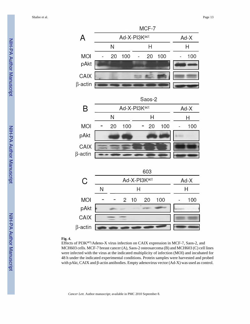
Fig. 4.Effects of PI3Kact/Adeno-X virus infection on CAIX expression in MCF-7, Saos-2, andMCH603 cells. MCF-7 breast cancer (A), Saos-2 osteosarcoma (B) and MCH603 (C) cell lineswere infected with the virus at the indicated multiplicity of infection (MOI) and incubated for48 h under the indicated experimental conditions. Protein samples were harvested and probedwith pAkt, CAIX and β-actin antibodies. Empty adenovirus vector (Ad-X) was used as control.
Shafee et al. Page 13
Cancer Lett. Author manuscript; available in PMC 2010 September 8.
NIH
-PA Author Manuscript
NIH
-PA Author Manuscript
NIH
-PA Author Manuscript