Diagnosis of immediate allergic reactions to beta-lactam antibiotics
Upload
independentCategory
view
1download
0
Biochemical Pharmacology 78 (2009) 396–405
Cyclic AMP enhances resolution of allergic pleurisy by promoting inflammatorycell apoptosis via inhibition of PI3K/Akt and NF-kB
Lirlandia P. Sousa a,b, Aline F. Carmo b, Barbara M. Rezende b, Fernando Lopes b,c, Douglas M. Silva b,Ana L. Alessandri b, Claudio A. Bonjardim d, Adriano G. Rossi e, Mauro M. Teixeira b,*, Vanessa Pinho b,c
a Setor de Patologia Clınica, Colegio Tecnico, Universidade Federal de Minas Gerais, Belo Horizonte, Brazilb Imunofarmacologia, Departamento de Bioquımica e Imunologia, ICB, Universidade Federal de Minas Gerais, Belo Horizonte, Brazilc Departamento de Morfologia, Universidade Federal de Minas Gerais, ICB, Belo Horizonte, Brazild Departamento de Microbiologia, Universidade Federal de Minas Gerais, ICB, Belo Horizonte, Brazile Medical Research Council Centre for Inflammation Research, Queen’s Medical Research Institute, University of Edinburgh, Edinburgh, United Kingdom
A R T I C L E I N F O
Article history:
Received 13 March 2009
Accepted 27 April 2009
Keywords:
cAMP
Eosinophil
Apoptosis
Inflammation
Akt
NF-kB
A B S T R A C T
Selective and timely induction of apoptosis is an effective means of resolving inflammation. The effects
and putative mechanisms by which cyclic AMP (cAMP) modulates leukocyte apoptosis in vivo are still
unclear. The present study aims at identifying intracellular pathways underlying the ability of cAMP
elevating agents to resolve eosinophilic inflammation in a model of allergic pleurisy in mice. Ovalbumin
(OVA) challenge of immunized mice induced eosinophil recruitment that peaked at 24 h and persisted
till 48 h. Treatment with the PDE4 inhibitor rolipram, cAMP mimetic db-cAMP or adenylate cyclase
activator forskolin, at 24 h after antigen-challenge resulted in profound resolution of eosinophilic
inflammation, without a decrease of mononuclear cell numbers. There was a concomitant increase in
number of apoptotic cells in the pleural cavity. The effects of rolipram and db-cAMP were inhibited by
the PKA inhibitor H89. Inhibition of PI3K/Akt or NF-kB induced resolution of inflammation that was
associated with increased apoptosis. OVA-challenge resulted in a time-dependent activation of Akt and
NF-kB, which was blocked by treatment with rolipram or PI3K/Akt pathway inhibitors. Thus, cAMP
elevating agents resolve established eosinophilic inflammation by inducing leukocyte apoptosis.
Mechanistically, the actions of cAMP are dependent on PKA and target a PI3K/Akt-dependent NF-kB
survival pathway.
� 2009 Elsevier Inc. All rights reserved.
Contents lists available at ScienceDirect
Biochemical Pharmacology
journa l homepage: www.e lsev ier .com/ locate /b iochempharm
1. Introduction
Eosinophils are effectors cells that play an important role in thepathophysiology of allergic diseases [1,2]. In allergic diseases, suchas asthma, eosinophils are a crucial source of cytotoxic proteins,lipid mediators, oxygen metabolites, and cytokines, which maycontribute to the severity of disease [3]. The accumulation ofeosinophils in tissue depends not only on the number of cells beingrecruited at any particular time, but also on the number of cellsthat are cleared or leave the tissue [4]. Thus, defective removal ofthese cells may play an important role in the initiation andpropagation of chronic inflammatory diseases. There are two main
Abbreviations: cAMP, cyclic adenosine monophosphate; db-cAMP, dibutyryl-cAMP;
i.pl., intrapleural; PDE4, phosphodiesterase 4; PI3K, phosphatidylinositol 3-kinase;
NF-kB, nuclear factor kappa B; Akt/PKB, protein kinase B; EMSA, electrophoretic
mobility shift assay.
* Corresponding author at: Av. Antonio Carlos, 6627, Pampulha, 31270-901, Belo
Horizonte, MG, Brazil. Tel.: +55 31 3409 2651; fax: +55 31 3409 2651.
E-mail address: [email protected] (M.M. Teixeira).
0006-2952/$ – see front matter � 2009 Elsevier Inc. All rights reserved.
doi:10.1016/j.bcp.2009.04.030
mechanisms that underlie the clearance of inflammatory cells fromtissues, namely apoptosis followed by their subsequent removal byphagocytes and necrosis. Whereas the latter is undoubtedlyassociated with enhanced inflammation and tissue injury, theformer is accompanied by shut down of cellular activity andinhibition of the inflammatory response [4]. Apoptosis ischaracterized by specific morphologic and biochemical eventsincluding cell shrinkage, cytoplasmic vacuolation, membraneblebbing, chromatin condensation and nuclear fragmentationassociated with endonucleolytic DNA cleavage [5].
More recently, there has been great interest in understanding ofthe signal transduction pathways relevant for induction of theapoptosis or survival of leukocytes in vivo [6–8]. Cyclic adenosine30,50 monophosphate (cAMP) is an important intracellular secondmessenger produced after adenylate cyclase activation thatregulates different cellular processes by cAMP effectors [9,10].Phosphodiesterases (PDEs) controls the intracellular cAMP levelsby catalyzing its hydrolysis and inactivating these secondmessengers [10]. PDE isoenzymes have been classified into elevendistinct families [10]. Of these, PDE3, PDE4 and PDE7 are the most
L.P. Sousa et al. / Biochemical Pharmacology 78 (2009) 396–405 397
important for the regulation of cAMP in various types of cells. Inneutrophils, eosinophils, mast cell and basophils, PDE4 isoenzymesappear to play a more important function in the regulation of cAMPin leukocyte [10]. Indeed, PDE4 inhibitors induce an increase in theintracellular levels of cAMP in leukocytes and have potent anti-inflammatory activity [11–14]. Intracellular actions of cAMP can bemimicked by administration of the cell permeable analoguedibutyryl-cAMP (db-cAMP) or the adenylate cyclase activatorforskolin. Similarly to rolipram, these compounds also inhibitleukocyte function and possess significant anti-inflammatoryeffects in vivo [15]. In vitro, inhibition of PDE4 enzymes andincrease of intracellular levels of cyclic AMP may modify thesurvival of eosinophils. Indeed, cAMP elevating agents mayenhance or prevent apoptosis of eosinophils depending on theiractivation status [16]. The effects of cAMP elevating/mimetics onleukocyte apoptosis and survival in vivo are not well-established.
The PI3K/Akt pathway has been also shown to mediate survivalin many cell types [17]. Recently, we have demonstrated [8] thatthe PI3K/Akt pathway was important for the survival of eosinophilsin vivo. It has been reported that there is a cross-talk between thecAMP-dependent and phosphatidylinositol 3-kinase (PI3K) path-ways, but the effects of cAMP on PI3K/Akt activity are quite varied[18,19] and cAMP can either stimulate or inhibit Akt activity. Forexample, cAMP activates PI3K/Akt in thyroid cells and hepatocytes[20,21], whereas inhibition of PI3K/Akt pathway by cAMP has beenreported in fibroblast and leukemia cells [22,23]. The transcriptionfactor nuclear factor kappa B (NF-kB) is a key regulator of severalcellular functions, including leukocyte activation and survival [24–26]. The pro-survival/anti-apoptotic effects of Akt can be mediatedby NF-kB. For example, Akt may phosphorylate IkB kinase (IKK)leading to NF-kB activation [27,28]. It is not known whether thepro-survival effect of the PI3K/Akt pathway during allergicinflammation is mediated via modification of NF-kB function.Hence, it is of interest to examine whether any resolving effect ofcAMP on allergic inflammation is mediated by prevention of thefunction of PI3K/Akt and consequent change in NF-kB function.
In the present study, we examined the ability of the PDE4inhibitor rolipram and of cAMP-inducers/mimetics, forskolin anddb-cAMP, to resolve eosinophilic inflammation in a model ofallergic pleurisy in mice [1,8,29,30]. We show that rolipram, db-cAMP and forskolin resolve established eosinophilic inflammationby promoting apoptosis of inflammatory cells and by inhibiting aPI3K/Akt-dependent NF-kB survival pathway.
2. Materials and methods
2.1. Animals
All procedures described here had prior approval from theAnimal Ethics Committee of Universidade Federal de Minas Gerais.Male C57/BL6 mice (8–10 weeks) obtained from the Bioscience Unitof Instituto de Ciencias Biologicas were housed under standardconditions and had free access to commercial chow and water.
2.2. Drugs, reagents and antibodies
Rolipram (Biomol1, Plymouth Meeting, PA), forskolin and Aktinhibitor-IV (both from Calbiochem, San Diego, CA), gliotoxin(Glio) (Fluka Biochemika, Switzerland), LY294002 (Alamone labs,Jerusalem, Israel), and pyrrolidine dithiocarbamate (PDTC, Sigma–Aldrich, St Louis, MO) were diluted in DMSO and further in PBS.Dibutyryl-cAMP was from Sigma and was diluted in PBS. Annexin-V Detection Kit was from Caltag Laboratories (Burlingame, CA).Rabbit anti-P-Akt (Ser 473), anti-Akt, anti cleaved caspase-3 andmouse anti-phospho-IkB-a were from Cell Signaling Technology(Beverly MA, USA). Rabbit anti-IkB-a (C-21/sc-371), anti p65/RelA
(C-20/sc-372), anti p50/NF-kB1 (H-119/sc-7178) and anti Bax (P-19/sc-526) or secondary anti-rabbit peroxidase conjugate anti-bodies were purchased from Santa Cruz Biotechnology (Santa Cruz,CA, EUA). Anti b-actin and anti-mouse peroxidase conjugateantibodies were from Sigma.
2.3. Induction of pleurisy
Animals were immunized with OVA (Sigma) adsorbed toaluminium hydroxide gel as described [1,8,29,30]. Briefly, micewere injected s.c. (subcutaneous) on days 1 and 8 with 0.2 ml of asolution containing 100 mg of OVA and 70 mg of aluminiumhydroxide (Reheiss, Dublin, Ireland). Sensitized mice werechallenged by i.pl. (intrapleural) administration of antigen (OVA)or PBS. The cells present in the pleural cavity were harvested atdifferent times after antigen-challenge by washing the cavity with2 ml of PBS and total cell counts performed in a modified Neubauerchamber using Turk’s stain. For the experiments evaluatingleukocyte apoptosis, infiltrating leukocytes were examined 2 h(annexin-V, DNA fragmentation assays, caspase-3 cleavage) and24 h (morphologic apoptosis) after drug treatment. Differential cellcounts were performed on cyto-centrifuge preparations (ShandonIII) stained with May–Grunwald–Giemsa using standard morpho-logical criteria to identify cell types. The results are presented asthe number of cells per cavity.
2.4. Treatment with drugs
The role of cAMP on eosinophil accumulation into pleural cavitywas investigated by using rolipram (a specific PDE4 inhibitor),forskolin (an adenylate cyclase activator), and db-cAMP (a cellpermeable cAMP analogue). Rolipram was administered systemi-cally (i.p.) at dose of 150 mg/mouse (6.0 mg/kg), 24 h after i.pl. OVA-challenge. This dose was shown to be effective in other experimentalsystem [14]. Forskolin 10 mg/mouse (0.4 mg/kg), Db-cAMP 100 mg/mouse (4 mg/kg), LY294002 (30 mg/mouse, 1.0 mg/kg), AKT inhi-bitor-IV 10 mg/mouse (0.4 mg/kg) and gliotoxin 20 mg/mouse(0.8 mg/kg) were gived i.pl. at a volume of the 100 ml, 24 h afterOVA-challenge. PDTC was administered systemically (i.p.) at a doseof 100 mg/kg, 24 h after the i.pl. administration of OVA. As a positivecontrol for anti-inflammatory activity, we used the syntheticglucocorticoid dexamethasone at dose of 2.0 mg/kg in PBS buffer.Glucocorticoids have been shown to induce eosinophil apoptosisand to enhance macrophage phagocytosis of apoptotic bodies [31].Drugs were dissolved in DMSO and further diluted in PBS. Controlmice received drug vehicle only.
2.5. Assessment of leukocyte apoptosis
2.5.1. Morphology
Apoptosis was assessed as previously described by us [8,24].Briefly, cells (5 � 104) collected 48 h after antigen-challenge werecyto-centrifuged, fixed and stained with May–Grunwald–Giemsaand counted using oil immersion microscopy (�100 objective) todetermine the proportion of cells with distinctive apoptoticmorphology (cells presented chromatin condensation, nuclearfragmentation and formation of apoptotic bodies out or insidemacrophages). Twenty-five fields were counted per slide andresults are expressed as the mean � S.E.M of number of apoptoticcells in 25 fields.
2.5.2. Annexin-V binding and propidium staining
Assessment of apoptosis was also performed by flow cytometryusing FITC-labeled annexin-V (Caltag laboratories), which binds tophosphatidylserine exposed on the surface of apoptotic cells, andpropidium iodide, as an index of loss of cell membrane integrity.
L.P. Sousa et al. / Biochemical Pharmacology 78 (2009) 396–405398
Annexin-V was added to 100 ml of 2.5 � 105 cells collected 2 h and6 h after drugs treatment, in binding buffer. Following 20 minincubation at room temperature, these samples were treated with5 ml of propidium iodide (50 mg/ml) and analyzed using a BectonDickenson FACScan (San Jose, CA) and FlowJo 7.2.2 software (TreeStar Company). Results are expressed as cells undergoing the earlystage of apoptosis quantified by staining with annexin-V but notpropidium iodide. The cells were selected based on size andgranularity, allowing separate analysis of granulocyte population.At the time point evaluated (26 h after OVA-challenge), morpho-logical analysis showed that granulocytes were eosinophils (>95%,data not shown).
2.6. Lysate preparation and Western blot analysis
Inflammatory cells harvested from the pleural cavity werewashed with PBS and total cell extracts or nuclear and cytoplasmiccell extracts were prepared, as described [32,33]. Protein amountswere quantified with the Bradford assay reagent from Bio-Rad(Bio-Rad, USA). Total cell extracts (40 mg), Nuclear (15 mg) andcytoplasmic (40 mg) extracts were separated by electrophoresis ona denaturing 10–15% polyacrylamide-SDS gel and transferred ontonitrocellulose membranes, as described [32]. Membranes wereblocked overnight at 4 8C with PBS containing 5% (w/v) nonfat dry
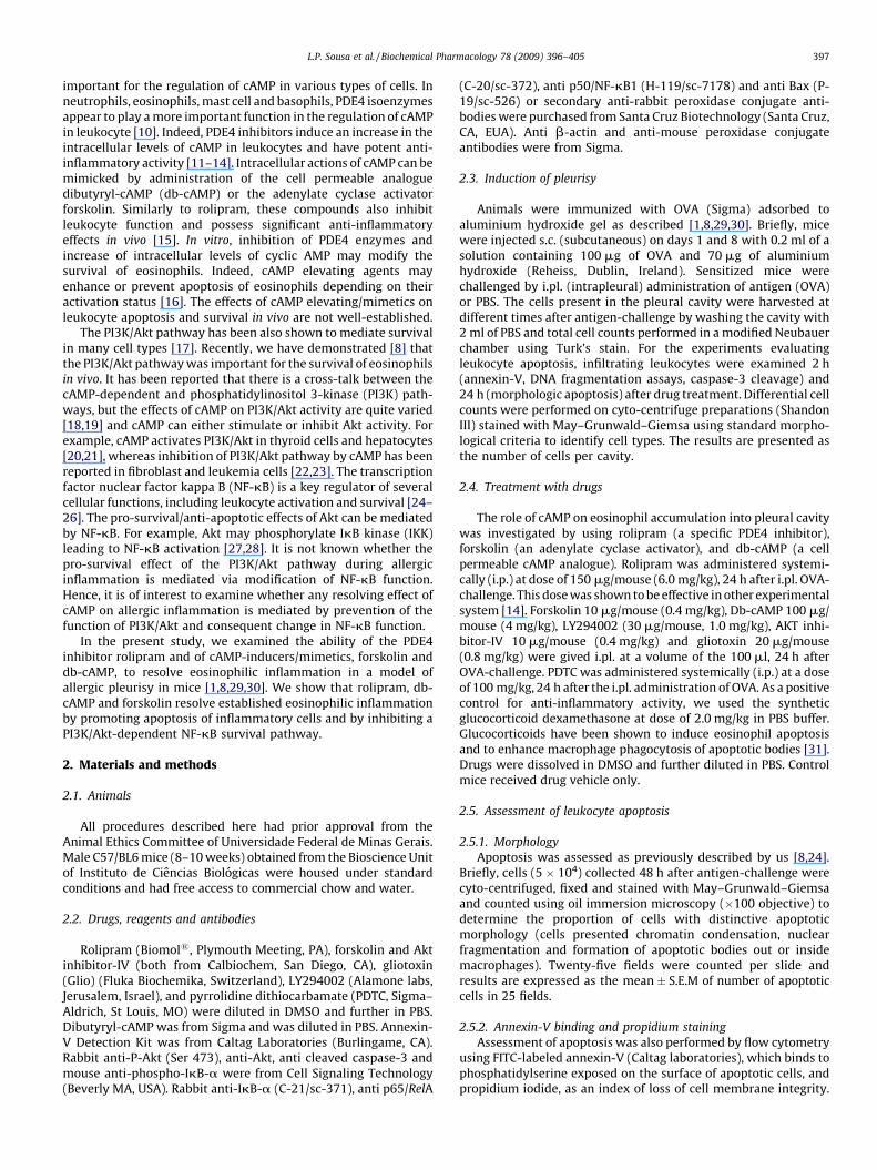
Fig. 1. Kinetics of leukocyte influx in allergic inflammation. Immunized mice were challe
the pleural cavity were collected at indicated times and processed for counting of leuk
expressed as the means � S.E.M of five mice in each group. *P < 0.05; **P < 0.01 and ***P
protocol of the induction of pleurisy and of the treatments used in this paper.
milk and 0.1% Tween-20, washed three times with PBS containing0.1% Tween-20 and then incubated with specifics antibodies(1:1000) in phosphate-buffered saline containing 5% (w/v) BSA and0.1% Tween-20. After washing, membranes were incubated withappropriated horseradish peroxidase-conjugated secondary anti-body (1:3000). Immunoreactive bands were visualized by usingECL detection system, as described by the manufacturer (GEHealthcare, Piscataway, NJ).
2.7. Electrophoretic mobility shift assay (EMSA)
Band shift assay was carried out of 10 mg nuclear extractsessentially as described [32,33], using a 50 [32P]-end-labeleddouble-stranded probe (only one strand is shown) correspondingto the consensus-binding site of NF-kB (50-AGT TGA GGG GAC TTTCCC AGG C-30). Heterologous competition assays were performedwith a 100-fold molar excess of cold oligonucleotide correspond-ing to c-fos SRE.
2.8. Statistical analysis
All results are presented as the mean � S.E.M. Normalized datawere analyzed by one-way ANOVA, and differences between groupswere assessed using the Student–Newman–Keuls post-test. A P-value
nged with an i.pl. injection of OVA (1 mg/cavity) or PBS. The cells that migrated into
ocytes (A), mononuclear cells (B), eosinophils (C), and neutrophils (D). Results are
< 0.001 when compared with PBS-injected mice. (E), schematic representation of the

L.P. Sousa et al. / Biochemical Pharmacology 78 (2009) 396–405 399
<0.05 was considered significant. Calculations were performed usingthe prism 4.0 software program for Windows (GraphPad software,San Diego, CA).
3. Results
3.1. Cyclic AMP elevating agents promote resolution of allergic
pleurisy by inducing leukocyte apoptosis
The model of allergic pleurisy used in the present experiment isa well-established model of acute eosinophilic inflammation
Fig. 2. Delayed treatment with rolipram resolves eosinophilic inflammation by promoting l
received a systemic (s.c) injection of rolipram (6 mg/kg, i.p.) or drug vehicle. The number of
(C) was assed 48 h after antigen-challenge as described in Section 2. Results are expres
morphology in 25 fields, and are shown as the mean� S.E.M of five mice in each group. **P <
challenged mice. In (D) 2.5� 105cells were collected 2 h and 6 h after drug or vehicle treatmen
as the means� S.E.M of the percentage of cells staining with annexin-V-FITC alone. There we
challenged mice. (E) Representative figures of apoptotic and non-apoptotic eosinophils. Vehicl
intact eosinophils, arrowheads indicate apoptotic eosinophils and double arrow indicates an
harvested from the pleural cavity 2 h after drug treatments (i.e. 26 h after OVA-challenge) w
previously described by our group [1,8,29,30] and by others[13]. Injection of 1 mg of OVA into the pleural cavity of sensitizedmice induced a time-dependent influx of leukocytes. As shown inFig. 1A–D, there was an increase in the total number of leukocytes,eosinophils, mononuclear cells and neutrophils in OVA-challengedmice. Total leukocyte influx reached a maximum at 48 h anddecreased at 72 h as compared with PBS-treated mice (Fig. 1A).Eosinophil influx was first detectable at 12 h, reached maximal at24–48 h and dropped thereafter (Fig. 1C). The time-course ofmononuclear infiltrate mirrored the total leukocyte influx (Fig. 1B).Antigen-challenge of sensitized mice also induced an early
eukocyte apoptosis. Immunized mice were challenged with OVA or PBS and 24 h later,
eosinophils (A), mononuclear cells (B) and cells with distinctive apoptotic morphology
sed as the number of cells per cavity, or number of cells with distinctive apoptotic
0.01 or ***P < 0.001 when compared with PBS-injected mice or with vehicle treated OVA-
t and incubated with FITC-labeled annexin-V and propidium iodide. Results are expressed
re at least five mice in each group. *P < 0.05, when compared with vehicle treated OVA-
e (upper panels) and rolipram-treated (lower panels) animals are shown. Arrows indicate
apoptotic body inside a macrophage. In (F), total extracts obtained of inflammatory cells
ere subjected to Western blot to analysis Bax accumulation as described in Section 2.
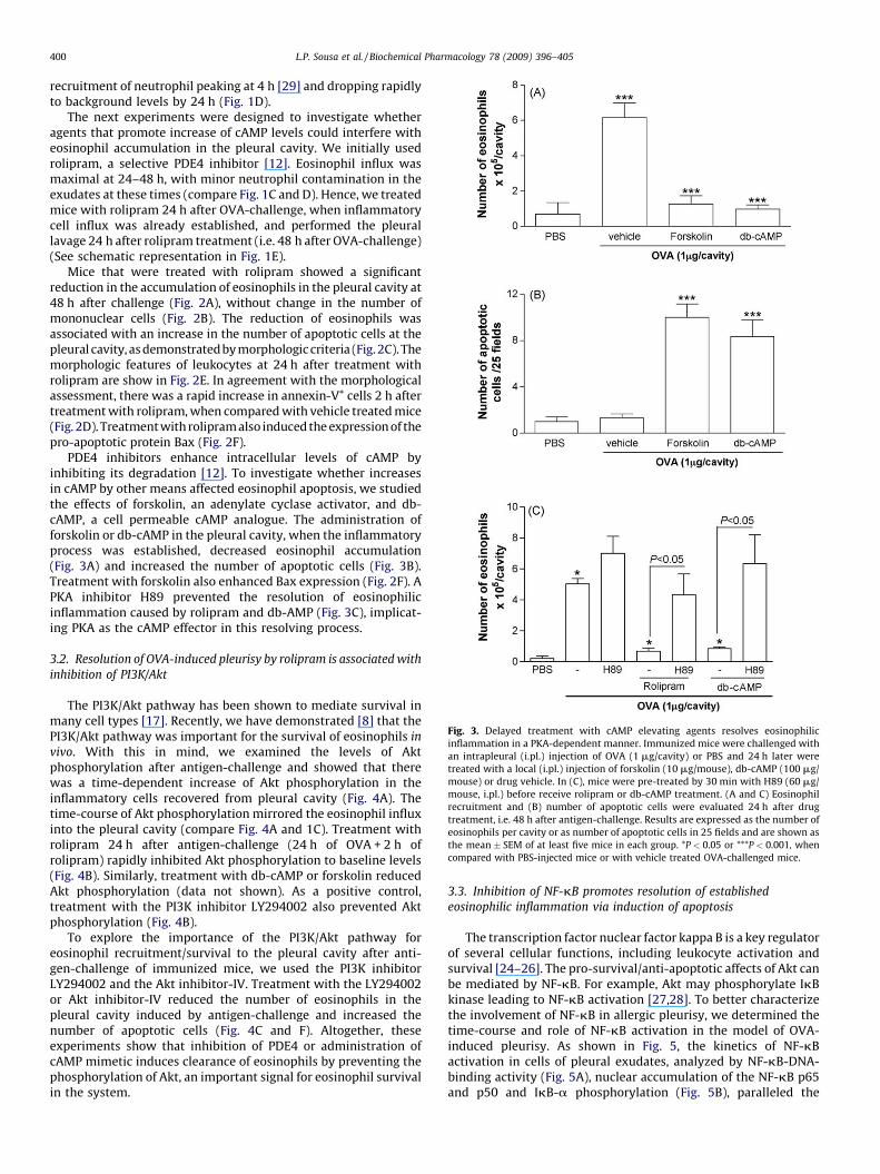
Fig. 3. Delayed treatment with cAMP elevating agents resolves eosinophilic
inflammation in a PKA-dependent manner. Immunized mice were challenged with
an intrapleural (i.pl.) injection of OVA (1 mg/cavity) or PBS and 24 h later were
treated with a local (i.pl.) injection of forskolin (10 mg/mouse), db-cAMP (100 mg/
mouse) or drug vehicle. In (C), mice were pre-treated by 30 min with H89 (60 mg/
mouse, i.pl.) before receive rolipram or db-cAMP treatment. (A and C) Eosinophil
recruitment and (B) number of apoptotic cells were evaluated 24 h after drug
treatment, i.e. 48 h after antigen-challenge. Results are expressed as the number of
eosinophils per cavity or as number of apoptotic cells in 25 fields and are shown as
the mean � SEM of at least five mice in each group. *P < 0.05 or ***P < 0.001, when
compared with PBS-injected mice or with vehicle treated OVA-challenged mice.
L.P. Sousa et al. / Biochemical Pharmacology 78 (2009) 396–405400
recruitment of neutrophil peaking at 4 h [29] and dropping rapidlyto background levels by 24 h (Fig. 1D).
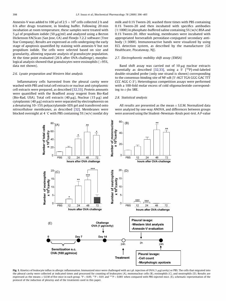
The next experiments were designed to investigate whetheragents that promote increase of cAMP levels could interfere witheosinophil accumulation in the pleural cavity. We initially usedrolipram, a selective PDE4 inhibitor [12]. Eosinophil influx wasmaximal at 24–48 h, with minor neutrophil contamination in theexudates at these times (compare Fig. 1C and D). Hence, we treatedmice with rolipram 24 h after OVA-challenge, when inflammatorycell influx was already established, and performed the pleurallavage 24 h after rolipram treatment (i.e. 48 h after OVA-challenge)(See schematic representation in Fig. 1E).
Mice that were treated with rolipram showed a significantreduction in the accumulation of eosinophils in the pleural cavity at48 h after challenge (Fig. 2A), without change in the number ofmononuclear cells (Fig. 2B). The reduction of eosinophils wasassociated with an increase in the number of apoptotic cells at thepleural cavity, as demonstrated by morphologic criteria (Fig. 2C). Themorphologic features of leukocytes at 24 h after treatment withrolipram are show in Fig. 2E. In agreement with the morphologicalassessment, there was a rapid increase in annexin-V+ cells 2 h aftertreatment with rolipram, when compared with vehicle treated mice(Fig. 2D). Treatment with rolipram also induced the expression of thepro-apoptotic protein Bax (Fig. 2F).
PDE4 inhibitors enhance intracellular levels of cAMP byinhibiting its degradation [12]. To investigate whether increasesin cAMP by other means affected eosinophil apoptosis, we studiedthe effects of forskolin, an adenylate cyclase activator, and db-cAMP, a cell permeable cAMP analogue. The administration offorskolin or db-cAMP in the pleural cavity, when the inflammatoryprocess was established, decreased eosinophil accumulation(Fig. 3A) and increased the number of apoptotic cells (Fig. 3B).Treatment with forskolin also enhanced Bax expression (Fig. 2F). APKA inhibitor H89 prevented the resolution of eosinophilicinflammation caused by rolipram and db-AMP (Fig. 3C), implicat-ing PKA as the cAMP effector in this resolving process.
3.2. Resolution of OVA-induced pleurisy by rolipram is associated with
inhibition of PI3K/Akt
The PI3K/Akt pathway has been shown to mediate survival inmany cell types [17]. Recently, we have demonstrated [8] that thePI3K/Akt pathway was important for the survival of eosinophils in
vivo. With this in mind, we examined the levels of Aktphosphorylation after antigen-challenge and showed that therewas a time-dependent increase of Akt phosphorylation in theinflammatory cells recovered from pleural cavity (Fig. 4A). Thetime-course of Akt phosphorylation mirrored the eosinophil influxinto the pleural cavity (compare Fig. 4A and 1C). Treatment withrolipram 24 h after antigen-challenge (24 h of OVA + 2 h ofrolipram) rapidly inhibited Akt phosphorylation to baseline levels(Fig. 4B). Similarly, treatment with db-cAMP or forskolin reducedAkt phosphorylation (data not shown). As a positive control,treatment with the PI3K inhibitor LY294002 also prevented Aktphosphorylation (Fig. 4B).
To explore the importance of the PI3K/Akt pathway foreosinophil recruitment/survival to the pleural cavity after anti-gen-challenge of immunized mice, we used the PI3K inhibitorLY294002 and the Akt inhibitor-IV. Treatment with the LY294002or Akt inhibitor-IV reduced the number of eosinophils in thepleural cavity induced by antigen-challenge and increased thenumber of apoptotic cells (Fig. 4C and F). Altogether, theseexperiments show that inhibition of PDE4 or administration ofcAMP mimetic induces clearance of eosinophils by preventing thephosphorylation of Akt, an important signal for eosinophil survivalin the system.
3.3. Inhibition of NF-kB promotes resolution of established
eosinophilic inflammation via induction of apoptosis
The transcription factor nuclear factor kappa B is a key regulatorof several cellular functions, including leukocyte activation andsurvival [24–26]. The pro-survival/anti-apoptotic affects of Akt canbe mediated by NF-kB. For example, Akt may phosphorylate IkBkinase leading to NF-kB activation [27,28]. To better characterizethe involvement of NF-kB in allergic pleurisy, we determined thetime-course and role of NF-kB activation in the model of OVA-induced pleurisy. As shown in Fig. 5, the kinetics of NF-kBactivation in cells of pleural exudates, analyzed by NF-kB-DNA-binding activity (Fig. 5A), nuclear accumulation of the NF-kB p65and p50 and IkB-a phosphorylation (Fig. 5B), paralleled the

Fig. 4. Akt phosphorylation is decreased by rolipram and delayed treatment with inhibitors of the PI3K/Akt pathway resolve eosinophilic inflammation. Immunized mice
were challenged with an intrapleural (i.pl.) injection of ovalbumin (OVA, 1 mg/cavity) or PBS at indicated times (A) or were treatment with rolipram (6 mg/kg, i.p.) or
LY294002 (30 mg/mouse, i.pl.) 24 h after OVA-challenge (B). Akt phosphorylation was evaluated at indicated times (A) or after 26 h after antigen-challenge (24 h OVA-
challenge + 2 h rolipram) (B) by Western blot analysis performed of total cell extracts obtained of pools of cells from at least five animals as shown in Section 2. Data shown are
representative of three independent experiments. In (C–F), the PI3K inhibitor LY294002 (30 mg/mouse, i.pl), Akt inhibitor-IV (10 mg/mouse, i.pl) or drug vehicle were
administered in 24 h-OVA-challenged mice and eosinophil recruitment (C and E) and number of apoptotic cells (D and F) were evaluated 24 h after drug treatment, i.e. 48 h
after antigen-challenge. Results are expressed as the number of eosinophils per cavity or as number of apoptotic cells in 25 fields and are shown as the mean � SEM of at least
five mice in each group. **P < 0.01 or ***P < 0.001, when compared with PBS-injected mice or with vehicle treated OVA-challenged mice.
L.P. Sousa et al. / Biochemical Pharmacology 78 (2009) 396–405 401
kinetics of total inflammatory cell influx into the pleural cavity, i.e.NF-kB activation was first detectable at 12 h, peaked at 24–48 h ofOVA-challenge and decreased thereafter (72 h).
We also evaluated whether the use of the NF-kB inhibitorsgiven in the same way as cAMP elevating agents, i.e. at 24 h afterantigen-challenge (Fig. 1E), could enhance resolution of eosino-philic inflammation. As seen in Fig. 6A, gliotoxin treatment given at24 h after OVA-challenge drastically reduced the accumulation ofeosinophils observed at 48 h but did not alter the number ofmononuclear cells (PBS, 23.4 � 0.8 � 105 mononuclear per cavity;OVA + vehicle, 43 � 3.3 � 105 mononuclear per cavity; OVA + Glio,
47.7 � 2.7 � 105 mononuclear per cavity; n = 5, P < 0.001 whencompare PBS � OVA). The reduction of eosinophil number at 48 h wasalso seen when another structurally distinct NF-kB inhibitor, PDTC,was given at 24 h (OVA + vehicle, 10.9 � 1.5 � 105 eosinophils percavity; OVA + PDTC, 2.5 � 0.8 � 105 eosinophils per cavity; n = 5,P < 0.001). For comparison, treatment with dexamethasone (Dexa), apotent anti-inflammatory drug with various cellular targets [31], at24 h after challenge diminished the accumulation of eosinophils inthe pleural cavity (Fig. 6A).
Next, we evaluated the efficacy of the compounds at blockingNF-kB activity at 2 h after compound administration (i.e. 26 h after
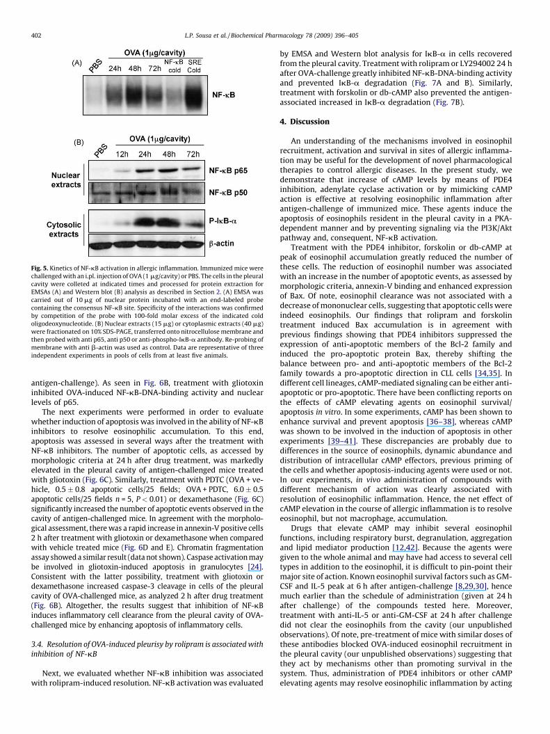
Fig. 5. Kinetics of NF-kB activation in allergic inflammation. Immunized mice were
challenged with an i.pl. injection of OVA (1 mg/cavity) or PBS. The cells in the pleural
cavity were colleted at indicated times and processed for protein extraction for
EMSAs (A) and Western blot (B) analysis as described in Section 2. (A) EMSA was
carried out of 10 mg of nuclear protein incubated with an end-labeled probe
containing the consensus NF-kB site. Specificity of the interactions was confirmed
by competition of the probe with 100-fold molar excess of the indicated cold
oligodeoxynucleotide. (B) Nuclear extracts (15 mg) or cytoplasmic extracts (40 mg)
were fractionated on 10% SDS-PAGE, transferred onto nitrocellulose membrane and
then probed with anti p65, anti p50 or anti-phospho-IkB-a antibody. Re-probing of
membrane with anti b-actin was used as control. Data are representative of three
independent experiments in pools of cells from at least five animals.
L.P. Sousa et al. / Biochemical Pharmacology 78 (2009) 396–405402
antigen-challenge). As seen in Fig. 6B, treatment with gliotoxininhibited OVA-induced NF-kB-DNA-binding activity and nuclearlevels of p65.
The next experiments were performed in order to evaluatewhether induction of apoptosis was involved in the ability of NF-kBinhibitors to resolve eosinophilic accumulation. To this end,apoptosis was assessed in several ways after the treatment withNF-kB inhibitors. The number of apoptotic cells, as accessed bymorphologic criteria at 24 h after drug treatment, was markedlyelevated in the pleural cavity of antigen-challenged mice treatedwith gliotoxin (Fig. 6C). Similarly, treatment with PDTC (OVA + ve-hicle, 0.5� 0.8 apoptotic cells/25 fields; OVA + PDTC, 6.0 � 0.5apoptotic cells/25 fields n = 5, P < 0.01) or dexamethasone (Fig. 6C)significantly increased the number of apoptotic events observed in thecavity of antigen-challenged mice. In agreement with the morpholo-gical assessment, there was a rapid increase in annexin-V positive cells2 h after treatment with gliotoxin or dexamethasone when comparedwith vehicle treated mice (Fig. 6D and E). Chromatin fragmentationassay showed a similar result (data not shown). Caspase activation maybe involved in gliotoxin-induced apoptosis in granulocytes [24].Consistent with the latter possibility, treatment with gliotoxin ordexamethasone increased caspase-3 cleavage in cells of the pleuralcavity of OVA-challenged mice, as analyzed 2 h after drug treatment(Fig. 6B). Altogether, the results suggest that inhibition of NF-kBinduces inflammatory cell clearance from the pleural cavity of OVA-challenged mice by enhancing apoptosis of inflammatory cells.
3.4. Resolution of OVA-induced pleurisy by rolipram is associated with
inhibition of NF-kB
Next, we evaluated whether NF-kB inhibition was associatedwith rolipram-induced resolution. NF-kB activation was evaluated
by EMSA and Western blot analysis for IkB-a in cells recoveredfrom the pleural cavity. Treatment with rolipram or LY294002 24 hafter OVA-challenge greatly inhibited NF-kB-DNA-binding activityand prevented IkB-a degradation (Fig. 7A and B). Similarly,treatment with forskolin or db-cAMP also prevented the antigen-associated increased in IkB-a degradation (Fig. 7B).
4. Discussion
An understanding of the mechanisms involved in eosinophilrecruitment, activation and survival in sites of allergic inflamma-tion may be useful for the development of novel pharmacologicaltherapies to control allergic diseases. In the present study, wedemonstrate that increase of cAMP levels by means of PDE4inhibition, adenylate cyclase activation or by mimicking cAMPaction is effective at resolving eosinophilic inflammation afterantigen-challenge of immunized mice. These agents induce theapoptosis of eosinophils resident in the pleural cavity in a PKA-dependent manner and by preventing signaling via the PI3K/Aktpathway and, consequent, NF-kB activation.
Treatment with the PDE4 inhibitor, forskolin or db-cAMP atpeak of eosinophil accumulation greatly reduced the number ofthese cells. The reduction of eosinophil number was associatedwith an increase in the number of apoptotic events, as assessed bymorphologic criteria, annexin-V binding and enhanced expressionof Bax. Of note, eosinophil clearance was not associated with adecrease of mononuclear cells, suggesting that apoptotic cells wereindeed eosinophils. Our findings that rolipram and forskolintreatment induced Bax accumulation is in agreement withprevious findings showing that PDE4 inhibitors suppressed theexpression of anti-apoptotic members of the Bcl-2 family andinduced the pro-apoptotic protein Bax, thereby shifting thebalance between pro- and anti-apoptotic members of the Bcl-2family towards a pro-apoptotic direction in CLL cells [34,35]. Indifferent cell lineages, cAMP-mediated signaling can be either anti-apoptotic or pro-apoptotic. There have been conflicting reports onthe effects of cAMP elevating agents on eosinophil survival/apoptosis in vitro. In some experiments, cAMP has been shown toenhance survival and prevent apoptosis [36–38], whereas cAMPwas shown to be involved in the induction of apoptosis in otherexperiments [39–41]. These discrepancies are probably due todifferences in the source of eosinophils, dynamic abundance anddistribution of intracellular cAMP effectors, previous priming ofthe cells and whether apoptosis-inducing agents were used or not.In our experiments, in vivo administration of compounds withdifferent mechanism of action was clearly associated withresolution of eosinophilic inflammation. Hence, the net effect ofcAMP elevation in the course of allergic inflammation is to resolveeosinophil, but not macrophage, accumulation.
Drugs that elevate cAMP may inhibit several eosinophilfunctions, including respiratory burst, degranulation, aggregationand lipid mediator production [12,42]. Because the agents weregiven to the whole animal and may have had access to several celltypes in addition to the eosinophil, it is difficult to pin-point theirmajor site of action. Known eosinophil survival factors such as GM-CSF and IL-5 peak at 6 h after antigen-challenge [8,29,30], hencemuch earlier than the schedule of administration (given at 24 hafter challenge) of the compounds tested here. Moreover,treatment with anti-IL-5 or anti-GM-CSF at 24 h after challengedid not clear the eosinophils from the cavity (our unpublishedobservations). Of note, pre-treatment of mice with similar doses ofthese antibodies blocked OVA-induced eosinophil recruitment inthe pleural cavity (our unpublished observations) suggesting thatthey act by mechanisms other than promoting survival in thesystem. Thus, administration of PDE4 inhibitors or other cAMPelevating agents may resolve eosinophilic inflammation by acting
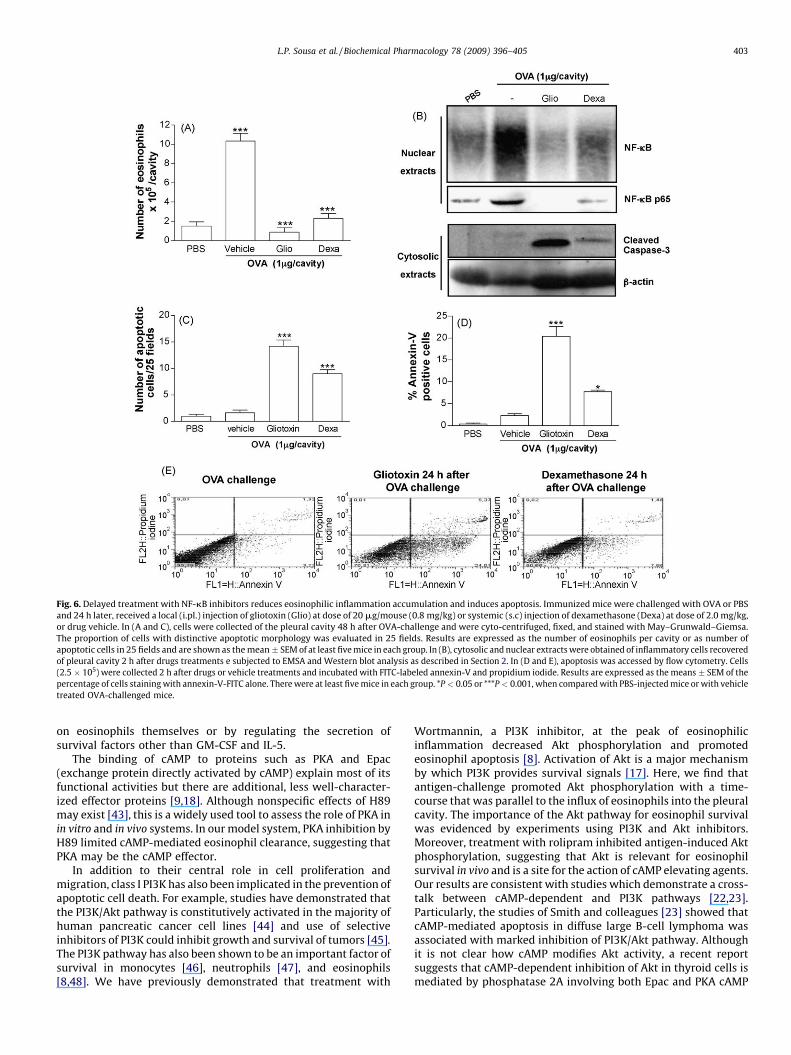
Fig. 6. Delayed treatment with NF-kB inhibitors reduces eosinophilic inflammation accumulation and induces apoptosis. Immunized mice were challenged with OVA or PBS
and 24 h later, received a local (i.pl.) injection of gliotoxin (Glio) at dose of 20 mg/mouse (0.8 mg/kg) or systemic (s.c) injection of dexamethasone (Dexa) at dose of 2.0 mg/kg,
or drug vehicle. In (A and C), cells were collected of the pleural cavity 48 h after OVA-challenge and were cyto-centrifuged, fixed, and stained with May–Grunwald–Giemsa.
The proportion of cells with distinctive apoptotic morphology was evaluated in 25 fields. Results are expressed as the number of eosinophils per cavity or as number of
apoptotic cells in 25 fields and are shown as the mean � SEM of at least five mice in each group. In (B), cytosolic and nuclear extracts were obtained of inflammatory cells recovered
of pleural cavity 2 h after drugs treatments e subjected to EMSA and Western blot analysis as described in Section 2. In (D and E), apoptosis was accessed by flow cytometry. Cells
(2.5 � 105) were collected 2 h after drugs or vehicle treatments and incubated with FITC-labeled annexin-V and propidium iodide. Results are expressed as the means � SEM of the
percentage of cells staining with annexin-V-FITC alone. There were at least five mice in each group. *P < 0.05 or ***P < 0.001, when compared with PBS-injected mice or with vehicle
treated OVA-challenged mice.
L.P. Sousa et al. / Biochemical Pharmacology 78 (2009) 396–405 403
on eosinophils themselves or by regulating the secretion ofsurvival factors other than GM-CSF and IL-5.
The binding of cAMP to proteins such as PKA and Epac(exchange protein directly activated by cAMP) explain most of itsfunctional activities but there are additional, less well-character-ized effector proteins [9,18]. Although nonspecific effects of H89may exist [43], this is a widely used tool to assess the role of PKA inin vitro and in vivo systems. In our model system, PKA inhibition byH89 limited cAMP-mediated eosinophil clearance, suggesting thatPKA may be the cAMP effector.
In addition to their central role in cell proliferation andmigration, class I PI3K has also been implicated in the prevention ofapoptotic cell death. For example, studies have demonstrated thatthe PI3K/Akt pathway is constitutively activated in the majority ofhuman pancreatic cancer cell lines [44] and use of selectiveinhibitors of PI3K could inhibit growth and survival of tumors [45].The PI3K pathway has also been shown to be an important factor ofsurvival in monocytes [46], neutrophils [47], and eosinophils[8,48]. We have previously demonstrated that treatment with
Wortmannin, a PI3K inhibitor, at the peak of eosinophilicinflammation decreased Akt phosphorylation and promotedeosinophil apoptosis [8]. Activation of Akt is a major mechanismby which PI3K provides survival signals [17]. Here, we find thatantigen-challenge promoted Akt phosphorylation with a time-course that was parallel to the influx of eosinophils into the pleuralcavity. The importance of the Akt pathway for eosinophil survivalwas evidenced by experiments using PI3K and Akt inhibitors.Moreover, treatment with rolipram inhibited antigen-induced Aktphosphorylation, suggesting that Akt is relevant for eosinophilsurvival in vivo and is a site for the action of cAMP elevating agents.Our results are consistent with studies which demonstrate a cross-talk between cAMP-dependent and PI3K pathways [22,23].Particularly, the studies of Smith and colleagues [23] showed thatcAMP-mediated apoptosis in diffuse large B-cell lymphoma wasassociated with marked inhibition of PI3K/Akt pathway. Althoughit is not clear how cAMP modifies Akt activity, a recent reportsuggests that cAMP-dependent inhibition of Akt in thyroid cells ismediated by phosphatase 2A involving both Epac and PKA cAMP

Fig. 7. Treatment with cAMP elevating agents and LY294002 prevents NF-kB
activation in allergic inflammation. Immunized mice were challenged with an
intrapleural (i.pl.) injection of OVA (1 mg/cavity) or PBS and 24 h later were treated
with rolipram (6 mg/kg, i.p.), LY294002 (30 mg/mouse, i.pl.), forskolin (10 mg/
mouse), db-cAMP (100 mg/mouse) or drug vehicle. Nuclear and cytoplasmic
extracts obtained of inflammatory cells harvested from the pleural cavity 2 h after
drug treatments (i.e. 26 h after OVA-challenge) were subjected to EMSA (A) and
Western blot (B) analysis, respectively. (A) EMSAs were carried out of 10 mg of
nuclear protein incubated with an end-labeled oligodeoxynucleotide probe
containing the consensus NF-kB site. Specificity of the interactions was
confirmed by competition of the probe with 100-fold molar excess of the
indicated cold oligodeoxynucleotide. In (B) cytoplasmic extracts (40 mg) were
fractionated on 10% SDS-PAGE, transferred onto nitrocellulose membrane and then
probed with anti-IkB-a antibody (upper panel) or with b-actin antibody (under
panel). Data are representative of two independent experiments in pools of cells
from at least five animals.
L.P. Sousa et al. / Biochemical Pharmacology 78 (2009) 396–405404
effectors [19]. Thus, cAMP may mediate its survival/pro-apoptoticeffects by modifying PI3K/Akt. Observations of opposing effects ofEpac and PKA on Akt activation can provide a potential mechanismfor the apparent cell type-specific effects of cAMP [18].
Akt/PKB has direct effects on the apoptosis pathway, forexample by phosphorylating pro-apoptotic proteins such ascaspase-9 and BAD. Akt also have effects in transcription factors,
Fig. 8. Model for the resolution inducing effects of the delayed treatment with cAMP
elevating agents in allergic eosinophilic inflammation. Increase in the intracellular
levels of cAMP by means of the delayed treatment with an adenylate cyclase
activator (forskolin), PDE4 inhibitor (rolipram) or db-cAMP promotes activation of a
cAMP effector (PKA), which then inhibits signaling thought PI3K/Akt pathway.
Down regulation of the Akt phosphorylation may itself lead to apoptosis or may
prevent NF-kB activation, which is a relevant signal for eosinophil survival.
Prevention of eosinophil survival leads to resolution of inflammation.
including the Forkhead transcription factor and NF-kB [17]. In thisregard, Akt can induce cell survival by phosphorylating IkB kinaseand, consequently, activating NF-kB [27–28]. The activated NF-kBmay then control cell survival via induction of the expression ofanti-apoptotic genes [24–26]. In our experiments, NF-kB activa-tion, as evaluated by DNA-binding activity, p65/p50 nuclearaccumulation and IkB-a phosphorylation correlated temporallywith the infiltration of leukocytes in the pleural cavity of antigen-challenge mice. Treatment with gliotoxin, PDTC or dexamethasoneat doses that inhibited NF-kB activation, induced resolution ofeosinophilic inflammation and increased leukocyte apoptosiswithout decreasing number of mononuclear cells. Importantly,cAMP elevation or PI3K inhibitors decreased antigen-induced NF-kB activation by preventing IkB-a degradation and NF-kB-DNA-binding activity in vivo. Previous studies have shown that PDE4inhibitors prevented NF-kB activation when given before orshortly after inflammatory stimulation [49–51], a finding con-sistent with the ability of PDE4 inhibitors to prevent leukocyteactivation and recruitment [12]. However, our results are first toshow the ability of delayed treatment with cAMP elevating agentsto resolve eosinophilic inflammation and emphasize the impor-tance of NF-kB for leukocyte survival in vivo. Moreover, our resultsare first to suggest that NF-kB activation is downstream of PI3K/Akt activation and resolution inducing effects in vivo.
Taken together, our data demonstrate that cAMP elevatingagents or mimetics promote resolution of established eosinophilicinflammation in a PKA-dependent manner and by inhibiting Aktphosphorylation and consequent NF-kB activation (see Fig. 8). Toour knowledge, this is the first observation that cAMP promotesapoptosis in vivo via inhibition of a PI3K/Akt/NF-kB pathway.Hence, we suggest that elevation of cAMP in vivo may represent apowerful anti-inflammatory strategy for the treatment of diseasesin which eosinophil accumulation is thought to play a relevantrole.
Acknowledgements
We thank Valdineria Borges and Ilma Marcal for technicalassistance. This work was supported by grants from EuropeanUnion FP6 (INNOCHEM, Grant number LSHB-CT-2005-518167),Fundacao de Amparo a Pesquisa do Estado de Minas Gerais(FAPEMIG) and Conselho Nacional de Desenvolvimento Cientıfico eTecnologico (CNPq). Lirlandia P. Sousa was a recipient of a post-doctoral fellowship from CNPq.
References
[1] Cara DC, Negrao-Correa D, Teixeira MM. Mechanisms underlying eosinophiltrafficking and their relevance in vivo. Histol Histopathol 2000;15:899–920.
[2] Lacy P, Moqbel R. Immune effector functions of eosinophils in allergic airwayinflammation. Curr Opin Allergy Clin Immunol 2001;1:79–84.
[3] Kita H, Gleich GJ. Chemokines active on eosinophils: potential roles in allergicinflammation. J Exp Med 1996;183:2421–6.
[4] Rossi AG, Hallett JM, Sawatzky DA, Teixeira MM, Haslett C. Modulation ofgranulocyte apoptosis can influence the resolution of inflammation. BiochemSoc Trans 2007;35:288–91.
[5] Cohen JJ. Apoptosis. Immunol Today 1993;14:126–30.[6] Sawatzky DA, Willoughby DA, Colville-Nash PR, Rossi AG. The involvement of
the apoptosis-modulating proteins ERK 1/2 Bcl-xL and Bax in the resolution ofacute inflammation in vivo. Am J Pathol 2006;168:33–41.
[7] Rossi AG, Sawatzky DA, Walker A, Ward C, Sheldrake TA, Riley NA, et al. Cyclin-dependent kinase inhibitors enhance the resolution of inflammation by pro-moting inflammatory cell apoptosis. Nat Med 2006;12:1056–64.
[8] Pinho V, Souza DG, Barsante MM, Hamer FP, De Freitas MS, Rossi AG, et al.Phosphoinositide-3 kinases critically regulate the recruitment and survival ofeosinophils in vivo: importance for the resolution of allergic inflammation. JLeukoc Biol 2005;77:800–10.
[9] Serezani CH, Ballinger MN, Aronoff DM, Peters-Golden M. Cyclic AMP: masterregulator of innate immune cell function. Am J Respir Cell Mol Biol2008;39:127–32.
[10] Soderling SH, Beavo JA. Regulation of cAMP and cGMP signaling: new phos-phodiesterases and new functions. Curr Opin Cell Biol 2000;12:174–9.

L.P. Sousa et al. / Biochemical Pharmacology 78 (2009) 396–405 405
[11] Souness JE, Aldous D, Sargent C. Immunosuppressive and anti-inflammatoryeffects of cyclic AMP phosphodiesterase (PDE) type 4 inhibitors. Immuno-pharmacology 2000;47:127–62.
[12] Teixeira MM, Gristwood RW, Cooper N, Hellewell PG. Phosphodiesterase(PDE)4 inhibitors: anti-inflammatory drugs of the future? Trends PharmacolSci 1997;18:164–71.
[13] Alves AC, Pires AL, Cruz HN, Serra MF, Diaz BL, Cordeiro RS, et al. Selectiveinhibition of phosphodiesterase type IV suppresses the chemotactic respon-siveness of rat eosinophils in vitro. Eur J Pharmacol 1996;312:89–96.
[14] Souza DG, Cassali GD, Poole S, Teixeira MM. Effects of inhibition of PDE4 andTNF-alpha on local and remote injuries following ischaemia and reperfusioninjury. Br J Pharmacol 2001;134:985–94.
[15] Diaz BL, Serra MF, Alves AC, Cordeiro SB, Martins MA, e Silva PM. Localexposure to salbutamol or Bt2 cyclic AMP inhibits pleural exudation andleukocyte influx caused by antigen in rats. Eur J Pharmacol 1996;296:173–80.
[16] Hallsworth MP, Giembycz MA, Barnes PJ, Lee TH. Cyclic AMP-elevating agentsprolong or inhibit eosinophil survival depending on prior exposure to GM-CSF.Br J Pharmacol 1996;117:79–86.
[17] Song G, Ouyang G, Bao S. The activation of Akt/PKB signaling pathway and cellsurvival. J Cell Mol Med 2005;9:59–71.
[18] Mei FC, Qiao J, Tsygankova OM, Meinkoth JL, Quilliam LA, Cheng X. Differentialsignaling of cyclic AMP: opposing effects of exchange protein directly acti-vated by cyclic AMP and cAMP-dependent protein kinase on protein kinase Bactivation. J Biol Chem 2002;277:11497–504.
[19] Hong K, Lou L, Gupta S, Ribeiro-Neto F, Altschuler DL. A novel Epac-Rap-PP2Asignaling module controls cAMP-dependent Akt regulation. J Biol Chem2008;283:23129–38.
[20] Cass LA, Summers SA, Prendergast GV, Backer JM, Birnbaum MJ, Meinkoth JL.Protein kinase A-dependent and -independent signaling pathways contributeto cyclic AMP-stimulated proliferation. Mol Cell Biol 1999;19:5882–91.
[21] Cullen KA, McCool J, Anwer MS, Webster CR. Activation of cAMP-guanineexchange factor confers PKA-independent protection from hepatocyte apop-tosis. Am J Physiol Gastrointest Liver Physiol 2004;287:G334–43.
[22] Kim S, Jee K, Kim D, Koh H, Chung J. Cyclic AMP inhibits Akt activity by blockingthe membrane localization of PDK1. J Biol Chem 2001;276:12864–70.
[23] Smith PG, Wang F, Wilkinson KN, Savage KJ, Klein U, Neuberg DS, et al. Thephosphodiesterase PDE4B limits cAMP-associated PI3K/AKT-dependent apop-tosis in diffuse large B-cell lymphoma. Blood 2005;105:308–16.
[24] Ward C, Chilvers ER, Lawson MF, Pryde JG, Fujihara S, Farrow SN, et al. NF-kappaB activation is a critical regulator of human granulocyte apoptosis invitro. J Biol Chem 1999;274:4309–18.
[25] Ward C, Walker A, Dransfield I, Haslett C, Rossi AG. Regulation of granulocyteapoptosis by NF-kappaB. Biochem Soc Trans 2004;32:465–7.
[26] Dutta J, Fan Y, Gupta N, Fan G, Gelinas C. Current insights into the regulation ofprogrammed cell death by NF-kappaB. Oncogene 2006;25:6800–16.
[27] Nair AS, Shishodia S, Ahn KS, Kunnumakkara AB, Sethi G, Aggarwal BB.Deguelin, an Akt inhibitor, suppresses IkappaBalpha kinase activation leadingto suppression of NF-kappaB-regulated gene expression, potentiation ofapoptosis, and inhibition of cellular invasion. J Immunol 2006;177:5612–22.
[28] Kane LP, Shapiro VS, Stokoe D, Weiss A. Induction of NF-kappaB by the Akt/PKBkinase. Curr Biol 1999;9:601–4.
[29] Klein A, Talvani A, Cara DC, Gomes KL, Lukacs NW, Teixeira MM. Stem cellfactor plays a major role in the recruitment of eosinophils in allergic pleurisyin mice via the production of leukotriene B4. J Immunol 2000;164:4271–6.
[30] Klein A, Talvani A, Silva PM, Martins MA, Wells TN, Proudfoot A, et al. Stem cellfactor-induced leukotriene B4 production cooperates with eotaxin to mediatethe recruitment of eosinophils during allergic pleurisy in mice. J Immunol2001;167:524–31.
[31] McColl A, Michlewska S, Dransfield I, Rossi AG. Effects of glucocorticoids onapoptosis and clearance of apoptotic cells. Sci World J 2007;7:1165–81.
[32] Sousa LP, Silva BM, Brasil BS, Nogueira SV, Ferreira PC, Kroon EG, et al.Plasminogen/plasmin regulates alpha-enolase expression through the MEK/ERK pathway. Biochem Biophys Res Commun 2005;337:1065–71.
[33] Souza DG, Vieira AT, Pinho V, Sousa LP, Andrade AA, Bonjardim CA, et al. NF-kappaB plays a major role during the systemic and local acute inflammatoryresponse following intestinal reperfusion injury. Br J Pharmacol 2005;145:246–54.
[34] Siegmund B, Welsch J, Loher F, Meinhardt G, Emmerich B, Endres S, et al.Phosphodiesterase type 4 inhibitor suppresses expression of anti-apoptoticmembers of the Bcl-2 family in B-CLL cells and induces caspase-dependentapoptosis. Leukemia 2001;15:1564–71.
[35] Moon EY, Lerner A. PDE4 inhibitors activate a mitochondrial apoptotic path-way in chronic lymphocytic leukemia cells that is regulated by proteinphosphatase 2A. Blood 2003;101:4122–30.
[36] Machida K, Inoue H, Matsumoto K, Tsuda M, Fukuyama S, Koto H, et al.Activation of PI3K-Akt pathway mediates antiapoptotic effects of beta-adre-nergic agonist in airway eosinophils. Am J Physiol Lung Cell Mol Physiol2005;288:L860–7.
[37] Parkkonen J, Hasala H, Moilanen E, Giembycz MA, Kankaanranta H. Phospho-diesterase 4 inhibitors delay human eosinophil and neutrophil apoptosis inthe absence and presence of salbutamol. Pulm Pharmacol Ther 2008;21:499–506.
[38] Chang HS, Jeon KW, Kim YH, Chung IY, Park CS. Role of cAMP-dependentpathway in eosinophil apoptosis and survival. Cell Immunol 2000;203:29–38.
[39] Takeuchi M, Tatsumi Y, Kitaichi K, Baba K, Suzuki R, Shibata E, et al. Selectivephosphodiesterase type 4 inhibitors reduce the prolonged survival of eosino-phils stimulated by granulocyte-macrophage colony-stimulating factor. BiolPharm Bull 2002;25:184–7.
[40] Wang W, Masu K, Tamura G, Suzuki K, Ohwada K, Okuyama K, et al. Inhibitionof eosinophil survival by a selective inhibitor of phosphodiesterase 4 via theinduction of apoptosis. Biol Pharm Bull 2005;28:515–9.
[41] Chung IY, Nam-Kung EK, Lee NM, Chang HS, Kim DJ, Kim YH, et al. Thedownregulation of Bcl-2 expression is necessary for theophylline-inducedapoptosis of eosinophil. Cell Immunol 2000;203:95–102.
[42] Giembycz MA, Lindsay MA. Pharmacology of the eosinophil. Pharmacol Rev1999;51:213–340.
[43] Murray AJ. Pharmacological PKA inhibition: all may not be what it seems. SciSignal 2008;1:re4.
[44] Bondar VM, Sweeney-Gotsch B, Andreeff M, Mills GB, McConkey DJ. Inhibitionof the phosphatidylinositol 30-kinase-AKT pathway induces apoptosis inpancreatic carcinoma cells in vitro and in vivo. Mol Cancer Ther 2002;1:989–97.
[45] Krystal GW, Sulanke G, Litz J. Inhibition of phosphatidylinositol 3-kinase-Aktsignaling blocks growth, promotes apoptosis, and enhances sensitivity ofsmall cell lung cancer cells to chemotherapy. Mol Cancer Ther 2002;1:913–22.
[46] Kelley TW, Graham MM, Doseff AI, Pomerantz RW, Lau SM, Ostrowski MC,et al. Macrophage colony-stimulating factor promotes cell survival throughAkt/protein kinase B. J Biol Chem 1999;274:26393–8.
[47] Cowburn AS, Cadwallader KA, Reed BJ, Farahi N, Chilvers ER. Role of PI3-kinase-dependent Bad phosphorylation and altered transcription in cytokine-mediated neutrophil survival. Blood 2002;100:2607–16.
[48] Myou S, Leff AR, Myo S, Boetticher E, Tong J, Meliton AY, et al. Blockade ofinflammation and airway hyperresponsiveness in immune-sensitized mice bydominant-negative phosphoinositide 3-kinase-TAT. J Exp Med 2003;198:1573–82.
[49] Sanchez AJ, Puerta C, Ballester S, Gonzalez P, Arriaga A, Garcia-Merino A.Rolipram impairs NF-kappaB activity and MMP-9 expression in experimentalautoimmune encephalomyelitis. J Neuroimmunol 2005;168:13–20.
[50] Schmitz T, Souil E, Herve R, Nicco C, Batteux F, Germain G, et al. PDE4 inhibitionprevents preterm delivery induced by an intrauterine inflammation. J Immu-nol 2007;178:1115–21.
[51] Kwak HJ, Song JS, Heo JY, Yang SD, Nam JY, Cheon HG. Roflumilast inhibitslipopolysaccharide-induced inflammatory mediators via suppression ofnuclear factor-kappaB, p38 mitogen-activated protein kinase, and c-JunNH2-terminal kinase activation. J Pharmacol Exp Ther 2005;315:1188–95.
Copyright © 2022 FDOKUMEN