MEKK3 is required for lysophosphatidic acid-induced NF-κB activation
7
MEKK3 is required for lysophosphatidic acid-induced NF-κB activation Wenjing Sun a,b , Hongxiu Li c , Yang Yu a , Yihui Fan a , Brian C. Grabiner c , Renfang Mao d , Ningling Ge a , Hong Zhang d , Songbin Fu b , Xin Lin c , Jianhua Yang a, ⁎ a Texas Children's Cancer Center, Department of Pediatrics, Dan L. Duncan Cancer Center, Baylor College of Medicine, Houston, TX 77030, United States b Laboratory of Medical Genetics, Harbin Medical University, Harbin 150081, China c Department of Molecular and Cellular Oncology, the University of Texas M.D. Anderson Cancer Center, Houston, TX 77030, United States d Department of Pathology, Baylor College of Medicine, Houston, TX 77030, United States abstract article info Article history: Received 1 May 2009 Accepted 18 May 2009 Available online 22 May 2009 Keywords: NF-κB LPA GPCR MEKK3 TAK1 Lysophosphatidic acid (LPA) is a potent agonist that exerts various cellular functions on many cell types through binding to its cognate G protein-coupled receptors (GPCRs). Although LPA induces NF-κB activation byacting on its GPCR receptor, the molecular mechanism of LPA receptor-mediated NF-κB activation remains to be well defined. In the present study, by using MEKK3-, TAK1-, and IKKβ-deficient murine embryonic fibro- blasts (MEFs), we found that MEKK3 but not TAK1 deficiency impairs LPA and protein kinase C (PKC)-induced IκB kinase (IKK)-NF-κB activation, and IKKβ is required for PKC-induced NF-κB activation. In addition, we demonstrate that LPA and PKC-induced IL-6 and MIP-2 production are abolished in the absence of MEKK3 but not TAK1. Together, our results provide the genetic evidence that MEKK3 but not TAK1 is required for LPA receptor-mediated IKK-NF-κB activation. © 2009 Elsevier Inc. All rights reserved. 1. Introduction The nuclear factor-κB (NF-κB) family of transcription factors plays an important role in regulating the expression of genes responsible for innate and adaptive immunity, stress responses, anti-apoptosis, cell proliferation, and differentiation [1–4]. In resting cells, NF-κB is retained in the cytosol in an inactive form through interaction with IκB inhibitory proteins. Release of NF-κB for translocation to the nucleus and activation of NF-κB dependent genes is accomplished through a signal-induced phosphorylation of IκB by IκB kinase (IKK) and sub- sequent IκBα degradation [5–8]. Upon binding to their agonist ligands, various cell surface receptors, including receptors for proinflamma- tory cytokines such as TNFα and IL-1, Toll-like receptors (TLRs), antigen receptors, and GPCRs, induce distinct signaling pathways that even- tually converge on IKK complex to activate NF-κB [9]. A major chal- lenge in the NF-κB field is to understand how these distinct receptor- mediated signaling effectors activate IKK/NF-κB in a signal-specific manner [10]. Lysophosphatidic acid (LPA) is a naturally occurring, water-soluble glycerophospholipid that exerts hormone- and growth factor-like acti- vities on many cell types including fibroblasts, endothelial cells, and smooth muscle cells [11–12]. LPA is involved in the regulation of various cellular responses such as cell proliferation, chemotaxis and survival through binding to its cognate G protein-coupled receptors (GPCRs) and activating LPA receptor-mediated multiple effector molecules, including NF-κB [11]. Recently, the adaptor and scaffold proteins β- arrestin2, Bcl10, MALT1 and CARMA3 were identified as essential signal transducers to mediate LPA-induced NF-κB activation [13–17]. Two members of MAP3K serine/threonine kinase family, MEKK3 and TAK1 have been demonstrated to be involved in regulating NF-κB activation through IKK [18–22]. Surprisingly, it has been suggested that TAK1 is not essential in the LPA-mediated NF-κB activation [15]. We therefore tested whether MEKK3 is required for LPA-induced NF- κB activation. Using MEKK3- and TAK1-deficient MEF cell lines, we demonstrate that MEKK3 but not TAK1 is required for LPA receptor- mediated IKK-NF-κB activation. These results reveal that MEKK3, but not TAK1, preferentially mediates GPCR-induced NF-κB activation. 2. Materials and methods 2.1. Antibodies, plasmids and reagents Antibodies against ERK1/2, phospho-ERK1/2, JNK, phospho-JNK, IκBα, phospho-IκBα, IKKβ, phospho-IKKα/β, TAK1, and secondary antibodies conjugated to horseradish peroxidase were purchased from Cell Signaling Technology (Beverly, MA). Antibodies against PCNA (PC- 10), NF-κB-p65 were from Santa Cruz Biotechnology, Inc. (Santa Cruz, CA). Antibody against MEKK3 was from BD Biosciences Pharmingen Cellular Signalling 21 (2009) 1488–1494 Abbreviations: NF-κB, nuclear factor-κB; IKK, IκB kinase; LPA, lysophosphatidic acid; GPCR, G protein-coupled receptor; PKC, protein kinase C; TAK1, TGF-β activated kinase 1; MEKK3, mitogen-activated protein kinase kinase kinase 3. ⁎ Corresponding author. Texas Children's Cancer Center, Department of Pediatrics, Baylor College of Medicine, Houston, TX 77030, United States. Tel.: +1 832 824 4572; fax: +1 832 825 4732. E-mail address: [email protected] (J. Yang). 0898-6568/$ – see front matter © 2009 Elsevier Inc. All rights reserved. doi:10.1016/j.cellsig.2009.05.007 Contents lists available at ScienceDirect Cellular Signalling journal homepage: www.elsevier.com/locate/cellsig
-
Upload
independent -
Category
Documents
-
view
0 -
download
0
Transcript of MEKK3 is required for lysophosphatidic acid-induced NF-κB activation

Cellular Signalling 21 (2009) 1488–1494
Contents lists available at ScienceDirect
Cellular Signalling
j ourna l homepage: www.e lsev ie r.com/ locate /ce l l s ig
MEKK3 is required for lysophosphatidic acid-induced NF-κB activation
Wenjing Sun a,b, Hongxiu Li c, Yang Yu a, Yihui Fan a, Brian C. Grabiner c, Renfang Mao d, Ningling Ge a,Hong Zhang d, Songbin Fu b, Xin Lin c, Jianhua Yang a,⁎a Texas Children's Cancer Center, Department of Pediatrics, Dan L. Duncan Cancer Center, Baylor College of Medicine, Houston, TX 77030, United Statesb Laboratory of Medical Genetics, Harbin Medical University, Harbin 150081, Chinac Department of Molecular and Cellular Oncology, the University of Texas M.D. Anderson Cancer Center, Houston, TX 77030, United Statesd Department of Pathology, Baylor College of Medicine, Houston, TX 77030, United States
Abbreviations:NF-κB, nuclear factor-κB; IKK, IκB kinaGPCR, G protein-coupled receptor; PKC, protein kinase C;MEKK3, mitogen-activated protein kinase kinase kinase⁎ Corresponding author. Texas Children's Cancer Cen
Baylor College of Medicine, Houston, TX 77030, Unitedfax: +1 832 825 4732.
E-mail address: [email protected] (J. Yang).
0898-6568/$ – see front matter © 2009 Elsevier Inc. Adoi:10.1016/j.cellsig.2009.05.007
a b s t r a c t
a r t i c l e i n f oArticle history:Received 1 May 2009Accepted 18 May 2009Available online 22 May 2009
Keywords:NF-κBLPAGPCRMEKK3TAK1
Lysophosphatidic acid (LPA) is a potent agonist that exerts various cellular functions on many cell typesthrough binding to its cognate G protein-coupled receptors (GPCRs). Although LPA induces NF-κB activationby acting on its GPCR receptor, the molecular mechanism of LPA receptor-mediated NF-κB activation remainsto be well defined. In the present study, by using MEKK3-, TAK1-, and IKKβ-deficient murine embryonic fibro-blasts (MEFs), we found that MEKK3 but not TAK1 deficiency impairs LPA and protein kinase C (PKC)-inducedIκB kinase (IKK)-NF-κB activation, and IKKβ is required for PKC-induced NF-κB activation. In addition, wedemonstrate that LPA and PKC-induced IL-6 and MIP-2 production are abolished in the absence of MEKK3 butnot TAK1. Together, our results provide the genetic evidence that MEKK3 but not TAK1 is required for LPAreceptor-mediated IKK-NF-κB activation.
© 2009 Elsevier Inc. All rights reserved.
1. Introduction
The nuclear factor-κB (NF-κB) family of transcription factors playsan important role in regulating the expression of genes responsiblefor innate and adaptive immunity, stress responses, anti-apoptosis,cell proliferation, and differentiation [1–4]. In resting cells, NF-κB isretained in the cytosol in an inactive form through interactionwith IκBinhibitory proteins. Release of NF-κB for translocation to the nucleusand activation of NF-κB dependent genes is accomplished through asignal-induced phosphorylation of IκB by IκB kinase (IKK) and sub-sequent IκBα degradation [5–8]. Upon binding to their agonist ligands,various cell surface receptors, including receptors for proinflamma-tory cytokines such as TNFα and IL-1, Toll-like receptors (TLRs), antigenreceptors, and GPCRs, induce distinct signaling pathways that even-tually converge on IKK complex to activate NF-κB [9]. A major chal-lenge in the NF-κB field is to understand how these distinct receptor-mediated signaling effectors activate IKK/NF-κB in a signal-specificmanner [10].
Lysophosphatidic acid (LPA) is a naturally occurring, water-solubleglycerophospholipid that exerts hormone- and growth factor-like acti-
se; LPA, lysophosphatidic acid;TAK1, TGF-β activated kinase 1;3.ter, Department of Pediatrics,States. Tel.: +1 832 824 4572;
ll rights reserved.
vities on many cell types including fibroblasts, endothelial cells, andsmoothmuscle cells [11–12]. LPA is involved in the regulation of variouscellular responses such as cell proliferation, chemotaxis and survivalthrough binding to its cognate G protein-coupled receptors (GPCRs)and activating LPA receptor-mediated multiple effector molecules,including NF-κB [11]. Recently, the adaptor and scaffold proteins β-arrestin2, Bcl10, MALT1 and CARMA3 were identified as essentialsignal transducers to mediate LPA-induced NF-κB activation [13–17].Two members of MAP3K serine/threonine kinase family, MEKK3 andTAK1 have been demonstrated to be involved in regulating NF-κBactivation through IKK [18–22]. Surprisingly, it has been suggestedthat TAK1 is not essential in the LPA-mediated NF-κB activation [15].We therefore tested whether MEKK3 is required for LPA-induced NF-κB activation. Using MEKK3- and TAK1-deficient MEF cell lines, wedemonstrate that MEKK3 but not TAK1 is required for LPA receptor-mediated IKK-NF-κB activation. These results reveal that MEKK3, butnot TAK1, preferentially mediates GPCR-induced NF-κB activation.
2. Materials and methods
2.1. Antibodies, plasmids and reagents
Antibodies against ERK1/2, phospho-ERK1/2, JNK, phospho-JNK,IκBα, phospho-IκBα, IKKβ, phospho-IKKα/β, TAK1, and secondaryantibodies conjugated to horseradish peroxidasewere purchased fromCell Signaling Technology (Beverly,MA). Antibodies against PCNA (PC-10), NF-κB-p65 were from Santa Cruz Biotechnology, Inc. (Santa Cruz,CA). Antibody against MEKK3 was from BD Biosciences Pharmingen

1489W. Sun et al. / Cellular Signalling 21 (2009) 1488–1494
(San Diego, CA). Antibody against β-actin were from Sigma (St. Louis,MO). pBabe-vector and pBabe-HA-MEKK3 expression vector has beendescribed previously [20]. The NF-κB-dependent firefly luciferasereporter plasmid and pCMV promoter-dependent Renilla-luciferasereporter were purchased from Clontech (Mountain View, California).LPA, phorbol-12-myristate-13-acetate (PMA) and ionomycin (Iono)were purchased from Sigma. Mouse IL-6 and MIP-2 ELISA kits werepurchased fromBD Biosciences and R & D Systems (Minneapolis, MN),respectively. The ECL-Plus Western blotting system was purchasedfrom GE Healthcare Bio-sciences Corp.
2.2. Cell culture and transfection
MEKK3−/−, TAK1−/− and IKKβ−/− as well as the reconstitutedMEF cell lines have been described previously [20,22,23]. These cellsare maintained in DMEM containing 10% FCS at 37 °C with 5% CO2,
and transfected with Lipofectamine 2000 (Invitrogen) following themanufacturer's protocol.
2.3. Luciferase reporter gene assay
Luciferase reporter gene assay was performed using a dual luci-ferase reporter assay system (Promega, Madison,WI) and aMonolight3010 luminometer (BD Pharmingen) as described previously [20].Briefly, targeted cells were transiently cotransfected with specificvectors and an NF-κB-dependent firefly luciferase reporter constructas well as a Renilla-luciferase control construct. Cellular extracts wereprepared 36 h post-transfection and the luciferase activities weredetermined. Relative NF-κB luciferase activity was normalized toRenilla-luciferase activity. Changes in luciferase activity with respectto control were calculated. Each experimentwas conducted in triplicate.
2.4. Preparation of nuclear and cytosolic fractions
Nuclear and cytosolic extracts were made as described [20]. Inbrief, cells were harvested in ice-cold PBS (pH 7.4) and were pelletedby centrifugation at 500 ×g for 3 min and then lysed for 30 min on icein buffer B (10 mM HEPES buffer, pH 7.9, containing 0.1 mM EDTA,10mMKCl, 0.4% (v/v) IGEPAL, 0.5 mM dithiothreitol (DTT), and 1 mMphenylmethylsulfonyl fluoride (PMSF)). Lysates were centrifuged at15,000 ×g for 10 min. The resulting supernatants constituted cytosolicfractions. The pellets were washed three times with buffer B andresuspended in buffer C (20 mM HEPES buffer, pH 7.9, containing400 mMNaCl, 1 mM EDTA, 1 mMDTT and 1 mM PMSF) and incubatedfor 30 min on ice and centrifuged at 15,000 ×g for 10 min. The super-natants were used as nuclear extracts.
2.5. Electrophoretic mobility shift assay (EMSA)
NF-κB oligonucleotide probes were labeled with [γ-32P]ATP. MEFcells (1×10⁶) were starved for 12 h and stimulated for the indicated
Fig. 1. MEKK3 is required for LPA-induced IKK-NF-κB activation. (A) MEKK3+/+ andMEKK3−/− MEF cells reconstituted with empty vector or HA-MEKK3 were either un-treated or treatedwith LPA (30µM) for 0, 30, and60min, then harvested.Nuclear extractswere prepared and subjected to EMSA by using 32P-labeled NF-κB probes. Whole celllysates (WCL) were subjected to SDS-PAGE and immunoblotting with antibodiesindicated. PCNA was used as a loading control for nuclear extracts and β-actin was de-tected as a loading control for WCL. (B) MEKK3+/+ and MEKK3−/− MEF cells reconsti-tuted with empty vector and MEKK3were stimulated as in A. Then nuclear extracts wereprepared and subjected to the immunoblotting analysis as indicated. (C)MEKK3−/−MEFcells reconstituted with empty vector and MEKK3 were either untreated or treated withLPA (30 µM) for 0, 5,15, 30, and 60min, then harvested.WCLwere subjected to SDS-PAGEand immunoblotting analysis as indicated. (D) One microgram of NF-κB luciferasereporter and 20 ng of Renilla-Luc plasmids were cotransfected into MEF cells indicated.Twenty-four hours after transfection, cells were starved for 12 h followed by the additionof LPA (30 µM) for 8 h. The relative luciferase activity wasmeasured and normalizedwiththe Renilla activity. Error bars indicate ±standard deviation in triplicate experiments.
1490 W. Sun et al. / Cellular Signalling 21 (2009) 1488–1494
time points. Nuclear extracts isolated from these cells were thenincubated with 32P-labeled probes in 10 mM HEPES (pH 7.9), 40 mMNaCl, 1 mM EDTA, 4% glycerol, 3 μg of poly(dI·dC), and 0.5 mM DTT
Fig. 2. MEKK3 is required for PKC-induced IKK/NF-κB activation. (A) MEKK3+/+ and MEKKtreated with PMA (40 ng/ml) plus Iono (100 ng/ml) for 0, 30, and 60 min. Nuclear extracts wand MEKK3−/− MEF cells reconstituted with empty vector and MEKK3 were stimulated asimmunoblotting analysis as indicated. (C) MEKK3−/− MEF cells reconstituted with empty(100 ng/ml) for 0, 5, 15, 30, and 60min, then harvested. WCL were subjected to SDS-PAGE anand 20 ng of Renilla-Luc plasmids were cotransfected into MEF cells indicated. Twenty-fou(40 ng/ml) plus Iono (100 ng/ml) for 8 h. The relative luciferase activity was measured and nexperiments. (E) IKKβ+/+ and IKKβ−/− MEF cells were either untreated or treated with PMnuclear extracts were subjected to SDS-PAGE and immunoblotting analysis as indicated. (Fcotransfected intoMEF cells indicated. Twenty-four hours after transfection, cells were starverelative luciferase activity was measured and normalized with the Renilla activity. Error bar
for 15 min at room temperature. The samples were then resolved ina nondenaturing polyacrylamide gel and exposed to X-ray film at−80 °C.
3−/− MEF cells reconstituted with empty vector and MEKK3 were either untreated orere prepared and subjected to EMSA by using 32P-labeled NF-κB probes. (B) MEKK3+/+
in A. WCL and nuclear extracts isolated from the cells were subjected to SDS-PAGE andvector and MEKK3 were either untreated or treated with PMA (40 ng/ml) plus Ionod immunoblotting analysis as indicated. (D) Onemicrogram of NF-κB luciferase reporterr hours after transfection, cells were starved for 12 h followed by the addition of PMAormalized with the Renilla activity. Error bars indicate ±standard deviation in triplicateA (40 ng/ml) plus Iono (100 ng/ml) for 0, 30, and 60 min, then harvested. WCL and
) One microgram of NF-κB luciferase reporter and 20 ng of Renilla-Luc plasmids wered for 12 h followed by the addition of PMA (40 ng/ml) plus Iono (100 ng/ml) for 8 h. Thes indicate ±standard deviation in triplicate experiments.
1491W. Sun et al. / Cellular Signalling 21 (2009) 1488–1494
2.6. Enzyme-linked immunosorbent assay (ELISA)
MEKK3-deficient MEF cell lines stably transfected with vectorcontrol or MEKK3 were treated with or without LPA (30 µM) or PMA(40 ng/ml) plus Iono (100 ng/ml), the supernatants were collectedat different time points. Mouse IL-6 or MIP-2 concentrations in themedium were determined by ELISA according to the manufacturer'sinstructions.
2.7. Immunoblotting
Cells were harvested in ice-cold PBS (pH 7.4) and spun down. Thepellet was dissolved in lysis buffer (50 mM Tris–HCl, pH 7.4, 150 mMNaCl, 1 mM EDTA, 1% IGEPAL, 0.25% Na-deoxycholate, 1 mM PMSF,1 mM DTT, 10 µg/ml aprotinin, 10 µg/ml leupeptin, 1 mM Ben-zamidine, 20 mM disodium p-nitrophenylphosphate (pNPP), 0.1 mMsodium orthovanadate (OV), 10 mM sodium fluoride (NaF), phospha-tase inhibitor cocktails A and B (Sigma Aldrich)). The cell lysates weresubjected directly to 10% SDS-PAGE and probed for the specific anti-body for immunoblotting analysis.
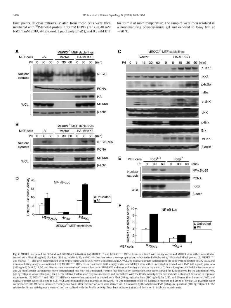
Fig. 3. TAK1 is not required for LPA and PMA/Iono-induced IKK-NF-κB activation. (A and C) Ttreated with LPA (A) or PMA/Iono (C) for 0, 30, and 60 min, then harvested. WCL and nuclearD) TAK1−/−MEF cells reconstituted with empty vector and TAK1 expression plasmid were eiharvested. WCL were subjected to SDS-PAGE and immunoblotting analysis as indicated.
3. Results
3.1. MEKK3 is required for LPA-induced IKK/NF-κB activation
Previous study suggests that TAK1 is not required for LPA-inducedNF-κB activation [15]. Therefore, we hypothesized that MEKK3, an-other NF-κB activating MAP3K, is playing an essential role in LPA-mediated NF-κB activation. To test this hypothesis, we used MEF cellsisolated from day 9.5 embryos of MEKK3 wild type (MEKK3+/+) andMEKK3-deficient (MEKK3−/−) mice. We first established MEKK3−/−
MEF stable cell lines reconstituted with empty vector or HA-MEKK3expression vector, respectively. The expression of HA-MEKK3 in thereconstituted MEF cell lines were verified by immunoblotting(Fig. 1A). Then MEKK3+/+ and MEKK3−/− MEF cell lines reconsti-tuted with vector control or HA-MEKK3 were treated with LPA at dif-ferent time points and nuclear translocation of NF-κB in these cellswere examined by EMSA and immunoblotting of the nuclear extractswith anti-NF-κB-p65 antibody (Fig.1A and B). In this assay, LPA stimu-lation effectively induced NF-κB nuclear translocation in the wild typeand HA-MEKK3-reconstituted MEFs, whereas LPA stimulation failed
AK1−/− MEF cells reconstituted with empty vector and TAK1 were either untreated orextracts were subjected to SDS-PAGE and immunoblotting analysis as indicated. (B andther untreated or treated with LPA (B) or PMA/Iono (D) for 0, 5, 15, 30, and 60 min, then
1492 W. Sun et al. / Cellular Signalling 21 (2009) 1488–1494
to induce nuclear translocation of NF-κB in MEKK3−/− MEFs (Fig. 1Aand B). These results suggest that MEKK3 is required for LPA-inducedNF-κB nuclear translocation in the cells.
MEKK3 has been shown to be involved in the regulation of IKK/NF-κB activation [18,20,23,24]. To further characterize the physiologicalrole of MEKK3 in LPA-mediated NF-κB activation, MEKK3−/− MEF celllines reconstituted with vector control or HA-MEKK3 were treatedwith LPA at different time points and subsequently lysed. The celllysates were immunoblotted with the indicated antibodies to examinethe LPA-induced IKK and IκBα phosphorylation. In this assay, LPA-induced IKK and IκBα phosphorylation were significantly impairedin the vector control MEKK3−/− MEF cells compared to the MEKK3reconstituted cells (Fig. 1C).
Previous studies have shown that MEKK3 is involved in JNK acti-vation [25–27]. We found that LPA failed to induce JNK2 phospho-rylation in the MEKK3−/− MEF cells with vector control compared tothe MEKK3 reconstituted cells whereas LPA-induced JNK1 phosphor-ylation was only slightly inhibited in the vector control MEKK3−/−
MEF cells at later time points. Interestingly, LPA-induced-ERK phos-phorylation was comparable in both cell lines (Fig. 1C).
Consistent with these results, luciferase analysis with NF-κB re-porter showed that LPA failed to induce the luciferase reporter geneexpression in MEKK3−/− MEF cells with the vector control whereasLPA induced higher level of reporter gene expression in the wild typeand/orMEKK3 reconstitutedMEF cells (Fig. 1D). Taken together, theseresults indicate that MEKK3 is required for LPA-induced optimal IKK/NF-κB activation.
3.2. MEKK3 is required for PMA/Iono-induced IKK/NF-κB activation
Protein kinase C (PKC)members have been reported to be involvedin LPA-induced NF-κB activation [15]. We therefore tested the role ofMEKK3 in PKC-mediated NF-κB nuclear translocation in MEF cells. Inour assays, the wild type, vector control and HA-MEKK3-reconstitutedMEKK3−/− MEF cells were treated with or without PKC pharmaco-logical agonists, PMA plus Iono at the indicated time points andnuclear translocation of NF-κB in these cells were examined by EMSA
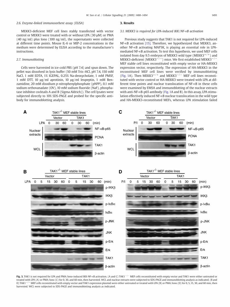
Fig. 4. MEKK3 is required for the LPA and PKC-induced IL-6 and MIP-2 production. (A and Btreated with LPA (A) or PMA/Iono (B) for the time points indicated. The supernatants fromaccording to the manufacturer's instructions.
and immunoblotting of nuclear extracts with anti-NF-κB-p65 anti-bodies. We found that PMA plus Iono (PMA/Iono) co-stimulationeffectively induced nuclear translocation of NF-κB in the wild typeMEFs and MEKK3−/− MEFs reconstituted with HA-MEKK3 whereasPMA/Iono stimulation failed to induce NF-κB nuclear translocation inMEKK3−/− MEFs with vector control (Fig. 2A and B). These resultssuggest that MEKK3 is required for PMA/Iono-induced NF-κB nucleartranslocation.
We further studied the role of MEKK3 in PMA/Iono-induced IKK-NF-κB activation and found that PMA/Iono-induced IKK and IκBαphosphorylation were significantly impaired in MEKK3−/− MEF cellswith the vector control compared to the HA-MEKK3 reconstitutedcells (Fig. 2C). We also found that PMA/Iono-induced JNK2 phosphor-ylation was completely blocked in MEKK3−/− MEF cells with thevector control and rescued in MEKK3 reconstituted MEF cells, where-as PMA/Iono-induced-JNK1 activation was only slightly inhibitedin MEKK3−/− MEF cells with the vector control at later time points(Fig. 2C). However, PMA/Iono-induced-ERK phosphorylation wascomparable in both cell lines (Fig. 2C). Consistent with these results,PMA/Iono failed to induce the NF-κB-responsive luciferase reportergene expression in the vector control MEKK3−/− MEF cells whereasPMA/Iono induced high level of reporter gene expression in the wildtype and MEKK3 reconstituted MEF cells (Fig. 2D). Taken together,these results indicate that MEKK3 is required for PKC-mediated IKK/NF-κB activation.
The IKKs and IKK-related kinases (IKKε/IKK-i and TBK1/NAK/T2K)play important roles in activation of the host defense system [28,29].IKKβ is essential for rapid NF-κB activation by proinflammatory signal-ing cascades, such as those triggered by TNFα or lipopolysaccharide(LPS), whereas IKKε/IKK-i and TBK1/NAK/T2K have been suggestedto be involved in PKC-induced NF-κB activation [29–34]. We there-fore tested the role of IKKβ in PKC-mediated NF-κB activation in MEFcells. In our assays, IKKβ wild type (IKKβ+/+) and IKKβ-deficient(IKKβ−/−) MEF cells were treated with or without PMA plus Iono atthe indicated time points and nuclear localization of NF-κB in thesecells were examined. We found that PMA/Iono co-stimulation effec-tively induced nuclear localization of NF-κB in IKKβ+/+MEFswhereas
) MEKK3−/− MEF cells reconstituted with empty vector and MEKK3 were untreated orthese cultures were collected and subjected to the mouse IL-6 and MIP-2 ELISA analysis
1493W. Sun et al. / Cellular Signalling 21 (2009) 1488–1494
PMA/Iono stimulation failed to induce nuclear localization of NF-κBin IKKβ−/− MEFs (Fig. 2E). Consistent with this result, PMA/Ionoinduced a higher NF-κB dependent luciferase activity in the IKKβ+/+
MEF cells compared to IKKβ−/− MEF cells (Fig. 2F). These resultssuggest that IKKβ is required for PMA/Iono-induced NF-κB activationin the MEF cells.
3.3. TAK1 is not required for LPA and PMA/Iono-induced IKK/NF-κBactivation
To further explore the role of TAK1 in the LPA and PMA/Iono-mediated NF-κB activation, we treated the TAK1-deficient (TAK1−/−)MEF cell lines reconstituted with vector control or TAK1 with LPA andPMA/Iono for the time points indicated. We found that LPA and PMA/Iono effectively induced IKK and IκBα phosphorylation as well asnuclear translocation of NF-κB in both vector control and TAK1 re-constituted TAK1−/− MEF cells (Fig. 3A–D). Thus, these results sug-gest that TAK1 is not required for the LPA and PMA/Iono-inducedNF-κB activation.
3.4. MEKK3 is required for LPA and PMA/Iono-induced IL-6 and MIP-2production
NF-κB activation has been shown to be required for LPA-inducedIL-6 and macrophage inflammatory protein-2 (MIP-2) production inMEF cells [13,15,16]. To determine the role of MEKK3 in LPA-induced
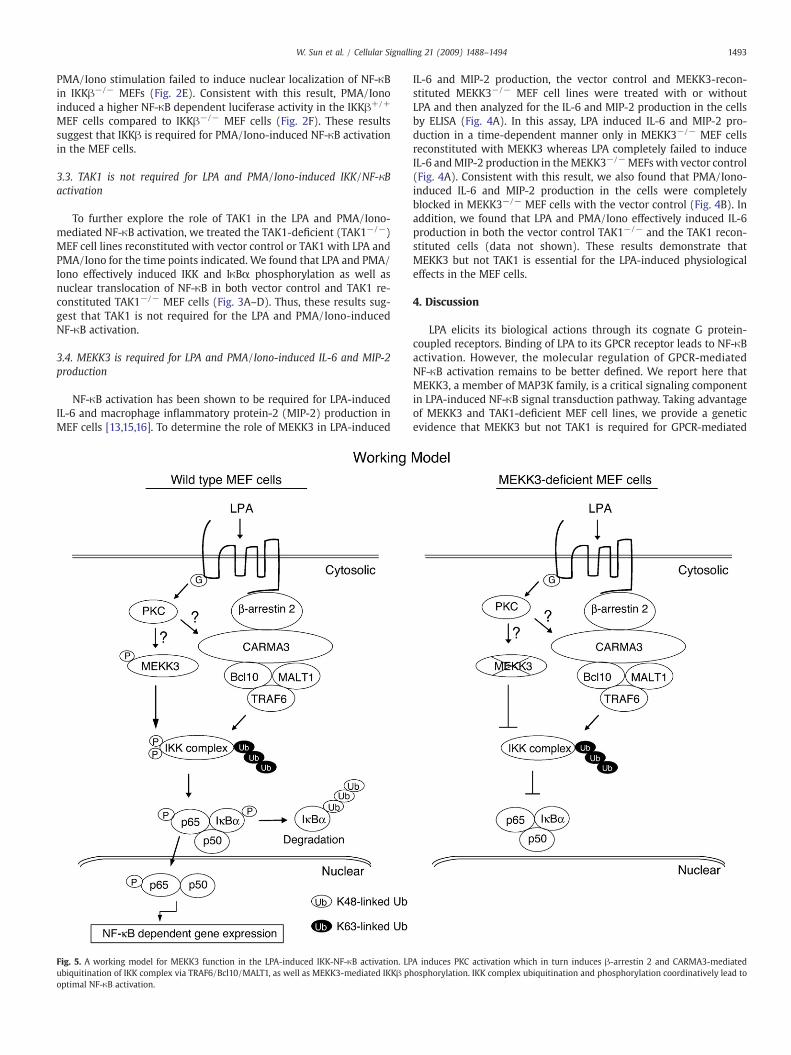
Fig. 5. A working model for MEKK3 function in the LPA-induced IKK-NF-κB activation. LPubiquitination of IKK complex via TRAF6/Bcl10/MALT1, as well as MEKK3-mediated IKKβ phoptimal NF-κB activation.
IL-6 and MIP-2 production, the vector control and MEKK3-recon-stituted MEKK3−/− MEF cell lines were treated with or withoutLPA and then analyzed for the IL-6 and MIP-2 production in the cellsby ELISA (Fig. 4A). In this assay, LPA induced IL-6 and MIP-2 pro-duction in a time-dependent manner only in MEKK3−/− MEF cellsreconstituted with MEKK3 whereas LPA completely failed to induceIL-6 andMIP-2 production in theMEKK3−/−MEFswith vector control(Fig. 4A). Consistent with this result, we also found that PMA/Iono-induced IL-6 and MIP-2 production in the cells were completelyblocked in MEKK3−/− MEF cells with the vector control (Fig. 4B). Inaddition, we found that LPA and PMA/Iono effectively induced IL-6production in both the vector control TAK1−/− and the TAK1 recon-stituted cells (data not shown). These results demonstrate thatMEKK3 but not TAK1 is essential for the LPA-induced physiologicaleffects in the MEF cells.
4. Discussion
LPA elicits its biological actions through its cognate G protein-coupled receptors. Binding of LPA to its GPCR receptor leads to NF-κBactivation. However, the molecular regulation of GPCR-mediatedNF-κB activation remains to be better defined. We report here thatMEKK3, a member of MAP3K family, is a critical signaling componentin LPA-induced NF-κB signal transduction pathway. Taking advantageof MEKK3 and TAK1-deficient MEF cell lines, we provide a geneticevidence that MEKK3 but not TAK1 is required for GPCR-mediated
A induces PKC activation which in turn induces β-arrestin 2 and CARMA3-mediatedosphorylation. IKK complex ubiquitination and phosphorylation coordinatively lead to

1494 W. Sun et al. / Cellular Signalling 21 (2009) 1488–1494
IKKβ and IκBα phosphorylation as well as NF-κB nuclear transloca-tion and activation. In addition, we also show that LPA fails to induceproduction of the pro-inflammatory cytokine IL-6 and MIP-2 onlyin MEKK3-deficient but not TAK1- deficient MEF cell lines. Theseresults suggest that MEKK3 but not TAK1 plays an essential role inLPA-induced cellular responses. In addition, we also found that IKKβ isrequired for PKC-mediated NF-κB activation in the MEF cells. Theseresults suggest that MEKK3-mediated IKKβ activation is required forLPA and PKC-induced NF-κB activation.
Previous studies using knockout MEF cells demonstrated thatβ-arrestin 2, CARMA3, Bcl10 and TRAF6 are required for LPA-inducedIKK-NF-κB activation whereas these proteins are not required forLPA-induced IKKβ phosphorylation [15,17]. These results suggestthat these proteins may be not required for LPA-mediated MEKK3activation. However, further studies are needed to determine howMEKK3 is activated by LPA and PKC-mediated signaling events innon-hematopoietic cell types.
Interestingly, in our studies, we failed to observe an obvious LPA-induced IκBα degradation eventhough LPA induced IκBα phospho-rylation. Consistent with our results, Qin et al. reported that MEKK3is required for TLR8-mediated NF-κB activation and TLR8-mediatedsignaling only causes IκBα phosphorylation but not degradation [26].Therefore, it is highly likely that the MEKK3-mediated IκBα phos-phorylation might lead to dissociation of IκBα from NF-κB withoutIκBα degradation [26].
In conclusion, our data provide evidence that MEKK3 but not TAK1is required for LPA and PKC-induced IKKβ/NF-κB activation in MEFcells. In view of the data presented here and in previous reports, wepropose a working model (Fig. 5), in which LPA binding to its cognateGPCR induces PKC activation that leads to MEKK3-mediated phospho-rylationof IKKβ andβ-arrestin2-CARMA3-Bcl10-MALT1-mediatedubi-quitination of IKK complex that coordinately lead to IKKβ-mediatedNF-κB activation in MEF cells.
Acknowledgements
We are grateful to Susan Burlingame for the excellent technicalassistance. We thank Drs. Paul Chiao, Bing Su and Sankar Ghosh forproviding IKKβ-, MEKK3- and TAK1-deficient MEFs. This researchwas supported in part by the National Institutes of Health Grant1R21CA106513-01A2 (to J.Y.), 5R01GM079451 (to X.L.), the AmericanCancer Society grant RSG-06-070-01-TBE (to J.Y.), and the Fleming andDavenport Award (to H.Z.).
References
[1] A.S. Baldwin Jr., Annu. Rev. Immunol. 14 (1996) 649.[2] M.J. May, S. Ghosh, Immunol. Today 19 (1998) 80.[3] Q. Li, I.M. Verma, Nat. Rev. Immunol. 2 (2002) 725.[4] M. Karin, Y. Ben-Neriah, Annu. Rev. Immunol. 18 (2000) 621.[5] A.A. Beg, A.S. Baldwin Jr., Genes Dev. 7 (1993) 2064.[6] A.A. Beg, T.S. Finco, P.V. Nantermet, A.S. Baldwin Jr., Mol. Cell Biol. 13 (1993) 3301.[7] S. Ghosh, M. Karin, Cell 109 (2002) S81 (Suppl).[8] I.M. Verma, J.K. Stevenson, E.M. Schwarz, A.D. Van, S. Miyamoto, Genes Dev. 9
(1995) 2723.[9] M.S. Hayden, S. Ghosh, Genes Dev. 18 (2004) 2195.[10] V. Dixit, T.W. Mak, Cell 111 (2002) 615.[11] R.D. Ye, J. Leukoc. Biol. 70 (2001) 839.[12] G.B. Mills, W.H. Moolenaar, Nat. Rev. Cancer 3 (2003) 582.[13] S. Klemm, S. Zimmermann, C. Peschel, T.W.Mak, J. Ruland, Proc. Natl. Acad. Sci. U. S. A.
104 (2007) 134.[14] L.M. lister-Lucas, J. Ruland, K. Siu, X. Jin, S. Gu, D.S. Kim, P. Kuffa, D. Kohrt, T.W. Mak,
G. Nunez, P.C. Lucas, Proc. Natl. Acad. Sci. U. S. A. 104 (2007) 139.[15] B.C. Grabiner, M. Blonska, P.C. Lin, Y. You, D. Wang, J. Sun, B.G. Darnay, C. Dong, X.
Lin, Genes Dev. 21 (2007) 984.[16] D. Wang, Y. You, P.C. Lin, L. Xue, S.W. Morris, H. Zeng, R. Wen, X. Lin, Proc. Natl.
Acad. Sci. U. S. A. 104 (2007) 145.[17] J. Sun, X. Lin, Proc. Natl. Acad. Sci. U. S. A. 105 (2008) 17085.[18] Q. Zhao, F.S. Lee, J. Biol. Chem. 274 (1999) 8355.[19] C.Wang, L. Deng, M. Hong, G.R. Akkaraju, J. Inoue, Z.J. Chen, Nature 412 (2001) 346.[20] J. Yang, Y. Lin, Z. Guo, J. Cheng, J. Huang, L. Deng, W. Liao, Z. Chen, Z. Liu, B. Su, Nat.
Immunol. 2 (2001) 620.[21] S. Sato, H. Sanjo, K. Takeda, J. Ninomiya-Tsuji, M. Yamamoto, T. Kawai, K.Matsumoto,
O. Takeuchi, S. Akira, Nat. Immunol. 6 (2005) 1087.[22] J.H. Shim, C. Xiao, A.E. Paschal, S.T. Bailey, P. Rao, M.S. Hayden, K.Y. Lee, C. Bussey,
M. Steckel, N. Tanaka, G. Yamada, S. Akira, K. Matsumoto, S. Ghosh, Genes Dev. 19(2005) 2668.
[23] C. Schmidt, B. Peng, Z. Li, G.M. Sclabas, S. Fujioka, J. Niu, M. Schmidt-Supprian, D.B.Evans, J.L. Abbruzzese, P.J. Chiao, Mol. Cell 12 (2003) 1287.
[24] C.W. Nho, P.J. O'Dwyer, J. Biol. Chem. 279 (2004) 26019.[25] K. Deacon, J.L. Blank, J. Biol. Chem. 274 (1999) 16604.[26] J. Qin, J. Yao, G. Cui, H. Xiao, T.W. Kim, J. Fraczek, P. Wightman, S. Sato, S. Akira, A.
Puel, J.L. Casanova, B. Su, X. Li, J. Biol. Chem. 281 (2006) 21013.[27] Q. Huang, J. Yang, Y. Lin, C.Walker, J. Cheng, Z.G. Liu, B. Su,Nat. Immunol. 5 (2004) 98.[28] E. Zandi, M. Karin, Mol. Cell Biol. 19 (1999) 4547.[29] T.L. Chau, R. Gioia, J.S. Gatot, F. Patrascu, I. Carpentier, J.P. Chapelle, L. O'Neill, R.
Beyaert, J. Piette, A. Chariot, Trends Biochem. Sci. 33 (2008) 171.[30] J. Hawiger, R.A. Veach, X.Y. Liu, S. Timmons, D.W. Ballard, Blood 94 (1999) 1711.[31] T. Shimada, T. Kawai, K. Takeda, M. Matsumoto, J. Inoue, Y. Tatsumi, A. Kanamaru, S.
Akira, Int. Immunol. 11 (1999) 1357.[32] Z.W. Li, W. Chu, Y. Hu, M. Delhase, T. Deerinck, M. Ellisman, R. Johnson, M. Karin,
J. Exp. Med. 189 (1999) 1839.[33] M. Tanaka,M.E. Fuentes, K. Yamaguchi,M.H. Durnin, S.A. Dalrymple, K.L. Hardy, D.V.
Goeddel, Immunity 10 (1999) 421.[34] H. Hacker, M. Karin, Sci. STKE (2006) re13.