A new multicolor bioluminescence imaging platform to investigate NF-κB activity and apoptosis in...
12
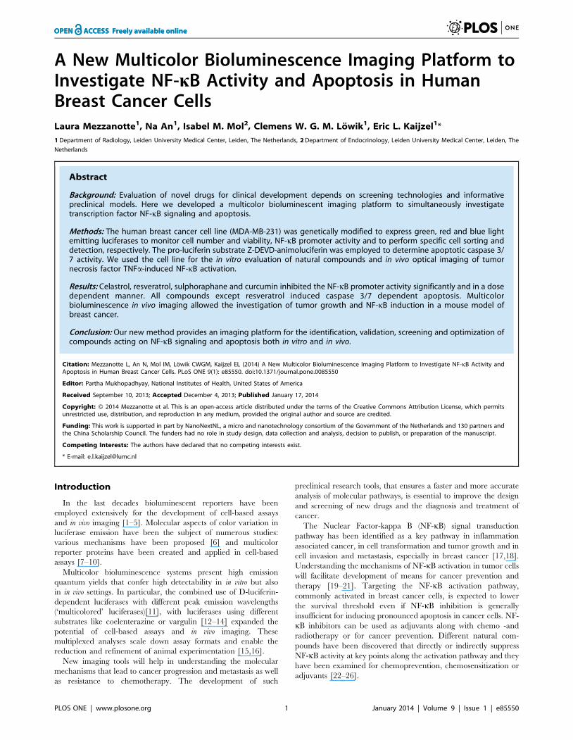
A New Multicolor Bioluminescence Imaging Platform to Investigate NF-kB Activity and Apoptosis in Human Breast Cancer Cells Laura Mezzanotte 1 , Na An 1 , Isabel M. Mol 2 , Clemens W. G. M. Lo ¨ wik 1 , Eric L. Kaijzel 1 * 1 Department of Radiology, Leiden University Medical Center, Leiden, The Netherlands, 2 Department of Endocrinology, Leiden University Medical Center, Leiden, The Netherlands Abstract Background: Evaluation of novel drugs for clinical development depends on screening technologies and informative preclinical models. Here we developed a multicolor bioluminescent imaging platform to simultaneously investigate transcription factor NF-kB signaling and apoptosis. Methods: The human breast cancer cell line (MDA-MB-231) was genetically modified to express green, red and blue light emitting luciferases to monitor cell number and viability, NF-kB promoter activity and to perform specific cell sorting and detection, respectively. The pro-luciferin substrate Z-DEVD-animoluciferin was employed to determine apoptotic caspase 3/ 7 activity. We used the cell line for the in vitro evaluation of natural compounds and in vivo optical imaging of tumor necrosis factor TNFa-induced NF-kB activation. Results: Celastrol, resveratrol, sulphoraphane and curcumin inhibited the NF-kB promoter activity significantly and in a dose dependent manner. All compounds except resveratrol induced caspase 3/7 dependent apoptosis. Multicolor bioluminescence in vivo imaging allowed the investigation of tumor growth and NF-kB induction in a mouse model of breast cancer. Conclusion: Our new method provides an imaging platform for the identification, validation, screening and optimization of compounds acting on NF-kB signaling and apoptosis both in vitro and in vivo. Citation: Mezzanotte L, An N, Mol IM, Lo ¨ wik CWGM, Kaijzel EL (2014) A New Multicolor Bioluminescence Imaging Platform to Investigate NF-kB Activity and Apoptosis in Human Breast Cancer Cells. PLoS ONE 9(1): e85550. doi:10.1371/journal.pone.0085550 Editor: Partha Mukhopadhyay, National Institutes of Health, United States of America Received September 10, 2013; Accepted December 4, 2013; Published January 17, 2014 Copyright: ß 2014 Mezzanotte et al. This is an open-access article distributed under the terms of the Creative Commons Attribution License, which permits unrestricted use, distribution, and reproduction in any medium, provided the original author and source are credited. Funding: This work is supported in part by NanoNextNL, a micro and nanotechnology consortium of the Government of the Netherlands and 130 partners and the China Scholarship Council. The funders had no role in study design, data collection and analysis, decision to publish, or preparation of the manuscript. Competing Interests: The authors have declared that no competing interests exist. * E-mail: [email protected] Introduction In the last decades bioluminescent reporters have been employed extensively for the development of cell-based assays and in vivo imaging [1–5]. Molecular aspects of color variation in luciferase emission have been the subject of numerous studies: various mechanisms have been proposed [6] and multicolor reporter proteins have been created and applied in cell-based assays [7–10]. Multicolor bioluminescence systems present high emission quantum yields that confer high detectability in in vitro but also in in vivo settings. In particular, the combined use of D-luciferin- dependent luciferases with different peak emission wavelengths (‘multicolored’ luciferases)[11], with luciferases using different substrates like coelenterazine or vargulin [12–14] expanded the potential of cell-based assays and in vivo imaging. These multiplexed analyses scale down assay formats and enable the reduction and refinement of animal experimentation [15,16]. New imaging tools will help in understanding the molecular mechanisms that lead to cancer progression and metastasis as well as resistance to chemotherapy. The development of such preclinical research tools, that ensures a faster and more accurate analysis of molecular pathways, is essential to improve the design and screening of new drugs and the diagnosis and treatment of cancer. The Nuclear Factor-kappa B (NF-kB) signal transduction pathway has been identified as a key pathway in inflammation associated cancer, in cell transformation and tumor growth and in cell invasion and metastasis, especially in breast cancer [17,18]. Understanding the mechanisms of NF-kB activation in tumor cells will facilitate development of means for cancer prevention and therapy [19–21]. Targeting the NF-kB activation pathway, commonly activated in breast cancer cells, is expected to lower the survival threshold even if NF-kB inhibition is generally insufficient for inducing pronounced apoptosis in cancer cells. NF- kB inhibitors can be used as adjuvants along with chemo -and radiotherapy or for cancer prevention. Different natural com- pounds have been discovered that directly or indirectly suppress NF-kB activity at key points along the activation pathway and they have been examined for chemoprevention, chemosensitization or adjuvants [22–26]. PLOS ONE | www.plosone.org 1 January 2014 | Volume 9 | Issue 1 | e85550
Transcript of A new multicolor bioluminescence imaging platform to investigate NF-κB activity and apoptosis in...
A New Multicolor Bioluminescence Imaging Platform toInvestigate NF-kB Activity and Apoptosis in HumanBreast Cancer CellsLaura Mezzanotte1, Na An1, Isabel M. Mol2, Clemens W. G. M. Lowik1, Eric L. Kaijzel1*
1 Department of Radiology, Leiden University Medical Center, Leiden, The Netherlands, 2 Department of Endocrinology, Leiden University Medical Center, Leiden, The
Netherlands
Abstract
Background: Evaluation of novel drugs for clinical development depends on screening technologies and informativepreclinical models. Here we developed a multicolor bioluminescent imaging platform to simultaneously investigatetranscription factor NF-kB signaling and apoptosis.
Methods: The human breast cancer cell line (MDA-MB-231) was genetically modified to express green, red and blue lightemitting luciferases to monitor cell number and viability, NF-kB promoter activity and to perform specific cell sorting anddetection, respectively. The pro-luciferin substrate Z-DEVD-animoluciferin was employed to determine apoptotic caspase 3/7 activity. We used the cell line for the in vitro evaluation of natural compounds and in vivo optical imaging of tumornecrosis factor TNFa-induced NF-kB activation.
Results: Celastrol, resveratrol, sulphoraphane and curcumin inhibited the NF-kB promoter activity significantly and in a dosedependent manner. All compounds except resveratrol induced caspase 3/7 dependent apoptosis. Multicolorbioluminescence in vivo imaging allowed the investigation of tumor growth and NF-kB induction in a mouse model ofbreast cancer.
Conclusion: Our new method provides an imaging platform for the identification, validation, screening and optimization ofcompounds acting on NF-kB signaling and apoptosis both in vitro and in vivo.
Citation: Mezzanotte L, An N, Mol IM, Lowik CWGM, Kaijzel EL (2014) A New Multicolor Bioluminescence Imaging Platform to Investigate NF-kB Activity andApoptosis in Human Breast Cancer Cells. PLoS ONE 9(1): e85550. doi:10.1371/journal.pone.0085550
Editor: Partha Mukhopadhyay, National Institutes of Health, United States of America
Received September 10, 2013; Accepted December 4, 2013; Published January 17, 2014
Copyright: ! 2014 Mezzanotte et al. This is an open-access article distributed under the terms of the Creative Commons Attribution License, which permitsunrestricted use, distribution, and reproduction in any medium, provided the original author and source are credited.
Funding: This work is supported in part by NanoNextNL, a micro and nanotechnology consortium of the Government of the Netherlands and 130 partners andthe China Scholarship Council. The funders had no role in study design, data collection and analysis, decision to publish, or preparation of the manuscript.
Competing Interests: The authors have declared that no competing interests exist.
* E-mail: [email protected]
Introduction
In the last decades bioluminescent reporters have beenemployed extensively for the development of cell-based assaysand in vivo imaging [1–5]. Molecular aspects of color variation inluciferase emission have been the subject of numerous studies:various mechanisms have been proposed [6] and multicolorreporter proteins have been created and applied in cell-basedassays [7–10].
Multicolor bioluminescence systems present high emissionquantum yields that confer high detectability in in vitro but alsoin in vivo settings. In particular, the combined use of D-luciferin-dependent luciferases with different peak emission wavelengths(‘multicolored’ luciferases)[11], with luciferases using differentsubstrates like coelenterazine or vargulin [12–14] expanded thepotential of cell-based assays and in vivo imaging. Thesemultiplexed analyses scale down assay formats and enable thereduction and refinement of animal experimentation [15,16].
New imaging tools will help in understanding the molecularmechanisms that lead to cancer progression and metastasis as wellas resistance to chemotherapy. The development of such
preclinical research tools, that ensures a faster and more accurateanalysis of molecular pathways, is essential to improve the designand screening of new drugs and the diagnosis and treatment ofcancer.
The Nuclear Factor-kappa B (NF-kB) signal transductionpathway has been identified as a key pathway in inflammationassociated cancer, in cell transformation and tumor growth and incell invasion and metastasis, especially in breast cancer [17,18].Understanding the mechanisms of NF-kB activation in tumor cellswill facilitate development of means for cancer prevention andtherapy [19–21]. Targeting the NF-kB activation pathway,commonly activated in breast cancer cells, is expected to lowerthe survival threshold even if NF-kB inhibition is generallyinsufficient for inducing pronounced apoptosis in cancer cells. NF-kB inhibitors can be used as adjuvants along with chemo -andradiotherapy or for cancer prevention. Different natural com-pounds have been discovered that directly or indirectly suppressNF-kB activity at key points along the activation pathway and theyhave been examined for chemoprevention, chemosensitization oradjuvants [22–26].
PLOS ONE | www.plosone.org 1 January 2014 | Volume 9 | Issue 1 | e85550
Effects of new candidate anticancer drugs on NF-kB signalinghave been extensively investigated by classic cell-based approacheslike transient transfection assays using NF-kB promoter-reporterplasmids [27] and electrophoresis mobility shift assays [28].Optical tools and transgenic bioluminescent animals have beenintroduced to study the effects on NF-kB signaling, like p65-GFPfusions [29], IkBa-luciferase (IkBa-FLuc) fusion reporters [30] andNF-kB luc reporter mice [31].
In this study we developed and validated a new triple colorcancer cell system generated by lentiviral transduction of thehuman breast cancer cell line MDA-MB-231 with differentbioluminescent reporters. In particular, the click beetle greenluciferase (CBG99) was used to monitor cell vitality while the redmutant of firefly luciferase (PpyRE9) monitored NF-kB promoteractivity. The blue extGluc, a transmembrane form of Gaussialuciferase, served as a reporter for in vitro cell sorting and ex vivo cellanalysis. Additionally, the use of the luciferase pro-substrate Z-DEVD-aminoluciferin, containing the DEVD tetrapeptide se-quence recognized by caspase-3 and -7, allows the non-invasiveimaging of apoptosis in luciferase expressing cells both in vitro andin vivo [32]. Upon activation of caspase-3 or -7 in apoptotic cells,the DEVD peptide is cleaved, and the liberated aminoluciferinreacts with luciferase to generate measurable light.
By means of this new imaging platform we investigated theeffects of plant-derived natural compounds like celastrol, resver-atrol, curcumin, betulinic acid and sulphoraphane on TNFa-induced NF-kB activation and apoptosis in human breast cancercells. For the first time, analysis of NF-kB activation and tumorprogression from cells to animal models using multicoloredreporter genes for drug testing is reported.
Experimental Procedures
Ethical statementAnimal experiments were reviewed and approved by the
Bioethics Committee of Leiden University, The Netherlands. Allanimals received human care in compliance with the ‘‘Code ofPractice Use of Laboratory Animals in Cancer Research’’(Inspectie W&V, July 1999).
Cell culture and reagentsMDA-MB-231 human breast cancer cells (ATCC number
HTB-26TM) were maintained in Dulbecco’s modified Eagle’smedium (Invitrogen, Grand Island, U.S.A.) supplemented with10% fetal bovine serum (Sigma, St. Louis, U.S.A.), 100 U/mlpenicillin (Sigma) and 0.1 mg/ml streptomycin (Sigma). The cellswere grown at 37uC in a 5% CO2 humidified incubator.Compounds were purchased from the following companies:celastrol and curcumin (Cayman, Michigan, U.S.A.), L-sulforaph-ane and resveratrol (Sigma), and betulinic acid (Tocris, Bristol,U.K.). Human recombinant TNFa was purchased from Sigma.
Lentiviral vectors constructionTo create the pRRL-PGKCBG99, the CBG99 luciferase gene
was excised with NheI and XbaI from pGL3-CBG99 (Promega,Leiden, The Netherlands) and cloned in the MCS of pRRL-PGK(a kind gift from Prof. R. Hoeben [33]).
The bicistronic pLM-NF-kB PpyRE9 vector was constructed byamplifying the PpyRE9 luciferase gene from the vector pGex-6p-2-PpyRE9 (provided by Prof. B. Branchini [34]) using thefollowing primers: forward primer 59-GGCGGCCATGGAA-GACGCCAAAAACATAAAG-39 with a NcoI restriction siteand reverse primer 59-GTCTAGATTAGATTTTCCGCCC-TTCTTGGCCTT-39 with XbaI restriction site. Next, the
PpyRE9 luciferase gene was cloned by replacing the luc gene inthe pGL3 control vector (Promega) to create the pGL3-PpyRE9vector. NF-kB promoter responsive elements were excised withKpnI and NcoI from the vector pGL4.32 [luc2P/NF-kB-RE/Hygro] (Promega) and cloned into the pGL3-PpyRE9 vector inorder to create the pGL3-NF-kB PpyRE9 vector. Subsequently,the NF-kB PpyRE9 cassette was excised with KpnI and XbaIrestriction enzymes, blunted and cloned into the bicistronicbidirectional pNFAT vector (kindly provided by Prof. M.R. Vande Brink [35]) in which the NFAT-CBRed cassette had beendeleted. The resulting bicistronic lentiviral vector named pLM-NF-kB PpyRE9 was checked for orientation. The new plasmidcontains the original Gaussia Luciferase [extGLuc] under thecontrol of PGK promoter in one direction and the NF-kB PpyRE9cassette in the other direction.
Cell Transduction and selectionSelf-inactivating lentiviruses were produced as previously
described [33]. MDA-MB-231 cells were transduced in sequentialsteps. First, MDA-MB-231 cells were transduced with a lentivirusexpressing pRRL-PGKCBG99 and selected by limiting dilutionmethods. Then, the obtained CBG99 luciferase positive cell linewas transduced with the pLM-NF-kB PpyRE9 lentivirus andFACS-sorted using a polyclonal anti-Gaussia luciferase antibody(Nanolight tech., Pinetop, AZ).
MTS and Luciferase assayTriple colored MDA-MB-231 cells were seeded in 96-well plates
and, 24 hours later, treated with different concentrations ofpuromycin (0.1 mg/ml – 10 mg/ml), a known selective antibiotictoxic to eukaryotic cells. After another 24 hours of incubation,plates were divided for the different analyses. An MTS colorimet-ric assay for assessing cell viability, CellTiter 96H AQueous OneSolution Cell Proliferation Assay (Promega, Leiden, The Nether-lands) was used according to manufacturers’ description. In brief,MTS solution was added to the medium for 2 hours after whichthe absorbance was measured at 490 nm with an ELISAmicroplate reader (VersaMax Molecular Devices, Sunnyvale,CA). To measure the luciferase activity biochemically, cells werewashed with PBS, harvested in reporter lysis buffer and assayed forluciferase activity with a luminometer (SpectraMax L lumines-cence microplate reader, Molecular Devices). The cell viabilitycurves were made for both MTS -and luciferase assays.
Reporter gene activity assayTriple colored MDA-MB-231 cells were seeded in 96-well dark
plates (Greiner bio-one, Germany) at a concentration of5000 cells/well, 24 hours prior treatment. The cell line was firstvalidated for NF-kB responses by adding TNFa (10 ng/ml) fordifferent time points of incubation (ranging from 2 to 72 hours).After establishing the optimal period of TNFa stimulation, theeffect of the plant-derived compounds was investigated on TNFa-induced NF-kB activation in the MDA-MB-231 cells. For that,cells were treated with medium (negative control), TNFa (10 ng/ml, positive control), TNFa together with chemopreventivenatural compounds: celastrol (0.1 mM–2.5 mM), sulforaphane(5 mM–100 mM), curcumin (10 mM–20 mM), resveratrol (1 mM–200 mM) or betulinic acid (0.1 mM–30 mM), followed by another24 hours of incubation. Luciferase activity was determined asfollows: the medium in plate was replaced with phosphate-bufferedsaline and D-Luciferin (Synchem, Germany) was added at a finalconcentration of 0.5 mM. Luciferase activity was measured usingan IVIS Spectrum (Caliper, Alameda, CA, USA). The instrumentstage was kept at 37uC and imaging set up was FOV C. Light
Multicolor Imaging of NF-kB Activity and Apoptosis
PLOS ONE | www.plosone.org 2 January 2014 | Volume 9 | Issue 1 | e85550
output was measured using an open filter and a series of band passfilter (20 nm) ranging from 500 nm to 700 nm each for 5 sec,5 min after substrate addition to live cells. All the data areexpressed in photon-flux and analyzed with Living ImageSoftware 4.0 (Caliper, Alameda, CA, USA). A spectral unmixingalgorithm was applied to the images to separate the red and greenluciferases as described previously [11]. Data were calculateddepicting multiple region of interests (ROIs) corresponding to thewell areas of the 96-well black plates in the images correspondingto the unmixing results. CBG luciferase activity in the greenspectrum (peak emission wavelength 540 nm) has been used as anindication of cell vitality and the PpyRE9 luciferase activity in thered spectrum (peak emission wavelength 620 nm) as an indicationof promoter activity. Measurement of extGLuc expression wascarried out using native coelenterazine (Nanolight tech.) at a finalconcentration of 20 mM.
Western blot analysis of P65 in nuclear extractsTo confirm the results of the reporter gene assays, western blot
analysis was performed to test NF-kB activity in the nuclearextracts. MDA-MB-231 cells (16106 cells/well) were seeded in 6-well plates for 24 hours, then treated with medium (negativecontrol), TNFa (10 ng/ml positive control), or TNFa (10 ng/ml)with celastrol (0.1 mM, 0.5 mM and 1 mM) or betulinic acid(0.5 mM, 1 mM, 10 mM and 30 mM). After 24 hours of treatmentcells were collected, and nuclear extracts were made using Nuclearand Cytoplasmic extraction reagents (Thermo Scientific, Rock-ford, U.S.A.). Total amount of protein of each sample wasdetermined by a Pierce BCA protein assay kit (Thermo Scientific,Rockford, U.S.A.). 15 mg of nuclear extracts was applied to a 10%SDS-PAGE and transferred onto a nitrocellulose membrane. Afterwashing, the membrane was incubated with an anti-P65polyclonal antibody in TPBS 1:1000 dilutions (Cell SignalingTechnology, Danvers, U.S.A.) overnight at room temperature. AGAPDH antibody (Cell Signaling Technology) was used to correctfor the amount of total protein. The blots were washed, exposed toan HRP-conjugated secondary antibody for 1 hour, and finallydetected using enhanced chemiluminescence (ECL) reagents(Thermo Scientific). Detection of ECL signals was performedwith the IVIS Spectrum and quantification of bands using LivingImage Software 4.0.
Caspase 3/7 activity apoptosis assayTriple colored MDA-MB-231 cells were seeded in a 96-well
dark plate (Greiner bio-one) for 24 hours to attach. The cells werethen treated with medium (negative control), or chemopreventivenatural compounds, followed by another 4 hours or 24 hours ofincubation. Rows of cells were then divided to estimate the effectof the different compounds on cell viability and apoptosis. Cellviability was established by the addition of D-luciferin to live cells;caspase 3/7 activity was determined by adding the luciferase pro-substrate, Z-DEVD-aminoluciferin using a Caspase-Glo 3/7 Assay(Promega). Upon activation of caspase-3 or -7, the DEVD peptideis cleaved off, releasing the aminoluciferin to react with luciferasegenerating measurable light [32]. The bioluminescent signalsgenerated by the addition of the Z-DEVD-aminoluciferin weredivided by the signals generated by the CBG99 luciferase uponaddition of D-luciferin in order to correct the data set for theamount of viable cells. Data are reported showing the correctedsignal for controls (only medium added) and the differenttreatments (medium containing the different plant-derived naturalcompounds).
Triple colored MDA-MB-231 xenograft modelTriple colored MDA-MB-231 cells (26106) were implanted in
the mammary fat pads of female athymic (BALB/c nu/nu) mice.D-luciferin (150 mg/kg) was injected intraperitoneally and imag-ing started 5 minutes later using a FOV C and a 30 sec acquisitiontime for both open filter and band pass filters acquisition. Micewere imaged weekly. Tumors reached a palpable size (around150 mm3) after two to three weeks.
Induction of NF-kBWhen tumor growth reached an exponential increase in CBG-
luc signals, mice were randomized in three groups. A controlgroup received an intratumoral injection of PBS and anintravenous injection of PBS. TNFa (20 mg/kg) was injectedintravenously in the second and third group. Next, these micewere given an intratumoral injection of celastrol (2 mg/kg) or PBS.Mice were imaged after the injection and after 24 hours with thesame settings. Evaluation of NF-kB induction was performed bycomparison of images before and 24 hours after treatment.
Induction of apoptosisTo measure induction of apoptosis, triple colored MDA-MB-
231 tumors were grown as described above, intratumorallyinjected with celastrol and after 4 and 24 hours Z-DEVD-aminoluciferin (50 mg/kg) was injected intraperitoneally. Activa-tion of caspase 3/7 was measured 20 min after injection ofsubstrate using an open filter and an exposure time of 30 sec.
ImmunohistochemistryTumors were surgically removed, fixed in 4% formaldehyde
and further processed for paraffin embedment. Sections (6–7 mm)were made using a standard microtome and placed on glass slides.A polyclonal rabbit anti-Gaussia luciferase antibody (Nanolighttech.) and an anti-rabbit–FITC antibody were used to reveal thepresence of Gaussia luciferase positive tumor cells.
Statistical analysisEach in vitro experiment was performed three times with six
replicate samples per data point. A Student’s t-test has beenapplied to determine statistically significant differences in thepromoter activity between positive controls and treated conditions.For in vivo experiments wherein more than two groups werecompared, one-way ANOVA followed by Tukey’s post-hoc testwas used to determine significant differences among treatedgroups.
Results
Generation of bioluminescent lentiviral vectorsThe pRRL-PGKCBG99 vector contained the CBG99 lucifer-
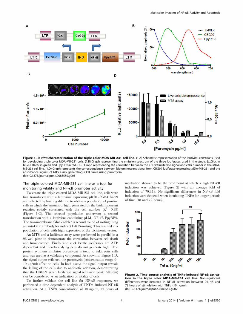
ase gene driven by a PGK promoter with a peak of emission at540 nm after addition of D-luciferin substrate. The bicistronicvector pLM-NF-kB PpyRE9 consisted of a NF-kB PpyRE9 and aPGK-extGLuc cassette separated by an insulator sequence toensure high induction of the promoters after integration of thevector (Figure 1.A). PpyRE9 luciferase has a peak of emission at620 nm using D-luciferin as a substrate. The presence of thetransmembrane Gluc reporter gene under the control of theconstitutive promoter in the bicistronic vector represents the ‘‘thirdcolor’’ since the peak of emission of Gluc luciferase is 480 nm andneeds the addition of a different substrate (e.g. coelenterazine)(Figure 1.B).
Multicolor Imaging of NF-kB Activity and Apoptosis
PLOS ONE | www.plosone.org 3 January 2014 | Volume 9 | Issue 1 | e85550
The triple colored MDA-MB-231 cell line as a tool formonitoring vitality and NF-kB promoter activity
To create the triple colored MDA-MB-231 cell line, cells werefirst transduced with a lentivirus expressing pRRL-PGKCBG99and selected by limiting dilution to obtain a population of positivecells in which the amount of light generated by the bioluminescentreaction strictly correlated with the cell number (R2 = 0.99)(Figure 1.C). The selected population underwent a secondtransduction with a lentivirus containing pLM- NF-kB PpyRE9.The transmembrane Gluc enabled a second round of sorting usingan anti-Gluc antibody for indirect FACS-sorting. This resulted in apopulation of cells with high expression of the bicistronic vector.
An MTS and a luciferase assay were performed in parallel in a96-well plate to demonstrate the correlation between cell deathand luminescence. Firefly and click beetle luciferases are ATPdependent and therefore dying cells do not generate light. Theprotein synthesis inhibitor puromycin is toxic to eukaryotic cellsand was used as a validating compound. As shown in Figure 1.D,the signal output reflected the puromycin (concentration range 0–10 mg/ml) effect on cells. In both assays the signal output revealsthe killing of the cells due to antibiotic addition, demonstratingthat the CBG99 green luciferase signal (emission peak: 540 nm)can be considered as an indication of vitality of cells.
To further validate the cell line for NF-kB responses, weperformed a time dependent analysis of TNFa- induced NF-kBactivation. At a TNFa concentration of 10 ng/ml, 24 hours of
incubation showed to be the time point at which a high NF-kBinduction was achieved (Figure 2) with an average fold ofinduction of 70613. No significant differences in NF-kB foldinduction were detected when incubating TNFa for longer periodsof time (48 and 72 hours).
Figure 1. In vitro characterization of the triple color MDA-MB-231 cell line. (1.A) Schematic representation of the lentiviral constructs usedfor developing triple color MDA-MB-231 cells. (1.B) Graph representing the emission spectrum of the three luciferases used in the study. ExtGluc inblue, CBG99 in green and PpyRE9 in red. (1.C) Graph representing the correlation between the CBG99 luciferase signal and cells number in the MDA-MB-231 cell line. (1.D) Graph represents the correspondence between bioluminescent signal from CBG99 luciferase expressing MDA-MB-231 and theabsorbance signals of MTS assay generating a kill curve using puromycin.doi:10.1371/journal.pone.0085550.g001
Figure 2. Time course analysis of TNFa-induced NF-kB activa-tion in the triple color MDA-MB-231 cell line. Non-significantdifferences were detected in NF-kB activation between 24, 48 and72 hours of stimulation with TNFa (10 ng/ml).doi:10.1371/journal.pone.0085550.g002
Multicolor Imaging of NF-kB Activity and Apoptosis
PLOS ONE | www.plosone.org 4 January 2014 | Volume 9 | Issue 1 | e85550
Effects of chemopreventive natural compounds on NF-kB promoter activity
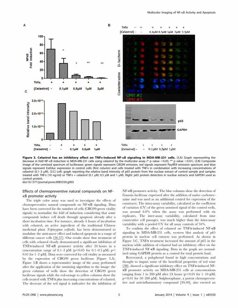
The triple color assay was used to investigate the effects ofchemopreventive natural compounds on NF-kB signaling. Datahave been corrected for the number of cells (CBG99-green vitalitysignals) to normalize the fold of induction considering that somecompounds induce cell death through apoptosis already after ashort incubation time. For instance, already 4 hours of incubationwith celastrol, an active ingredient of the traditional Chinesemedicinal plant Tripterygium wilfordii, has been demonstrated tomodulate the anticancer effect and induced apoptosis in a range ofdifferent cancer cells [36,37]. Our results show that treatment ofcells with celastrol clearly demonstrated a significant inhibition ofTNFa-induced NF-kB promoter activity after 24 hours in aconcentration range of 0.1–2 mM (p,0.05 for 0.1–0.5 mM; p,0.01 for 1–2 mM). Data were corrected for cell vitality as measuredby the expression of CBG99 green luciferase (Figure 3.A).Figure 3.B shows a representative image of the assay performedafter the application of the unmixing algorithm to the image: thegreen columns of wells show the detection of CBG99 greenluciferase signals while the red-orange to yellow columns show thecells treated with TNFa plus increasing concentrations of celastrol.The decrease of the red signal is indicative for the inhibition of
NF-kB promoter activity. The blue columns show the detection ofGaussia luciferase expressed after the addition of native coelenter-azine and was used as an additional control for expression of theconstructs. The intra-assay variability, calculated as the coefficientof variation (CV) of the green unmixed signal of the control wells,was around 6.0% when the assay was performed with sixreplicates. The inter-assay variability, calculated from nineconsecutive cell passages, was much higher than the intra-assayvariability with a pooled CV for all assay controls of 34%.
To confirm the effect of celastrol on TNFa-induced NF-kBsignaling in MDA-MB-231 cells, western blot analysis of p65protein in nuclear cell extracts was performed. As shown inFigure 3.C, TNFa treatment increased the amount of p65 in thenucleus while addition of celastrol had an inhibitory effect on theTNFa-induced NF-kB signaling. Data in the graph are normal-ized using GAPDH protein as a control for total protein load.
Resveratrol, a polyphenol found in high concentrations andthought to impart some of the beneficial properties of red wine[38], showed a significant inhibitory effect on TNFa-induced NF-kB promoter activity on MDA-MB-231 cells at concentrationsranging from 1 to 200 mM after 24 hours (p,0.05 for 1–10 mM;p,0.01 for 50–200 mM). Sulphoraphane, a potent chemopreven-tive and anti-inflammatory compound [39,40], also exerted an
Figure 3. Celastrol has an inhibitory effect on TNFa-induced NF-kB signaling in MDA-MB-231 cells. (3.A) Graph representing thedecrease in fold NF-kB induction in MDA-MB-231 cells using celastrol by the multicolor assay (* p value ,0.05; ** p value ,0.01). (3.B) Compositeimage of the unmixed spectrum of luciferases: green signals represent CBG99 emission, red signals represent PpyRE9 emission spectrum and bluesignals represent ExtGluc expression in control cells (first column) and cells treated with TNFa in combination with increasing concentrations ofcelastrol (0.1–3 mM). (3.C) Left: graph reporting the relative band intensity of p65 protein from the nuclear extract of control sample and samplestreated with TNFa (10 ng/ml) or TNFa + celastrol (0.1 mM; 0.5 mM and 1 mM). Right: p65 protein detection in nuclear extracts and GAPDH used ascontrol protein.doi:10.1371/journal.pone.0085550.g003
Multicolor Imaging of NF-kB Activity and Apoptosis
PLOS ONE | www.plosone.org 5 January 2014 | Volume 9 | Issue 1 | e85550
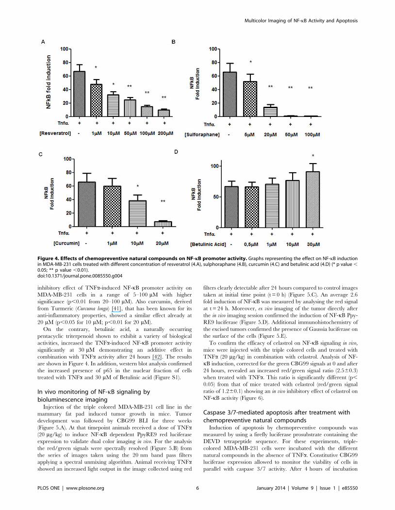
inhibitory effect of TNFa-induced NF-kB promoter activity onMDA-MB-231 cells in a range of 5–100 mM with highersignificance (p,0.01 from 20–100 mM). Also curcumin, derivedfrom Turmeric (Curcuma longa) [41], that has been known for itsanti-inflammatory properties, showed a similar effect already at20 mM (p,0.05 for 10 mM; p,0.01 for 20 mM).
On the contrary, betulinic acid, a naturally occurringpentacyclic triterpenoid shown to exhibit a variety of biologicalactivities, increased the TNFa-induced NF-kB promoter activitysignificantly at 30 mM demonstrating an additive effect incombination with TNFa activity after 24 hours [42]. The resultsare shown in Figure 4. In addition, western blot analysis confirmedthe increased presence of p65 in the nuclear fraction of cellstreated with TNFa and 30 mM of Betulinic acid (Figure S1).
In vivo monitoring of NF-kB signaling bybioluminescence imaging
Injection of the triple colored MDA-MB-231 cell line in themammary fat pad induced tumor growth in mice. Tumordevelopment was followed by CBG99 BLI for three weeks(Figure 5.A). At that timepoint animals received a dose of TNFa(20 mg/kg) to induce NF-kB dependent PpyRE9 red luciferaseexpression to validate dual color imaging in vivo. For the analysisthe red/green signals were spectrally resolved (Figure 5.B) fromthe series of images taken using the 20 nm band pass filtersapplying a spectral unmixing algorithm. Animal receiving TNFashowed an increased light output in the image collected using red
filters clearly detectable after 24 hours compared to control imagestaken at initial time point (t = 0 h) (Figure 5.C). An average 2.6fold induction of NF-kB was measured by analysing the red signalat t = 24 h. Moreover, ex vivo imaging of the tumor directly afterthe in vivo imaging session confirmed the induction of NF-kB Ppy-RE9 luciferase (Figure 5.D). Additional immunohistochemistry ofthe excised tumors confirmed the presence of Gaussia luciferase onthe surface of the cells (Figure 5.E).
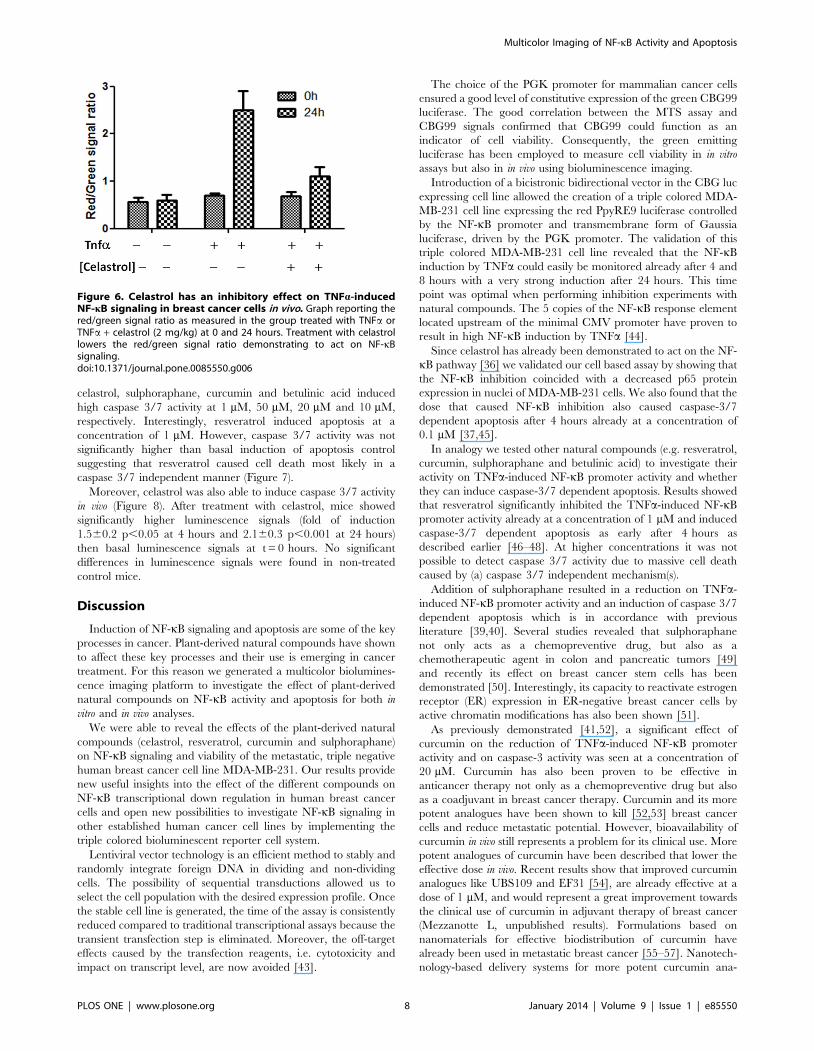
To confirm the efficacy of celastrol on NF-kB signaling in vivo,mice were injected with the triple colored cells and treated withTNFa (20 mg/kg) in combination with celastrol. Analysis of NF-kB induction, corrected for the green CBG99 signals at 0 and after24 hours, revealed an increased red/green signal ratio (2.560.3)when treated with TNFa. This ratio is significantly different (p,0.05) from that of mice treated with celastrol (red/green signalratio of 1.260.1) showing an in vivo inhibitory effect of celastrol onNF-kB activity (Figure 6).
Caspase 3/7-mediated apoptosis after treatment withchemopreventive natural compounds
Induction of apoptosis by chemopreventive compounds wasmeasured by using a firefly luciferase prosubstrate containing theDEVD tetrapeptide sequence. For these experiments, triple-colored MDA-MB-231 cells were incubated with the differentnatural compounds in the absence of TNFa. Constitutive CBG99luciferase expression allowed to monitor the viability of cells inparallel with caspase 3/7 activity. After 4 hours of incubation
Figure 4. Effects of chemopreventive natural compounds on NF-kB promoter activity. Graphs representing the effect on NF-kB inductionin MDA-MB-231 cells treated with different concentration of resveratrol (4.A), sulphoraphane (4.B), curcumin (4.C) and betulinic acid (4.D) (* p value ,0.05; ** p value ,0.01).doi:10.1371/journal.pone.0085550.g004
Multicolor Imaging of NF-kB Activity and Apoptosis
PLOS ONE | www.plosone.org 6 January 2014 | Volume 9 | Issue 1 | e85550
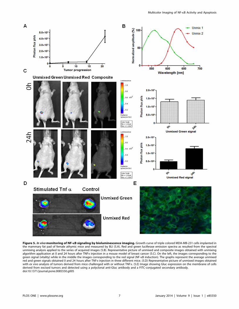
Figure 5. In vivo monitoring of NF-kB signaling by bioluminescence imaging. Growth curve of triple colored MDA-MB-231 cells implanted inthe mammary fat pad of female athymic mice and measured by BLI (5.A). Red and green luciferase emission spectra as resulted from the spectralunmixing analysis applied to the series of acquired images (5.B). Representative picture of unmixed and composite images obtained with unmixingalgorithm application at 0 and 24 hours after TNFa injection in a mouse model of breast cancer (5.C). On the left, the images corresponding to thegreen signal (vitality) while in the middle the images corresponding to the red signal (NF-kB induction). The graphs represent the average unmixedred and green signals obtained 0 and 24 hours after TNFa injection in three different mice. (5.D) Representative picture of unmixed images obtainedwith ex vivo analysis of tumors derived from mice challenged with or without TNFa. (5.E) Image showing Gluc expression on the membrane of cellsderived from excised tumors and detected using a polyclonal anti-Gluc antibody and a FITC-conjugated secondary antibody.doi:10.1371/journal.pone.0085550.g005
Multicolor Imaging of NF-kB Activity and Apoptosis
PLOS ONE | www.plosone.org 7 January 2014 | Volume 9 | Issue 1 | e85550
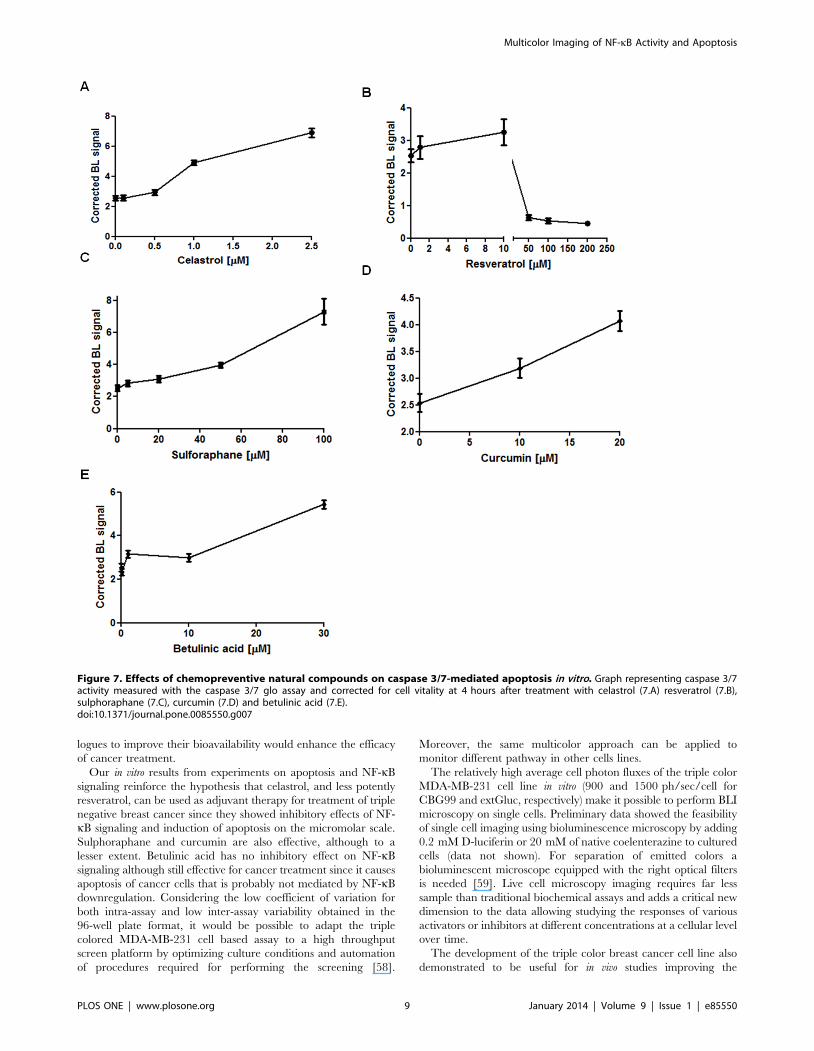
celastrol, sulphoraphane, curcumin and betulinic acid inducedhigh caspase 3/7 activity at 1 mM, 50 mM, 20 mM and 10 mM,respectively. Interestingly, resveratrol induced apoptosis at aconcentration of 1 mM. However, caspase 3/7 activity was notsignificantly higher than basal induction of apoptosis controlsuggesting that resveratrol caused cell death most likely in acaspase 3/7 independent manner (Figure 7).
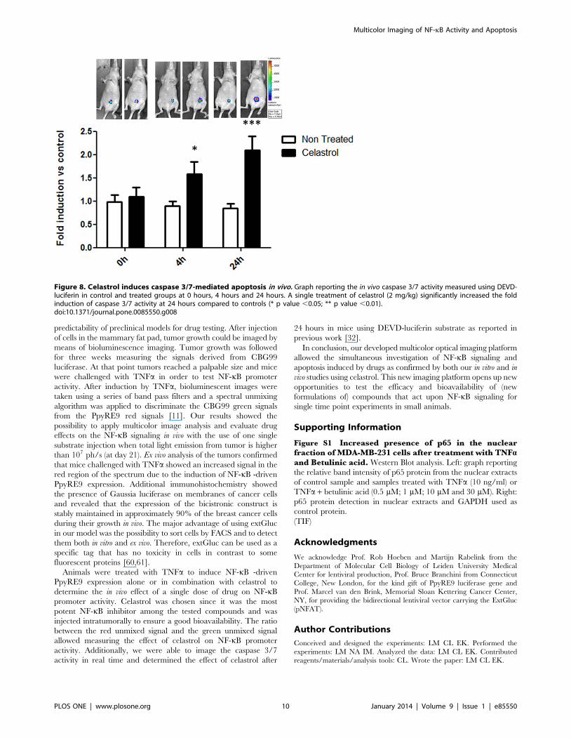
Moreover, celastrol was also able to induce caspase 3/7 activityin vivo (Figure 8). After treatment with celastrol, mice showedsignificantly higher luminescence signals (fold of induction1.560.2 p,0.05 at 4 hours and 2.160.3 p,0.001 at 24 hours)then basal luminescence signals at t = 0 hours. No significantdifferences in luminescence signals were found in non-treatedcontrol mice.
Discussion
Induction of NF-kB signaling and apoptosis are some of the keyprocesses in cancer. Plant-derived natural compounds have shownto affect these key processes and their use is emerging in cancertreatment. For this reason we generated a multicolor biolumines-cence imaging platform to investigate the effect of plant-derivednatural compounds on NF-kB activity and apoptosis for both invitro and in vivo analyses.
We were able to reveal the effects of the plant-derived naturalcompounds (celastrol, resveratrol, curcumin and sulphoraphane)on NF-kB signaling and viability of the metastatic, triple negativehuman breast cancer cell line MDA-MB-231. Our results providenew useful insights into the effect of the different compounds onNF-kB transcriptional down regulation in human breast cancercells and open new possibilities to investigate NF-kB signaling inother established human cancer cell lines by implementing thetriple colored bioluminescent reporter cell system.
Lentiviral vector technology is an efficient method to stably andrandomly integrate foreign DNA in dividing and non-dividingcells. The possibility of sequential transductions allowed us toselect the cell population with the desired expression profile. Oncethe stable cell line is generated, the time of the assay is consistentlyreduced compared to traditional transcriptional assays because thetransient transfection step is eliminated. Moreover, the off-targeteffects caused by the transfection reagents, i.e. cytotoxicity andimpact on transcript level, are now avoided [43].
The choice of the PGK promoter for mammalian cancer cellsensured a good level of constitutive expression of the green CBG99luciferase. The good correlation between the MTS assay andCBG99 signals confirmed that CBG99 could function as anindicator of cell viability. Consequently, the green emittingluciferase has been employed to measure cell viability in in vitroassays but also in in vivo using bioluminescence imaging.
Introduction of a bicistronic bidirectional vector in the CBG lucexpressing cell line allowed the creation of a triple colored MDA-MB-231 cell line expressing the red PpyRE9 luciferase controlledby the NF-kB promoter and transmembrane form of Gaussialuciferase, driven by the PGK promoter. The validation of thistriple colored MDA-MB-231 cell line revealed that the NF-kBinduction by TNFa could easily be monitored already after 4 and8 hours with a very strong induction after 24 hours. This timepoint was optimal when performing inhibition experiments withnatural compounds. The 5 copies of the NF-kB response elementlocated upstream of the minimal CMV promoter have proven toresult in high NF-kB induction by TNFa [44].
Since celastrol has already been demonstrated to act on the NF-kB pathway [36] we validated our cell based assay by showing thatthe NF-kB inhibition coincided with a decreased p65 proteinexpression in nuclei of MDA-MB-231 cells. We also found that thedose that caused NF-kB inhibition also caused caspase-3/7dependent apoptosis after 4 hours already at a concentration of0.1 mM [37,45].
In analogy we tested other natural compounds (e.g. resveratrol,curcumin, sulphoraphane and betulinic acid) to investigate theiractivity on TNFa-induced NF-kB promoter activity and whetherthey can induce caspase-3/7 dependent apoptosis. Results showedthat resveratrol significantly inhibited the TNFa-induced NF-kBpromoter activity already at a concentration of 1 mM and inducedcaspase-3/7 dependent apoptosis as early after 4 hours asdescribed earlier [46–48]. At higher concentrations it was notpossible to detect caspase 3/7 activity due to massive cell deathcaused by (a) caspase 3/7 independent mechanism(s).
Addition of sulphoraphane resulted in a reduction on TNFa-induced NF-kB promoter activity and an induction of caspase 3/7dependent apoptosis which is in accordance with previousliterature [39,40]. Several studies revealed that sulphoraphanenot only acts as a chemopreventive drug, but also as achemotherapeutic agent in colon and pancreatic tumors [49]and recently its effect on breast cancer stem cells has beendemonstrated [50]. Interestingly, its capacity to reactivate estrogenreceptor (ER) expression in ER-negative breast cancer cells byactive chromatin modifications has also been shown [51].
As previously demonstrated [41,52], a significant effect ofcurcumin on the reduction of TNFa-induced NF-kB promoteractivity and on caspase-3 activity was seen at a concentration of20 mM. Curcumin has also been proven to be effective inanticancer therapy not only as a chemopreventive drug but alsoas a coadjuvant in breast cancer therapy. Curcumin and its morepotent analogues have been shown to kill [52,53] breast cancercells and reduce metastatic potential. However, bioavailability ofcurcumin in vivo still represents a problem for its clinical use. Morepotent analogues of curcumin have been described that lower theeffective dose in vivo. Recent results show that improved curcuminanalogues like UBS109 and EF31 [54], are already effective at adose of 1 mM, and would represent a great improvement towardsthe clinical use of curcumin in adjuvant therapy of breast cancer(Mezzanotte L, unpublished results). Formulations based onnanomaterials for effective biodistribution of curcumin havealready been used in metastatic breast cancer [55–57]. Nanotech-nology-based delivery systems for more potent curcumin ana-
Figure 6. Celastrol has an inhibitory effect on TNFa-inducedNF-kB signaling in breast cancer cells in vivo. Graph reporting thered/green signal ratio as measured in the group treated with TNFa orTNFa + celastrol (2 mg/kg) at 0 and 24 hours. Treatment with celastrollowers the red/green signal ratio demonstrating to act on NF-kBsignaling.doi:10.1371/journal.pone.0085550.g006
Multicolor Imaging of NF-kB Activity and Apoptosis
PLOS ONE | www.plosone.org 8 January 2014 | Volume 9 | Issue 1 | e85550
logues to improve their bioavailability would enhance the efficacyof cancer treatment.
Our in vitro results from experiments on apoptosis and NF-kBsignaling reinforce the hypothesis that celastrol, and less potentlyresveratrol, can be used as adjuvant therapy for treatment of triplenegative breast cancer since they showed inhibitory effects of NF-kB signaling and induction of apoptosis on the micromolar scale.Sulphoraphane and curcumin are also effective, although to alesser extent. Betulinic acid has no inhibitory effect on NF-kBsignaling although still effective for cancer treatment since it causesapoptosis of cancer cells that is probably not mediated by NF-kBdownregulation. Considering the low coefficient of variation forboth intra-assay and low inter-assay variability obtained in the96-well plate format, it would be possible to adapt the triplecolored MDA-MB-231 cell based assay to a high throughputscreen platform by optimizing culture conditions and automationof procedures required for performing the screening [58].
Moreover, the same multicolor approach can be applied tomonitor different pathway in other cells lines.
The relatively high average cell photon fluxes of the triple colorMDA-MB-231 cell line in vitro (900 and 1500 ph/sec/cell forCBG99 and extGluc, respectively) make it possible to perform BLImicroscopy on single cells. Preliminary data showed the feasibilityof single cell imaging using bioluminescence microscopy by adding0.2 mM D-luciferin or 20 mM of native coelenterazine to culturedcells (data not shown). For separation of emitted colors abioluminescent microscope equipped with the right optical filtersis needed [59]. Live cell microscopy imaging requires far lesssample than traditional biochemical assays and adds a critical newdimension to the data allowing studying the responses of variousactivators or inhibitors at different concentrations at a cellular levelover time.
The development of the triple color breast cancer cell line alsodemonstrated to be useful for in vivo studies improving the
Figure 7. Effects of chemopreventive natural compounds on caspase 3/7-mediated apoptosis in vitro. Graph representing caspase 3/7activity measured with the caspase 3/7 glo assay and corrected for cell vitality at 4 hours after treatment with celastrol (7.A) resveratrol (7.B),sulphoraphane (7.C), curcumin (7.D) and betulinic acid (7.E).doi:10.1371/journal.pone.0085550.g007
Multicolor Imaging of NF-kB Activity and Apoptosis
PLOS ONE | www.plosone.org 9 January 2014 | Volume 9 | Issue 1 | e85550
predictability of preclinical models for drug testing. After injectionof cells in the mammary fat pad, tumor growth could be imaged bymeans of bioluminescence imaging. Tumor growth was followedfor three weeks measuring the signals derived from CBG99luciferase. At that point tumors reached a palpable size and micewere challenged with TNFa in order to test NF-kB promoteractivity. After induction by TNFa, bioluminescent images weretaken using a series of band pass filters and a spectral unmixingalgorithm was applied to discriminate the CBG99 green signalsfrom the PpyRE9 red signals [11]. Our results showed thepossibility to apply multicolor image analysis and evaluate drugeffects on the NF-kB signaling in vivo with the use of one singlesubstrate injection when total light emission from tumor is higherthan 107 ph/s (at day 21). Ex vivo analysis of the tumors confirmedthat mice challenged with TNFa showed an increased signal in thered region of the spectrum due to the induction of NF-kB -drivenPpyRE9 expression. Additional immunohistochemistry showedthe presence of Gaussia luciferase on membranes of cancer cellsand revealed that the expression of the bicistronic construct isstably maintained in approximately 90% of the breast cancer cellsduring their growth in vivo. The major advantage of using extGlucin our model was the possibility to sort cells by FACS and to detectthem both in vitro and ex vivo. Therefore, extGluc can be used as aspecific tag that has no toxicity in cells in contrast to somefluorescent proteins [60,61].
Animals were treated with TNFa to induce NF-kB -drivenPpyRE9 expression alone or in combination with celastrol todetermine the in vivo effect of a single dose of drug on NF-kBpromoter activity. Celastrol was chosen since it was the mostpotent NF-kB inhibitor among the tested compounds and wasinjected intratumorally to ensure a good bioavailability. The ratiobetween the red unmixed signal and the green unmixed signalallowed measuring the effect of celastrol on NF-kB promoteractivity. Additionally, we were able to image the caspase 3/7activity in real time and determined the effect of celastrol after
24 hours in mice using DEVD-luciferin substrate as reported inprevious work [32].
In conclusion, our developed multicolor optical imaging platformallowed the simultaneous investigation of NF-kB signaling andapoptosis induced by drugs as confirmed by both our in vitro and invivo studies using celastrol. This new imaging platform opens up newopportunities to test the efficacy and bioavailability of (newformulations of) compounds that act upon NF-kB signaling forsingle time point experiments in small animals.
Supporting Information
Figure S1 Increased presence of p65 in the nuclearfraction of MDA-MB-231 cells after treatment with TNFaand Betulinic acid. Western Blot analysis. Left: graph reportingthe relative band intensity of p65 protein from the nuclear extractsof control sample and samples treated with TNFa (10 ng/ml) orTNFa + betulinic acid (0.5 mM; 1 mM; 10 mM and 30 mM). Right:p65 protein detection in nuclear extracts and GAPDH used ascontrol protein.(TIF)
Acknowledgments
We acknowledge Prof. Rob Hoeben and Martijn Rabelink from theDepartment of Molecular Cell Biology of Leiden University MedicalCenter for lentiviral production, Prof. Bruce Branchini from ConnecticutCollege, New London, for the kind gift of PpyRE9 luciferase gene andProf. Marcel van den Brink, Memorial Sloan Kettering Cancer Center,NY, for providing the bidirectional lentiviral vector carrying the ExtGluc(pNFAT).
Author Contributions
Conceived and designed the experiments: LM CL EK. Performed theexperiments: LM NA IM. Analyzed the data: LM CL EK. Contributedreagents/materials/analysis tools: CL. Wrote the paper: LM CL EK.
Figure 8. Celastrol induces caspase 3/7-mediated apoptosis in vivo. Graph reporting the in vivo caspase 3/7 activity measured using DEVD-luciferin in control and treated groups at 0 hours, 4 hours and 24 hours. A single treatment of celastrol (2 mg/kg) significantly increased the foldinduction of caspase 3/7 activity at 24 hours compared to controls (* p value ,0.05; ** p value ,0.01).doi:10.1371/journal.pone.0085550.g008
Multicolor Imaging of NF-kB Activity and Apoptosis
PLOS ONE | www.plosone.org 10 January 2014 | Volume 9 | Issue 1 | e85550
References
1. Michelini E, Cevenini L, Mezzanotte L, Coppa A, Roda A (2010) Cell-basedassays: fuelling drug discovery. Anal Bioanal Chem 398, 227–238.
2. Brasier AR, Ron D (1992) Luciferase reporter gene assay in mammalian cells.Methods Enzymol. 216, 386–97.
3. Wood KV (1990) Luc genes: introduction of colour into bioluminescence assays.J Biolumin Chemilumin. 5,107–114.
4. Contag CH, Jenkins D, Contag PR, Negrin RS (2000) Use of reporter genes foroptical measurements of neoplastic disease in vivo. Neoplasia 2, 41–52.
5. De A, Lewis XZ, Gambhir SS (2003) Noninvasive imaging of lentiviral-mediatedreporter gene expression in living mice. Mol Ther. 7, 681–691.
6. Hosseinkhani S (2011) Molecular enigma of multicolor bioluminescence of fireflyluciferase. Cell Mol Life Sci. 7, 1167–82.
7. Branchini BR, Ablamsky DM, Murtiashaw MH, Uzasci L, Fraga H, et al. (2007)Thermostable red and green light-producing firefly luciferase mutants forbioluminescent reporter applications. Anal Biochem 361, 253–262.
8. Nakajima Y, Kimura T, Sugata K, Enomoto T, Asakawa A, et al. (2005)Multicolor luciferase assay system: one-step monitoring of multiple geneexpressions with a single substrate. Biotechniques 38, 891–894.
9. Michelini E, Cevenini L, Mezzanotte L, Ablamsky D, Southworth T, et al.(2008) Spectral-resolved gene technology for multiplexed bioluminescent cell-based assays and high-content screening. Anal Chem 80, 260–267.
10. Gammon ST, Leevy WM, Gross S, Gokel GW, Piwnica-Worms D (2006)Spectral unmixing of multicolored bioluminescence emitted from heterogeneousbiological sources. Anal Chem 78, 1520–1527.
11. Mezzanotte L, Que I, Kaijzel EL, Branchini B, Roda A, et al. (2011) Sensitivedual color in vivo bioluminescence imaging using a new red codon optimizedfirefly luciferase and a green click beetle luciferase. PLoS ONE 6, e19277.
12. Wurdinger T, Badr C, Pike L, de Kleine R, Weissleder R, et al. (2008) Asecreted luciferase for ex vivo monitoring of in vivo processes. Nat Methods 5,171–173.
13. Loening AM, Wu AM, Gambhir SS (2007) Red-shifted Renilla reniformisluciferase variants for imaging in living subjects. Nat Methods. 4, 641–643.
14. Yamagishi K, Enomoto T, Ohmiya Y (2006) Perfusion-culture-based secretedbioluminescence reporter assay in living cells. Anal Biochem 354, 15–21.
15. Fan F, Wood KV (2007) Bioluminescent assays for high-throughput screening.Assay Drug Dev Technol 5, 127–136.
16. Stell A, Belcredito S, Ramachandran B, Biserni A, Rando G, et al. (2007)Multimodality imaging: novel pharmacological applications of reporter systems.Q J Nucl Med Mol. Imaging 51, 127–138.
17. Liu M, Sakamaki T, Casimiro MC, Willmarth NE, Quong AA, et al. (2010) Thecanonical NF-kappaB pathway governs mammary tumorigenesis in transgenicmice and tumor stem cell expansion. Cancer Res. 70, 10464–10473.
18. Huber MA, Beug H, Wirth T (2004) Epithelial-mesenchymal transition: NF-kappaB takes center stage. Cell Cycle 3, 1477–1480.
19. Karin M, Greten FR (2005) NF-kappaB: linking inflammation and immunity tocancer development and progression. Nat Rev Immunol. 5, 749–759.
20. Prasad S, Ravindran J, Aggarwal BB (2010) NF-kappaB and cancer: howintimate is this relationship. Mol Cell Biochem. 336, 25–37.
21. Lin Y, Bai L, Chen W, Xu S (2010) The NF-kappaB activation pathways,emerging molecular targets for cancer prevention and therapy. Expert OpinTher Targets. 14, 45–55.
22. Haefner B. (2006) Targeting NF-kappaB in anticancer adjunctive chemother-apy. Cancer Treat Res. 130, 219–245.
23. Katula KS, McCain JA, Radewicz AT (2005) Relative ability of dietarycompounds to modulate nuclear factor-kappaB activity as assessed in a cell-based reporter system. J. Med. Food 8, 269–274.
24. Ralhan R, Pandey MK, Aggarwal BB (2009) Nuclear factor-kappa B linkscarcinogenic and chemopreventive agents. Front. Biosci.(School ed) 1, 45–60.
25. Castillo-Pichardo L, Martınez-Montemayor MM, Martınez JE, Wall KM,Cubano LA, et al. (2009) Inhibition of mammary tumor growth and metastasesto bone and liver by dietary grape polyphenols. Clin. Exp. Metastasis 26, 505–516.
26. Jeong WS, Kim IW, Hu R, Kong AN (2004) Modulatory properties of variousnatural chemopreventive agents on the activation of NF-kappaB signalingpathway. Pharm. Res. 21, 661–670.
27. Robbins D, Zhao Y (2011) Imaging NF-kB signaling in mice for screeninganticancer drugs. Methods Mol. Biol. 716, 169–177.
28. Ashall L, Horton CA, Nelson DE, Paszek P, Harper CV, et al. (2009) Pulsatilestimulation determines timing and specificity of NF-kappaB-dependent tran-scription. Science 324, 242–246.
29. Bartfeld S, Hess S, Bauer B, Machuy N, Ogilvie LA, et al. (2010) High-throughput and single-cell imaging of NF-kappaB oscillations using monoclonalcell lines. BMC Cell Biol. 16, 11:21.
30. Gross S, and Piwnica-Worms D (2005) Real-time imaging of ligand-inducedIKK activation in intact cells and in living mice. Nat Methods. 2, 607–614.
31. Carlsen H, Moskaug JØ, Fromm SH, Blomhoff R (2002). In vivo imaging of NF-kappa B activity. J Immunol. 1; 1441–6.
32. Scabini M, Stellari F, Cappella P, Rizzitano S, Texido G, et al. (2011) In vivoimaging of early stage apoptosis by measuring real-time caspase-3/7 activatio-n.Apoptosis. 16:198–207.
33. Carlotti F, Bazuine M, Kekarainen T, Seppen J, Pognonec P, et al. (2004)Lentiviral vectors efficiently transduce quiescent mature 3T3-L1 adipocytes.Mol. Ther. 9, 209–217.
34. Branchini BR, Ablamsky DM, Davis AL, Southworth TL, Butler B, et al. (2010)Red-emitting luciferases for bioluminescence reporter and imaging applications.Anal. Biochem. 396, 290–297.
35. Na IK, Markley JC, Tsai JJ, Yim NL, Beattie BJ, et al. (2010) Concurrentvisualization of trafficking, expansion, and activation of T lymphocytes and T-cell precursors in vivo. Blood 116: e18–25.
36. Sethi G, Ahn KS, Pandey MK, Aggarwal BB (2007) Celastrol, a noveltriterpene, potentiates TNF-induced apoptosis and suppresses invasion of tumorcells by inhibiting NF-kappaB-regulated gene products and TAK1-mediatedNF-kappaB activation. Blood. 109, 2727–2735.
37. Kannaiyan R, Manu KA, Chen L, Li F, Rajendran P, et al. (2011) Celastrolinhibits tumor cell proliferation and promotes apoptosis through the activation ofc-Jun N-terminal kinase and suppression of PI3 K/Akt signaling pathways.Apoptosis 16, 1028–1041.
38. Pozo-Guisado E, Merino JM, Mulero-Navarro S, Lorenzo-Benayas MJ,Centeno F, et al. (2005) Resveratrol-induced apoptosis in MCF-7 human breastcancer cells involves a caspase-independent mechanism with downregulation ofBcl-2 and NF-kappaB. Int. J. Cancer. 116, 1004.
39. Moon DO, Kim MO, Kang SH, Choi YH, Kim GY (2009) Sulforaphanesuppresses TNF-alpha-mediated activation of NF-kappaB and induces apoptosisthrough activation of reactive oxygen species-dependent caspase-3. Cancer Lett.274, 132–142.
40. Kallifatidis G, Rausch V, Baumann B, Apel A, Beckermann BM, et al. (2009)Sulforaphane targets pancreatic tumour-initiating cells by NF-kappaB inducedanti-apoptotic signaling. Gut 58, 949–963.
41. Chiu TL, Su CC (2009) Curcumin inhibits proliferation and migration byincreasing the Bax to Bcl-2 ratio and decreasing NF-kappaBp65 expression inbreast cancer MDA-MB-231 cells. Int. J. Mol. Med. 23,469–475.
42. Kasperczyk H, La Ferla-Bruhl K, Westhoff MA, Behrend L, Zwacka RM, et al.(2005) Betulinic acid as new activator of NF-kappaB: molecular mechanisms andimplications for cancer therapy. Oncogene 24, 6945–6956.
43. Jacobsen L, Calvin S, Lobenhofer E (2009) Transcriptional effects oftransfection: the potential for misinterpretation of gene expression datagenerated from transiently transfected cells. Biotechniques 47,825.
44. Badr CE, Niers JM, Tjon-Kon-Fat LA, Noske DP, Wurdinger T, et al. (2009)Real-time monitoring of nuclear factor kappaB activity in cultured cells and inanimal models. Mol. Imaging 8, 278–290.
45. Sung B, Park B, Yadav VR, Aggarwal BB (2010) Celastrol, a triterpene,enhances TRAIL-induced apoptosis through the down-regulation of cell survivalproteins and up-regulation of death receptors. J. Biol. Chem. 285, 11498–11507.
46. Scarlatti F, Sala G, Somenzi G, Signorelli P, Sacchi N, et al. (2003) Resveratrolinduces growth inhibition and apoptosis in metastatic breast cancer cells via denovo ceramide signaling. FASEB J. 17, 2339–2341.
47. Nguyen TH, Mustafa FB, Pervaiz S, Ng FS, Lim LH (2008) ERK1/2 activationis required for resveratrol-induced apoptosis in MDA-MB-231 cells. Int. J.Oncol. 33, 81–92.
48. Tang HY, Shih A, Cao HJ, Davis FB, Davis PJ, et al. (2006) Resveratrol-inducedcyclooxygenase-2 facilitates p53-dependent apoptosis in human breast cancercells. Mol Cancer Ther. 5, 2034–2042.
49. Lampe JW. (2009) Sulforaphane: from chemoprevention to pancreatic cancertreatment? Gut 58, 900–902.
50. Li Y, Zhang T, Korkaya H, Liu S, Lee HF, et al. (2010) Sulforaphane, a dietarycomponent of broccoli/broccoli sprouts, inhibits breast cancer stem cells. ClinCancer Res. 16, 2580–2590.
51. Meeran SM, Patel SN, Li Y, Shukla S, Tollefsbol TO (2012) Bioactive dietarysupplements reactivate ER expression in ER-negative breast cancer cells byactive chromatin modifications. PLoS ONE 7, e37748.
52. Zong H, Wang F, Fan QX, Wang LX (2012) Curcumin inhibits metastaticprogression of breast cancer cell through suppression of urokinase-typeplasminogen activator by NF-kappa B signaling pathways. Mol. Biol. Rep. 39,4803–4808.
53. Kim SR, Park HJ, Bae YH, Ahn SC, Wee HJ, et al. (2012) Curcumin down-regulates visfatin expression and inhibits breast cancer cell invasion. Endocri-nology 153, 554–563.
54. Sun A, Lu YJ, Hu H, Shoji M, Liotta DC, et al.(2009) Curcumin analogcytotoxicity against breast cancer cells: exploitation of a redox-dependentmechanism. Bioorg Med Chem Lett. 19, 6627–31.
55. Anand P, Kunnumakkara AB, Newman RA, Aggarwal BB (2007) Bioavailabilityof curcumin : problems and promises. Mol. Pharm. 4, 807–818.
56. Yallapu MM, Gupta BK, Jaggi M, Chauhan SC (2010) Fabrication of curcuminencapsulated PLGA nanoparticles for improved therapeutic effects in metastaticcancer cells. J. Colloid. Interface. Sci. 351, 19–29.
57. Thamake SI, Raut SL, Gryczynski Z, Ranjan AP, Vishwanatha JK (2012)Alendronate coated poly-lactic-co-glycolic acid (PLGA) nanoparticles for activetargeting of metastatic breast cancer. Biomaterials 33, 7164–7173.
58. Maddox CB, Rasmussen L, White EL (2008) Adapting Cell-Based Assays to theHigh Throughput Screening Platform: Problems Encountered and LessonsLearned. JALA Charlottesv. Va. 13, 168–173.
Multicolor Imaging of NF-kB Activity and Apoptosis
PLOS ONE | www.plosone.org 11 January 2014 | Volume 9 | Issue 1 | e85550
59. Kwon H, Enomoto T, Shimogawara M, Yasuda K, Nakajima Y, et al. (2010)Bioluminescence imaging of dual gene expression at the single-cell level.Biotechniques 48, 460–462.
60. Liu HS, Jan MS, Chou CH, Chen PH, Ke NJ (1999) Is green fluorescent proteintoxic to the living cells? Biochem. and Biophys.l Res. Comm.260, 712–717.
61. Shaner NC, Steinbach PA, Tsien RY (2005). A guide to choosing fluorescentproteins. Nat Methods. 2, 905–9.
Multicolor Imaging of NF-kB Activity and Apoptosis
PLOS ONE | www.plosone.org 12 January 2014 | Volume 9 | Issue 1 | e85550