MEKK3 is required for lysophosphatidic acid-induced NF-κB activation
Upload
independentCategory
view
0download
0
Differential activation of NF-κB and nitric oxide in lymphocytesregulates in vitro and in vivo radiosensitivity
Deepak Sharmaa,1, Santosh K. Sandura, R. Rashmia, D.K. Mauryaa, Shweta Suryavanshia,Rahul Checkera, Sunil Krishnanb, and K.B. Sainisa,*a Radiation Biology & Health Sciences Division, Bio-Medical Group, Bhabha Atomic ResearchCentre, Modular Laboratories, Trombay, Mumbai, Maharashtra 400 085, Indiab Division of Radiation Oncology, Univ. of Texas M. D. Anderson Cancer Center, Houston, TX77030 USA
AbstractLymphocytes are more sensitive to radiation in vivo than in vitro. However, the mechanism of thisdifferential response is poorly understood. In the present study, it was found that the lipidperoxidation and cell death were significantly higher in lymphocytes following whole bodyirradiation (WBI) as compared to lymphocytes exposed to radiation in vitro. EL-4 cellstransplanted in mice were also more sensitive to radiation than EL-4 cells irradiated in vitro. DNArepair, as assessed by comet assay, was significantly faster in lymphocytes exposed to 4 Gyradiation in vitro as compared to that in lymphocytes obtained from whole body irradiated miceexposed to the same dose of radiation. This was associated with increased NF-κB activation inresponse to genotoxic stress and lesser activation of caspase in lymphocytes in vitro compared toin vivo. To explain the differential radiosensitivity, we postulated a role of nitric oxide, anextrinsic diffusible mediator of radiosensitivity that has also been implicated in DNA repairinhibition. Nitric oxide levels were significantly elevated in the plasma of whole body irradiatedmice but not in the supernatant of cells irradiated in vitro. Addition of sodium nitroprusside (SNP),a nitric oxide donor to cells irradiated in vitro inhibited the repair of DNA damage and enhancedapoptosis (increased Bax to Bcl-2 ratio). Administration of L-NAME, a nitric oxide synthaseinhibitor, to mice significantly protected lymphocytes against WBI-induced DNA damage andinhibited in vivo radiation-induced production of nitric oxide. These results confirm that theobserved differential radiosensitivity of lymphocytes was due to slow repair of DNA due to nitricoxide production in vivo.
KeywordsNitric oxide; Radiosensitivity; NF-κB; Apoptosis; Proliferation; Lipid peroxidation
© 2010 Elsevier B.V. All rights reserved.*Corresponding author. Tel.: +91 22 25593638, fax: +91 22 25505326/25505151., [email protected], [email protected](K.B. Sainis).1Present address: McArdle Laboratory for Cancer Research, University of Wisconsin-Madison, 1400 University Avenue, Madison,WI 53706, USA.Conflict of interest statementNo financial or personal conflict of interests.
NIH Public AccessAuthor ManuscriptMutat Res. Author manuscript; available in PMC 2011 April 6.
Published in final edited form as:Mutat Res. 2010 December 21; 703(2): 149–157. doi:10.1016/j.mrgentox.2010.08.010.
NIH
-PA Author Manuscript
NIH
-PA Author Manuscript
NIH
-PA Author Manuscript

1. IntroductionCellular damage by radiation can occur by direct transfer of energy to biomolecules as wellas by free radical mediated indirect mechanisms. The generation of highly reactive oxygenspecies through radiolysis of water brings about oxidation of crucial cellular componentsleading to DNA strand breaks, peroxidation of lipid moieties in cellular membranes andprotein oxidation [1,2]. Our previous studies clearly demonstrated that higher the levels ofROS generated, greater is the extent of radiation induced cell death and vice versa [3]. DNAis considered to be a crucial target for radiation-induced damage. Damage to DNA can leadto activation of various genes resulting in cell cycle arrest and recruitment of DNA repairenzymes to the damaged site [4,5]. The residual damage or misrepair can cause genomicinstability and higher frequency of mutations resulting in either clonogenic cell deathsubsequent to several mitotic cycles or results into a transformed cell [6,7]. The othermechanism of radiation-induced cell death is by alterations occurring in the plasmamembrane and further activation of different signal transduction pathways [8–10].
Higher radiosensitivity of tumor cells compared to normal cells is a very importantrequirement for success of cancer radiotherapy [11,12]. Multiple factors like quiescence, cellcycle stage, stage of differentiation, DNA repair and types of cells are known to influenceradiosensitivities [13]. The concentration of oxygen also contributes to the radiosensitivityof cells [14].
It is widely accepted that cells irradiated in vitro are less radiosensitive than the cellsirradiated in vivo. Both nitric oxide (NO) and activation of NF-κB have been implicated insurvival of irradiated cells [15–17]. The present study was focused on examining theircontribution to the differences in the damage to lymphocytes and tumor cells under in vitroand in vivo irradiation conditions.
2. Materials and methods2.1. Chemicals
Fetal calf serum (FCS) was obtained from GIBCO BRL. NG-nitro-L-arginine-methyl-ester(L-NAME) was purchased from Calbiochem, USA. Carboxy fluorescein diacetatesuccinimidyl ester (CFSE) was procured from Molecular Probes, The Netherlands. RPMI1640 tissue culture medium, propidium iodide (PI), tris base, EDTA, DMSO, agarose andsodium nitroprusside (SNP) were obtained from Sigma Chemical Company, USA.Dexamethasone was procured from Wockhardt Limited, Mumbai, India. PE labeled Bcl2and Bax were purchased from Santacruz Biotechnology. Antibodies against caspase-3 andactin-β were procured from Cell Signaling Technology.
2.2. AnimalsEight to ten-week-old inbred C57BL/6 mice, weighing approximately 20–25 g and reared inthe animal house of Bhabha Atomic Research Centre were used. The guidelines issued bythe Institutional Animal Ethics Committee of Bhabha Atomic Research Centre, Governmentof India, regarding the maintenance and dissections of small animals were strictly followed.Ether anesthesia as permitted by BARC animal ethics committee was used in all ourexperiments. Animals were sacrificed by cervical dislocation.
2.3. Irradiation scheduleWhole body irradiation (WBI) and in vitro irradiation were carried out using Gamma Cell220 irradiator machine (AECL, Canada). The dose rate as estimated by chemical dosimetrywas 6.20 Gy/min. Animals were placed in perspex boxes and exposed to 4 Gy whole body
Sharma et al. Page 2
Mutat Res. Author manuscript; available in PMC 2011 April 6.
NIH
-PA Author Manuscript
NIH
-PA Author Manuscript
NIH
-PA Author Manuscript

γ-irradiation (WBI). In all the experiments, mice were sacrificed 1, 4, 24 or 96 h after WBI.For in vitro experiments tumor (EL-4) cells and lymphocytes were resuspended in RPMImedium without FCS at a cell density of 5 × 10⁶ cells/ml and exposed to 4 Gy radiation.
2.4. Preparation of cellsSpleen cells were obtained by squeezing the spleen through a nylon mesh in a petri platecontaining RPMI medium. The RBC was lysed by brief hypotonic shock. Viablelymphocytes were enumerated by trypan blue dye exclusion. Peripheral blood mononuclearcells (PBMNCs) were obtained from the blood taken from lateral tail vein of live mice usinga syringe (26.5 G). They were used for comet assay.
2.5. Analysis of lipid peroxidation following irradiationDamage to lymphocyte cell membranes was assessed in terms of lipid peroxidation. The invitro irradiated cells were plated in 24 well culture plates (2.5 × 10⁶ cells/ml) in threereplicates and were incubated at 37 °C in 5% CO2 for 2, 4 or 24 h. Similarly, lymphocyteswere obtained 2, 4 or 24 h after 4 Gy WBI. Cells isolated from unirradiated control miceserved as control. Subsequently, lymphocytes were suspended in 300 μl 10 mM potassiumphosphate buffer pH 7.4, and 900 μl TBA reagent (0.375% thiobarbituric acid (TBA), 0.25M HCl, 15% trichloroacetic acid (TCA) and 6 mM EDTA) was added. The reaction mixturewas incubated at 85 °C for 20 min, cooled to ambient temperature and centrifuged at 12,000× g for 10 min at 25 °C. Thiobarbituric acid reactive substances (TBARS) in the supernatantwere estimated by measuring the absorption at 535 nm using Varian DMS 200 UV-Visiblespectrophotometer. Lipid peroxidation values are expressed as nanomoles of TBARS per 2.5× 10⁶ cells.
2.6. Measurement of cell deathFor estimation of cell survival, CFSE staining (1 μM, 5 min, 37 °C) was carried out to studythe cellular esterase activity following irradiation. CFSE is a non-fluorescent dye that istaken up by live as well as dead cells. The live cells contain esterase enzymes, whichconverts non-fluorescent CFSE to a fluorescent derivative carboxyfluorescein, which bindsto amino groups in cellular proteins. The dead cells do not have esterase activity and henceremain non-fluorescent. After CFSE staining, 50,000 cells were acquired in a fluorescenceactivated cell sorter (FACS Vantage, Becton Dickinson Immunocytometry Systems, USA)and the fraction of non-fluorescent cells was gated using Cellquest® software and CFSEnegative cells considered as dead cells.
For estimation of radiation induced death, lymphocytes were either exposed to 4 Gyradiation in vitro or obtained from mice after 4 Gy WBI and further cultured for 20 h invitro. The unirradiated cells served as control. These cells were washed with 10 mMphosphate-buffered saline (PBS) and incubated overnight with 1 ml of staining solutioncontaining 0.5 μg/ml propidium iodide, 0.1% sodium citrate and 0.1% Triton X-100 [18]. Atotal of 20,000 cells were acquired on a Partec Cyflow flowcytometer and were analyzedusing Flomax software. Undivided cells were in G1 phase of cell cycle (2n DNA content).The cells containing less than 2n DNA (sub-G1 population) represented the apoptotic cells.
2.7. Western blot analysisCell lysates were prepared from lymphocytes exposed to radiation in vitro or fromlymphocytes obtained from WBI mice [19]. Briefly, the cells were washed with ice-coldphosphate-buffered saline and suspended in 0.1 ml lysis buffer (10 mM HEPES, pH 7.9, 10mM KCl, 0.1 mM EDTA, 0.1 mM EGTA, 1 mM dithiothreitol, 0.5 mM PMSF, 2 μg/mlleupeptin, 2 μg/ml aprotinin, and 0.5 mg/ml benzamidine). The cells were allowed to swell
Sharma et al. Page 3
Mutat Res. Author manuscript; available in PMC 2011 April 6.
NIH
-PA Author Manuscript
NIH
-PA Author Manuscript
NIH
-PA Author Manuscript

on ice for 15 min, after which 25 μl of 10% nonidet P-40 was added and tubes werevortexed. The supernatants containing proteins from cytosolic fraction were collected bycentrifuging the cells at 8000 rpm for 6 min at 4 °C. Proteins were fractionated by SDS-PAGE, electrotransferred on to a nitrocellulose membranes, probed with the appropriateantibodies, and detected by enhanced chemiluminescence (Roche, Germany). Ponceaustaining of the blot or actin-β served as the loading control.
2.8. Immuofluorescence microscopyLymphocytes were seeded on glass cover slips at a density of 1 × 10⁶ cells/ml after exposingthem to radiation in vitro and in vivo. Cells were then fixed in 4% paraformaldehyde at 4 °Cand then immunofluorescently labeled as described earlier [20] to visualize phosphorylatedform of P65. DNA was stained with Hoechst33258. The coverslips were mounted in 80%glycerol in PBS containing 1,4-diazabicyclo[2.2.2]octane (DABCO) and observed with a60× water immersion objective in LSM510 scanning module (Carl Zeiss Microscopy, JenaGmbH, Germany) with a Krypton–Argon laser, coupled to an Orthoplan Zeiss confocalphotomicroscope using a 488 nm laser line and a 510 band pass filter. The images wereprocessed and analyzed using Adobe Photoshop software.
2.9. Evaluation of the effect of radiation on proliferation EL-4 tumor cellThe cell proliferation was measured by dye dilution using a flowcytometer. The EL-4 cellswere stained with CFSE (20 μM, 5 min, 37 °C) and washed three times with RPMI medium.For estimation of the effect of radiation on proliferation of tumor cells in vitro, CFSElabeled EL-4 cells were resuspended in RPMI medium (5 × 10⁶ cells/ml) and exposed to 4Gy γ-radiation. Cells were further cultured for 96 h at 37 °C in RPMI medium containing10% FCS (complete medium, CM) in 5% CO2. Untreated cells served as a control. The cellproliferation was measured by dye dilution using a flowcytometer. For estimation of theeffect of irradiation in vivo, 1 × 10⁷ CFSE labeled EL-4 cells were injected i.p. into C57BL/6 mice and the mice were exposed to 4 Gy WBI. The unirradiated mice bearing EL-4 tumorserved as control. These mice were sacrificed after 96 h and the tumor cells were recoveredby the peritoneal flush using 5 ml ice-cold RPMI medium. The percentage of daughter cellswas determined using Cellquest® software.
2.10. Estimation of radiation induced DNA damage by single cell gel electrophoresis(SCGE) (Alkaline comet assay)
For estimation of DNA damage in vivo, peripheral blood mononuclear cells were isolatedfrom whole body irradiated mice at different time intervals (5 min to 4 h) after irradiationand also from unirradiated control mice. For in vitro experiments, lymphocytes wereresuspended in RPMI 1640 medium at a density of 5 × 10⁶ cells/ml and exposed to 4 Gyradiation. The cells were further cultured for 5 min to 4 h at 37 °C. Unirradiated cells servedas control. In one of the groups, SNP was added to the in vitro irradiated lymphocytes 30min after irradiation. For studying the DNA damage in these cells alkaline single cell gelelectrophoresis (comet assay) was carried out according to the method of Tice et al. [21].The images were captured with high-performance GANZ (Model: ZC-Y11PH 4) color videocamera. The quantification of the DNA strand breaks (comet tail length) in the stored imageswas done using the CASP software [22].
2.11. Estimation of radiation induced generation of nitric oxideNO levels were estimated according to the method of Green et al. [23]. Blood was drawnfrom tail vein of live animals after respective treatments. Briefly plasma samples wereobtained from control or whole body irradiated mice 2, 4 or 24 h after irradiation. For invitro experiments, lymphocytes were irradiated in vitro (4 Gy). These cells were further
Sharma et al. Page 4
Mutat Res. Author manuscript; available in PMC 2011 April 6.
NIH
-PA Author Manuscript
NIH
-PA Author Manuscript
NIH
-PA Author Manuscript

cultured in CM (5 × 10⁶ cells/ml) for 2, 4 or 24 h at 37 °C in 5% CO2 and the culturesupernatants were collected. Plasma samples (100 μl) or undiluted culture supernatants (100μl) were incubated with 100 μl of Griess reagent (1% sulfanilamide, 0.1% N-(1-Napthyl)ethylenediamine dihydrochloride (Sigma), 2.5% phosphoric acid) in distilled water. Theabsorbance was measured at 550 nm using an ELISA plate reader (Bio-Tek Instruments,USA). Amount of nitrite in each sample was calculated from a standard curve generatedwith known dilutions of sodium nitrite.
2.12. Intracellular antibody staining by flow cytometryLymphocytes (3 × 10⁶ cells) were treated with SNP (200 μM) following exposure to 4 Gyradiation and were incubated for 24 h at 37 °C. Cultured cells were fixed with 4%paraformaldehyde for 10 min at room temperature and excess of paraformaldehyde wasremoved by washing with wash buffer (PBS containing 1%BSA). Cells were permeabilizedby washing with PBST (PBS containing 0.02% Tween-20) thrice for 5 min each at roomtemperature followed by two washes with wash buffer and then incubated with antibodiesagainst Bcl-2 or Bclxl for 30 min. Cells were then washed twice and analyzed on a PartecCyflow flowcytometer using Flomax software.
2.13. Treatment with L-NAME, dexamethasone and SNPFor in vitro experiments, sodium nitroprusside (200 μM) was added in culture medium 30min after irradiation of cells. For in vivo experiments, L-NAME (150 μg/mouse) ordexamethasone (200 μg/mouse) was injected i.p. 2 h before irradiation. Normal saline (0.9%sodium chloride) was injected as a vehicle control.
2.14. Statistical analysisThe statistical significance of the differences in respect of all parameters studied between invitro irradiated cells and cells recovered from whole body irradiated mice was assessed byStudent’s ‘t’-test.
3. Results3.1. Effect of radiation on lipid peroxidation, and viability of lymphocytes
Fig. 1a shows lipid peroxidation in lymphocytes as estimated by TBARS method. The invitro irradiated (4 Gy) lymphocytes cultured for 2, 4 or 24 h did not show any enhancementin lipid peroxidation as compared to their unirradiated controls (Fig. 1a). However, thelymphocytes obtained from mice 2, 4 or 24 h after exposure to 4 Gy WBI treatment showedsignificantly elevated TBARS levels as compared to those from control untreated animals.
Fig. 1b shows the loss of esterase activity in lymphocytes 4 h after irradiation. Thelymphocytes obtained from WBI exposed mice 0, 1 or 4 h after irradiation showed asignificantly higher loss of esterase activity as compared to in vitro irradiated lymphocytes(Fig. 1b).
Radiation induced cell death as estimated by propidium iodide staining followed by flowcytometric analysis showed that there is a significant increase in sub-G1 population inlymphocytes obtained from WBI mice (4 Gy) as compared to lymphocytes irradiated in vitroand cultured further for 24 h (Fig. 1c and d).
Fig. 1e shows degradation of pro-caspase-3 under in vitro and in vivo irradiation conditionsin lymphocytes after 24 h of culture. Lymphocytes isolated from WBI (4 Gy) treated miceshowed higher activation of caspase-3 as evinced from degradation of pro-caspase-3 as
Sharma et al. Page 5
Mutat Res. Author manuscript; available in PMC 2011 April 6.
NIH
-PA Author Manuscript
NIH
-PA Author Manuscript
NIH
-PA Author Manuscript

compared to that in lymphocytes irradiated (4 Gy) in vitro suggesting that radiation inducedapoptosis is higher in vivo than in vitro (Fig. 1e).
3.2. Effect of in vitro and in vivo radiation on EL-4 tumor cell proliferationFig. 2a shows the comparison of flowcytometric histograms of EL-4 cells 96 h after in vitroirradiation and EL-4 cells taken from whole body irradiated mice 96 h after EL-4transplantation. Fig. 2b shows percent daughter cells (M3 + M4) in both in vitro irradiatedEL-4 cells and those isolated from WBI exposed tumor bearing mice. Even though theinhibition of proliferation was significant at 4 Gy in vitro irradiated EL-4 cells (65.8 +0.69% daughter cells in 4 Gy irradiated group as compared to 72.5 + 3.56% daughter cells incontrol group) it was much more profound in the EL-4 cells that were stained with CFSEand injected into C57BL/6 mice and exposed to 4 Gy WBI (57.4 + 0.38% daughter cells inwhole body irradiated group as compared to 81.2 + 0.55% daughter cells in control group)(Fig. 2b).
3.3. Effect of radiation on NO production in vitro and in vivoNO production by lymphocytes exposed to 4 Gy γ-radiation in vitro was estimated in theculture supernatants 2, 4 or 24 h after irradiation. In vivo NO production was measured inthe plasma obtained from mice 2, 4 or 24 h after 4 Gy WBI. Fig. 3 shows nitrite levels in theculture supernatants vis-à-vis in plasma obtained after in vitro or in vivo irradiationrespectively. There was a marginal difference in nitrite levels in the culture supernatantsobtained from lymphocytes irradiated in vitro at all the time points as compared tosupernatants obtained from unirradiated lymphocytes. However, a significant increase in thenitrite levels was seen in the plasma obtained from mice 2, 4 or 24 h after WBI treatment ascompared to that in the plasma from unirradiated mice (Fig. 3).
3.4. Effect of radiation on DNA damage in vitro and in vivoFig. 4a shows representative comet images of lymphocytes irradiated in vitro or in vivo. Fig.4a and b depicts DNA damage and kinetics of repair in lymphocytes after in vitro irradiationor WBI as estimated by alkaline SCGE method. The tail length of the comet is a measure ofDNA strand breaks [22]. Cells exhibited maximum DNA strand breaks 5 min after in vitroirradiation as shown by increase in tail length (Fig. 4b). Cells examined at later time pointsshowed a decrease in the DNA damage, which is due to repair of DNA strand breaks. Mostof the DNA damage (~90%) was repaired within first 60 min after in vitro irradiation.Almost complete repair was observed 4 h after irradiation. However, the DNA damage inlymphocytes isolated from mice at different time intervals following 4 Gy WBI showedslower repair (Fig. 4c) as compared to in vitro irradiated lymphocytes. About 40% of theDNA damage was repaired within first 60 min as compared to 90% repair in the in vitroirradiated cells (Fig. 4b and c). The lymphocytes isolated from mice 4 h after WBI showed~80% DNA repair, which was still lower than the repair in vitro.
3.5. Effect of radiation on activation of NF-κB in vitro and in vivoFig. 5a shows degradation of IκB-α within 1 h after exposure of lymphocytes to radiation invitro. However, the levels of IκB-α were restored 4 h after of irradiation suggesting thatmaximum activation of NF-κB was observed at early time points. Interestingly, IκB-αdegradation was not seen in lymphocytes taken from WBI mice at both 1 and 4 h. Animmunocytochemical assay demonstrated the nuclear translocation of phosphorylated formof P65 1 h after lymphocytes exposed to in vitro radiation (2 Gy). Interestingly, there was noactivation of P65 in lymphocytes at 4 h after 2 Gy in vitro irradiation. On the other hand P65translocation was not detected in lymphocytes taken from WBI mice at both 1 and 4 h (Fig.
Sharma et al. Page 6
Mutat Res. Author manuscript; available in PMC 2011 April 6.
NIH
-PA Author Manuscript
NIH
-PA Author Manuscript
NIH
-PA Author Manuscript

5b). These results corroborated with pattern of IκB-α degradation as shown in Fig. 5a.Lymphocytes treated with TNF-α were used as positive control.
3.6. Effect of SNP treatment on radiation induced DNA damageFig. 6a shows the effect of addition of SNP to cells after in vitro irradiation. SNP was addedto lymphocytes 30 min after irradiation. The extent of DNA damage as estimated by cometassay in irradiated cells was significantly higher as compared to that in unirradiated cells.Addition of SNP to unirradiated lymphocytes did not induce significant DNA damage. Theradiation induced DNA damage was also not influenced by SNP addition 1 h afterirradiation. However, after 4 h a significantly higher DNA damage was seen in irradiatedcells treated with SNP as compared to only irradiated cells. This difference indicated that theirradiated cells repaired their DNA damage, but the irradiated cells treated with SNP failedto repair the DNA damage. This was further supported by the changes in the ratio ofintracellular levels of Bax to that of Bcl-2 (Fig. 6b). The ratio of Bax to Bcl-2 determines thefate of the cell following exposure to ionizing radiation. This ratio increased whenlymphocytes were exposed to in vitro radiation and cultured for 24 h suggesting an increasein apoptotic population. Addition of SNP to irradiated lymphocytes significantly enhancedthe ratio of Bax to Bcl-2 as compared to irradiated lymphocytes cultured in the absence ofSNP (Fig. 6b).
3.7. Effect of dexamethasone and L-NAME on radiation induced DNA damage and NOFurther support to the role of NO in enhancing the radiosensitivity of lymphocytes wasprovided using inhibitors of NO synthase. Fig. 7a shows the DNA damage in lymphocytesisolated from control and 4 Gy WBI exposed mice 1 and 4 h after irradiation. The mice wereinjected with dexamethasone (200 μg/mice), L-NAME (150 μg/mice) or saline 2 h prior toWBI treatment. Lymphocytes isolated from both dexamethasone as well as L-NAME treatedmice did not show any increase in DNA damage as compared to that in lymphocytes isolatedfrom saline injected mice. The lymphocytes isolated from saline treated mice 1 h after 4 GyWBI treatment showed a significantly higher DNA damage as compared to lymphocytesisolated from unirradiated mice. The cells from dexamethasone treated WBI exposed miceshowed slightly higher DNA damage as compared to only whole body irradiated mice.However, the lymphocytes from L-NAME treated WBI exposed mice showed asignificantly less DNA damage as compared to only whole body irradiated mice. Thelymphocytes isolated from dexamethasone or L-NAME or saline treated mice 4 h after WBItreatment showed a decrease in the total DNA damage due to repair. The cells from salinetreated mice 4 h after WBI as well as cells from dexamethasone treated mice 4 h after WBIshowed significantly higher DNA damage as compared to lymphocytes isolated from L-NAME treated mice 4 h after WBI.
Fig. 6b shows the plasma nitrite levels in control, 4 Gy WBI or L-NAME injected miceexposed to WBI. L-NAME was injected 2 h prior to irradiation and the plasma nitrite levelswere estimated 24 h after exposure to WBI. The plasma nitrite levels in whole bodyirradiated mice were significantly higher than that in the control unirradiated mice. The L-NAME treated mice exposed to WBI showed significantly lower plasma nitrite levels ascompared to only whole body irradiated mice (Fig. 7b).
4. DiscussionIt has been generally accepted since long that radiation causes more deleterious effects tocellular systems in vivo as compared to in vitro. Availability of techniques to study the DNAdamage and repair at single cell level allows us to revisit the differences from a mechanisticpoint of view. The present studies are an effort in that direction using lymphocytes which
Sharma et al. Page 7
Mutat Res. Author manuscript; available in PMC 2011 April 6.
NIH
-PA Author Manuscript
NIH
-PA Author Manuscript
NIH
-PA Author Manuscript
are known to be sensitive to radiation. Extent of lipid peroxidation in lymphocytes fromWBI exposed mice increased significantly over the control unirradiated mice but no suchincrease was observed when lymphocytes were exposed to in vitro radiation indicatinghigher oxidative damage in vivo as compared to in vitro (Fig. 1a). Earlier we had shown thathigher oxidative damage is directly related to cell death in lymphocytes as well as tumorcells [3]. The differences in in vitro and in vivo radiation effects were also seen in terms ofcell death, which was measured in terms of loss of esterase activity (viability), increase insub-G1 population and increase in degradation of pro-caspase-3 (apoptosis) (Fig. 1b–e).Lymphocytes isolated from mice 1 h after exposure to WBI did not show significant loss ofviability. But the lymphocytes isolated from mice 4 h after WBI treatment showed asignificantly higher loss of viability (Fig. 1b). We have also observed (data not shown) thatexposure of whole spleen in vitro to 4 Gy of radiation did not alter the esterase activitysignificantly as compared to that in cells irradiated in vitro. This suggested that the greatersensitivity of lymphocytes in vivo than in vitro may be attributable to extrinsic signalsemanating from outside the spleen.
To examine whether this differential in vitro and in vivo radiation effect would also be seenin cells implanted into an animal’s body, EL-4 tumor cells were injected into the peritonealcavity of the mice. EL-4 cells are T lymphoma cells which can divide in vitro in culture andgrow as ascitic tumors in MHC matched mice. The EL-4 cells recovered from whole bodyirradiated mice showed significantly higher inhibition of proliferation as compared to invitro irradiated EL-4 cells (Fig. 2a and b). This further supports the conclusion thatradiosensitivity of lymphocytes in vivo may be driven by a signal extrinsic to thelymphocytes, possibly a diffusible cytokine or chemokine or endocrine molecule.
One such candidate molecule is NO, a gaseous signaling molecule that serves as anendogenous regulator of blood flow and thrombosis, a principal neurotransmitter mediatingerectile function, and a dominant mediator of inflammation and host defense. Multiple linesof evidence suggest that NO is a modulator of radiation response. First, generation of NOhas been reported in leukocytes and in skin cells following WBI [15,16]. Second, solublefactors released from irradiated lymphocytes initiate a signaling cascade in unirradiatedlymphocytes resulting in immunomodulation [24]. Third, radiosensitization of bystandertumor cells by NO has been reported [25]. Fourth, ionizing radiation and NO have beenshown to synergistically promote apoptosis and growth inhibition of cancer [26]. Fifth, NOis a potent radiosensitizer of hypoxic tumor cells [25,27], a key determinant of radiationresistance. Lastly, NO donors such as nitroglycerin have been demonstrated to improve theeffects of radiation therapy in solid cancers. Taken together, these reports support ourhypothesis that NO may contribute to the greater radiosensitivity of lymphocytes in vivo ascompared to in vitro. This hypothesis was further strengthened by the documentation of asignificant increase in NO levels in the sera from whole body irradiated mice, but not in thesupernatants of in vitro irradiated lymphocytes (Fig. 3).
NO can enhance radiation-induced damage by reacting with superoxide ions generatedduring radiolysis of water to produce peroxynitrite. Peroxynitrite in turn can inducecytotoxicity in cells by initiation of lipid peroxidation and direct inhibition of mitochondrialrespiratory chain enzymes [28]. Alternatively, NO can inhibit the DNA repair machinery ofthe cells activated after irradiation [29] similar to its effect on DNA repair following UV andchemical induced damage [17]. In our studies, the kinetics of DNA repair was slowerfollowing radiation induced DNA damage in vivo as compared to that in vitro (Fig. 4b andc). This greater DNA repair efficiency in vitro was corroborated by the demonstration ofgreater NF-κB activation in vitro than in vivo. NF-κB is a key mediator of the cellularresponse to genotoxic stress downstream of DNA damage sensors. It counteracts apoptosisand allows cells to escape from lethal effects of DNA damage [30–32]. Interestingly, there
Sharma et al. Page 8
Mutat Res. Author manuscript; available in PMC 2011 April 6.
NIH
-PA Author Manuscript
NIH
-PA Author Manuscript
NIH
-PA Author Manuscript
are reports that nitric oxide donating compounds are capable of suppressing DNA bindingability of NF-κB [33]. In our study, nitric oxide which is generated in plasma may suppressNF-κB under WBI conditions due to which IκB-α degradation and P65 activation was notobserved in lymphocytes obtained from whole body irradiated mice. On the other hand,there is no nitric oxide production under in vitro conditions, hence, NF-κB activation wasobserved in lymphocytes. On the contrary, there are also reports which show that nitricoxide can activate NF-κB [34].
NF-κB, a prosurvival transcription factor, has been shown to be responsible forradioresistance of cells during radiotherapy by several investigators [35,36]. It was alsoshown that overexpression of an upstream kinase IKK led to radioprotection in mice [37]. Arecent report by Burdelya et al. [38] showed that activation of NF-κB by a peptide fromflagellin offered protection against radiation induced lethality in mice and baboons. Theseresults suggested that this transcription factor plays a critical role in survival of cells/organism. Our studies demonstrated activation of NF-κB following in vitro irradiation (2Gy) of lymphocytes but not in lymphocytes taken from WBI (2 Gy) treated mice (Fig. 5aand b). Activation of this transcription factor following in vitro irradiation may beresponsible for the higher radioresistance under these conditions.
Consistent with the notion that inhibition of DNA repair contributes to radiosensitization oflymphocytes in vivo, L-NAME a competitive inhibitor of NO synthase protectedlymphocytes against radiation induced DNA damage (Fig. 7a) and decreased plasma nitritelevels (Fig. 7b). Dexamethasone, a specific iNOS inhibitor did not protect these cells fromradiation induced DNA damage (Fig. 7a). A direct involvement of NO in enhancingradiosensitivity of lymphocytes was confirmed when SNP, a NO donor, was added to invitro irradiated cells and it impaired the repair of DNA damage in those cells (Fig. 6a).Addition of SNP to in vitro irradiated cells enhanced extent of apoptosis as evinced by anincrease in the ration of Bax to Bcl-2 as compared to cells irradiated in vitro in the absenceof SNP (Fig. 6b). These results clearly demonstrated the role of nitric oxide in enhancingradiation induced cell death. Taken together with all the observations on nitric oxide, weinfer that endothelial NOS may be contributing to the radiation induced elevation of plasmanitrite levels. However this assumption needs to be verified.
Our studies thus confirm that radiation induced damage is higher when cells receiveradiation under in vivo conditions than in vitro and demonstrate that the more gradual repairof DNA and pronounced cell death were mediated by extrinsically derived NO. Theseresults advance a role for exogenous NO-donating drugs (such as nitroglycerin) forenhancement of radiation induced depletion of lymphocytes in whole body irradiationprotocols. Accidental whole body radiation exposure to 2 Gy leads to a radiation prodromethat can be managed conservatively, doses of 4 Gy or greater are associated with thedevelopment of a hematological syndrome that is often fatal without aggressive medicalintervention requiring a bone marrow transplant. The dose of radiation, 4 Gy, which is usedin our study, sits at the threshold of conservative management vs. aggressive management(possibly requiring bone marrow transplantation). To some extent, the biological insightgained from these experiments provide a conceptual framework for investigation of meansto abrogate one potential mechanism of heightened radiosensitivity of lymphocytes andthereby, enhance survival following accidental exposure.
Our results suggest that NOS inhibitors may be therapeutic in the management of thehematopoietic syndrome following accidental whole body radiation exposure. It remains tobe determined whether such agents can serve as post-exposure radioprotectors or mitigatorsof hematopoietic syndrome following accidental exposure.
Sharma et al. Page 9
Mutat Res. Author manuscript; available in PMC 2011 April 6.
NIH
-PA Author Manuscript
NIH
-PA Author Manuscript
NIH
-PA Author Manuscript

AcknowledgmentsWe thank Ms Vasumathy and Mr. Manjoor Ali for their technical assistance in confocal microscopy. We alsoacknowledge Shri K. S. Munankar, Shri Narendra Sidnalkar and Mr. Deepak Kathole for their laboratoryassistance.
References1. Hall EJ, Marchese M, Hei TK, Zaider M. Radiation response characteristics of human cells in vitro.
Radiat Res. 1988; 114:415–424. [PubMed: 3375434]2. Goodhead DT. Initial events in the cellular effects of ionizing radiations: clustered damage in DNA.
Int J Radiat Biol. 1994; 65:7–17. [PubMed: 7905912]3. Shankar B, Kumar SS, Sainis KB. Generation of reactive oxygen species and radiation response in
lymphocytes and tumor cells. Radiat Res. 2003; 160:478–487. [PubMed: 12968927]4. Price A. The repair of ionising radiation-induced damage to DNA. Semin Cancer Biol. 1993; 4:61–
71. [PubMed: 8513149]5. Ward JF. DNA damage as the cause of ionizing radiation-induced gene activation. Radiat Res.
1994; 138:S85–88. [PubMed: 8146335]6. Hutchinson F. Molecular biology of mutagenesis of mammalian cells by ionizing radiation. Semin
Cancer Biol. 1993; 4:85–92. [PubMed: 8513151]7. Szumiel I. Ionizing radiation-induced cell death. Int J Radiat Biol. 1994; 66:329–341. [PubMed:
7930835]8. Wallace M, Coates PJ, Wright EG, Ball KL. Differential post-translational modification of the
tumour suppressor proteins Rb and p53 modulate the rates of radiation-induced apoptosis in vivo.Oncogene. 2001; 20:3597–3608. [PubMed: 11439323]
9. Kolesnick R, Fuks Z. Radiation and ceramide-induced apoptosis. Oncogene. 2003; 22:5897–5906.[PubMed: 12947396]
10. Zhan M, Han ZC. Phosphatidylinositide 3-kinase/AKT in radiation responses. Histol Histopathol.2004; 19:915–923. [PubMed: 15168354]
11. Caffo O. Radiosensitization with chemotherapeutic agents. Lung Cancer. 2001; 34(Suppl 4):S81–S90. [PubMed: 11742709]
12. McKenna WG, Muschel RJ. Targeting tumor cells by enhancing radiation sensitivity. GenesChromosomes Cancer. 2003; 38:330–338. [PubMed: 14566853]
13. Maity A, McKenna WG, Muschel RJ. The molecular basis for cell cycle delays following ionizingradiation: a review. Radiother Oncol. 1994; 31:1–13. [PubMed: 8041894]
14. Stone HB, Brown JM, Phillips TL, Sutherland RM. Oxygen in human tumors: correlationsbetween methods of measurement and response to therapy. Summary of a workshop heldNovember 19–20, 1992, at the National Cancer Institute, Bethesda, Maryland. Radiat Res. 1993;136:422–434. [PubMed: 8278585]
15. Vodovotz Y, Coffin D, DeLuca AM, McKinney L, Cook JA, Wink D, Mitchell JB. Induction ofnitric oxide production in infiltrating leukocytes following in vivo irradiation of tumor-bearingmice. Radiat Oncol Investig. 1999; 7:86–97.
16. Chi C, Ozawa T, Anzai K. In vivo nitric oxide production and iNOS expression in X-ray irradiatedmouse skin. Biol Pharm Bull. 2006; 29:348–353. [PubMed: 16462044]
17. Chien YH, Bau DT, Jan KY. Nitric oxide inhibits DNA-adduct excision in nucleotide excisionrepair. Free Radic Biol Med. 2004; 36:1011–1017. [PubMed: 15059641]
18. Sharma D, Kumar SS, Sainis KB. Antiapoptotic and immunomodulatory effects of chlorophyllin.Mol Immunol. 2007; 44:347–359. [PubMed: 16616780]
19. Checker R, Sharma D, Sandur SK, Khanam S, Poduval TB. Anti-inflammatory effects ofplumbagin are mediated by inhibition of NF-kappa;B activation in lymphocytes. IntImmunopharmacol. 2009; 9:949–958. [PubMed: 19374955]
20. Sandur SK, Ichikawa H, Sethi G, Ahn KS, Aggarwal BB. Plumbagin (5-hydroxy-2-methyl-1,4-naphthoquinone) suppresses NF-kappa;B activation and NF-kappa;B-regulated gene productsthrough modulation of p65 and IkappaBalpha kinase activation, leading to potentiation of
Sharma et al. Page 10
Mutat Res. Author manuscript; available in PMC 2011 April 6.
NIH
-PA Author Manuscript
NIH
-PA Author Manuscript
NIH
-PA Author Manuscript
apoptosis induced by cytokine and chemotherapeutic agents. J Biol Chem. 2006; 281:17023–17033. [PubMed: 16624823]
21. Tice RR, Agurell E, Anderson D, Burlinson B, Hartmann A, Kobayashi H, Miyamae Y, Rojas E,Ryu JC, Sasaki YF. Single cell gel/comet assay: guidelines for in vitro and in vivo genetictoxicology testing. Environ Mol Mutagen. 2000; 35:206–221. [PubMed: 10737956]
22. Konca K, Lankoff A, Banasik A, Lisowska H, Kuszewski T, Gozdz S, Koza Z, Wojcik A. A cross-platform public domain PC image-analysis program for the comet assay. Mutat Res. 2003;534:15–20. [PubMed: 12504751]
23. Green LC, Wagner DA, Glogowski J, Skipper PL, Wishnok JS, Tannenbaum SR. Analysis ofnitrate, nitrite, and [15N]nitrate in biological fluids. Anal Biochem. 1982; 126:131–138. [PubMed:7181105]
24. Shankar B, Pandey R, Sainis K. Radiation-induced bystander effects and adaptive response inmurine lymphocytes. Int J Radiat Biol. 2006; 82:537–548. [PubMed: 16966181]
25. De Ridder M, Van Esch G, Engels B, Verovski V, Storme G. Hypoxic tumor cellradiosensitization: role of the iNOS/NO pathway. Bull Cancer. 2008; 95:282–291. [PubMed:18390408]
26. Cook T, Wang Z, Alber S, Liu K, Watkins SC, Vodovotz Y, Billiar TR, Blumberg D. Nitric oxideand ionizing radiation synergistically promote apoptosis and growth inhibition of cancer byactivating p53. Cancer Res. 2004; 64:8015–8021. [PubMed: 15520210]
27. De Ridder M, Verellen D, Verovski V, Storme G. Hypoxic tumor cell radiosensitization throughnitric oxide. Nitric Oxide. 2008; 19:164–169. [PubMed: 18474256]
28. Szabo C. Multiple pathways of peroxynitrite cytotoxicity. Toxicol Lett. 2003; 140–141:105–112.29. Jaiswal M, LaRusso NF, Nishioka N, Nakabeppu Y, Gores GJ. Human Ogg1, a protein involved in
the repair of 8-oxoguanine, is inhibited by nitric oxide. Cancer Res. 2001; 61:6388–6393.[PubMed: 11522631]
30. Janssens S, Tschopp J. Signals from within: the DNA-damage-induced NF-kappa;B response. CellDeath Differ. 2006; 13:773–784. [PubMed: 16410802]
31. Karin M, Greten FR. NF-kappa;B: linking inflammation and immunity to cancer development andprogression. Nat Rev Immunol. 2005; 5:749–759. [PubMed: 16175180]
32. Wu ZH, Miyamoto S. Many faces of NF-kappa;B signaling induced by genotoxic stress. J MolMed. 2007; 85:1187–1202. [PubMed: 17607554]
33. Matthews JR, Botting CH, Panico M, Morris HR, Hay RT. Inhibition of NF-kappa;B DNA bindingby nitric oxide. Nucleic Acids Res. 1996; 24:2236–2242. [PubMed: 8710491]
34. Lander HM, Sehajpal P, Levine DM, Novogrodsky A. Activation of human peripheral bloodmononuclear cells by nitric oxide-generating compounds. J Immunol. 1993; 150:1509–1516.[PubMed: 8432991]
35. Ahmed KM, Li JJ. NF-kappa; B-mediated adaptive resistance to ionizing radiation. Free RadicBiol Med. 2008; 44:1–13. [PubMed: 17967430]
36. Sandur SK, Deorukhkar A, Pandey MK, Pabon AM, Shentu S, Guha S, Aggarwal BB, Krishnan S.Curcumin modulates the radiosensitivity of colorectal cancer cells by suppressing constitutive andinducible NF-kappa;B activity. Int J Radiat Oncol Biol Phys. 2009; 75:534–542. [PubMed:19735878]
37. Egan LJ, Eckmann L, Greten FR, Chae S, Li ZW, Myhre GM, Robine S, Karin M, Kagnoff MF.Ikappa;B-kinasebeta-dependent NF-kappa;B activation provides radioprotection to the intestinalepithelium. Proc Natl Acad Sci USA. 2004; 101:2452–2457. [PubMed: 14983030]
38. Burdelya LG, Krivokrysenko VI, Tallant TC, Strom E, Gleiberman AS, Gupta D, Kurnasov OV,Fort FL, Osterman AL, Didonato JA, Feinstein E, Gudkov AV. An agonist of toll-like receptor 5has radioprotective activity in mouse and primate models. Science. 2008; 320:226–230. [PubMed:18403709]
Sharma et al. Page 11
Mutat Res. Author manuscript; available in PMC 2011 April 6.
NIH
-PA Author Manuscript
NIH
-PA Author Manuscript
NIH
-PA Author Manuscript

Fig. 1.Radiation induced lipid peroxidation and cell death in lymphocytes and inhibition ofproliferation in EL-4 cells. (a) Lymphocytes were exposed to 4 Gy γ-radiation in vitro andcultured for 2, 4, or 24 h. Lymphocytes were also isolated from mice 2, 4 or 24 h after 4 GyWBI (in vivo). Lipid peroxidation in lymphocytes was estimated by TBARS method. Eachvalue represents mean ± S.E.M. from three replicates and two such experiments were carriedout. *p < 0.05 as compared to cells obtained from control mice. (b) Viability of lymphocytesas estimated by CFSE staining and flowcytometry. Lymphocytes were exposed to 4 Gy γ-radiation in vitro, or taken from mice 1 or 4 h after 4 Gy WBI and stained with CFSE. Fiftythousand cells were acquired in a flowcytometer and the percentage of non-fluorescent cellswas calculated using Cellquest® software. Each value represents mean ± S.E.M. from fourreplicates and two such experiments were carried out. *p < 0.01, as compared to unirradiatedcells and #p < 0.01 as compared to in vitro irradiated cells. (c) Flow cytometric histogramsof propidium iodide labeled lymphocytes exposed to either in vitro radiation (4 Gy) or takenfrom WBI mice and cultured for 24 h. (d) Bar diagram showing percent cell death under invitro and in vivo irradiation conditions as estimated by propidium iodide staining. Eachvalue represents mean ± S.E.M. from three replicates and two such experiments were carriedout. *p < 0.05, as compared to in vitro irradiated cells. (e) Apoptosis in lymphocytes
Sharma et al. Page 12
Mutat Res. Author manuscript; available in PMC 2011 April 6.
NIH
-PA Author Manuscript
NIH
-PA Author Manuscript
NIH
-PA Author Manuscript

following in vitro irradiation and WBI. Lymphocytes were either exposed to 4 Gy radiationand cultured for 24 h in vitro or obtained from mice 4 h after 4 Gy WBI and further culturedfor 20 h. Cells were lysed and proteins were fractionated on 10% SDS-PAGE, andelectrotransferred to nitrocellulose membrane. Western blot analysis was performed usinganti-caspase-3 antibody. Ponceau staining served as loading control.
Sharma et al. Page 13
Mutat Res. Author manuscript; available in PMC 2011 April 6.
NIH
-PA Author Manuscript
NIH
-PA Author Manuscript
NIH
-PA Author Manuscript

Fig. 2.Comparison of in vitro irradiation and WBI induced suppression of EL-4 cell proliferation.(a) Flowcytometric histograms show dilution of CFSE fluorescence in EL-4 cells followingin vitro irradiation as well as WBI treatment. EL-4 tumor cells were stained with CFSE andexposed to 4 Gy γ-radiation in vitro. Alternatively 10 million CFSE stained EL-4 cells wereinjected i.p. into C57BL/6 mice and the mice were exposed to 4 Gy WBI. Control and invitro irradiated cells were cultured for 96 h. The EL-4 cells from WBI exposed mice wererecovered by peritoneal flush 96 h after irradiation. Cell proliferation was monitored byCFSE dye dilution using flowcytometer. A total of 20,000 labeled cells were acquired. (b)The percentage of daughter cells (M3 + M4) in each histogram from (a). The data from arepresentative experiment is shown and two such experiments were carried out. The data
Sharma et al. Page 14
Mutat Res. Author manuscript; available in PMC 2011 April 6.
NIH
-PA Author Manuscript
NIH
-PA Author Manuscript
NIH
-PA Author Manuscript
points represent mean ± S.E.M. of four replicates. *p < 0.01, as compared to unirradiatedcontrol cells, #p < 0.01 as compared to in vitro irradiated cells.
Sharma et al. Page 15
Mutat Res. Author manuscript; available in PMC 2011 April 6.
NIH
-PA Author Manuscript
NIH
-PA Author Manuscript
NIH
-PA Author Manuscript

Fig. 3.Radiation induced NO production in vitro and in vivo. NO was estimated in the culturesupernatants of in vitro irradiated lymphocytes cultured for 2, 4 or 24 h or plasma obtainedfrom mice 2, 4 or 24 h after WBI. Concentration of NO was estimated using Griess reagent.Each value represents mean ± S.E.M. from three replicates and two such experiments werecarried out. *p < 0.05, as compared to control unirradiated mice.
Sharma et al. Page 16
Mutat Res. Author manuscript; available in PMC 2011 April 6.
NIH
-PA Author Manuscript
NIH
-PA Author Manuscript
NIH
-PA Author Manuscript

Fig. 4.Radiation induced DNA damage and its repair in vitro and in vivo. (a) Representativeimages of comet assay performed on lymphocytes irradiated in vitro or obtained from wholebody irradiated mice. (b) Lymphocytes were exposed to 4 Gy γ-radiation in vitro andaliquots of irradiated cells were drawn 5, 15, 30, 60 and 240 min after irradiation. DNAdamage was estimated by alkaline single cell gel electrophoresis. Each value representsmean ± S.E.M. from 50 cells and two such experiments were carried out. *p < 0.01, ascompared to unirradiated cells. (c) Lymphocytes were recovered from mice at different timeintervals after 4 Gy WBI. DNA damage was monitored by single cell gel electrophoresismethod. Each bar represents mean ± S.E.M. from 50 cells. The data from a representativeexperiment are shown and two such independent experiments with three replicates in eachgroup were carried out. *p < 0.01, as compared to unirradiated mice.
Sharma et al. Page 17
Mutat Res. Author manuscript; available in PMC 2011 April 6.
NIH
-PA Author Manuscript
NIH
-PA Author Manuscript
NIH
-PA Author Manuscript
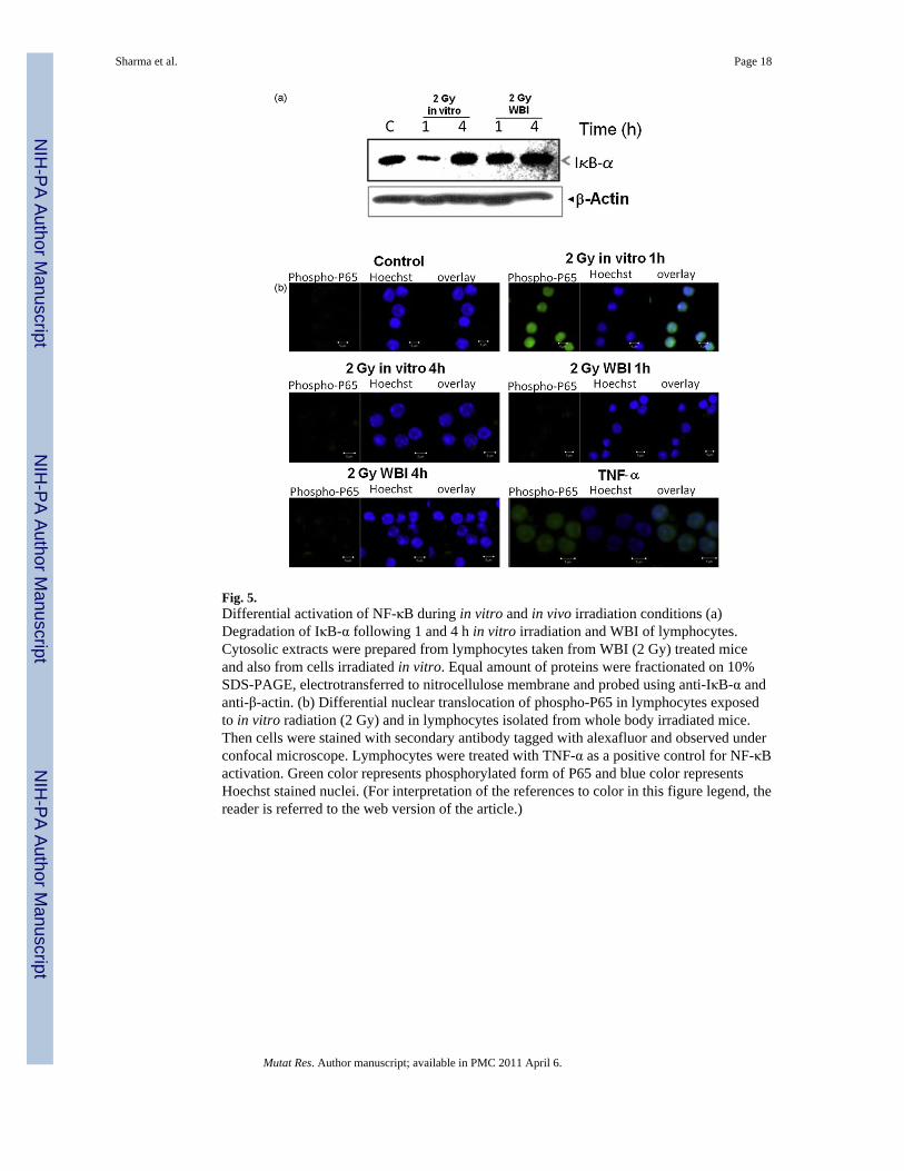
Fig. 5.Differential activation of NF-κB during in vitro and in vivo irradiation conditions (a)Degradation of IκB-α following 1 and 4 h in vitro irradiation and WBI of lymphocytes.Cytosolic extracts were prepared from lymphocytes taken from WBI (2 Gy) treated miceand also from cells irradiated in vitro. Equal amount of proteins were fractionated on 10%SDS-PAGE, electrotransferred to nitrocellulose membrane and probed using anti-IκB-α andanti-β-actin. (b) Differential nuclear translocation of phospho-P65 in lymphocytes exposedto in vitro radiation (2 Gy) and in lymphocytes isolated from whole body irradiated mice.Then cells were stained with secondary antibody tagged with alexafluor and observed underconfocal microscope. Lymphocytes were treated with TNF-α as a positive control for NF-κBactivation. Green color represents phosphorylated form of P65 and blue color representsHoechst stained nuclei. (For interpretation of the references to color in this figure legend, thereader is referred to the web version of the article.)
Sharma et al. Page 18
Mutat Res. Author manuscript; available in PMC 2011 April 6.
NIH
-PA Author Manuscript
NIH
-PA Author Manuscript
NIH
-PA Author Manuscript

Fig. 6.Effect of SNP on radiation induced DNA damage in lymphocytes. (a) Lymphocytes wereexposed to 4 Gy γ-radiation in vitro and SNP (200 μM) was added 30 min after irradiation.Cellular DNA damage (comet tail length) was estimated by SCGE method 1 and 4 h afterirradiation. Each value represents mean ± S.E.M. from four replicates and two suchexperiments were carried out. *p < 0.01, as compared to unirradiated respective controls. #p< 0.01, as compared to respective 4 Gy irradiated lymphocytes. (b) Lymphocytes weretreated with SNP (200 μM) following irradiation (4 Gy) and cultured for 24 h. Cells werelabeled with PE tagged Bcl-2 or Bax antibodies and acquired on a flowcytometer. The ratioof Bax to Bcl-2 was estimated as an index of apoptosis. Two such independent experimentswere carried out. *p < 0.01, as compared to 4 Gy irradiated lymphocytes.
Sharma et al. Page 19
Mutat Res. Author manuscript; available in PMC 2011 April 6.
NIH
-PA Author Manuscript
NIH
-PA Author Manuscript
NIH
-PA Author Manuscript
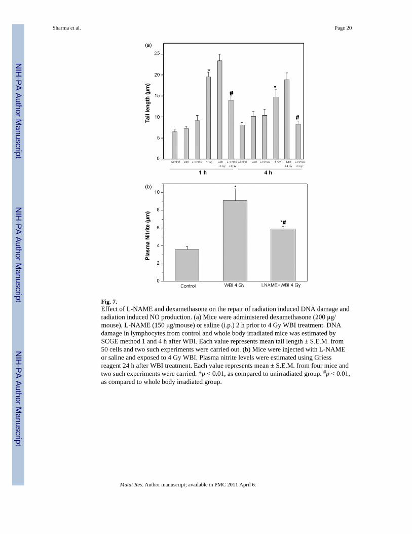
Fig. 7.Effect of L-NAME and dexamethasone on the repair of radiation induced DNA damage andradiation induced NO production. (a) Mice were administered dexamethasone (200 μg/mouse), L-NAME (150 μg/mouse) or saline (i.p.) 2 h prior to 4 Gy WBI treatment. DNAdamage in lymphocytes from control and whole body irradiated mice was estimated bySCGE method 1 and 4 h after WBI. Each value represents mean tail length ± S.E.M. from50 cells and two such experiments were carried out. (b) Mice were injected with L-NAMEor saline and exposed to 4 Gy WBI. Plasma nitrite levels were estimated using Griessreagent 24 h after WBI treatment. Each value represents mean ± S.E.M. from four mice andtwo such experiments were carried. *p < 0.01, as compared to unirradiated group. #p < 0.01,as compared to whole body irradiated group.
Sharma et al. Page 20
Mutat Res. Author manuscript; available in PMC 2011 April 6.
NIH
-PA Author Manuscript
NIH
-PA Author Manuscript
NIH
-PA Author Manuscript
Copyright © 2022 FDOKUMEN