pathogenic importance of oxidative stress - SciELO
21
Anais da Academia Brasileira de Ciências (2005) 77(4): 695-715 (Annals of the Brazilian Academy of Sciences) ISSN 0001-3765 www.scielo.br/aabc An overview of chagasic cardiomyopathy: pathogenic importance of oxidative stress MICHELE A. ZACKS 1 , JIAN-JUN WEN 2 , GALINA VYATKINA 2 , VANDANAJAY BHATIA 2 and NISHA GARG 3 1 Department of Pathology, University of Texas Medical Branch, Galveston TX 77555, USA 2 Department of Microbiology and Immunology, University of Texas Medical Branch, Galveston TX 77555, USA 3 Department of Microbiology and Immunology and Pathology, Center for Biodefense and Emerging Infectious Diseases and Sealy Center for Vaccine Development, 3.142C Medical Research Building, University of Texas Medical Branch 301, University Boulevard, Galveston TX 77555-1070, USA Manuscript received on August 8, 2005; accepted for publication on August 15, 2005; presented by GEORGE ADOSREIS ABSTRACT There is growing evidence to suggest that chagasic myocardia are exposed to sustained oxidative stress- induced injuries that may contribute to disease progression. Pathogen invasion- and replication-mediated cellular injuries and immune-mediated cytotoxic reactions are the common source of reactive oxygen species (ROS) in infectious etiologies. However, our understanding of the source and role of oxidative stress in chagasic cardiomyopathy (CCM) remains incomplete. In this review, we discuss the evidence for increased oxidative stress in chagasic disease, with emphasis on mitochondrial abnormalities, electron trans- port chain dysfunction and its role in sustaining oxidative stress in myocardium. We discuss the literature reporting the consequences of sustained oxidative stress in CCM pathogenesis. Key words: chagasic cardiomyopathy, reactive oxygen species, inflammation, mitochondria, oxidant/anti- oxidant status, oxidative damage. PARASITE, VECTOR AND TRANSMISSION Trypanosoma cruzi, a parasitic protozoan of the ancient branch of eukaryotes (Kingdom Eukaryota, Order Kinetoplastida), is the etiological agent of Chagas disease in humans (Miles 2003). Currently, the World Health Organization estimates that 11– 18 million individuals are infected worldwide (WHO 2002). Transmission of T. cruzi occurs predomi- nantly via insect vectors of the subfamily Triatoma, family Reduviidae, referred to as “ kissing bugs”. Residing in the peridomestic habitat of mud-thatch Correspondence to: Nisha Garg Ph.D. E-mail: [email protected] houses in rural areas (Mott et al. 1978), Triatoma infestans in South America, Rhodnius prolixus in Venezuela, Colombia and Central America (Scho- field and Dias 1999), and Triatoma barberi in Mexico (Guzman 2001), are the most common species responsible for transmission. Improvements in housing conditions and vector control measures instituted by the Southern Cone Initiative in 1991 have contributed to a decline in transmission in endemic countries (Schofield and Dias 1999). How- ever, concern remains that reinfestation of homes by secondary sylvatic vectors, e.g. Triatoma sor- dida, will compromise the long-term efficacy of An Acad Bras Cienc (2005) 77 (4)
-
Upload
khangminh22 -
Category
Documents
-
view
0 -
download
0
Transcript of pathogenic importance of oxidative stress - SciELO

Anais da Academia Brasileira de Ciências (2005) 77(4): 695-715(Annals of the Brazilian Academy of Sciences)ISSN 0001-3765www.scielo.br/aabc
An overview of chagasic cardiomyopathy:pathogenic importance of oxidative stress
MICHELE A. ZACKS1, JIAN-JUN WEN2, GALINA VYATKINA2,VANDANAJAY BHATIA2 and NISHA GARG3
1Department of Pathology, University of Texas Medical Branch, Galveston TX 77555, USA2Department of Microbiology and Immunology, University of Texas Medical Branch, Galveston TX 77555, USA
3Department of Microbiology and Immunology and Pathology,Center for Biodefense and Emerging Infectious Diseases and Sealy Center for Vaccine Development,
3.142C Medical Research Building, University of Texas Medical Branch301, University Boulevard, Galveston TX 77555-1070, USA
Manuscript received on August 8, 2005; accepted for publication on August 15, 2005;
presented by GEORGE A DOSREIS
ABSTRACT
There is growing evidence to suggest that chagasic myocardia are exposed to sustained oxidative stress-
induced injuries that may contribute to disease progression. Pathogen invasion- and replication-mediated
cellular injuries and immune-mediated cytotoxic reactions are the common source of reactive oxygen
species (ROS) in infectious etiologies. However, our understanding of the source and role of oxidative
stress in chagasic cardiomyopathy (CCM) remains incomplete. In this review, we discuss the evidence for
increased oxidative stress in chagasic disease, with emphasis on mitochondrial abnormalities, electron trans-
port chain dysfunction and its role in sustaining oxidative stress in myocardium. We discuss the literature
reporting the consequences of sustained oxidative stress in CCM pathogenesis.
Key words: chagasic cardiomyopathy, reactive oxygen species, inflammation, mitochondria, oxidant/anti-
oxidant status, oxidative damage.
PARASITE, VECTOR AND TRANSMISSION
Trypanosoma cruzi, a parasitic protozoan of the
ancient branch of eukaryotes (Kingdom Eukaryota,
Order Kinetoplastida), is the etiological agent of
Chagas disease in humans (Miles 2003). Currently,
the World Health Organization estimates that 11– 18
million individuals are infected worldwide (WHO
2002). Transmission of T. cruzi occurs predomi-
nantly via insect vectors of the subfamily Triatoma,
family Reduviidae, referred to as “ kissing bugs”.
Residing in the peridomestic habitat of mud-thatch
Correspondence to: Nisha Garg Ph.D.E-mail: [email protected]
houses in rural areas (Mott et al. 1978), Triatoma
infestans in South America, Rhodnius prolixus in
Venezuela, Colombia and Central America (Scho-
field and Dias 1999), and Triatoma barberi in
Mexico (Guzman 2001), are the most common
species responsible for transmission. Improvements
in housing conditions and vector control measures
instituted by the Southern Cone Initiative in 1991
have contributed to a decline in transmission in
endemic countries (Schofield and Dias 1999). How-
ever, concern remains that reinfestation of homes
by secondary sylvatic vectors, e.g. Triatoma sor-
dida, will compromise the long-term efficacy of
An Acad Bras Cienc (2005) 77 (4)

696 MICHELE A. ZACKS ET AL.
vector control measures in Brazil and other South-
ern American countries (Monteiro et al. 2001, WHO
2002). Blood transfusion and organ transplanta-
tion represent further routes of T. cruzi transmission.
Although many countries in Latin America screen
blood donations for T. cruzi, infection rates ranging
from 0.1% to 24.4% are estimated to occur through
transfusion (WHO 2002).
In the U.S., vector-transmitted human infec-
tions have been reported in the southern States
(Ochs et al. 1996, Herwaldt et al. 2000). Several
studies have shown the presence of the insect vec-
tor as well as infection of domestic dogs and wild
animals in the US (Meurs et al. 1998, Bradley et
al. 2000, Beard et al. 2003, Yabsley and Noblet
2002). Further, due to significant increases in im-
migration to the USA and Canada from endemic
countries and perinatal transmission, it is estimated
that ∼ 100, 000 people residing in USA may be
infected with T. cruzi. Currently, the US does not
screen blood donations, as no approved screening
test is available (Dodd and Leiby 2004). These
conditions, taken together, could contribute to the
transmission of T. cruzi by blood-borne, congenital,
and to a lesser extent, vector-borne routes, indicating
the potential for the emergence of Chagas disease as
a disease of public health importance in the USA
(Leiby et al. 2000, Dodd and Leiby 2004).
CHAGASIC DISEASE
CLINICAL SYMPTOMS
Infection by T. cruzi elicits acute non-specific
symptoms e.g. fever, malaise, edema and/or enlar-
ged liver or spleen to the characteristic swelling
at the site of entry- a chagoma (of the skin), or
Romana’s sign (of the conjuctiva or eyelid). In
spite of the high blood parasitemia in the acute
phase, clinical symptoms do not warranty hospital
visit, and therefore, anti-parasitic treatment is
often not initiated. In few (<5%) acute patients,
sudden death due to congestive heart failure associ-
ated with myocarditis or meningoencephalitis may
occur. The majority of patients enter an “ indeter-
minate” phase that is defined by the presence of T.
cruzi specific antibodies but the absence of clini-
cal signs of cardiac abnormalities. Between 10– 30
years after initial infection, an estimated 30– 40%
of “ indeterminate”patients show recognizable signs
and/or symptoms of a unique form of heart disease
referred to as chagasic cardiomyopathy (CCM).
Generally, patients develop a complex con-
stellation of symptoms and signs; their presence
or absence and severity have been used to create
a diagnostic scale for CCM (Rocha et al. 2003).
The non-specific symptoms suggestive of heart
problems include palpitations, dizziness, and syn-
cope. Clinical manifestations of CCM are conges-
tive heart failure, thromboembolism in brain, limbs
or lungs, and ventricular fibrillation (Rassi et al.
2000). The principal defects in the conduction sys-
tem present as a mixture of arrhythmias – e.g. tachy-
cardia, bradycardia, ventricular fibrillation – and
electrical impulse blockages – e.g. right bundle
branch and left anterior fasicle block. These are
identified by electrocardiography as well as via 24-
hour Holter monitoring or stress (exercise) testing
(Elizari 2002). Myocardial defects, that is, impair-
ment in the architecture of the heart muscle, are
mainly cardiomegaly with hypertrophy and dilation
of the chambers; and aneurism, particularly in the
apical portion of the left ventricle, which may rup-
ture. These attributes can be identified via echo-
cardiography and chest X-ray and are frequently ob-
served at autopsy. Clinical manifestations of CCM
may also be correlated with defective innervation
and defective contraction within the myocardium.
Pathogenic changes in the parasympathetic branch
of the autonomic nervous system are sometimes ob-
served at autopsy (Koberle 1968, Mott and Hags-
trom 1965, Oliveira 1985). Specifically, a reduction
– or in some cases, a complete absence – of cells
in the neuronal ganglia has been described and may
be indicative of a role for impaired heart innerva-
tion in development of CCM (Marin-Neto 1998).
Further, the basement membrane of the myocardial
capillaries is abnormally thick, suggestive of micro-
circulation defects (Rossi et al. 2003). Summariz-
An Acad Bras Cienc (2005) 77 (4)

CCM AND OXIDATIVE STRESS 697
ing, the dilation and hypertrophy of the left ventri-
cle combined with fibrosis at its apex are considered
pathognomonic of CCM, while the distinctive clin-
ical syndrome described above accounts for heart
failure and subsequent death that occurs in chagasic
patients.
PATHOLOGY
Several specific heart changes – at the gross and mi-
croscopic levels – are distinguishing in CCM. At au-
topsy, the most commonly observed abnormalities
in the heart structure are global (biventricular) heart
enlargement, apical aneurism, dilation/ thinning and
thrombi of the heart walls (Rossi et al. 2003). The
histological examination of sections from both au-
topsy and biopsy specimens revealed tissue fibro-
sis, inflammation, and hypertrophy of cardiac fibers
are the chief pathological consequences of chaga-
sic heart disease. Fibrosis is most pronounced in
the left ventricle and apex. This varies in severity
and its location may be interstitial and/or diffuse. It
corresponds to increased collagen fiber deposition
around the muscle bundles that resembles reparative
fibrosis resulting from microinfarction. These mi-
croscopic observations bear relationship to the gross
pathological observations of heart enlargement of
the hypertrophic and dilated form.
PATHOGENIC MECHANISMS(HISTORICAL PERSPECTIVE)
Extensive studies utilizing human biopsy and au-
topsy specimens- and experimental investigations
have provided data on the clinical and pathologi-
cal manifestations of chagasic disease. However,
the mechanism(s) leading up to CCM remain un-
certain. There is currently no universally accepted
model to predict which individuals in the “ inde-
terminate” phase will progress to chronic chagasic
disease. Several fundamental enigmas remain un-
resolved. First, why does the parasite, which is
shown to infect a wide variety of organs and cell
types, primarily cause disease in the heart? Second,
why does only a subset of infected (seropositive)
individuals progress to CCM? Third, what accounts
for the long latency period between infection and
appearance of heart disease? Nevertheless, a sig-
nificant body of literature supports the hypothesis
that immune responses, consistently triggered by
parasite persistence or by the host response to self-
antigens (autoimmunity) or both play a role in the
development and/or propagation of pathological
lesions. The concept of parasite persistence is that a
few persisting parasites continually trigger immune
responses, leading to chronic inflammation and cell
death. Alternatively, the concept of autoimmunity is
that the parasite has antigens that mimic the human
host. In effort to control T. cruzi infection, the host
produces antibodies and T cells that subsequently
recognize self-antigens, and destroy the myocardial
tissue.
PARASITE PERSISTENCE
Several lines of evidence establish that a low level of
parasites remain in the blood and/or the heart tissue.
First, direct detection in autopsy specimens and my-
ocardial biopsies has been possible using new meth-
ods e.g. immunohistochemical and immunofluores-
cence techniques (Mortara et al. 1999, Higuchi et
al. 2003) and PCR (Jones et al. 1993, Salomone
et al. 2000), including in situ PCR (Jones et al.
1992). Second, in chronically infected individuals,
the reactivation of acute disease, particularly menin-
goencephalitis, that occurs following immunosup-
pression due to AIDS (Rocha et al. 1994, Sartori et
al. 1995) or drug therapy (Jardim and Takayanagui
1994, D’Almeida et al. 1996) illustrates that par-
asites persist in previously undiagnosed individu-
als for years after initial infection. Thirdly, trans-
mission of T. cruzi can occur via blood transfusion
and transplantation of infected organs obtained from
asymptomatic individuals (Leiby et al. 2000). Fi-
nally, parasite persistence would explain the clinical
benefit of anti-parasitic drug treatment in chronic in-
fection, which has been observed in a limited num-
ber of studies (Andrade et al. 1992, Viotti et al.
1994). Altogether, these studies support the con-
cept of parasite persistence and its likely pathologic
role in CCM development.
An Acad Bras Cienc (2005) 77 (4)

698 MICHELE A. ZACKS ET AL.
IMMUNE RESPONSES: PROTECTIVE OR
PATHOLOGICAL
Sterilizing immunity does not appear to exist in T.
cruzi infection. Nevertheless, the immune response
that is mounted against T. cruzi after initial infection
is capable of controlling the parasite, as evidenced
by the eventual resolution of acute parasitemia. As
mentioned above, these immune responses may con-
tribute to pathology through either or a combination
of two general mechanisms: i) a process of auto-
immune destruction of host cardiac tissue (auto-
immune hypothesis) or ii) through specific media-
tors of the immune response to T. cruzi itself or to
its antigenic components that linger in the tissues
(parasite persistence hypothesis).
Experimental studies
Assorted experimental approaches using animal
and tissue culture models of T. cruzi infection and
disease have provided a portrait of the immune re-
sponse to T. cruzi infection. In addition, various
knockout or immunocompromised animals have
been utilized to dissect the relative contribution of
different features of the immune response that
mediate protection versus pathology. Briefly, in
acutely infected animals (mice and rats), it is shown
that macrophages (Mφ) and natural killer (NK) cells
provide the first line of defense (Nogueira and Cohn
1978). T. cruzi elicits IFN-γ production by NK
cells, activating Mφ (Torrico et al. 1991, Gazzinelli
et al. 1992). In turn, Mφ produce TNF-α, induc-
ing nitric oxide (NO), which is toxic to parasites
in vitro (Vespa et al. 1994, Martins et al. 1998).
Interestingly, GPI-anchored macromolecules, abun-
dantly expressed by the infective and intracellular
stages of the parasite, have been characterized as
the prime inducers of the proinflammatory cytokines
(e.g. TNF-α, IL-1 and IL-6) and chemokines (e.g.
IP-10, MCP-1, MIG, and RANTES) that may be
key regulators of the innate and adaptive immune
responses responsible for the control of acute infec-
tion. CD4+ T cells assist in the control of T. cruzi
through secretion of Th1 cytokines, amplification
of phagocytic activity of macrophages, and stimula-
tion of B cell proliferation and antibody production
(Aliberti et al. 1996, Brener and Gazzinelli 1997).
CD8+ T cells are shown to exhibit specific cyto-
toxicity in vitro in response to T. cruzi-expressed
antigens (Garg et al. 1997, Wizel et al. 1997, Garg
and Tarleton 2002) and are suggested to control T.
cruzi either by cytolysis of the infected cells or by
secretion of Th1 cytokines that induce trypanocidal
activity. Altogether, these studies conclude that an
efficient protective response to acute parasitemia is
provided by Th1 cytokines, lytic antibodies and the
concerted activities of macrophages, T helper cells
and cytotoxic T lymphocytes.
With progression to indeterminate - chronic
phase, parasite nests are not detectable by conven-
tional histological analysis. However, a low level of
inflammatory response constituted by macrophages
and CD8+ T lymphocytes persist in the heart. Al-
though often they are not associated with amastig-
ote nests or trypomastigotes, T. cruzi antigens may
be present (Higuchi 1995, Reis et al. 1997). CD4+
T cells, though less prominent in the chronic stage,
are suggested to be associated with myocyte death
and increased animal mortality (Soares et al. 2001)
and appear to represent an autoreactive phenotype
that contributes to tissue destruction (Soares and
Santos 1999). At this later stage, continued pro-
duction of IFN-γ and IL-2 is believed to stimulate
lytic antibody production by B cells (Brener and
Gazzinelli 1997). Several investigators have utilized
mice treated with antibody to immune mediators
and murine models in which genes for various im-
mune mediators have been deleted to evaluate the
significance of inflammatory response in chagasic
disease. These include mice deficient in inducible
NO synthase (iNOS), IFN-γ , TNF-α, TNF-α re-
ceptor, and CD4+ and/or CD8+ T cells (Minoprio et
al. 1987, Tarleton 1990, Tarleton et al. 1992, Bach-
maier et al. 1997, Martins et al. 1998, Aliberti et al.
2001, Chandra et al. 2002). The overall observation
from these studies is that despite a general increase
in parasite burden, the extent of cell death and tissue
damage are diminished in mice deficient in inflam-
An Acad Bras Cienc (2005) 77 (4)

CCM AND OXIDATIVE STRESS 699
matory mediators compared to the wild-type con-
trols, thus suggesting the pathological significance
of persistent inflammation in chagasic disease.
Human studies
Many of the above experimental observationsagree with the more limited number of humanstudies. In acute infection, hypergammaglobuline-mia occurs and the continued presence of antibodyis used to diagnose infection with T. cruzi. In hu-man patients’ cardiac biopsies, among the mono-nuclear cells, macrophages and CD8+ T cells rep-resent the majority of the infiltrate (Higuchi et al.1997). IFN-γ can be detected in tissues via in situ
immunohistochemistry (Bahia-Oliveira et al. 1998,Correa-Oliveira et al. 1999) and appears to be cor-related with the presence of CD8+ T cells (Higuchiet al. 1997, Reis et al. 1997). In addition, T. cruzi-specific human CTL have been identified (Thomsonet al. 1998, Wizel et al. 1998). While parasitesare rarely seen in cardiac tissue, pseudocysts areassociated with relatively sparse infiltrate of IL-2+
and IL-4+ lymphocytes (Higuchi et al. 1997, Reiset al. 1997). Again, CD8+ CTL are suggested tobe likely the major effectors of pathogenesis, con-tributing to the development of fibrosis in responseto cytopathology (Brener and Gazzinelli 1997).
To sum up, until recently, autoimmunity wasbelieved to be the primary mechanism of chaga-sic disease development. A variety of autoantigenshave been described and autoimmune-mediatedprocesses have been demonstrated in experimentalmodels (Leon et al. 2001, Pontes-de-Carvalho etal. 2002). Increasingly, with the demonstration ofparasite antigens in the heart tissue, parasite per-sistence has gained favor among several investiga-tors. We surmise that the contribution of parasitepersistence, auto-antigens and chronic inflammationis not mutually exclusive and all these mechanismsmay contribute, in part, to pathogenesis of CCM.The mechanistic studies identifying i) the media-tors produced by cardiomyocytes in response to T.
cruzi infection, that may trigger the migration ofleukocytes and other cells to the heart; ii) the sig-
naling mechanisms regulated by the inflammatorycytokines (e.g. TNF-α and IL-1) that may evokecell survival/cell growth or cell death responses inchagasic myocardium; and iii) the destructive ef-fects of “ oxidative burst” of activated inflammatorycells in CCM would be discussed elsewhere.
VASCULAR MEDIATORS IN ENDOTHELIUM
In addition to cardiomyocytes, T. cruzi infects en-dothelial cells in the heart. The role of vascularmediators e.g. endothelin-1 (ET-1) and thrombox-ane A2 (TXA2) in CCM has recently been explored(Petkova et al. 2001). ET-1 is a vasoconstrictorproduced by endothelial cells as well as cardiomy-ocytes and other cell types (Yanagisawa et al. 1988).Its synthesis by endothelial cells is increased duringin vitro infection with T. cruzi (Wittner et al. 1995)and in the myocardium of infected mice (Petkovaet al. 2000b). This appears to be regulated bythe transcription factors, NFκB (Huang et al. 1999)and AP-1, in conjunction with the MAPK signaltransduction pathway (Miyauchi and Masaki 1999,Petkova et al. 2000a). T. cruzi infected endothelialcells have also been shown to induce cytokine geneexpression (Tanowitz et al. 1992). This may con-tribute to CCM development, as cytokines stimulateET-1 production (Kurihara et al. 1989, Martins etal. 1998). TXA2 induces vasospasm and plateletaggregation, which is observed in CCM (Rossi andRamos 1996). Thus, TXA2 and ET-1 may poten-tiate CCM by promoting microvascular pathology,e.g., vasospasm, focal ischemia and microthrombi(Petkova et al. 2001). Vascular mediators, in con-junction with cytokines induced by T. cruzi infec-tion, may be significant to the development of micro-circulatory abnormalities that are well documentedin CCM.
OXIDANTS, ANTIOXIDANTS AND OXIDATIVE STRESS(AN OVERVIEW)
REACTIVE OXYGEN SPECIES (ROS) AND
REACTIVE NITROGEN SPECIES (RNS)
Broadly defined, ROS are derivatives of molecularoxygen (O2) (Nordberg and Arner 2001), e.g. su-
An Acad Bras Cienc (2005) 77 (4)

700 MICHELE A. ZACKS ET AL.
peroxide (O·−2 ), hydroxyl radical (HO·), and hydro-
gen peroxide (H2O2). ROS are produced throughthe action of specific oxidases and oxygenases, theFenton reaction and are also by-products of the elec-tron transport chain (Turrens 2003, 2004). RNS in-clude ·NO and its derivatives. ·NO is produced bythe enzymatic activity of NOS. Different NOS iso-forms have been identified in the cytoplasm (iNOS),in mitochondria (mtNOS), or in specific cell typese.g. endothelial NOS (eNOS) (Andrew and Mayer1999). ROS are unstable and react rapidly with otherfree radicals and macromolecules in chain reactionsto generate increasingly harmful oxidants (Kirkine-zos and Moraes 2001). The toxic effects of ROS arebelieved to vary in proportion to the quantity andtheir oxidant strength e.g. HO· > > > O·
2 > H2O2.While H2O2 is not excessively reactive, it is highlydiffusible and is a precursor of HO·. HO· is highlyreactive at its site of production. H2O2 and ·NOreadily cross membranes and thus, are capable ofaffecting distant cellular targets. Interaction of O·−
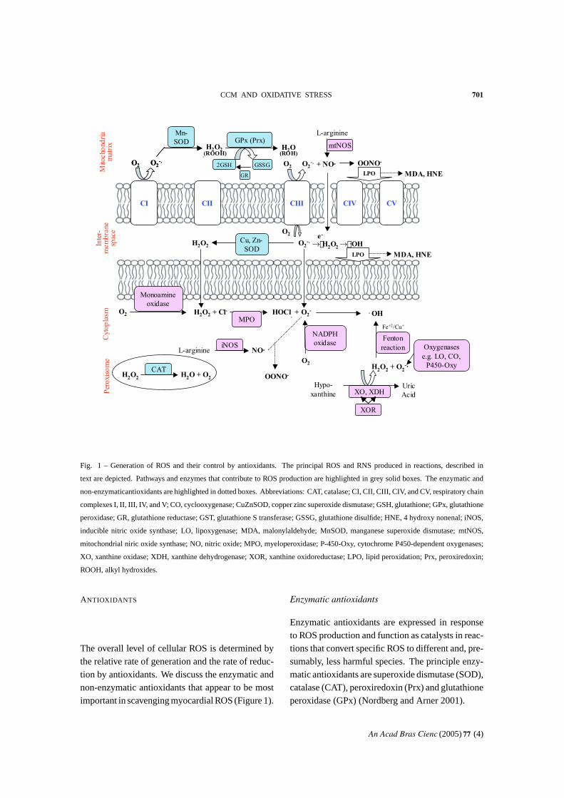
2and ·NO results in highly toxic, stable peroxynitrite(O=NOO−) (Figure 1).
SOURCES OF ROS
Oxidases and oxygenases
Numerous oxidases and oxygenases expressed indifferent cell types and locations within the cell con-tribute to the formation of ROS. By definition, oxi-dases reduce O2 whereas oxygenases (oxidoreduc-tases) transfer O2 to substrates. ROS production,termed the “ oxidative burst” of activated phagocyticcells e.g. macrophages and neutrophils, results fromNADPH oxidase and/or by myeloperoxidase activ-ity (Halliwell 1991, Eiserich et al. 2002). This ROSproduction is critical to anti-microbial function,contributing either directly or indirectly to the killingof intracellular organisms. Myeloperoxidase, pro-duced by neutrophils, converts H2O2 and chlorideions into hypochlorous (HOCl) acid (Winterbournet al. 2000). NADPH oxidase, produced by manytypes of phagocytes, reduces O2 to O·−
2 (Cross et al.1994, Babior 1999). Subsequently, O·−
2 and HOClfurther can react to form HO· (Candeias et al. 1993).
Other oxidases are more generally expressedin mammalian cells but differ in their basal level ofexpression and subcellular locations. Monoamineoxidase, present in the outer mitochondria mem-brane, converts O2 to H2O2 on the cytoplasmic face(Hauptmann et al. 1996). Both xanthine oxidaseand xanthine dehydrogenase, derived from xanthineoxidoreductase, produce H2O2 and O·−
2 in the pro-cess of degrading the purine hypoxanthine to uricacid (Berry and Hare 2004). In rats and some lowereukaryotes, xanthine oxidase is demonstrated to beboth cytoplasmic and peroxisomal. O·−
2 may also beproduced by lipoxygenase, cyclooxygenase (McIn-tyre et al. 1999) and cytochrome P450-dependentoxygenases (Coon et al. 1992).
Fenton chemical reaction
Besides production by oxidases and oxygenases, theFenton reaction is another mechanism of ROS for-mation. This reaction results in the Fe+2- or Cu+-mediated conversion of H2O2 to HO· (Goldstein etal. 1993).
Electron transport chain (ETC)
Mitochondria are considered a major source ofROS production in heart (Loschen et al. 1971). Inthe mitochondria, the partial reduction of O2 occursas a result of leakage of electrons from the ETC,contributing one, two or three electrons to form O·−
2 ,H2O2, or HO·, respectively. Electron leakage canarise at a number of points in the ETC, produc-ing O·−
2 (Turrens 2004). As much as 2– 4% of thereducing equivalents escape the respiratory chain,leading to O·−
2 formation. O·−2 is dismutated by
MnSOD to H2O2 that may then be converted tohighly reactive and harmful HO· radicals. Gen-erally, the leakage of electrons at CI flavoproteingenerates O·−
2 in mitochondrial matrix while CIIIubisemiquinones (UQ−.) generated at Q1 (UQ−1.)and Qo (UQ−o.) sites release O·−
2 in the matrix andintermembrane space of the mitochondria, respec-tively (Han et al. 2001, 2003).
An Acad Bras Cienc (2005) 77 (4)
CCM AND OXIDATIVE STRESS 701Inter-
mem
brane
sp
ace
Mitochondri
a m
atrix
Cytoplasm H2O2 + Cl- HOCl + O2
-.
NADPH
oxidase
Monoamine
oxidaseO2
O2
MPO. OH
Mn-
SOD
O2-.
GPx (Prx)
H2O2
H2O2,(ROOH)
H2O(ROH)
CII
O2-.O2 O2-.O2
CI CIII CIV
O2-.O2
Cu, Zn-
SOD
O2
mtNOS
L-arginine
OONO-+ NO·
Fe+2/Cu+
Fenton
reactioniNOSL-arginine NO·
OONO-
→ H2O2→ ·OH
e-
Oxygenases
e.g. LO, CO,
P450-Oxy
XO, XDH
XOR
Uric
Acid
Hypo-
xanthine
H2O2
+ O2-·
CV
LPO
H2O + O
2
CATH2O2
Peroxiso
me
2GSH GSSG
GR LPO
MDA, HNE
MDA, HNE
Fig. 1 – Generation of ROS and their control by antioxidants. The principal ROS and RNS produced in reactions, described in
text are depicted. Pathways and enzymes that contribute to ROS production are highlighted in grey solid boxes. The enzymatic and
non-enzymaticantioxidants are highlighted in dotted boxes. Abbreviations: CAT, catalase; CI, CII, CIII, CIV, and CV, respiratory chain
complexes I, II, III, IV, and V; CO, cyclooxygenase; CuZnSOD, copper zinc superoxide dismutase; GSH, glutathione; GPx, glutathione
peroxidase; GR, glutathione reductase; GST, glutathione S transferase; GSSG, glutathione disulfide; HNE, 4 hydroxy nonenal; iNOS,
inducible nitric oxide synthase; LO, lipoxygenase; MDA, malonylaldehyde; MnSOD, manganese superoxide dismutase; mtNOS,
mitochondrial niric oxide synthase; NO, nitric oxide; MPO, myeloperoxidase; P-450-Oxy, cytochrome P450-dependent oxygenases;
XO, xanthine oxidase; XDH, xanthine dehydrogenase; XOR, xanthine oxidoreductase; LPO, lipid peroxidation; Prx, peroxiredoxin;
ROOH, alkyl hydroxides.
ANTIOXIDANTS
The overall level of cellular ROS is determined bythe relative rate of generation and the rate of reduc-tion by antioxidants. We discuss the enzymatic andnon-enzymatic antioxidants that appear to be mostimportant in scavenging myocardial ROS (Figure 1).
Enzymatic antioxidants
Enzymatic antioxidants are expressed in responseto ROS production and function as catalysts in reac-tions that convert specific ROS to different and, pre-sumably, less harmful species. The principle enzy-matic antioxidants are superoxide dismutase (SOD),catalase (CAT), peroxiredoxin (Prx) and glutathioneperoxidase (GPx) (Nordberg and Arner 2001).
An Acad Bras Cienc (2005) 77 (4)

702 MICHELE A. ZACKS ET AL.
SOD converts O·−2 to H2O2 (Fridovich 1974).
MnSOD, the mitochondrial isoform makes up∼ 70% of the SOD activity in heart, and 90%in cardiomyocytes. The remaining fraction con-sists primarily of cytoplasmic CuZnSOD with <1%extracellular SOD (ECSOD). The importance ofMnSOD in regulating O·−
2 in myocardium isdemonstrated by the fact that MnSOD– /– mice diesoon after birth with dilated cardiomyopathy (Liet al. 1995). GPx (isoforms GPx1 – GPx5), usingglutathione (GSH), reduces H2O2 or ROOH to H2Oor alcohols (ROH), respectively. GPx1 and GPx3are the most abundant intracellular isoforms andGPx4 is a mitochondrial isoform. Unlike MnSOD,mice deficient in GPx develop normally and show nomarked pathologic changes under normal physi-ologic conditions, and exhibit a pronounced suscep-tibility to myocardial ischemia-reperfusion injury(Ho et al. 1997). CAT, located in peroxisomes, ishighest in the liver and erythrocytes, and convertsH2O2 to H2O and O2. Prx reduces peroxides, in-cluding H2O2 and alkyl hydroperoxides (ROOH).
Non-enzymatic antioxidants
The role of GSH in maintaining cellular redox stateis complex. GSH cooperates with GPx in the detox-ification of H2O2 to 2H2O. In addition, GSH par-ticipates in reactions with glutathione S-transferase(GST) to bind ROS e.g. attachment of ·NO to formS-nitrosoglutathione adducts. Glutathione reduc-tase (GR) functions to regenerate antioxidant capac-ity, converting from glutathione disulfide (GSSG)to GSH. Vitamins and other chemical antioxidantsplay an important role in the control of ROS cas-cades. Vitamin E (α-tocopherol) is active in mem-branes where it functions to reduce ROS and lipidperoxy radicals. Vitamin C (ascorbate) serves pre-dominantly as an antioxidant in plasma due to its wa-ter solubility. It functions by reducing α-tocopherol-lipid peroxide radicals to normal form (Nordbergand Arner 2001). Uric acid, found in extracellularfluids, detoxifies HO·. Metal ions (Fe+2 or Cu+) con-tribute to ROS-mediated peroxidation of lipids viathe Fenton reaction that produces H2O2. The se-
questration of these ions in protein-bound form, e.g.as iron-transferrin or copper-ceruloplasmin boundcomplexes, also adds indirectly to the antioxidantcapacity of cells (Turrens 2004).
OXIDATIVE STRESS
When produced transiently in limited quantities,ROS and RNS play critical roles in normal devel-opmental processes; ROS and RNS, control signaltransduction mechanisms that regulate cell prolifer-ation, differentiation and death (Droge 2002, Finkel2003). However, when produced in excess or forsustained periods, or when the antioxidant systemis compromised, cells are unable to efficiently scav-enge free radicals, leading to ROS accumulation.ROS can rapidly oxidize proteins, lipids and DNA(Butterfield et al. 1998, Marnett 2000), thereby re-sulting in dysfunction of physiological processesand cellular damage leading to cell/tissue death. Re-sults of a variety of studies on the mammalian systemhave shown that oxidative stress can be reliably mea-sured by oxidative-damage biomarkers, such as lipidperoxides, protein carbonyls, and oxidative DNAmodifications.
Lipid peroxidation (LPO)
Lipid peroxidation is the major biochemical con-sequence of oxidative deterioration of polyunsatu-rated lipids in cell membranes and causes damageto membrane integrity and loss of membrane pro-tein function. Peroxidation, in general, is initiatedby oxidative attack-mediated removal of a H· atomresulting in carbon centered radical, that in aerobiccells undergoes molecular rearrangement followingexposure to O2 to give a peroxyl radical. Peroxylradicals can combine with each other, attack mem-brane proteins, or abstract H· from adjacent fattyacids side chains in a membrane thereby propagatinga chain reaction of lipid peroxidation. The ETC andmembrane phospholipids such as those in the mito-chondrial membrane are particularly susceptible toLPO (Halliwell 1991, Cardoso et al. 1999). Amonga wide range of aldehydic compounds, 4-hydroxy-2-nonenal (4-HNE) and malonylaldehyde (MDA) are
An Acad Bras Cienc (2005) 77 (4)

CCM AND OXIDATIVE STRESS 703
the most common reactive products of the peroxi-dation of membrane phospholipids (Zarkovic 2003).These aldehydic products are relatively stable com-pounds, and are able to diffuse and attack targets inthe near vicinity as well as those distant from theirsite of origin (Esterbauer 1982).
Protein oxidative modifications
Many different types of protein oxidative modifi-cations may be induced by direct or indirect attackby ROS/RNS and secondary by-products of oxida-tive stress (Dalle-Donne et al. 2003). These mod-ifications result in protein carbonyl (PCO) deriva-tives (aldehydes and ketones), nitrative adducts (e.g.3-nitrotyrosine) and Michael-adducts formation.Cys, His or Lys amino acids are the prime targetof 4-HNE, a highly reactive αβ unsaturated alde-hyde, resulting in Michael adducts formation andirreversible alkylation and introduction of carbonylgroups into proteins (Uchida and Stadtman 1992).Arg, Lys, Pro, and Thr residues may be directlyderivatized by ROS leading to formation of proteincarbonyls (Butterfield et al. 1998, Chevion et al.2000). Carbonyl groups can also be introduced intoproteins by oxidative reaction of sugar derivatives(ketoamines, ketoaldehydes and deoxyosones) withLys residue in a process called glycation or glyoxida-tion. As a consequence of oxidative modifications,functional impairment of proteins occurs and fur-thermore, leads to protein turnover e.g. degradationby proteases via the proteasome (Floyd et al. 2001).
DNA damage
DNA can be oxidized by a variety of mechanisms,resulting in nucleotide damage e.g. formation of8-oxoguanine lesions. As a result, DNA replica-tion may be inaccurate leading to mutations andtranscription errors. While mechanisms exist to re-pair these DNA lesions, the level of DNA dam-age may exceed the capacity of the cellular repairmechanisms. Furthermore, mtDNA is believed to beparticularly susceptible to sustained damage, sincemitochondria may lack appropriate DNA repair
mechanisms (Evans and Cooke 2004).
MITOCHONDRIAL ABNORMALITIES ANDOXIDATIVE STRESS IN CCM
Oxidative stress is generally viewed as a pro-tective defense mechanism employed by the hostto control parasitic infection. There is, however,growing evidence to suggest that sustained oxidativestress may contribute to CCM pathology. Pathogeninvasion- and replication-mediated cellular injuriesand immune-mediated cytotoxic reactions are thecommon source of ROS in infectious etiologies. Asmentioned in section “ Immune responses: Protec-tive or pathological”, experimental studies haveshown that T. cruzi infection elicits inflammatorycytokines, NO production, and oxidative burst, all ofwhich, though essential for controlling acute para-sitemia, may have toxic effects on host cellular com-ponents and be of pathological significance in CCM.
Mitochondria represent 30% of the total vol-ume of cardiomyocytes and provide ∼90% of thecellular energy through the oxidative phosphory-lation pathway. As discussed in section “ Electrontransport chain (ETC)”, the CI and CIII complexesof the respiratory chain are the prime site forelectron leakage to oxygen, and free radical pro-duction in mitochondria. The rate of mitochondrialROS release is inversely proportional to the rateof electron transport, exponentially increasing whenCI or CIII complexes of ETC function at a sub-optimal level (Ide et al. 1999, Wallace 2000, Les-nefsky et al. 2001, Chen et al. 2003). Interestingly,mitochondria are targets to a variety of endogenousand exogenous insults that may affect ETC func-tion. Further, CI and CIII are redox sensitive, asthey contain Fe4S4 clusters which when oxidized,release one iron atom, resulting in the inactivationof important functional Fe-S centers and enzymeactivity. The released ferrous ions, when partici-pating in the Fenton reaction, produce highly re-active HO·− radicals. Consequently, ROS as a byproduct of respiratory chain is considered themajor source of free radicals in the heart and mito-chondrial dysfunction-mediated oxidative stress has
An Acad Bras Cienc (2005) 77 (4)
704 MICHELE A. ZACKS ET AL.
been shown in many cardiac pathologies (Sawyeret al. 2002). The importance of these findings asit relates to chagasic disease is that an early andconsistent repression of CI and/or CIII activities as-sociated with sustained oxidative stress is shownin mitochondria isolated from the myocardium ofmice infected by T. cruzi (Vyatkina et al. 2004).A consistent decline in MnSOD activity, the ma-jor oxygen radical scavenger in the mitochondrialmatrix, during progression of infection and diseasein chagasic myocardium was also shown (Wen etal. 2004). These studies have led to the suggestionthat a catastrophic cycle of mitochondrial func-tional decline and ROS generation, coupled with aninability to efficiently scavenge the mitochondrialROS (due to MnSOD deficiency), predisposes thechagasic hearts to sustained oxidative stress duringinfection and disease development. We discuss theliterature related to role of mitochondrial dysfunc-tion and inadequate antioxidant defenses in sustain-ing the oxidative stress in CCM.
MITOCHONDRIAL DYSFUNCTION IN CCM
The early insights suggesting mitochondrial alter-
ations in CCM were provided by electron micro-
scopic analysis of biopsies from cardiac tissue of
chagasic patients (Carrasco Guerra et al. 1987,
Palacios-Pru et al. 1989, Parada et al. 1997) and ex-
perimental models (Uyemura et al. 1995, Garg et al.
2003). Microscopic examination of biopsy samples
from the patients showed that degenerative myo-
cardial changes occur very early during the indeter-
minate phase and become more pronounced with in-
creased severity of clinical disease. In experimental
models of chagasic disease, the ultrastructural eval-
uation of the morphological alterations in the myo-
cardium has illustrated an accumulation of large, ir-
regular nuclei, swollen and displaced mitochondria,
and myofibrillar degeneration, that progress in par-
allel with the severity of disease (Garg et al. 2003).
These studies have revealed two important observa-
tions. First, nuclear and mitochondrial structural
damage occurs much earlier than do the clinical
symptoms of disease. Second, the severity of these
aberrations increase with the evolution of chronic
disease. Jointly, these observations imply that a
correlation exists between the extent of specific or-
ganelle abnormalities and clinical severity of chaga-
sic disease.
Recent molecular studies have profiled the
changes in mitochondrial function-related gene ex-
pression in experimental models of T. cruzi infection
and disease development (Garg et al. 2003, Mukher-
jee et al. 2003, Garg et al. 2004). These studies
utilized global and custom-designed arrays and con-
firmed the array data by traditional and real-time RT-
PCR and Northern blotting approaches. The over-
all picture that emerged from these studies was that
the myocardial transcripts encoding metabolic en-
zymes involved in fatty acid β-oxidation were up-
regulated, while the mRNAs for a majority of the
subunits of the complexes of the ETC pathway were
repressed in response to infection. It is important
to note that the mtDNA-encoded transcripts for the
subunits of the ETC complexes were repressed in
response to infection (Vyatkina et al. 2004), be-
fore the alterations in nDNA-encoded transcripts
were detected. The expression level of mtDNA-
encoded ETC components that were examined (9
of 13) as substantially reduced (up to 80%) with
progression to chronic disease phase. A loss in
mtDNA-encoded transcripts (and presumably pro-
teins) below the threshold level is likely to result
in a deficiency of respiratory complexes in chagasic
hearts.
We and others have demonstrated a substan-
tial decline in respiratory chain complexes CI, CIII,
and CV activities in the myocardium of infected
experimental animals (Uyemura et al. 1995, Vyatk-
ina et al. 2004). Along with a decline in total spe-
cific activity of respiratory complexes (determined
by spectrophotometry assays), inactivation of the
assembled complexes (determined by catalytic
staining on blue-native gels) was also noted, sug-
gesting that multiple mechanisms are likely to
be involved in inhibition of the respiratory com-
plexes in the chagasic myocardium. Interestingly,
the finding of a loss in CI activity early in response
An Acad Bras Cienc (2005) 77 (4)

CCM AND OXIDATIVE STRESS 705
to parasite infection led to a suggestion that CI
might be the potential site for ROS generation in
acute murine myocardium. CIII deficiencies were
consistent in cardiac mitochondria, but were not
observed in skeletal muscle mitochondria at all
stages of infection and disease progression, lead-
ing to an implication that CIII is the likely site for
sustained ROS generation in chagasic myocardium
(Vyatkina et al. 2004). Future studies would, hope-
fully, address the pathophysiological significance of
mtROS in CCM.
In human patients, direct demonstration of mi-
tochondrial dysfunction awaits further investigation.
However, biochemical analysis have provided indi-
rect evidence to suggest mitochondrial abnormal-
ities in chagasic patients. For example, Carrasco
et al. showed a reduction in the activities of suc-
cinate dehydrogenase and myosine ATPase by his-
tochemical staining of small endomyocardial biop-
sies obtained from the chagasic patients (Carrasco
Guerra et al. 1987). The histochemical alteration
index, while increased in seropositive patients in
the so-called “ indeterminate” phase, was highest in
chronic patients. In another study of chagasic pa-
tients, changes in the serum pattern of metabolic
enzymes was documented (Alarcon-Corredor et al.
2002). The main finding in this study was a sub-
stantial increase in glutamate-oxaloacetate transam-
inase (GOT) and 3-hydroxy butyrate dehydrogenase
(HBDH) activity. Importantly, high serum levels
of GOT and HBDH were detected in indeterminate
patients, and remained consistently high in patients
advancing to clinical cardiac dysfunction. Consider-
ing the site (coronary sinus) and the extent of release
of GOT and HBDH in indeterminate-to-chronic pa-
tients, it was surmised that mitochondrial and cell
membrane injuries are the earliest events in chaga-
sic disease, and the degenerative mitochondrial and
cellular events persist with advanced disease. The
detection of inorganic phosphorus and isocitrate de-
hydrogenase molecules in the serum of indetermi-
nate patients, followed by a positive coronary sinus-
femoral artery inorganic phosphate gradient with ad-
vancement of chronic disease further supports the
hypothesis of very early and progressive manifes-
tations of mitochondrial metabolic abnormalities in
chagasic myocarditis (Carrasco et al. 1997). Collec-
tively, these studies support the hypothesis that
mitochondrial metabolic abnormalities are mani-
fested very early in infection and exacerbated during
CCM progression.
FACTORS CONTRIBUTING TO MITOCHONDRIAL
DYSFUNCTION
T. cruzi infection elicits substantial biochemical,
molecular and immunological insults all of which
may adversely affect mitochondria in chagasic my-
ocardium. Invasion by parasite elicits transient ele-
vations in intracellular Ca+2 (Burleigh and Andrews
1995) followed by release of the parasite from the
parasitophorous vacuole. The acute phase of in-
fection is then marked by massive replication of
the parasite in host cell cytoplasm and activation
of potent inflammatory reactions (section “ Immune
responses: Protective or pathological”). In mam-
malian cells, Ca+2 overload is known to induce the
opening of mitochondrial permeability transition
pores, leading to dissipation of the proton gradient
(Korge et al. 2001, Kanno et al. 2002). Given, that
maintenance of mitochondrial membrane potential
and ion transport is essential for oxidative phospho-
rylation it is likely that T. cruzi-induced Ca+2 over-
load might be the primary signal in mitochondrial
dysfunction. This notion is supported by others,
who indicate that the elevated levels of Ca+2 con-
tribute to impairment of mitochondrial respiratory
enzyme activity (Medrano and Fox 1994, Liang
and Molkentin 2002).
Following initial injuries, it is likely that mi-
tochondria may be damaged by ROS originated via
inflammatory mechanisms or respiratory chain im-
pairment. The first evidence of oxidative damage to
mitochondria was reported utilizing an experimen-
tal model of infection and chronic disease (Wen and
Garg 2004). This study demonstrated a substantial
increase in LPO and PCO derivatives in cardiac mi-
tochondria of infected mice, compared to controls.
The LPO derivatives of mitochondrial membranes
An Acad Bras Cienc (2005) 77 (4)

706 MICHELE A. ZACKS ET AL.
were detectable as early as 3 days post-infection, and
gradually increased by >2-fold during the course of
disease development. The PCO content in cardiac
mitochondria became evident during the acute in-
fection phase and remained consistently enhanced
throughout the chronic phase of disease progres-
sion. ROS-induced LPO and PCO derivatives de-
position in mitochondrial membranes is shown to
cause increased permeability and loss of mitochon-
drial membrane potential and protonmotive force
(Piper et al. 1994, Vercesi et al. 1997, Cardoso et
al. 1999). We anticipate future studies would de-
lineate the mechanistics of mitochondrial oxidative
modifications of membrane lipids and proteins in
alterations of mitochondrial integrity, dissipation of
the mitochondrial membrane potential and proton-
motive force, and respiratory chain inefficiency
in CCM.
Direct oxidative modification of specific sub-
units of respiratory complexes may be an underly-
ing means of inactivation of assembled mitochon-
drial complexes in chagasic hearts (Wen and Garg
2004). Cardiac mitochondria from infected mice
were subjected to two-dimensional blue-native gel
electrophoresis to resolve the subunits of the res-
piratory complexes. Carbonylated subunits deriva-
tized with dinitrophenylhydrazine were then de-
tected by immunoblotting and identified by N-termi-
nal Edman sequencing. On the basis of the identity
of subunits that were oxidatively modified, differ-
ent mechanisms were proposed to participate in in-
activation of CI and CIII respiratory complexes in
chagasic hearts. Of the >42 subunits of CI, carbonyl
adducts were primarily detected with NDUFS1,
NDUFS2, and NDUFV1, the core subunits of CI
complex (Papa et al. 2002, Carroll et al. 2003).
Considering that genetic mutations in genes encod-
ing NDUFS1 (Benit et al. 2001), NDUFS2 (Loef-
fen et al. 2001), and NDUFV1 (Schuelke et al.
1999) and oxidation/nitration of NDUFS2 (Murray
et al. 2003) are linked to CI deficiencies in human
and bovine hearts, it was surmised that oxidatively
modified structural subunits contribute to the inac-
tivation of the assembled CI complex in chagasic
hearts. Among the 11 components of CIII, consis-
tent carbonylation of core proteins (UQCRC1 and
most likely UQCRC2), thought to be involved in
the cleavage and processing of the targeting pre-
sequence of Reiske 2 Fe-2S protein (ISP) (Iwata et
al. 1998), was noted in chagasic hearts. The in-
appropriate processing of ISP by oxidatively mod-
ified core proteins may result in incorporation of
the misfolded ISP in CIII. Ultimately, the conse-
quences would potentially be the mis-assembly of
the catalytic site and inhibition of the enzymatic ac-
tivity of complex. Further studies would confirm the
mechanistics of oxidative stress-induced CI and CIII
inactivation in CCM. Nevertheless, the observation
of dose-dep endent HNE-mediated inhibition of res-
piratory complexes in the same study supports the
idea that oxidative modifications contribute to inac-
tivation of respiratory complexes in chagasic my-
ocardium.
In chronically infected murine hearts, a sub-
stantial depletion of mtDNA was demonstrated and
in addition was associated with reduced levels of
mtDNA-encoded transcripts. These observations
imply that a limited biosynthesis of mitochondria-
encoded protein subunits may contribute to reduced
assembly of respiratory chain complexes in chaga-
sic myocardium (Vyatkina et al. 2004). What may
cause mtDNA depletion in chagasic myocardium
is not known. Numerous studies strongly support
reactive species as playing a prominent role in
mtDNA deletions through oxidative damage. Under
conditions of oxidative stress, accumulation of sig-
nificantly higher levels of DNA oxidation product
8-hydroxydeoxyguanosine in mtDNA compared to
nuclear DNA and increased degradation of the mu-
tated mtDNA is shown in a variety of in vitro and
in vivo conditions (Palmeira et al. 1997, Williams
et al. 1998, Serrano et al. 1999). It is postulated
that increased ROS production may lead to muta-
tions and eventually degradation of oxidatively dam-
aged mtDNA, thus accounting for decreased assem-
bly and activity of respiratory complexes in chagasic
myocardium.
Taken together, in the animal model of CCM,
An Acad Bras Cienc (2005) 77 (4)

CCM AND OXIDATIVE STRESS 707
mitochondria dysfunction was evidenced at RNA,
protein, and possibly DNA levels using several ex-
perimental approaches while indirect evidence for
mitochondrial dysfunction are provided in human
patients. Increased LPO and PCO deposition in
mitochondria with disease severity, and oxidative
modifications of subunits of the mitochondrial com-
plexes provide strong evidence in support of sus-
tained oxidative stress of mitochondrial origin in
chagasic hearts. Future studies would determine
whether impaired mitochondrial tolerance due to
oxidative stress result in increased vulnerability of
mitochondrial DNA and energetics and thereby
constitute a mechanism in myocardial dysfunction
in CCM.
INADEQUATE ANTIOXIDANT RESPONSE ANDOXIDATIVE DAMAGE IN CHAGASIC HEARTS
The data discussed so far provides evidence to
support the idea that chagasic hearts are likely
to be exposed to ROS of inflammatory and mito-
chondrial origin. The myocardium contains high
concentrations of various non-enzymatic and enzy-
matic scavengers of ROS (Antioxidants) which pro-
tects from oxidative damage. However, ROS pro-
duction may overwhelm the ability of the cell to
detoxify these radicals, resulting in ROS-induced
oxidative stress. The myocardial cells, when oxida-
tively stressed, may exhibit saturation of the antiox-
idant defenses, loss of intracellular redox homeosta-
sis, alterations in cellular signaling, and induction of
pathological processes (Hensley et al. 2000, Mar-
tindale and Holbrook 2002, Ueda et al. 2002).
A series of recent studies have addressed the
oxidative status and antioxidant defense capabili-
ties during the course of infection and progression
of chagasic disease in human patients and experi-
mental models. The demonstration of a selenium
deficiency that increased with severity of chronic
disease in chagasic patients (Rivera et al. 2002) was
probably the first observation suggesting that
antioxidant deficiencies may be related to the pro-
gression of disease pathology. Further studies in
experimental CCM models showed that selenium-
depletion was associated with increased susceptibil-
ity, myocarditis severity, and heart damage (Gomez
et al. 2002), leading to higher mortality rate (de
Souza et al. 2002). The myocardial damage in in-
fected mice was arrested or reversed upon dietary
supplementation with low doses of selenium (de
Souza et al. 2003). In other studies, the detection
of inflammatory cytotoxic mediators (TNF-α and
NO) along with a reduction in plasma levels of GPx
and SOD in patients led to a suggestion that an oxi-
dant/antioxidant imbalance might drive the chagasic
disease pathology (Perez-Fuentes et al. 2003). We
have shown in an animal model that when antiox-
idant defense responses (constituted by GPx, GR,
and GSH) were of sufficient magnitude (e.g. in
skeletal muscle), T. cruzi-induced oxidative stress
and damage was controlled (Wen et al. 2004). How-
ever, myocardium appeared to be poorly equipped
with antioxidant defenses. In response to T. cruzi,
a transient increase in antioxidant enzyme activities
(GPx, GR) and reductant (GSH) level was noted in
the myocardium of acutely infected mice. How-
ever, these antioxidants were static (similar to con-
trol level) or decreased during disease development.
Consequently, myocardium of infected animals sus-
tained oxidative damage evidenced by consistent in-
crease in oxidative stress biomarkers (LPO, PCO,
GSSG) during the course of infection and chronic
disease (Wen et al. 2004). It was concluded that the
glutathione antioxidant reserve is not depleted in the
myocardium, but is not adequate to limit oxidative
stress-induced damage during CCM development.
Finally, a consistent decline in MnSOD activity
with progression of infection and disease is shown
in a murine model (Wen et al. 2004). MnSOD cou-
pled with GPx (mitochondrial and cytosolic) and
CAT (cytosolic) is important in minimizing O·−2 and
H2O2 levels in the heart. When produced in ex-
cess, or when MnSOD activity is not sufficient,
O2·− participates in iron-catalyzed pathways form-
ing highly reactive and damaging ·OH for which
no antioxidant enzyme system exists (Tokoro et al.
1996). Additionally, O·−2 enhances the toxicity of
phagocyte-induced NO that is elicited for parasitic
An Acad Bras Cienc (2005) 77 (4)

708 MICHELE A. ZACKS ET AL.
control in an infected host, by forming peroxyni-
trite (ONOO– ) (Sato et al. 1993). Both ONOO– and·OH are known to induce substantial tissue injury
and dysfunction by virtue of their ability to nitro-
sylate and/or oxidize biomolecules (Ide et al. 2001,
Katsanos et al. 2002). Authors (Wen et al. 2004) de-
duced that repression of MnSOD’s protective ability
contributes to oxidative modifications and dysfunc-
tion of respiratory complexes which could lead to a
vicious cycle of uncontrolled ROS production. This
hypothesis is supported by the observations of a de-
crease in CI complex-mediated respiration, and an
increase in oxidative damage, DNA fragmentation,
and cytochrome c release in MnSOD– /+ mice which
exhibit a 50% reduction in MnSOD activity com-
pared to normal controls ( Williams et al. 1998, Van
Remmen et al. 2001). In MnSOD– /– mice neona-
tal lethality associated with the development of di-
lated cardiomyopathy and mitochondrial dysfunc-
tion provides further evidence for the importance of
MnSOD activity in maintaining the integrity of the
mitochondrial enzymes susceptible to direct inacti-
vation by O·−2 (Li et al. 1995).
SUMMARY
Sustained ROS generation (of inflammatory and mi-
tochondrial origin) coupled with inadequate antiox-
idant response resulting in inefficient scavenging of
ROS in the heart leads to long-term oxidative stress
and subsequently oxidative damage of the cardiac
cellular components during chagasic disease. Fu-
ture studies testing the usefulness of therapies ca-
pable of enhancing mitochondrial function, antiox-
idant efficiency, or ROS scavenging in combination
with anti-parasite drugs will provide convincing ev-
idence to link oxidative stress as a causative mech-
anism in the development of CCM.
ACKNOWLEDGMENTS
The work in NG laboratory is supported in part
by grants from the American Heart Association
(0160074Y), John Sealy Memorial Endowment
Fund for Biomedical Research, American Health
Assistance Foundation, and National Institutes of
Health (AI053098-01, AI054578-01). MZ has been
awarded James W. McLaughlin predoctoral fellow-
ship for infection and immunity. VB is an awardee
of a postdoctoral fellowship from the Sealy Center
of Vaccine Development. Our thanks are due to Ms.
Mardelle Susman for proof-reading and editing of
the manuscript.
RESUMO
Há evidências que sugerem que as miocardites chagásicas
são devidas aos danos induzidos pelo estresse oxidativo,
podendo contribuir para a evolução da doença de Chagas.
Em doenças infecciosas, a formação de espécies reativas
do oxigênio (ROS) é, principalmente, derivada de danos
celulares mediados pela invasão e replicação do patógeno
e por reações citotóxicas mediadas pelo sistema imune.
No entanto, como as ROS são formadas e sua função
no estresse oxidativo na cardiomiopatia chagásica (CCM)
não estão completamente elucidadas. Nesta revisão, nós
discutimos as evidências para o aumento do estresse oxi-
dativo na doença de Chagas, com ênfase nas anormali-
dades mitocondriais, na disfunção da cadeia de transporte
de elétrons e seu papel na manutenção do estresse oxi-
dativo no miocárdio. Discutimos ainda, os resultados da
literatura que relatam as conseqüências da manutenção do
estresse oxidativo na patogênese da CCM.
Palavras-chave: cardiomiopatia chagásica, espécies rea-
tivas do oxigênio, inflamação, mitocôndria, relação oxi-
dante/antioxidante, danos oxidativos.
REFERENCES
ALARCON-CORREDOR OM, CARRASCO-GUERRA H,
RAMIREZ DE FERNANDEZ M AND LEON W. 2002.
Serum enzyme pattern and local enzyme gradients
in chronic chagasic patients. Acta Cient Venez 53:
210– 217.
ALIBERTI JC, CARDOSO MA, MARTINS GA, GAZ-
ZINELLI RT, VIEIRA LQ AND SILVA JS. 1996.
Interleukin-12 mediates resistance to Trypanosoma
cruzi in mice and is produced by murine macrophages
in response to live trypomastigotes. Infect Immun 64:
1961– 1967.
ALIBERTI JC, SOUTO JT, MARINO AP, LANNES-
An Acad Bras Cienc (2005) 77 (4)

CCM AND OXIDATIVE STRESS 709
VIEIRA J, TEIXEIRA MM, FARBER J, GAZZINEL-
LI RT AND SILVA JS. 2001. Modulation of chemo-
kine production and inflammatory responses in inter-
feron-gamma- and tumor necrosis factor-R1-defi-
cient mice during Trypanosoma cruzi infection. Am
J Pathol 158: 1433– 1440.
ANDRADE SG, RASSI A, MAGALHAES JB, FERRIOL-
LI FILHO F AND LUQUETTI AO. 1992. Specific
chemotherapy of Chagas disease: a comparison be-
tween the response in patients and experimental an-
imals inoculated with the same strains. Trans R Soc
Trop Med Hyg 86: 624– 626.
ANDREW PJ AND MAYER B. 1999. Enzymatic function
of nitric oxide synthases. Cardiovasc Res 43: 521–
531.
BABIOR BM. 1999. NADPH oxidase: an update. Blood
93: 1464– 1476.
BACHMAIER K, NEU N, PUMMERER C, DUNCAN GS,
MAK TW, MATSUYAMA T AND PENNINGER JM.
1997. iNOS expression and nitrotyrosine formation
in the myocardium in response to inflammation is
controlled by the interferon regulatory transcription
factor 1. Circulation 96: 585– 591.
BAHIA-OLIVEIRA LM ET AL. 1998. IFN-gamma in hu-
man Chagas’ disease: protection or pathology? Braz
J Med Biol Res 31: 127– 131.
BEARD CB, PYE G, STEURER FJ, RODRIGUEZ R,
CAMPMAN R, PETERSON AT, RAMSEY J, WIRTZ
RA AND ROBINSON LE. 2003. Chagas disease in a
domestic transmission cycle, southern Texas, USA.
Emerg Infect Dis 9: 103– 105.
BENIT P ET AL. 2001. Large-scale deletion and point
mutations of the nuclear NDUFV1 and NDUFS1
genes in mitochondrial complex I deficiency. Am
J Hum Genet 68: 1344– 1352.
BERRY CE AND HARE JM. 2004. Xanthine oxidore-
ductase and cardiovascular disease: molecular mech-
anisms and pathophysiological implications. J Phys-
iol 555: 589– 606.
BRADLEY KK, BERGMAN DK, WOODS JP, CRUT-
CHER JM AND KIRCHHOFF LV. 2000. Preva-
lence of American trypanosomiasis (Chagas dis-
ease) among dogs in Oklahoma. J Am Vet Med Assoc
217: 1853– 1857.
BRENER Z AND GAZZINELLI RT. 1997. Immunological
control of Trypanosoma cruzi infection and patho-
genesis of Chagas’ disease. Int Arch Allergy Im-
munol 114: 103– 110.
BURLEIGH BA AND ANDREWS NW. 1995. The mecha-
nisms of Trypanosoma cruzi invasion of mammalian
cells. Annu Rev Microbiol 49: 175– 200.
BUTTERFIELD DA ET AL. 1998. Structural and func-
tional changes in proteins induced by free radical-
mediated oxidative stress and protective action of
the antioxidants N - tert - butyl - alpha - phenylnitrone
and vitamin E. Ann N Y Acad Sci 854: 448– 462.
CANDEIAS LP, PATEL KB, STRATFORD MR AND
WARDMAN P. 1993. Free hydroxyl radicals are
formed on reaction between the neutrophil-derived
species superoxide anion and hypochlorous acid.
FEBS Lett 333: 151– 153.
CARDOSO SM, PEREIRA C AND OLIVEIRA R. 1999.
Mitochondrial function is differentially affected upon
oxidative stress. Free Radic Biol Med 26: 3– 13.
CARRASCO GUERRA HA, PALACIOS-PRU E, DAGERT
DE SCORZA C, MOLINA C, INGLESSIS G AND
MENDOZA RV. 1987. Clinical, histochemical, and
ultrastructural correlation in septal endomyocardial
biopsies from chronic chagasic patients: detection of
early myocardial damage. Am Heart J 113: 716– 724.
CARRASCO HA, ALARCON M, OLMOS L, BURGUERA
J, BURGUERA M, DIPAOLO A AND CARRASCO
HR. 1997. Biochemical characterization of myocar-
dial damage in chronic Chagas’ disease. Clin Cardiol
20: 865– 869.
CARROLL J, FEARNLEY IM, SHANNON RJ, HIRST J
AND WALKER JE. 2003. Analysis of the subunit
composition of complex I from bovine heart mito-
chondria. Mol Cell Proteomics 2: 117– 126.
CHANDRA M ET AL. 2002. Significance of inducible
nitric oxide synthase in acute myocarditis caused by
Trypanosoma cruzi (Tulahuen strain). Int J Parasitol
32: 897– 905.
CHEN Q, VAZQUEZ EJ, MOGHADDAS S, HOPPEL CL
AND LESNEFSKY EJ. 2003. Production of reac-
tive oxygen species by mitochondria: Central role
of complex III. J Biol Chem 278: 36027– 36031.
CHEVION M, BERENSHTEIN E AND STADTMAN ER.
2000. Human studies related to protein oxidation:
protein carbonyl content as a marker of damage. Free
Radic Res 33 (Suppl.): S99– 108.
An Acad Bras Cienc (2005) 77 (4)

710 MICHELE A. ZACKS ET AL.
COON MJ, DING XX, PERNECKY SJ AND VAZ AD.
1992. Cytochrome P450: progress and predictions.
Faseb J 6: 669– 673.
CORREA-OLIVEIRA R ET AL. 1999. The role of the im-
mune response on the development of severe clinical
forms of human Chagas disease. Mem Inst Oswaldo
Cruz 94 (Suppl.1): 253– 255.
CROSS AR, YARCHOVER JL AND CURNUTTE JT.
1994. The superoxide-generating system of human
neutrophils possesses a novel diaphorase activ-
ity. Evidence for distinct regulation of electron flow
within NADPH oxidase by p67-phox and p47-phox.
J Biol Chem 269: 21448– 21454.
DALLE-DONNE I, GIUSTARINI D, COLOMBO R, ROS-
SI R AND MILZANI A. 2003. Protein carbonylation
in human diseases. Trends Mol Med 9: 169– 176.
D’ALMEIDA P, KEITEL E, BITTAR A, GOLDANI J,
SANTOS A, NEUMANN J AND GARCIA V. 1996.
Long-term evaluation of kidney donors. Transplant
Proc 28: 93– 94.
DE SOUZA AP, MELO DE OLIVEIRA G, NEVE J, VAN-
DERPAS J, PIRMEZ C, DE CASTRO SL, ARAUJO-
JORGE TC AND RIVERA MT. 2002. Trypanosoma
cruzi: host selenium deficiency leads to higher mor-
tality but similar parasitemia in mice. Exp Parasitol
101: 193– 199.
DE SOUZA AP, DE OLIVEIRA GM, VANDERPAS J, DE
CASTRO SL, RIVERA MT AND ARAUJO-JORGE
TC. 2003. Selenium supplementation at low doses
contributes to the decrease in heart damage in exper-
imental Trypanosoma cruzi infection. Parasitol Res
91: 51– 54.
DODD RY AND LEIBY DA. 2004. Emerging infectious
threats to the blood supply. Annu Rev Med 55: 191–
207.
DROGE W. 2002. Free radicals in the physiological con-
trol of cell function. Physiol Rev 82: 47– 95.
EISERICH JP ET AL. 2002. Myeloperoxidase, a leuko-
cyte-derived vascular NO oxidase. Science 296:
2391– 2394.
ELIZARI MV. 2002. Arrhythmias associated with Cha-
gas’ heart disease. Card Electrophysiol Rev 6: 115–
119.
ESTERBAUER H. 1982. Aldehydic producs of lipid peox-
idation. In: Free radicals, lipid peroxidation and can-
cer. McBrien DCH and Slater TF (Eds), Academic
Press, London, p. 101– 128.
EVANS MD AND COOKE MS. 2004. Factors contribut-
ing to the outcome of oxidative damage to nucleic
acids. Bioessays 26: 533– 542.
FINKEL T. 2003. Oxidant signals and oxidative stress.
Curr Opin Cell Biol 15: 247– 254.
FLOYD RA, WEST M AND HENSLEY K. 2001. Oxi-
dative biochemical markers; clues to understanding
aging in long-lived species. Exp Gerontol 36: 619–
640.
FRIDOVICH I. 1974. Superoxide dismutases. Adv En-
zymol Relat Areas Mol Biol 41: 35– 97.
GARG N AND TARLETON RL. 2002. Genetic immu-
nization elicits antigen-specific protective immune
responses and decreases disease severity in Trypa-
nosoma cruzi infection. Infect Immun 70: 5547–
5555.
GARG N, NUNES MP AND TARLETON RL. 1997. De-
livery by Trypanosoma cruzi of proteins into the
MHC class I antigen processing and presentation
pathway. J Immunol 158: 3293– 3302.
GARG N, POPOV VL AND PAPACONSTANTINOU J.
2003. Profiling gene transcription reveals a defi-
ciency of mitochondrial oxidative phosphoryla-
tion in Trypanosoma cruzi-infected murine hearts:
implications in chagasic myocarditis development.
Biochim Biophys Acta 1638: 106– 120.
GARG N, BHATIA V, GERSTNER A, DEFORD J AND
PAPACONSTANTINOU J. 2004. Gene expression
analysis in mitochondria from chagasic mice: Alter-
ations in specific metabolic pathways. Biochemical
J 381: 743– 752.
GAZZINELLI RT, OSWALD IP, HIENY S, JAMES SL
AND SHER A. 1992. The microbicidal activity of
interferon-gamma-treated macrophages against Try-
panosoma cruzi involves an L-arginine-dependent,
nitrogen oxide-mediated mechanism inhibitable by
interleukin-10 and transforming growth factor-beta.
Eur J Immunol 22: 2501– 2506.
GOLDSTEIN S, MEYERSTEIN D AND CZAPSKI G.
1993. The Fenton reagents. Free Radic Biol Med
15: 435– 445.
GOMEZ RM, SOLANA ME AND LEVANDER OA. 2002.
Host selenium deficiency increases the severity of
chronic inflammatory myopathy in Trypanosoma
cruzi-inoculated mice. J Parasitol 88: 541– 547.
An Acad Bras Cienc (2005) 77 (4)

CCM AND OXIDATIVE STRESS 711
GUZMAN B. 2001. Epidemiology of Chagas disease in
Mexico: an update. Trends Parasitol 17: 372– 376.
HALLIWELL B. 1991. Reactive oxygen species in living
systems: source, biochemistry, and role in human
disease. Am J Med 91: 14S– 22S.
HAN D, WILLIAMS E AND CADENAS E. 2001. Mito-
chondrial respiratory chain-dependent generation of
superoxide anion and its release into the intermem-
brane space. Biochem J 353: 411– 416.
HAN D, CANALI R, RETTORI D AND KAPLOWITZ N.
2003. Effect of glutathione depletion on sites and
topology of superoxide and hydrogen peroxide pro-
duction in mitochondria. Mol Pharmacol 64: 1136–
1144.
HAUPTMANN N, GRIMSBY J, SHIH JC AND CADENAS
E. 1996. The metabolism of tyramine by monoamine
oxidase A/B causes oxidative damage to mitochon-
drial DNA. Arch Biochem Biophys 335: 295– 304.
HENSLEY K, ROBINSON KA, GABBITA SP, SALSMAN
S AND FLOYD RA. 2000. Reactive oxygen species,
cell signaling, and cell injury. Free Radic Biol Med
28: 1456– 1462.
HERWALDT BL, GRIJALVA MJ, NEWSOME AL, MC-
GHEE CR, POWELL MR, NEMEC DG, STEURER
FJ AND EBERHARD ML. 2000. Use of polymerase
chain reaction to diagnose the fifth reported US case
of autochthonous transmission of Trypanosoma
cruzi, in Tennessee, 1998. J Infect Dis 181: 395–
399.
HIGUCHI MD. 1995. Endomyocardial biopsy in Chagas’
heart disease: pathogenetic contributions. São Paulo
Med J 113: 821– 825.
HIGUCHI MD, RIES MM, AIELLO VD, BENVENUTI
LA, GUTIERREZ PS, BELLOTTI G AND PILEGGI
F. 1997. Association of an increase in CD8+ T cells
with the presence of Trypanosoma cruzi antigens in
chronic, human, chagasic myocarditis. Am J Trop
Med Hyg 56: 485– 489.
HIGUCHI M DE L, BENVENUTI LA, MARTINS REIS
M AND METZGER M. 2003. Pathophysiology of
the heart in Chagas’ disease: current status and new
developments. Cardiovasc Res 60: 96– 107.
HO YS, MAGNENAT JL, BRONSON RT, CAO J, GAR-
GANO M, SUGAWARA M AND FUNK CD. 1997.
Mice deficient in cellular glutathione peroxidase de-
velop normally and show no increased sensitivity to
hyperoxia. J Biol Chem 272: 16644– 16651.
HUANG H, CALDERON TM, BERMAN JW, BRAUN-
STEIN VL, WEISS LM, WITTNER M AND TANO-
WITZ HB. 1999. Infection of endothelial cells with
Trypanosoma cruzi activates NF-kappaB and induces
vascular adhesion molecule expression. Infect Im-
mun 67: 5434– 5440.
IDE T, TSUTSUI H, KINUGAWA S, UTSUMI H, KANG
D, HATTORI N, UCHIDA K, ARIMURA K, EGA-
SHIRA K AND TAKESHITA A. 1999. Mitochondrial
electron transport complex I is a potential source of
oxygen free radicals in the failing myocardium. Circ
Res 85: 357– 363.
IDE T, TSUTSUI H, HAYASHIDANI S, KANG D, SUE-
MATSU N, NAKAMURA K, UTSUMI H, HAMA-
SAKI N AND TAKESHITA A. 2001. Mitochondrial
DNA damage and dysfunction associated with ox-
idative stress in failing hearts after myocardial in-
farction. Circ Res 88: 529– 535.
IWATA S, LEE JW, OKADA K, LEE JK, IWATA M,
RASMUSSEN B, LINK TA, RAMASWAMY S AND
JAP BK. 1998. Complete structure of the 11-subunit
bovine mitochondrial cytochrome bc1 complex. Sci-
ence 281: 64– 71.
JARDIM E AND TAKAYANAGUI OM. 1994. Chagasic
meningoencephalitis with detection of Trypano-
soma cruzi in the cerebrospinal fluid of an immun-
odepressed patient. J Trop Med Hyg 97: 367– 370.
JONES EM, COLLEY DG, TOSTES S, LOPES ER,
VNENCAK-JONES CL AND MCCURLEY TL. 1992.
A Trypanosoma cruzi DNA sequence amplified from
inflammatory lesions in human chagasic cardiomy-
opathy. Trans Assoc Am Physicians 105: 182– 189.
JONES EM, COLLEY DG, TOSTES S, LOPES ER,
VNENCAK-JONES CL AND MCCURLEY TL. 1993.
Amplification of a Trypanosoma cruzi DNA se-
quence from inflammatory lesions in human chagasic
cardiomyopathy. Am J Trop Med Hyg 48: 348– 357.
KANNO T, FUJITA H, MURANAKA S, YANO H, UT-
SUMI T, YOSHIOKA T, INOUE M AND UTSUMI
K. 2002. Mitochondrial swelling and cytochrome
c release: sensitivity to cyclosporin A and calcium.
Physiol Chem Phys Med NMR 34: 91– 102.
KATSANOS KH, PAPPAS CJ, PATSOURAS D, MICHA-
LIS LK, KITSIOS G, ELISAF M AND TSIANOS EV.
2002. Alarming atrioventricular block and mitral
valve prolapse in the Kearns-Sayre syndrome. Int
J Cardiol 83: 179– 181.
An Acad Bras Cienc (2005) 77 (4)

712 MICHELE A. ZACKS ET AL.
KIRKINEZOS IG AND MORAES CT. 2001. Reactive
oxygen species and mitochondrial diseases. Semin
Cell Dev Biol 12: 449– 457.
KOBERLE F. 1968. Chagas’ disease and Chagas’ syn-
dromes: the pathology of American trypanosomia-
sis. Adv Parasitol 6: 63– 116.
KORGE P, HONDA HM AND WEISS JN. 2001. Regula-
tion of the mitochondrial permeability transition by
matrix Ca(2+) and voltage during anoxia/reoxyge-
nation. Am J Physiol Cell Physiol 280: C517– 526.
KURIHARA H, YOSHIZUMI M, SUGIYAMA T, TAKAKU
F, YANAGISAWA M, MASAKI T, HAMAOKI M,
KATO H AND YAZAKI Y. 1989. Transform-
ing growth factor-beta stimulates the expression of
endothelin mRNA by vascular endothelial cells. Bio-
chem Biophys Res Commun 159: 1435– 1440.
LEIBY DA ET AL. 2000. Evidence of Trypanosoma
cruzi infection (Chagas’ disease) among patients un-
dergoing cardiac surgery. Circulation 102: 2978–
2982.
LEON JS, GODSEL LM, WANG K AND ENGMAN DM.
2001. Cardiac myosin autoimmunity in acute Cha-
gas’ heart disease. Infect Immun 69: 5643– 5649.
LESNEFSKY EJ, GUDZ TI, MIGITA CT, IKEDA-SAITO
M, HASSAN MO, TURKALY PJ AND HOPPEL CL.
2001. Ischemic injury to mitochondrial electron
transport in the aging heart: damage to the iron-
sulfur protein subunit of electron transport complex
III. Arch Biochem Biophys 385: 117– 128.
LI Y ET AL. 1995. Dilated cardiomyopathy and neonatal
lethality in mutant mice lacking manganese superox-
ide dismutase. Nat Genet 11: 376– 381.
LIANG Q AND MOLKENTIN JD. 2002. Divergent sig-
naling pathways converge on GATA4 to regulate car-
diac hypertrophic gene expression. J Mol Cell Car-
diol 34: 611– 616.
LOEFFEN J, ELPELEG O, SMEITINK J, SMEETS R,
STOCKLER-IPSIROGLU S, MANDEL H, SENGERS
R, TRIJBELS F AND VAN DEN HEUVEL L. 2001.
Mutations in the complex I NDUFS2 gene of patients
with cardiomyopathy and encephalomyopathy. Ann
Neurol 49: 195– 201.
LOSCHEN G, FLOHE L AND CHANCE B. 1971. Res-
piratory chain linked H(2)O(2) production in pigeon
heart mitochondria. FEBS Lett 18: 261– 264.
MARIN-NETO JA. 1998. Cardiac dysautonomia and
pathogenesis of Chagas’ heart disease. Int J Cardiol
66: 129– 131.
MARNETT LJ. 2000. Oxyradicals and DNA damage.
Carcinogenesis 21: 361– 370.
MARTINDALE JL AND HOLBROOK NJ. 2002. Cellular
response to oxidative stress: signaling for suicide and
survival. J Cell Physiol 192: 1– 15.
MARTINS GA, CARDOSO MA, ALIBERTI JC AND SIL-
VA JS. 1998. Nitric oxide-induced apoptotic cell
death in the acute phase of Trypanosoma cruzi in-
fection in mice. Immunol Lett 63: 113– 120.
MCINTYRE M, BOHR DF AND DOMINICZAK AF.
1999. Endothelial function in hypertension: the role
of superoxide anion. Hypertension 34: 539– 545.
MEDRANO CJ AND FOX DA. 1994. Substrate-depen-
dent effects of calcium on rat retinal mitochondrial
respiration: physiological and toxicological studies.
Toxicol Appl Pharmacol 125: 309– 321.
MEURS KM, ANTHONY MA, SLATER M AND MILLER
MW. 1998. Chronic Trypanosoma cruzi infection in
dogs: 11 cases (1987-1996). J Am Vet Med Assoc
213: 497– 500.
MILES M. 2003. American trypanosomiasis (Chagas dis-
ease). In: Manson’s tropical disease, 2nd Ed. (Cook
G and Zumla A Eds), Elsevier Science, London, p.
1325– 1337.
MINOPRIO P, EISEN H, JOSKOWICZ M, PEREIRA P
AND COUTINHO A. 1987. Suppression of poly-
clonal antibody production in Trypanosoma cruzi-
infected mice by treatment with anti-L3T4 antibod-
ies. J Immunol 139: 545– 550.
MIYAUCHI T AND MASAKI T. 1999. Pathophysiology
of endothelin in the cardiovascular system. Annu
Rev Physiol 61: 391– 415.
MONTEIRO FA, ESCALANTE AA AND BEARD CB.
2001. Molecular tools and triatomine systematics: a
public health perspective. Trends Parasitol 17: 344–
347.
MORTARA RA ET AL. 1999. Imaging Trypanosoma
cruzi within tissues from chagasic patients using con-
focal microscopy with monoclonal antibodies. Para-
sitol Res 85: 800– 808.
MOTT KE AND HAGSTROM JW. 1965. The Pathologic
Lesions of the Cardiac Autonomic Nervous System in
An Acad Bras Cienc (2005) 77 (4)

CCM AND OXIDATIVE STRESS 713
Chronic Chagas’ Myocarditis. Circulation 31: 273–
286.
MOTT KE, MUNIZ TM, LEHMAN JR JS, HOFF R,
MORROW JR RH, DE OLIVEIRA TS, SHERLOCK
I AND DRAPER CC. 1978. House construction, tri-
atomine distribution, and household distribution of
seroreactivity to Trypanosoma cruzi in a rural com-
munity in northeast Brazil. Am J Trop Med Hyg 27:
1116– 1122.
MUKHERJEE S ET AL. 2003. Microarray analysis of
changes in gene expression in a murine model of
chronic chagasic cardiomyopathy. Parasitol Res 91:
187– 196.
MURRAY J, TAYLOR SW, ZHANG B, GHOSH SS AND
CAPALDI RA. 2003. Oxidative damage to mito-
chondrial complex I due to peroxynitrite: identifi-
cation of reactive tyrosines by mass spectrometry. J
Biol Chem 278: 37223– 37230.
NOGUEIRA N AND COHN ZA. 1978. Trypanosoma
cruzi: in vitro induction of macrophage microbici-
dal activity. J Exp Med 148: 288– 300.
NORDBERG J AND ARNER ES. 2001. Reactive oxy-
gen species, antioxidants, and the mammalian thiore-
doxin system. Free Radic Biol Med 31: 1287– 1312.
OCHS DE, HNILICA VS, MOSER DR, SMITH JH AND
KIRCHHOFF LV. 1996. Postmortem diagnosis of
autochthonous acute chagasic myocarditis by poly-
merase chain reaction amplification of a species-
specific DNA sequence of Trypanosoma cruzi. Am
J Trop Med Hyg 54: 526– 529.
OLIVEIRA JS. 1985. A natural human model of intrinsic
heart nervous system denervation: Chagas’ cardiopa-
thy. Am Heart J 110: 1092– 1098.
PALACIOS-PRU E, CARRASCO H, SCORZA C AND ES-
PINOZA R. 1989. Ultrastructural characteristics of
different stages of human chagasic myocarditis. Am
J Trop Med Hyg 41: 29– 40.
PALMEIRA CM, SERRANO J, KUEHL DW AND WAL-
LACE KB. 1997. Preferential oxidation of cardiac
mitochondrial DNA following acute intoxication
with doxorubicin. Biochim Biophys Acta 1321:
101– 106.
PAPA S, SARDANELLI AM, SCACCO S, PETRUZZELLA
V, TECHNIKOVA-DOBROVA Z, VERGARI R AND
SIGNORILE A. 2002. The NADH: ubiquinone ox-
idoreductase (complex I) of the mammalian respi-
ratory chain and the cAMP cascade. J Bioenerg
Biomembr 34: 1– 10.
PARADA H, CARRASCO HA, ANEZ N, FUENMAYOR
C AND INGLESSIS I. 1997. Cardiac involvement is
a constant finding in acute Chagas’ disease: a clini-
cal, parasitological and histopathological study. Int
J Cardiol 60: 49– 54.
PEREZ-FUENTES R ET AL. 2003. Severity of chronic
Chagas disease is associated with cytokine/antioxi-
dant imbalance in chronically infected individuals.
Int J Parasitol 33: 293– 299.
PETKOVA SB, ASHTON A, BOUZAHZAH B, HUANG
H, PESTELL RG AND TANOWITZ HB. 2000a. Cell
cycle molecules and diseases of the cardiovascular
system. Front Biosci 5: D452– 60.
PETKOVA SB ET AL. 2000b. Myocardial expression of
endothelin-1 in murine Trypanosoma cruzi infection.
Cardiovasc Pathol 9: 257– 265.
PETKOVA SB ET AL. 2001. The role of endothelin in
the pathogenesis of Chagas’ disease. Int J Parasitol
31: 499– 511.
PIPER HM, NOLL T AND SIEGMUND B. 1994. Mito-
chondrial function in the oxygen depleted and reoxy-
genated myocardial cell. Cardiovasc Res 28: 1– 15.
PONTES-DE-CARVALHO L, SANTANA CC, SOARES
MB, OLIVEIRA GG, CUNHA-NETO E AND RIBEI-
RO-DOS-SANTOS R. 2002. Experimental chronic
Chagas’ disease myocarditis is an autoimmune dis-
ease preventable by induction of immunological tol-
erance to myocardial antigens. J Autoimmun 18:
131– 138.
RASSI JR A, RASSI A AND LITTLE WC. 2000. Chagas’
heart disease. Clin Cardiol 23: 883– 889.
REIS MM, HIGUCHI MDL, BENVENUTI LA, AIELLO
VD, GUTIERREZ PS, BELLOTTI G AND PILEGGI
F. 1997. An in situ quantitative immunohistochemi-
cal study of cytokines and IL-2R+ in chronic human
chagasic myocarditis: correlation with the presence
of myocardial Trypanosoma cruzi antigens. Clin Im-
munol Immunopathol 83: 165– 172.
RIVERA MT ET AL. 2002. Progressive Chagas’ car-
diomyopathy is associated with low selenium levels.
Am J Trop Med Hyg 66: 706– 712.
ROCHA A ET AL. 1994. Pathology of patients with
Chagas’ disease and acquired immunodeficiency
syndrome. Am J Trop Med Hyg 50: 261– 268.
An Acad Bras Cienc (2005) 77 (4)

714 MICHELE A. ZACKS ET AL.
ROCHA MO, RIBEIRO AL AND TEIXEIRA MM. 2003.
Clinical management of chronic Chagas cardiomy-
opathy. Front Biosci 8: e44– 54.
ROSSI MA AND RAMOS SG. 1996. Coronary microvas-
cular abnormalities in Chagas’ disease. Am Heart J
132: 207– 210.
ROSSI MA, RAMOS SG AND BESTETTI RB. 2003.
Chagas’ heart disease: clinical-pathological corre-
lation. Front Biosci 8: e94– 109.
SALOMONE OA, JURI D, OMELIANIUK MO, SEM-
BAJ A, AGUERRI AM, CARRIAZO C, BARRAL JM
AND MADOERY R. 2000. Prevalence of circulating
Trypanosoma cruzi detected by polymerase chain re-
action in patients with Chagas’ cardiomyopathy. Am
J Cardiol 85: 1274– 1276.
SARTORI AM, LOPES MH, CARAMELLI B, DUARTE
MI, PINTO PL, NETO V AND AMATO SHIKANAI-
YASUDA M. 1995. Simultaneous occurrence of
acute myocarditis and reactivated Chagas’ disease in
a patient with AIDS. Clin Infect Dis 21: 1297– 1299.
SATO W ET AL. 1993. Deletion of mitochondrial DNA
in a patient with conduction block. Am Heart J 125:
550– 552.
SAWYER DB, SIWIK DA, XIAO L, PIMENTEL DR,
SINGH K AND COLUCCI WS. 2002. Role of ox-
idative stress in myocardial hypertrophy and failure.
J Mol Cell Cardiol 34: 379– 388.
SCHOFIELD CJ AND DIAS JC. 1999. The Southern
Cone Initiative against Chagas disease. Adv Para-
sitol 42: 1– 27.
SCHUELKE M, SMEITINK J, MARIMAN E, LOEFFEN J,
PLECKO B, TRIJBELS F, STOCKLER-IPSIROGLU S
AND VAN DEN HEUVEL L. 1999. Mutant NDUFV1
subunit of mitochondrial complex I causes leukodys-
trophy and myoclonic epilepsy. Nat Genet 21: 260–
261.
SERRANO J, PALMEIRA CM, KUEHL DW AND WAL-
LACE KB. 1999. Cardioselective and cumulative ox-
idation of mitochondrial DNA following subchronic
doxorubicin administration. Biochim Biophys Acta
1411: 201– 205.
SOARES MB AND SANTOS RR. 1999. Immunopathol-
ogy of cardiomyopathy in the experimental Chagas
disease. Mem Inst Oswaldo Cruz 94: 257– 262.
SOARES MB, PONTES-DE-CARVALHO L AND RI-
BEIRO-DOS-SANTOS R. 2001. The pathogenesis
of Chagas’ disease: when autoimmune and parasite-
specific immune responses meet. An Acad Bras
Cienc 73: 547– 559.
TANOWITZ HB ET AL. 1992. Cytokine gene expression
of endothelial cells infected with Trypanosoma cruzi.
J Infect Dis 166: 598– 603.
TARLETON RL. 1990. Depletion of CD8+ T cells in-
creases susceptibility and reverses vaccine-induced
immunity in mice infected with Trypanosoma cruzi.
J Immunol 144: 717– 724.
TARLETON RL, KOLLER BH, LATOUR A AND POS-
TAN M. 1992. Susceptibility of beta 2-microglo-
bulin-deficient mice to Trypanosoma cruzi infection.
Nature 356: 338– 340.
THOMSON SA, SHERRITT MA, MEDVECZKY J, EL-
LIOTT SL, MOSS DJ, FERNANDO GJ, BROWN LE
AND SUHRBIER A. 1998. Delivery of multiple CD8
cytotoxic T cell epitopes by DNA vaccination. J Im-
munol 160: 1717– 1723.
TOKORO T, ITO H AND SUZUKI T. 1996. Alterations
in mitochondrial DNA and enzyme activities in hy-
pertrophied myocardium of stroke-prone SHRS. Clin
Exp Hypertens 18: 595– 606.
TORRICO F, HEREMANS H, RIVERA MT, VAN MARCK
E, BILLIAU A AND CARLIER Y. 1991. Endogenous
IFN-gamma is required for resistance to acute Try-
panosoma cruzi infection in mice. J Immunol 146:
3626– 3632.
TURRENS JF. 2003. Mitochondrial formation of reactive
oxygen species. J Physiol 552: 335– 344.
TURRENS JF. 2004. Oxidative stress and antioxidant
defenses: a target for the treatment of diseases caused
by parasitic protozoa. Mol Aspects Med 25: 211–
220.
UCHIDA K AND STADTMAN ER. 1992. Modification
of histidine residues in proteins by reaction with 4-
hydroxynonenal. Proc Natl Acad Sci USA 89: 4544–
4548.
UEDA S, MASUTANI H, NAKAMURA H, TANAKA T,
UENO M AND YODOI J. 2002. Redox control of
cell death. Antioxid Redox Signal 4: 405– 414.
UYEMURA SA, ALBUQUERQUE S AND CURTI C.
1995. Energetics of heart mitochondria during acute
phase of Trypanosoma cruzi infection in rats. Int J
Biochem Cell Biol 27: 1183– 1189.
An Acad Bras Cienc (2005) 77 (4)

CCM AND OXIDATIVE STRESS 715
VAN REMMEN H, WILLIAMS MD, GUO Z, ESTLACK
L, YANG H, CARLSON EJ, EPSTEIN CJ, HUANG
TT AND RICHARDSON A. 2001. Knockout mice
heterozygous for Sod2 show alterations in cardiac
mitochondrial function and apoptosis. Am J Physiol
Heart Circ Physiol 281: H1422– 1432.
VERCESI AE, KOWALTOWSKI AJ, GRIJALBA MT,
MEINICKE AR AND CASTILHO RF. 1997. The role
of reactive oxygen species in mitochondrial perme-
ability transition. Biosci Rep 17: 43– 52.
VESPA GN, CUNHA FQ AND SILVA JS. 1994. Nitric
oxide is involved in control of Trypanosoma cruzi-
induced parasitemia and directly kills the parasite in
vitro. Infect Immun 62: 5177– 5182.
VIOTTI R, VIGLIANO C, ARMENTI H AND SEGURA
E. 1994. Treatment of chronic Chagas’ disease with
benznidazole: clinical and serologic evolution of pa-
tients with long-term follow-up. Am Heart J 127:
151– 162.
VYATKINA G, BHATIA V, GERSTNER A, PAPACON-
STANTINOU J AND GARG N. 2004. Impaired mi-
tochondrial respiratory chain and bioenergetics dur-
ing chagasic cardiomyopathy development. Biochim
Biophys Acta 1689: 162– 173.
WALLACE DC. 2000. Mitochondrial defects in car-
diomyopathy and neuromuscular disease. Am Heart
J 139: S70– 85.
WEN J-J AND GARG N. 2004. Oxidative modifications
of mitochondrial respiratory complexes in response
to the stress of Trypanosoma cruzi infection. Free
Radic Biol Med 37: 2072– 2081.
WEN J-J, VYATKINA G AND GARG N. 2004. Oxida-
tive damage during chagasic cardiomyopathy devel-
opment: Role of mitochondrial oxidant release and
inefficient antioxidant defense. Free Radic Biol Med
37: 1821– 1833.
WILLIAMS MD, VAN REMMEN H, CONRAD CC,
HUANG TT, EPSTEIN CJ AND RICHARDSON A.
1998. Increased oxidative damage is correlated to
altered mitochondrial function in heterozygous man-
ganese superoxide dismutase knockout mice. J Biol
Chem 273: 28510– 28515.
WINTERBOURN CC, VISSERS MC AND KETTLE AJ.
2000. Myeloperoxidase. Curr Opin Hematol 7: 53–
58.
WITTNER M ET AL. 1995. Trypanosoma cruzi induces
endothelin release from endothelial cells. J Infect Dis
171: 493– 497.
WIZEL B, NUNES M AND TARLETON RL. 1997. Iden-
tification of Trypanosoma cruzi trans-sialidase fam-
ily members as targets of protective CD8+ TC1 re-
sponses. J Immunol 159: 6120– 6130.
WIZEL B, GARG N AND TARLETON RL. 1998. Vac-
cination with trypomastigote surface antigen 1-enco-
ding plasmid DNA confers protection against lethal
Trypanosoma cruzi infection. Infect Immun 66:
5073– 5081.
WORLD HEALTH ORGANIZATION. 2002. Control of
chagas disease: second report of the WHO expert
committee Strategic Direction for Research. Geneva
Switzerland, p 1– 159.
YABSLEY MJ AND NOBLET GP. 2002. Seroprevalence
of Trypanosoma cruzi in raccoons from South Car-
olina and Georgia. J Wildl Dis 38: 75– 83.
YANAGISAWA M, KURIHARA H, KIMURA S, TOMOBE
Y, KOBAYASHI M, MITSUI Y, YAZAKI Y, GOTO K
AND MASAKI T. 1988. A novel potent vasoconstric-
tor peptide produced by vascular endothelial cells.
Nature 332: 411– 415.
ZARKOVIC N. 2003. 4-Hydroxynonenal as a bioactive
marker of pathophysiological processes. Mol As-
pects Med 24: 281– 291.
An Acad Bras Cienc (2005) 77 (4)