To die or not to die: that is the autophagic question
14
78 Current Molecular Medicine 2008, 8, 78-91 1566-5240/08 $55.00+.00 © 2008 Bentham Science Publishers Ltd. To Die or Not to Die: That is the Autophagic Question Lorenzo Galluzzi 1,2,3 , José Miguel Vicencio 1,2,3 , Oliver Kepp 1,2,3 , Ezgi Tasdemir 1,2,3 , Maria Chiara Maiuri 1,2,3,4 and Guido Kroemer* ,1,2,3 1 INSERM, U848, 94805 Villejuif, France; 2 Institut Gustave Roussy, 94805 Villejuif, France; 3 Université Paris- Sud 11, 94805 Villejuif, France; 4 Università degli studi di Napoli Federico II, Facoltà di Scienze Biotecnologiche, Dipartimento di Farmacologia Sperimentale, 80131 Napoli, Italy Abstract: Macroautophagy (commonly referred to as autophagy) is the process by which intact organelles and/or large portions of the cytoplasm are engulfed within double-membraned autophagic vacuoles for degradation. Whereas basal levels of autophagy ensure the physiological turnover of old and damaged organelles, the massive accumulation of autophagic vacuoles may represent either an alternative pathway of cell death or an ultimate attempt for cells to survive by adapting to stress. The activation of the autophagic pathway beyond a certain threshold may promote cell death directly, by causing the collapse of cellular functions as a result of cellular atrophy (autophagic, or type II, cell death). Alternatively, autophagy can lead to the execution of apoptotic (type I) or necrotic (type III) cell death programs, presumably via common regulators such as proteins from the Bcl-2 family. On the other hand, limited self-eating can provide cells with metabolic substrates to meet their energetic demands under stressful conditions, such as nutrient deprivation, or favor the selective elimination of damaged (and potentially dangerous) organelles. In these instances, autophagy operates as a pro-survival mechanism. The coordinate regulation of these opposite effects of autophagy relies upon a complex network of signal transducers, most of which also participate in non-autophagic signaling cascades. Thus, autophagy occupies a crucial position within the cell’s metabolism, and its modulation may represent an alternative therapeutic strategy in several pathological settings including cancer and neurodegeneration. Here, we present a general outline of autophagy followed by a detailed analysis of organelle-specific autophagic pathways and of their intimate connections with cell death. INTRODUCTION Autophagy (i.e. self-eating, from the greek words auto = self and phagein = to eat) is a general term used to identify multiple intracellular processes that converge on a common degradation pathway mediated by lysosomes. Despite their common outcome, such processes (i.e. chaperone-mediated autophagy, microautophagy and macroautophagy) exhibit specific features and are mediated by distinct mechanisms. During chaperone-mediated autophagy, cytosolic proteins that contain a specific pentapeptide domain are unfolded and translocated directly across the lysosomal membrane by cytosolic and lysosomal chaperones [1, 2]. On the contrary, both micro- and macroautophagy involve the segregation of parts of the cytoplasm mediated by a sequestering membrane. However, whereas in microautophagy the engulfment occurs directly at the lysosomal membrane (via invagination/septation), a sequestering membrane distinct from the lysosome intervenes during macroautophagy [2-4]. Since microautophagy and chaperone-mediated autophagy are not involved in the selective degradation of intracellular organelles and since their contribution to cell death has not been extensively characterized in mammalian cells, the present review will focus on macroautophagy (hereafter referred to as autophagy). *Address correspondence to this author at the INSERM U848, Institut Gustave Roussy, PR1, 39, rue Camille Desmoulins, 94805 Villejuif, France; Tel: +33-1-4211-6046; Fax: +33-1-4211-6047; E-mail: [email protected] In physiological conditions, baseline levels of autophagy contribute to the maintenance of cellular homeostasis, by balancing the continuous biosynthetic processes that characterize healthy cells. In this context, autophagy guarantees the turnover of long- lived proteins and aging cytoplasmic components (including entire organelles) by ensuring their degradation within lysosomes and by rendering the resulting macromolecules available for anabolic reactions. Moreover, autophagy takes part in development, differentiation and tissue remodeling of various multicellular eukaryotes [5]. Autophagy has also been implicated in a plethora of human diseases including neurodegeneration, cancer, muscular disorders, liver disease and pathogen infection [6-11]. In all these pathological settings, autophagy can exert a dual role: on one hand it may provide beneficial effects by favoring healing (or at least by slowing down disease progression); on the other hand it may aggravate the disease, either by promoting (as for liver and neurodegenerative disorders) or by inhibiting (e.g. during cancer therapy or infection by intracellular pathogens) cell death [4]. MOLECULAR MECHANISMS AND REGUL- ATION OF AUTOPHAGY At a morphological level, autophagy is characterized by the formation of double- or multi-membraned vesicles, known as autophagosomes or autophagic vacuoles (AVs), which sequester cytoplasmic components and deliver them to lysosomes for
-
Upload
independent -
Category
Documents
-
view
8 -
download
0
Transcript of To die or not to die: that is the autophagic question
78 Current Molecular Medicine 2008, 8, 78-91
1566-5240/08 $55.00+.00 © 2008 Bentham Science Publishers Ltd.
To Die or Not to Die: That is the Autophagic Question
Lorenzo Galluzzi1,2,3, José Miguel Vicencio1,2,3, Oliver Kepp1,2,3, Ezgi Tasdemir1,2,3, Maria Chiara Maiuri1,2,3,4 and Guido Kroemer*,1,2,3
1INSERM, U848, 94805 Villejuif, France;
2Institut Gustave Roussy, 94805 Villejuif, France;
3Université Paris-
Sud 11, 94805 Villejuif, France; 4Università degli studi di Napoli Federico II, Facoltà di Scienze
Biotecnologiche, Dipartimento di Farmacologia Sperimentale, 80131 Napoli, Italy
Abstract: Macroautophagy (commonly referred to as autophagy) is the process by which intact organelles
and/or large portions of the cytoplasm are engulfed within double-membraned autophagic vacuoles for
degradation. Whereas basal levels of autophagy ensure the physiological turnover of old and damaged
organelles, the massive accumulation of autophagic vacuoles may represent either an alternative pathway of
cell death or an ultimate attempt for cells to survive by adapting to stress. The activation of the autophagic
pathway beyond a certain threshold may promote cell death directly, by causing the collapse of cellular
functions as a result of cellular atrophy (autophagic, or type II, cell death). Alternatively, autophagy can lead to
the execution of apoptotic (type I) or necrotic (type III) cell death programs, presumably via common regulators
such as proteins from the Bcl-2 family. On the other hand, limited self-eating can provide cells with metabolic
substrates to meet their energetic demands under stressful conditions, such as nutrient deprivation, or favor
the selective elimination of damaged (and potentially dangerous) organelles. In these instances, autophagy
operates as a pro-survival mechanism. The coordinate regulation of these opposite effects of autophagy relies
upon a complex network of signal transducers, most of which also participate in non-autophagic signaling
cascades. Thus, autophagy occupies a crucial position within the cell’s metabolism, and its modulation may
represent an alternative therapeutic strategy in several pathological settings including cancer and
neurodegeneration. Here, we present a general outline of autophagy followed by a detailed analysis of
organelle-specific autophagic pathways and of their intimate connections with cell death.
INTRODUCTION
Autophagy (i.e. self-eating, from the greek words auto = self and phagein = to eat) is a general term used to identify multiple intracellular processes that converge on a common degradation pathway mediated by lysosomes. Despite their common outcome, such processes (i.e. chaperone-mediated autophagy, microautophagy and macroautophagy) exhibit specific features and are mediated by distinct mechanisms. During chaperone-mediated autophagy, cytosolic proteins that contain a specific pentapeptide domain are unfolded and translocated directly across the lysosomal membrane by cytosolic and lysosomal chaperones [1, 2]. On the contrary, both micro- and macroautophagy involve the segregation of parts of the cytoplasm mediated by a sequestering membrane. However, whereas in microautophagy the engulfment occurs directly at the lysosomal membrane (via invagination/septation), a sequestering membrane distinct from the lysosome intervenes during macroautophagy [2-4]. Since microautophagy and chaperone-mediated autophagy are not involved in the selective degradation of intracellular organelles and since their contribution to cell death has not been extensively characterized in mammalian cells, the present review will focus on macroautophagy (hereafter referred to as autophagy).
*Address correspondence to this author at the INSERM U848, Institut Gustave Roussy, PR1, 39, rue Camille Desmoulins, 94805 Villejuif,
France; Tel: +33-1-4211-6046; Fax: +33-1-4211-6047; E-mail: [email protected]
In physiological conditions, baseline levels of autophagy contribute to the maintenance of cellular homeostasis, by balancing the continuous biosynthetic processes that characterize healthy cells. In this context, autophagy guarantees the turnover of long-lived proteins and aging cytoplasmic components (including entire organelles) by ensuring their degradation within lysosomes and by rendering the resulting macromolecules available for anabolic reactions. Moreover, autophagy takes part in development, differentiation and tissue remodeling of various multicellular eukaryotes [5]. Autophagy has also been implicated in a plethora of human diseases including neurodegeneration, cancer, muscular disorders, liver disease and pathogen infection [6-11]. In all these pathological settings, autophagy can exert a dual role: on one hand it may provide beneficial effects by favoring healing (or at least by slowing down disease progression); on the other hand it may aggravate the disease, either by promoting (as for liver and neurodegenerative disorders) or by inhibiting (e.g. during cancer therapy or infection by intracellular pathogens) cell death [4].
MOLECULAR MECHANISMS AND REGUL-ATION OF AUTOPHAGY
At a morphological level, autophagy is characterized by the formation of double- or multi-membraned vesicles, known as autophagosomes or autophagic vacuoles (AVs), which sequester cytoplasmic components and deliver them to lysosomes for
Autophagy and Cell Death Current Molecular Medicine, 2008, Vol. 8, No. 2 79
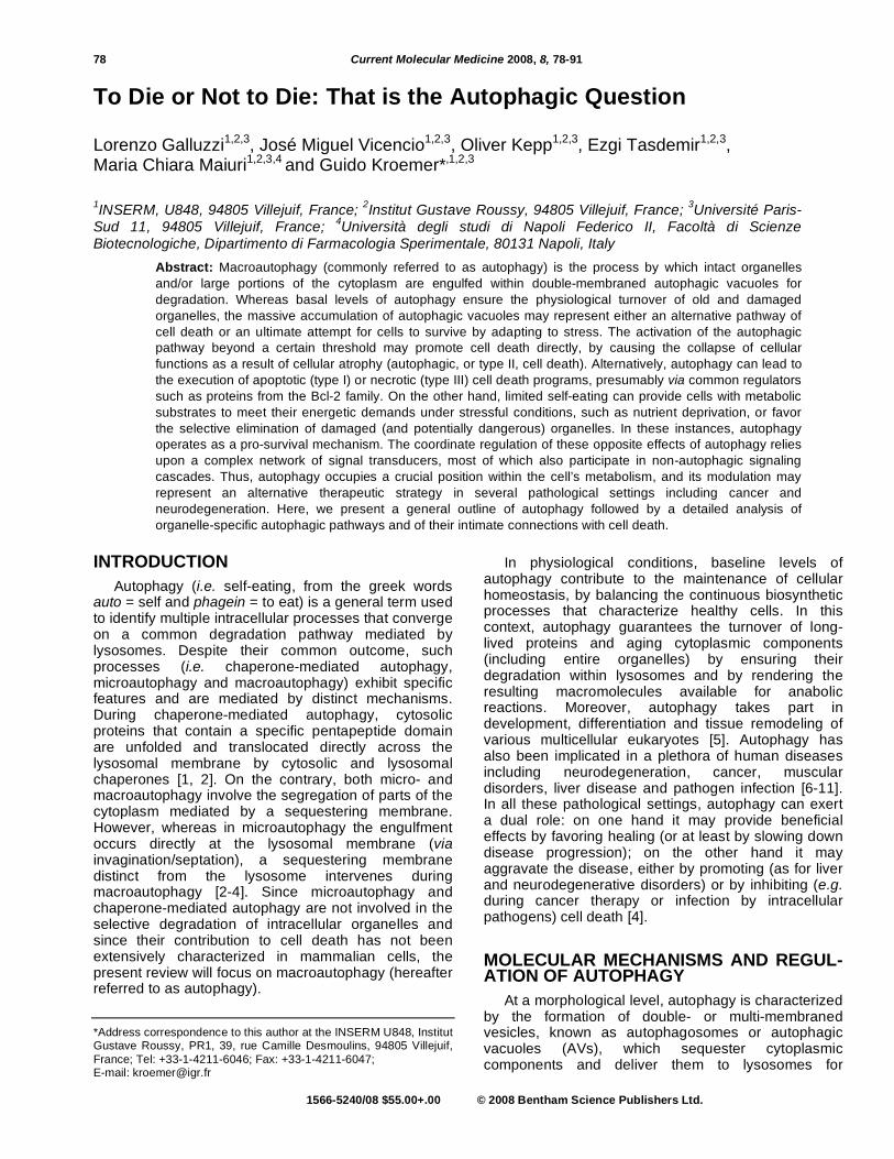
degradation. The autophagosome originates from a membrane of still unknown derivation (usually termed phagophore or isolation membrane) [12] that progressively expands, surrounds the material to be degraded and finally forms a double-membraned AV [3, 13]. At this stage, the AV lumen contains undegraded components, and is referred to as early autophagic vacuole (AVI) [14]. The subsequent fusion between AVIs and lysosomes then allows lysosomal acidic hydrolases to gain access to the AV lumen and promotes the degradation of its content. Such vesicles, formed upon the AVI-lysosomal fusion and containing partially digested material, are called late autophagic vacuoles (AVII), autophagolysosomes or autolysosomes [3, 13]. When observed by transmission electron microscopy, AVIIs appear as vacuoles with an electron-dense content and lack the double-membraned structure typical of AVIs [14]. This derives from the degradation of the AVI inner vesicle (known also as autophagic body) as a whole, occurring after the fusion of the AVs outer membrane with lysosomes. Fig. (1). Eventually, the metabolites generated by the degradation of the autophagic body are liberated into the cytosol, where they can reenter metabolic reactions [3, 13].
The molecular determinants of autophagy were first characterized in yeast models, and several of these autophagy-related (atg) proteins have known homologs in higher eukaryotes, and notably in humans [15-18]. Most of the proteins encoded by atg genes serve non-redundant roles within the autophagic pathway, mainly at the step of autophagosome formation, and the inhibition of one of such components usually suffices to block autophagy [13]. This may be attained by genetic and/or pharmacological means and may result in: (1) overall blockade of the autophagic cascade, when the activity of proteins involved in the initiation step is inhibited; (2) accumulation of immature phagophores, following the knockdown of proteins involved in the vesicle elongation process; (3) accumulation of AVIs, if
the fusion with lysosomes is blocked; or (4) accumulation of AVIIs, when the pH-dependent activation of lysosomal hydrolases is prevented [3]. At present, almost 40 atg genes have been identified in yeasts, among which only 11 have clear orthologs in mammals [2, 13, 17].
In mammalian cells, the nucleation of phagophores is regulated by the balance between class III and class I phosphatidylinositol 3-kinase (PI3K) complexes [19]. hVps34, the class III PI3K, catalyzes the phosphorylation of phosphatidylinositol (PtdIns) into phosphatidylinositol-3-phosphate (PtdIns(3)P), a molecular signal that promotes the recruitment of essential Atg proteins (Atg4, Atg7 and Atg10) [20, 21]. Its activation occurs upon the assembly of a multiprotein complex including also Beclin 1 (Bcn-1, the mammalian ortholog of Atg6), UVRAG (UV irradiation resistance-associated tumor suppressor gene), and the myristylated kinase p150 (ortholog of Vps15) [3, 22]. Conversely, the class I enzymatic complex plays an inhibitory function, although one of its products is PtdIns(3). Presumably, this inhibitory effect is mediated by other phosphoinositides generated by class I PI3K, in particular phosphatidylinositol-3,4,5-trisphoshate (PtdIns(3,4,5)P3), which promote the activation of mTOR (mammalian target of rapamycin) by its upstream regulator Akt1 (also known as PKB, i.e. protein kinase B) [13, 23]. The exact mechanism by which PtdIns(3)P favors autophagy in mammalian cells has not been established yet. As a possibility, PtdIns(3)P might act as a docking site to recruit soluble components of the autophagic machinery to the sites of vesicle formation [3, 13]. Such proteins may include also Atg9, whose retrieval transport from isolation membranes is governed by the Atg1-Atg13 signaling complex, a target of regulation by mTOR [24, 25].
TOR (target of rapamycin) is the major negative regulator of autophagy in yeasts, and acts by maintaining the hyperphosphorylation of Atg13, one of the proteins necessary for autophagy [26-28]. In this
Fig. (1). General overview of autophagy. The principal steps involved in the formation of early (AVI) and late (AVII) autophagic
vacuoles and in their processing along the autophagic pathway are depicted, together with the main gene products that regulate
each phase. For a detailed description of the process, please refer to the section “Molecular mechanisms and regulation of
autophagy”.
80 Current Molecular Medicine, 2008, Vol. 8, No. 2 Galluzzi et al.
state, Atg13 displays a reduced affinity for the serine/threonine kinase Atg1 and autophagy occurs only at baseline levels [29]. When TOR senses adverse environmental changes such as nutrient deprivation, its activity is quickly shut down and Atg13 undergoes rapid dephosphorylation, in turn allowing for the interaction with Atg1 and upregulation of autophagy [28]. It remains to be determined whether the kinase activity of Atg1 (the only serine/threonine kinase among the Atg proteins) is required or not for the induction of autophagy in yeast [28, 30]. Similar to yeast, the ortholog of TOR (mTOR) represents the crucial modulator of autophagy in mammalian cells [31]. However, the precise mechanisms underlying mTOR activity have not yet been fully elucidated [32].
In all eukaryotes, at least two distinct TOR-containing multiprotein complexes exist. These complexes are referred to as TORC1 (i.e. TOR complex 1, also known as CRTC1, i.e. CREB-regulated transcription coactivator 1) and TORC2 (CRTC2) [33]. The role of TORCs is not limited to the control of autophagy but extends to the regulation of protein translation and cell-cycle progression in response to growth factors and nutrient availability [34-36]. Interestingly, TORC2 (but not TORC1) catalyzes the activating-phosphorylation of Akt1/PKB, a serine/ threonine kinase whose activity is regulated also by the levels of PtdIns(3,4,5)P3 at the plasma membrane. mTOR autophagy-inhibitory functions are stimulated by Akt1. In this context, the tumor suppressor PTEN (phosphatase and tensin homologue) positively stimulates autophagy via its PtdIns(3,4,5)P3-3-phosphatase activity [37, 38]. As compared to TORC1, TORC2 exhibits a reduced sensitivity to acute pharmacological inhibition by rapamycin (a lipophilic macrolide that is commonly employed in experimental settings to promote autophagy) [33, 39]. Nonetheless, it seems that part of the sensitivity to rapamycin is restored by prolonged exposure or by the use of derivatives, at least in some cell types [40, 41]. As a note, rapamycin and its derivatives act by binding to the immunophilin FK506-binding protein 12 (FKBP12), which in turn becomes able to sequestrate TOR within an inactive molecular complex [42, 43].
Vesicle elongation is mediated by two separated but related ubiquitin-like conjugation systems [44]. The first pathway relies upon the conjugation between Atg5 and Atg12, mediated by the E1-like and E2-like enzymes Atg7 and Atg10, respectively [45-47]. As an alternative, phosphatidylethanolamine is conjugated to LC3 (i.e. microtubule-associated protein light chain 3, the mammalian ortholog of Atg8) after a proteolytic cleavage mediated by Atg4, followed by the action of Atg7 and Atg3, an additional E2-like enzyme [48-50]. In this context, the cleavage and lipidation of LC3 lead to its conversion from a soluble (LC3-I) to an AV-bound form (LC3-II). LC3-I to LC3-II conversion provides a specific marker for autophagy, since it results in a shift from a diffuse to a punctuate immunofluorescence staining pattern (corresponding to the relocalization of LC3-II to AVs) as well as in an increase of
electrophoretic mobility [51]. In addition, green fluorescent protein (GFP)-LC3 chimeras may be employed to monitor AVs by fluorescence videomicroscopy [3, 50].
The final step of AVI maturation involves their fusion with lysosomal vesicles. In yeast, this process is well characterized and depends on the activity of Ccz1 and Mon1 [52]. These proteins participate in a common fusion machinery that mediates several other membrane-trafficking pathways, but the implication in autophagy of their mammalian orthologs has not yet been elucidated [32, 53]. How the fusion between AVIs and lysosomes is controlled in mammals has only recently begun to emerge. Regulators of this process include the small GTP binding protein Rab7, the AAA-type ATPase SKD1 and presumably presenilin 1 (which is also linked to Alzheimer disease) [54, 55]. As a note, the pH within lysosomes may affect not only the activation of acidic hydrolases but also the AVI-lysosomal fusion [56].
CROSSTALK BETWEEN AUTOPHAGY AND APOPTOSIS
Autophagy and cell death are deeply intertwined at multiple levels. Often, similar stimuli are able to induce either an autophagic or an apoptotic response, which seem however to develop in a mutually exclusive fashion. Mixed phenotypes in response to such common triggers are rarely observed, and the prevalence of one phenotype over the other may be accounted for by various models. As a possibility, both phenomena may manifest only upon the achievement of a certain threshold level for activation, which may vary according to the stimulus and the specific experimental context. Alternatively, conditions that are known to promote both autophagy and apoptosis may prompt the cells with a sort of “decision” between the two pathways, which may be related to their mutual inhibition. Irrespective of the molecular mechanisms that underlie the cross-regulation between autophagy and apoptosis, their functional interrelationship is complex and is subject of an intense debate. On one hand, autophagy represents an alternative route to cell death (termed autophagic, or type II, cell death) that is known to occur in multiple pathophysiological scenarios [57]. On the other hand, in several settings autophagy inhibits apoptosis and allows for adaptation to environmental stress, thus acting as a strong pro-survival mechanism [3, 58].
Often, type II cell death manifests when the cell’s apoptotic machinery is hijacked. For instance, the pan-caspase inhibitor Z-VAD.fmk has been shown to induce per se autophagic cell death in some cell types (including mouse L929 fibrosarcoma cells and human Jurkat T-cell lymphoma cells). Such an effect is probably correlated to the combined inhibition of multiple members of the caspase family (including caspase-8) as well as to that of a calpain-like protease, which may act as an autophagic cell death repressor [59]. Similarly, type II cell death (that could be partially
Autophagy and Cell Death Current Molecular Medicine, 2008, Vol. 8, No. 2 81
suppressed by RNAi-mediated depletion of Bcn-1) was elicited by Z-VAD.fmk in U937 monocytoid cells and macrophages treated with lipopolysaccharide [60]. Rather unexpectedly, such lethal effects of Z-VAD.fmk have been associated with an overproduction of reactive oxygen species (ROS), due to the selective degradation (mediated by the autophagic pathway, and hence suppressed by autophagy inhibitors or by RNAi-mediated knockdown of Atg7 and Atg8) of the ROS-scavenger enzyme catalase [61, 62]. In a different experimental setting, the treatment of wild-type (wt) mouse embryonic fibroblasts (MEFs) with Z-VAD.fmk failed to modify their lethal response to the DNA-damaging agent etoposide [63]. On the contrary, MEFs from bax
-/--bak
-/- double-knockout mice (that are
resistant to several pro-apoptotic stimuli), but not from animals in which other components of the apoptotic machinery (such as Apaf-1 and caspase-9) were knocked-out, exhibited a delayed response to etoposide, that manifested with a massive autophagic vacuolization rather than with the morphological features of apoptosis. In this context, the etoposide-induced death of bax
-/--bak
-/- MEFs was efficiently
inhibited by the small-interfering RNA (siRNA)-mediated downregulation of Atg6/Bcn-1 and Atg5, as well as by pharmacological inhibitors of autophagy [63]. The fact that the autophagic response to etoposide was specifically observed in bax
-/--bak
-/-MEFs, but not in
apaf-1-/-
nor in caspase-9-/-
cells, indicates that these (and possibly other) Bcl-2-like proteins may directly or indirectly modulate autophagy independently of their well-known role in apoptosis.
Recently, autophagy has been postulated to play a lethal role also in the necrotic (type III) cell death of Caenorhabditis elegans neurons [64]. In this context, it has been demonstrated that larvae carrying the neurotoxic alleles mec-4(d) or deg-3(d) (that induce per se neuronal cell death with the macroscopic and ultrastructural features of necrosis) exhibited an enhanced survival (and a reduced number of neuronal corpses) when genes involved in distinct steps of autophagy were concomitantly knocked-out (or when the corresponding transcripts were depleted via RNAi). This applied to unc-51, bec-1, igg-1 and atgr-18 (the nematode orthologs of mammalian atg1, atg6/bcn-1, atg8/LC3 and atg18, respectively) and was also observed following pharmacological inhibition of autophagy with the hVps34 inhibitor 3-methyladenine (3-MA). Interestingly, while mec-4(d)-induced neuronal degeneration was ameliorated in both lysosomal aspartyl protease- (Cad-1)- and calpain- (Clp-1)-deficient animals, only in the former the concomitant knockout of igg-1 further improved cell survival, suggesting that Clp-1 and autophagy function in the same pathway to mediate necrotic cell death [64].
When the apoptotic machinery is inhibited, the depletion of intracellular metabolite stores (for instance following the withdrawal of growth factors) promotes a pro-survival, rather than a lethal autophagic response. This was first demonstrated with immortalized interleukin-3 (IL3)-dependent cell lines derived from
bax-/-
-bak-/-
mice [65]. As opposite to their wt counterparts, these cells did not succumb to apoptosis upon IL3 withdrawal, but underwent a prolonged autophagic response that resulted in progressive cellular atrophy and (only after several weeks) cell death. In this context, autophagy allowed growth factor-deprived cells (that are unable to internalize nutrients in sufficient quantity to sustain cellular bioenergetics, despite their abundance in the medium) to maintain ATP synthesis from the catabolism of intracellular substrates and to remain responsive to the readdition of IL3 for long periods. However, growth factor-deprived bax
-/--bak
-/- cells died upon RNAi-mediated
downregulation of Atg5 or Atg7, as well as upon pharmacological inhibition of autophagy with 3-MA or the lysosomal acidification inhibitor chloroquine [65]. This strongly corroborates the hypothesis that, in specific scenarios, autophagy mobilizes nutrients by promoting the catabolism of intracellular stores, in an attempt to counteract the progressive drop of ATP levels typical of starving cells, and to prevent a bioenergetic collapse that would eventually result in cell death.
Accordingly, the inhibition of autophagy (either at early or late stages) may result in the failure of cells to adapt to nutrient deprivation, thus precipitating cell death (that may manifest or not with an apoptotic phenotype). For instance, distinct human cancer cell lines (including HeLa cervical carcinoma and HCT116 colon carcinoma cells) were shown to die under starvation conditions if autophagy was concomitantly inhibited either via RNAi (targeting Atg5, Atg6/Bcn-1, Atg10 or Atg12) or by means of pharmacological inhibitors. This cell death occurred indeed via a type I pathway, since it exhibited the classical features of apoptosis (including mitochondrial membrane permeabilization, i.e. MMP, caspase activation and chromatin condensation) and it could be retarded by the stabilization of mitochondrial membranes (through the depletion of Bax and Bak or by overexpressing Bcl-2 or vMIA, i.e. the mitochondrion-targeted viral modulator of apoptosis encoded by Cytomegalovirus) as well as by caspase inhibition [58, 66]. Notably, before the appearance of typical signs of apoptosis, the phenotype of cells heavily depended on the stage at which autophagy was blocked. Thus, whereas no AVs were observed in cells depleted for proteins necessary for the first steps of autophagy (e.g. Atg5, Atg6/Bcn-1, Atg10, Atg12 or hVps34), a massive autophagic vacuolization resulted from the inhibition of the AVI-lysosomal fusion (as obtained with chloroquine or by RNAi targeting the lysosome-associated membrane protein 2, i.e. LAMP-2). In the latter case, a mixed type I-type II phenotype characterized the cells before their demise, thus demonstrating that autophagic vacuolization may co-exist with an apoptotic morphology and that it may result also from the failure of cells to dispose of AVs rather than solely from their enhanced generation [58, 66]. However, the inhibition of autophagy does not always promote type I cell death [67]. For instance, non-apoptotic cell death associated
82 Current Molecular Medicine, 2008, Vol. 8, No. 2 Galluzzi et al.
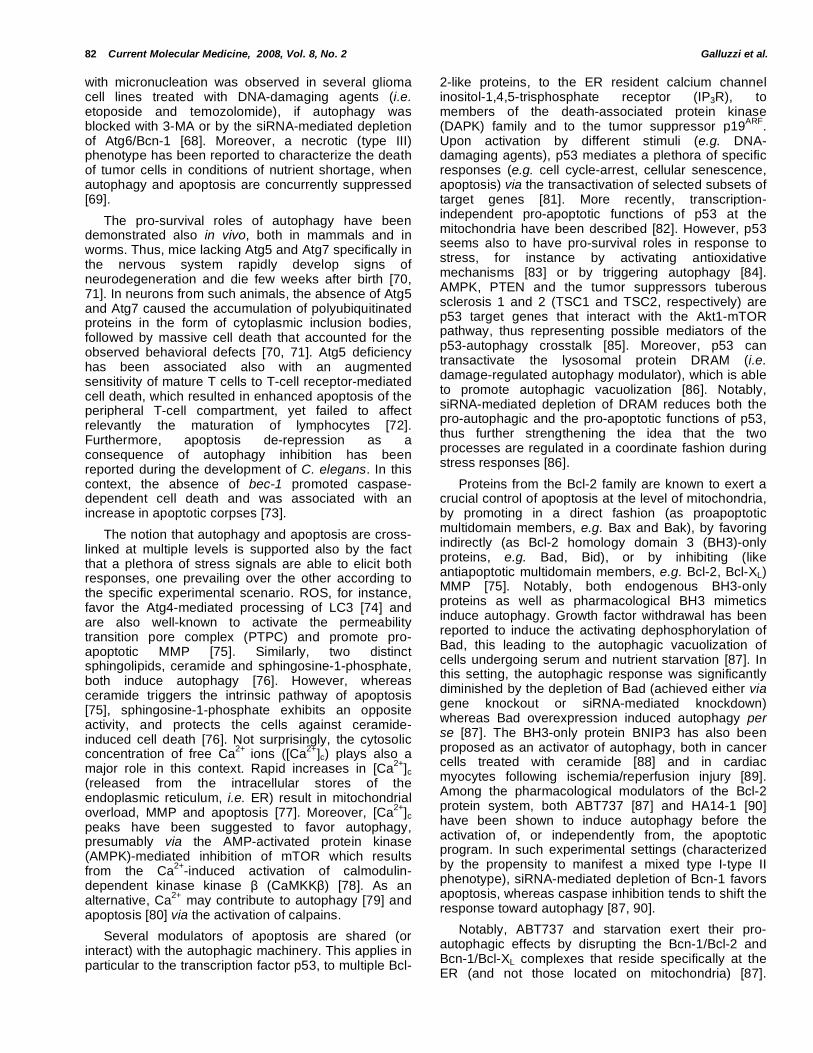
with micronucleation was observed in several glioma cell lines treated with DNA-damaging agents (i.e. etoposide and temozolomide), if autophagy was blocked with 3-MA or by the siRNA-mediated depletion of Atg6/Bcn-1 [68]. Moreover, a necrotic (type III) phenotype has been reported to characterize the death of tumor cells in conditions of nutrient shortage, when autophagy and apoptosis are concurrently suppressed [69].
The pro-survival roles of autophagy have been demonstrated also in vivo, both in mammals and in worms. Thus, mice lacking Atg5 and Atg7 specifically in the nervous system rapidly develop signs of neurodegeneration and die few weeks after birth [70, 71]. In neurons from such animals, the absence of Atg5 and Atg7 caused the accumulation of polyubiquitinated proteins in the form of cytoplasmic inclusion bodies, followed by massive cell death that accounted for the observed behavioral defects [70, 71]. Atg5 deficiency has been associated also with an augmented sensitivity of mature T cells to T-cell receptor-mediated cell death, which resulted in enhanced apoptosis of the peripheral T-cell compartment, yet failed to affect relevantly the maturation of lymphocytes [72]. Furthermore, apoptosis de-repression as a consequence of autophagy inhibition has been reported during the development of C. elegans. In this context, the absence of bec-1 promoted caspase-dependent cell death and was associated with an increase in apoptotic corpses [73].
The notion that autophagy and apoptosis are cross-linked at multiple levels is supported also by the fact that a plethora of stress signals are able to elicit both responses, one prevailing over the other according to the specific experimental scenario. ROS, for instance, favor the Atg4-mediated processing of LC3 [74] and are also well-known to activate the permeability transition pore complex (PTPC) and promote pro-apoptotic MMP [75]. Similarly, two distinct sphingolipids, ceramide and sphingosine-1-phosphate, both induce autophagy [76]. However, whereas ceramide triggers the intrinsic pathway of apoptosis [75], sphingosine-1-phosphate exhibits an opposite activity, and protects the cells against ceramide-induced cell death [76]. Not surprisingly, the cytosolic concentration of free Ca
2+ ions ([Ca
2+]c) plays also a
major role in this context. Rapid increases in [Ca2+
]c (released from the intracellular stores of the endoplasmic reticulum, i.e. ER) result in mitochondrial overload, MMP and apoptosis [77]. Moreover, [Ca
2+]c
peaks have been suggested to favor autophagy, presumably via the AMP-activated protein kinase (AMPK)-mediated inhibition of mTOR which results from the Ca
2+-induced activation of calmodulin-
dependent kinase kinase (CaMKK ) [78]. As an alternative, Ca
2+ may contribute to autophagy [79] and
apoptosis [80] via the activation of calpains.
Several modulators of apoptosis are shared (or interact) with the autophagic machinery. This applies in particular to the transcription factor p53, to multiple Bcl-
2-like proteins, to the ER resident calcium channel inositol-1,4,5-trisphosphate receptor (IP3R), to members of the death-associated protein kinase (DAPK) family and to the tumor suppressor p19
ARF.
Upon activation by different stimuli (e.g. DNA-damaging agents), p53 mediates a plethora of specific responses (e.g. cell cycle-arrest, cellular senescence, apoptosis) via the transactivation of selected subsets of target genes [81]. More recently, transcription-independent pro-apoptotic functions of p53 at the mitochondria have been described [82]. However, p53 seems also to have pro-survival roles in response to stress, for instance by activating antioxidative mechanisms [83] or by triggering autophagy [84]. AMPK, PTEN and the tumor suppressors tuberous sclerosis 1 and 2 (TSC1 and TSC2, respectively) are p53 target genes that interact with the Akt1-mTOR pathway, thus representing possible mediators of the p53-autophagy crosstalk [85]. Moreover, p53 can transactivate the lysosomal protein DRAM (i.e. damage-regulated autophagy modulator), which is able to promote autophagic vacuolization [86]. Notably, siRNA-mediated depletion of DRAM reduces both the pro-autophagic and the pro-apoptotic functions of p53, thus further strengthening the idea that the two processes are regulated in a coordinate fashion during stress responses [86].
Proteins from the Bcl-2 family are known to exert a crucial control of apoptosis at the level of mitochondria, by promoting in a direct fashion (as proapoptotic multidomain members, e.g. Bax and Bak), by favoring indirectly (as Bcl-2 homology domain 3 (BH3)-only proteins, e.g. Bad, Bid), or by inhibiting (like antiapoptotic multidomain members, e.g. Bcl-2, Bcl-XL) MMP [75]. Notably, both endogenous BH3-only proteins as well as pharmacological BH3 mimetics induce autophagy. Growth factor withdrawal has been reported to induce the activating dephosphorylation of Bad, this leading to the autophagic vacuolization of cells undergoing serum and nutrient starvation [87]. In this setting, the autophagic response was significantly diminished by the depletion of Bad (achieved either via gene knockout or siRNA-mediated knockdown) whereas Bad overexpression induced autophagy per se [87]. The BH3-only protein BNIP3 has also been proposed as an activator of autophagy, both in cancer cells treated with ceramide [88] and in cardiac myocytes following ischemia/reperfusion injury [89]. Among the pharmacological modulators of the Bcl-2 protein system, both ABT737 [87] and HA14-1 [90] have been shown to induce autophagy before the activation of, or independently from, the apoptotic program. In such experimental settings (characterized by the propensity to manifest a mixed type I-type II phenotype), siRNA-mediated depletion of Bcn-1 favors apoptosis, whereas caspase inhibition tends to shift the response toward autophagy [87, 90].
Notably, ABT737 and starvation exert their pro-autophagic effects by disrupting the Bcn-1/Bcl-2 and Bcn-1/Bcl-XL complexes that reside specifically at the ER (and not those located on mitochondria) [87].
Autophagy and Cell Death Current Molecular Medicine, 2008, Vol. 8, No. 2 83
Conversely, both BH3 mimetics and BH3-only proteins are known to promote apoptosis at mitochondria, by inducing MMP [75]. These observations suggest that only a subset of Bcl-2 family members with a particular subcellular localization (notably the ER) may be involved in the regulation of autophagy, whereas Bcl-2-like proteins localized at mitochondria may preferentially control apoptosis [91]. This hypothesis, however, is incompatible with data on BH3-only proteins that exhibit a near-to-total mitochondrial localization (such as BNIP3 and hSPIN1, the human ortholog of the Drosophila melanogaster protein spinster), yet have been reported to induce autophagy and non-apoptotic cell death [92, 93]. Thus, the mechanism by which BH3-only proteins trigger autophagy versus apoptosis remains to be clarified. In this regard, it has been hypothesized that “activator” BH3-only proteins (e.g. Bid) – that interact with and activate directly the proapoptotic multidomain members of the Bcl-2 family – would promote only apoptosis, whereas the so-called “derepressors” (e.g. Bad) – which are known to promote cell death indirectly, by neutralizing the activity of antiapoptotic Bcl-2-like proteins – would trigger both programs [3, 94].
Recently, it has been found that Bcn-1 contains a BH3 domain and that it can be de facto included among the BH3-only proteins [87, 95]. As other BH3-
containing proteins, Bcn-1 is sequestered by the antiapoptotic multidomain proteins from the Bcl-2 family (in particular by Bcl-2 and Bcl-XL) due to the interaction of its amphipatic BH3 helix with the hydrophobic groove of Bcl-2/Bcl-XL that serves as a BH3 receptor [95]. This notion supports a model to explain the pro-autophagic effects of other BH3-only proteins and BH3-mimetic compounds. Accordingly, both activated endogenous BH3-only proteins and exogenous BH3 mimetics would favor autophagy by competitively displacing Bcn-1 from its inhibitory interaction with Bcl-2/Bcl-XL, thereby allowing it to allosterically activate hVps34 [21, 91] Fig. (2). In this context, it has been shown that Bcl-2 downregulation (by the inducible expression of antisense oligonucleotides) promotes massive vacuolization and type II cell death independently from MMP and caspase activation, in human leukemic HL-60 cells [96]. This further corroborates the idea that the preponderant function of Bcl-2 may be either anti-apoptotic [75] or anti-autophagic [97, 98], depending on the specific experimental setting.
Extra-mitochondrial activities of Bcl-2 like proteins may contribute indirectly to their apoptosis-modulatory role [99-102]. In this context, Bcl-2 family proteins have been demonstrated to operate at the ER to control the steady-state luminal Ca
2+ concentration and the
agonist-induced Ca2+
release, by interacting with
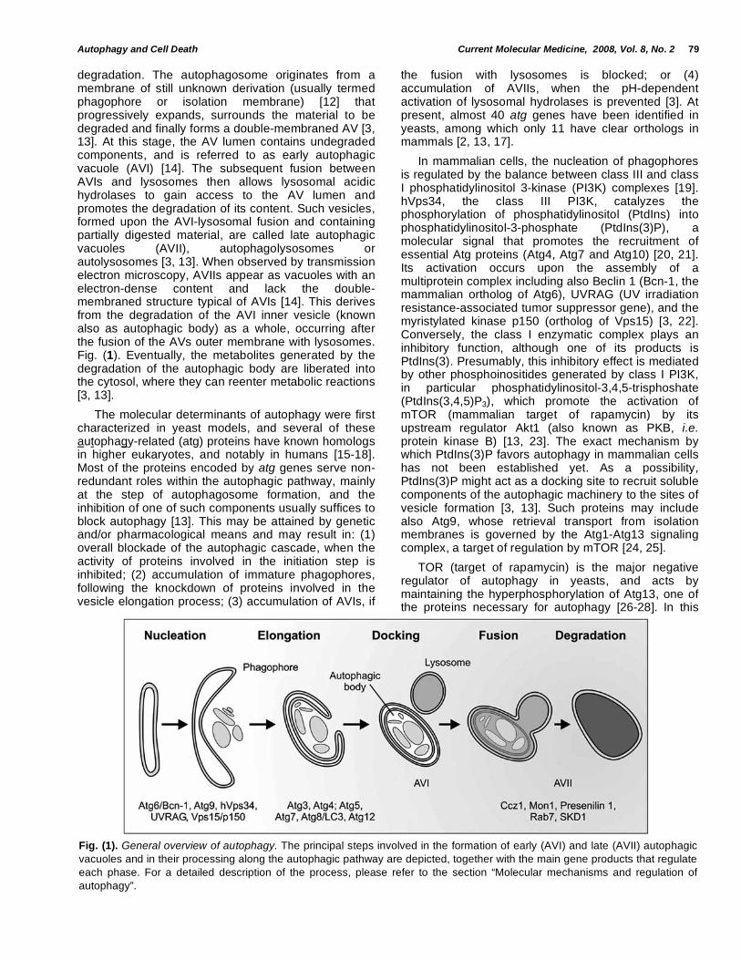
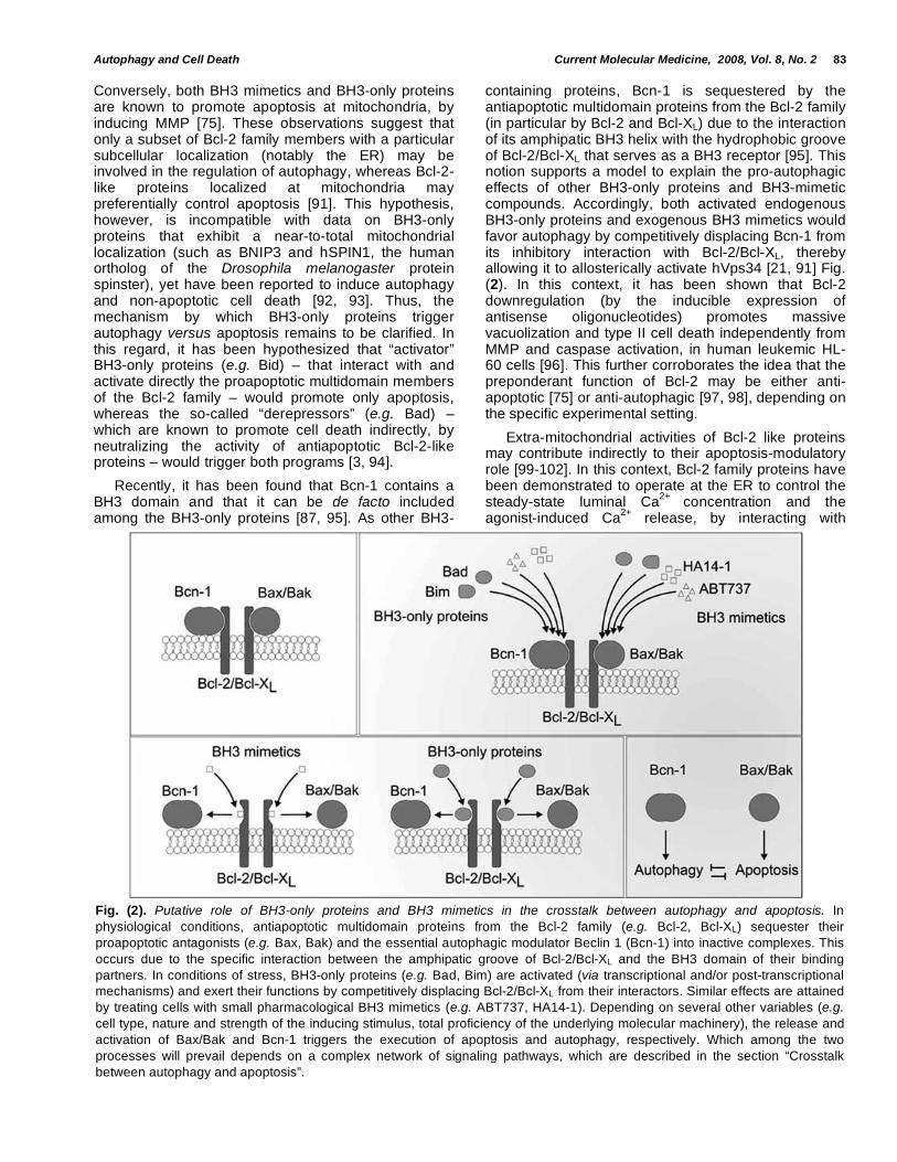
Fig. (2). Putative role of BH3-only proteins and BH3 mimetics in the crosstalk between autophagy and apoptosis. In
physiological conditions, antiapoptotic multidomain proteins from the Bcl-2 family (e.g. Bcl-2, Bcl-XL) sequester their
proapoptotic antagonists (e.g. Bax, Bak) and the essential autophagic modulator Beclin 1 (Bcn-1) into inactive complexes. This
occurs due to the specific interaction between the amphipatic groove of Bcl-2/Bcl-XL and the BH3 domain of their binding
partners. In conditions of stress, BH3-only proteins (e.g. Bad, Bim) are activated (via transcriptional and/or post-transcriptional
mechanisms) and exert their functions by competitively displacing Bcl-2/Bcl-XL from their interactors. Similar effects are attained
by treating cells with small pharmacological BH3 mimetics (e.g. ABT737, HA14-1). Depending on several other variables (e.g.
cell type, nature and strength of the inducing stimulus, total proficiency of the underlying molecular machinery), the release and
activation of Bax/Bak and Bcn-1 triggers the execution of apoptosis and autophagy, respectively. Which among the two
processes will prevail depends on a complex network of signaling pathways, which are described in the section “Crosstalk
between autophagy and apoptosis”.
84 Current Molecular Medicine, 2008, Vol. 8, No. 2 Galluzzi et al.
several Ca2+
-related proteins including the IP3R as well as different isoforms of the sarco/endoplasmic reticulum Ca
2+ ATP-ase (SERCA) [103-106]. It is still
controversial whether Bcl-2/Bcl-XL act on the IP3R by modulating its gating in response to IP3 or by promoting a Ca
2+ leak into the cytosol [102, 107]. Independently
from the model of interaction with the IP3R, Bcl-2/Bcl-XL exert their antiapoptotic functions by diminishing the IP3R-mediated Ca
2+ release [108]. In HeLa cells
overexpressing Bcl-2, the apoptotic response could be restored by the concomitant overexpression of SERCA, which corrected the ER Ca
2+ levels, correlating with an
increase in mitochondrial Ca2+
uptake [105]. Contrarily to Bcl-2/Bcl-XL, the proapoptotic proteins Bax and Bak have been shown to promote the transfer of Ca
2+ from
the ER to mitochondria and cell death [109, 110]. Moreover, bax
-/--bak
-/- MEFs exhibited a defective
mobilization of Ca2+
in response to various pro-apoptotic stimuli, which could be partially restored upon SERCA overexpression [100]. As previously mentioned, [Ca
2+]c increases seem to be required for
autophagy, and Bcl-2 might act as an inhibitor of autophagy by dampening [Ca
2+]c elevation [78]. Thus,
Bcl-2 modulates Ca2+
signaling to reduce both apoptotic and autophagic responses.
The IP3R is deeply involved in the regulation of autophagy [111]. Both pharmacological inhibition (with xestospongins) and siRNA-mediated downregulation of the IP3R have been shown to promote the autophagic process [111]. In contrast to the mechanism elicited by Ca
2+ mobilization [78], IP3R inhibition-induced
autophagy did not involve Ca2+
itself, yet was inhibited by Bcl-2/Bcl-XL specifically targeted to the ER [111]. Moreover, the inhibition of IP3R did not provoke ER stress and the absence of the major ER stress transducer transmembrane kinase/endoribonuclease inositol-requiring enzyme 1 (IRE1) did not affect IP3R inhibition-induced autophagy [112]. Altogether, these observations point to an autophagic pathway that is directly activated by the inhibition IP3R (independently from Ca
2+ and ER stress), but also to a controversial
role for the IP3R in autophagy. Indeed, the Ca2+
channel activity of the IP3R is necessary to induce autophagy, yet the same outcome derives from its depletion/inhibition. Notably, Bcl-2 is able to inhibit the autophagic response in both instances, outlining once again the deep connection between apoptotic and autophagic machineries.
Several inducers of the extrinsic pro-apoptotic pathway (e.g. interferon- , cross-linking of the death receptor Fas/CD95, tumor necrosis factor , transforming growth factor , and detachment from the extracellular matrix) [113-115], as well as signals that trigger the intrinsic pathway (e.g. oncogenic stress) [116], are able to promote the activation of the serine/threonine kinase DAPK1. In response to such stimuli, DAPK1 mediates either apoptosis or autophagy via several mechanisms [117, 118] including the activation of p53 through the p19
ARF-mediated inhibition
of MDM2 (the main negative regulator of p53) [116], the inhibition of integrin-mediated adhesion [115], and
the induction of a mitochondrial apoptotic pathway independent from p53 [114]. Notably, DAPK1 exhibits a prominent tumor suppressor activity [116], and its expression is lost due to epigenetic mechanisms (DNA methylation) in several cancers [117]. Other members of the DAPK family (in particular DAPK2, also known as DAPK-related protein kinase 1, i.e. DRP1, and DAPK3) have also been implicated in the coordinate regulation of type I and type II cell death [118, 119]. Of note, whereas DAPK1 is mainly localized to the actin cytoskeleton, overexpressed DRP1 concentrates within the AV lumen, suggesting a direct involvement of this latter kinase in the control of autophagy [118].
p19ARF
is a potent tumor suppressor that acts from a nucleolar localization by blocking MDM2-induced p53 degradation and by regulating ribosomal biogenesis [120, 121]. Its expression is lost in several tumors, including small cell lung cancer, T-cell acute lymphoblastic leukemia and colon carcinoma, due to both genetic and epigenetic mechanisms [122-125]. A short isoform of p19
ARF (known as smARF, i.e. small
mitochondrial ARF) results from internal translation at Met45 and localizes to a proteinase K-resistant mitochondrial compartment [126]. Following ectopic expression, smARF (that lacks the nucleolar functional domains of p19
ARF) favors the dissipation of the
mitochondrial transmembrane potential ( m) independently from p53 and Bcl-2 family proteins, followed by massive autophagic vacuolization and caspase-independent cell death [126, 127]. Notably, this autophagic response can be partially suppressed by the siRNA-mediated depletion of Atg5 or Bcn-1 [126].
ORGANELLE-SPECIFIC AUTOPHAGY
The functions of autophagy include the selective removal of old, unnecessary and/or damaged cytoplasmic organelles that may represent a serious threat for cellular homeostasis if they are not disposed of. This organelle-specific autophagic response was first identified in yeast cells that adapt their metabolism to different nutritional sources [128]. For instance, in Saccharomyces cerevisiae pexophagy (the selective degradation of peroxisomes) takes places when cells are shifted from oleic acid as the sole carbon source (that promotes peroxisome biogenesis) to glucose-rich or nitrogen-limiting conditions, thus eliminating metabolically superfluous organelles [129]. The specificity of pexophagy has been attributed to the presence on the peroxisomal membrane of the protein peroxin 14 [130, 131]. In yeasts, also nonessential nuclear components are selectively sent to degradation and recycling in the vacuole. However, this phenomenon (known as piecemeal microautophagy of the nucleus, i.e. PMN) occurs also in atg7
-/- cells, and
hence does not depend on the general macroautophagic machinery [132]. Organelle-specific autophagy is not limited to unicellular eukaryotes, but has also been described in mammals. For instance, under conditions of elevated glucose demand,

Autophagy and Cell Death Current Molecular Medicine, 2008, Vol. 8, No. 2 85
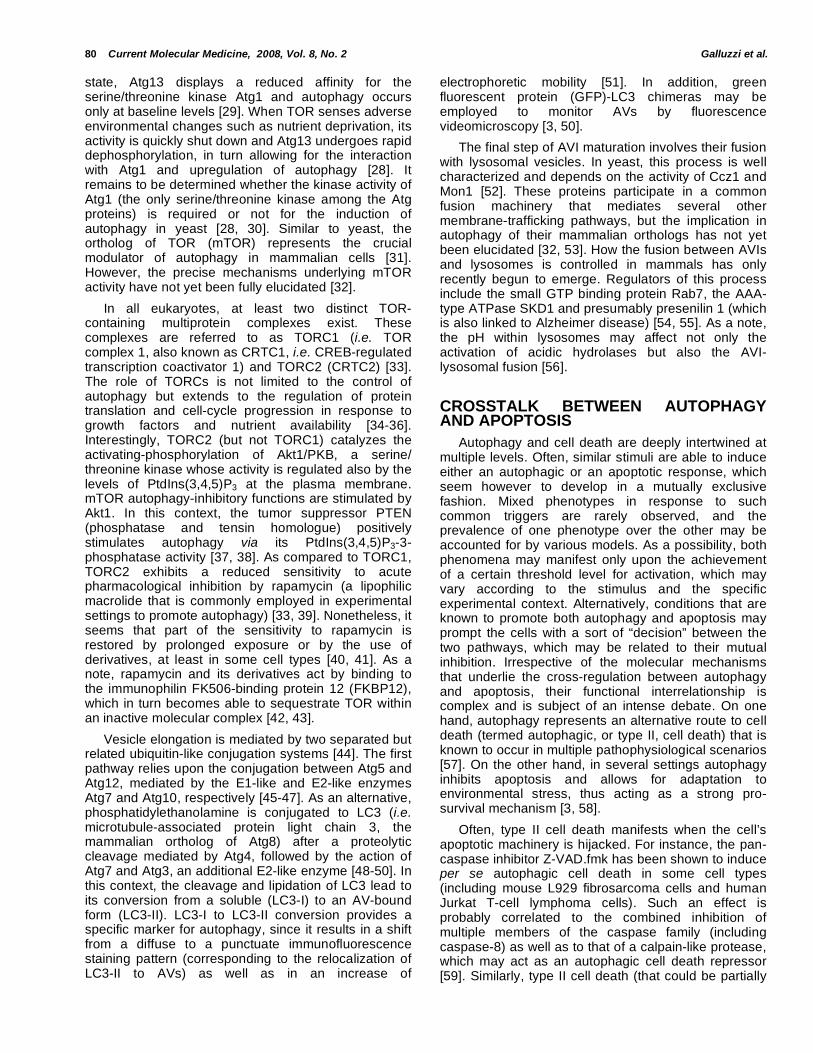
cytoplasmic glycogen granules are able to undergo a selective, highly regulated form of macroautophagic degradation. This process provides a main contribution to glucose homeostasis, particularly in newborn animals, which have to face extreme metabolic demands concurring with the interruption of transplacental supplies [133]. Notably, glycogen autophagy is promoted by glucagon secreted during postnatal hypoglycemia, yet is inhibited by parental glucose (and insulin) through the Akt1/mTOR axis of autophagy regulation [134]. Mitochondria and ER from mammalian cells (similarly to their yeast counterpart) may also be specifically eliminated by autophagic degradation [3]. The pathways leading to the selective removal of mitochondria and ER have been named mitophagy and reticulophagy (or ER-phagy), respectively [135, 136] Fig. (3).
Mitophagy
In eukaryotic cells, the vast majority of ATP is synthesized by mitochondria in a process called oxidative phosphorylation. Enzymatic complexes of the mitochondrial inner membrane (IM) catalyze the sequential oxidation of respiratory substrates coupled to the extrusion of protons from the mitochondrial matrix. In turn, this generates an electrochemical gradient whose regulated dissipation allows the F0F1 ATP synthase complex to produce ATP [137]. Redox reactions normally generate ROS, including hydrogen peroxide (H2O2) and superoxide anion (O2
-), at
multiple sites along the respiratory chain [138]. Whereas physiological levels of ROS are buffered by
intra- and extra-mitochondrial antioxidant systems [139] and are involved in multiple intracellular signaling pathways [140], ROS overgeneration (that occurs for instance upon uncoupling or inhibition of the respiratory complexes) may represent a major threat for cell survival [141]. ROS-mediated cytotoxicity involves the oxidation of theoretically all cell constituents, including DNA, proteins and lipids. This leads to DNA strand breaks, base modifications as well as to the functional impairment of lipids and proteins, which also lose their correct folding and tend to form toxic aggregates [142]. However, since mitochondria account for the majority of intracellular ROS generation, they represent the preferential site of ROS damage. Notably, oxidative stress is known to promote the opening of the PTPC, causing a sudden increase in IM permeability to low molecular weight solutes [75]. Mitochondrial permeability transition (MPT) leads to m dissipation, uncoupling of the oxidative phosphorylation, arrest of ATP synthesis and complete functional collapse of mitochondria. In addition, it results in an osmotic imbalance that drives the unregulated entry of water into the mitochondrial matrix, physical rupture of the outer mitochondrial membrane, and release into the cytosol of toxic proteins normally retained in the intermembrane space. Altogether, these events activate caspase-dependent and –independent apoptotic executioner mechanisms and lead to cell death [75]. ROS-mediated mitochondrial damage involves also mtDNA. As a matter of fact, mtDNA is more susceptible to oxidative injury than its nuclear counterpart, in part because it lacks the protective effect mediated by histones, but also because DNA
Fig. (3). Examples of organelle-specific pathways of autophagy. As opposite to a general, rather unspecific autophagic program,
several pathways have been described that deliver selected organelles to autophagosomes for degradation. These include (but
are not limited to) the macroautophagic degradation of the endoplasmic reticulum (reticulophagy) and of mitochondria
(mitophagy), as well as the specific autophagy of peroxisomes in yeasts (known as pexophagy). The so-called “piecemeal
microautophagy of the nucleus” (PMN), that has been described in yeast cells, might also be considered as an example of
organelle-specific autophagy, yet it is mediated by a microautophagic route. For additional details, please see the section
“Organelle-specific autophagy”.

86 Current Molecular Medicine, 2008, Vol. 8, No. 2 Galluzzi et al.
repair mechanisms are less effective in mitochondria than in the nucleus [143, 144]. mtDNA mutations may further exacerbate mitochondrial dysfunction by leading to the synthesis of faulty mitochondrial proteins or by blocking synthesis altogether. This accounts, at least partially, for the morphological and functional abnormalities of mitochondria associated with aging [145]. Given their multifaceted toxic potential, it is not surprising that cells have evolved specific mechanisms to ensure the timely elimination of damaged, old and/or dysfunctional mitochondria that prominently occurs via the activation of a targeted macroautophagic program [135].
MPT involving the vast majority of mitochondria promotes cell death [75]. On the contrary, permeabilized mitochondria are selectively targeted by mitophagy, if only a small fraction of a cell’s mitochondria is affected (for instance if the stimulus is administered at low, sub-apoptotic doses). This was first documented in cultured rat hepatocytes during nutrient deprivation [146]. In this setting, serum deprivation and glucagon treatment were employed to mimic fasting and promoted a limited increase in the rate of mitochondria undergoing depolarization to about 1.5% per hour. Notably, such depolarized mitochondria colocalized with autophagosomes and autolysosomes, which also increased in number upon starvation. Cyclosporine A (CsA, a pharmacological inhibitor of the PTPC) and its non-immunosuppressive analogue NIM811 suppress both m dissipation and autophagic vacuolization induced by nutrient depletion [146, 147]. As assessed by time-lapse confocal microscopy of hepatocytes expressing a GFP-LC3 fusion protein subjected to starvation, phagophores sequester polarized mitochondria, and only 10 minutes after ring closure depolarization occurs (as monitored via the potential-sensitive probe tetramethylrhodamine methyl ester, i.e. TMRM), followed by acidification of the newly generated AVI (revealed by the uptake of LysoTracker
®
Red) and fusion with lysosomal precursors (to form AVII). Interestingly, mitochondrial sequestration is unaffected by CsA, that would rather inhibit depolarization and the ensuing events [135, 148]. Similar scenarios are elicited by the BH3-only protein BNIP3 (that is activated by hypoxia) and by the BH3 mimetic ABT737, which are both able to cause m dissipation and mitophagy [89, 91, 92].
Mitophagy has also been shown to occur as a result of irreversible laser-induced photodamage [135]. When selected portions of the cytoplasm of living hepatocytes were exposed to a 488-nm laser beam, the mitochondria of the photodamaged zone dissipated their m in a light dose-dependent fashion. Whereas low light intensities promoted a reversible damage (mitochondrial TMRM fluorescence was indeed recovered within a few minutes), with longer exposures
m was irreversibly dissipated, followed by a specific mitophagic response disposing of photodamaged mitochondria, but not of intact neighboring organelles [135]. At difference with starvation-induced mitophagy, the autophagic response to photodynamic damage
could not be blocked by the inhibition of PI3K with 3-MA or wortmannin. Altogether, this data suggest that photodamage induces mitophagy downstream of PI3K-mediated signaling yet pointed again to the involvement of MPT (already proposed as the effector mechanism of photodynamic damage at mitochondria [149-151]).
Mitochondrial dysfunction, mitophagy and aging are intimately connected [145, 152, 153]. In post-mitotic non-proliferating tissues (e.g. liver, brain, heart, kidney), mitochondrial half-life has been estimated around 20-25 days. New mitochondria form continuously from the fission of pre-existing ones (in a process closely resembling bacterial division), while old ones are disposed of via mitophagy. mtDNA mutations tend to accumulate with time, and mitophagy might serve as a “quality control” mechanism, to get rid of dysfunctional mitochondria that harbor mutated mtDNA. In this context, the accumulation of mtDNA defects may result from reduction of autophagic activity due to age [154, 155]. Conversely, some mutations may be selected and amplified by making a mitochondrion less likely to be degraded, for instance by reducing its potential to generate ROS [153, 156].
An interesting link between mitophagy, oxidative stress and aging is provided by the protein Uth1p. In yeast, deletion of the uth1 gene confers resistance to several stress stimuli including heat shock, pro-oxidants (e.g. paraquat-generated superoxide anion and H2O2) as well as carbon and nitrogen starvation [157]. Loss of Uth1p has been shown to interfere with mitochondrial biogenesis, in particular by reducing the levels of cytochromes aa3, b and c as well as those of citrate synthase [158]. More recently, it has been shown that Uth1p is required in yeast for cell death induced by the ectopic expression of Bax as well as for the mitophagic response induced by rapamycin [157, 159]. Interestingly, Uth1p was first identified in a genetic screen aimed to isolate yeast strains that withstand long-term starvation [160]. Taken together, these observations suggest that an enhanced mitophagic elimination of damaged mitochondria may account, at least partially, for the well-known extension of lifespan resulting from caloric restriction (that indeed activates mitophagy) in organisms from different taxa [161]. Unfortunately, in mammals there is no obvious homolog of uth1, and the identification of Uth1p-like proteins remains elusive.
Beyond speculations, it is clear that the multifaceted contribution of mitophagy to different cellular processes (including response to stress, aging and cell death) awaits further clarification. At least theoretically, the specific modulation of mitophagy might represent a novel therapeutic approach for human pathologies that are directly or indirectly related to oxidative stress, such as ischemic injuries and cancer.
Reticulophagy
ER fulfills multiple metabolic functions, including the regulation of intracellular Ca
2+ stores [162], the
Autophagy and Cell Death Current Molecular Medicine, 2008, Vol. 8, No. 2 87
detoxification of potentially dangerous xenobiotics [163] as well as the synthesis, folding and post-translational modifications of secreted or integral membrane proteins [164, 165]. In this context, the finely regulated homeostatic mechanism known as unfolded protein response (UPR) operates to adapt the ER protein folding capacity to changing environmental conditions [165]. UPR senses the accumulation of misfolded proteins within the ER lumen and activates a complex, multi-branched signaling pathway that (via a massive transcriptional program and post-transcriptional mechanisms) aims at re-establishing homeostasis [164, 166]. In the ER, one of the principal sensors for misfolded proteins is IRE1 [167, 168]. Through an unusual, yet evolutionarily conserved mechanism, IRE1 catalyzes the unconventional splicing of a single mRNA species, HAC1, thus allowing for the synthesis of the active transcription factor HAC1 [169, 170]. HAC1 (and its metazoan ortholog X-box binding protein 1, i.e. XBP1) transactivates a large set of genes (in yeast including 5% of all genes), most of which encode for chaperones, folding enzymes and other factors involved in the secretory pathway, ER-associated protein degradation (ERAD) and the synthesis of phospholipids [171]. To favor the clearance and degradation of misfolded proteins, autophagy is also induced in the context of UPR [172, 173]. In mammals, the protein kinase RNA (PKR)-like ER kinase (PERK) and the activating transcription factor 6 (ATF6) operate in parallel to IRE1 for the activation of the UPR [165, 166]. Altogether, these factors promote a comprehensive program that attempts to adjust the protein processing machinery in order to overcome folding stress. When this adaptation response fails and ER stress persists, the intrinsic apoptotic pathway is activated and drives cells to death [174].
In yeast cells, oxidative stress-induced UPR provokes a massive increase in ER volume (3- to 5-fold as compared to untreated cells), which favors the accommodation of newly synthesized enzymes and possibly inhibits the aggregation of misfolded proteins by lowering their luminal concentration [175]. In this setting, ER expansion is frequently followed by the appearance of double-membraned vesicles exhibiting a diameter of 300-700 nm and containing stacked membrane cisternae, but no other organelles. As assessed via immunogold labeling of the ER marker Sec63, sequestered membranes represent indeed portions of the ER, despite the fact that they are almost completely free of ribosomes. Accordingly, these autophagic vacuoles have been named “ER-containing autophagosomes” (ERAs). Apparently, also the sequestering membrane derives at least partially from the ER (since it is associated with hundreds of ribosomes), suggesting that ERAs form by de facto self-engulfment of the ER [175]. ERAs generation is prevented in yeast strains knockout for different atg genes (including atg1, atg8, atg9, atg16 and atg20), that are also unable to grow under strong UPR-inducing conditions (as opposite to wt cells). Interestingly, a yeast mutant lacking vps4 and pep4
(that both code for vacuolar proteases) retained a significant growing potential even in the presence of severe ER stress (though in normal conditions the same strain displays an impaired growth, as compared to wt cells). Taken together, these observations suggest that ERA formation relies (at least in part) on the general machinery for autophagy and that, under severe folding stress, they provide a survival advantage primarily by the sequestration (rather than the degradation) of parts of the ER [175]. However, it has been recently reported that autophagic degradation of the ER is not affected by the knockout of atg5 and atg12, arguing against the sole involvement of the general autophagic effectors in reticulophagy or suggesting the existence of yet uncharacterized backup mechanisms [176].
Reticulophagic responses have been described also in different experimental scenarios. For instance, a strikingly high frequency of ERAs (as compared to vacuoles containing only cytoplasmic portions or Golgi fragments) has been reported in yeast cells undergoing carbon starvation-induced autophagy [177]. Moreover, early studies on the reversal of phenobarbital-induced smooth ER expansion in rat liver associated the decrease in membrane levels with an increased number of AVs [178]. Similarly, a massive increase in autophagic activity has been proposed to mediate the second phase of ER degradation in zebrafish hepatocytes following the withdrawal of oestradiol E2 (previously administered to promote the enlargement of the ER) [179]. With regard to mammalian systems, it has been reported that AVs induced by ER stress in human SK-N-SH neuroblastoma cells contained “multi-lamellar structures” that could not be found in starvation-induced AVs [180]. Moreover, aggregates of misfolded proteins in the ER lumen have been shown to promote the activating lipidation of LC3 [181] and to require a proficient autophagic machinery for clearance [182]. These observations suggest that ERAs might represent an evolutionarily conserved response to folding stress.
At least hypothetically, reticulophagy may contribute to restore homeostasis via a dual effect, exerted at different stages of the UPR. During the actual response to folding stress, reticulophagy may promote the selective degradation of ER portions that are clogged with aggregates of misfolded proteins and hence are unable to sustain their disposal. In addition, reticulophagy may have an important role in the extinguishment of the UPR, by reducing the ER mass back to normal levels [136]. Similarly to mitophagy, reticulophagy has been recognized as a specific form of autophagy only in recent times, and the precise characterization of its molecular determinants is still incipient. Nonetheless, it is tempting to envisage scenarios in which pharmacological modulators of reticulophagy might be used for the treatment of human pathologies associated with an intra-ER accumulation of protein aggregates, such as Alzheimer and Parkinson diseases.
88 Current Molecular Medicine, 2008, Vol. 8, No. 2 Galluzzi et al.
CONCLUDING REMARKS
At present, a considerable amount of data has been collected on the general mechanisms of autophagy, in several model organisms ranging from yeasts to humans. This has led to the detailed comprehension of an evolutionarily conserved mechanism that activates a coordinated, cell-wide program to cope with adverse environmental conditions (like nutrient starvation), and has opened the possibility for its pharmacological modulation. There are only two possible outcomes of the autophagic stress response: either cells succeed in re-establishing homeostasis and survive, or they die. Thus, autophagy represents both a pro-survival and a pro-death mechanisms. Moreover, autophagy is implicated in several human pathologies including neurodegeneration, cancer and microbial infection, implying that therapeutic approaches based on its modulation may be developed. More recently, pathways that lead to the degradation of specific intracellular organelles, while sparing others, have been described. Despite the characterization of such organelle-specific pathways is only at the beginning, reticulophagy and mitophagy (the selective autophagic removal of the endoplasmic reticulum and mitochondria, respectively) may represent promising therapeutic targets, for instance in ageing-related conditions or in pathologies characterized by the accumulation of misfolded protein aggregates within the ER. To this aim, a better understanding of the underlying molecular mechanisms and key regulators is urgently awaited.
ACKNOWLEDGEMENTS
This work has been supported by a special grant from Ligue National contre le cancer (èquipe labellisée), as well as by grants from Agence Nationale de Recherche, Agence Nationale pour la Recherche sur le Sida, Cancéropôle Ile-de-France, Fondation pour la Recherche Médicale, Institut National du Cancer, European Commission (ApoSys, RIGHT, Active p53, Trans-Death, Death-Train, ChemoRes) and Sidaction. OK is the recipient of an EMBO post-doctoral fellowship.
ABBREVIATIONS
3-MA = 3-methyladenine
[Ca2+
]c = cytosolic free Ca2+
ion concentration
m = mitochondrial transmembrane potential
AMPK = AMP-activated protein kinase
ATF6 = activating transcription factor 6
AV = autophagic vacuole
AVI = early autophagic vacuole
AVII = late autophagic vacuole
atg = autophagy-related
Bcn-1 = Beclin 1
BH3 = Bcl-2 homology domain 3
CRTC = CREB-regulated transcription coactiva-tor
CsA = cyclosporine A
DAPK = death-associated protein kinase
DRP1 = DAPK-related protein kinase 1
ER = endoplasmic reticulum
ERAD = ER-associated protein degradation
ERA = ER-containing autophagosome
FKBP12 = immunophilin FK506-binding protein 12
hSPIN1 = human spinster homologue 1
hVps34 = human vacuole protein sorting factor protein 34
IL3 = interleukin-3
IM = mitochondrial inner membrane
IP3 = inositol-1,4,5-trisphosphate
IP3R = inositol-1,4,5-trisphosphate receptor
IRE1 = inositol-requiring enzyme 1
LAMP-2 = lysosome-associated membrane protein
LC3 = microtubule-associated protein light chain 3
MEF = mouse embryonic fibroblast
MMP = mitochondrial membrane permeabiliza-tion
MPT = mitochondrial permeability transition
mTOR = mammalian target of rapamycin
PERK = protein kinase RNA (PKR)-like ER kinase
PI3K = phosphatydilinositol 3-kinase
PKB = protein kinase B
PMN = piecemeal microautophagy of the nucleus
PTEN = phosphatase and tensin homologue
PtdIns = phosphatidylinositol
PtdIns(3)P = phosphatidylinositol-3-phosphate
PtdIns = phosphatidylinositol-3,4-bisphoshate (3,4)P2
PtdIns = phosphatidylinositol-3,4,5-trisphoshate (3,4,5)P3
PTPC = permeability transition pore complex
RNAi = RNA interference
SERCA = sarco/endoplasmic reticulum Ca2+
ATP-ase
siRNA = small-interfering RNA
smARF = small mitochondrial ARF
Autophagy and Cell Death Current Molecular Medicine, 2008, Vol. 8, No. 2 89
TMRM = tetramethylrhodamine methyl ester
TOR = target of rapamycin
TORC = TOR complex
TSC1 = tuberous sclerosis 1
TSC2 = tuberous sclerosis 2
UPR = unfolded protein response
UVRAG = UV irradiation resistance-associated tumor suppressor gene
vMIA = mitochondrion-targeted viral modulator of apoptosis
Vps15 = vacuole protein sorting factor protein 15
wt = wild-type
XBP1 = X-box binding protein 1
Z-VAD.fmk = N-benzyloxycarbonyl-Val-Ala-Asp.fluoro-methylketone
REFERENCES [1] Dice, J.F. (2007) Autophagy, 3, 295-299. [2] Rubinsztein, D.C., Gestwicki, J.E., Murphy, L.O. and
Klionsky, D.J. (2007) Nat. Rev. Drug Discov., 6, 304-312. [3] Maiuri, M.C., Zalckvar, E., Kimchi, A. and Kroemer, G. (2007)
Nat. Rev. Mol. Cell Biol., 8, 741-752. [4] Shintani, T. and Klionsky, D.J. (2004) Science, 306, 990-995. [5] Levine, B. and Klionsky, D.J. (2004) Dev. Cell, 6, 463-477. [6] Rubinsztein, D.C., DiFiglia, M., Heintz, N., Nixon, R.A., Qin,
Z.H., Ravikumar, B., Stefanis, L. and Tolkovsky, A. (2005) Autophagy, 1, 11-22.
[7] Kroemer, G. and Jaattela, M. (2005) Nat. Rev. Cancer, 5, 886-897.
[8] Levine, B. (2006) Autophagy, 2, 65-66. [9] Nishino, I. (2006) Semin Pediatr. Neurol., 13, 90-95. [10] Vergne, I., Singh, S., Roberts, E., Kyei, G., Master, S.,
Harris, J., de Haro, S., Naylor, J., Davis, A., Delgado, M. and Deretic, V. (2006) Autophagy, 2, 175-178.
[11] Belanger, M., Rodrigues, P.H., Dunn, W.A., Jr. and Progulske-Fox, A. (2006) Autophagy, 2, 165-170.
[12] Juhasz, G. and Neufeld, T.P. (2006) PLoS Biol., 4, e36. [13] Yorimitsu, T. and Klionsky, D.J. (2005) Cell Death Differ., 12
Suppl 2, 1542-1552. [14] Talloczy, Z., Jiang, W., Virgin, H.W.t., Leib, D.A., Scheuner,
D., Kaufman, R.J., Eskelinen, E.L. and Levine, B. (2002) Proc Natl. Acad. Sci. U S A, 99, 190-195.
[15] Thumm, M., Egner, R., Koch, B., Schlumpberger, M., Straub, M., Veenhuis, M. and Wolf, D.H. (1994) FEBS Lett., 349, 275-280.
[16] Tsukada, M. and Ohsumi, Y. (1993) FEBS Lett., 333, 169-174.
[17] Klionsky, D.J., Cregg, J.M., Dunn, W.A., Jr., Emr, S.D., Sakai, Y., Sandoval, I.V., Sibirny, A., Subramani, S., Thumm, M., Veenhuis, M. and Ohsumi, Y. (2003) Dev. Cell, 5, 539-545.
[18] Reggiori, F. and Klionsky, D.J. (2002) Eukaryot. Cell, 1, 11-21.
[19] Petiot, A., Ogier-Denis, E., Blommaart, E.F., Meijer, A.J. and Codogno, P. (2000) J. Biol. Chem., 275, 992-998.
[20] Yan, Y. and Backer, J.M. (2007) Biochem. Soc. Trans., 35, 239-241.
[21] Nobukuni, T., Kozma, S.C. and Thomas, G. (2007) Curr. Opin. Cell Biol., 19, 135-141.
[22] Kihara, A., Kabeya, Y., Ohsumi, Y. and Yoshimori, T. (2001) EMBO Rep., 2, 330-335.
[23] Codogno, P. and Meijer, A.J. (2005) Cell Death Differ., 12 Suppl 2, 1509-1518.
[24] He, C. and Klionsky, D.J. (2007) Autophagy, 3, 271-274.
[25] Reggiori, F., Tucker, K.A., Stromhaug, P.E. and Klionsky, D.J. (2004) Dev. Cell, 6, 79-90.
[26] Noda, T. and Ohsumi, Y. (1998) J. Biol. Chem., 273, 3963-3966.
[27] Funakoshi, T., Matsuura, A., Noda, T. and Ohsumi, Y. (1997) Gene, 192, 207-213.
[28] Kamada, Y., Funakoshi, T., Shintani, T., Nagano, K., Ohsumi, M. and Ohsumi, Y. (2000) J. Cell Biol., 150, 1507-1513.
[29] Matsuura, A., Tsukada, M., Wada, Y. and Ohsumi, Y. (1997) Gene, 192, 245-250.
[30] Abeliovich, H., Zhang, C., Dunn, W.A., Jr., Shokat, K.M. and Klionsky, D.J. (2003) Mol. Biol. Cell, 14, 477-490.
[31] Klionsky, D.J., Meijer, A.J. and Codogno, P. (2005) Autophagy, 1, 59-60; discussion 60-51.
[32] Klionsky, D.J. (2005) J. Cell Sci., 118, 7-18. [33] Loewith, R., Jacinto, E., Wullschleger, S., Lorberg, A.,
Crespo, J.L., Bonenfant, D., Oppliger, W., Jenoe, P. and Hall, M.N. (2002) Mol. Cell, 10, 457-468.
[34] Beugnet, A., Tee, A.R., Taylor, P.M. and Proud, C.G. (2003) Biochem. J., 372, 555-566.
[35] Castedo, M., Ferri, K.F. and Kroemer, G. (2002) Cell Death Differ., 9, 99-100.
[36] Fingar, D.C. and Blenis, J. (2004) Oncogene, 23, 3151-3171. [37] Arico, S., Petiot, A., Bauvy, C., Dubbelhuis, P.F., Meijer, A.J.,
Codogno, P. and Ogier-Denis, E. (2001) J. Biol. Chem., 276, 35243-35246.
[38] Feng, Z., Zhang, H., Levine, A.J. and Jin, S. (2005) Proc. Natl. Acad. Sci. U S A, 102, 8204-8209.
[39] Bhaskar, P.T. and Hay, N. (2007) Dev. Cell, 12, 487-502. [40] Sarbassov, D.D., Ali, S.M., Sengupta, S., Sheen, J.H., Hsu,
P.P., Bagley, A.F., Markhard, A.L. and Sabatini, D.M. (2006) Mol. Cell, 22, 159-168.
[41] Zeng, Z., Sarbassov dos, D., Samudio, I.J., Yee, K.W., Munsell, M.F., Ellen Jackson, C., Giles, F.J., Sabatini, D.M., Andreeff, M. and Konopleva, M. (2007) Blood, 109, 3509-3512.
[42] Huang, S., Bjornsti, M.A. and Houghton, P.J. (2003) Cancer Biol. Ther., 2, 222-232.
[43] Brown, E.J., Albers, M.W., Shin, T.B., Ichikawa, K., Keith, C.T., Lane, W.S. and Schreiber, S.L. (1994) Nature, 369, 756-758.
[44] Ohsumi, Y. (2001) Nat. Rev. Mol. Cell Biol., 2, 211-216. [45] Hanada, T. and Ohsumi, Y. (2005) Autophagy, 1, 110-118. [46] Mizushima, N., Noda, T., Yoshimori, T., Tanaka, Y., Ishii, T.,
George, M.D., Klionsky, D.J., Ohsumi, M. and Ohsumi, Y. (1998) Nature, 395, 395-398.
[47] Shintani, T., Mizushima, N., Ogawa, Y., Matsuura, A., Noda, T. and Ohsumi, Y. (1999) EMBO J., 18, 5234-5241.
[48] Kirisako, T., Ichimura, Y., Okada, H., Kabeya, Y., Mizushima, N., Yoshimori, T., Ohsumi, M., Takao, T., Noda, T. and Ohsumi, Y. (2000) J. Cell Biol., 151, 263-276.
[49] Tanida, I., Tanida-Miyake, E., Komatsu, M., Ueno, T. and Kominami, E. (2002) J. Biol. Chem., 277, 13739-13744.
[50] Tanida, I., Ueno, T. and Kominami, E. (2004) Int. J. Biochem. Cell Biol., 36, 2503-2518.
[51] Tasdemir, E., Galluzzi, L., Maiuri, C., Criollo, A., Vitale, I., Hangen, E., Modjtahedi, N. and Kroemer, G. (2008) Methods Mol. Biol., in press.
[52] Wang, C.W., Stromhaug, P.E., Shima, J. and Klionsky, D.J. (2002) J. Biol. Chem., 277, 47917-47927.
[53] Wang, C.W. and Klionsky, D.J. (2003) Mol. Med., 9, 65-76. [54] Eskelinen, E.L. (2005) Autophagy, 1, 1-10. [55] Jager, S., Bucci, C., Tanida, I., Ueno, T., Kominami, E.,
Saftig, P. and Eskelinen, E.L. (2004) J. Cell Sci., 117, 4837-4848.
[56] Kawai, A., Uchiyama, H., Takano, S., Nakamura, N. and Ohkuma, S. (2007) Autophagy, 3, 154-157.
[57] Tsujimoto, Y. and Shimizu, S. (2005) Cell Death Differ., 12 Suppl 2, 1528-1534.
[58] Boya, P., Gonzalez-Polo, R.A., Casares, N., Perfettini, J.L., Dessen, P., Larochette, N., Metivier, D., Meley, D., Souquere, S., Yoshimori, T., Pierron, G., Codogno, P. and Kroemer, G. (2005) Mol. Cell Biol., 25, 1025-1040.
[59] Madden, D.T., Egger, L. and Bredesen, D.E. (2007) Autophagy, 3, 519-522.
90 Current Molecular Medicine, 2008, Vol. 8, No. 2 Galluzzi et al.
[60] Xu, Y., Kim, S.O., Li, Y. and Han, J. (2006) J. Biol. Chem., 281, 19179-19187.
[61] Yu, L., Alva, A., Su, H., Dutt, P., Freundt, E., Welsh, S., Baehrecke, E.H. and Lenardo, M.J. (2004) Science, 304, 1500-1502.
[62] Yu, L., Wan, F., Dutta, S., Welsh, S., Liu, Z., Freundt, E., Baehrecke, E.H. and Lenardo, M. (2006) Proc. Natl. Acad. Sci. U S A, 103, 4952-4957.
[63] Shimizu, S., Kanaseki, T., Mizushima, N., Mizuta, T., Arakawa-Kobayashi, S., Thompson, C.B. and Tsujimoto, Y. (2004) Nat. Cell Biol., 6, 1221-1228.
[64] Samara, C., Syntichaki, P. and Tavernarakis, N. (2008) Cell Death Differ., 15, 105-112.
[65] Lum, J.J., Bauer, D.E., Kong, M., Harris, M.H., Li, C., Lindsten, T. and Thompson, C.B. (2005) Cell, 120, 237-248.
[66] Gonzalez-Polo, R.A., Boya, P., Pauleau, A.L., Jalil, A., Larochette, N., Souquere, S., Eskelinen, E.L., Pierron, G., Saftig, P. and Kroemer, G. (2005) J. Cell Sci., 118, 3091-3102.
[67] Golstein, P. and Kroemer, G. (2007) Trends Biochem. Sci., 32, 37-43.
[68] Katayama, M., Kawaguchi, T., Berger, M.S. and Pieper, R.O. (2007) Cell Death Differ., 14, 548-558.
[69] Degenhardt, K., Mathew, R., Beaudoin, B., Bray, K., Anderson, D., Chen, G., Mukherjee, C., Shi, Y., Gelinas, C., Fan, Y., Nelson, D.A., Jin, S. and White, E. (2006) Cancer Cell, 10, 51-64.
[70] Hara, T., Nakamura, K., Matsui, M., Yamamoto, A., Nakahara, Y., Suzuki-Migishima, R., Yokoyama, M., Mishima, K., Saito, I., Okano, H. and Mizushima, N. (2006) Nature, 441, 885-889.
[71] Komatsu, M., Waguri, S., Chiba, T., Murata, S., Iwata, J., Tanida, I., Ueno, T., Koike, M., Uchiyama, Y., Kominami, E. and Tanaka, K. (2006) Nature, 441, 880-884.
[72] Pua, H.H., Dzhagalov, I., Chuck, M., Mizushima, N. and He, Y.W. (2007) J. Exp. Med., 204, 25-31.
[73] Takacs-Vellai, K., Vellai, T., Puoti, A., Passannante, M., Wicky, C., Streit, A., Kovacs, A.L. and Muller, F. (2005) Curr. Biol., 15, 1513-1517.
[74] Scherz-Shouval, R., Shvets, E., Fass, E., Shorer, H., Gil, L. and Elazar, Z. (2007) EMBO J., 26, 1749-1760.
[75] Kroemer, G., Galluzzi, L. and Brenner, C. (2007) Physiol. Rev., 87, 99-163.
[76] Lavieu, G., Scarlatti, F., Sala, G., Carpentier, S., Levade, T., Ghidoni, R., Botti, J. and Codogno, P. (2006) J. Biol. Chem., 281, 8518-8527.
[77] Rizzuto, R. and Pozzan, T. (2006) Physiol. Rev., 86, 369-408.
[78] Hoyer-Hansen, M., Bastholm, L., Szyniarowski, P., Campanella, M., Szabadkai, G., Farkas, T., Bianchi, K., Fehrenbacher, N., Elling, F., Rizzuto, R., Mathiasen, I.S. and Jaattela, M. (2007) Mol. Cell, 25, 193-205.
[79] Demarchi, F., Bertoli, C., Copetti, T., Tanida, I., Brancolini, C., Eskelinen, E.L. and Schneider, C. (2006) J. Cell Biol., 175, 595-605.
[80] Yousefi, S., Perozzo, R., Schmid, I., Ziemiecki, A., Schaffner, T., Scapozza, L., Brunner, T. and Simon, H.U. (2006) Nat. Cell Biol., 8, 1124-1132.
[81] Haupt, S., Berger, M., Goldberg, Z. and Haupt, Y. (2003) J. Cell Sci., 116, 4077-4085.
[82] Moll, U.M., Wolff, S., Speidel, D. and Deppert, W. (2005) Curr. Opin. Cell Biol., 17, 631-636.
[83] Vousden, K.H. and Lane, D.P. (2007) Nat. Rev. Mol. Cell Biol., 8, 275-283.
[84] Zeng, X., Yan, T., Schupp, J.E., Seo, Y. and Kinsella, T.J. (2007) Clin. Cancer Res., 13, 1315-1321.
[85] Feng, Z., Hu, W., de Stanchina, E., Teresky, A.K., Jin, S., Lowe, S. and Levine, A.J. (2007) Cancer Res., 67, 3043-3053.
[86] Crighton, D., Wilkinson, S., O'Prey, J., Syed, N., Smith, P., Harrison, P.R., Gasco, M., Garrone, O., Crook, T. and Ryan, K.M. (2006) Cell, 126, 121-134.
[87] Maiuri, M.C., Le Toumelin, G., Criollo, A., Rain, J.C., Gautier, F., Juin, P., Tasdemir, E., Pierron, G., Troulinaki, K., Tavernarakis, N., Hickman, J.A., Geneste, O. and Kroemer, G. (2007) EMBO J., 26, 2527-2539.
[88] Daido, S., Kanzawa, T., Yamamoto, A., Takeuchi, H., Kondo, Y. and Kondo, S. (2004) Cancer Res., 64, 4286-4293.
[89] Hamacher-Brady, A., Brady, N.R., Logue, S.E., Sayen, M.R., Jinno, M., Kirshenbaum, L.A., Gottlieb, R.A. and Gustafsson, A.B. (2007) Cell Death Differ., 14, 146-157.
[90] Kessel, D. and Reiners, J.J., Jr. (2007) Cancer Lett., 249, 294-299.
[91] Maiuri, M.C., Criollo, A., Tasdemir, E., Vicencio, J.M., Tajeddine, N., Hickman, J.A., Geneste, O. and Kroemer, G. (2007) Autophagy, 3, 374-376.
[92] Vande Velde, C., Cizeau, J., Dubik, D., Alimonti, J., Brown, T., Israels, S., Hakem, R. and Greenberg, A.H. (2000) Mol. Cell Biol., 20, 5454-5468.
[93] Yanagisawa, H., Miyashita, T., Nakano, Y. and Yamamoto, D. (2003) Cell Death Differ., 10, 798-807.
[94] Willis, S.N. and Adams, J.M. (2005) Curr. Opin. Cell Biol., 17, 617-625.
[95] Oberstein, A., Jeffrey, P.D. and Shi, Y. (2007) J. Biol. Chem., 282, 13123-13132.
[96] Saeki, K., Yuo, A., Okuma, E., Yazaki, Y., Susin, S.A., Kroemer, G. and Takaku, F. (2000) Cell Death Differ., 7, 1263-1269.
[97] Pattingre, S. and Levine, B. (2006) Cancer Res., 66, 2885-2888.
[98] Pattingre, S., Tassa, A., Qu, X., Garuti, R., Liang, X.H., Mizushima, N., Packer, M., Schneider, M.D. and Levine, B. (2005) Cell, 122, 927-939.
[99] Pinton, P. and Rizzuto, R. (2006) Cell Death Differ., 13, 1409-1418.
[100] Scorrano, L., Oakes, S.A., Opferman, J.T., Cheng, E.H., Sorcinelli, M.D., Pozzan, T. and Korsmeyer, S.J. (2003) Science, 300, 135-139.
[101] Bassik, M.C., Scorrano, L., Oakes, S.A., Pozzan, T. and Korsmeyer, S.J. (2004) EMBO J., 23, 1207-1216.
[102] Chen, R., Valencia, I., Zhong, F., McColl, K.S., Roderick, H.L., Bootman, M.D., Berridge, M.J., Conway, S.J., Holmes, A.B., Mignery, G.A., Velez, P. and Distelhorst, C.W. (2004) J. Cell Biol., 166, 193-203.
[103] Rong, Y. and Distelhorst, C.W. (2008) Annu. Rev. Physiol., in press.
[104] Lam, M., Dubyak, G., Chen, L., Nunez, G., Miesfeld, R.L. and Distelhorst, C.W. (1994) Proc. Natl. Acad. Sci. U S A, 91, 6569-6573.
[105] Pinton, P., Ferrari, D., Rapizzi, E., Di Virgilio, F., Pozzan, T. and Rizzuto, R. (2001) EMBO J., 20, 2690-2701.
[106] Patterson, R.L., Boehning, D. and Snyder, S.H. (2004) Annu. Rev. Biochem., 73, 437-465.
[107] Oakes, S.A., Scorrano, L., Opferman, J.T., Bassik, M.C., Nishino, M., Pozzan, T. and Korsmeyer, S.J. (2005) Proc. Natl. Acad. Sci. U S A, 102, 105-110.
[108] Oakes, S.A., Opferman, J.T., Pozzan, T., Korsmeyer, S.J. and Scorrano, L. (2003) Biochem. Pharmacol., 66, 1335-1340.
[109] Nutt, L.K., Chandra, J., Pataer, A., Fang, B., Roth, J.A., Swisher, S.G., O'Neil, R.G. and McConkey, D.J. (2002) J. Biol. Chem., 277, 20301-20308.
[110] Nutt, L.K., Pataer, A., Pahler, J., Fang, B., Roth, J., McConkey, D.J. and Swisher, S.G. (2002) J. Biol. Chem., 277, 9219-9225.
[111] Criollo, A., Maiuri, M.C., Tasdemir, E., Vitale, I., Fiebig, A.A., Andrews, D., Molgo, J., Diaz, J., Lavandero, S., Harper, F., Pierron, G., di Stefano, D., Rizzuto, R., Szabadkai, G. and Kroemer, G. (2007) Cell Death Differ., 14, 1029-1039.
[112] Criollo, A., Vicencio, J.M., Tasdemir, E., Maiuri, M.C., Lavandero, S. and Kroemer, G. (2007) Autophagy, 3, 350-353.
[113] Cohen, O., Inbal, B., Kissil, J.L., Raveh, T., Berissi, H., Spivak-Kroizaman, T., Feinstein, E. and Kimchi, A. (1999) J. Cell Biol., 146, 141-148.
[114] Jang, C.W., Chen, C.H., Chen, C.C., Chen, J.Y., Su, Y.H. and Chen, R.H. (2002) Nat. Cell Biol., 4, 51-58.
[115] Wang, W.J., Kuo, J.C., Yao, C.C. and Chen, R.H. (2002) J. Cell Biol., 159, 169-179.
[116] Raveh, T., Droguett, G., Horwitz, M.S., DePinho, R.A. and Kimchi, A. (2001) Nat. Cell Biol., 3, 1-7.
Autophagy and Cell Death Current Molecular Medicine, 2008, Vol. 8, No. 2 91
[117] Bialik, S. and Kimchi, A. (2006) Annu. Rev. Biochem., 75, 189-210.
[118] Inbal, B., Bialik, S., Sabanay, I., Shani, G. and Kimchi, A. (2002) J. Cell Biol., 157, 455-468.
[119] Shani, G., Marash, L., Gozuacik, D., Bialik, S., Teitelbaum, L., Shohat, G. and Kimchi, A. (2004) Mol. Cell Biol., 24, 8611-8626.
[120] Sugimoto, M., Kuo, M.L., Roussel, M.F. and Sherr, C.J. (2003) Mol. Cell, 11, 415-424.
[121] Pomerantz, J., Schreiber-Agus, N., Liegeois, N.J., Silverman, A., Alland, L., Chin, L., Potes, J., Chen, K., Orlow, I., Lee, H.W., Cordon-Cardo, C. and DePinho, R.A. (1998) Cell, 92, 713-723.
[122] Gazzeri, S., Della Valle, V., Chaussade, L., Brambilla, C., Larsen, C.J. and Brambilla, E. (1998) Cancer Res., 58, 3926-3931.
[123] Gardie, B., Cayuela, J.M., Martini, S. and Sigaux, F. (1998) Blood, 91, 1016-1020.
[124] Kim, J.H., Yoon, S.Y., Kim, C.N., Joo, J.H., Moon, S.K., Choe, I.S., Choe, Y.K. and Kim, J.W. (2004) Cancer Lett., 203, 217-224.
[125] Vonlanthen, S., Heighway, J., Altermatt, H.J., Gugger, M., Kappeler, A., Borner, M.M., van Lohuizen, M. and Betticher, D.C. (2001) Br. J. Cancer, 84, 1372-1376.
[126] Reef, S., Zalckvar, E., Shifman, O., Bialik, S., Sabanay, H., Oren, M. and Kimchi, A. (2006) Mol. Cell, 22, 463-475.
[127] Reef, S. and Kimchi, A. (2006) Autophagy, 2, 328-330. [128] Tuttle, D.L., Lewin, A.S. and Dunn, W.A., Jr. (1993) Eur. J.
Cell Biol., 60, 283-290. [129] Hutchins, M.U., Veenhuis, M. and Klionsky, D.J. (1999) J.
Cell Sci., 112 ( Pt 22), 4079-4087. [130] Bellu, A.R. and Kiel, J.A. (2003) Microsc. Res. Tech., 61,
161-170. [131] Bellu, A.R., Komori, M., van der Klei, I.J., Kiel, J.A. and
Veenhuis, M. (2001) J. Biol. Chem., 276, 44570-44574. [132] Roberts, P., Moshitch-Moshkovitz, S., Kvam, E., O'Toole, E.,
Winey, M. and Goldfarb, D.S. (2003) Mol. Biol. Cell, 14, 129-141.
[133] Kondomerkos, D.J., Kalamidas, S.A., Kotoulas, O.B. and Hann, A.C. (2005) Histol. Histopathol., 20, 689-696.
[134] Kotoulas, O.B., Kalamidas, S.A. and Kondomerkos, D.J. (2006) Pathol. Res. Pract., 202, 631-638.
[135] Kim, I., Rodriguez-Enriquez, S. and Lemasters, J.J. (2007) Arch Biochem. Biophys., 462, 245-253.
[136] Bernales, S., Schuck, S. and Walter, P. (2007) Autophagy, 3, 285-287.
[137] Saraste, M. (1999) Science, 283, 1488-1493. [138] Adam-Vizi, V. and Chinopoulos, C. (2006) Trends
Pharmacol. Sci., 27, 639-645. [139] Kern, J.C. and Kehrer, J.P. (2005) Front Biosci., 10, 1727-
1738. [140] Finkel, T. (2003) Curr. Opin. Cell Biol., 15, 247-254. [141] Fleury, C., Mignotte, B. and Vayssiere, J.L. (2002)
Biochimie., 84, 131-141. [142] Scherz-Shouval, R. and Elazar, Z. (2007) Trends Cell Biol., [143] Bohr, V.A. (2002) Free Radic. Biol. Med., 32, 804-812. [144] Yakes, F.M. and Van Houten, B. (1997) Proc. Natl. Acad.
Sci. U S A, 94, 514-519. [145] Kujoth, G.C., Hiona, A., Pugh, T.D., Someya, S., Panzer, K.,
Wohlgemuth, S.E., Hofer, T., Seo, A.Y., Sullivan, R., Jobling, W.A., Morrow, J.D., Van Remmen, H., Sedivy, J.M., Yamasoba, T., Tanokura, M., Weindruch, R., Leeuwenburgh, C. and Prolla, T.A. (2005) Science, 309, 481-484.
[146] Elmore, S.P., Qian, T., Grissom, S.F. and Lemasters, J.J. (2001) FASEB J., 15, 2286-2287.
[147] Rodriguez-Enriquez, S., Kim, I., Currin, R.T. and Lemasters, J.J. (2006) Autophagy, 2, 39-46.
[148] Kim, I., Rodriguez-Enriquez, S., Mizushima, N., Ohsumi, Y. and Lemasters, J.J. (2004) Hepatology, 40, 291A.
[149] Lam, M., Oleinick, N.L. and Nieminen, A.L. (2001) J. Biol. Chem., 276, 47379-47386.
[150] Belzacq, A.S., Jacotot, E., Vieira, H.L., Mistro, D., Granville, D.J., Xie, Z., Reed, J.C., Kroemer, G. and Brenner, C. (2001) Cancer Res., 61, 1260-1264.
[151] Zorov, D.B., Juhaszova, M. and Sollott, S.J. (2006) Biochim. Biophys. Acta., 1757, 509-517.
[152] Jin, S. (2006) Autophagy, 2, 80-84. [153] Lemasters, J.J. (2005) Rejuvenation Res., 8, 3-5. [154] Bergamini, E. (2006) Mol. Aspects Med., 27, 403-410. [155] Terman, A. (1995) Gerontology, 41 Suppl 2, 319-326. [156] de Grey, A.D. (1997) Bioessays, 19, 161-166. [157] Camougrand, N., Kissova, I., Velours, G. and Manon, S.
(2004) FEMS Yeast Res., 5, 133-140. [158] Camougrand, N.M., Mouassite, M., Velours, G.M. and
Guerin, M.G. (2000) Arch. Biochem. Biophys., 375, 154-160. [159] Camougrand, N., Grelaud-Coq, A., Marza, E., Priault, M.,
Bessoule, J.J. and Manon, S. (2003) Mol. Microbiol., 47, 495-506.
[160] Kennedy, B.K., Austriaco, N.R., Jr., Zhang, J. and Guarente, L. (1995) Cell, 80, 485-496.
[161] Bergamini, E., Cavallini, G., Donati, A. and Gori, Z. (2003) Biomed. Pharmacother., 57, 203-208.
[162] Meldolesi, J. and Pozzan, T. (1998) Trends. Biochem. Sci., 23, 10-14.
[163] Cribb, A.E., Peyrou, M., Muruganandan, S. and Schneider, L. (2005) Drug Metab. Rev., 37, 405-442.
[164] Harding, H.P., Calfon, M., Urano, F., Novoa, I. and Ron, D. (2002) Annu. Rev. Cell Dev. Biol., 18, 575-599.
[165] Schroder, M. and Kaufman, R.J. (2005) Annu. Rev. Biochem., 74, 739-789.
[166] Bernales, S., Papa, F.R. and Walter, P. (2006) Annu. Rev. Cell Dev. Biol., 22, 487-508.
[167] Cox, J.S. and Walter, P. (1996) Cell, 87, 391-404. [168] Cox, J.S., Shamu, C.E. and Walter, P. (1993) Cell, 73, 1197-
1206. [169] Urano, F., Bertolotti, A. and Ron, D. (2000) J. Cell Sci., 113
Pt 21, 3697-3702. [170] Back, S.H., Lee, K., Vink, E. and Kaufman, R.J. (2006) J.
Biol. Chem., 281, 18691-18706. [171] Travers, K.J., Patil, C.K., Wodicka, L., Lockhart, D.J.,
Weissman, J.S. and Walter, P. (2000) Cell, 101, 249-258. [172] Yorimitsu, T. and Klionsky, D.J. (2007) Autophagy, 3, 160-
162. [173] Yorimitsu, T., Nair, U., Yang, Z. and Klionsky, D.J. (2006) J.
Biol. Chem., 281, 30299-30304. [174] Xu, C., Bailly-Maitre, B. and Reed, J.C. (2005) J. Clin.
Invest., 115, 2656-2664. [175] Bernales, S., McDonald, K.L. and Walter, P. (2006) PLoS
Biol., 4, e423. [176] Mijaljica, D., Prescott, M. and Devenish, R.J. (2006) Traffic,
7, 1590-1595. [177] Hamasaki, M., Noda, T., Baba, M. and Ohsumi, Y. (2005)
Traffic, 6, 56-65. [178] Feldman, D., Swarm, R.L. and Becker, J. (1980) J.
Histochem. Cytochem., 28, 997-1006. [179] Veranic, P. and Pipan, N. (1995) Biol. Cell, 83, 69-76. [180] Ogata, M., Hino, S., Saito, A., Morikawa, K., Kondo, S.,
Kanemoto, S., Murakami, T., Taniguchi, M., Tanii, I., Yoshinaga, K., Shiosaka, S., Hammarback, J.A., Urano, F. and Imaizumi, K. (2006) Mol. Cell Biol., 26, 9220-9231.
[181] Kouroku, Y., Fujita, E., Tanida, I., Ueno, T., Isoai, A., Kumagai, H., Ogawa, S., Kaufman, R.J., Kominami, E. and Momoi, T. (2007) Cell Death Differ., 14, 230-239.
[182] Kamimoto, T., Shoji, S., Hidvegi, T., Mizushima, N., Umebayashi, K., Perlmutter, D.H. and Yoshimori, T. (2006) J. Biol. Chem., 281, 4467-4476.
Received: November 15, 2007 Revised: December 01, 2007 Accepted: December 05, 2007