Follicular Helper T Cell Differentiation Requires Continuous Antigen Presentation that Is...
24
Follicular helper T cell differentiation requires continuous antigen presentation that is independent of unique B cell signaling Elissa K. Deenick 1,2 , Anna Chan 1 , Cindy S. Ma 1,2 , Dominique Gatto 1,2 , Pamela L Schwartzberg 3 , Robert Brink 1,2,4 , and Stuart G. Tangye 1,2,4 1 Immunology Program, Garvan Institute of Medical Research, Darlinghurst, 2010, NSW, Australia 2 St Vincent’s Clinical School, University of NSW, Kensington, 2033, NSW, Australia 3 National Human Genome Research Institute, National Institutes of Health, Bethesda, Maryland, 20892 USA Summary Effective humoral immunity depends on the support of B cell responses by T-follicular helper (Tfh) cells. Whilst it has been proposed that Tfh cell differentiation requires T-B interactions, the relative contribution of specific populations of Ag presenting cells remains unknown. We employed three independent strategies that compromised interactions between CD4 + T cells and activated B cells in vivo. Whereas the expansion of CD4 + T cells was relatively unaffected, Tfh cell differentiation was completely blocked in all scenarios. Surprisingly, augmenting antigen presentation by non-B cells rescued Tfh cell differentiation, as determined by surface phenotype, gene expression and germinal center localization. We conclude that although Ag presentation by responding B cells is typically required for the generation of Tfh cells, this does not result from the provision of a unique B cell-derived signal, but rather because responding B cells rapidly become the primary source of antigen. Introduction B cell responses, such as germinal center (GC) formation and the generation of high affinity long-lived plasma cells and memory cells, are dependent on help provided by CD4 + T cells. T follicular helper (Tfh) cells are a specialized subset of T cells that provide help to B cells (Breitfeld et al., 2000; Schaerli et al., 2000). Tfh cells are characterized by increased expression of numerous molecules including the surface markers CXCR5, PD1, ICOS and CD40 ligand (CD40L), the cytokine IL-21 and the transcription factor Bcl-6 (King et al., 2008). These serve not only as markers of Tfh cells but also play important roles in their generation and function. The coordinated upregulation of CXCR5 and downregulation of CCR7 is important for positioning of Tfh cells in the B cell follicle (Ansel et al., 1999; Hardtke et al., 2005; Haynes et al., 2007). Similarly, CD40L and IL-21 are potent modulators of B cell differentiation (Armitage et al., 1992; Bryant et al., 2007; Ettinger et al., 2005; Noelle et al., 1992; Ozaki et al., 2002), while ICOS-ICOS-ligand (ICOS-L) interactions are required for eliciting T-dependent (TD) B cell responses (Mak et al., 2003; McAdam et al., 2001; Tafuri et al., 2001). Several recent studies have also demonstrated that Bcl-6 controls the commitment of CD4 + T cells to a Tfh fate in the same way that Th1, Th2, Correspondence should be addressed to E.K.D, [email protected], Phone: +61 2 9295 8509, Fax: +61 2 9295 8404, Or S.G.T, [email protected], Phone: +61 2 9295 8455, Fax: +61 2 9295 8404. 4 These authors contributed equally to this work. NIH Public Access Author Manuscript Immunity. Author manuscript; available in PMC 2012 September 04. Published in final edited form as: Immunity. 2010 August 27; 33(2): 241–253. doi:10.1016/j.immuni.2010.07.015. NIH-PA Author Manuscript NIH-PA Author Manuscript NIH-PA Author Manuscript
-
Upload
independent -
Category
Documents
-
view
0 -
download
0
Transcript of Follicular Helper T Cell Differentiation Requires Continuous Antigen Presentation that Is...
Follicular helper T cell differentiation requires continuousantigen presentation that is independent of unique B cellsignaling
Elissa K. Deenick1,2, Anna Chan1, Cindy S. Ma1,2, Dominique Gatto1,2, Pamela LSchwartzberg3, Robert Brink1,2,4, and Stuart G. Tangye1,2,4
1Immunology Program, Garvan Institute of Medical Research, Darlinghurst, 2010, NSW, Australia2St Vincent’s Clinical School, University of NSW, Kensington, 2033, NSW, Australia3National Human Genome Research Institute, National Institutes of Health, Bethesda, Maryland,20892 USA
SummaryEffective humoral immunity depends on the support of B cell responses by T-follicular helper(Tfh) cells. Whilst it has been proposed that Tfh cell differentiation requires T-B interactions, therelative contribution of specific populations of Ag presenting cells remains unknown. Weemployed three independent strategies that compromised interactions between CD4+ T cells andactivated B cells in vivo. Whereas the expansion of CD4+ T cells was relatively unaffected, Tfhcell differentiation was completely blocked in all scenarios. Surprisingly, augmenting antigenpresentation by non-B cells rescued Tfh cell differentiation, as determined by surface phenotype,gene expression and germinal center localization. We conclude that although Ag presentation byresponding B cells is typically required for the generation of Tfh cells, this does not result fromthe provision of a unique B cell-derived signal, but rather because responding B cells rapidlybecome the primary source of antigen.
IntroductionB cell responses, such as germinal center (GC) formation and the generation of high affinitylong-lived plasma cells and memory cells, are dependent on help provided by CD4+ T cells.T follicular helper (Tfh) cells are a specialized subset of T cells that provide help to B cells(Breitfeld et al., 2000; Schaerli et al., 2000). Tfh cells are characterized by increasedexpression of numerous molecules including the surface markers CXCR5, PD1, ICOS andCD40 ligand (CD40L), the cytokine IL-21 and the transcription factor Bcl-6 (King et al.,2008). These serve not only as markers of Tfh cells but also play important roles in theirgeneration and function. The coordinated upregulation of CXCR5 and downregulation ofCCR7 is important for positioning of Tfh cells in the B cell follicle (Ansel et al., 1999;Hardtke et al., 2005; Haynes et al., 2007). Similarly, CD40L and IL-21 are potentmodulators of B cell differentiation (Armitage et al., 1992; Bryant et al., 2007; Ettinger etal., 2005; Noelle et al., 1992; Ozaki et al., 2002), while ICOS-ICOS-ligand (ICOS-L)interactions are required for eliciting T-dependent (TD) B cell responses (Mak et al., 2003;McAdam et al., 2001; Tafuri et al., 2001). Several recent studies have also demonstrated thatBcl-6 controls the commitment of CD4+ T cells to a Tfh fate in the same way that Th1, Th2,
Correspondence should be addressed to E.K.D, [email protected], Phone: +61 2 9295 8509, Fax: +61 2 9295 8404, Or S.G.T,[email protected], Phone: +61 2 9295 8455, Fax: +61 2 9295 8404.4These authors contributed equally to this work.
NIH Public AccessAuthor ManuscriptImmunity. Author manuscript; available in PMC 2012 September 04.
Published in final edited form as:Immunity. 2010 August 27; 33(2): 241–253. doi:10.1016/j.immuni.2010.07.015.
NIH
-PA Author Manuscript
NIH
-PA Author Manuscript
NIH
-PA Author Manuscript
Th17 and Treg cells are controlled by T-bet, GATA3, RORγt and FoxP3, respectively(Johnston et al., 2009; Nurieva et al., 2009; Yu et al., 2009b).
Uncertainty exists in the steps involved in Tfh cell differentiation, although roles for severaldifferent molecules in their generation have been elucidated. For example, Tfh cells arereduced in mice deficient in ICOS (Akiba et al., 2005; Bossaller et al., 2006) and patientswith immune deficiencies caused by mutations in ICOS and CD40LG (Bossaller et al.,2006) suggesting that these molecules play key roles in their generation and/or maintenance.It has also been proposed that Tfh cell generation is a multi-step process involving initialactivation on dendritic cells (DC) within the T cell zone followed by interactions with Bcells at the T-B border or within the follicle (King et al., 2008; Yu et al., 2009a).
X-linked lymphoproliferative disease (XLP) is a rare immunodeficiency caused bymutations in SH2D1A, which encodes SLAM-associated protein (SAP). SAP is anintracellular adaptor molecule that binds members of the SLAM family of surface receptorsand mediates downstream signaling. The SLAM family of receptors is widely expressed onhematopoietic cells, including T and B cells. Both XLP patients and SAP-deficient micedisplay impaired TD Ab responses due to an inability of SAP-deficient CD4+ T cells toprovide help to B cells (Crotty et al., 2003; Czar et al., 2001; Hron et al., 2004; Ma et al.,2005; Ma et al., 2007; Yin et al., 2003). Although several groups have recently examinedTfh cells in SAP-deficient mice, their conclusions have been contradictory, with somegroups noting normal Tfh cell development but compromised function (Kamperschroer etal., 2008; Qi et al., 2008) yet others observing impaired development (Cannons et al., 2010;Linterman et al., 2009). Insight into the mechanism of defective help by SAP-deficientCD4+ T cells was provided by the finding that SAP-deficient CD4+ T cells had a reducedability to form stable conjugates with B cells resulting in dramatically reduced interactiontimes between CD4+ T cells and B cells (Qi et al., 2008).
We sought to clarify the role of SAP in the different stages of CD4+ T cell activation andTfh cell development, particularly in light of the observed defect in T-B interactions. Wefound that SAP-deficient CD4+ T cells were incapable of generating normal numbers ofantigen (Ag) specific Tfh cells, thus revealing an important role for SAP in Tfh celldevelopment. Similarly, when Ag was limiting, Ag-presentation by activated B cells wasrequired for Tfh cell generation. Boosting with Ag, however, could substantially restore thisdefect inasmuch that CD4+ T cells no longer required Ag-presentation by B cells to becomeTfh cells. Rather, this “rescue” of Tfh cell development reflected ongoing Ag-presentationby DC. Hence, the differences observed in Tfh cell development in SAP-deficient mice(Cannons et al., 2010; Kamperschroer et al., 2008; Linterman et al., 2009; Qi et al., 2008)could be explained by differences in Ag-presentation between DC and activated B cells.
Thus, this study not only provides an explanation for the disparate results observed for Tfhcell generation in the absence of SAP but also demonstrates that the requirement forinteractions with B cells during Tfh cell formation reflects Ag availability rather than aunique B-cell derived activation signal. Together, our findings reveal an alternative pathwayfor Tfh cell development that is B-cell independent.
ResultsSAP-deficient mice show a severe defect in the generation of Tfh cells
To study the role of SAP in Tfh cell development SAP-deficient mice were crossed withmice expressing the OT-II T cell receptor (TCR), which recognizes ovalbumin (OVA).Wild-type (WT) or SAP-deficient OT-II CD4+ T cells were then transferred into congenicWT hosts, which were immunized with OVA plus Alum. WT OT-II cells reached their peak
Deenick et al. Page 2
Immunity. Author manuscript; available in PMC 2012 September 04.
NIH
-PA Author Manuscript
NIH
-PA Author Manuscript
NIH
-PA Author Manuscript
of expansion at day 5 after which their numbers began to contract (Figure 1A). SAP-deficient OT-II cells also showed maximal expansion on day 5, however, the degree ofexpansion was only half that observed for WT cells (Figure 1A). To determine the kineticsof Tfh cell formation, expression of CXCR5 and PD1 was examined. Three days afterimmunization no CXCR5hiPD1hi cells were generated from either WT or SAP-deficient OT-II cells. However, by day 5 a population of CXCR5hiPD1hi (Tfh) cells could bedistinguished in the WT OT-II cells (Figure 1B, C). Strikingly, this population was not seenfor SAP-deficient OT-II cells (Figure 1B, C). Thus, whereas WT CD4+ T cells generated arobust Tfh cell response that peaked around day 5–7 and could still be observed at day 10,SAP-deficient CD4+ T cells were largely unable to form Tfh cells.
Next, we studied changes in expression of ICOS and CCR7, which are also associated withCD4+ T cell activation. Early in the response, before CXCR5hiPD1hi cells appear, WT andSAP-deficient OT-II cells downregulated CCR7 (Figure 1D) and upregulated ICOS (Figure1E) to a similar degree. Although SAP-deficient CD4+ T cells failed to generate Tfh cells byday 7, their expression of ICOS was similar to WT OT-II cells (Figure 1E) and theymaintained substantial CCR7 downregulation (Figure 1D). Hence, early activation eventsare intact in SAP-deficient CD4+ T cells.
Tfh cells are characterized not only by expression of CXCR5 and PD1 but also by alteredexpression of other markers (King et al., 2008). Thus, to better understand the developmentof Tfh cells, their phenotype was determined (Figure S1). As previously described(Chtanova et al., 2004; Kim et al., 2004; Ma et al., 2009; Rasheed et al., 2006) theyexpressed high amounts of CXCR4, ICOS, CD200 and CD272 (BTLA), although theselatter three were upregulated on all WT OT-II cells, albeit to a slightly lower degree than onTfh cells. Tfh cells also showed decreased expression of CCR7, CD62L and CD127(IL-7Rα). Importantly, the SLAM family members SLAM, CD84 and Ly108 were presenton activated OT-II cells (both non Tfh and Tfh) at higher amounts than on endogenous(mostly naive) CD4+ T cells.
We next determined whether the detection of CXCR5hiPD1hi cells corresponded with theappearance of OT-II cells in the B-cell follicle. At day 3 the majority of WT and SAP-deficient OT-II cells localized to the T cell zone while only a few were observed in thefollicle (Figure 1F). By day 7, GC were established and WT OT-II cells could be foundwithin these structures, in addition to the T cell zone and B-cell follicle. In contrast, SAP-deficient OT-II cells could not be found in the GC (Figure 1F,G) with the majority of themremaining in the T cell zone. Thus, both phenotypic and histological analyses demonstratedthat SAP-deficient T cells failed to form Tfh cells, particularly those localizing to GC.
Restoring GC does not induce Tfh cell development from SAP-deficient CD4+ T cellsAlthough we could find GC in mice receiving SAP-deficient OT-II cells, GC formation wasreduced compared to mice that received WT OT-II cells (Figure 2A, 3D). These residual GCobserved in mice receiving SAP-deficient OT-II cells are largely supported by theendogenous SAP-sufficient CD4+ T cells that also respond to OVA, as demonstrated by aconsiderable decrease in GC cells when OT-II cells were transferred into SAP-deficientrecipients (Figure S2). Thus, it was possible that the lack of GC-related Tfh celldevelopment was secondary to poor GC development, and a reduction in GC B cells, tosupport these Tfh cells. This scenario would be consistent with the proposed role of B cellsin providing appropriate signals for Tfh cell development (King et al., 2008; Yu et al.,2009a). To test this possibility Thy1.1− WT or Thy1.1+ SAP-deficient OT-II cells weretransferred either separately or together into CD45.1-congenic recipients followed bychallenge with OVA plus Alum. The co-transfer of WT OT-II cells was able to rescue thepoor GC response induced by SAP-deficient OT-II cells as measured by the presence of
Deenick et al. Page 3
Immunity. Author manuscript; available in PMC 2012 September 04.
NIH
-PA Author Manuscript
NIH
-PA Author Manuscript
NIH
-PA Author Manuscript
IgG1+GL7+ cells (Figure 2A). Nevertheless, SAP-deficient CD4+ T cells still showeddecreased expansion (Figure 2B) and the increased GC response did not induce formation ofCXCR5hiPD1hi Tfh cells from SAP-deficient CD4+ T cells (Figure 2C, D). Therefore,defective formation of Tfh cells in the absence of SAP is not merely a consequence of thelack of a strong GC response.
SAP-deficient T cells can differentiate into Tfh cells following boosting with specific AgSeveral phenotypic markers of Tfh cells, such as high PD1 and CXCR5 expression and lowCD127 expression, have been associated with ongoing T cell activation (Franchimont et al.,2002; Hammerbeck and Mescher, 2008) suggesting that Tfh cells may require sustainedstimulation for their development or maintenance. Therefore, we determined whether theprovision of a second dose of Ag might restore Tfh cell development from SAP-deficientCD4+ T cells. Accordingly, some mice were given OVA peptide three days after the primaryOVA plus Alum challenge. Mice receiving the peptide boost showed increased numbers ofOT-II cells regardless of whether or not the transferred cells expressed SAP (Figure 3A).More strikingly this peptide boost dramatically restored Tfh cell development from SAP-deficient precursors such that the number of CXCR5hiPD1hi CD4+ T cells approached thatseen for WT cells in recipients immunized with OVA plus Alum alone (Figure 3B, C). Thisrescue of CXCR5hiPD1hi Tfh cells was not associated with an increase in GC B cells(Figure 3D), nor a substantial change in downregulation of CD62L (Figure 3E, F). Thissecond dose of Ag did, however, result in an increase in the proportion of cells thatdownregulated CD127 (Figure 3G, H). The increase in CD127lo cells was particularlyprominent in the SAP-deficient OT-II population, which had less than half the WT numbersof CD127lo cells if they received only OVA plus alum, but reached approximately 90% ofWT numbers following the boost. Because loss of CD127 is associated with sustained TCRsignaling (Franchimont et al., 2002; Hammerbeck and Mescher, 2008) SAP deficient T cellsmay lack the persistent Ag-stimulation necessary for Tfh cell formation.
SAP-deficient Tfh cell express Bcl-6 and localize to germinal centersAlthough boosting with peptide increased the number of CXCR5hiPD1hi cells it wasnecessary to determine whether these cells possessed other characteristics of Tfh cells suchas localization within the follicle and GC, and expression of Tfh cell-associated genes. Thiswas particularly important as the maximum degree of CXCR5 and PD1 expressed by WTand SAP-deficient OT-II cells following the peptide boost was decreased compared to WTcells that had not received the boost (Figure 3C). In contrast to the initial OVA plus alumchallenge, in which no SAP-deficient cells were found in the GC (Figure 1 F, G), followingpeptide boost both WT and SAP-deficient OT-II cells were detected in the follicle and theGC (Figure 3I). Thus, in terms of positioning within secondary lymphoid tissues, these cellsgenerated from SAP-deficient CD4+ T cells following peptide boost appear to be bona fideTfh cells.
The Tfh cell phenotype has also been associated with expression of the transcription factorBcl-6 (Chtanova et al., 2004; Kim et al., 2004; Rasheed et al., 2006). Therefore, we isolatedthe three different subsets of OT-II cells (Figure 3J) generated in response to OVA plusAlum with or without the peptide boost – CD62Lhi, CD62LloPD1lo and CD62LloPD1hi (i.e.Tfh cells) - and determined their expression of Bcl6 (Figure 3K). Irrespective of theimmunization strategy and genotype of the transferred OT-II cells, high expression of Bcl6was only detected in the CD62LloPD1hi population (Figure 3K). This confirmed that theinability of SAP-deficient OT-II cells to form Tfh cells in the absence of the peptide Agboost was not simply a consequence of a lack of surface CXCR5 and PD1 expression butalso a failure to upregulate the Tfh cell “master regulator” Bcl-6. Interestingly, even thoughthe peptide-boosted OT-II cells displayed reduced amounts of PD1 and CXCR5 compared to
Deenick et al. Page 4
Immunity. Author manuscript; available in PMC 2012 September 04.
NIH
-PA Author Manuscript
NIH
-PA Author Manuscript
NIH
-PA Author Manuscript

those responding to OVA plus Alum alone, Bcl-6 expression by Tfh (i.e. CD62LloPD1hi)cells generated by these different immunization strategies was similar. We also examinedexpression of IL-21 and SAP in the sorted populations. We found high Il21 expression in allof the CD62LloPD1hi populations consistent with a Tfh phenotype. However, we alsoobserved elevated Il21 in the CD62Lhi and CD62LloPD1lo populations from mice thatreceived a peptide boost (Figure 3L). Sh2d1a (encoding SAP) expression was alsoupregulated in the WT CD62LloPD1hi populations (Figure 3M) consistent with previousreports of increased expression of SAP mRNA or protien in Tfh cells (Chtanova et al., 2004;Ma et al., 2009; Rasheed et al., 2006).
Thus, although in the absence of SAP there is a paucity of Tfh cells, this deficiency can bepartially rescued by the provision of peptide Ag. This rescue was not limited to peptide Agalone as it was also achieved by boosting with whole OVA protein (Figure S3A, B) orpeptide-pulsed in vitro activated B cells (data not shown). Interestingly, in both of thesealternate boosting protocols, the effect on WT cells was superior than with peptide, therebyresulting in a much greater difference in the generation of Tfh cells between WT and SAP-deficient cells (Figure S3A, B). Consistent with the ability of SAP-deficient T cells todevelop into Tfh cells if sufficient Ag is available, we also observed less severe defects inthe numbers of SAP-deficient Tfh cells in response to alternative immunization strategies(Figure S3C, D). Similarly, Tfh-like CD4+CXCR5+ cells could be detected in the blood ofXLP patients (Figure S3E).
Peptide boost overcomes the requirement for B cell activation via CD40 for Tfh celldevelopment
What does this ability of Ag boost to rescue SAP-deficient CD4+ T cells reveal about thegeneration of Tfh cells in general? It was recently shown that SAP-deficient CD4+ T cellsare able to interact normally with DC, but not B cells (Qi et al., 2008). This is consistentwith our observation that initial activation of SAP-deficient T cells, such as expansion,ICOS upregulation and CCR7 downregulation, was relatively normal. Thus, one explanationfor the reduced expansion, CD127 downregulation and Tfh cell development observed atlater time-points for SAP-deficient CD4+ T cells is that there is a lack of ongoing Ag-stimulation that would normally be provided by activated Ag-presenting B cells. To moreclosely examine the role of B cells in Tfh cell development we generated mixed BMchimeras in which the B cells lacked CD40 (µMT: Cd40−/−), and therefore could not beactivated by CD40L-expressing CD4+ T cells, but CD40+ DC were present (Figure 4A,S4A). Chimeras received WT OT-II cells and were challenged with OVA plus Alum with orwithout the peptide boost. In mice that did not receive peptide boost we observed similarexpansion of OT-II cells regardless of whether the B cells expressed CD40 (Figure 4B).However, in the absence of CD40+ B cells there was a complete absence of Tfh cells (Figure4C, D). This was associated with a lack of GC B cells (Figure 4E) consistent with the needfor CD40L-CD40 interactions for TD B-cell responses (Renshaw et al., 1994; Xu et al.,1994). When mice received a peptide boost there was an increase in the number of OT-IIcells recovered. Strikingly, this was associated with the appearance of CXCR5hiPD1hi Tfhcells in the µMT: Cd40−/− chimeras, although GC still failed to be generated. In contrast, thepeptide boost was unable to efficiently rescue Tfh cell development from CD40L-deficientOT-II cells (Figure S4B–G), indicating that CD40L:CD40 interactions, with either B cells orother APC, are required for Tfh cell development. OT-II cells in the µMT: Cd40−/− chimericmice also showed decreased CD127 downregulation in response to OVA plus Alum alone,however, this could be increased by peptide injection (Figure 4F) mirroring the observationsfor SAP-deficient OT-II cells (Figure 3H). Thus, Tfh cells could be generated independentlyof TD B-cell activation and GC formation when mice received an Ag boost.
Deenick et al. Page 5
Immunity. Author manuscript; available in PMC 2012 September 04.
NIH
-PA Author Manuscript
NIH
-PA Author Manuscript
NIH
-PA Author Manuscript
B cell mediated Ag-presentation is not required for Tfh cell development in the presence ofexcess Ag
These results with the µMT:Cd40−/−chimeras suggested that B cells may not be as critical toTfh cell development as previously suggested. Hence, we generated another set of mixedBM chimeras in which the B cells - but not DC - lacked MHC class II (MHC II) and weretherefore incapable of presenting Ag to CD4+ T cells (Figure 5A). As expected, OT-II cellstransferred into B6: MHCII−/− control chimeras expanded and formed Tfh cells in responseto OVA plus Alum (Figure 5B–D) and these numbers were further increased followingpeptide boost. Moreover, these mice developed strong GC responses (Figure 5E). Incontrast, when B cells were unable to present Ag (i.e. µMT: MHCII−/−chimeras) thegeneration of Tfh cells in response to OVA plus Alum alone was abolished (Figure 5B–C).This lack of Tfh cell development was associated with a reduced proportion of CD127lo
cells (Figure 5E). The peptide boost at day 3, however, induced greater downregulation ofCD127 and was able to restore formation of Tfh cells in these mice as judged by surfacephenotype (Figure 5D) and Bcl6 expression (Figure S5C). Boosting with whole OVA wasable to rescue Tfh cell development in a similar manner (Figure S5A, B). Thus, responses ofWT OT-II CD4+ T cells in the absence of B-cell mediated Ag presentation replicatedresponses of SAP-deficient OT-II cells, which is consistent with the reduced ability of SAP-deficient CD4+ T cells to interact with B cells (Qi et al., 2008). More strikingly, this resultreveals that Tfh cell formation does not strictly require Ag presentation by B cells, but rathercan be driven by other APC when there is abundant Ag. This is further supported by thefinding that Tfh-like CD4+CXCR5+ cells can be detected in the blood of B-cell deficientpatients with X-linked agammaglobulinemia due to mutations in BTK (Figure S5D).
Limited Ag-presentation by DC is extended by peptide boostOur data using mixed BM chimeric mice extends that of other groups that have established akey role for B cells in Tfh cell generation under conditions of limiting Ag (Haynes et al.,2007; Johnston et al., 2009; Zaretsky et al., 2009). Because Tfh cells could be formed in thepresence of excess Ag when Ag presentation by B cells was abolished, we hypothesized thatDC were responsible for prolonged Ag presentation (and thus Tfh cell development)following the peptide boost. To test this we immunized mice and then boosted some withpeptide. Two days later we determined whether there was ongoing Ag presentation bysorting DC and culturing them with naïve CFSE-labeled OT-II cells. When peptide wasadded to the in vitro cultures DC from all three groups of mice could promote proliferation,demonstrating that all the DC had equal APC capabilities (Figure 6A, B). In contrast, onlyDC isolated from mice that received the peptide boost induced OT-II cell proliferation in theabsence of exogenous peptide. Peptide boost did not result in prolonged Ag-presentation byall MHC class II-expressing cells as non-activated B cells from boosted mice did not induceproliferation of OT-II cells in the absence of exogenous peptide (Figure S6). Thus, injectionof Alum plus OVA i.p. results in only brief Ag presentation by DC that can no longer bedetected by day 6. However, expression of peptide: MHCII complexes can be prolonged byi.v. administration of peptide thereby facilitating ongoing stimulation of CD4+ T cells onDC.
Boosting the Tfh response has limited effect on the B cell responseTD B cell responses require the generation and function of Tfh cells. It is unclear, however,whether increasing the number of Tfh cells is sufficient to increase the subsequent Abresponse. Indeed, a recent study found that dramatically increasing the generation of Tfhcells by ectopically expressing Bcl6 in naïve CD4+ T cells only augmented the Ag-specificB-cell response by ~2-fold (Johnston et al., 2009). Thus, we examined the effect thatboosting with peptide (and thus increasing the number of Ag-specific Tfh cells) would haveon the B cell response. OT-II CD4+ T cells and hen egg lysozyme (HEL)-specific B cells
Deenick et al. Page 6
Immunity. Author manuscript; available in PMC 2012 September 04.
NIH
-PA Author Manuscript
NIH
-PA Author Manuscript
NIH
-PA Author Manuscript

(SWHEL) were adoptively transferred into recipient mice that were then challenged withHEL coupled to OVA peptide (HEL-OVApep). In order to also study any effect on affinitymaturation we used a variant of HEL (HEL2X) that has approximately 200-times loweraffinity for the transgenic B cell receptor than WT HEL (Paus et al., 2006). Boosting themice with OVA peptide at day 3 and 6 resulted in a significant increase in both total OT-IIcells (p=0.0003) and OT-II Tfh (p=0.0074) cells at day 10 (Figure 7A,B). However, eventhough Tfh cell numbers were augmented, no increase in the total number of HEL-binding Bcells (Figure 7C), nor in the titers of serum anti-HEL IgG1 (Figure 7D) were observed.Because boosting Tfh cell numbers may alter the quality of the response, we stained theresponding B cells with a different variant of HEL (ie HEL3X) that identifies cells that hadundergone affinity maturation (Paus et al., 2006) (Figure 7E). This staining revealed thataffinity maturation of the major isotype switched population (IgG1) was unaltered followingpeptide boost (Figure 7F). Similarly, titers of affinity matured IgG1 in the serum were notaltered (Figure 7G). Thus, boosting Tfh cells during the course of an ongoing Ab responsehas little effect on the outcome of Ab production suggesting that the initial immunization inthis case had already produced optimal numbers of these cells
DiscussionThe generation of Tfh cells is crucial to the development of long-lived effector B cells.Dysregulated Tfh cell development and/or function has been associated with both immunedeficiencies and autoimmune diseases (King et al., 2008). Consequently, it is important tounderstand the mechanisms that regulate the differentiation of naïve CD4+ T cells to a Tfhcell fate. Recent studies have identified requirements for CD40-CD40L, ICOS-ICOS-L(Akiba et al., 2005; Bossaller et al., 2006; Nurieva et al., 2008), IL-21 (Nurieva et al., 2008;Vogelzang et al., 2008), IL-6 (Eddahri et al., 2009; Nurieva et al., 2008) and B cells (Hayneset al., 2007; Johnston et al., 2009; Zaretsky et al., 2009) in Tfh cell formation. However,many uncertainties about the differentiation pathway of Tfh cells remain. For instance, therelative roles of DC and B cells in inducing Tfh cell differentiation are unclear, whereas therole of IL-21 is controversial (Bessa et al., 2010; Linterman et al., 2010; Nurieva et al.,2008; Vogelzang et al., 2008; Zotos et al., 2010). Initial activation of T cells occurs on DCwithin the T cell zone. This interaction is thought to induce the concomitant upregulation ofCXCR5 and downregulation of CCR7, which mediates the migration of T cells to the T-Bborder or follicle (Ansel et al., 1999; Hardtke et al., 2005; Haynes et al., 2007). Indeed it hasbeen shown that entry of CD4+ T cells into the follicle can occur independently ofinteractions with B cells (Fillatreau and Gray, 2003).
Following this initial activation of CD4+ T cells by DC, however, subsequent interactionswith B cells are suggested to provide the requisite signals for Tfh cell development (King etal., 2008). A key role for B cells in inducing Tfh cells is supported by several studies inwhich Tfh cell numbers were severely diminished in mice where B cells were absent orunable to present Ag to the responding CD4+ T cells (Haynes et al., 2007; Johnston et al.,2009; Zaretsky et al., 2009). Such a two-stage model of Tfh cell development raises thequestion of what the unique signal is that B cells provide to induce Tfh cell differentiation.For example, it was recently shown that in the absence of ICOS-L expression by B cells Tfhcell numbers were reduced (Nurieva et al., 2008). However, because many other cells -including DC - also express ICOS-L (Aicher et al., 2000), this does not fulfill the role of aunique B-cell specific signal. We also observed a severe defect in Tfh cell developmentfollowing immunization of mice whose B cells were unable to present Ag. However, Tfhcell development could be rescued when these same mice were boosted with OVA peptide.This demonstrates that B cells do not express a unique set of co-stimulatory molecules orsoluble signals that are required for Tfh cell generation. Rather, these data suggest that otherAPC, namely DC, can drive Tfh cell development autonomously.
Deenick et al. Page 7
Immunity. Author manuscript; available in PMC 2012 September 04.
NIH
-PA Author Manuscript
NIH
-PA Author Manuscript
NIH
-PA Author Manuscript

We were able to demonstrate that boosting mice with peptide prolonged Ag presentation byDC. Thus, our findings imply that non-B cell APC possess all of the signals required forinducing Tfh cells, but the limiting factor is Ag availability. Tfh cells possess manyhallmarks of cells receiving strong TCR stimulation, such as expression of CXCR5, PD1,IL-21 and downregulation of CD127 (Franchimont et al., 2002; Hammerbeck and Mescher,2008). Recent work has also suggested a role for Ag affinity in Tfh cell generation(Fazilleau et al., 2009). These findings are consistent with the idea that Tfh cell developmentand/or maintenance requires ongoing Ag stimulation. Hence, the ability of any APC to directTfh cell differentiation will be determined by its ongoing capacity to present Ag. Inconditions of limiting Ag availability it would be predicted that B cells would have anadvantage over other APC in the acquisition of Ag due to the ability of the high affinityBCR to efficiently take up Ag (Macaulay et al., 1997) and their access to Ag depots in theform of immune complexes on FDC (Szakal et al., 1989).
This model also provides an explanation for the range of results observed for Tfh celldevelopment in SAP-deficient mice. Whereas some groups have reported relatively normalTfh cell development (Kamperschroer et al., 2008; Qi et al., 2008), others observed asubstantial numerical defect (Cannons et al., 2010; Linterman et al., 2009). We alsoobserved varying severity in the defect in Tfh cell numbers depending on the immunizationstrategy used. SAP-deficient CD4+ T cells are unable to form stable conjugates with B cells(Qi et al., 2008). Strikingly, we were able to modulate whether or not Tfh cells developedfrom SAP-deficient CD4+ T cells by using different immunization strategies that altered theamount of Ag presented by DC. Thus, the spectrum of Tfh cell responses seen in SAP-deficient mice likely reflects the relative ratio of B cell to DC Ag presentation with modelsthat show relatively normal Tfh cells representing situations where APC other than B cellscontinue to present Ag for extended periods of time. An example of this is the finding ofnormal frequencies of Tfh cells in SAP-deficient mice infected with influenza virus, asetting that would presumably result in persistent Ag presentation by multiple types of APC(Kamperschroer et al., 2008). It is important to note that regardless of the increasedgeneration of Tfh cells with increased Ag, these SAP-deficient CD4+ cells continued to beunable to support robust B cells responses, as evidenced by a lack of GC and Ag-specific Abresponses (Crotty et al., 2003; Czar et al., 2001; Hron et al., 2004; Ma et al., 2007; Yin et al.,2003), because of their inability to interact with the B cells to provide appropriate helpersignals (Qi et al., 2008). This may explain the normal frequencies of circulating CXCR5+
Tfh-like cells in XLP patients despite the inability of these patients to elicit efficient TD Abresponses in vivo and their CD4+ T cells to provide help to B cells in vitro (Ma et al., 2005;Ma et al., 2006).
IL-21 has also been shown to be expressed by Tfh cells (Chtanova et al., 2004; Nurieva etal., 2008) and may have a role in their development (Nurieva et al., 2008; Vogelzang et al.,2008). Interestingly, we showed that a peptide boost was able to not only induce formationof IL-21-producing Tfh cells from SAP-deficient cells but also increase IL-21 productionfrom all responding OT-II cells. Thus, it is conceivable that part of the ability of the peptideboost to rescue Tfh cell development is due to the ability of the increased TCR signaling toincrease the amount of IL-21 available to the differentiating cells (Fazilleau et al., 2009).
Regulation of Tfh cell development is critical for generating appropriate B cell responses.Our studies shed further light on the mechanisms involved in their generation. Importantly,they reveal the role of ongoing Ag stimulation, which has particular relevance to situationsof chronic Ag exposure, such as autoimmunity, where it has been shown that Tfh cellnumbers are increased (Hu et al., 2009; Victoratos and Kollias, 2009; Vinuesa et al., 2005).Further, they reveal the ability of DC to fully drive Tfh cell development from naïve CD4+ Tcells even in the absence of T-B cell interactions. Surprisingly, however, we found that
Deenick et al. Page 8
Immunity. Author manuscript; available in PMC 2012 September 04.
NIH
-PA Author Manuscript
NIH
-PA Author Manuscript
NIH
-PA Author Manuscript
increasing Tfh cells was not sufficient to boost Ab production. This may be because the GCresponse in the model investigated is already “saturated” with Tfh cells and thus furtherincreasing Tfh cell numbers has no discernable effect on the behavior of the B cells. Thus, incases where it might be desirable to boost Ab responses, such as vaccination, boosting Tfhcell numbers may only be an effective strategy when this component of the response issuboptimal, for example in immunocompromised individuals or immunodeficient conditionswhere Tfh cell generation is affected.
Experimental ProceduresMice
OT-II (Barnden et al., 1998), SWHEL (Phan et al., 2003), MHCII−/− (Madsen et al., 1999),Cd40−/− (Kawabe et al., 1994), Sh2d1a−/− (Czar et al., 2001), Cd40lg−/− (Renshaw et al.,1994), Icos−/− (Tafuri et al., 2001) and Igh-6−/− (i.e. µMT) (Kitamura et al., 1991) mice wereon C57Bl/6 backgrounds and have been described previously. Mice were bred and housed inspecific pathogen-free conditions in the Garvan Institute Biological Testing Facility orAustralian BioResources. C57BL/6 and SJL-Ptprca (CD45.1 congenic) C57BL/6 mice werepurchased from the Animal Resources Centre or Australian BioResources. Experimentswere approved by the Garvan Institute-St. Vincent’s Animal Experimentation EthicsCommittee.
OT-II Adoptive transfersFor OT-II experiments spleen cells containing 3 × 10⁴ Va2+CD4+ OT-II T cells wereinjected i.v. into recipient mice. Recipient mice were also immunized i.p. with 100 µg ofOVA (Sigma-Aldrich) in Alum (Pierce) on the day of transfer. For BM chimera experimentsCD4+ T cells were negatively selected from OT-II splenocytes using a MACS CD4+
isolation kit (Miltenyi). Some mice also received 10µg of OVA323–339 peptide (Mimotopes)intravenously.
Immunofluorescence histologySections (6–7 µm) were cut using a Leica CM1900 cryostat, fixed in acetone, and blockedwith 30% normal horse serum. T cells were stained with anti-CD4 FITC, follicular B cellswith anti-IgD Alexa Fluor 647, GC with PNA-FITC (Vector Laboratories) and OT-IItransgenic T cells were detected with anti-CD45.2 biotin, followed by SA-A555(Invitrogen). Slides were analyzed with a Zeiss Axiovert 200M microscope and AdobePhotoshop software.
Quantitative PCRSpleens were taken from mice and stained for sorting. OT-II cells were identified as CD4+
B220− CD45.2+. Three different populations of OT-II cells were sorted based on CD62Land PD1 expression using a FACSAria or FACSVantage (BD). RNA was extracted usingQiagen RNeasy kit and transcribed into cDNA with SuperScript™ III using first strandsynthesis protocol by Invitrogen. Expression of Bcl6, Il21 and Sh2d1a was then determinedby Real Time PCR using the Roche LightCycler® 480 Probe Master Mix and System. AllReal Time PCR primers were from Integrated DNA Technologies and designed using RocheUPL Primer Design Program. All reactions were standardized to the expression of GAPDH.
Bone Marrow chimerasSJL-Ptprca (CD45.1 congenic) recipients were lethally irradiated (2 doses of 425 rads 4hours apart) using an x-ray irradiator (General Electric). Mice then received an 80/20 mix ofB6/MHCII−/−, Igh-6−/−/MHCII−/−, B6/ Cd40−/− or Igh-6−/−/ Cd40−/− BM cells (6–10 × 106
Deenick et al. Page 9
Immunity. Author manuscript; available in PMC 2012 September 04.
NIH
-PA Author Manuscript
NIH
-PA Author Manuscript
NIH
-PA Author Manuscript
cells) intravenously. Mice were allowed to reconstitute for at least 8 weeks beforeexperiments were performed. Reconstitution was tested by staining for CD45.1, B220,CD11c, CD40 and MHCII.
Isolation of DC and in vitro Ag presentation assaySpleens were removed and digested in Collagenase D (Roche) for 30 minutes at 37°C. RBCwere removed by centrifugation over Ficoll and cells were stained for CD11c and B220.CD11c+B220+ cells were sorted using a FACSAria or FACSVantage (BD). CD4+ cells wereisolated from the spleens of OT-II mice using a MACS CD4+ selection kit (Miltenyi) andwere labeled with 5µM CFSE. 4×10⁴ CD11c+ cells were cultured together with 5×10⁴ OT-IIcells in 96-round bottom plates. To some cultures 3µM OVA323–339 was added. Three dayslater cells were harvested, stained for CD4 and run on a flow cytometer.
SWHEL experimentsProduction of recombinant HEL proteins and adoptive transfer procedures have beendescribed previously (Paus et al., 2006). For collaborative responses between SWHEL B cellsand OT-II T cells, HEL2X was chemically conjugated to OVA323–339 peptide(CGGISQAVHAAHAEINEAGR) using the cross-linking agent succinimidyl-6- [β-maleimidopropionamido] hexanoate (Pierce). A mixture of spleen cells from SWHEL andOT-II mice containing 3 × 10⁴ HEL-binding B cells and 3 × 10⁴ Vα2+ CD4+ OT-II T cellswas injected i.v. into recipient mice together with 4–6µg of HEL2X-OVApep conjugate. Micealso received 10µg of OVA emulsified in Alum i.p. To detect HEL-binding cells,splenocytes were stained with saturating amounts of HEL (100 ng/ml), followed by AlexaFluor 647-conjugated anti-HEL (HyHEL9). To detect affinity-matured SWHEL cells,splenocytes were stained with HEL3X (50ng/ml) followed by HyHEL9 Alexa Fluor 647 aspreviously described (Phan et al., 2006). Serum levels of affinity-matured antibody weredetected by coating plates with HEL WT or HEL4X (which is bound by the HyHEL10 withthe Y53D mutation but not unmutated HyHEL10) and detected with an anti-IgG1 (seeSupplemental methods).
Supplementary MaterialRefer to Web version on PubMed Central for supplementary material.
AcknowledgmentsWe thank the Garvan Flow Facility for cell sorting, T. Chan for the HEL4X, D. Fulcher, B. Gaspar and S. Rimintonfor patient samples, L. Corcoran for the Bcl-6 antibody and T. Phan for critically reviewing this manuscript. Thiswork was funded by grants and fellowships awarded by the Australian NHMRC to EKD, CSM, DG, RB and SGT.
ReferencesAicher A, Hayden-Ledbetter M, Brady WA, Pezzutto A, Richter G, Magaletti D, Buckwalter S,
Ledbetter JA, Clark EA. Characterization of human inducible costimulator ligand expression andfunction. J Immunol. 2000; 164:4689–4696. [PubMed: 10779774]
Akiba H, Takeda K, Kojima Y, Usui Y, Harada N, Yamazaki T, Ma J, Tezuka K, Yagita H, OkumuraK. The role of ICOS in the CXCR5+ follicular B helper T cell maintenance in vivo. J Immunol.2005; 175:2340–2348. [PubMed: 16081804]
Ansel KM, McHeyzer-Williams LJ, Ngo VN, McHeyzer-Williams MG, Cyster JG. In vivo-activatedCD4 T cells upregulate CXC chemokine receptor 5 and reprogram their response to lymphoidchemokines. J Exp Med. 1999; 190:1123–1134. [PubMed: 10523610]
Deenick et al. Page 10
Immunity. Author manuscript; available in PMC 2012 September 04.
NIH
-PA Author Manuscript
NIH
-PA Author Manuscript
NIH
-PA Author Manuscript
Armitage RJ, Fanslow WC, Strockbine L, Sato TA, Clifford KN, Macduff BM, Anderson DM, GimpelSD, Davis-Smith T, Maliszewski CR. Molecular and biological characterization of a murine ligandfor CD40. Nature. 1992; 357:80–82. [PubMed: 1374165]
Barnden MJ, Allison J, Heath WR, Carbone FR. Defective TCR expression in transgenic miceconstructed using cDNA-based alpha- and beta-chain genes under the control of heterologousregulatory elements. Immunol Cell Biol. 1998; 76:34–40. [PubMed: 9553774]
Bessa J, Kopf M, Bachmann MF. Cutting Edge: IL-21 and TLR Signaling Regulate Germinal CenterResponses in a B Cell-Intrinsic Manner. J Immunol. 2010; 184:4615–4619. [PubMed: 20368279]
Bossaller L, Burger J, Draeger R, Grimbacher B, Knoth R, Plebani A, Durandy A, Baumann U,Schlesier M, Welcher AA, et al. ICOS deficiency is associated with a severe reduction ofCXCR5+CD4 germinal center Th cells. J Immunol. 2006; 177:4927–4932. [PubMed: 16982935]
Breitfeld D, Ohl L, Kremmer E, Ellwart J, Sallusto F, Lipp M, Forster R. Follicular B helper T cellsexpress CXC chemokine receptor 5, localize to B cell follicles, and support immunoglobulinproduction. J Exp Med. 2000; 192:1545–1552. [PubMed: 11104797]
Bryant VL, Ma CS, Avery DT, Li Y, Good KL, Corcoran LM, de Waal Malefyt R, Tangye SG.Cytokine-mediated regulation of human B cell differentiation into Ig-secreting cells: predominantrole of IL-21 produced by CXCR5+ T follicular helper cells. J Immunol. 2007; 179:8180–8190.[PubMed: 18056361]
Cannons JL, Qi H, Lu KT, Dutta M, Gomez-Rodriguez J, Cheng J, Wakeland EK, Germain RN,Schwartzberg PL. Optimal germinal center responses require a multistage T cell:B cell adhesionprocess involving integrins, SLAM-associated protein, and CD84. Immunity. 2010; 32:253–265.[PubMed: 20153220]
Chtanova T, Tangye SG, Newton R, Frank N, Hodge MR, Rolph MS, Mackay CR. T follicular helpercells express a distinctive transcriptional profile, reflecting their role as non-Th1/Th2 effector cellsthat provide help for B cells. J Immunol. 2004; 173:68–78. [PubMed: 15210760]
Crotty S, Kersh EN, Cannons J, Schwartzberg PL, Ahmed R. SAP is required for generating long-termhumoral immunity. Nature. 2003; 421:282–287. [PubMed: 12529646]
Czar MJ, Kersh EN, Mijares LA, Lanier G, Lewis J, Yap G, Chen A, Sher A, Duckett CS, Ahmed R,Schwartzberg PL. Altered lymphocyte responses and cytokine production in mice deficient in theX-linked lymphoproliferative disease gene SH2D1A/DSHP/SAP. Proc Natl Acad Sci U S A.2001; 98:7449–7454. [PubMed: 11404475]
Eddahri F, Denanglaire S, Bureau F, Spolski R, Leonard WJ, Leo O, Andris F. Interleukin-6/STAT3signaling regulates the ability of naive T cells to acquire B-cell help capacities. Blood. 2009;113:2426–2433. [PubMed: 19020307]
Ettinger R, Sims GP, Fairhurst AM, Robbins R, da Silva YS, Spolski R, Leonard WJ, Lipsky PE.IL-21 induces differentiation of human naive and memory B cells into antibody-secreting plasmacells. J Immunol. 2005; 175:7867–7879. [PubMed: 16339522]
Fazilleau N, McHeyzer-Williams LJ, Rosen H, McHeyzer-Williams MG. The function of follicularhelper T cells is regulated by the strength of T cell antigen receptor binding. Nat Immunol. 2009;10:375–384. [PubMed: 19252493]
Fillatreau S, Gray D. T cell accumulation in B cell follicles is regulated by dendritic cells and isindependent of B cell activation. J Exp Med. 2003; 197:195–206. [PubMed: 12538659]
Franchimont D, Galon J, Vacchio MS, Fan S, Visconti R, Frucht DM, Geenen V, Chrousos GP,Ashwell JD, O'Shea JJ. Positive effects of glucocorticoids on T cell function by up-regulation ofIL-7 receptor alpha. J Immunol. 2002; 168:2212–2218. [PubMed: 11859107]
Hammerbeck CD, Mescher MF. Antigen controls IL-7R alpha expression levels on CD8 T cells duringfull activation or tolerance induction. J Immunol. 2008; 180:2107–2116. [PubMed: 18250416]
Hardtke S, Ohl L, Forster R. Balanced expression of CXCR5 and CCR7 on follicular T helper cellsdetermines their transient positioning to lymph node follicles and is essential for efficient B-cellhelp. Blood. 2005; 106:1924–1931. [PubMed: 15899919]
Haynes NM, Allen CDC, Lesley R, Ansel KM, Killeen N, Cyster JG. Role of CXCR5 and CCR7 infollicular Th cell positioning and appearance of a programmed cell death gene-1high germinalcenter-associated subpopulation. J Immunol. 2007; 179:5099–5108. [PubMed: 17911595]
Deenick et al. Page 11
Immunity. Author manuscript; available in PMC 2012 September 04.
NIH
-PA Author Manuscript
NIH
-PA Author Manuscript
NIH
-PA Author Manuscript

Hron JD, Caplan L, Gerth AJ, Schwartzberg PL, Peng SL. SH2D1A regulates T-dependent humoralautoimmunity. J Exp Med. 2004; 200:261–266. [PubMed: 15263031]
Hu Y-L, Metz DP, Chung J, Siu G, Zhang M. B7RP-1 blockade ameliorates autoimmunity throughregulation of follicular helper T cells. J Immunol. 2009; 182:1421–1428. [PubMed: 19155489]
Johnston RJ, Poholek AC, DiToro D, Yusuf I, Eto D, Barnett B, Dent AL, Craft J, Crotty S. Bcl6 andBlimp-1 are reciprocal and antagonistic regulators of T follicular helper cell differentiation.Science. 2009; 325:1006–1010. [PubMed: 19608860]
Kamperschroer C, Roberts DM, Zhang Y, Weng N-P, Swain SL. SAP enables T cells to help B cellsby a mechanism distinct from Th cell programming or CD40 ligand regulation. J Immunol. 2008;181:3994–4003. [PubMed: 18768854]
Kawabe T, Naka T, Yoshida K, Tanaka T, Fujiwara H, Suematsu S, Yoshida N, Kishimoto T, KikutaniH. The immune responses in CD40-deficient mice: impaired immunoglobulin class switching andgerminal center formation. Immunity. 1994; 1:167–178. [PubMed: 7534202]
Kim CH, Lim HW, Kim JR, Rott L, Hillsamer P, Butcher EC. Unique gene expression program ofhuman germinal center T helper cells. Blood. 2004; 104:1952–1960. [PubMed: 15213097]
King C, Tangye SG, Mackay CR. T follicular helper (TFH) cells in normal and dysregulated immuneresponses. Annu Rev Immunol. 2008; 26:741–766. [PubMed: 18173374]
Kitamura D, Roes J, Kuhn R, Rajewsky K. A B cell-deficient mouse by targeted disruption of themembrane exon of the immunoglobulin mu chain gene. Nature. 1991; 350:423–426. [PubMed:1901381]
Linterman MA, Beaton L, Yu D, Ramiscal RR, Srivastava M, Hogan JJ, Verma NK, Smyth MJ, RigbyRJ, Vinuesa CG. IL-21 acts directly on B cells to regulate Bcl-6 expression and germinal centerresponses. J Exp Med. 2010; 207:353–363. [PubMed: 20142429]
Linterman MA, Rigby RJ, Wong RK, Yu D, Brink R, Cannons JL, Schwartzberg PL, Cook MC,Walters GD, Vinuesa CG. Follicular helper T cells are required for systemic autoimmunity. J ExpMed. 2009; 206:561–576. [PubMed: 19221396]
Ma CS, Hare NJ, Nichols KE, Dupre L, Andolfi G, Roncarolo MG, Adelstein S, Hodgkin PD, TangyeSG. Impaired humoral immunity in X-linked lymphoproliferative disease is associated withdefective IL-10 production by CD4+ T cells. J Clin Invest. 2005; 115:1049–1059. [PubMed:15761493]
Ma CS, Nichols KE, Tangye SG. Regulation of cellular and humoral immune responses by the SLAMand SAP families of molecules. Annu Rev Immunol. 2007; 25:337–379. [PubMed: 17201683]
Ma CS, Pittaluga S, Avery DT, Hare NJ, Maric I, Klion AD, Nichols KE, Tangye SG. Selectivegeneration of functional somatically mutated IgM+CD27+, but not Ig isotype-switched, memory Bcells in X-linked lymphoproliferative disease. J Clin Invest. 2006; 116:322–333. [PubMed:16424938]
Ma CS, Suryani S, Avery DT, Chan A, Nanan R, Santner-Nanan B, Deenick EK, Tangye SG. Earlycommitment of naïve human CD4(+) T cells to the T follicular helper (T(FH)) cell lineage isinduced by IL-12. Immunol Cell Biol. 2009; 87:590–600. [PubMed: 19721453]
Macaulay AE, DeKruyff RH, Goodnow CC, Umetsu DT. Antigen-specific B cells preferentiallyinduce CD4+ T cells to produce IL-4. J Immunol. 1997; 158:4171–4179. [PubMed: 9126977]
Madsen L, Labrecque N, Engberg J, Dierich A, Svejgaard A, Benoist C, Mathis D, Fugger L. Micelacking all conventional MHC class II genes. Proc Natl Acad Sci USA. 1999; 96:10338–10343.[PubMed: 10468609]
Mak TW, Shahinian A, Yoshinaga SK, Wakeham A, Boucher L-M, Pintilie M, Duncan G, GajewskaBU, Gronski M, Eriksson U, et al. Costimulation through the inducible costimulator ligand isessential for both T helper and B cell functions in T cell-dependent B cell responses. Nat Immunol.2003; 4:765–772. [PubMed: 12833154]
McAdam AJ, Greenwald RJ, Levin MA, Chernova T, Malenkovich N, Ling V, Freeman GJ, SharpeAH. ICOS is critical for CD40-mediated antibody class switching. Nature. 2001; 409:102–105.[PubMed: 11343122]
Noelle RJ, Roy M, Shepherd DM, Stamenkovic I, Ledbetter JA, Aruffo A. A 39-kDa protein onactivated helper T cells binds CD40 and transduces the signal for cognate activation of B cells.Proc Natl Acad Sci USA. 1992; 89:6550–6554. [PubMed: 1378631]
Deenick et al. Page 12
Immunity. Author manuscript; available in PMC 2012 September 04.
NIH
-PA Author Manuscript
NIH
-PA Author Manuscript
NIH
-PA Author Manuscript
Nurieva RI, Chung Y, Hwang D, Yang XO, Kang HS, Ma L, Wang YH, Watowich SS, Jetten AM,Tian Q, Dong C. Generation of T follicular helper cells is mediated by interleukin-21 butindependent of T helper 1, 2, or 17 cell lineages. Immunity. 2008; 29:138–149. [PubMed:18599325]
Nurieva RI, Chung Y, Martinez GJ, Yang XO, Tanaka S, Matskevitch TD, Wang YH, Dong C. Bcl6mediates the development of T follicular helper cells. Science. 2009; 325:1001–1005. [PubMed:19628815]
Ozaki K, Spolski R, Feng CG, Qi C-F, Cheng J, Sher A, Morse HC, Liu C, Schwartzberg PL, LeonardWJ. A critical role for IL-21 in regulating immunoglobulin production. Science. 2002; 298:1630–1634. [PubMed: 12446913]
Paus D, Phan TG, Chan TD, Gardam S, Basten A, Brink R. Antigen recognition strength regulates thechoice between extrafollicular plasma cell and germinal center B cell differentiation. J Exp Med.2006; 203:1081–1091. [PubMed: 16606676]
Phan TG, Amesbury M, Gardam S, Crosbie J, Hasbold J, Hodgkin PD, Basten A, Brink R. B cellreceptor-independent stimuli trigger immunoglobulin (Ig) class switch recombination andproduction of IgG autoantibodies by anergic self-reactive B cells. J Exp Med. 2003; 197:845–860.[PubMed: 12668643]
Phan TG, Paus D, Chan TD, Turner ML, Nutt SL, Basten A, Brink R. High affinity germinal center Bcells are actively selected into the plasma cell compartment. J Exp Med. 2006; 203:2419–2424.[PubMed: 17030950]
Qi H, Cannons JL, Klauschen F, Schwartzberg PL, Germain RN. SAP-controlled T-B cell interactionsunderlie germinal centre formation. Nature. 2008; 455:764–769. [PubMed: 18843362]
Rasheed AU, Rahn HP, Sallusto F, Lipp M, Muller G. Follicular B helper T cell activity is confined toCXCR5(hi)ICOS(hi) CD4 T cells and is independent of CD57 expression. Eur J Immunol. 2006;36:1892–1903. [PubMed: 16791882]
Renshaw BR, Fanslow WC, Armitage RJ, Campbell KA, Liggitt D, Wright B, Davison BL,Maliszewski CR. Humoral immune responses in CD40 ligand-deficient mice. J Exp Med. 1994;180:1889–1900. [PubMed: 7964465]
Schaerli P, Willimann K, Lang AB, Lipp M, Loetscher P, Moser B. CXC chemokine receptor 5expression defines follicular homing T cells with B cell helper function. J Exp Med. 2000;192:1553–1562. [PubMed: 11104798]
Szakal AK, Kosco MH, Tew JG. Microanatomy of lymphoid tissue during humoral immuneresponses: structure function relationships. Annu Rev Immunol. 1989; 7:91–109. [PubMed:2653380]
Tafuri A, Shahinian A, Bladt F, Yoshinaga SK, Jordana M, Wakeham A, Boucher LM, Bouchard D,Chan VS, Duncan G, et al. ICOS is essential for effective T-helper-cell responses. Nature. 2001;409:105–109. [PubMed: 11343123]
Victoratos P, Kollias G. Induction of autoantibody-mediated spontaneous arthritis critically dependson follicular dendritic cells. Immunity. 2009; 30:130–142. [PubMed: 19119026]
Vinuesa CG, Cook MC, Angelucci C, Athanasopoulos V, Rui L, Hill KM, Yu D, Domaschenz H,Whittle B, Lambe T, et al. A RING-type ubiquitin ligase family member required to repressfollicular helper T cells and autoimmunity. Nature. 2005; 435:452–458. [PubMed: 15917799]
Vogelzang A, McGuire HM, Yu D, Sprent J, Mackay CR, King C. A fundamental role forinterleukin-21 in the generation of T follicular helper cells. Immunity. 2008; 29:127–137.[PubMed: 18602282]
Xu J, Foy TM, Laman JD, Elliott EA, Dunn JJ, Waldschmidt TJ, Elsemore J, Noelle RJ, Flavell RA.Mice deficient for the CD40 ligand. Immunity. 1994; 1:423–431. [PubMed: 7882172]
Yin L, Al-Alem U, Liang J, Tong WM, Li C, Badiali M, Medard JJ, Sumegi J, Wang ZQ, Romeo G.Mice deficient in the X-linked lymphoproliferative disease gene sap exhibit increasedsusceptibility to murine gammaherpesvirus-68 and hypo-gammaglobulinemia. J Med Virol. 2003;71:446–455. [PubMed: 12966553]
Yu D, Batten M, Mackay C, King C. Lineage specification and heterogeneity of T follicular helpercells. Curr Opin Immunol. 2009a; 21:619–625. [PubMed: 19896816]
Deenick et al. Page 13
Immunity. Author manuscript; available in PMC 2012 September 04.
NIH
-PA Author Manuscript
NIH
-PA Author Manuscript
NIH
-PA Author Manuscript

Yu D, Rao S, Tsai LM, Lee SK, He Y, Sutcliffe EL, Srivastava M, Linterman M, Zheng L, SimpsonN, et al. The Transcriptional Repressor Bcl-6 Directs T Follicular Helper Cell LineageCommitment. Immunity. 2009b; 31:457–468. [PubMed: 19631565]
Zaretsky AG, Taylor JJ, King IL, Marshall FA, Mohrs M, Pearce EJ. T follicular helper cellsdifferentiate from Th2 cells in response to helminth antigens. J Exp Med. 2009; 206:991–999.[PubMed: 19380637]
Zotos D, Coquet JM, Zhang Y, Light A, D'Costa K, Kallies A, Corcoran LM, Godfrey DI, ToellnerKM, Smyth MJ, et al. IL-21 regulates germinal center B cell differentiation and proliferationthrough a B cell-intrinsic mechanism. J Exp Med. 2010; 207:365–378. [PubMed: 20142430]
Deenick et al. Page 14
Immunity. Author manuscript; available in PMC 2012 September 04.
NIH
-PA Author Manuscript
NIH
-PA Author Manuscript
NIH
-PA Author Manuscript
Highlights
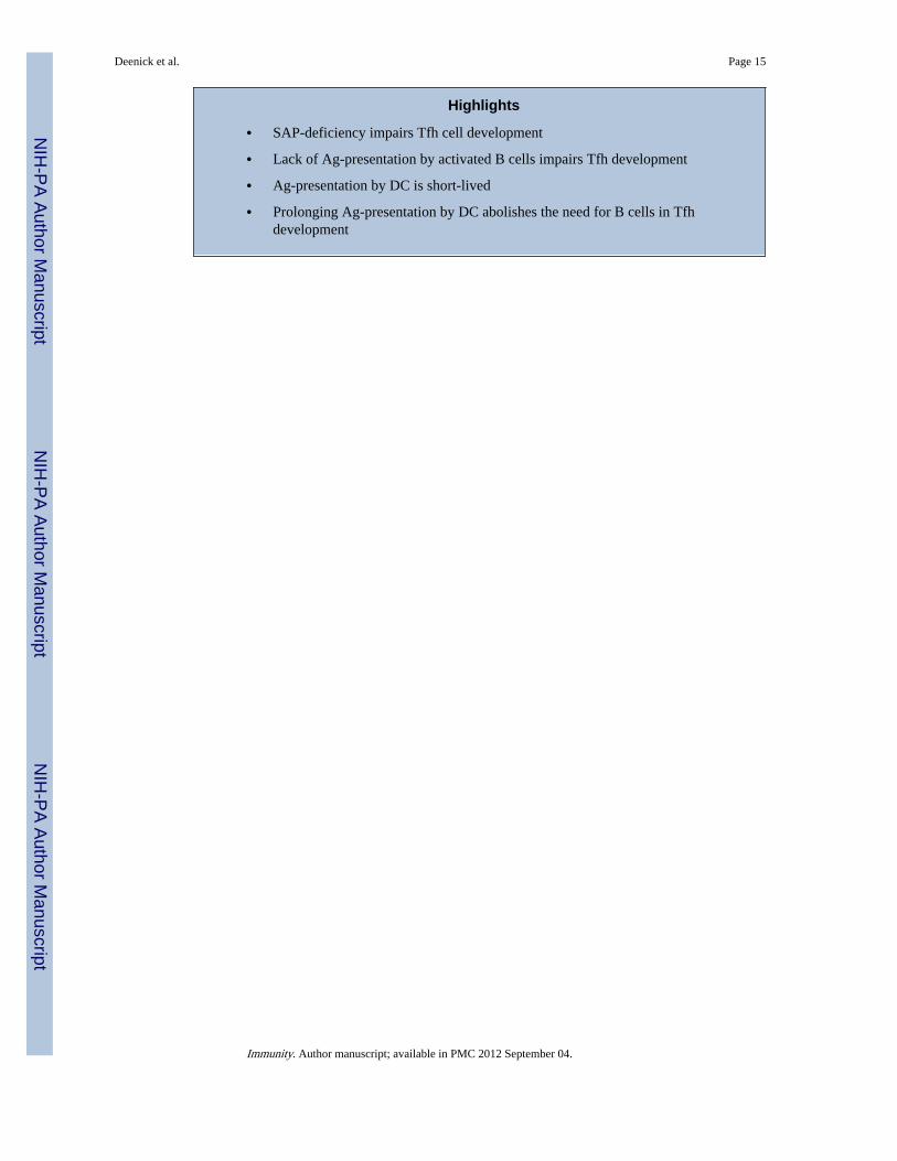
• SAP-deficiency impairs Tfh cell development
• Lack of Ag-presentation by activated B cells impairs Tfh development
• Ag-presentation by DC is short-lived
• Prolonging Ag-presentation by DC abolishes the need for B cells in Tfhdevelopment
Deenick et al. Page 15
Immunity. Author manuscript; available in PMC 2012 September 04.
NIH
-PA Author Manuscript
NIH
-PA Author Manuscript
NIH
-PA Author Manuscript
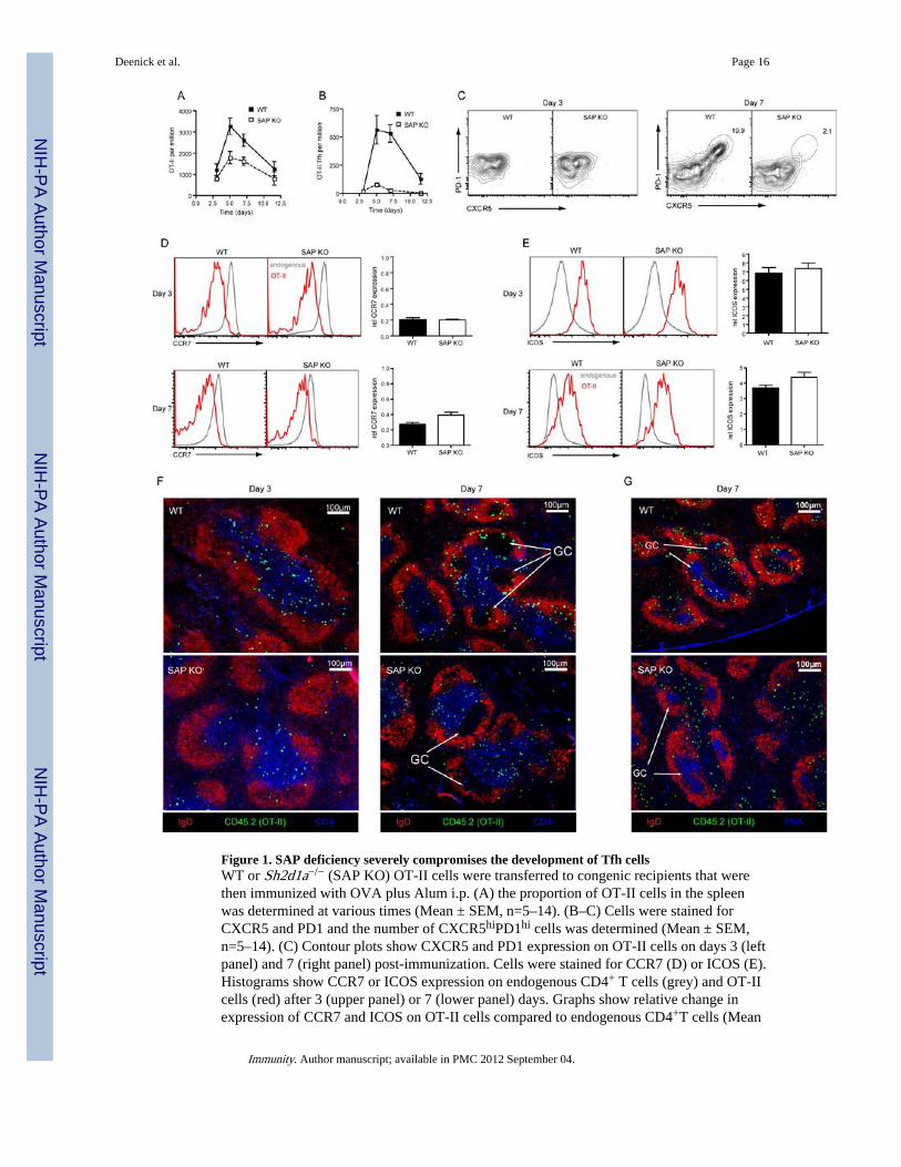
Figure 1. SAP deficiency severely compromises the development of Tfh cellsWT or Sh2d1a−/− (SAP KO) OT-II cells were transferred to congenic recipients that werethen immunized with OVA plus Alum i.p. (A) the proportion of OT-II cells in the spleenwas determined at various times (Mean ± SEM, n=5–14). (B–C) Cells were stained forCXCR5 and PD1 and the number of CXCR5hiPD1hi cells was determined (Mean ± SEM,n=5–14). (C) Contour plots show CXCR5 and PD1 expression on OT-II cells on days 3 (leftpanel) and 7 (right panel) post-immunization. Cells were stained for CCR7 (D) or ICOS (E).Histograms show CCR7 or ICOS expression on endogenous CD4+ T cells (grey) and OT-IIcells (red) after 3 (upper panel) or 7 (lower panel) days. Graphs show relative change inexpression of CCR7 and ICOS on OT-II cells compared to endogenous CD4+T cells (Mean
Deenick et al. Page 16
Immunity. Author manuscript; available in PMC 2012 September 04.
NIH
-PA Author Manuscript
NIH
-PA Author Manuscript
NIH
-PA Author Manuscript
± SEM, n=7–8). (F) Immunofluorescence staining of spleen sections was carried out todetermine the positioning of OT-II T cells (CD45.2+) within the follicle (IgD) and T cellszone (CD4) after 3 (left panel) or 7 days (right panel). (G) Further staining confirmedpositioning within the GC (PNA) at day 7.
Deenick et al. Page 17
Immunity. Author manuscript; available in PMC 2012 September 04.
NIH
-PA Author Manuscript
NIH
-PA Author Manuscript
NIH
-PA Author Manuscript
Figure 2. Rescuing germinal centre formation does not facilitate Tfh cell development from SAP-deficient CD4+ T cellsCD45.1-congenic mice were transferred with CD45.2+ OT-II cells. Mice received eitherThy1.1− WT OT-II or Thy1.1+ SAP KO OT-II alone or a combination of equal numbers ofThy1.1− WT OT-II or Thy1.1+ SAP KO OT-II and were then immunized i.p with OVA plusAlum. The responses were examined 7 days later. (A) The proportion of IgG1+GL7+ (GC) Bcells in recipient spleens. (B) The proportion of OT-II cells from each donor was determined– WT OT-II: filled bars, SAP KO OT-II: open bars. (C) The proportion of CXCR5hiPD1hi
Tfh cells from each donor was determined. All graphs show Mean ± SEM, n=4 (D) Contourplots show expression of CXCR5 and PD1 on OT-II cells of each genotype.
Deenick et al. Page 18
Immunity. Author manuscript; available in PMC 2012 September 04.
NIH
-PA Author Manuscript
NIH
-PA Author Manuscript
NIH
-PA Author Manuscript

Figure 3. Treatment with peptide can rescue Tfh cell development from SAP-deficient T cellsWT or SAP KO OT-II cells were transferred to congenic recipients that were then givenOVA plus Alum i.p. at day 0. On day 3 some mice received additional OVA peptide i.v. andthe mice then sacrificed on day 7. (A) The proportion of total OT-II cells in the spleen. (B)The proportion of CXCR5hiPD1hi Tfh OT-II cells was determined by (C) staining forCXCR5 and PD1 on OT-II cells. (D) Proportion of GC B cells as determined by staining forexpression of GL7 and Fas. (E) Expression of CD62L and PD1 on OT-II cells. (F)Percentage of OT-II cells that are CD62Llo. (G) Expression of CD127 and PD1 on OT-IIcells. (H) Percentage of cells OT-II cells that are CD127lo. All graphs show Mean±SEM,n=7–9, except (F) n=4–6. (I) Immunofluorescence staining of spleen sections was performed
Deenick et al. Page 19
Immunity. Author manuscript; available in PMC 2012 September 04.
NIH
-PA Author Manuscript
NIH
-PA Author Manuscript
NIH
-PA Author Manuscript
to determine the positioning of OT-II T cells (CD45.2+) within the follicle (IgD) and GC(PNA). (J-M) CD62Lhi, CD62LloPD1lo and CD62LloPD1hi populations of OT-II CD4+ Tcells (CD4+B220−CD45.2+) were isolated by sorting. Expression of Bcl6 (K), Il21 (L) andSh2d1a (M) in each of these populations was then determined by quantitative PCR. N.D. -Not done. The values represent the (Mean±SEM of 4 experiments for peptide boost, 3experiments for non-boost).
Deenick et al. Page 20
Immunity. Author manuscript; available in PMC 2012 September 04.
NIH
-PA Author Manuscript
NIH
-PA Author Manuscript
NIH
-PA Author Manuscript

Figure 4. B cell activation via CD40 is not required for Tfh developmentChimeras were generated as described in Experimental Procedures (see also Figure S4A)using 80:20 mix of µMT:Cd40−/− BM (to generate mice with B cells lacking CD40) or B6:Cd40−/− BM (controls). Thy1.1+ WT OT-II cells were transferred into the chimeras, whichwere then given OVA plus Alum i.p. at day 0. On day 3 some mice received additionalOVA peptide i.v. and the mice were sacrificed on day 7 for analysis. (A) Reconstitution wasdetermined by staining B cells (B220+) and DC (CD11c+) for CD40 expression. (B) Theproportion of total OT-II cells in the spleen. (C) The proportion of CXCR5hiPD1hi Tfh OT-II cells was determined by (D) staining for CXCR5 and PD1 on OT-II cells. (E) Proportionof GC B cells as assessed by staining for expression of GL7 and Fas. (F) Percentage of cellsOT-II cells that are CD127lo. All plots show Mean ± SEM, n=7–14.
Deenick et al. Page 21
Immunity. Author manuscript; available in PMC 2012 September 04.
NIH
-PA Author Manuscript
NIH
-PA Author Manuscript
NIH
-PA Author Manuscript
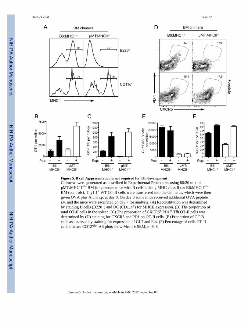
Figure 5. B cell Ag-presentation is not required for Tfh developmentChimeras were generated as described in Experimental Procedures using 80:20 mix ofµMT:MHCII−/− BM (to generate mice with B cells lacking MHC class II) or B6:MHCII−/−
BM (controls). Thy1.1+ WT OT-II cells were transferred into the chimeras, which were thengiven OVA plus Alum i.p. at day 0. On day 3 some mice received additional OVA peptidei.v. and the mice were sacrificed on day 7 for analysis. (A) Reconstitution was determinedby staining B cells (B220+) and DC (CD11c+) for MHCII expression. (B) The proportion oftotal OT-II cells in the spleen. (C) The proportion of CXCR5hiPD1hi Tfh OT-II cells wasdetermined by (D) staining for CXCR5 and PD1 on OT-II cells. (E) Proportion of GC Bcells as assessed by staining for expression of GL7 and Fas. (F) Percentage of cells OT-IIcells that are CD127lo. All plots show Mean ± SEM, n=6–8.
Deenick et al. Page 22
Immunity. Author manuscript; available in PMC 2012 September 04.
NIH
-PA Author Manuscript
NIH
-PA Author Manuscript
NIH
-PA Author Manuscript
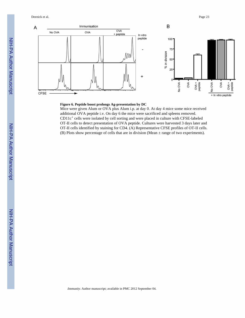
Figure 6. Peptide boost prolongs Ag-presentation by DCMice were given Alum or OVA plus Alum i.p. at day 0. At day 4 mice some mice receivedadditional OVA peptide i.v. On day 6 the mice were sacrificed and spleens removed.CD11c+ cells were isolated by cell sorting and were placed in culture with CFSE-labeledOT-II cells to detect presentation of OVA peptide. Cultures were harvested 3 days later andOT-II cells identified by staining for CD4. (A) Representative CFSE profiles of OT-II cells.(B) Plots show percentage of cells that are in division (Mean ± range of two experiments).
Deenick et al. Page 23
Immunity. Author manuscript; available in PMC 2012 September 04.
NIH
-PA Author Manuscript
NIH
-PA Author Manuscript
NIH
-PA Author Manuscript
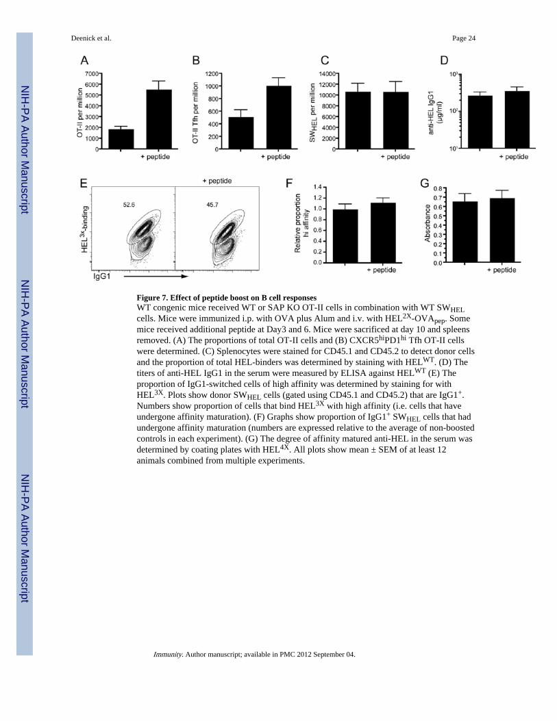
Figure 7. Effect of peptide boost on B cell responsesWT congenic mice received WT or SAP KO OT-II cells in combination with WT SWHELcells. Mice were immunized i.p. with OVA plus Alum and i.v. with HEL2X-OVApep. Somemice received additional peptide at Day3 and 6. Mice were sacrificed at day 10 and spleensremoved. (A) The proportions of total OT-II cells and (B) CXCR5hiPD1hi Tfh OT-II cellswere determined. (C) Splenocytes were stained for CD45.1 and CD45.2 to detect donor cellsand the proportion of total HEL-binders was determined by staining with HELWT. (D) Thetiters of anti-HEL IgG1 in the serum were measured by ELISA against HELWT (E) Theproportion of IgG1-switched cells of high affinity was determined by staining for withHEL3X. Plots show donor SWHEL cells (gated using CD45.1 and CD45.2) that are IgG1+.Numbers show proportion of cells that bind HEL3X with high affinity (i.e. cells that haveundergone affinity maturation). (F) Graphs show proportion of IgG1+ SWHEL cells that hadundergone affinity maturation (numbers are expressed relative to the average of non-boostedcontrols in each experiment). (G) The degree of affinity matured anti-HEL in the serum wasdetermined by coating plates with HEL4X. All plots show mean ± SEM of at least 12animals combined from multiple experiments.
Deenick et al. Page 24
Immunity. Author manuscript; available in PMC 2012 September 04.
NIH
-PA Author Manuscript
NIH
-PA Author Manuscript
NIH
-PA Author Manuscript