The pathogenic role of interleukin-27 in autoimmune diabetes
Upload
independentCategory
view
2download
0
IntroductionExperimental autoimmune uveitis (EAU) is a T cell–mediated autoimmune disease model that targets theneural retina and related tissues. EAU is induced byimmunization with preparations of purified retinalantigens or their fragments, of which the retinal solu-ble antigen (S-Ag, also known as arrestin) and theinterphotoreceptor retinoid-binding protein (IRBP)are the best known. This experimental model is usedto represent a series of human inflammatory diseases
collectively known as uveitis, which are characterizedby a frequent presence of immune response to retinalantigens, particularly S-Ag. EAU can also be inducedby adoptive transfer of T cells from immunized recip-ients to naive, genetically compatible hosts (1). In mostcases, the pathogenesis of the disease implicates a Th1-type cytokine response (1).
Genetic studies have documented associationbetween the presence or absence of certain HLA alle-les and susceptibility to particular autoimmune dis-orders (2). Autoimmune uveitis is a spectrum of dis-eases that show differences in clinical manifestationsand course, and differences in associations with spe-cific HLA loci. The associated class I or class II allelesmay also depend on the ethnic origin of the popula-tion studied. Among the class II–associated uveiticsyndromes, the HLA-DRB1*0405 allele, encoding fora variant of the HLA-DR4 antigen, was found to besignificantly increased in a Japanese population ofVogt-Koyanagi-Harada patients (3), and this was alsoconfirmed in other populations (4, 5). Clinically sim-ilar to Vogt-Koyanagi-Harada syndrome, sympathet-ic ophthalmia is also associated with HLA-DR4 sub-types in Japanese, British, and Irish populations (6,7). Intermediate uveitis not associated with multiplesclerosis was associated with the HLA-DR3 antigen,
The Journal of Clinical Investigation | April 2003 | Volume 111 | Number 8 1171
A humanized model of experimental autoimmune uveitis in HLA class II transgenic mice
Giuseppina Pennesi,1 Mary J. Mattapallil,1 Shu-Hui Sun,1 Dody Avichezer,1
Phyllis B. Silver,1 Zaruhi Karabekian,1 Chella S. David,2 Paul A. Hargrave,3
J. Hugh McDowell,3 W. Clay Smith,3 Barbara Wiggert,4 Larry A. Donoso,5
Chi-Chao Chan,1 and Rachel R. Caspi1
1Laboratory of Immunology, National Eye Institute, NIH, Bethesda, Maryland, USA2Department of Immunology, Mayo Clinic, Rochester, Minnesota, USA3Department of Ophthalmology, University of Florida, Gainesville, Florida, USA 4Laboratory of Retinal Cell and Molecular Biology, National Eye Institute, NIH, Bethesda, Maryland, USA5Wills Eye Hospital, Philadelphia, Pennsylvania, USA
Experimental autoimmune uveitis (EAU) is a disease of the neural retina induced by immuniza-tion with retinal antigens, such as interphotoreceptor retinoid-binding protein (IRBP) and arrestin(retinal soluble antigen, S-Ag). EAU serves as a model for human autoimmune uveitic diseases asso-ciated with major histocompatibility complex (HLA) genes, in which patients exhibit immuno-logical responses to retinal antigens. Here we report the development of a humanized EAU modelin HLA transgenic (TG) mice. HLA-DR3, -DR4, -DQ6, and -DQ8 TG mice were susceptible to IRBP-induced EAU. Importantly, HLA-DR3 TG mice developed severe EAU with S-Ag, to which wild-typemice are highly resistant. Lymphocyte proliferation was blocked by anti-HLA antibodies, con-firming that antigen is functionally presented by the human MHC molecules. Disease could betransferred by immune cells with a Th1-like cytokine profile. Antigen-specific T cell repertoire, asmanifested by responses to overlapping peptides derived from S-Ag or IRBP, differed from that ofwild-type mice. Interestingly, DR3 TG mice, but not wild-type mice, recognized an immunodomi-nant S-Ag epitope between residues 291 and 310 that overlaps with a region of S-Ag recognized byuveitis patients. Thus, EAU in HLA TG mice offers a new model of uveitis that should representhuman disease more faithfully than currently existing models.
J. Clin. Invest. 111:1171–1180 (2003). doi:10.1172/JCI200315155.
Received for publication January 28, 2002, and accepted in revised formFebruary 11, 2003.
Address correspondence to: Rachel R. Caspi, Laboratory ofImmunology, National Eye Institute, National Institutes ofHealth, 10 Center Drive, Bethesda, Maryland 20892, USA. Phone: (301) 435-4555; Fax: (301) 480-6668; E-mail: [email protected] Pennesi’s present address is: Laboratorio diDifferenziamento Cellulare, Istituto Nazionale per la Ricerca sulCancro, Genova, Italy.Giuseppina Pennesi and Mary J. Mattapallil contributed equallyto this work.Conflict of interest: The authors have declared that no conflict ofinterest exists.Nonstandard abbreviations used: experimental autoimmuneuveitis (EAU); retinal soluble antigen (S-Ag); interphotoreceptorretinoid-binding protein (IRBP); transgenic (TG); bovine IRBP(bIRBP); bovine retinal soluble antigen (bS-Ag).

while panuveitis showed association with HLA-DR4in an Italian population (8). Association with HLA-DQalleles was also described (3, 7, 9), but the stronglinkage disequilibrium between some DR and DQalleles makes it difficult to distinguish whether therewas a prevalent role of one molecule or whether theDR and DQ genes acted in an epistatic manner.Genetic associations in uveitis have recently beenreviewed in depth (10).
The antigen or antigens that might be involved in theetiology of human uveitis have not been elucidated.Although uveitis patients frequently display cellularand humoral responses to the retinal antigens that areuveitogenic in animals, it is not known whether theseresponses are causally involved in their disease or rep-resent an epiphenomenon (11). This information is keyfor future development of antigen-specific therapiesfor these blinding autoimmune diseases. If indeedthese responses are causally related, the immunodom-inant epitopes are expected to vary in different HLAtypes and will need to be defined in order for sophisti-cated therapies to be possible.
Similarly to uveitis in the human, EAU in animals isgenetically controlled (10). There are clear species-spe-cific differences in sensitivity to uveitogenic proteins.For example, IRBP is a much more potent uveitogenthan S-Ag for mice, while the reverse is true in guineapigs, and both proteins are strongly uveitogenic in theLewis rat (12–15). Within each species, strain depend-ence of susceptibility is apparent that is at least in partdue to MHC control. Susceptible H2 haplotypes areH2b, H2d, H2k, and H2r. MHC control of susceptibilityto EAU in the H2k haplotype was tentatively mapped tothe IA subregion (a homologue of human HLA-DQ),with modifying effects from the IE locus (homologousto human HLA-DR) (15).
To better study the role of HLA molecules in thepathogenesis of uveitis, we undertook to develop a“humanized” model of EAU. To this end, we inducedEAU in mice expressing transgenic (TG) HLA-DR3, -DR4, -DQ6, or -DQ8 molecules in the absence of theendogenous class II (Aβ0) (16). These mice positivelyselect a repertoire of T cells expressing various Vβ T cellreceptors that can identify immunogenic peptide epi-topes similar or identical to human subjects of thesame HLA-DR or -DQ genotype (16, 17). We show thatall the tested HLA TG mice developed EAU afterimmunization with IRBP. In addition, HLA-DR3 TGmice developed severe uveitis after immunization withS-Ag, to which wild-type mice are highly resistant. Anti-body blocking studies confirmed that the antigenswere being recognized by the T cells in the context ofthe human HLA molecules. Furthermore, recognitionof peptides derived from S-Ag and peptides derivedfrom IRBP by HLA TG mice differs from recognition bywild-type mice and has some similarity to that ofuveitis patients. Finally, as in classical EAU, the diseasecan be transferred by immune cells having a Th1 phe-notype, but not by immune serum.
This new humanized model of uveitis offers a morerelevant approximation of human uveitis than hither-to available rodent models and will facilitate the char-acterization of uveitogenic epitopes presented by dif-ferent HLA class II types.
MethodsAnimals. HLA-DR4, -DQ6, and -DQ8 single TG micewere developed at the Mayo Clinic and have beendescribed previously (18–20). HLA-DR3 mice origi-nally developed by Günter Hammerling and associates(21) were bred onto the Aβ0 background at the MayoClinic. The HLA-DR3 TG mice that carry the HLA-DRA*0103 and DRB1*0301 genes in an MHCclass II–negative H2-Aβ0 background (Aβ0.DR3)express the human DR3 antigen as their only MHCclass II molecule (22). HLA-DR4 TG mice had theHLA-DRB1*0401 gene in an Aβ0 background, modi-fied to express IE molecules by insertion of the genefor the IE α chain from the H2k haplotype (Eαk/Eβb.Aβ0.DR4) (18). Thus, they coexpressed murine H2-IE and human HLA-DR4 molecules. HLA-DQ6TG mice carried the DQA1*0103 and DQB1*0601transgenes in an Aβ0 background (Aβ0.DQ6) (19).Similarly, HLA-DQ8 TG mice had the DQA1*0301 andDQB1*0302 human genes (Aβ0.DQ8) (20, 23). BothHLA-DQ TG mice expressed the human DQ moleculeas their only MHC class II antigen (Table 1). Controlswere DR3- and DR4-negative littermates, Aβ0 mice,Aβ0 mice expressing IE antigens (Eαk/Eβb.Aβ0), andC57BL/10 or C57BL/6 mice. DR3-negative littermatesdid not express any MHC class II molecules, theirgenotype being equivalent to that of Aβ0 mice. DR4-negative littermates expressed the IE molecule, havinga genotype comparable to Eαk/Eβb.Aβ0. These micehad the H2-IE molecule as their only MHC class IIantigen. H2-IAb is the only MHC class II moleculeexpressed by the parental strain C57BL/10, which hasa defective gene encoding the Eβ chain and fails toexpress IE molecules (Table 1).
Animals were bred and maintained at the NIH or atthe Mayo Clinic under specific pathogen–free condi-tions and were given water and chow ad libitum. Thecare and use of the animals was in compliance withinstitutional guidelines.
Genotyping. Mice were screened for presence of thehuman HLA molecule by PCR as described (24, 25).Expression of the human or murine MHC antigenswas quantified by flow cytometric analysis of lymphnode cells and splenocytes using FITC-conjugatedanti-DR (clone L243), anti-DQ (clones Tü 39 and Tü169), anti-IA (clone AF6-120.1), or anti-IE (clone 14-4-4S) antibodies (BD Pharmingen, FranklinLakes, New Jersey, USA) as recommended by themanufacturer. Mice that did not demonstrate expres-sion of HLA molecules by flow cytometry wereexcluded from analysis.
Antigens and reagents. Bovine IRBP (bIRBP) was puri-fied from retinal extracts as described (26) by affinity
1172 The Journal of Clinical Investigation | April 2003 | Volume 111 | Number 8

chromatography on ConA followed by ion exchangechromatography on a Pharmacia MonoQ column.Bovine S-Ag (bS-Ag) was prepared from the ConA col-umn flowthrough as follows. The extract was dialyzedagainst 10 volumes of 10 mM HEPES, 15 mM NaCl, 1mM EDTA, 1 mM benzamidine, pH 7.0, with thebuffer changed once. S-Ag was purified by the methodof Buczylko and Palczewski (27) with modificationsdescribed by Puig et al. (28). The final elution fromthe heparin-agarose column was via a gradient from10 mM HEPES and 15 mM NaCl, pH 7.0, to 10 mMHEPES and 400 mM NaCl, pH 7.0. Purified bS-Ag waspooled based on an OD of 278 nm. Preparation of therecombinant human S-Ag (hS-Ag) was described pre-viously (29). Pertussis toxin and CFA were purchasedfrom Sigma-Aldrich (St. Louis, Missouri, USA).Mycobacterium tuberculosis strain H37RA was pur-chased from Difco Laboratories (Detroit, Michigan,USA). Twenty-residue peptides overlapping by tenresidues and spanning the first homologous repeat ofhuman IRBP (hIRBP) (35 kDa) (30) and the entire hS-Ag (48 kDa; AnaSpec Inc., San Jose, California,USA) were synthesized by conventional solid-phasetechniques as described (30, 31).
EAU induction and scoring. EAU induction and scor-ing was performed two ways: by active immunizationand by adoptive transfer. For active immunization,mice were injected subcutaneously with 200 µg ofbIRBP, bS-Ag, or recombinant hS-Ag emulsified (1:1,vol/vol) with CFA (Sigma-Aldrich) that had been sup-plemented with M. tuberculosis strain H37RA (DifcoLaboratories) to a final concentration of 2.5 mg/ml.Concurrent with immunization, 0.2 µg of pertussistoxin was injected intraperitoneally. Eyes from IRBP-immunized mice were collected 21–28 days afterimmunization, and eyes from S-Ag–immunized ani-mals were collected after 28–35 days. Eyes were fixedfor 1 hour in 4% phosphate-buffered glutaraldehydeand transferred into 10% phosphate-bufferedformaldehyde. Fixed and dehydrated tissue wasembedded in methacrylate. Sections (4–6 µm) were
cut through the pupillary–optic nerve plane and thenstained with standard H&E.
Quantitation of disease was performed in a maskedfashion using criteria described previously (32). Briefly,eyes were assigned a score ranging from 0 to 4 depend-ing on the extent of inflammation and tissue damage.The minimal criterion to score an eye as positive byhistopathology was inflammatory cell infiltration ofthe ciliary body, choroid, vitreous, or retina (EAU grade0.5). Progressively higher grades were assigned for thepresence of discrete lesions in the tissue, such as vas-culitis, granuloma formation, retinal folding and/ordetachment and photoreceptor damage. The maximalgrade of 4 reflects extensive retinal damage with com-plete destruction of the photoreceptor cell layer.
For induction of EAU by adoptive transfer, HLA-DQ8 and HLA-DQ6 mice were immunized with IRBPand HLA-DR3 mice were immunized with S-Ag usingthe uveitogenic protocol described above. On day 14after immunization, immune serum was collected,titrated for antibody content by ELISA, and stored at4°C until infusion into naive recipients. Lymph nodeand spleen cells were pooled within a group and werecultured for 3 days as described previously (33) with anoptimal concentration of the immunizing antigen (30µg/ml IRBP or 10 µg/ml S-Ag) and 5 ng/ml recombi-nant IL-12. Naive recipient mice of the appropriategenotype were infused intraperitoneally with 60 × 106
to 100 × 106 cultured cells. One milliliter of immuneserum was injected intravenously as two doses of 0.5ml administered concurrently with the cells and 48hours later. Disease development was evaluated byfundoscopy and scored on a scale of 0–4 as describedpreviously (32). EAU was confirmed by histopatholo-gy in eyes harvested after 16–18 days.
Delayed-type hypersensitivity responses. Two days beforethe termination of an experiment, mice received 10 µgof the appropriate antigen in a volume of 10 µl intra-dermally into the pinna of one ear. The other ear wasinjected with PBS. Ear swelling was measured 48 hourslater with a spring-loaded micrometer.
The Journal of Clinical Investigation | April 2003 | Volume 111 | Number 8 1173
Table 1MHC class II genotype and phenotype of mouse strains used in this study
Strain HLA transgeneA MHC class II phenotype MHC class II restriction element Reference
Test strains
DR3 DRA*0103; DRB1*0301 Aβ0.DR3+ HLA-DR3 (21)DR4 DRB1*0401 Eαk/Eβb.Aβ0.DR4+ HLA-DR4/H2-IEk/b (18)DQ6 DQA1*0103; DQB1*0601 Aβ0.DQ6+ HLA-DQ6 (19) DQ8 DQA1*0301; DQB1*0302 Aβ0.DQ8+ HLA-DQ8 (20, 23)
Control strains
DR3– littermates – Aβ0.DR3– –DR4– littermates – Eαk/Eβb.Aβ0.DR4– H2-IEk/b
Aβ0 – Aβ0 – (51)Eαk/Eβb.Aβ0 – Eαk/Eβb.Aβ0 H2-IEk/b (52)C57BL/10 or C57BL/6 – H2-IAb H2-IAb
AThe TG strains are on the C57BL/10 background.

Lymphocyte proliferation assay and antibody blocking.Lymph nodes draining the immunization site(inguinal and iliac) were collected at the terminationof each experiment and were pooled within eachgroup. Triplicate 0.2-ml cultures containing 5 × 10⁵
cells/well were stimulated with 30 µg of IRBP or 20 µgof S-Ag in 96-well round-bottomed plates in RPMI1640 (BioWhittaker Inc., Walkersville, Maryland,USA) supplemented as described and containing 1%mouse serum (32). For proliferation to bIRBP, 20mg/ml α-methyl-mannopyranoside (Sigma-Aldrich)was included to neutralize any possible traces of ConA that might leach from the column used in the ini-tial stages of IRBP purification. This concentration ofα-methyl-mannopyranoside had no adverse effect oncell proliferation. Results are presented as stimulationindexes, calculated as average cpm of triplicate cul-tures with antigen, divided by the average cpm of trip-licate cultures with medium, ± SE.
The restriction element involved in antigen presenta-tion was assessed by antibody blocking assays usingpurified antibodies to DR (clone L243), DQ (clone Tü39 or Tü 169), IA (clone AF6-120.1), or IE (clone 14-4-4S) (BD Pharmingen). The antibodies were added at aconcentration of 10 µg/ml to wells containing the cellsin the presence or absence of the appropriate stimulat-ing antigen. The cultures were incubated for 60 hoursand were pulsed with 3H thymidine (1.0 µCi/10 µl/well)for the last 18 hours.
T cell epitope mapping. Twenty-residue peptidesoverlapping by 10 residues and representing the lin-ear sequence of the first repeat of hIRBP or theentire sequence of the hS-Ag molecule were used(30) (AnaSpec Inc.). HLA-DQ8 TG, HLA-DR3 TG,
or wild-type C57BL/6 mice (H2b) were immunized witha uveitogenic regimen of IRBP or S-Ag, respectively.Spleens were harvested on day 10 after immunizationand were pooled from several mice within each group.Cells were tested in a standard proliferation assay, asdescribed above, against a 10-µM concentration of pep-tides derived from the immunizing antigen. Specificcounts (∆ cpm) were calculated after subtraction ofbackground cpm.
Determination of lymphokine content in culture super-natants and antibodies in immune serum by ELISA.Cytokines were determined in supernatants of lymphnode cells collected on day 14 after immunizationand cultured with an optimal concentration of theimmunizing antigen as for the proliferation assaysabove. Supernatants were collected after 48 hours.The presence of IL-2, IL-4, IL-5, IL-6, IL-10, IL-12, IL-13, IFN-γ, and TNF-α in the supernatant of thestimulated cells was measured by multiplex ELISAusing the Pierce SearchLight technology (PierceBoston Technology, Woburn, Massachusetts, USA)(34) (http://www.searchlightonline.com). Serumantibodies were assayed on IRBP- or S-Ag–coatedplates (5 µg/100 µl/well) using HRP-labeled goatanti-mouse IgG (Zymed Laboratories Inc., San Fran-cisco, California, USA) as developing antibody and3,3´5,5´-tetramethylbenzidine substrate (100 µl/well;Endogen Inc., Woburn, Massachusetts, USA).
Statistical analysis. Statistical significance of differ-ences in disease scores were calculated usingSnedecor and Cochran’s test (35) for linear trend inproportions, with each mouse (average of both eyes)as one statistical event. This is a nonparametric testthat generates its P values by frequency analysis ofthe number of individuals at each possible score,thus taking into account both severity and incidenceof disease. Delayed hypersensitivity and lymphocyte
1174 The Journal of Clinical Investigation | April 2003 | Volume 111 | Number 8
Figure 1Histopathological features of EAU in HLA TG mice. (a) Normal eye.The retinal layers are ordered and well preserved. (b) Disease score2 in C57BL/10 mice immunized with IRBP. The retinal architectureis disorganized, inflammatory cell infiltration is present, and the pho-toreceptor layer is damaged. (c) Disease score 3 in DR4 TG miceimmunized with IRBP. Note extensive destruction of the photore-ceptor cell layer. (d) Very severe disease (score 4) in DR3 TG miceimmunized with S-Ag. Note complete destruction of the photore-ceptor cell layers including photoreceptor cells (H&E, ×200).
Figure 2Susceptibility of HLA TG mice to IRBP-induced EAU. Bars are aver-age of disease scores. Disease incidence (positive among totalmice) is next to each bar. The HLA TG strains are in the top paneland the control strains are in the bottom panel. If only one eyeshowed disease the animal was scored as positive and its score wasrecorded as the average of both eyes. Shown are combined resultsof five experiments. Significant difference (P ≤ 0.05) from C57BLcontrol is indicated by an asterisk.
proliferation data were analyzed using an independ-ent t test. Probability values of P ≤ 0.05 were consid-ered to be significant.
ResultsHLA TG mice are susceptible to IRBP-induced uveitis. HLATG mice were immunized with a uveitogenic proto-col of IRBP and eyes were harvested for histopathol-ogy 21–28 days after immunization. Typical EAU wasinduced in all HLA TG strains. Histopathology (Fig-ure 1, b and c) was grossly similar to that observed inwild-type mice on the C57BL/10 background, show-ing mononuclear and polymorphonuclear cell infil-tration, vitritis, choroiditis, and varying degrees ofphotoreceptor cell damage. Disease severity in indi-vidual mice varied from 0.5 to 3, and incidence wasabout 80% (Figure 2). These results werenot different from incidence and severityscores observed in the control strains.Aβ0 mice, which do not express any MHCclass II molecules but do express murineclass I molecules, had little to no disease,with only one mouse of 11 showing traceinfiltration in one eye, most likely repre-senting a mild spontaneous inflamma-tion unrelated to the experimentalmanipulations. We do not favor the pos-sibility that this represents presentationof IRBP through the class I pathwaybecause these mice had negligible IRBP-specific delayed-type hypersensitivity andproliferative responses (data not shown),suggesting that in the absence of class II,IRBP immunization fails to elicit a signif-icant response.
Since no apparent differences in diseaseparameters were observed in HLA TG miceexpressing or not expressing the murine IE
molecules, their role when a human class II molecule ispresent appears to be subordinate. Nevertheless, whena human class II is absent, such as in the Eαk/Eβb.Aβ0mice, the presence of murine IE alone is sufficient topermit EAU development.
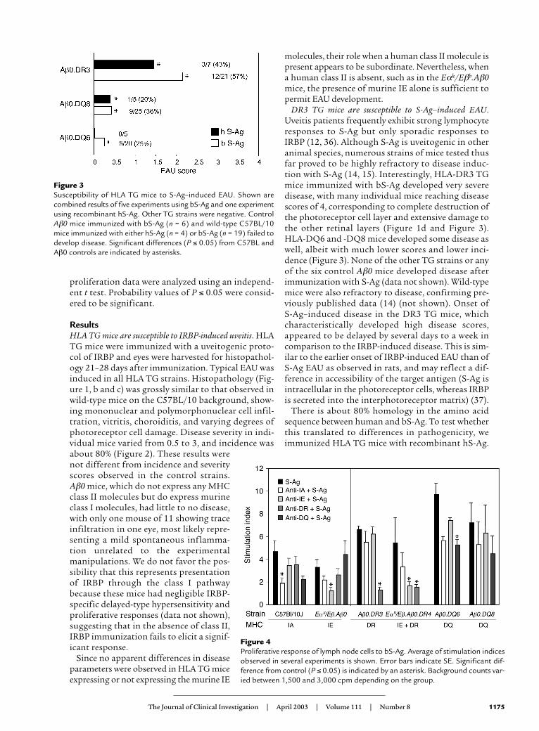
DR3 TG mice are susceptible to S-Ag–induced EAU.Uveitis patients frequently exhibit strong lymphocyteresponses to S-Ag but only sporadic responses toIRBP (12, 36). Although S-Ag is uveitogenic in otheranimal species, numerous strains of mice tested thusfar proved to be highly refractory to disease induc-tion with S-Ag (14, 15). Interestingly, HLA-DR3 TGmice immunized with bS-Ag developed very severedisease, with many individual mice reaching diseasescores of 4, corresponding to complete destruction ofthe photoreceptor cell layer and extensive damage tothe other retinal layers (Figure 1d and Figure 3).HLA-DQ6 and -DQ8 mice developed some disease aswell, albeit with much lower scores and lower inci-dence (Figure 3). None of the other TG strains or anyof the six control Aβ0 mice developed disease afterimmunization with S-Ag (data not shown). Wild-typemice were also refractory to disease, confirming pre-viously published data (14) (not shown). Onset of S-Ag–induced disease in the DR3 TG mice, whichcharacteristically developed high disease scores,appeared to be delayed by several days to a week incomparison to the IRBP-induced disease. This is sim-ilar to the earlier onset of IRBP-induced EAU than ofS-Ag EAU as observed in rats, and may reflect a dif-ference in accessibility of the target antigen (S-Ag isintracellular in the photoreceptor cells, whereas IRBPis secreted into the interphotoreceptor matrix) (37).
There is about 80% homology in the amino acidsequence between human and bS-Ag. To test whetherthis translated to differences in pathogenicity, weimmunized HLA TG mice with recombinant hS-Ag.
The Journal of Clinical Investigation | April 2003 | Volume 111 | Number 8 1175
Figure 3Susceptibility of HLA TG mice to S-Ag–induced EAU. Shown arecombined results of five experiments using bS-Ag and one experimentusing recombinant hS-Ag. Other TG strains were negative. ControlAβ0 mice immunized with bS-Ag (n = 6) and wild-type C57BL/10mice immunized with either hS-Ag (n = 4) or bS-Ag (n = 19) failed todevelop disease. Significant differences (P ≤ 0.05) from C57BL andAβ0 controls are indicated by asterisks.
Figure 4Proliferative response of lymph node cells to bS-Ag. Average of stimulation indicesobserved in several experiments is shown. Error bars indicate SE. Significant dif-ference from control (P ≤ 0.05) is indicated by an asterisk. Background counts var-ied between 1,500 and 3,000 cpm depending on the group.

DR3 and DQ8 TG mice developed EAU after immu-nization with the recombinant hS-Ag with scoressimilar to the EAU developed in response to bS-Ag(Figure 3). This finding is of particular importance inview of the strong responses of human patients to S-Ag, that had presumably developed as responses tothe autologous human molecule. HLA-DR4 TG miceand all the control strains, including the wild type,were resistant (data not shown).
The retinal antigen is presented and recognized on humanMHC molecules. Because DR (though not DQ) strainsretain the mouse Eβ, and in the case of DR4 also theEα molecule, it was necessary to verify that antigencan be presented productively on the human class IImolecules. This was done by lymphocyte prolifera-tion blocking studies using appropriate monoclonal
antibodies. Draining lymph node cells from miceimmunized with IRBP or bS-Ag were collected andwere stimulated in culture with the corresponding anti-gen. Proliferative responses were observed in all strainsexcept Aβ0, which is devoid of any class II molecules. InHLA-DR3, -DQ6, and -DQ8 TG mice, proliferation inresponse to S-Ag was blocked by specific monoclonalantibodies to human, but not to mouse, MHC class IImolecules (Figure 4). In Eαk/Eβb.Aβ0.DR4 TG mice,which coexpress DR and IE molecules, both anti-DRand anti-IE antibodies abrogated proliferation, indi-cating that both class II molecules were functionallyinvolved in antigen presentation. A similar pattern wasobserved with HLA TG lymph node cells of miceimmunized with IRBP (data not shown). A cytotoxiceffect of antibodies was excluded because the anti-HLAantibodies did not interfere with proliferation of wild-type lymphocytes to antigen and because backgroundcounts in the presence of antibodies without antigendid not differ from background in presence of mediumalone (data not shown).
It should be pointed out that strong proliferativeresponses were seen in all genotypes except Aβ0, irre-spective of whether or not they developed disease.Similarly, all strains except Aβ0 mounted delayed-type hypersensitivity responses against S-Ag andIRBP (data not shown). This indicates that not onlypathogenic but also nonpathogenic epitopes of theretinal antigens in question were functionally pre-sented by human class II molecules.
Pathogenesis of EAU in HLA TG mice: a cell-mediatedresponse with a Th1-predominant cytokine profile and a mod-ifying role for antibodies. Studies of EAU in wild-typemice indicated a central role for a Th1-type responsein pathogenesis of disease (1). To evaluate the respons-es of HLA TG mice to uveitogenic immunization, wemeasured the cytokines in supernatants of lymphnode and spleen cells from immunized mice that werestimulated in vitro with the immunizing antigen. Weselected as representative strains the DQ8 TG mice
1176 The Journal of Clinical Investigation | April 2003 | Volume 111 | Number 8
Figure 5Cytokine responses. Cytokine production by lymph node cells fromHLA-DQ8 TG mice stimulated with bIRBP (a), and lymph nodecells from HLA-DR3 TG mice stimulated with bS-Ag (b), measuredby multiplexed ELISA. Shown are titers in pg/ml averaged fromtwo experiments.
Figure 6Adoptive transfer of cells and/or serum. Recipient mice were infused with serum, cells, or cells and serum from immunized syngeneic donors. EAUdevelopment was followed by fundus examination. (a and b) Disease scores of HLA-DQ8 and HLA-DQ6 recipients whose donors were immu-nized with bIRBP. (c) Disease scores of HLA-DR3 recipients whose donors were immunized with bS-Ag. Shown is one of two representative exper-iments with five mice per group. *Statistically significant difference in scores from cells alone (P ≤ 0.05). †Trend (P ≤ 0.1).

immunized with bIRBP and the DR3 TG mice immu-nized with bS-Ag, which were high responders for therespective antigens. Both strains produced very highlevels of IL-6, which was also typical of wild-typeC57BL/6 mice (not shown). Of the cytokines typifyinga Th1 or a Th2 response, IFN-γ and TNF-α were pre-dominant over IL-4, IL-5, and IL-13. DR3 mice pro-duced relatively more IFN-γ than did DQ8 mice (Fig-ure 5, a and b). Thus, EAU induced in HLA TG miceappears to be associated with a Th1-predominantcytokine response profile, analogous to the “classical”model of EAU in wild-type mice.
In wild-type mice, EAU can be adoptively transferredby immune cells but not by immune serum. To testwhether the pathogenesis of EAU in HLA TG micewas cell- or antibody-mediated, we adoptively trans-ferred cells, serum, or cells and serum together fromimmunized to naive syngeneic mice which were thenfollowed by fundus examination. Activated lympho-cytes from immunized mice cultured with antigen inthe presence of IL-12 (33) were able to transfer EAU tonaive syngeneic recipients (Figure 6). None of themice that received high-titer immune serum alone(antibody titers between 1 × 10⁻⁶ and 1 × 10⁻⁷, see
Methods) developed disease. Interestingly, when cellsand serum were combined, the serum was able toslightly but consistently raise disease scores comparedwith cells alone, suggesting that antibodies can mod-ify the course of disease.
T cell epitope recognition of IRBP and S-Ag epitopes inHLA TG mice. The results described above indicatedthat T cells responding to retinal antigen presentedon HLA molecules were involved in the diseaseprocess in HLA TG mice. Because the class II–restrict-ed T cell repertoire is selected in these mice by humanclass II, we decided to examine epitope recognition ofHLA TG mice. We chose as representative strainsthose with high disease susceptibility for each anti-gen: DQ8 for IRBP and DR3 for S-Ag. Mice wereimmunized with native bovine antigen, and spleencell responses were recalled in vitro with a panel ofoverlapping peptides representing the entire firstrepeat of hIRBP or the entire sequence of hS-Ag. Theresponses were compared with those of H2b haplo-type wild-type mice (C57BL/6). Our decision toimmunize with bovine proteins and recall withhuman peptides (although human recombinant pro-teins are available) was based on the reasoning that
The Journal of Clinical Investigation | April 2003 | Volume 111 | Number 8 1177
Figure 7Mapping of T cell epitopes. Epitope recognition by DQ8 mice immunized with bIRBP and DR3 TG mice immunized with bS-Ag was comparedwith that of C57BL/6 (IAb) wild-type controls. Antigen-specific responses were recalled with overlapping peptides representing the first repeatof human IRBP or the whole hS-Ag. Shown are specific counts as averaged from two to three repeat experiments for each strain of mice. Back-ground counts varied from 450 to 3,000 cpm depending on the group.

in a human-human or a bovine-bovine combination,the strongest responses would represent recognitionof nonconserved epitopes foreign to the mouse. Inthe bovine-human combination, the responses wouldbe more likely to represent recognition of conservedepitopes shared with the autologous antigen.
The results showed distinct differences betweenepitope recognition of HLA TG and wild-type mice,suggesting that a different T cell repertoire had beenselected (Figure 7). IRBP peptides 11-30 and 61-80were immunodominant in DQ8TG mice but not inwild-type mice. S-Ag peptide 291-310 (NRERRG-IALDGKIKHEDTNL) appeared to be immunodomi-nant in DR3TG mice. This peptide was weakly recog-nized by wild-type mice, which instead respondedstrongly to peptide 51-70. It is of interest to note thatpeptide 291-310 of S-Ag partly overlaps with the pre-viously characterized S-Ag peptides M 303–320 andN 281–302, which elicit responses in lymphocytes ofpatients affected by different uveitic diseases (11, 31).
DiscussionThe present study describes a new, “humanized”model of EAU developed in HLA TG mice in whichdisease-relevant epitopes appear to be largely restrict-ed by the human class II molecules. Evidence that thisis indeed the case is provided by (a) lack of class II mol-ecules other than the human one in the HLA-DQ6,HLA-DQ8, and HLA-DR3 mice; (b) lack of disease inthe control Aβ0 mice lacking the human class II; (c)the ability of the appropriate anti-HLA antibodies toblock lymphocyte proliferation of HLA TG mice inresponse to the immunizing retinal antigen; and (d)distinct differences in epitope recognition betweenHLA TG and wild-type mice, suggesting differences inthe selected Ag-specific repertoire.
Ours is not the first attempt at establishing ahumanized model of uveitis. Uveitis is a heteroge-neous group of diseases affecting different parts ofthe eye and showing association with various HLAclass I or class II molecules (38). HLA-B27 is stronglyassociated with anterior uveitis that accompaniesankylosing spondylitis. HLA-A29 confers an in-creased risk of birdshot chorioretinopathy that is insome cases up to 224 times higher than the generalpopulation (39). Sympathetic ophthalmia is associ-ated with HLA-DR4, and intermediate uveitis (unre-lated to multiple sclerosis) with HLA-DR3 (3–8, 39,40). The relevant human HLA molecule does notnecessarily precipitate the appearance of symptomsin the animal carrying it as a transgene. AlthoughHLA-B27 TG rats and mice do develop spontaneousspondyloarthropathies, they do not develop eitherspontaneous or bacterially induced anterior uveitis(41–43). In contrast, the recently described HLA-A29TG mice develop spontaneous uveitis with strikinghistological similarity to the HLA-A29–associateddisease in humans. This supports the conclusionthat the HLA-A29 molecule itself participates in the
pathogenesis of birdshot chorioretinopathy (41). TheEAU model in HLA class II TG mice described hereprovides evidence in favor of an involvement of classII–restricted responses in human uveitis. No lessimportantly, this new model suggests that the sameretinal antigens that are uveitogenic in animals mayalso be causally involved in human uveitic diseasesand validates antigen-specific immunotherapiesbased on these antigens, such as the recent oral tol-erance trial in which uveitis patients were fed S-Agand in fact appeared to receive a clinical benefit (44).
Arguably the most interesting observation in thepresent study is the susceptibility of HLA-DR3 TGmice to disease induced with S-Ag. Of particular noteis the finding that the T cell epitope recognition inthese mice takes on a similarity to that of uveitispatients. DR3 TG mice but not wild-type miceresponded to S-Ag peptide 291–310. This sequencepartly overlaps with peptide M, a promiscuous S-Agepitope spanning residues 303–320 that is uveito-genic in rats, guinea pigs, and primates, and is recog-nized by lymphocytes from uveitis patients but notby healthy controls (11, 12, 31, 45). Although humanpatients appear to exhibit much more frequent cel-lular responses to S-Ag than to IRBP, wild-type micedevelop EAU with IRBP but are highly resistant toEAU induced with S-Ag. It has been proposed thatthe reason for this is central tolerance to S-Ag, owingto its abundance in the murine thymus (46), suchthat T cells capable of recognizing S-Ag are efficient-ly deleted from the repertoire. The dramatic changein susceptibility of HLA-DR3 TG mice to S-Ag,apparently stemming solely from substitution of therestricting class II molecule from mouse to human,suggests that other mechanisms may be at play.Thus, in addition to and independently of its poten-tial to bring better understanding of the involvementof S-Ag in human uveitic disease, the uveitis model inHLA-DR3 mice also offers the opportunity toapproach basic questions in the development of self-tolerance to retinal antigens. These questions will bethe subject of a separate study.
The Eαk/Eβb.Aβ0 mice (which served here as con-trols for the Eαk/Eβb.Aβ0.DR4 TG mice) providesome new information about the role of IE moleculesin the EAU model. It is apparent that IA moleculesare sufficient to permit induction of EAU in the H2b
haplotype, as mice on the C57BL background, whichdo not express IE molecules, develop disease. Ourprevious data in intra-H2 congenics tentativelymapped control of EAU to IAk, with only modifyinginfluence from IEk (15). The present data show forthe first time that the IE molecule by itself, withoutthe presence of IA, is sufficient to confer the abilityto develop EAU.
The interaction between the T cell coreceptor CD4and MHC class II molecules plays a crucial role inintrathymic selection as well as peripheral activationof CD4+ T cells. In our humanized EAU model,
1178 The Journal of Clinical Investigation | April 2003 | Volume 111 | Number 8

murine CD4 molecules bind to the β2 domain ofhuman MHC class II. It has been argued that species-specific amino acid differences might alter the inter-action between mouse CD4 and human class II,resulting in reduced or modified responses (18, 47).Since we do not have human CD4 TG mice at our dis-posal, we attempted to partly address the effect of ahomologous versus heterologous CD4–class II inter-action by using DR4 chimeric mice. These miceexpress a hybrid DR4 molecule formed from theinvariant portion of the murine IE molecule thatcontains the CD4 binding site, fused with the vari-able region that contains the antigen-binding pock-et of the human HLA-DR4 molecule. UnlikeEαk/Eβb.Aβ0.DR4 mice, these DR4 chimeric micehave no murine class II molecules and thus have onlya homologous CD4–class II interaction. DR4chimeric mice developed IRBP-induced EAU with anincidence and average score similar to that ofnonchimeric DR4 TG mice and were resistant to S-Ag(data not shown). Thus, presence of a heterologousCD4–class II interaction in our model appears not tocause an altered pattern of susceptibility to disease.
The pathogenesis of EAU in the HLA TG miceappears to be similar to that of EAU in wild-type miceand reinforces the notion that cell-mediated respons-es may also be causally involved in human uveitis. Inwild-type mice, uveitogenicity is associated with theTh1 response (1). Both IRBP-induced EAU in DQ8 TGmice and S-Ag–induced EAU in DR3 TG mice wereassociated with an IFN-γ–dominant response to theirrespective uveitogen. Interestingly, IL-2– and IFN-γ–producing CD4+ and CD8+ cells are detected in periph-eral blood from patients with Behçet disease (48, 49).Disease could be transferred with Th1-polarized cellsfrom primed individuals into naive recipients.Immune serum alone did not transfer disease, but inconjunction with the cells, injection of serum was ableto slightly, but consistently, raise disease scores. Thissuggests that although the antibodies by themselvesare unable to enter the eye through an intact blood-retinal barrier, once the blood-retinal barrier isbreached by activated T cells, antibodies serve to mod-ify the course of disease.
In summary, this is the first report of a humanized classII–restricted model of uveitis that includes a new mousemodel for S-Ag–induced uveitis. This model validates theclass II involvement in human uveitis and supports an eti-ological role for retinal antigens, which are uveitogenic inanimals, in human disease. Identification of T cell deter-minants restricted to MHC molecules known to predis-pose to uveitis will permit better understanding of diseasemechanisms and promises to facilitate development ofantigen-specific therapies tailored to particular HLA hap-lotypes. Importantly, once autoantigenic epitopes havebeen identified, the disease model generated in these micewill permit manipulations to mechanistically elucidatethe development of autoimmunity that are not possiblein human patients (50).
AcknowledgmentsWe thank Julie Hanson and her staff at the Immuno-genetic Mouse Colony, Mayo Clinic, for providingsome of the HLA-TG mice used in this study. We thankRajeev Agarwal and Angelia Viley for support in geno-typing the HLA TG mice bred at NIH. We are gratefulto Rafael Grajewski for assistance in fundoscopic eval-uations and useful discussions, and to Shao-Bo Su forflawless intravenous injections.
1. Caspi, R.R. 2002. Th1 and Th2 responses in pathogenesis and regulationof experimental autoimmune uveoretinitis. Int. Rev. Immunol.21:197–208.
2. Klein, J., and Sato, A. 2000. The HLA system — second of two parts. N. Engl. J. Med. 343:782–786.
3. Shindo, Y., Inoko, H., Yamamoto, T., Nakamura, S., and Ohno, S. 1994.HLA-DRB1 typing of Vogt-Koyanagi-Harada’s disease by PCR-RFLP andthe strong association with DRB1*0405 and DRB1*0410. Br. J. Ophthal-mol. 78:223–226.
4. Alaez, C., et al. 1999. Strong association of HLA class II sequences inMexicans with Vogt-Koyanagi-Harada’s disease. Hum. Immunol.60:875–882.
5. Kim, M.H., et al. 2000. Association of HLA with Vogt-Koyanagi-Haradasyndrome in Koreans. Am. J. Ophthalmol. 129:173–177.
6. Shindo, Y., et al. 1997. Immunogenetic study of sympathetic oph-thalmia. Tissue Antigens. 49:111–115.
7. Kilmartin, D.J., et al. 2001. Immunogenetics and clinical phenotype ofsympathetic ophthalmia in British and Irish patients. Br. J. Ophthalmol.85:281–286.
8. Cuccia Belvedere, M., et al. 1986. Genetic heterogeneity in uveitis. Dis.Markers. 4:243–246.
9. Melin-Aldana, H., et al. 1992. Human leukocyte antigen-DRB1*1104 inthe chronic iridocyclitis of pauciarticular juvenile rheumatoid arthritis.J. Pediatr. 121:56–60.
10. Pennesi, G., and Caspi, R.R. 2002. Genetic control of susceptibility inclinical and experimental uveitis. Int. Rev. Immunol. 21:67–88.
11. Adamus, G., and Chan, C.C. 2002. Experimental autoimmune uveitides:multiple antigens, diverse diseases. Int. Rev. Immunol. 21:209–230.
12. Gery, I., Mochizuki, M., and Nussenblatt, R.B. 1986. Retinal specific anti-gens and immunopathogenic processes they provoke. In Progress in reti-nal research. Volume 5. N. Osborne and G.J. Chader, editors. PergamonPress. Oxford, United Kingdom. 5:75–109.
13. Faure, J.P. 1980. Autoimmunity and the retina. Curr. Top. Eye Res. 2:215–301.14. Caspi, R.R., et al. 1988. A new model of autoimmune disease. Experi-
mental autoimmune uveoretinitis induced in mice with two differentretinal antigens. J. Immunol. 140:1490–1495.
15. Caspi, R.R., Grubbs, B.G., Chan, C.C., Chader, G.J., and Wiggert, B. 1992.Genetic control of susceptibility to experimental autoimmune uveore-tinitis in the mouse model. Concomitant regulation by MHC and non-MHC genes. J. Immunol. 148:22384–22389.
16. Taneja, V., and David, C.S. 1998. HLA transgenic mice as humanizedmodels of disease and immunity. J. Clin. Invest. 101:921–926.
17. Yang, H., et al. 2002. Mapping myasthenia gravis–associated T cell epi-topes on human acetylcholine receptors in HLA transgenic mice. J. Clin.Invest. 109:1111–1120. doi:10.1172/JCI200214255.
18. Pan, S., Trejo, T., Hansen, J., Smart, M., and David, C.S. 1998. HLA-DR4(DRB1*0401) transgenic mice expressing an altered CD4-binding site:specificity and magnitude of DR4-restricted T cell response. J. Immunol.161:925–929.
19. Kong, Y.M., et al. 1997. Role of mouse and human class II transgenes insusceptibility to and protection against mouse autoimmune thyroiditis.Immunogenetics. 46:312–317.
20. Neeno, T., et al. 1996. HLA-DQ8 transgenic mice lacking endogenousclass II molecules respond to house dust allergens: identification of anti-genic epitopes. J. Immunol. 156:3191–3195.
21. Strauss, G., Vignali, D.A., Schonrich, G., and Hammerling, G.J. 1994.Negative and positive selection by HLA-DR3(DRw17) molecules intransgenic mice. Immunogenetics. 40:104–108.
22. Kong, Y.C., et al. 1996. HLA-DRB1 polymorphism determines suscepti-bility to autoimmune thyroiditis in transgenic mice: definitive associa-tion with HLA-DRB1*0301 (DR3) gene. J. Exp. Med. 184:1167–1172.
23. Nabozny, G.H., et al. 1996. HLA-DQ8 transgenic mice are highly sus-ceptible to collagen-induced arthritis: a novel model for human poly-arthritis. J. Exp. Med. 183:27–37.
24. Olerup, O., and Zetterquist, H. 1992. HLA-DR typing by PCR amplifi-cation with sequence-specific primers (PCR-SSP) in 2 hours: an alterna-tive to serological DR typing in clinical practice including donor-recip-ient matching in cadaveric transplantation. Tissue Antigens. 39:225–235.
The Journal of Clinical Investigation | April 2003 | Volume 111 | Number 8 1179

25. Olerup, O., Aldener, A., and Fogdell, A. 1993. HLA-DQB1 and -DQA1typing by PCR amplification with sequence-specific primers (PCR-SSP)in 2 hours. Tissue Antigens. 41:119–134.
26. Pepperberg, D.R., Okajima, T.L., Ripps, H., Chader, G.J., and Wiggert, B.1991. Functional properties of interphotoreceptor retinoid-binding pro-tein. Photochem. Photobiol. 54:1057–1060.
27. Buczylko, J., and Palczewski, K. 1993. Purification of arrestin frombovine retinas. In Photoreceptor cells. P.A. Hargrave, editor. Academic PressInc. San Diego, California, USA. 226–236.
28. Puig, J., et al. 1995. Synthetic phosphopeptide from rhodopsin sequenceinduces retinal arrestin binding to photoactivated unphosphorylatedrhodopsin. FEBS Lett. 362:185–188.
29. Smith, W.C. 1996. A splice variant of arrestin from human retina. Exp.Eye Res. 62:585–592.
30. Donoso, L.A., et al. 1989. Human interstitial retinoid binding protein. A potent uveitopathogenic agent for the induction of experimentalautoimmune uveitis. J. Immunol. 143:79–83.
31. de Smet, M.D., et al. 1990. Cellular immune responses of patients withuveitis to retinal antigens and their fragments. Am. J. Ophthalmol.110:135–142.
32. Caspi, R.R. 1997. Experimental autoimmune uveoretinitis in the rat andmouse. In Current protocols in immunology. Volume 3. J.E. Coligan, A.M.Kruisbeek, D.H. Margulies, E.M. Shevach, and W. Strober, editors. JohnWiley and Sons Inc. Hoboken, New Jersey, USA. 15.6.1.
33. Tarrant, T.K., Silver, P.B., Chan, C.C., Wiggert, B., and Caspi, R.R. 1998.Endogenous IL-12 is required for induction and expression of experi-mental autoimmune uveitis. J. Immunol. 161:122–127.
34. Moody, M.D., Van Arsdell, S.W., Murphy, K.P., Orencole, S.F., and Burns,C. 2001. Array-based ELISAs for high-throughput analysis of humancytokines. Biotechniques. 31:186–190, 192–194.
35. Snedecor, G.W., and Cochran, W.G. 1967. Statistical methods. Iowa StateUniversity Press. Ames, Iowa, USA. 248 pp.
36. Hirose, S., et al. 1988. Lymphocyte responses to retinal-specific antigensin uveitis patients and healthy subjects. Curr. Eye Res. 7:393–402.
37. Fox, G.M., et al. 1987. Experimental autoimmune uveoretinitis (EAU)induced by retinal interphotoreceptor retinoid-binding protein (IRBP):differences between EAU induced by IRBP and by S-antigen. Clin.Immunol. Immunopathol. 43:256–264.
38. Nussenblatt, R.B., Whitcup, S.M., and Palestine, A.G. 1996. Uveitis:
fundamentals and clinical practice. 2nd edition. Mosby-Year Book Inc. St. Louis, Missouri, USA. 22–26.
39. Baarsma, G.S., Priem, H.A., and Kijlstra, A. 1990. Association of birdshotretinochoroidopathy and HLA-A29 antigen. Curr. Eye Res. 9:63–68.
40. Feltkamp, T.E. 1990. Ophthalmological significance of HLA associateduveitis. Eye. 4:839–844.
41. Szpak, Y., et al. 2001. Spontaneous retinopathy in HLA-A29 transgenicmice. Proc. Natl. Acad. Sci. U. S. A. 98:2572–2576.
42. Baggia, S., et al. 1997. A novel model of bacterially-induced acute anteri-or uveitis in rats and the lack of effect from HLA-B27 expression. J. Inves-tig. Med. 45:295–301.
43. Khare, S.D., Bull, M.J., Hanson, J., Luthra, H.S., and David, C.S. 1998.Spontaneous inflammatory disease in HLA-B27 transgenic mice is inde-pendent of MHC class II molecules: a direct role for B27 heavy chainsand not B27-derived peptides. J. Immunol. 160:101–106.
44. Nussenblatt, R.B., et al. 1997. Treatment of uveitis by oral administra-tion of retinal antigens: results of a phase I/II randomized masked trial.Am. J. Ophthalmol. 123:583–592.
45. Hirose, S., et al. 1989. An 18-mer peptide derived from the retinal S antigeninduces uveitis and pinealitis in primates. Clin. Exp. Immunol. 77:106–111.
46. Egwuagu, C.E., Charukamnoetkanok, P., and Gery, I. 1997. Thymicexpression of autoantigens correlates with resistance to autoimmunedisease. J. Immunol. 159:3109–3112.
47. Chapoval, S.P., et al. 2002. Allergic inflammatory response to short rag-weed allergenic extract in HLA-DQ transgenic mice lacking CD4 gene. J. Immunol. 168:890–899.
48. Sakaguchi, M., Sugita, S., Sagawa, K., Itoh, K., and Mochizuki, M. 1998.Cytokine production by T cells infiltrating in the eye of uveitis patients.Jpn. J. Ophtalmol. 42:262–268.
49. de Kozak, Y., and Verwaerde, C. 2002. Cytokine in immunotherapy ofexperimental uveitis. Int. Rev. Immunol. 21:231–253.
50. Abraham, R.S., and David, C.S. 2000. Identification of HLA-class-II-restricted epitopes of autoantigens in transgenic mice. Curr. Opin.Immunol. 12:122–129.
51. Gosgrove, D., et al. 1991. Mice lacking MHC class II molecules. Cell.66:1051–1066.
52. Lawrance, S.K., et al. 1989. Transgenic HLA-DR alpha faithfully recon-stitutes IE-controlled immune functions and induces cross-tolerance toE alpha in E alpha 0 mutant mice. Cell. 58:583–594.
1180 The Journal of Clinical Investigation | April 2003 | Volume 111 | Number 8
Copyright © 2022 FDOKUMEN