Inhibition of experimental autoimmune orchitis by fossil diatoms
Upload
independentCategory
view
0download
0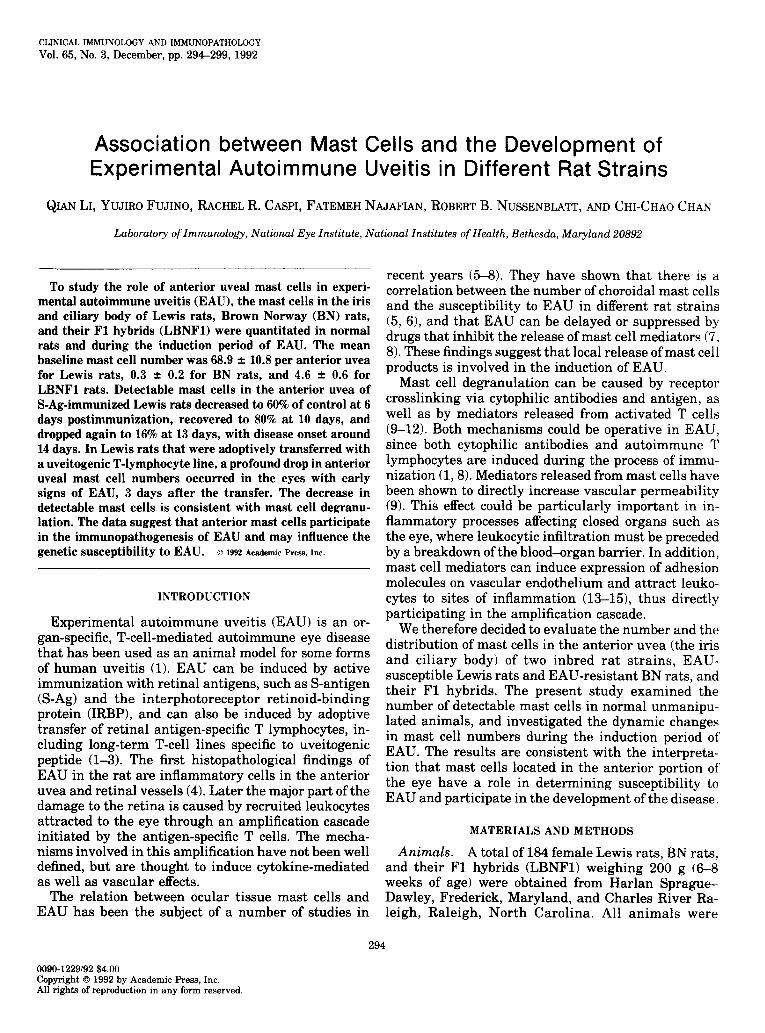
CLINICAL IMMUNOLOGY AND IMMUNOPATHOLOGY
Vol. 65,No. 3,December, pp. 294-299, 1992
Association between Mast Cells and the Development of Experimental Autoimmune Uveitis in Different Rat Strains
QIANLI,YUJIROFUJINO,RACHEL R. CASPI,FATEMEHNAJAFIAN,ROBERT B. NUSSENBLA~, AND CHI-CHAOCHAN
Laboratory of Immunology, National Eye Institute, National Institutes of Health, Bethesda, Maryland 20892
To study the role of anterior uveal mast cells in experi- mental autoimmune uveitis (EAU), the mast cells in the iris and ciliary body of Lewis rats, Brown Norway (BN) rats, and their Fl hybrids (LBNFl) were quantitated in normal rats and during the induction period of EAU. The mean baseline mast cell number was 68.9 f 10.8 per anterior uvea for Lewis rats, 0.3 + 0.2 for BN rats, and 4.6 + 0.6 for LBNFl rats. Detectable mast cells in the anterior uvea of S-Ag-immunized Lewis rats decreased to 60% of control at 6 days postimmunization, recovered to 80% at 10 days, and dropped again to 16% at 13 days, with disease onset around 14 days. in Lewis rats that were adoptively transferred with a uveitogenic T-lymphocyte line, a profound drop in anterior uveal mast cell numbers occurred in the eyes with early signs of EAU, 3 days after the transfer. The decrease in detectable mast cells is consistent with mast cell degranu- lation. The data suggest that anterior mast cells participate in the immunopathogenesis of EAU and may influence the genetic susceptibility to EAU. D 1st~ Academic PM+, IM.
INTRODUCTION
Experimental autoimmune uveitis (EAU) is an or- gan-specific, T-cell-mediated autoimmune eye disease that has been used as an animal model for some forms of human uveitis (1). EAU can be induced by active immunization with retinal antigens, such as S-antigen (S-Ag) and th e interphotoreceptor retinoid-binding protein (IRBP), and can also be induced by adoptive transfer of retinal antigen-specific T lymphocytes, in- cluding long-term T-cell lines specific to uveitogenic peptide (l-3). The first histopathological findings of EAU in the rat are inflammatory cells in the anterior uvea and retinal vessels (4). Later the major part of the damage to the retina is caused by recruited leukocytes attracted to the eye through an amplification cascade initiated by the antigen-specific T cells. The mecha- nisms involved in this amplification have not been well defined, but are thought to induce cytokine-mediated as well as vascular effects.
The relation between ocular tissue mast cells and EAU has been the subject of a number of studies in
recent years (5-8). They have shown that there is a correlation between the number of choroidal mast cells and the susceptibility to EAU in different rat strains (5, 6), and that EAU can be delayed or suppressed by drugs that inhibit the release of mast cell mediators (7, 8). These findings suggest that local release of mast cell products is involved in the induction of EAU.
Mast cell degranulation can be caused by receptor crosslinking via cytophilic antibodies and antigen, as well as by mediators released from activated T cells (9-12). Both mechanisms could be operative in EAU, since both cytophilic antibodies and autoimmune T lymphocytes are induced during the process of immu- nization cl,@. Mediators released from mast cells have been shown to directly increase vascular permeability (9). This effect could be particularly important in in- flammatory processes affecting closed organs such as the eye, where leukocytic infiltration must be preceded by a breakdown of the blood-organ barrier. In addition, mast cell mediators can induce expression of adhesion molecules on vascular endothelium and attract leuko- cytes to sites of inflammation (13-151, thus directly participating in the amplification cascade.
We therefore decided to evaluate the number and the distribution of mast cells in the anterior uvea (the iris and ciliary body) of two inbred rat strains, EAU- susceptible Lewis rats and EAU-resistant BN rats, and their Fl hybrids. The present study examined the number of detectable mast cells in normal unmanipu- lated animals, and investigated the dynamic changes in mast cell numbers during the induction period of EAU. The results are consistent with the interpreta- tion that mast cells located in the anterior portion of the eye have a role in determining susceptibility to EAU and participate in the development of the disease.
MATERIALSANDMETHODS
Animals. A total of 184 female Lewis rats, BN rats, and their Fl hybrids (LBNFl) weighing 200 g (6-8 weeks of age) were obtained from Harlan Sprague- Dawley, Frederick, Maryland, and Charles River Ra- leigh, Raleigh, North Carolina. All animals were
294
0090-1229192 $4.00 C opyright 0 1992 by Academic Press, Inc. All rights of reproduction in any form reserved.
MAST CELLS IN EXPERIMENTAL AUTOIMMUNE UVEITIS 295
housed in environmentally controlled rooms with a 12- hr light and dark cycle. They were provided food and water ad libitum.
Antigens and reagents. S-antigen was prepared from bovine retinas according to the method of Dorey et al. (16). R16 (a 15-mer synthetic peptide containing the major pathogenic epitope of interphotoreceptor reti- noid-binding protein) was synthesized and purified on a peptide synthesizer (3).
T-cell lines. An RlG-specific helper-T-cell line (LR16) was established from draining lymph nodes of RlG-immunized Lewis rats, essentially as previously described (2, 3). The cells were maintained in culture by a weekly regimen of stimulation with R16 on anti- gen presenting cells, followed by expansion in interleu- kin-a-containing medium. The LR16 lymphocyte line is highly uveitogenic, and transfers EAU to otherwise unmanipulated syngeneic recipients. Disease onset in the recipient usually occurs on the fourth day after injection of 1 to 2 million LR16 cells.
Znduction of EAU. Rats were immunized with pu- rified bovine S-Ag emulsified (l:l, v/v) in complete Freund’s adjuvant (CFA, Difco) that had been enriched with Mycobacterium tuberculosis strain H37Ra (Difco) to a concentration of 2.5 mg/ml. Each rat was immu- nized by a single injection of 30 pg of S-Ag in a total volume of 0.1 ml into one hind footpad.
Adoptive transfer was performed as follows: 3 x lo6 LR16 cells and 1 x lo8 thymocytes (irradiated 3000 R) were cocultured with R16 (3 kg/ml) in a volume of 10 ml supplemented RPM1 1640 medium in lo-cm Petri dishes for 48 hr at 37°C. The activated cells were washed twice and injected intraperitoneally into naive Lewis recipients (1.5 x lo6 cells into each animal).
Tissue preparation. Animals were sacrificed by COz inhalation. Enucleated eyes were immediately im- mersed in 10% neutral buffered formalin for 15 min. Under a dissecting microscope, the globes were opened by an equatorial incision at the limbus. The iris and ciliary body were isolated carefully with a dissecting needle, then floated in distilled water and flat-mounted on poly-L-lysine-coated slides. Slides were air-dried and stored at 4°C for 2 days before staining.
Staining. The slides were depigmented in aqueous potassium permanganate solution for 60 min. A 1% concentration was used for the BN and LBNFl rats, and a 0.25% concentration was used for the Lewis rats, which have no uveal pigmentation. Slides were washed three times in distilled water and bleached with 5% aqueous oxalic acid solution for approximately 10 min until the tissue became clear. The slides were again washed in distilled water, and 0.15% toluidine blue solution was applied for 3 min. The slides were covered with gelvatol (20/30 Resin, Indian Orchard Plant,
Springfield, MA), and the total mast cell numbers on each flat-mounted iris and ciliary body were counted under a microscope by two independent observers.
EAU occurrence and presentation of results. Ani- mals were followed daily for signs of EAU, starting 10 days after immunization. Iris hyperemia, exudation and hypopyon were taken as clinical evidence of EAU onset. Incidence is shown as the percent of positive animals, or the number of positive out of total animals in each group. All experiments were repeated at least twice. Statistics on the data were analyzed using stu- dents’ t-test.
RESULTS
Anterior mast cells in normal rats of different strains. Total mast cells in the anterior uvea of unmanipu-
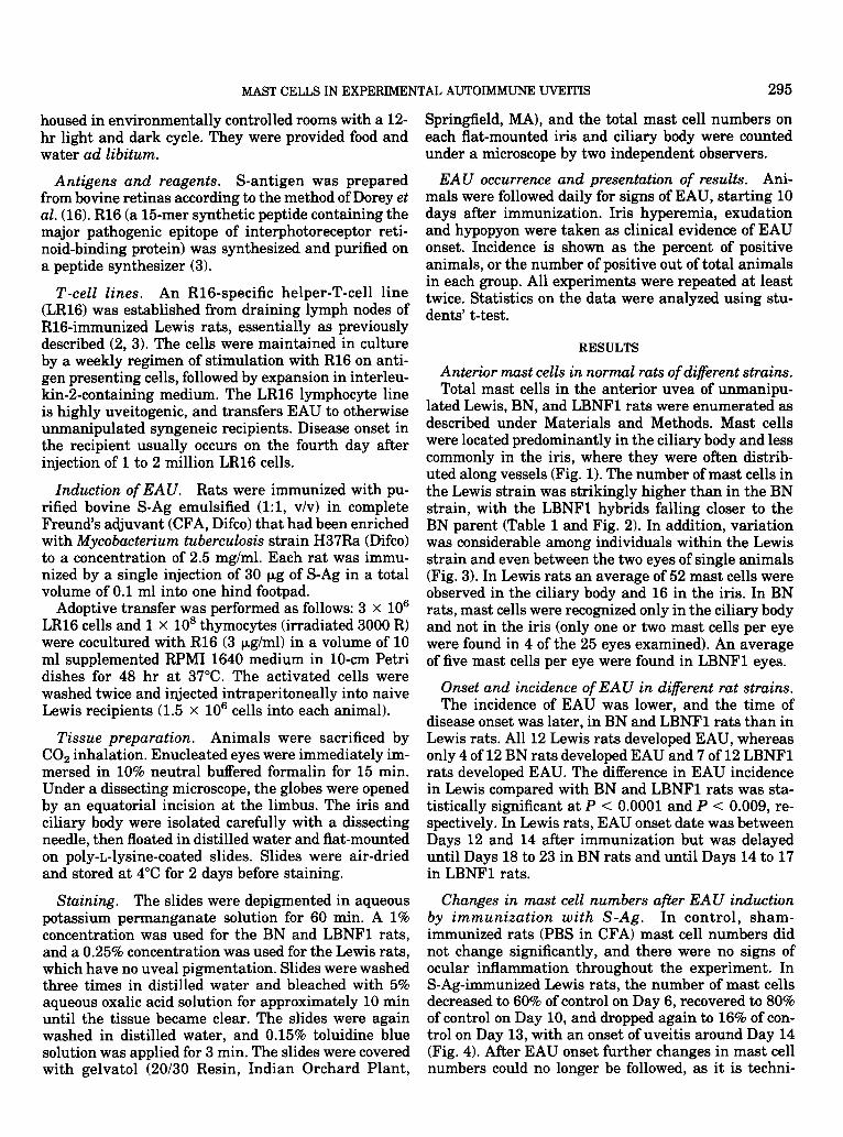
lated Lewis, BN, and LBNFl rats were enumerated as described under Materials and Methods. Mast cells were located predominantly in the ciliary body and less commonly in the iris, where they were often distrib- uted along vessels (Fig. 1). The number of mast cells in the Lewis strain was strikingly higher than in the BN strain, with the LBNFl hybrids falling closer to the BN parent (Table 1 and Fig. 2). In addition, variation was considerable among individuals within the Lewis strain and even between the two eyes of single animals (Fig. 3). In Lewis rats an average of 52 mast cells were observed in the ciliary body and 16 in the iris. In BN rats, mast cells were recognized only in the ciliary body and not in the iris (only one or two mast cells per eye were found in 4 of the 25 eyes examined). An average of five mast cells per eye were found in LBNFl eyes.
Onset and incidence of EAU in different rat strains. The incidence of EAU was lower, and the time of
disease onset was later, in BN and LBNFl rats than in Lewis rats. All 12 Lewis rats developed EAU, whereas only 4 of 12 BN rats developed EAU and 7 of 12 LBNFl rats developed EAU. The difference in EAU incidence in Lewis compared with BN and LBNFl rats was sta- tistically significant at P < 0.0001 and P < 0.009, re- spectively. In Lewis rats, EAU onset date was between Days 12 and 14 after immunization but was delayed until Days 18 to 23 in BN rats and until Days 14 to 17 in LBNFl rats.
Changes in mast cell numbers after EAU induction by immunization with S-Ag. In control, sham- immunized rats (PBS in CFA) mast cell numbers did not change significantly, and there were no signs of ocular inflammation throughout the experiment. In S-Ag-immunized Lewis rats, the number of mast cells decreased to 60% of control on Day 6, recovered to 80% of control on Day 10, and dropped again to 16% of con- trol on Day 13, with an onset of uveitis around Day 14 (Fig. 4). After EAU onset further changes in mast cell numbers could no longer be followed, as it is techni-

296 LI ET AL
FIG. 1. Photomicrograph of mast cells in the iris of a Lewis rat. The detail morphologies of mast cells are poor due to the thickness of the uvea (toluidine blue, x200; inset, x400).
tally difficult to obtain adequate tissue preparations from severely inflamed eyes. It was impossible to reli- ably evaluate changes in mast cell number in LBNFl and BN rats after S-Ag immunization, because there were very few mast cells present at baseline (Table 2).
. l
MeanGE: Lewis 68.9f10.8 LBNFl 4.6fO 6
EN 0.3f0.2
z . . L 40 : . ’
f 20 : t : 8
l . l 0 4t m
Lewis LBNFl EN
Rat Strains
FIG. 2. Total mast cells in the anterior uvea (iris plus ciliary body) of individual eyes of various rat strains. Each symbol repre- sents a single eye. Note the high variability in the counts for mast cells.
Changes in mast cell numbers after EAU inductton by adoptive transfer of uveitogenic T cells. Table 3 shows the mast cell numbers in the anterior uvea of Lewis rats injected with the pathogenic LR16 lympho- cyte line. No statistical difference relative to baseline was found on Day 1 or 2 after uveitogenic cell chal- lenge, and no eyes had either clinical or histopatholog- ical evidence of EAU. On Day 3, however, 5 of 16 eyes had early clinical signs of EAU (i.e., miosis, iris con- gestion, and edema). In these 5 eyes only a single mast cell in one eye could be identified, constituting a highly significant decrease in mast cell number. In the 11 of 16 eyes without clinical signs of EAU, there was a mild
TABLE 1 Numbers and Distribution of Mast Cells in Anterior Uvea
of Normal Rats
Number Number of mast cells”
Strain of rats Iris Ciliary body __-..-~~~ -~-~ .- ~.. --. _. -~ Lewis 15 16.1 t 3.3* 51.5 f 11.3* LBNFl 9 0.3 t 0.2 4.3 f 0.8 BN 13 0 0.3 i- 0.2
a Values are means t SE. * Significantly different from BN and LBNFl (P < 0.0001).

MAST CELLS IN EXPERIMENTAL AUTOIMMUNE UVEITIS 297
0 1 :
4
l- . I -
10 11 12 13 I 14
- I 5
Individual Rat
FIG. 3. Variation in mast cell numbers between the two eyes in single animals.
reduction in mast cell number which did not reach sta- tistical significance compared with baseline. By Day 4, all eyes had clinical evidence of severe inflammation, making quantitation of mast cells impossible by this flat-mount method.
DISCUSSION
The present study demonstrates that the number of mast cells present in the anterior uveal tract is much higher in Lewis rats than in BN or LBNFl rats, and is correlated with a higher incidence of EAU and an ear-
& 60- .- ; E a
70-
60 - iii tn K 50
D = 3 40
r 30-
: Z 20 -
ii ! - 10 -
” .
da; 0 day 6 day 10 day 13
Days after lmmunlzatlon
FIG. 4. Kinetics of change in mast cell numbers (*SE) in S-Ag immunized Lewis rats during the induction period of EAU. Asterisks denote statistically significant differences from controls (PBS in CFA).
lier onset of disease. These results are in agreement with a previous study, in which the number of mast cells in the choroid of various rat strains was associ- ated with their respective susceptibility to EAU (6). Our study also points to a similarity in the number of mast cells in the anterior and posterior portions of the eye within the strains studied: high in the Lewis, in- termediate in LBNFl, and low in BN. We hypothesize that in the EAU-resistant BN strain, the overall small number of local mast cells may be insufficient to pro- vide efficient amplification of the inflammatory cas- cade, resulting in a delay or absence of disease induc- tion.
The considerable variation in mast cell number be- tween the two eyes of most Lewis rats is of interest. The severity and the onset of EAU sometimes vary between the two eyes of the same animal, and in some cases the disease is unilateral. The unequal distribu- tion of mast cells between the two eyes could explain this phenomenon.
Although the target antigen (S-Ag or IRBP) is lo- cated in the posterior portion of the eye, the clinical onset of EAU is almost invariably accompanied by acute inflammation in the anterior segment. In fact, anterior inflammation may precede posterior involve- ment, since the first inflammatory cells entering the eye are frequently observed around the ciliary body (Ghan and Caspi, unpublished data). One possible ex- planation for this phenomenon is that the mast cells in the anterior segment may be involved in the early stage of anterior uveitis.
In the Lewis strain, there was a biphasic drop in detectable mast cell numbers during the induction pe- riod: a decrease to 60% on Day 6, with an apparent recovery to 80% on Day 10, and a second profound de- crease to 16% on Day 13 just prior to EAU induction which occurred around Day 14 (17, IS>. The apparent drop in mast cell numbers is most consistent with de- granulation, which would decrease their detectability for about 24 hr, while regeneration of the granules occurs (9,19). The striking drop in mast cell number 24 hr before EAU onset could be directly involved in fa- cilitating the subsequent infiltration of inflammatory cells. A similar observation was made earlier in exper- imental allergic encephalomyelitis (EAE), where a de- crease in number and functional activity of mast cells was noted at the onset of EAE, just before and during the development of clinical signs in the Lewis rat (20- 22). Mast cell involvement and subsequent release of vasoactive amines to alter the integrity of the blood- brain barrier were proposed to play a role in the devel- opment of actively induced EAE (23, 24).
Recent studies of adhesion molecules in EAE dem- onstrated their expression in a stage-specific manner within the central nervous system (CNS) and sug- gested specific homing and attachment of lymphocytes to the CNS vasculature as being the earliest features of

298 LI ET AL.
TABLE 2 Numbers of Mast Cells in Anterior Uvea in Different Strains of Rats during Induction Period of EAU
~- -. .-. -. -.
Days after Lewis
LBNFl BN immunization S-AG + CFA PBS + CFA S-Ag t CFA S-Ag -+ CFA
Baseline 68.9 t 10.8 (15)” 4.6 r 0.6 (9) 0.3 f 0.2 (13) 6 33.2 t- 6.7 (12)* 54.8 t 13.4 (8) 3.3 t 0.5 (8) I 3 ‘- 0.3 i8)
10 48.9 2 11.1 (8) 61.7 + 15.2 (5) 1.6 I+_ 0.5 ill) 0.6 t 0.3 16) 13 10.0 ” 2.5 (6)* 61.8 + 15.7 (6) ND ND
a Values are mean counts t SE, with number of rats in parentheses. ND, not done. * Statistically significant reduction (P < 0.003) in comparison to control.
the disease process (25-27). Thus, lymphocytes may immunization, in the adoptively transferred animals induce local changes at the blood-brain barrier favor- we did not observe a biphasic change in mast cell num- ing further influx of inflammatory cells. We speculate ber. Mast cells became undetectable only in eyes that that the initial moderate decrease in mast cells ob- were already showing early clinical signs of disease. served on Day 6 in the present study may be involved This suggests that the early drop in mast cell counts in in the elevated expression of adhesion molecules occur- S-Ag-immunized animals is connected to an event(s) ring in the eyes of Lewis rats 7 days after S-Ag immu- not represented in the adoptive transfer model, such as nization (28). induction of serum antibodies.
Our results for mast cellular kinetics in EAU differs from those reported by de Kozak et al. (8) for choroidal mast cells, where the numbers were doubled on Day 6 and were lowest on Day 10. Two nonmutually exclu- sive hypotheses can be proposed to reconcile this ap- parent discrepancy: either the number of mast cells in the eye is constant and the reciprocal shifts in their numbers represent migration of mast cells between the anterior and posterior compartments, or the changes represent different kinetics of degranulation in the an- terior and posterior portions of the eye.
Although there are other genetic differences between Lewis and BN strains, our study points to an associa- tion between the genetically determined number of mast cells in the anterior uvea of rats and their sus- ceptibility to EAU. These results strongly suggest par- ticipation of anterior uveal mast cells in the immuno- pathogenesis of ocular autoimmunity, particularly in the early stages of EAU.
ACKNOWLEDGMENT
We next examined changes in detectable mast cell numbers during the course of EAU induction by adop- tive transfer of uveitogenic T lymphocytes in the Lewis strain (uveitogenic T-cell lines are not available in the other strains). It should be noted that EAU induced by adoptive transfer circumvents the early steps of anti- gen presentation and recognition, as well as induction of high titers of antibodies, and is considered to repre- sent only the cell-mediated, efferent mechanisms of disease (2, 29). In contrast to the situation in active
The authors thank Dr. Louis Kasner for his excellent editorial assistance.
REFERENCES
1. Nussenblatt, R. B., and Palestine, A. G. (Eds.1, Ocular autoim- munity. In “UVEITIS Fundamentals and Clinical Practice,” pp. 38-46. Year Book Medical, Chicago, 1989.
2. Caspi, R. R., Roberge, F. G., Mcallister, C. G., El-Saied, M., Ku- wabara, T., Gery, I., Hanna, E., and Nussenblatt, R. B., T cell lines mediating experimental autoimmune uveoretinitis (EAU) in the rat. J. Zmmunol. 136, 928-933, 1986.
3. Sanui, H., Redmond, T. M., Kotakem, S., Wiggert, B., Hu, L-H., Margalit, H., Berzofsky, J. A., Chader, G. J., and Gery, I., Iden- tification of an immunodominant and highly immunopathogenic determinant in the retinal interphotoreceptor retinoid-binding protein (IRBP). J. Exp. Med. 169, 1947-1960, 1989.
4. de Kozak, Y., Sakai, J., Thillaye, B., and Faure, J-P., S antigen- induced experimental autoimmune uveo-retinitis in rats. Curr.
Eye Res. 1, 327-337, 1981.
TABLE 3 Number of Mast Cells in Iris and Ciliary Body of Lewis
Rats after Adoptive Transfer of LR16 Cell Line
Days after Rats without EAU Rats with EAU
adoptive Number of Number of Number of Number of transfer eyes/total mast cells” eyes/total mast cells”
1 14/14 2 16116 3 11/16
54.8 + 6.9 o/14 54.1 * 10.3 0116 44.4 t 7.7 5116 0.2 + 0.3*
(a total of only one mast cell was identified)
a Values are means * SE. * Statistically significant reduction (P < 0.0001) in comparison to
animals that did not exhibit signs of EAU.
5. Gery, I., Robinson, W. G., Jr., Shichi, H., El-Saied, M., Mochi- zuki, M., Nussenblatt, R. B., and Williams, R. M., Differences in susceptibility to experimental autoimmune uveitis among rats of various strains In “Advances in Immunology and Immunopa- thology of the Eye” (G. R. O’Connor and J. W. Chandler, Eds.), pp. 242-245, Masson, New York, 1982.
6. Mochizuki, M., Kuwabara, T., Chan, C C., Nussenblatt, R. B., Metcalfe, D. D., and Gery, I., An association between suscepti-

MAST CELLS IN EXPERIMENTAL AUTOIMMUNE UVEITIS 299
7.
8.
9.
10.
11.
12.
13.
14.
15.
bility to experimental autoimmune uveitis and choroidal mast cell numbers. J. Zmmunol. 133, 1699-1701, 1984. de Kozak, Y., Sakai, J., Sainte-Laudy, J., Faure, J-P., and Ben- veniste, J., Pharmacological modulation of IgE-dependent mast cell degranulation in experimental autoimmune uveoretinitis. Jpn. J. Ophthnlmol. 27, 598-608, 1983. de Kozak, Y., Sainte-Laudy, J., Benveniste, J., Faure, J-P., Ev- idence for immediate hypersensitivity phenomena in experi- mental autoimmune uveoretinitis. Eur. J. Zmmunol. 11, 612 617,198l. Galli, S. J., Dvorak, A. M., and Dvorak, H. F., Basophils and mast cells: Morphologic insights into their biology, secretory patterns, and function. Prog. Allergy 34, l-84, 1984. Plaut, M., Pierce, J. H., Watson, C. J., Hanley-Hyde, J., Nordan, R. P., and Paul, W. E., Mast cell lines produce lymphokines in response to cross-linkage of FceRI or to calcium ionophores. Nu- ture 339, 64-67, 1989. Marshall, J. S., and Bienenstock, J., Mast cells. Springer Semin. Zmmunopathol. 12, 191-202. Askenase, P. W., Rosenstein, R. W., and Ptak, W., T cells pro- duce an antigen-binding factor with in vivo activity antalogous to IgE antibody. J. Exp. Med. 157, 862-873, 1983. Maggiano, N., Colotta, F., Castellino, F., Ricci, R., Valitutti, S., Larocca, L. M., and Musiani, P., Interleukin-2 receptor expres- sion in human mast cells and basophils. Znt. Arch. Allergy Appl. Zmmunol. 91, 8-14, 1990. Valent, P., Bevec, D., Maurer, D., Besemer, J., Di Padova, F., Butterfield, J. H., Speiser, W., Majdic, O., Lechner, K., and Bet- telheim, P., Interleukin 4 promotes expression of mast cell ICAM- antigen. Proc. Natl. Acad. Sci. USA 88, 3339-3342, 1991. Klein, L. M., Lavker, R. M., Matis, W. L., and Murphy, F., De- granulation of human mast cells induces an endothelial antigen central to leukocvte adhesion. Proc. N&l. Acud. Sci. USA 86. 8972-8976, 1989.”
16. Dorey, C., Cozette, J., and Faure, J-P., A simple and rapid method for isolation of retina S antigen. Ophthalmic Res. 14, 249,1982.
17. Chan, C-C., Hooks, J. J., Nussenblatt, R. B., and Detrick, B., Expression of Ia antigen on retinal pigment epithelium in ex- perimental autoimmune uveoretinitis. CUFF. Eye Res. 5, 325- 330, 1986.
18. Fujikawa, L. S., Chan, C C., McAllister, C., Gery, I., Hooks, J. J., Betrick, B., and Nussenblatt, R. B., Class II histocompatibility
Received April 17, 1992; accepted with revision August 19, 1992
19.
20.
21.
22.
23.
24.
25.
26.
27.
28.
29.
complex antigens (I-A and I-E) in experimental autoimmune uveitis. In “Modern Trends in Immunology and Immunopathol- ogy of the Eye” (A. G. Secchi and I. A. Fregona, Eds.), pp. 69-75, Masson, Milan, 1989. Allansmith, M. R., Baird, R. S., Henriquez, A. S., and Bloch, K. J., Regeneration of mast cells after ocular anaphylaxis In “Advances in Immunology and Immunopathology of the Eye” (G. R. O’Connor and J. W. Chandler, Eds.), pp. 173-175, Masson, New York, 1982. Stanley, N. C., Jackson, F. L., and Orr, E. L., Attenuation of ex- perimental autoimmune encephalomyelitis by compound 48/80 in Lewis rats. J. Neuroimmunol. 29,223-228, 1990. Be, L., Olsson, T., Nyland, H., Kruger, P. G., Taule, A., and Merk, S., Mast cells in brains during experimental allergic en- cephalomyelitis in Lewis rats. J. Neural. Sci. 105, 135-142, 1991. Levi-Schaffer, F., Riesel, N., Soffer, D., Abramsky, O., and Bren- ner, T., Mast cell activity in experimental allergic encephalomy- elitis. Mol. Chem. Neuropathol. 15, 173-184, 1991. Linthicum, D. S., Development of acute autoimmune encephalo- myelitis in mice: Factors regulating the effector phase of the disease. Zmmunobiology 162, 211-220, 1982. Dietsch, G. N., and Hinrichs, D. J., The role of mast cells in the elicitation of experimental allergic encephalomyelitis. J. Zmmu- nol. 142, 14761481, 1989. Cannelia, B., Cross, A. H., and Raine, C. S., Adhesion-related molecules in the central nervous system. Upregulation corre- lates with inflammatory cell influx during relapsing experimen- tal autoimmune encephalomyelitis. Lab. Invest. 65,23-31,1991. O’Neill, J. K., Butter, C., Baker, D., Gschmeissner, S. E., Kraal, G., Butcher, E. C., and Turk, J. L., Expression of vascular ad- dressins and ICAM- by endothelial cells in the spinal cord dur- ing chronic relapsing experimental allergic encephalomyelitis in the Biozzi AB/H mouse. Immunology 72, 520-525, 1991. Yednock, T. A., Cannon, C., Fritz, L. C., Sanchez-Madrid, F., Steinman, L., and Karin, N., Prevention of experimental au- toimmune encephalomyelitis by antibodies against a431 inte- grin. Nature 356, 63-66, 1992. DeBarge, L. R., Chan, C-C., Caspi, R. R., Nussenblatt, R. B., and Whitcup, S., Expression of cell adhesion molecules in mice with experimental autoimmune uveitis. Invest. Ophthulmol. Vis. Sci. 33 (Suppl.), 796, 1992. Caspi, R. R., Basic mechanisms in immune mediated uveitic dis- ease. In “Immunology of Eye Diseases” (S. Lightman, Ed.), pp. 61-86, Kluwer Academic, Boston, 1989.
Copyright © 2022 FDOKUMEN