A REPORT ON APHAKIC VISION FOLLOWING PHACOEMULSIFICATION AND INTRAOCULAR lens power requirements in...
102
-
Upload
tamilnaduveterinaryanimalsciences -
Category
Documents
-
view
0 -
download
0
Transcript of A REPORT ON APHAKIC VISION FOLLOWING PHACOEMULSIFICATION AND INTRAOCULAR lens power requirements in...


INDIAN JOURNAL OF CANINE PRACTICEAn Official Organ of Indian Society for Advancement of Canine Practice
Vol. 06 No. 1 Half Yearly JUNE 2014Chief Editor
Prof. (Dr.) A.K. SrivastavaLucknow
EditorProf. J.C. Jena
COVAS, Bhubneswar
Associate EditorsDr. Amarpal
I.V.R.I., IzatnagarDr. V. C. Murthy
COVAS, KVAFSU, Banglore
Editorial BoardChairman
Prof. S.S. HonnappagolAnimal Husbandry
Commissioner, G.O.I., NewdelhiMembers
Prof. A. C. VarshneyV.C., DUVASU, Mathura
Maj. Gen. Shrikant SharmaV.C., LLRUVAS, HisarProf. S. D. Sharma
Ex-Dean,COVAS, MathuraProf. A. K. Sinha
Ex-Dean, COVAS, RanchiProf. D. B. Sarode
FVM, Mekale, EthiopiaProf. S.S. Randhawa
Director Research, GADVASU,Ludhiana
Prof. M. S. VasanthDean, COVAS, Puttur
Prof. S.YathirajDean, COVAS, Banglore
Prof. G.K. SinghDean, COVAS, Pantnagar
Prof. S. PrathabanDean, COVAS, Tirunelveli
Prof. S.P. ShuklaDean, COVAS, Rewa
Editorial OfficeI.S.A.C.P.
21/5, Sector-21, Indira Nagar,Lucknow – 226 016, India.
Telephone: 0091-522-4071826;919839025350
E-mail: [email protected];
CHIEF PATRONPATRONS
APEX ADVISORY BOARD
EXECUTIVESPresidentVice President
Secretary General
Treasurer
Joint Secretary
Executive Members
Ex- Officio Members
P.R.O.
Prof. P.N. BhatFormer Director & V.C., I.V.R.IProf. R.M. AcharyaFormer D.D.G.(AS), I.C.A.R.,Prof. P.K. UppalFormer Director, NRCE,Prof. R.R. ShuklaEx-President, N.A.V.Sc., New DelhiProf. Nagendra SharmaFormer Vice Chancellor, SKUAT, JammuProf. M.P. YadavSecretary, N.A.A.S., GurgaonProf. J.M. NigamFormer Dean COVAS,Palampur (H.P.)Prof. Harpal SinghFormer Dean COVAS, Pantnagar (Uttr.)Prof. S. K. DwivediFormer Director, N.R.C.E., HisarProf. A.K.GahlotVice Chancellor, RAJUVAS, BikanerProf. Anil Kumar SrivastavaDirector, N.D.R.I., KarnalProf. Aditya Kumar MishraVice Chancellor, MAFSU, NagpurLt. Gen. J.K. SrivastavaEx-Director General, R.V.S., New DelhiProf. S.A. JagdishFormer Dean COVAS MumbaiProf. Amlendu ChakrabortyFormer Dean, COVAS, Kolkata.Dr. Anup BhaumikSecretary, V.C.I. New DelhiProf. V. K. SinhaPatnaProf. N.A.SudhanJammuProf. A.K. SrivastavaLucknow.Dr. Shrish ChandraAllahabadDr. Hemant Tilakshi JainNagpur (MHS)Prof. Dipak Kumar DeCOVAS, Kolkata (W.B.)Prof. Shamshul HaqueCOVAS, Ranchi (Jharkhand)Prof. Anil Kumar AhujaCOVAS, Bikaner (Rajasthan)Dr. V.K. SharmaDehradoon (Uttranchal)Dr. O.S. PraskashShimoga (Karnataka)Dr. Rajesh RohiCOVAS, Mumbai (MHS)Dr. Shyam K. VenugopalCOVAS, Trissur (Kerala)Prof. S.K. RayFounder President; Bhubneswar (Orissa)Dr. Rajesh VarshneyFounder Treasurer; Lucknow (U.P.)
Ashok ChopraNew Delhi

INSTRUCTIONS TO AUTHORSIndian Journal of Canine Practice will be Published half yearly (biannually) in a size 19.5cm x 25cm in 2columns, set solid, In a print area of 17.5cm x 23.5cm.The official language is English.
All articles should be sent to:
Prof. A.K. Srivastava; M.V.Sc., Ph.D., Chief Editor, I.J.C.P.;
I.S.A.C.P., 21/5, Sector-21, Indira Nagar, Lucknow -226 016, INDIA.
([email protected] or [email protected] or ak.srivastava [email protected])
Nature of Coverage: Contributors are welcome which fulfill the following requirements.
Contributor should be a life member of the society (ISACP).
All research / clinical contributions in the field of Canine Practice are accepted.
Original full articles should not exceed 12 typed pages.
Short notes based on experimental evidence, clinical work should not exceed 4 typed pages. Abstract and heatedsections are not necessary.
Clinical reviews will be accepted only on invitation.
Book review by subject-matter specialist will be for the book sent to the journal for review.
The spelling should be that of the Oxford English Dictionary
Papers are accepted for publication on the understanding that they have not been published and are not beingconsidered for publication elsewhere and that they have not be republished without prior permission from thepublisher.
The authors should send two copies of manuscript which should be typed on A-4 size of Paper either in PageMaker or in Words (Hard Copy) along with the C.D. Manuscript must be typed in double space including title(informative and short), authors name and address, and short title (not more than 15 words) figures and tablecaptions, table and literature citations. Do not use italic type or words of expressions such as et aI., per se, Ibid, invitro, in vivo etc. Underline when italic is required. At least 2.5 cm margin should be on all sides of the paper.Paper may be sent by Email ([email protected] or [email protected] or [email protected]).Full papers must be divided into the following sections -
ABSTRACT not exceeding 150words,KEY WORDS, INTRODUCTION, MATERIALS AND METHODS,RESULTS AND DISCUSSION (to avoid repetition), if any followed by ACKNOWLEDGEMENT andREFERENCES (Not more than 2500 words).
SHORT COMMUNICATIONS should be continuous with references at the end of the paper (Not more than1200 words).
TABLE - Only one table is to be placed on a sheet of paper. It must be typed with its caption and fullycomprehensive without reference to the text.
FIGURES - should be minimum, line drawings, photographs and graphs should be referred as Fig. 1, 2 and so on.
PHOTOGRAPHS - should be sharp, glossy print (black and white). If coloured then properly scanned / printcopy. If transmitted through post, illustrations / photograph should not be folded but should be protected by cardboard paper. The authors have to bear the cost of printing of coloured photographs at the rate of Rs.1,000.00/- perpage.
REFERENCES - Correct reference is the - responsibility of the authors. It should be given in alphabetical orderwith the abbreviations according to world list of Scientific Periodicals. The style should be as follows -
Madhu, B. and Lal, S. S. (1987). The in vitro effects of anthelmintics on phosphatases of sheep nodular worm.Indian J. Paras't., 11: 201-203 " References to cited books and monographs should' include the name of authors,year of publication, title, edition, town of publication and publisher e g., Faust, E. C, and Russell, P. F. (1864).Craig and Faust clinical parasitology. 7th ed., Lea and Fabiger, Philadelphia p 4-9. Personal communicationshould be cited in the text and not in reference list.
REFEREE - Article received will be sent to the referee and authors should improve them by going through thecomments of referees.
REPRINTS- Twenty reprints may be supplied to the first author on the payment of nominal charges. Order forreprints will be received with paper. Order for the reprint after receipt of paper will not be entertained.
Note: It is essential to obtain IAEC / CPCSEA approval in all the projects involving animal experimentation.

C O N T E N T SCANINE MEDICINECONCURRENT TOXOCARA CATI AND ISOSPORA FELIS INFECTION IN TWO PERSIAN CATSManju K. Mathew, Usha Narayana Pillai, Thomas Edison D’Sa, Anahita Anil Kumar and S YogeshpriyaDIROFILARIASIS IN A DOGA.K. Srivastava and B. SyedVERTEBRAL HEART SCALE SCORE OF MONGREL DOGSMukesh Srivastava, R.P. Pandey, Ashish Srivastava, Deepesh Kumar, S. Purohit and V. MalikTHERAPEUTIC MANAGEMENT OF OTITIS EXTERNA IN DOGSChandan Lodh, Suprabha Choudhury and Surojit DasDIAGNOSIS AND TREATMENT OF CHOLANIGO-HEPATITIS WITH URSODEOXYCHOLIC ACID INA DOGR.K. Bhardwaj, A.K. Gupta, J.S. Soodan and R. SinghRENAL FAILURE – A CLINICAL REPORT OF THREE CASESLalita Kumari and S. HaqueNEPHROTIC SYNDROME IN A SIAMESE CATA.K. Srivastava and B. Syed
……
……
……
……
……
……
……
01
03
05
09
13
16
19
CANINE MICRBIOLOGYVACCINE FAILURE AGAINST CANINE PARVOVIRUS INFECTIONS IN DOGSS. Nandi and Manoj Kumar
…… 21
CANINE REPRODUCTIONFOLLICULAR CYSTIC OVARIES AND CEH-PYOMETRA IN A DOGC. Jayakumar, Abhignya Krishna, K.S. Shwetha and G. SudhaULTRASONOGRAPHIC PREGNANCY DIAGNOSIS AND LANDMARKS IN DOMESTIC CATSS.U. Gulavane, M.N. Rangnekar, S.A. Bakshi , R.J. Chaudhari and P.J. Thakur
……
…….
31
33
CANINE SURGERYDIAGNOSTIC AND THERAPEUTIC MANAGEMENT OF CANINE OSTEOARTHRITISV.P. Chandrapuria, Apra Shahi, H.S. Dhakare and Somil RaiA REPORT ON APHAKIC VISION FOLLOWING PHACOEMULSIFICATION AND INTRAOCULARLENS POWER REQUIREMENTS IN CATSC. Ramani, N.J. D’Souza, M.K. Ahirwar, L. Nagarajan and B.J. WilliamSURGICAL MANAGEMENT OF SOFT TISSUE SARCOMA IN A DOGRamesh Rathod, A.S. Patil, L. Ranganath, B.N. Nagaraja, A. Anirudh and RavikumarSURGICAL MANAGEMENT OF OSTEOSARCOMA IN A DOGA. Anirudh, K.M. Srinivasa Murthy and L. RanganathA CASE OF CANINE TRANSMISIBLE VENEREAL TUMOUR IN A MALE DOG AND ITS SURGICALMANAGEMENTJayakrushna Das, Sidhartha Sankar Behera, S.K. Panda, P.K. Rath, M. Behera and S. PatiSURGICAL MANAGEMENT OF INGUINAL HERNIA IN A MALE DOGS. Ravikumar and L. RanganathMANDIBULAR FRACTURE AND ITS SURGICAL MANAGEMENT IN A DOGJayakrushna Das, Sidhartha Sankar Behera and Ananta HembramCUNEIFORM ULNAR OSTEOTOMY FOR VALGUS DEFORMITY OF RADIUS AND ULNA IN AGREAT DANE PUPS. Ravikumar, V. Mahesh and L. RanganathBILATERAL HYGROMA IN A GREAT DANE DOG AND ITS SURGICAL MANAGEMENTI. Nath, Jasmeet Singh, Sidhartha Sanker Behera, Prasant Kumar Sika and B.K DwibedyMASSIVELY ENLARGED MAMMARY TUMOUR IN A DOG AND ITS SURGICAL MANAGEMENTI. Nath, Jasmeet Singh, S.S. Behera, Samir Kumar Das, Sujit Prasad Das and Sripati SethiMEGAESOPHAGUS AND ITS MANAGEMENT IN THREE DIFFERENT LARGE BREED ADULTBITCHESA.K. Maji and Arnab Kumar MajieURETHROCYSTOSCOPIC DIAGNOSIS AND THERAPEUTIC MANAGEMENT OF URINARY TRACTDISORDER IN FEMALE DOGSV.P. Chandrapuria, Dinesh Gupta, Apra Shahi, Dharmendra Kumar and Somil Rai
......
......
......
......
......
......
......
......
......
......
......
......
38
42
45
47
49
53
56
60
62
65
69
72
CANINE NUTRITIONCOMPARATIVE GROWTH PERFORMANCE OF PUPPIES ON HOMEMADE AND COMMERCIALNON – VEGETARIAN FOODG.M. Gadegaonkar S,.A. Kale, M.B. Patil and V.D. Kank
…… 80
CANINE APPLIED SUBJECTSPREVALENCE OF SPONTANEOUSLY OCCURRING NEOPLASMS AMONGST CANINES IN JAMMUAsma Hamid, Shagufta Azmi, Shafiqur Rahman and Maneesh SharmaA STUDY ON CLINICAL AND HAEMATOBIOCHEMICAL PARAMETERS IN CANINEDEMODICOSISA. Janus, P.V. Tresamol, K.A. Mercey, Biju P. Habeeb and H. ShameemCANINE METASTATIC MAMMARY CARCINOMA IN LUNGS: A CASE REPORTS. Roshini, G.N. Patil, A.K. Mhase, G.K. Sawale, S.D. Moregaonkar and D.P. Kadam
……
……
……
87
92
95
Note: The Authors and editors do not accept any responsibility for any loss or damage arising from actions or decisions basedon information contained in this publication. The opinion expressed are those of the authors and the inclusion in thispublication does not amount to its endorsement.

Indian Journal of Canine Practice Volume 6 Issue 1, June, 20141
CONCURRENT TOXOCARA CATI AND ISOSPORA FELISINFECTION IN TWO PERSIAN CATS
Manju K. Mathew, Usha Narayana Pillai, Thomas Edison D’Sa, Anahita AnilKumar and S Yogeshpriya
Department of Clinical Veterinary Medicine,College of Veterinary and Animal Sciences, Mannuthy, Thrissur, 680651.
[Received: 06.2.2014; Accepted: 08.6.2014]
Isospora genera of the protozoancoccidia is found to affect dogs and cats(Soulsby, 1982). Two species infect cats Ifelis and I rivolta. The organism isworldwide in distribution and thedevelopmental stages occur in the smallintestine. Animals become infected byingesting sporulated oocysts. Toxocaracati occurs in the small intestine of cats.Paratenic hosts, rats, play an important rolein the life cycle of this organism.
Case History and ObservationsTwo Persian cats, one six months of
age and other fourteen months old (Fig.1)were presented at University VeterinaryHospital, Mannuthy in September 2011
with a complaint of anorexia since threedays and foul smelling diarrhoea. Cats weretransported from Bangalore one week back.Animals were fed both commercial cat foodand homely food. On clinical examinationintestinal loops were found to be thickened.Toxocara cati (Fig.2) worm was found infaeces. On microscopic examination offaeces sporulated and unsporulatedIsospora felis (Fig.3) oocyst could be seen.Animals were treated with Banminthsuspension @ 7.5mg/kg orally and advisedBactrisol bolus @ 15mg/kg per oral for fivedays along with supportive multivitamintherapy.
Fig.1: 6 months old kitten Fig.2 : Toxocara cati worms from faeces Fig.3: sporulated andunsporulated coccidial oocysts
ResultsOwner reported improvement in
condition of the cats after one week. Foodintake came to normal with formed faecesand faecal samples were negative forparasitic ova after two weeks.
Discussion

Indian Journal of Canine Practice Volume 6 Issue 1, June, 20142
Isosporosis in kittens occursprimarily during weaning stress. Clinicalsigns in the case discussed includediarrhoea, weight loss and dehydration asdescribed by Tomimura (1957).Coccidiosis is usually associated with otherinfectious agents, immunosuppression orstress. In the present case cats weresubjected to transportation stress one weekearlier. Diagnosis of the case is by clinicalsymptoms and demonstration of oocysts inthe feces. Isospora oocysts consists of 2sporocysts each with 4 sporozoites.Treatment may be unnecessary in cats sincethey usually spontaneously eliminate theinfection. In clinically affected animalssulfa- trimethoprim could be used.
Prenatal infection does not occurwith Toxocara cati and the infection is byingestion of eggs containing infective
second stage larvae. Majority of infectionsin kittens are derived from the milk ofinfected queens. Clinical signs includedgeneral unthriftiness, pot belliedappearance, intermittent diarrhoea andanaemia. Diagnosis is by presence of eggsin faeces and clinical signs. Treatment canbe done with benzimidazoles, pyrantelpamoate, piperazine, DEC etc.
ReferencesSoulsby, E.J.L., (1982). Helminths,
arthropods and protozoa ofdomesticated animals, ElsevierSaunder, St.Louis, Missouri, USA809p.
Tomimura, T.(1957) . Experimental studieson coccidiosis in dogs and cats.Riseichugaku Zasshi.6: 12-14.
*****

Indian Journal of Canine Practice Volume 6 Issue 1, June, 20143
DIROFILARIASIS IN A DOG
A.K. Srivastava and B. SyedFaculty of Veterinary Medicine, Jigjiga University, Jigjiga, Ethiopia.
[Received: 22.6.2013; Accepted: 03.1.2014]
Commonly known as heartwormdisease, dirofilariasis is a parasitic diseasecaused by a nematode called “Dirofilariaimmitis”, which in its adult form lives inthe right side of the heart and pulmonaryarteries. Dirofilariasis is a serious illnesswhich can affect cats and dogs that get itfrom mosquito bites, which inoculatemicrofilariae through the skin from aninfected animal into a healthy one (ArnoldP. et al.,1994 and Levy J.K. et al., 2011). Normally after 6 months from the timethe contagion occurred, the tinymicrofilariae that are traveling through thebloodstream reach their adult form andaccumulate in the heart’s right ventricle andpulmonary arteries. Most frequent signs indogs associated to the parasite’s locationare: loss of appetite, weight loss, cough,laboured breathing and the presence ofblood in respiratory secretions, amongothers. Cats, on the other hand, can alsodevelop asthma and tachycardia. Theseverity of these and other clinical signsdepends on the number of worms, theanimal’s activity and its immune responsethe worm. Therefore, in some animals itcan derive in a congestive heart failure,abdominal against oedema or oedema ofthe extremities, and even death due to alarge number of worms in the heart (HochH. and Strickland K., 2008). A novel approach for the treatment ofcardiopulmonary dirofilariosis is targetingthe Wolbachia rickettsial endosymbionts.Treatment with tetracyclines has beenreported to damage D. immitis, evencausing death of adult worms (Kramer L. etal., 2008 and Colby K.N. et al., 2011).Long-lasting administration of bothDoxycycline and Ivermectin before or inthe place of melarsomine injections can
eliminate adult worms and reduce the riskof thromboembolism. Therefore, it hasbeen suggested that a combination ofDoxycycline for 30 days and Ivermectin for6 months has a potential efficacy, as highas 73%, in the adulticide therapy in dogsinfested with D immitis (Bazzocchi C. etal., 2008 and Giannelli A. et al., 2013). A G.S.D. of 3 years old from theSomali region was referred to the FacultyClinic of Jigjiga University, Jigjiga with ahistory of weight loss and rapid fatigue. Itwas having the symptoms of chroniccough, occasional dyspnoea and poorexercise tolerance with normal appetite.The dog had the temperature of 39.50C. Onclinical examination increased vesicularsounds and dyspnoea after physicalexcitement and effort were the onlyabnormal findings demonstrable.Radiologically demonstrated changes of thepulmonary arteries led to a tentativediagnosis of Dirofilariosis. The stoolexamination was negative for parasiticinfestation. The parasitological diagnosisbased on serology and the morphology ofmicrofilaria isolated from the bloodindicated an infection by microfilariae andadult stages of Dirofilaria immitis. Onhaematological examination, thehaemogram of the dog revealed mildanaemia. However in present caseneutrophilia was also recorded. After premedication with Aspirin,the patient was treated against adult filariaewith Caparsolate, and a month later withIvermectin against the microfilariae. At thetime of reexamination, 5 months afterinitiation of therapy, the dog was clinicallyhealthy and free of any demonstrableinfection with Dirofilaria.

Indian Journal of Canine Practice Volume 6 Issue 1, June, 20144
Left: Aedes albopictus mosquito, a potential vector for Dirofilariasis; Center: Dirofilariasand Right: Picture of affected dog.
ReferencesArnold P., Deplazes P., Ruckstuhl H., and
Flückiger M. (1994).Case report:dirofilariasis in a dog. Schweiz ArchTierheilkd.;136(8):265-9.
Bazzocchi C., Mortarino M., Grandi G., etal. (2008).Combined Ivermectin andDoxycycline treatment hasmicrofilaricidal and adulticidalactivity against Dirofilaria immitis inexperimentally infected dogs. Int. J.Parasitol. 38(12):1401-10.
Colby K.N., Levy J.K., Dunn K.F. et al.(2011). Diagnostic, treatment and
prevention protocols for canine heartworm infection in animal sheltring agencies. Vet. Parasitol.
176(4):333-341.Giannelli A., Ramos R.A., Traversa D., et
al. (2013). Treatment of Dirofilariarepens microfilariae-mia with acombination of Doxycycline hyclateand Ivermectin. Vet Parasitol. 178
(May 21, 2013).Hoch H. and Strickland K.(2008).Canine
and feline dirofilariasis: prophylaxis,treatment, and complications oftreatment. Compend Contin EducVet.: 30(3):146-51.
Kramer L., Grandi G., Leoni M., et al.(2008). Wolbachia and its influenceDirofilaria immitis infection. VetParasitol.158(3):191-5.
Levy J.K., Lappin M.R., Glaser A.L. et al.,(2011). Prevalence of infectious
diseases in Cats and Dogs rescued following Hurricane Katrina.
JAVMA; 238(3):311-317.*****

Indian Journal of Canine Practice Volume 6 Issue 1, June, 20145
VERTEBRAL HEART SCALE SCORE OF MONGREL DOGS
Mukesh Srivastava, R.P. Pandey, Ashish Srivastava, Deepesh Kumar, S. Purohitand V. Malik
Department of Clinical Medicine, College of Veterinary Science & Animal HusbandryDUVASU, Mathura, Uttar Pradesh (India).
[Received: 23.2.2014; Accepted: 08.6.2014]
Nineteen mature healthy mongrel dogs with mean age of 2.6 years underwent lateral thoracicradiography for evaluation of Vertebral Heart Score (VHS). The long axis of the heart was measured from theventral border of the left main stem bronchus to the cardiac apex and short axis was measured at the widest pointof the cardiac image on a line perpendicular to the long axis at the level of the caudal vena cava. Long and shortaxis measurements were compared to the vertebrae starting at the cranial edge of T4 to caudally. Result expressedin units of vertebral lengths was obtained for each axis and the two numbers were then added together to give thevalue of the VHS. The vertebral heart score in clinically healthy native dogs was 10.13 ± 0.18 vertebrae with arange of 8.9 to 11.5 vertebrae. The mean ± SE of short axes were 4.57 ± 0.10 vertebrae and of long axes were 5.55± 0.09 vertebrae. Significant difference was not observed in male and female dogs in relation to VHS,longitudinal axis and short axis.Keywords: Cardiomegaly, Mongrel dogs, Thoracic radiography, Vertebral Heart Score.
IntroductionCardiomegaly as a consistent sign
of heart disease can be seen in cases ofhypertrophic or dilated cardiomyopathy(Litster and Buchanan, 2000). Thoracicradiography is an integral part of thediagnosis and management of cardiacdisease (Root and Bahr, 2002). Studiesusing planimetry and various cardiothoracicratios have been reported, a guideline of 2.5to 3.5 intercostal spaces for dogs wasintroduced but limitations of this methodare variations of the heart size and shape,conformation of the thorax, phase ofrespiration, superimposition of ribs, andimprecise measurement points (Lamb andBoswood, 2002). Method for measuring thecanine cardiac silhouette that involvesmeasuring its long and short axes in alateral radiograph and comparing the sumof these measurements to the mid thoracicvertebral bodies, to produce a unitlessindex called the vertebral heart (VHS)score is described by Buchanan andBucheler (1995) as there is good relationswere known to exist between body lengthand heart weight. As there are variousfactors that influence the VHSmeasurement, interbreed differences inregards to normal heart size and shape is
important to consider whenever the heart isevaluated (Hansson, 2005). Lamb et al.,(2001) have documented the VHS score forvarious breeds like Doberman (10 ±0.6),German shepherd (9.7 ± 0.7), CavalierKing Charles spaniel (10.6 ± 0.5), Labradorretriever (10.8 ± 0.6) and Boxer (11.6 ±0.8). Lamb et al. (2001) suggested the useof breed-specific VHS values to have ahigh specificity for normal heart size. VHSscore can be used to assess progressivecardiomegaly (Buchanan and Bucheler,1995), pacing induce experimental heartfailure (Nakayama et al., 2001),hypoadrenocorticism (Melian et al., 1999).Large populations of mongrel dogs are stillused as companion animals in India,therefore, the present study was planned toestablish the reference range of VHS inmongrel dog.
Materials and methodsTen males and nine females
healthy mongrel dogs (19), presented fordiseases other than cardiovascular systemwith mean age 2.6 years were selected forthe study after clinical (physicalexamination and auscultation) andlaboratory evaluation (haemato-biochemical and urinalysis). Left lateral

Indian Journal of Canine Practice Volume 6 Issue 1, June, 20146
thoracic radiographs were taken duringinspiration as suggested by Gulanber et al.,(2005). The radiographs were taken in leaststress condition without use of anyanesthetic agents. The long axis of theheart, reflecting the combined size of theleft atrium and left ventricle was measuredfrom the ventral border of the left mainstem bronchus to the cardiac apex. Theshort axis was measured at the widest pointof the cardiac image on a line perpendicularto the long axis at the level of the caudalvena cava (Buchanan and Bucheler, 1995).
Measurements were recorded incentimeters for statistical analyses. Longand short axis measurements werecompared to the vertebrae starting at thecranial edge of T4 to caudally and resultexpressed in units of vertebral lengths wasobtained for each axis (fig-1). Themeasurements of the long and short axeswere recorded in terms of the numbers ofvertebrae covered and the two numberswere then added together to give the valueof the VHS and data are expressed as mean± SE.
Fig-1 The short axis measurement of the cardiac silhouette (A) and the long axis measurement(B) gives VHS on a lateral thoracic radiograph.
ResultsThe vertebral heart score in
clinically healthy native dogs was 10.13 ±0.18 vertebrae. Distribution of VHS rangewas 8.9 to 11.5 vertebrae. The Mean ± SEof short axes were 4.57 ± 0.10 vertebraeand of long axes were 5.55 ± 0.09vertebrae. The longitudinal axis in bitcheswas 5.61± 0.13, while in dogs was 5.61±
0.13 vertebrae. The short axis in bitcheswas 4.63 ± 0.12; while in dogs was 4.51 ±0.15 vertebrae. VHS in females was 10.14± 0.24 and in males was 10.12 ± 0.26(table-1). Data were analyzed through SASenterprise guide version 4.4 and nosignificant difference between males andfemales was found in relation to long axis,short axis and Vertebral heart Score.
Table-1 (Mean ± SE) Vertebral heart score (VHS) and various axis in mongrel dogs
Parameters Long axis Short axis VHSMale (N=10) 5.61± 0.13 NS 4.51 ± 0.15 NS 10.12 ± 0.26 NS
Female(N=9) 5.51 ± 0.13NS 4.63 ± 0.12NS 10.14 ± 0.24 NS
Total (N=19) 5.55 ± 0.09 4.57 ± 0.10 10.13 ± 0.18

Indian Journal of Canine Practice Volume 6 Issue 1, June, 20147
DiscussionThe recognition of interbreed
variations of cardiac dimensions has led tothe development of breed-specific rangesfor echocardiography in dogs (Nakayama etal., 2001), in the same direction Lamb etal., (2001) published breed-specific VHSranges for some popular breeds of dogs likeDoberman Labrador retriever and Boxeretc. Mongrel dogs are not a registered breedin our country, and there is no referencerange for such parameters. In present studythe range of VHS for native dogs was 10.09± 0.76 vertebrae, which is found closer toLabrador retriever having VHS score of10.8 ± 0.6 vertebrae (Lamb et al., 2001),but was slightly above than VHS in initialstudy of Buchanan and Bucheler, (1995),who reported the mean VHS of 9.7 ± 0.5vertebrae. Distribution of VHS range was8.9 to 11.5 vertebrae; this finding was inagreement with Buchanan and Bucheler,(1995), who reported a clinical range of 8.5to 10.5 vertebrae. VHS range of presentstudy was indicative of great variation inthe VHS score. More researches in thisdirection are required as cardiacmeasurements are likely to be useful onlywhen the normal range is relatively narrow(Lamb et al., 2000). In the present studysex dependant VHS score was non-significant and may be used for diagnosisof cardiomegaly in dogs. Similarobservations were reported in dogs byBavegems et al., (2005). However, Sleeperand Buchanan (2001) recommended thatsex should be taken into account whenevaluating the possibility of cardiomegalyon the basis of the VHS. VHS values forheart size can be affected by several factorslike individual variations in the actual heartsize, vertebral length between breeds ofdog need to be considered, as well as thepresence of narrowed disc spaces. Hanssonet al., (2005) have suggested the possibilityof inter individual variation inmeasurement. Gulanber et al., (2005)suggested one positional fault as a cause ofwrong interpretation of VHS, they
suggested that in lateral radiography if X-ray beams and thorax are not exactlyvertical on each other, VHS will bechanged and this fact should be taken intoconsideration while interpretatingradiograph. In the present study theradiographs were taken and evaluated in thestandard position to rectify the positionalvariation in VHS. Besides shape and size ofheart, thoracic radiography can be used toget the status regarding pulmonarycirculation and amount of pulmonaryedema (Richard et al., 2007).
On the basis of above study it wasconcluded that cardiomegaly can be easilydetermined on the basis on left lateralthoracic radiography using VHSapplication. Although the VHS range havebeen reported by various researcher forsome popular breeds of dog for clinicalpurpose, still most of the breeds are notstandardized in this relation. Cardiacmeasurements are likely to be useful onlywhen the normal range is relatively narrow.Thoracic radiography is proved integralpart of the diagnosis and therapeuticevaluation of cardiac disease. Studies usingplanimetry and various cardiothoracicratios have been reported but limitations ofthis method must be recognized.
ReferencesLitster, A. and Buchanan, J.W.(2000).
Vertebral scale system to measureheart size in radiographs of cats. J.
Am. Vet. Med.Assoc.,216:210-214.Root, C.R. and Bahr, R.J.(2002). The heart
and great vessels in Textbook ofdiagnostic veterinary radiology,Thrall DE (ed) 4 edition, W.B.Saunders Company, Philadelphia.pp: 402-419.
Lamb, C.R. and Boswood, A.(2002). Roleof survey radiography in diagnosingcanine cardiac disease. Comp. Cont.Ed. Prac. Vet., 24: 316-326.
Buchanan, J.W. and Bucheler, J.(1995).Vertebral scale system to measurecanine heart size in radiographs. J.

Indian Journal of Canine Practice Volume 6 Issue 1, June, 20148
Am.Vet.Med. Assoc., 206:194-199.Hansson, K., Haggstrom, J., Kvart, C. and
Lord, P.(2005). Interobservervariability of vertebral heart sizemeasurements in dogs with normaland enlarged hearts. Vet.Radiol.Ultrasound. 46: 122-30.
Lamb, C.R., Wilkeley, H. and Boswood,A.(2001). Use of breed-specificranges for the vertebral heart scaleas an aid to the radiographicdiagnosis of cardiac disease indogs.Vet. Rec., 148: 707–711.
Nakayama, H., Nakayama, T. and Hamlin,R.L.(2001). Correlation of cardiacenlargement as assessed byvertebral heart size andechocardiographic andelectrocardiographic findings indogs with evolving cardiomegalydue to rapid ventricular pacing. J.Vet. Intern. Med. 15: 217–221.
Melian, C., Stefenacci, J. and Peterson,M.(1999). Radiographic findings indogs with naturally occurringprimary hypoadrenocorticism. J AmAnim Hosp Assoc., 35:208-212.
Gulanber, E.G. and Ramazan, G.(2005).Vertebral Scale System to Measure
Heart Size in Thoracic Radiographsof Turkish Shepherd (Kangal)Dogs. Turk J Vet Anim Sci. 2005:29723-726.
Lamb, C.R., Tyler, M., Boswood, A.,Skelly, B.J. and Cain, M.(2000).Assessment of the value of the vert-ebral heart scale in the rdiographicdiagnosis of cardiac disease in dogsVeterinary Record.146:687-690.
Bavegems, V., Van Caelenberg, A., andDuchateau, L. (2005). Vertebralheart size ranges specific forwhippets. Vet. Radiol. Ultrasound.;46, 5: 400-403.
Sleeper, M.M. and Buchanan, J.W.(2001).Vertebral scale system to measureheart size in growing puppies. J.Am. Vet. Med. Assoc. 219: 57-59.
Richard, Woolley., Paul, Smith. and Elizab-eth Munro. (2007). Effects ofTreatment Type on Vertebral HeartSize in dogs with MyxomatousMitral Valve Disease. Intern J ApplRes. Vet. Med. 5 (1): 43-48.
*****

Indian Journal of Canine Practice Volume 6 Issue 1, June, 20149
THERAPEUTIC MANAGEMENT OF OTITIS EXTERNAIN DOGS
Chandan Lodh, Suprabha Choudhury and Surojit DasDepartment of Veterinary Medicine, Ethics and Jurisprudence, Faculty of Veterinary and Animal
Sciences, West Bengal University of Animal and Fishery Sciences, Kolkata-700037. West Bengal.[Received: 24.2.2014; Accepted: 07.6.2014]
Otitis externa is an acute or chronicinflammation of external ear, are relativelycommon canine diseases, occurring inapproximately 10 to 20% of the dogs (Scottet al.,2001). Dogs with long pendulus earare most commonly affected with otitisexterna (August,1988). A number ofpredisposing factor are identified such asanatomical ear cannel stenosis, hair in theear cannel, pendulous ears, increasedhumidity moisture retention, washinginjury during manipulation foreign bodies,prolong antibiotic treatment, obstructivedisease, systemic immune suppressivecondition (Rosser, 2004). Other causesinclude bacteria, yeast as well asprogressing pathological alteration. Otitiscan cause considerable pain, distress anddiscomfort to the affected dogs. Thedifficulty in treating otitis is due to thecomplexity and multiple etiological agentsand emergence of drug resistance (Kumaret al., 2002). Relax or recurrence of earinfection is often noticed in otitic dogs evenwith symptomatic treatment.Microorganisms are consideredperpetuating as they are responsible for theaggravation of the otitis. Commonorganism are isolated from dogs with otitisexterna include Staphylococcus spp.,Pseudomonas spp., E. coli., Pasteurellaspp., Streptococcus spp. (August,1988).Pseudomonas aeruginosa is gram negativeglucose non fermented aerobic bacteria.Pseudomonas aeruginosa either alone orcombination with other micro organism isthe dominating otitis externa. The purpose of the present studywas to determine the clinical,
microbiological and therapeutic evalutionof canine otitis.
Materials and Methods A total number of 82 dogs fromboth genders from different breeds andfrom 1yr to 10 years old, all with clinicalsigns specific for otitis externa wereincluded for the presence study. Clinicalcases were presented with unilateral orbilateral droppings of ears, head shaking,pruritis, pain, when palpated erythematicand swelling of the ear shoot or ear cannelwith increased amount of serumin like waxmaterial. The study was carried out in thedepartment of Veterinary Medicine, Ethicsand Jurisprudence, Faculty of Veterinaryand Animal Sciences, West BengalUniversity of Animal and Fishery Sciences,Kolkata-37. And the sample was collectedfrom the suspected clinical cases presentedin the clinics, Belgachia, TeachingVeterinary Clinical Complex, West BengalUniversity of Animal and Fishery Sciences,Various private clinics and individualhouseholds of Kolkata, West Bengal.
Samples were cultivated in selectedagar media, blood agar containing 5%sheep blood and Mac Konkeys agar, andother standard media for isolation andidentification of bacteria from suspectedanimal using standard technique(cruickshank, 1975). Cultures wereincubated aerobically for 24 to 28 hours at37°C. After incubation the bacteria wereidentified on the basis of culturalmorphological and biochemicalcharacteristics (Quinn et al., 1994). Theisolates were regarded as significant incausing the condition if there was seriousgrowth of single bacterial species in a

Indian Journal of Canine Practice Volume 6 Issue 1, June, 201410
mixture and a moderate growth in a pureculture. The bacterial isolates toantimicrobial drugs were tested by the discdiffusion methods and the results wereinterpreted by the three score system of(Bauer et al., 1966). The invitro antibiotic sensitivitytest was done using with the antibioticsAmpicillin (10 µg), Steptomycin (10 µg),Lincomycin (15 µg), Gentamycin (10 µg),Tobramycin (10 µg), Doxycyclin (30 µg),Enrofloxacin (5 µg), PolymyxinB (10 µg),Chlorumphenical (30 µg), Ciprofloxacin(50 µg), Ofloxacin (10 µg), Amoxicillinclavulanic (30 µg). Out of 82 clinical casesinvestigated in the study total number of 40dogs showed positive for bacterialinfection. For therapeutic evaluation all thepositive dogs were divided into 4 equalgroups, each group comprising 10 animalsand treatment was done for a period of 7days after cleaning the affected ears withH2O2 (Hydrogen peroxide). The dogs ofgroup-1 treated with Enrofloxacin 5 mg/kg.b. wt. orally once daily along withwokazole ear lotion topically. Group-IIanimals were treated with Cifran® ear dropalong with Cifran tablet at the dose rate of250 mg/ animal twice daily orally. GroupIII were treated with Ofloxacin ear dropand Ofloxacin tablet 400 mg orally twicedaily. Group-IV animals were treated withTobramycin ear drop and along withKetoconazole tablet at the dose rate of 10mg/ kg b. wt. The results were analyzedemploying chi square test (Snedecor andCochran 1969).
Results and Discussion Out of 40 cases subjected to thecultural examination showed gram negativebacteria 52.5% (21 cases) and grampositive 32.5% (13 cases) both gramnegative and gram positive 12.5% (5 cases)and 2 cases found mixed infection offungus along with bacteria. The isolatesincluded Staphylococcus Spp,Streptococcus Spp, Pseudomonasaeruginosa, Escherichia coli, ProteusSpp. and Bacillus Spp. (Table: 2). Themicroorganism isolated from ear swab werein accordance with the findings of ChrisLittle, 1996 and Senthil kumar et al., 2010.The highest prevalence of PseudomonasSpp. Found in chronic otitic externa casesin this studies was in accordance with thefindings of Kumar, 2002, Vikas et al.,2003, Petrov et al., 2013 who also reportedthat 40% of dogs with otitis have infectionwith Pseudomonas Spp. The antibiogram of the gramnegative bacteria isolates revealed thatciprofloxacin, Enrofloxacin, Oflaxacin andPolymixin B were the highly sensitivedrug. (Table : 1) This findinings wascorroborated with the findings of Kumar,2002, Vikas et al., 2003 and Senthil kumaret al., 2010. Gram positive bacteria isolatedin this study found to be sensitive toAmoxycillin-clavulanic acid, followed byCiprofloxacin and Doxycycline. (Table: 3)These findings are in close agreement withthe findings of Bywater et al., 1985 andSenthil kumar et al., 2010.
Table 1 : Results of antibiotic susceptibility testing of six most common bacterial isolates fromdog with otitis externa
Antibiotic StaphylococcusSpp.
StreptococcusSpp.
Pseudomonasaeruginosa
Escherichiacoli
ProteusSpp.
BacillusSpp.
Enrofloxacin 74% 10% 63% 85% 76% 25%Ampicillin 66% 92% 5% 6% 5% 7%Gentamycin 80% 88% 98% 81% 70% 26%Tobramycin 72% 77% 82% 92% 81% 35%Doxycycline 51% 68% 3% 75% 20% 25%Polymycin B 34% 4% 100% 96% 25% 26%Chloramphenicol 60% 85% 3% 85% 25% 42%Ciprofloxacin 70% 68% 90% 92% 82% 55%Ofloxacin 58% 59% 82% 85% 85% 62%

Indian Journal of Canine Practice Volume 6 Issue 1, June, 201411
Amoxicillin-clavulanic acid
90% 92% 5% 60% 51% 25%
Table 2 : Isolation of common bacterial isolatesAntibiotics Number of cases affected Percentage
Staphylococcus Spp 8 20%Streptococcus Spp 5 12.5%Pseudomonas aeruginosa 26 65%Escherichia coli 7 17.5%Proteus Spp. 2 5%Bacillus Spp. 3 7.5%
Table : 3 Clinical recovery of Otitic dogs after different combination of drugs therapyGroups Group-I Group-II Group-III Group- IV
Number of animals 10 10 10 10Number of animals recovery 7 10 7 8Percentage of recovery 70% 100% 70% 80%
Out of 10 dogs in Gr-I animals 7dogs showed clinical recovery after 7 daysof treatment. This finding in agreementwith earlier observation of Lakshmi etal.,(2010). In Gr-II, all the dogs showedcomplete clinically recovery after 7 days oftreatment. This finding is in closeagreement with the findings of Lakshmi etal., 2010 who also reported 91% clinicalrecovery. In Gr-III animals out of 10 dogs 7dogs were cured with topical and oraltherapy with Ofloxacin. Similarobservation is also made by Lakshmi etal.,(2010). Group-IV animals showedclinical recovery after 7 days of treatmentin 8 animals. The comparative efficacy ofdifferent combination of drug revealed thatCiprofloxacin topical and oral combinationtherapy was highly efficacious againstchronic otitis externa in dog compare toother combination of drugs, used in thepresent study. This finding is in closeagreement with the findings of Lakshmi etal.,(2010).
References:August, J.R.(1988). Otitis externa. A
disease of multifactorial etiology.Vet. Clin. North Am. Small Animal.Pract., 18: 731-742.
Bauer, A.W., Kirby, W.M., Sherris, J.C andTurk, M. (1966). Antibiotic suceptibi-lity testing by a standardized singledisc method. Am. J. Clin. Pathol, 45:
493.Bywater, R.J., Palmer, G.H., Buswell, J.F.
and Stanton, A. (1985). Clavulanate-potentiated amoxicillin activityinvitro and bioavailability in the dog.Vet Rec. 116: 33.
Chris Little (1996). A clinician’s approachto the investigation of Otitis externa.In Practice, 17(1):9-16.
Cruickshank, R., Dagi, J.P., Marmian, B.P.and Swain, R.H.A. (1975), Medicalmicrobiology, 12th edn. Vol-2,churchil living stone, Edinburgh,London and New York.
Kumar, A., Sing, k. and Sharma, A. (2002).Treatment of otitis externa in dogsassociated with Malassezia pachyder-matitis. Indian vet. J. 79: 727-729.
Lakshmi, K., and Tirumala, D.S.(2010).Therapeutic management of otitis indogs. Indian J. Vet. Med. 30(2):122-123.
Petrov, V., Mihaylov, G., Tsachev, I.,Zhelev, G., Marutsov, P., Koev, K.(2013). Otitis externa in dogs:micro-biology and antimicrobial suscepti-bility. Med. Vet., 164(1): 18-22.
Quinn, P.J., Carter, M.E., Markey, B.,Carter, G.R. (1994). ClinicalVeterinary Microbiology, London:Wolfe/ Mosby. 237-242.
Rosser, E.J. (2004). Causes of otitisexterna. Vet. Clin. North Am. SmallAnimal. Pract., 34: 459-468.
Indian Journal of Canine Practice Volume 6 Issue 1, June, 201412
Scott, D.W., Miller, W.H. and Griffin, C.E.(2001). Diseases of the eyelids, clawsanal sacs and ears. In: Muller andKrick’s Small Animal Dermatology.
W.B.Saunders,PA,USA. 1185-1236.Senthil Kumar,K., Selvaraj,P., Vairamuthu,
S., Shammi, M., and Kathiresan, D.(2010). Antibiogram patterns ofmicrobes isolated from otitis externaof dogs, J. Vety and Am. Sci. 6(3):
145-147.Snedecor, G.W. and Cochran, W.G. (1969).
Statistical Methods. 6th edn. Oxfordand IBM Publishing Co. Pvt. Ltd.,Delhi.
Vikas, K., Pal, D., and Aggarwal, A. (2003)Antibiogram of microorganismisolated from ears of dogs havingotitis externa. Indian vet. J. 80: 1316-1317.
*****
SUBSCRIPTION
The Indian Journal of Canine Practice is a biannual Journal. Subscribers will receive June andDecember Issues during the period of their subscription.
The Subscription rates are as under:-
India Abroad
For Institutions Rs.1000.00 $100 or £ 72
(Annual subscription only)
Corporate Members Free Free
For Canine Practitioners, Scientists,
Academicians and Students
Annual Subscription Rs.500.00 $25 or £ 18
Three Year Subscription Rs.900.00 $50 or £ 35
Life Members Free Free
Currency abbreviations used are Rs. (Indian Rupees), $ (US Dollars) and £ (British Pounds)
For subscription, please write to The Chief Editor, I.J.C.P., Advancement of Canine Practice,21/5, Sector – 21, Indiranagar, Lucknow – 226 016; India.
E-mail: [email protected]; [email protected]

Indian Journal of Canine Practice Volume 6 Issue 1, June, 201413
DIAGNOSIS AND TREATMENT OFCHOLANIGO-HEPATITIS WITH URSODEOXYCHOLIC
ACID IN A DOG
R.K. Bhardwaj, A.K. Gupta, J.S. Soodan and R. SinghDivision of Veterinary Medicine,
Faculty of Veterinary Science and Animal Husbandry, SKUAST(J), R.S.Pura, Jammu -181102,.[Received: 01.2.2014; Accepted: 01.6.2014]
Cholangiohepatitis, inflammationof the gall bladder, bile ducts and the liver,is usually caused by bacteria. Manybacteria have been isolated by differentresearchers: Enterococcus faecium (Presselet al., 2005), Escherichia coli, Clostridiumspecies and a faecal Streptococcus spps(Neill et al., 2006 and Ramery et al., 2012).The prognosis is good if timely diagnosisand treatment is adopted. Treatment withfluids, antibiotics and hepato-protectantdrugs is generally effective. The presentcommunications reports treatment ofcholangio-hepatitis with ursodeoxycholicacid in a dog.
A four year-old male Labradordog weighing 25 kg, was presented at theuniversity clinic with history of lethargy,fever, anorexia, vomition, abdominaldiscomfort and icterus from 15-days.
Physiological parameters wereelevated; temperature- 104.5ºF, heart rate-96/minute and respiration rate- 49/minute.Clinical examination revealed ictericconjunctiva, sclera, buccal mucousmembrane, ventral abdominal skin andpinna of ears. Skin tent test showed 8-10%dehydration. Abdominal palpation revealedtense abdomen and pain was evincedbehind the right costal arch. Ultrasoundexamination revealed mild distension ofgall bladder with hyperechoic contents andhypoechoic liver parenchyma around thegall bladder and spleenomegaly. Liverfunction test parameters were altered.Alanine aminotransferase (ALT), Aspartateamino transferase (AST), Alkalinephosphatase (ALP), γ-glutamyl transferase(GGT), Total bilirubin, Direct and Indirectbilirubin were increased above the normalrange (Table-1).
Table-1: Biochemical parameters of cholangio-hepatitis suffering dog following treatment Number of days of treatementS.No
.Parameters
1st day 7th day 21st day 30th day.1 ALT(U/L) 281 177 104 712 AST(U/L) 428 284 156 623 ALP (U/L) 2340 1248 893 3144 GGT(U/L) 257 169 107 295 T.Bilirubin (mg/dl) 8.97 6.78 4.85 2.756 Direct bilirubin (mg/dl) 7.39 5.78 3.54 1.457 Indirect bilirubin(mg/dl) 1.58 1.00 1.31 1.308 Total protein (g/dl) 8.72 9.36 8.99 8.769 Albumin (g/dl) 1.95 2.43 2.46 2.8910 Globulins (g/dl) 6.77 6.93 6.53 5.87
Dog was diagnosed to be sufferingfrom infectious cholangio-hepatitis on thebasis of history, clinical signs, ultrasoundfindings and hemato-biochemical
parameters. Increase in the values of ALT,AST, ALP, GGT, total bilirubin andhyperglobulinemia associated withleukocyosis and neutrophilia were

Indian Journal of Canine Practice Volume 6 Issue 1, June, 201414
indicative of bacterial cholangio-hepatitis,However, definitive diagnosis requiredcollection and culture of liver and bilewhich was not done in the present case.Neill et al., (2006) also reported that fourdogs suffering from bacterialcholangitis/cholangiohepatitis had historyof leukocytosis or neutrophilia associatedwith increased total bilirubin, ALT andALP. Dog was treated with Inj. Dextrose(10%), Inj. DNS, Inj. Roscillin (Ampicillin)@ 20 mg/kg b.wt, Inj. Aciloc (Ranitidine)@ 0.5 mg/kg b.wt Inj. Eldervit-12 (VitaminB1, B6, B12 & C) I/V twice daily for sevendays. Suspension Silybon (Silymarin)-1tsftwice daily and Tab. Udiliv(ursodeoxycholic acid) @ 15 mg/kg oncedaily were given orally for 30 days.
After a week of treatment animalshowed clinical improvement in term ofreduced discoloration of mucousmembranes, disappearance of clinical signslike vomition, abdominal discomfort andregaining of appetite. Antibiotic(Ampicillin) was continued orally for onemonth along with Tab. Sporolac(Lactobacillius sporogens) to preventsuprainfections in intestinal tract. Totalleukocyte count decreased and came tonormal by 21st day of treatment, howeveranimal became anemic with treatment andvalues of Hb, PCV, TEC and plateletscount decreased by 21st day and thenshowed increment on 30th day. Values ofALT, AST, ALP, GGT and total bilirubindecreased significantly and reach near tonormal level by 30th day of treatment.
Table-2:Hematological parameters of cholangio-hepatitis suffering dog followingtreatment
Number of days of treatementS.No.
Parameters1st day 7th day 21st day 30th day.
1 Hemoglobin (g/dl) 11.2 10.10 8.60 9.502 PCV (%) 35.0 34.00 29.00 31.003 TEC × 106/µl 5.40 5.20 4.00 4.254 TLC × 103/µl 19.43 12.54 8.57 7.455 Platelets 103/µl 156 115 850 6906 Differential Leucocytes Count (%)6(a) Neutophils 78 73 68 706(b) Lymphocytes 19 22 29 286(c) Monocytes 1 3 2 16(d) Eosinophils 1 2 1 16(e) Basophils - - - -
Symptoms like, anorexia, vomition,abdominal discomfort and icterus seen inthe present case were also reported byForrester et al., (1992) in a dog sufferingcholangio-hepatitis. However, fever inaddition to anorexia, vomition and icterusin the present case was in agreement withthe findings of Neill et al., (2006). Increasein the ALP and GGT associated with liverdiseases is secondary to varying degree ofcholestasis. In the present case elevatedlevels of ALP, ALT, AST, GGT and totalbilirubin are attributed to extra hepatic
cholestasis and hepatitis. Fuentealba et al.,(1997) reported increase in ALP, ALT,AST, and SDH activity, with variablevalues of GGT and total bilirubin in dogssuffering from cholangio-hepatitis. Pillai etal., (2009) also reported increase in ALT,ALP and bilirubin in a dog suffering fromcholangio-hepatitis.
Ultrasound findings in the presentcase are in agreement with Pillai et al.,(2009). However, Neill et al., (2006)reported non-specific ultrasound findings incholangio-hepatitis in dogs. Treatment of

Indian Journal of Canine Practice Volume 6 Issue 1, June, 201415
cholangio-hepatitis with fluids, antibioticsand hepato-protectant drugs has beenindicated by many scientists. Ampicillin,semisynthetic penicillin is safest and goodchoice of antibiotic for hepatic diseases.Ursodeoxycholic acid derived originallyfrom bile of black bear, which latercommercialized as a synthetic product isused to treat cholangio-hepatitis and itcompetitively replaces endogenous bileacids that accumulate in cholestatic hepaticdiseases and prevent the cell membranedamage, induction of apoptosis andnecrosis of liver. It is also used as apowerful cholerectic agent to treat sludgedbile and cholelithiasis. Bellentani et al.,(1993) and Pillai et al., (2009) reported thatthe use of ursodeoxycholic acid results indecrease of hepatic enzymes in patient withchronic liver disease and improves liverhistology in the patients with primarybiliary cirrhosis.
ReferencesBellentani, S., Podda, M., Tiribelli, C.,
Callea, F.and Marazzi, M. (1993).Ursodiol in the long-term treatmentof chronic hepatitis: a double-blindmulticenter clinical trial. J. Hepatol.19:459.
Forrester, S. G., Rogers, K.S. and Relford,
R.L. (1992)..J .Am. Vet. Med.Assoc.200:1704.
Fuentealba, C., Guest, S., Haywood, S. andHorney, B. (1997). Chronic hepatitis:a retrospective study in 34 dogs. Can.Vet. J. 38: 365.
Neill, E. J.O., Day, M.J., Hall, E.J., Holden,D.J., Murphy, K.F., Barr, F.J andPearson, G.R. (2006) Bacterialcholangitis/cholangiohepatitis with orwithout concurrent cholecystitis infour dogs.J.Small Ani. Pract. 47:325.
Pillai, U. N., Jabina, M.P., Pandian, S. J.V., Premni, E and Baby, P.G. (2009).Ursodexycholic acid treatment ofcholangitis in a dog. Indian Vet. J.86:506
Pressel, M.A., Fox, L.E., Apley, M.D. andSimutis, F.Jhttp://www.sciencedirect.com/science/article/pii/S1098612X05000240-aff3. (2005). Vancomycinfor multidrugresistant Enterococcusfaecium cholangiohepatitis in a cat.J.Feline and Canine Surgery.7:317.
Ramery, E., Papakonstantinou, S., Pinilla,M., McAllister, H., Jahns, H.,Gallagher,B and O’Brien, P.J (2012)Bacterial cholangiohepatitis in a dog.Can. Vet. J.53:423–425
Sparkes, A.H. (2003). Feline hepatic diseas-es where are we now? 28th Worldcongress of WSAVA, Thailand.

Indian Journal of Canine Practice Volume 6 Issue 1, June, 201416
RENAL FAILURE – A CLINICAL REPORT OF THREE CASES
Lalita Kumari1 and S. Haque2
1PG research scholar and 2Professor & H.O.D., Department of Veterinary Medicine, Ranchi VeterinaryCollege, B.A.U., Ranchi-834006.
[Received: 26.2.2014; Accepted: 04.6.2014]
IntroductionRenal failure have always remain a
major area of concern for clinicians of bothHuman and animal practice. Incidence ofkidney failure in dogs is increasing day byday with the changing life style of today’sworld. Acute renal failure is defined as anacute decrease in renal function, resultingin a lack of excretion of nitrogenous waste(urea and creatinine ) and leading toincrease in serum creatinine above therefrence range with no evidence ofchronicity (Langston, 2010). When kidneydamage occurs, body becomes unable toget rid of excess urine and wastes fromthe body and blood electrolytes (such aspotassium and magnesium) elevated(Gaikwad et al.,2012). Kidney failure notonly has significant morbidity, but a highmortality rate (Javaid et al., 2012).Mortality of dogs due to renal failure inRanchi has been a problem for quite a longtime. A considerably large number of casessuffering from renal failure are regularlybrought and admitted to Ranchi VeterinaryCollege Hospital, Kanke, Ranchi fortreatment.
Case History and ObservationA total of 3 clinical cases between
2-5 years were presented at Clinicalcomplex of Ranchi Veterinary CollegeHospital, Kanke, Ranchi for treatment withcomplaint of anorexia, weakness andvomiting. On detailed examination, dogswere found to have subnormal temperature,elevated pulse rate and respiratory rate,dehydration and uremic breath. Haemato-Biochemical, Urine analysis andUltrasonographic examination revealeddogs suffering from acute renal failure.
Biochemical examination showed BUNlevel >100mg/dl and Serum creatinine level> 3.0mg/dl. Ultrasonographic observationrevealed hyperechoic kidney comparable toliver in 2 cases and no clear-cutdemarcation in cortex and medulla wasnoticed in the other dog. On Urine analysisproteinuria, dark colour urine and presenceof cast cells was observed in all the 3 dogs.
Treatment givenAll the renal failure dogs were
treated with Nefroliv capsule (marketed byIndian Herbs, Saharan Pur) @ 2 capsulesp.o., Ringer lactate @ 15ml/kg b.wt. i.v.,Dextrose 5% @ 15ml/kg b.wt. i.v., Rantac@ 0.5mg/kg b.wt. im, Ondem 0.3mg/kgb.wt. i.v., Inimox forte @ 300mg i.m andalso Peritoneal dialysis was done daily untilSerum creatinine and BUN level returnedto normal. Haemato-Biochemical,Urineanalysis and Ultrasonographic evaluationwere performed on day 0, 3, 9 and 15of observation.
Result and DiscussionIn all the 3 cases subnormal
temperature, elevated pulse rate andrespiratory rate returned to normal aftertreatment which is in accordance toMugford et.al. (2013), Kumar et al. (2011),Ross (2006), Cowgill and Francy (2006).Higher mean values of Hb was observed inall the ARF dogs before treatment due todehydration and haemoconcentration whichin turn may be due to vomiting, diarrhoeaand possibly haemorrhage also observed byMugford et.al. (2013), Stanley andLangston (2008), Ross (2006). Neutrophiliaand lymphopaenia were consistent findings.Leucocytosis was observed before

Indian Journal of Canine Practice Volume 6 Issue 1, June, 201417
treatment in renal failure cases due toinflammation (Mugford et. al., 2013 andRoss, 2006) and typical stress reaction ofuremia(Coles, 1986). TLC returned tonormal after treatment. The serumcreatinine level decreased gradually inall the three dogs on 3rd, 9th and 15th
day of treatment. Increase in BUN levelwas observed on 0 day of observationwhich significantly decreased astreatment
Physical and Haemato-biochemical parameters in Dogs with ARF.DOG DAY 0 DAY 3RD DAY 9TH DAY 15TH
TEMPERATURE (ºF) 1 98.20 99.60 100.80 101.20 2 99.40 99.80 100.60 101.00 3 98.00 99.20 100.40 101.60 PULSE RATE ( /min) 1 107 99 93 87 2 103 96 90 89 3 106 100 91 88 RESPIRATORY RATE (/min) 1 28 26 26 21 2 26 24 24 22 3 27 23 22 20 HAEMOGLOBIN ( gm%) 1 16.80 11.20 13.60 13.80 2 14.20 13.20 13.40 13.40 3 15.00 12.40 13.60 14.00 TOTAL LEUCOCYTE COUNT (x103/µl) 1 31.60 20.40 14.20 11.60
2 20.00 11.20 9.10 7.20 3 21.00 10.60 8.40 7.60 BUN (mg/dl) 1 218.50 72.43 13.33 9.02 2 104.20 45.86 9.58 8.10 3 132.00 51.40 11.24 8.61 SERUM CREATININE (mg/dl) 1 11.10 7.65 3.43 1.46 2 3.24 2.33 1.42 0.84 3 5.88 4.97 1.87 0.85
Nefroliv is a polyherbalformulation in which its constituentsespecially B. diffusa, C. nurvala, T.terrestris, B. ligulata and S. nigrum havediuretic, anti-inflammatory andnephroprotective effects (Nandy andPradhan, 2006 and Dey et. al., 2004) thathelp in correction of damaged renal tubularcells of dogs and enhancing functioning ofkidney.
Peritoneal dialysis furtherdecreases serum BUN levels. Scrutu et al.,(2008) reported decrease of BUN was
14.8% /dialysis cycle. Hence, it can beconcluded that synergistic effect of bothNefroliv and Peritoneal Dialysis results intosignificantly greater decrease in serumBUN level.
SummaryARF dogs treated with Nefroliv +
Peritoneal dialysis + Fluid therapy showedgood and faster recovery which is veryimportant in acute renal failure cases sothat dogs

Indian Journal of Canine Practice Volume 6 Issue 1, June, 201418
could be recovered in initiation phase ofrenal failure and do not enter into chronicstage. Also there is rapid decline in renalfunction over a period of hours to days sofaster recovery is a great challenge intreatment of renal failure dogs.
RefrencesColes, E. H. (1986). Veterinary Clinical
Pathology. 4th edn. W. B. SaundersCompany, Philadelphia.
Cowgwill, L. D. and Francy, T. (2005).Acute uremia. In: Text Book ofVeterinary internal medicine: Disease ofthe Dog and Cat, 6th ed. (eds.Stephen J.Ettinger and Edward C. Feldman), W.B.Saunder’s Company, Philadelphia, pp.1731-1751.
Dey, P. C., Nath, B., Nayak, D. C. andMukherjee, S. K. (2004). Clinical asses-sment of NephTone for renal disordersin dogs. Phytomedica, 5: 125-128.
Gaikwad, K., Dagle, P., Choughule, P.,Joshi, Y. M. and Kadam, V. (2012). Areview on some nephroprotectivemedicinal plants. International Journalof Pharmaceutical Sciences andResearch. 3(8): 2451-2454.
Javaid, R., Aslam, M., Nizami, Q. &Javaid, R. (2012). Role of AntioxidantHerbal Drugs in Renal Disorders : AnOverview. Free radicals and
Antioxidants. 2(1): 2-5.Kumar, M., Haque, S. and Sharma, A. K.
(2011). Continuous AmbulatoryPeritoneal Dialysis in cases of AcuteRenal Failure in Dogs. The IndianVeterinary Journal. 88(10): 32-34.
Langston, C. (2010) Acute uremia. InTextb-ook of Veterinary Internal Medicne.7thedn. Eds S. J. Ettinger, E. C. Feldman.Saunders Elsevier. pp 1969-1985.
Mugford, A., Li, R. and Humm, K. (2013).Acute kidney injury in dogs and cats1.Pathogenesis and diagnosis. In Practice.35 : 253-264.
Nandy, K. and Pradhan, N. R. (2006). Clini-cal and haemato-biochemical changes ingentamicin induced renal failure in dogsand its therapeutic management. IndianJ. Vet. Med. 26(1): 16-21.
Ross, L. A. (2006) Acute Renal Failure.Standards of Care : Emergency AndCritical Care Medicine, 8(4): 1-9.
Scurtu I., Giurgiu, G., Mircean, M.,Livitchi, L. and Niculae, M. (2008).Peritoneal dialysis in dogs and cats.Buletin USAMV Veterinary Medicine.65(2): 369.
Stanley, S. W. and Langston, C. E (2008)Hemodialysis in a dog with acute renalfailure from currant toxicity. Can Vet J,49:63–66.
*****

Indian Journal of Canine Practice Volume 6 Issue 1, June, 201419
NEPHROTIC SYNDROME IN A SIAMESE CAT
A.K. Srivastava and B. SyedFaculty of Veterinary Medicine, Jigjiga University, Jigjiga, Ethiopia.
[Received: 02.7.2013; Accepted: 03.1.2014]
Nephrotic syndrome is a relativelyrare end-stage renal disease of cats, definedas the combination of four hallmark clinicalsigns:1) significant protein loss in urine(proteinuria)2) low serum albumin (hypoalbuminemia)3) edema and/or other abnormal fluidaccumulation (Ascites)4) elevated blood cholesterol level(hyperlipidemia, particularlyhypercholester-olemia)
The average age of onset in catsvaries, but middle-aged and old cats aremost commonly affected. In most cases, aproliferative glomerulonephritis is anunderlying cause of nephrotic syndrome(Bishop S.A. et al., 1992). There appears toa sex predilection in cats with 75% of casesoccurring in males. Animals with proteinuria,hypoalbuminemia, and hyperlipidemiawithout edema or ascites are considered tohave a nephrotic tendency. Dogs and catsare less likely to develop overt dependentedema or ascites as compared with humanbeings. Development of edema or ascitesrequires sodium retention leading to fluidoverload in addition to thehypoalbuminemia; it may be that domesticspecies are less prone to sodium retentionthan are humans. It should be rememberedthat nephrotic tendency results from a lossof glomerular permselectivity notglomerular permeability. Azotemia is not arequisite clinical component of nephrotictendency (Katherine M. James, 1998). Common causes of nephrotic syndro-me in cats include: Glomerulonephritis(White J.D. et al.,2008 and Cavana P. etal.,2008); Amyloidosis; Chronic interstitialnephritis (Bishop S.A. et al.,1992a); Feline
infectiousperitonitis;Renal lymphosarcoma;Potassium depletion nephropathy.
Nephrotic syndrome is a set of abovementioned clinical signs that may developas a secondary to the above effects ofglomerular injury.
A 5year old castrated male Siamesecat was brought to the Faculty Clinics withthe history of subcutaneous oedema of face,body and hind legs (Fig.1), vomition andanorexia since a week. The cat wasdepressed, emaciated and inactive and haddry and lustreless coat, regular andpredominantly costal respiration, pendulousabdomen, polyuria, pale mucousmembrane, slightly sunken eye balls withdry cornea, and regular, hard andincompressible pulse. Examination of heartrevealed decrease in point of maximumimpulse and mild heart sounds, but noadventitious heart sounds. Lungs werealmost normal. Palpation and percussion ofabdomen indicated presence of fluid thrill.Radiograph of Chest and abdomen showedimportant nephroitic lesions (Fig.2). Urineanalysis revealed massive proteinuria (0.7g/dl) and epithelial casts. There wasnormocytic anaemia with normalleucogram. Biochemical analysis ofblood/serum indicated elevated blood urea(160 mg/dl) and serum creatinine level (7.5mg/dl), hypoalbuminaemia (2.1 g/dl) andnormal levels of total serum protein (6.7g/dl) and cholesterol (175 mg/dl).
The Cat was treated with Ampicillin 6mg/kg bid, 5% Dextrose @25mg/kg ivdaily, Vitamin – E 100 IU per day, B-Complex 0.5 ml i/m daily and Furosemide2 mg/kg daily were given for a weak. Forproteinuria and Hypertension Enalapril @0.1 - 0.5 mg/kg q 24 hrs was administered.For Hypercoagulability treatment, Aspirin

Indian Journal of Canine Practice Volume 6 Issue 1, June, 201420
0.5 mg/kg (q 72 hrs) was also administered.Dietary sodium restriction and high-quality,restricted-quantity protein diets wererecommended as referred by Grauer G.F.
and DiBartola S.P.(1995). The cat couldnot be saved even after all efforts and diedon 10th day.
Fig.1:The classic appearance of a nephrotic Fig.2 Radiograph of the Cat with nephrotic syndromeSyndrome cat with facial oedema and pot-bellied appearance
Postmortem examination revealed,about 1 litre of straw coloured fluid in theabdominal cavity, congested andoedematous lungs, hard and rounded borderliver and enlarged and brownish colourkidneys containing multiple haemorrhagicareas in cortex and severe congestion atcortico-medullary junction. Thehistopathological changes of membranousglomerulonephrit are microthrombi inglomerular capillaries, haemorrhagicnecrosis and infarction indicates a vascularpathology as a probable cause.Membranous nephropathies are generallyassociated with heavy proteinuria andfrequently an insidiously progressivecourse leading to renal failure. Proteinuria,presence of urinary casts, elevated bloodurea and serum creatinine indicated renalfailure. Very few studies of companion animalpatients are published. When anunderlying disease is found, correction ofthat disorder is often the most criticalaspect of therapy. Unfortunately, such asunderlying disorder is rarely found.Supportive care for patients with renal
failure, hypertension, ascites, or edema isalways indicated.
ReferencesBishop S.A. et al., (1992) Experimental
proliferative glomerulonephritis in thecat. J Comp Pathol 106(1):49-60.
Bishop S.A. et al., (1992)a Antibody respo-nse and antibody affinity maturation incats with experimental proliferativeimmune complex glomerulonephritis. JComp Pathol 107(1):91-102.
Cavana P. et al., (2008) Noncongophilicfibrillary glomerulonephritis in a cat.Vet Pathol 45(3):347-351.
Grauer G.F. and DiBartola S.P. (1995)Glomerular Disease. In: Textbook ofVeterinary Internal Medicine. EttingerSJ and Feldman EF, Eds. p. 1760-1775.
Katherine M. James, (1998).Proteinuria andDiseases of the Glomerulus. Small Ani-mal Nephrology and Urology;June,1998
White J.D. et al., (2008) Persistent haematuriaand proteinuria due to glomerular disease inrelated Abyssinian cats. J Feline Med Surg10(3):219-229.
*****

Indian Journal of Canine Practice Volume 6 Issue 1, June, 201421
VACCINE FAILURE AGAINST CANINE PARVOVIRUSINFECTIONS IN DOGS
S. Nandi1 and Manoj Kumar2
1Principal Scientist and Incharge, 2Ph.D.Scholar, Virology Laboratory, Centre for Animal DiseaseResearch and Diagnosis (CADRAD), Indian Veterinary Research Institute (IVRI),
Izatnagar, U.P. (243122), India.[Received: 28.6.2013; Accepted: 02.1.2014]
Canine parvovirus 2 (CPV 2) has been considered to be an important pathogen of domestic and wildcanids and has spread worldwide since its emergence in 1978. It has been reported from Asia, Australia, NewZealand, the Americas and Europe. There are two distinct parvoviruses known to infect dogs – the pathogenicCPV-2 and CPV-1 or the minute virus of canine (MVC). The disease is characterized by two prominent clinicalforms, enteritis with vomition and diarrhea in dogs of all ages and myocarditis and subsequent heart failure inpups of less than 3 months of age with high morbidity (100%) and frequent mortality up to 10% in adult dogs and91% in pups. The disease condition has been complicated further due to emergence of a number of variantsnamely CPV-2a, CPV-2b and CPV-2c over the years and involvement of domestic and wild canines.Vaccination is the most cost effective and ideal method to control the canine parvovirus infections in canines.Both live attenuated and inactivated vaccines are available to control the disease in animals. Vaccines used duringthe late 1970s and early 1980s were of feline panleukopenia virus (FPV) origin followed by canine-origin,inactivated and live attenuated vaccines of CPV-2, CPV-2a and CPV-2b. High-titer, low-passage CPV vaccinescontaining a canine-origin, attenuated virus are currently the vaccines of choice for use in pups of any breed. Inspite of large scale vaccination to control the disease in dogs, the disease has been reported both in vaccinated andthe unvaccinated dogs despite the progress in the field of diagnostics and immunoprophylactic agents.Considering the enormous importance of the disease, many reasons behind the vaccine failure in canineparvovirus infections have been discussed in this review for the benefit of the scientific fraternity, dog owners,veterinary practitioners, students, researchers and diagnosticians which in turn help in the better and effectivemanagement and ultimately control of the disease .Keywords: Canine parvovirus 2 (CPV-2), CPV-2a, CPV-2b, CPV-2c, Hemorrhagicgastroenteritis, Myocarditis, Vaccination, Vaccination failure, Maternal antibodies, Inactivatedvaccine, Live attenuated vaccine.
IntroductionCanine parvovirus 2, the causative
agent of acute haemorrhagic enteritis andmyocarditis in dogs, is one of the mostimportant pathogenic viruses. It is a highlyinfectious and often fatal disease. CPV-2was first recognized in 1977 and since thenit has been well established as an entericpathogen of dogs throughout the worldwith high morbidity (100%) and frequentmortality up to 10% (Appel et al., 1978).The disease is characterized by twoprominent clinical forms (i) enteritis withvomition and diarrhea in dogs of all ages(ii) myocarditis and subsequent heartfailure in pups of less than 3 months of age(Appel and Parrish, 1987). The virus wasnamed CPV-2 in order to differentiate itfrom a closely related parvovirus of canineknown as CPV-1 or minute virus of canine
(MVC). CPV is believed to have originatedas a host range variant from felinepanleucopenia virus (FPV), include a directmutation from FPV, a mutation from a FPVvaccine virus and the adaptation to the newdog host via non-domestic carnivores, likemink and foxes. The original type (CPV-2)which emerged in the late 1970s wasrapidly replaced by two antigenic variants,CPV-2a in 1979 and CPV-2b in 1983(Parrish et al., 1985; Parrish et al., 1991).Further in 2000, a third type CPV-2c wasfirst detected in Italy and found to beprogressively replacing other variants inmany countries of the European Union,South America, North America and Asia(Buonavoglia et al., 2001; Martella et al.,2004; Nakamura et al., 2004; Decaro et al.,2006, 07; Perez et al., 2007; Hong et al.,2007; Calderon et al., 2009; Nandi et al.,

Indian Journal of Canine Practice Volume 6 Issue 1, June, 201422
2010) but Australia has been declared freeof CPV-2c. Vaccination is the most costeffective and efficient method to control thecanine parvovirus infections in dogs. Bothlive attenuated and inactivated CPVvaccines are available to control thedisease in dogs. Current vaccinations havehelped to control the spread of this diseasebut despite being vaccinated, some dogsstill contract and die from parvovirusinfections. Further, a large pool ofunvaccinated apparently healthy stray dogsmay act as carriers without showing anysymptoms and become source of infectionto other susceptible dogs. In spite of greatdevelopment in the field of virology,immunology, biotechnology, genetics,genomics, proteomics etc. many things arestill unexplored and the best way tocontrol the disease. There are differencesin opinion about the efficacy of the existingCPV vaccine in controlling the newvariants of CPV and a variety ofvaccination regime are adopted by theveterinarians against the CPV infections.Some are in opinion that the currentvaccine based on CPV-2 is still effectiveagainst all the CPV variants (Spibey et al.,2008; Larson and Schultz, 2008). Othersopine that as there is no incidence of CPV-2 outbreaks now-a-days, the vaccine strain(CPV-2) must be replaced by new variantsof CPV-2a/2b/2c based on the prevalencein a particular region (Decaro et al., 2007c;Decaro et al., 2008). This article is aimedto provide detailed information about thedifferent causes of vaccine failure and thebest way to control the disease. With abetter understanding of the disease, crossprotective activity of the different mutantsand various causes of vaccine failure, itwould be possible for the veterinarypractitioners to discharge the best possiblemanagement and the immunoprophylacticmeasures which in turn help in theprevention and the control of the CPVinfections in dogs.Etiology
Canine parvovirus belongs to thegenus Parvovirus and family Parvoviridae.CPV has icosahedral symmetry, 25 nm indiameter and non-enveloped with a linear,single stranded DNA genome of 5.2 Kb.The infectious capsid contains ~55 copiesof VP2 and ~5 copies of the VP1 proteinwhich contains both the VP2 sequence and143 additional N-terminal residues (Tsao etal., 1991; Xie and Chapman, 1996). VP2(64 kDa) is an NH2-terminally truncatedform of VP1 (84 kDa) and is the majorcomponent of the capsid. Elaborate loopsforming most of the capsid surface make upmost of the functional sites of the capsid,including those involved in receptor andantibody binding (Agbandje et al., 1993;Strassheim et al., 1994; Govindasamy etal., 2003; Hueffer et al., 2003). The cellreceptor for CPV is the transferrin receptor(TfR), and appropriate TfR binding leads tocell infection followed by generation oflarge number of progeny virus particles(Hueffer et al., 2003b; Parker et al., 2001).There are at least 5 or 6 amino acidchanges between the variants CPV-2a/2band the original CPV-2 in the VP2 capsidproteins while the variant CPV-2a differsfrom the variant CPV-2b only in the 426Asn-Asp within the major antigenic site ofthe capsid whereas in CPV-2c it is Glu-426(Parrish et al., 1991; Buonavoglia et al.,2001). The few amino acid differences inCPV-2 and its variants have alteredantigenic features of the virus and modifiedimportant biological properties such as thein vivo and in vitro host ranges, theinteraction with the cellular receptor andthe virulence (Cavalli et al., 2008).
Epidemiology of CPVCanine parvovirus infection occurs
worldwide in domestic dogs and othermembers of the dog family. Incidence ishigher in animal shelters, pet shops, andbreeding kennels. CPV can affect dogs atany age. Severe infection is most commonin puppies between 6 weeks and 4 monthsold. All breeds of dogs are susceptible. The

Indian Journal of Canine Practice Volume 6 Issue 1, June, 201423
crossbreds are less susceptible incomparison to pure breeds like Rottweilers,Doberman Pinchers, English SpringerSpaniels and German Shepherd, theexception to this being Toy Poodles andCocker Spaniels (Houston et al., 1996).The CPV infection is more severe in youngpuppies especially those younger than threemonths of age (Appel et al., 1979; Jacob etal., 1980). All infected dogs may notnecessarily exhibit clinical manifestationsbut they may shed the virus in feces duringthe acute phase of enteric fever and showsignificant rise in the serum antibody titers(Stann et al., 1984).
The different antigenic variants ofCPV-2 are prevalent in varying proportionin different countries. The prevalence ofCPV-2b has been reported by variousauthors in several countries namely Brazil(Pereira et al., 2000), USA (Parrish et al.,1988), Japan (Hirasawa et al., 1996),Switzerland (Truyen et al, 2000) and SouthAfrica (Steinel et al., 1998). Contrastingly,CPV-2a was found to be the prevalentantigenic type in France, Taiwan and Italy(Chang et al., 1992; Martella et al., 2004).However both CPV-2a and CPV-2b havebeen found to be distributed in equalproportion in Spain (de Ybanez et al.,1995) and U.K. (Greenwood et al., 1996).CPV-2c has also been found in Vietnam(Nakamura et al., 2004), Spain (Decaroet al., 2006b), United Kingdom (Decaroet al., 2007a), South America (Perezet al., 2007), North America (Kapilet al., 2007) and India ( Nandi et al.,2010b) . CPV-2 for the first time isolated inIndia in 1982 (Ramadass and Khadher,1982). After that, the incidence of CPV-2variants in dogs were reported fromdifferent states viz. Kerala (Deepa andSaseendranath, 2000), Assam (Phukan etal., 2004), Tamil Nadu (Sanjukta et al.,2008), Orissa (Banja et al., 2002), WestBengal (Biswas et al., 2006), Pondicherry(Panneer et al., 2008), Haryana (Savi et al.,2009) and Uttar Pradesh (Panda et al.,
2009; Nandi et al., 2010 a, b). Theprevalence of CPV-2a has beendocumented in 2001 in Southern India(Narayanan et al., 2001( Chinchikar et al.,2006-7)). However, the incidence of CPV-2b is more comared to other mutants inNorthern India (Kumar and Nandi, 2010).Occurrence of CPV-2c was first reported inIndia by Nandi et al. (2010b) based on thesequence analysis of CPV-2b positivesample. Its presence in India supports theassumption that CPV-2c is going to reach aworldwide distribution and provides newinformation to understand the evolution ofantigenic variants of CPV-2 (Nandi et al.,2010b).
SymptomsThere is a broad range in the
severity of symptoms shown by dogsinfected with parvovirus. Many adult dogsexposed to the virus remain apparentlyhealthy but act as a carrier to transmit thevirus to the susceptible animals. Thedisease in majority of the cases is seen indogs less than 6 months of age with severesymptoms in puppies younger than 3months of age. The most common form ofthe disease is enteritis. It is characterizedby vomiting, diarrhea, dehydration, dark orbloody faeces and in severe cases fever andlowered WBC counts. Early symptoms aredepression, loss of appetite, vomiting, highfever and severe diarrhea. There is slightrise of temperature in the initial stage of thedisease but gradually turn to subnormallevel with advancement of vomiting anddiarrhoea (Kramer et al., 1980). There is noconsistent character of the stool, it may bewatery, yellow in color or tinged with frankblood in severe cases. Rapid dehydration isa danger, and dogs may continue to vomitand have diarrhoea until they die, usuallythree days after onset of symptoms. Thecourse of illness is also highly variabledepending on the infectious dose of thevirus and clinical signs usually developfrom 3 to 5 days following infection andtypically persist for 5 to 7 days (Fletcher et

Indian Journal of Canine Practice Volume 6 Issue 1, June, 201424
al., 1979). The acute parvovirus enteritiscan be seen in dogs of any breed, sex orage. However, cross bred dogs are lesssusceptible than pure breed dogs such asRottweilers, Doberman Pinchers, EnglishSpringer Spaniels, Labrador Retrievers andGerman Shepherd with the exception ofToy Poodles and Cocker Spaniels. Thedisease will progress rapidly and deathoccurs as early as 2 days after the onset ofthe disease. The presence of Gram –vebacteria, parasites or other viruses canworsen the condition and slow down theprocess of recovery. The second form ofCPV is cardiac syndrome, or myocarditis,which can affect puppies under threemonths old (Appel et al., 1979). Within aninfected litter, 70% pups will die in heartfailure by 8 weeks of age and the remaining30% will have pathological changes whichmay result in death many months or evenyears later. The most dramaticmanifestation of CPV-2 myocarditis is thesudden death in young pups usually about 4weeks of age (Mochizuki et al., 1996).
The tissue distribution of CPVwas found to have similar patterns in dogsinfected by types 2a, 2b and 2c, revealingthat the variants have the same biologicalbehaviour. Parvovirus replication in dogsand cats takes place mainly in highlymitotically active tissues, such as bonemarrow, lymphoid organs and intestinalcrypts (Appel and Parrish, 1987). Thenervous tissue involvement has beendescribed in cats (Csiza et al., 1972;Wilcox et al., 1984; Url et al., 2003),whereas in dogs CPV antigen has neverbeen detected in neurons, despite thepresence of neurodegeneration(Agungpriyono et al., 1999; Url andSchmidt, 2005) but these results were incontrast with the Decaro et al. (2007b),who demonstrated the presence of CPVnucleic acid in all tissues including brain,cerebellum and bulb.
Vaccines and Immunity
Puppies get protected during thefirst few weeks of their life throughcolostrums. The duration of immunitydepends on how much colostrums a puppyreceived in its first 2-3 days of life with anaverage half life of 9-11 days (Decaro etal., 2005b). The decline of maternalantibody level starts from first week to 13weeks in the pups. Immunity to CPVinfection appears directly related toantibody titre. Vaccines used to date areunreliable when given in the presence ofmaternal antibodies. Effective vaccines areavailable for the prevention of CPV -2infections. Both modified live andinactivated parvovirus vaccines have beenused to fully susceptible sero-negativepups. Attenuated strains of CPV have beenderived by repeated passage of the virusesin cell culture. The vaccine viruses areshed at much lower titres in the faecessuggesting that the absence of enteritisresults from decreased viral replication inthe intestine. Experimentally live virusvaccines have been shown to protect dogsfor at least 3 years or longer. Inactivatedvaccines however, provide only a limitedduration of immunity to infection and dogsare protected against disease for severalmonths (Carmichael et al., 1993; Schultz,2006). For parvoviral prophylaxis,modified live virus (MLV) vaccines haveproved to be much more effective thaninactivated vaccines. MLV vaccines havebeen shown to be safer and neither vaccineinduced diseases, reversion of virulence orthe involvement of vaccine viruses in thegeneration of new viruses have beenconfirmed (Carmichael et al., 1993).
There is a strong correlationbetween HI or serum neutralizing antibodytiters and resistance to infection with CPV.The HI test has been useful to measureantibodies which correlated with immunity.Dogs vaccinated with killed vaccinedeveloped a serum antibody titre of lessthan 1:80 in HI test and shed virulent CPVwhen challenged orally. It is indicated thatdogs with low antibody titre support viral

Indian Journal of Canine Practice Volume 6 Issue 1, June, 201425
replication in the intestine and are a sourceof infection for susceptible contacts. Dogsthat recover from the infection havestandard HI titres ranging from 1: 2560 to1: 20480 which persist for at least one yearand are solidly immune (Appel et al., 1980;Carmichael et al., 1993). Pups are fullysusceptible to challenge CPV infectionwhen the HI titre falls below 80(Carmichael et al., 1993). This leaves aperiod of several weeks where the youngpups are susceptible to infection butrefractory to vaccination. This period hasbeen termed as “immunity gap”. In 1985, avaccine became available to use in dogsagainst CPV infection and when given atthe age of 12 weeks 90% overcame theimmunity gap. As the CPV evolves veryrapidly the question arises about theefficacy of such vaccines against currentfield strains (Greenwood et al., 1995).Based on this observation, O’Brien (1994)suggested that all the susceptible petpopulation should be vaccinated at aninterval of 3 weeks with a low passageMLV-CPV until they attain an age of 18-20weeks.
The HI titre > 1:80 is consideredprotective. The highest rate of infection isreported in pups older than 6 weeks of age(Pratelli et al., 2000). Passively acquiredantibody titers below 80 are not consideredprotective against infection but theycommonly interfere with immunization.There is a critical period where maternalantibodies are no longer present insufficient quantity to confer protection. But90% of the pups from vaccinatedpopulations respond to vaccines at 12weeks of age (Decaro et al., 2005b;Schultz, 2006).
Vaccination of dogs is generallyperformed using multivalent vaccines,which contain CDV, CPV, leptospirabacterin and inactivated rabies virus.Monovalent CPV-2 vaccines are alsoavailable, some of them containing veryhigh titer (107 TCID50) virus and widelyrecommended for initial vaccination of
pups. About 60% of all puppies sero-converted after a single vaccination eitherat 6 weeks of age with a CPV monovalentvaccine or at 8 weeks of age with amultivalent vaccine. At 12 weeks of ageanother shot is given when all pups hadreceived 2-3 inoculation at this age butnearly 10% pups still had not sero-converted (Pratelli et al., 2000). Theprincipal reason for the non-responders wasthe persistence of interfering levels ofmaternal antibodies. None of the vaccinestested were capable of breaking through amaternal antibody titer of 1:160 or higher,regardless whether the vaccines were hightittered or not (Decaro et al., 2005b).The following general vaccination scheduleis recommended.1. Vaccination at 6 weeks of age witha CPV-2 monovalent vaccine.2. Vaccination at 8 weeks of age witha multivalent vaccine CPV, CDV, canineadenovirus (CAV), leptospira and rabiesantigen.3. Vaccination at 15 of 16 weeks ofage with a multivalent vaccine CPV, CDV,CAV, leptospira and rabies antigen.If it is necessary to develop an individualvaccination schedule, determination of theantibody titer of one or two pups in thelitter could be determined at 5 or 6 weeksof age, then vaccination of the litter may becalculated on the basis of titer, using anestimated antibody half life of 9.5 days.Vaccination is likely to be successful whenthe maternal antibody titer has declined toless than 1:10. Titer below 1:40 is variablyprotective, but they may interfere withvaccination.
Antigenic variation and cross-protectionThere is a growing concern that
the vaccines used currently to prevent CPVinfection in dogs may fail to effectivelyprotect pups against the new CPV antigenicvariants (Martella et al., 2005; Truyen,2006). Although the original CPV-2 wascompletely replaced by the antigenicvariants a few years after its appearance,

Indian Journal of Canine Practice Volume 6 Issue 1, June, 201426
the original CPV-2 is still used in mostcommercial vaccines (Nandi et al., 2010).Several studies have demonstrated thatCPV-2 vaccines are still effective to induceprotection against CPV variants(Greenwood et al., 1995). The antigenicrelationships among the original CPV-2and the variants CPV-2a, CPV-2b andCPV-2c were evaluated by HI and SNusing the sera of immune dogs and rabbits(Cavalli et al., 2008). Cross-antigenicevaluation of the CPV-2 variants revealedclear differences, which were moreappreciable by SN than by HI. Thesefindings confirm preliminary observationsand deserve particular attention, as HI is thegold standard test used in diagnosticlaboratories for evaluation of humoralimmunity to CPV-2 (Pratelli et al., 2001).Accordingly, the results obtained with HImay tend to overrate the real immune statusof the animals. The greatest antigenicdifferences were found between theoriginal CPV-2, which is still largelyemployed in vaccine formulations and thevariants. The original CPV-2 differs in atleast five or six amino acid changes fromthe recent CPV-2 variants (Parrish et al.,1991). However, it was also possible toobserve antigenic differences among theCPV-2a, CPV-2b and CPV-2c variants,which may differ from each other even by asingle amino acid change (Martella et al.,2005). In the animals immunized withCPV-2, the SN titers to the antigenicvariants CPV-2a, CPV-2b and CPV-2cwere significantly lower than thehomologous titers (Cavalli et al., 2008). Itis improbable that these differences mayaccount for decreased protection against thevariants in dogs that are protected by astrong active immune response, since afterrepeated immunizations the antibody titersin young dogs appear to be markedlyhigher than the minimum levels requiredfor protection against disease and infection.However, it is possible that thesedifferences may allow escape from thelimited antibody repertoire of maternal
origin in young, unvaccinated pups (Cavalliet al., 2008). Severe parvovirus outbreakshave been observed in pups with HI titersof maternally derived antibodies above thethreshold (1:80) related to protectionagainst disease and infection. Likewise,experimental infection by virulent CPV-2bstrains of unvaccinated pups with highmaternally derived antibody HI titers (>80)which are usually expected to prevent CPVinfection and disease, resulted in clinicalsigns, virus shedding and an antibodyresponse (Decaro et al., 2005b; Elia et al.,2005). Although animals immunizedcorrectly with CPV-2 vaccines are fullyprotected clinically, there is evidence thatthe active immunity elicited by the vaccinesmay sometimes fail to protect adult dogs,and the reasons for this may rely on aphysiological decline of the protectiveimmunity or on the increasedvirulence/tropism inherent to some CPVstrains (Greenwood et al., 1995). Thesporadic cases of CPV-2c infection in adultdogs (>1 year) have been diagnosed. Thedisease outbreak caused by CPV-2c in adultdogs immunized 3 times with a vaccinecontaining the original CPV-2 has beenreported. Marked antigenic differenceswere observed by SN in the sera of dogsand rabbits immunized with the CPV-2bvaccine, as the heterologous SN titers(versus CPV-2a and 2c) were significantlylower than the homologous SN titer(versus CPV-2b) (Cavalli et al., 2008).
The evaluation of the antigenicfeatures of CPV-2c by cross-neutralizationrevealed a unique pattern for the variantCPV-2c. The CPV-2c variant was lesseffectively recognized by SN by the sera ofdogs inoculated with the heterologous(CPV-2, CPV-2a and CPV-2b) viruses.Conversely, in dogs infected/inoculatedwith CPV-2c, the homologous (versusCPV-2c) titers tended to be lower than theheterologous titers, notably versus CPV-2b.To a lesser extent, a similar inconsistentpattern was observed in rabbits inoculatedwith the variant CPV-2a, as the

Indian Journal of Canine Practice Volume 6 Issue 1, June, 201427
homologous (versus CPV-2a) titers tendedto be lower than the heterologus titers toCPV-2b. The antigenic paradox exhibitedby CPV-2c may generate a differentselective pressure in the dog population andmay have contributed to the spread of thevariant CPV-2c. These findings warrantstudies to evaluate the opportunity todevelop ML vaccines based on the CPV-2cvariant (Cavalli et al., 2008). It is indicatedthat the discrepancies between the HI andSN titers, suggesting that HI is notadequate to evaluate the real protectiveimmunity of dogs, in particular against theantigenic variants. Also, by SN there aresignificant differences in the homologousand heterologous antibody titers, thesedifferences were more marked between theoriginal CPV-2 and the recent variantsCPV-2a, CPV-2b and CPV-2c (Cavalli etal., 2008).
Vaccine failureThe primary cause of CPV
vaccine is an interfering level of maternallyderived antibodies against CPV. Thegenetic constitution of dogs also influencesthe susceptibility of particular breeds toCPV infections. Further,immunocompetence of the host at the timeof vaccination also influences to elicit theeffective immune response. Mismatchingbetween vaccine strain and field strain ofCPV may have variable protection levelagainst various strains prevailing in thefield. Maintenance of cold chain is animportant parameter particularly in thetropical conditions to maintain the potencyand efficacy of the vaccines. Improperadministration of vaccine in the host mayalso play an important role in the vaccinefailure. Other reasons behind vaccinefailure in canine parvovirus infections maybe improper zoosanitory measures,disinfection practices, follow up ofimproper vaccination schedule, presence ofother intercurrent diseases of bacterial andviral origin, antigenic mass present in thevaccine, etc.
TherapyThe restoration of the electrolyte
and fluid balance is the most important goalof therapy. The affected dogs should be putunder broad spectrum antibiotic umbrella(Ampicillin, Chloramphenicol,Erythromycin, Gentamycin etc.).Norfloxacin and Nalidixic acid have beenproved to be effective against caninehaemorrhagic gastroenteritis. Thesymptomatic treatment with steroid, broadspectrum antibiotic, fluid and electrolytemay save the life of the animal (Woods etal., 1980). During the early phase of thedisease, the application of hyperimmuneserum may help to reduce the virus loadand render the infection less dramatic.Such treatment has been shown to reducethe mortality and shorten the length of thedisease however hyperimmune serum isdifficult to obtain. In case of vomition,Reglan @ 0.5 mg /kg body weight(Metaclopromide) may be given at 8 hoursinterval. To correct the gastric problemCimetidine, Ranitidine, Famotidine and tocheck diarrhea, Loparamide or bismuthsubnitrate or other astringent preparationsmay be given (Kramer et al., 1980). A dogwith persistent vomition should not begiven any food until the diarrhea andvomition subsides.
Prevention and ControlAs the canine parvovirus is not
enveloped, it is especially hardy in theenvironment. It is able to withstand winterfreezing temperatures in the groundoutdoors and many household disinfectantsare not capable of killing it indoors.Infected dogs shed virus in their stool ingigantic amounts during the 2 weeksfollowing exposure. A typical/averageinfectious dose for an unvaccinated dog is1000 viral particles. An infected dog sheds35 million viral particles (35,000 times thetypical infectious dose) per ounce of stool.Virus loses its infectivity within one month,therefore, it should be safe to introduce anew puppy indoors one month after the

Indian Journal of Canine Practice Volume 6 Issue 1, June, 201428
active infection has ended. If the outdoorsis contaminated and is frozen, one mustwait for it to thaw out before safelyintroducing a new puppy. Shaded areasshould be considered contaminated forseven months. Areas with good sunlightexposure should be consideredcontaminated for five months. Althoughmost disinfectants cannot kill it, chlorinebleach (1 part bleach and 30 part water) isquite effective. There is no way tocompletely disinfect contaminated dirt andgrass, although sunlight and drying hassome effect. Mechanical decontaminationthrough irrigation may also be helpful, butthe area must be allowed to dry thoroughlybetween applications. Potassiumperoxymonosulfate has relatively goodactivity in the face of organic matter, andcan be sprayed on contaminated areas usinga pesticide sprayer or other applicator.
Another strategy to reduce riskfor parvoviral outbreaks is to segregatejuvenile animals from adults. Puppies andkittens should not be housed with adults.Puppies or kittens can be housed togetherusing a planned all in-all out co-housingapproach. In this approach, littermates canbe housed together in small groups (3 pergroup), and unrelated puppies or kittensthat were already living together beforeadmission can also be housed together.Dogs and cats should be housed in separateareas because CPV-2b has the potential toinfect cats and cause panleukopenia.Finally, all efforts to reduce stress shouldbe pursued. The most effective way toreduce stress on animals is to preventcrowding by practicing populationmanagement principles. Limiting run andcage occupancy to 1–2 compatible animalseach results in less stress and substantiallyreduces risk of contracting infectiousdisease.
ReferencesAppel MJG and Parrish CR (1987). Canine
parvovirus type 2, In M. J. G. Appel(ed.), Virus infections of carnivores.
Elsevier, Amsterdam, TheNetherlands, pp. 69–92.
Appel MJG, Cooper BJ, Greisen H andCarmichael LE (1978). Status report:canine viral enteritis. Journal ofAmerican Veterinary MedicineAssociation 173: 1516-1518.
Buonavoglia C, Martella V, Pratelli A,Tempesta M, Cavalli A, BuonavogliaD, Bozzo G, Elia G, Decaro N andCarmichael L (2001). Evidence forevolution of canine parvovirus type 2in Italy. Journal of General Virology82: 3021–3025.
Calderon MG, Mattino N, Bucafusco D,Fogel F, Rumorini P and La Torre J(2009). Molecular characterization ofcanine parvovirus strains in Argentina
:detection of the pathogenic variantCPV2c in vaccinated dogs. Journal ofVirological Methods 159: 141–145.
Carmichael LE, Joubert JC and Pollock RV(1993). A modified live canine parvo-virus vaccine and Immune response.Cornell Veterinarian 73: 13-29.
Cavalli A, Martella V, Desario C, CameroM, Bellacicco AL, De Palo P, DecaroN, Elia G and Buonavoglia C (2008).Evaluation of the antigenic relationshi-ps among canine parvovirus type 2variants. Clinical and VaccineImmunology 15: 534–539.
Decaro N, Campolo M, Desario C, Elia G,Martella V, Ricci D, Lorusso E andBuonavoglia C (2005b). Maternally-derived antibodies in pups and protect-ion from canine parvovirus infection.Biologicals. 33: 261–267.
Decaro N, Desario C, Addie DD, MartellaV, Vieira MJ, Elia G, Davis C, Thom-son G, Truyen U and Buonavoglia C(2007a). The study of molecular epide-miology of canine parvovirus,
Europe. Emerging Infect. Dis.13:1222–1224.
Decaro N, Desario C, Campolo M, Elia G,Martella V, Ricci D, Lorusso E andBuonavoglia C (2005a). Clinical andvirological findings in pups naturally

Indian Journal of Canine Practice Volume 6 Issue 1, June, 201429
infected by canine parvovirus type 2Glu-426 mutant. Journal of VeterinaryDiagnostic Investigation 17: 133–138.
Decaro N, Desario C, Elia G, Martella V,Mari V, Lavazza A, Nardi M andBuonavoglia C (2008). Evidence forimmunisation failure in vaccinatedadult dogs infected with canineparvovirus type 2c. NewMicrobiologica. 31: 125-130.
Decaro N, Elia G, Martella V, Campolo M,Desario C and Camero M (2006). Firstdetection of canine parvovirus type 2cin pups with haemorrhagic enteritis inSpain. Journal of Veterinary MedicineB 53: 468–472.
Decaro N, Elia G, Martella V, Campolo M,Lorusso A, Desario C, Mari V, CameroM and Buonavoglia C (2007c).Occurrence of severe gastroenteritis inpups after canine parvovirus vaccineadministration: A clinical andlaboratory diagnostic dilemma. Vaccine25: 1161–1166.
Decaro N, Martella V, Elia G, Desario C,Campolo M, Lorusso E and Buonavogli-a C (2007b). Tissue distribution of theantigenic variants of canine parvovirustype 2 in dogs. VeterinaryMicrobiology 121: 39–44.
Desario C, Decaro N, Campolo M, CavalliA, Cirone F, Elia G, Martella V, Loruss-o E, Camero M and Buonavoglia C(2005). Canine parvovirus infection:which diagnostic test for virus? Journalof Virological Methods 121: 179–185.
Elia G, Cavalli A, Cirone F, Lorusso E,Camero M, Buonavoglia D and Tempes-ta M (2005). Antibody levels and prote-ction to canine parvovirus type 2.Journal of Veterinary Medicine B 52:320-322.
Greenwood NM, Chalmers SK, BaxendaleW and Thompson H (1995). Compariso-n of isolates of canine parvovirus byrestriction enzyme analysis andvaccine efficacy against field strains.Veterinary Record 136: 63–67.
Hong C, Decaro N, Desario C, Tanner P,
Pardo MC, Sanchez S, Buonavoglia Cand Saliki JT (2007). Occurrence ofcanine parvovirus type 2c in the UnitedStates, Journal of VeterinaryDiagnostic Investigation 19: 535-539.
Kramer JM, Meunter PC and Pollock RVH(1980). Canine parvovirus: update.Veterinary Medicine Small AnimalClinic 175: 1541-1555.
Kumar M. and Nandi, S. (2010).Molecular Typing of canine parvovirusvariants by polymerase chain reactionand restriction enzyme analysis.Transbound. Emerg. Dis. 57: 6 :458–463.
Larson LJ and Schultz RD (2008). Do twocurrent canine parvovirus type 2 and2b vaccines provide protection againstthe new type 2c variant. VeterinaryTherapeutics 9: 94-101.
Martella V, Cavalli A, Decaro N, Elia G,Desario C, Campolo M, Bozzo G,Tarsitano E and Buonavoglia C (2005).Immunogenicity of an intranasallyadministered modified live canineparvovirus type 2b vaccine in pupswith maternally derived antibodies.Clinical Diagnostic LaboratoryImmunology 12: 1243–1245.
Moon HS. Lee SA, Lee SG, Choi R,Jeoung SY, Kim D and Hyun C(2008). Veterinary Comparison of thepathogenicity in three different Koreancanine parvovirus 2 (CPV-2) isolates.Microbiology 131: 47–56.
Nakamura M, Tohya Y, Miyazawa T,Mochizuki M, Phung HT, Nguyen NH,Huynh LM, Nguyen LT, Nguyen NPand Akashi H (2004). A novelantigenic variant of canine Parvo virusfrom a Vietnamese dog. Archives ofVirology 149: 2261–2269.
Nandi S, Anbazhagan R and Kumar M(2010a). Molecular characterizationand nucleotide sequence analysis ofcanine parvovirus strains in vaccine inIndia. Veterinary Italiana46(1):69–81.
Nandi S, Chidri S, Kumar M and ChauhanRS (2010b). Occurrence of canine

Indian Journal of Canine Practice Volume 6 Issue 1, June, 201430
parvovirus type 2c in the dogs withhaemorrhagic enteritis in India.Research in Veterinary Science 88:169-171.
O’Brien SE (1994). Serologic response ofpuppies to the low-passage modified-live canine parvovirus-2 component in acombination vaccine. Journal ofAmerican Veterinary MedicineAssociation 204:1207–1209.
Parrish CR, O'Connell PH, Evermann JFand Carrmichael LE (1985). Naturalvariation of canine parvovirus. Science230: 1046-1048.
Parrish CR, Aquadro CF, Strassheim ML,Evermann JF, Sgro JY and MohammedHO (1991). Rapid antigenic typereplacement and DNA sequenceevolution of canine parvovirus. Journalof Virology 65: 6544-6552.
Perez R, Francia L, Romero V, Maya L,Lopez I and Hernandez M (2007). Firstdetection of canine parvovirus type 2c inSouth America. VeterinaryMicrobiology 124: 147-152.
Pollock RV and Carmichael LE (1983).Use of modified live felinepanleukopenia virus vaccine toimmunize dogs against canineparvovirus. American Journal ofVeterinary Research 44: 169–175.
Pratelli A, Cavalli A, Martella V, Tempesta
M, Decaro N, Carmichael LE andBuonavoglia C (2001). Canineparvovirus (CPV) vaccination:comparison of neutralizing antibodyresponse in pups after inoculation withCPV2 or CPV2b modified live virusvaccine. Clinical DiagnosticLaboratory Immunology 8: 612–615.
Pratelli AA, Cavalli G, Normanno MG,Palma GD, Buonavoglia C and MartellaV (2000). Immunization of pups withmaternally derived antibodies to canineparvovirus (CPV) using a modified-livevariant (CPV-2b). Journal ofVeterinary Medicine 47: 273–276.
Schultz R (2006). Duration of immunity forcanine and feline vaccines: a review.Veterinary Microbiology117(1):75–79.
Spibey N, Greenwood NM, Sutton D,Chalmers WS and Tarpey I (2008)Canine parvovirus type 2 vaccineprotects against virulent challenge withtype 2c virus. Veterinary Microbiology128: 48-55.
Truyen U (2006). Evolution of canineparvovirus—A need for new vaccines?Veterinary Microbiology 117: 9–13.
Woods CB, Pollock RVH and CarmichaelLE (1980). Canine parvoviral enteritis.Journal of American Animal HospitalAssociation 16: 171-179.
*****

Indian Journal of Canine Practice Volume 6 Issue 1, June, 201431
FOLLICULAR CYSTIC OVARIES AND CEH-PYOMETRAIN A DOG
C. Jayakumar, Abhignya Krishna, K.S. Shwetha and G. SudhaDepartment of Veterinary Gynaecology and Obstetrics, Veterinary College, Bangalore
[Received: 31.1.2014; Accepted: 01.6.2014]
IntroductionOvarian follicular cysts are fluid
filled structures in the ovary with a distinctwall that may be secreting estrogen withsubsequent estrogen mediated effects onthe female reproductive tract and extrareproductive system (Post et al., 1991).Ovarian follicular structures greater than8mm in diameter present during proestrusor estrus prior to ovulation, or follicles ofany size present during late estrus (postovulation), diestrus or anestrus are definedas follicular cysts (Johnston et al., 2001).Ultrasonographically, follicular cystsappear as focal hypoechoic and anechoicstructures that may show far enhancement.Follicular cysts may be single or multiple;if multiple cysts are present in one ovary,the cysts do not communicate. Pathogenesisof follicular cystic disease in the dog ovaryis unknown. Clinical signs of cystic ovarianfollicular disease are referable to increasedserum estrogen concentration. The mostcommon presenting complaint is estrouscycle irregularity exhibited as irregularinterestrous intervals and prolongedproestrus or estrus. Concurrent diseasesreported in dogs with follicular cyst includeCEH-pyometra complex,mammary, ovarianand uterine neoplasia and skin changes(Johnston et al., 2001).
The present paper places onrecord Cystic Endometrial Hyperplasia–Pyometra complex associated withfollicular cyst in a dog.
Case History and ObservationsA nine year old, nulliparous,
Lhasa apso dog presented to theDepartment of Veterinary Gynaecology &Obstetrics, Veterinary College, Bangalore
was evaluated for prolongedserosanguinous vaginal discharge sincemore than two weeks. Vaginal cytologicalexamination revealed predominance ofcornified vaginal epithelial cells (>80%).Vaginoscopy exposed presence ofedematous billows of vaginal mucosa.Abdominal ultrasonography with a 5 MHztransducer revealed enlarged uterus withvarying sized hypoechoic areas and focalanechoic areas caudal to both kidneys.Laboratory findings indicated peripheralleukocytosis and moderately elevated BUNand creatinine levels. Based on the clinical,ultrasonographic, vaginoscopic andlaboratory findings, the condition waspresumptively diagnosed as open cervixpyometra associated with follicular cyst. Asthe dog has passed its fertile age and notintended for breeding, ovariohysterectomywas performed.
Under intravenous anaestheticprotocol with Ketamine and Propofol,ovario-hysterectomy was performedthrough a mid ventral laparotomy incision.The right ovary exposed a large follicularcyst measuring 4.1 x 3.7 cm in diameterand containing 10 ml of clear cystic fluid.The left ovary also exposed multiple cystsand the biggest one had a diameter of 2.2 x1.6 cm (Fig.1). Though both uterine hornswere enlarged, the left horn enlargementwas prominent than right. The hornsexpressed serosanguinous discharge and theendometrium was moderately thickened bymultiple fluid filled cavities significantlycystic endometrial hyperplasia. Thelaparotomy wound was approximated asper standard procedures and post operativeantibiotic and supportive treatmentsinstituted. Skin sutures were removed on

Indian Journal of Canine Practice Volume 6 Issue 1, June, 201432
tenth day and the animal had an uneventfulrecovery.
Fig.1- Cystic Ovary after Surgical Removal of Uterus and Ovaries
Histologically, the cyst lined bycuboidal to columnar cells and thatcontained no ovum was identified asfollicular type. Cystic endometrialhyperplasia and endometritis wasconfirmed on histopathologicalexamination of the thickened area of theuterine wall.
DiscussionOvarian follicular cysts, usually a
condition of the older bitch, are thin walledstructures containing clear, serous fluid andmay be single or multiple and unilateral orbilateral (Roberts, 1982). Granulosa cellslining follicular cysts may be productive,secreting estrogen with subsequent estrogenmediated effects on the dog’s reproductivetract and extra reproductive systems(Johnston et al., 2001). Prolonged periodsof secretion of estrogen from follicular cystpredispose to cystic endometrialhyperplasia (Harvey, 1998). Limitedsuccess has been reported with hormonaltherapy for ovarian follicular cysts andovariectomy or ovario-hysterectomy is thetreatment of choice (Johnston et al., 2001).Follicular cysts complicate cysticendometrial hyperplasia – pyometra (BangSil Kim et al., 2008). The presence offollicular cysts in both ovaries of thereported dog and the prolonged exposure toestrogen from follicular cyst might have
predisposed to Cystic endometrialhyperplasia – pyometra complex correlativeto the findings of Harvey (1998) andJohnston et al., (2001) who reported a closerelationship between ovarian follicularcysts; ovarian, mammary, uterine neoplasiaand co- existence of Cystic endometrialhyperplasia- pyometra complex.
ReferencesBang-Sil Kim, Hel Su Kim, Ki Chul Kim,
Chul Ho Park, Ki Seok Oh and ChangHo Son (2008). Vaginal prolapse byovarian follicular cysts in a female Jin-dodog. Korean J Vet Res., 48 : 223-225.
Harvey, M. (1998). Condition of the non-pregnant female. In: BSAVA manual ofsmall animal reproduction andneonatology. Simpson G., British SmallAnimal Veterinary Association: 36-39.
Johston, S.D., Kustritz, M.V., Olson, P. N.(2001). Disorders of the canine ovary.In: Canine and feline theriogenology. 1st
Edn. Saunders, Philadelphia, pp. 195-235.
Post,K., Van Haaften, B., Okkens, A.C.(1991). Vaginal hyperplasia in the bitch-Literature and commentary. Can Vet J.,32: 35-37.
Roberts, S. J. (1982). Veterinary obstetricsand genital diseases (Theriogenology),2nd ed. CBS publishers and distributors,India, pp. 590.
*****

Indian Journal of Canine Practice Volume 6 Issue 1, June, 201433
ULTRASONOGRAPHIC PREGNANCY DIAGNOSIS ANDLANDMARKS IN DOMESTIC CATS
S.U. Gulvaane, M.N. Rangnekar, S.A. Bakshi, R.J. Chaudhari and P.J. ThakurDepartment of Animal Reproduction, Gynaecology, and Obstetrics,
Bombay Veterinary College, Parel, Mumbai -12.[Received: 28.2.2014; Accepted: 08.6.2014]
Twenty two queens of Persian breed were divided into two groups depending on the history of mating.Group A (n = 13) queens had confirmed history of mating and Group B (n = 9) queens had unknown history ofmating. The queens were scanned below day 40 {A(I) and B(I)} and above day 40 {(AII) and (BII)} of gestation.A real time, B mode, portable ultrasonographic machine (SSD – 500 Aloka Co.Ltd., Japan) with 3.5 MHztransabdominal sector transducer was used for scanning of the uterus. All the queens were scanned 20 days andabove to avoid false negatives. In group A (I) and B(I) the gestational sacs were visible on day 20 when the studywas started, with a C-shaped embryo. Other landmarks like cardiac activity, ossification, movements of embryowere observed on day 20, 32 and 35, respectively. In group A (II) and B (II) biparietal diameter and bodydiameter were measured as the head was distinctly observed and the rib cage was also seen. Foetal structures likestomach, eye was observed on day 47 and 42 respectively whereas, pronounced foetal movements were observedfrom day 44. The different chambers of the heart with valve movements were observed at day 53.Keywords: Cats, Ultrasonography, Pregnancy, landmarks
IntroductionCat breeding is a growing field but
there is limited data available on breeding,pregnancy diagnosis, queening and otherreproductive indices. No method iscommercially available to assess the felinephysiologic response during pregnancy.Behavioral changes seen in queens duringmetestrus do not allow differentiationbetween pregnancy and pseudopregnancyfollowing fertilization failure. Thedetection of pregnancy by abdominalpalpation can usually be performed in atractable individual at around 30 gestationaldays. Much greater manual skills arerequired for earlier detection of pregnancy.Radiographic studies of the pregnant queenbecome reliable only after skeletalossification begins, generally from 35 to 39days of gestation. Pregnancy diagnosis byabdominal palpation or radiography mayconfirm the presence of foetuses at onepoint in time, but these methods cannot beused to assess foetal viability untilprofound morphologic changes haveoccurred (Davidson et al., 1986). Hence,the present study was conducted to performultrasonographic pregnancy diagnosis indomestic cats and study the foetal
landmarks during gestation at differentphases.
Materials and MethodsTotal twenty two clinically healthy
queens of Persian breed aged between 1.5to 6 years and weighing around 1.5 to 3 kgwere scanned between 20 to 60 days postmating in the Department of AnimalReproduction, Gynaecology and Obstetrics,Bombay Veterinary Collage, Parel,Mumbai 400 012 and private clinics locatedin Mumbai. All the scans were performed20 days and above to avoid false negatives.The queens were divided in to two groupsas follows: Group A (n= 13): in this group,queens had confirmed dates of matingrecorded by the owner. Group B (n=9): Inthis group, queens had history of matingbut dates of mating were unknown or notrecorded by the owner.
The queens in Group A were furtherdivided into two groups depending on thestage of gestation in which they werescanned. Thirteen queens were scanned inGroup A (I) below day 40 of gestation andthese thirteen queens were scanned againabove day 40 of gestation in Group A (II).

Indian Journal of Canine Practice Volume 6 Issue 1, June, 201434
Group A (I): 13 scans less than day 40 ofgestation.Group A (II): 13 scans above day 40 ofgestation.
Group B had nine queens in total, ofwhich six queens were scanned in Group B(I). One queen (K2) was scanned twice inGroup B (I). Therefore, the total number ofscans in Group B (I) was seven. Eightqueens were scanned in Group B (II), ofwhich three queens were scanned twice inGroup B(II). Therefore, the total number ofscans in Group B (II) was eleven. Thequeens from Group B were further dividedin to two groups depending on the stage ofgestation in which they were scanned.
Group B (I): 7 scans less than day 40 ofgestation.Group B (II): 11 scans above day 40 ofgestation.
A real time, B mode, portableultrasonographic machine (SSD-500 AlokaCo. Ltd., Japan) with 3.5 MHztransabdominal sector transducer was usedfor scanning of the uterus (Plate 1). The fullbladder was used as an acoustic imagingwindow to locate the uterus. The selectedqueens were prepared by clipping the hairon the ventral abdomen. Queens were heldin the dorsal recumbency (Plate 2) forbetter ultrasonographic image.
Plate 1: Ultrasonography machine with attached Plate 2: Restraining of the queen in dorsalthermal printer recumbency
Results and DiscussionGroup A (I) and B (I)Pregnancy diagnosis using ultrasoundand landmark observed before day 40 ofgestation:
In this group 14 queens werescanned of which 13 were diagnosedpregnant and one queen was diagnosed asnon-pregnant. From the 13 pregnant queensmeasurements of 35 foetuses and 38gestational sacs were taken. Pregnancy wasconfirmed after the anechoic gestationalsacs were visible on the screen of theultrasound machine. These anechoicgestational sacs were circular to ovoid inshape and were surrounded hypoechoic tohyperechoic wall of the gestational sac. Inthe present study, the gestational sacs werevisible on day 20 of gestation in one queen(C10) who was scanned at that time. Other
queens were scanned above twenty days ofgestation depending on their availability.Davidson et al .(1986), Pineda (1989),Grifin (2001), Baker et al. (2002), Mattonand Nyland (2002), Zambelli et al. (2002a), Zambelli et al. (2002 b) , Tibary et al.(2003) , Dickie (2006) and Zambelli et al.(2006) recorded earliest pregnancydiagnosis after 10th day post breeding (day11- 14). Their findings could not becompared with the findings of the presentstudy as the scanning in the current studycommenced from 20th day of gestation toavoid false negatives. The observationswere taken from 20th day of gestationconsidering the findings of Burke et al.(1986) who found that it was difficult to getcorrect observations with real time unitsprior to 18 day of gestation.

Indian Journal of Canine Practice Volume 6 Issue 1, June, 201435
In the present study, the embryo wasvisible at day 20 as a ‘C’ shaped structure.This ‘C’ shaped structure was maintainedtill day 24. Zambelli et al. (2002b) andZambelli et al. (2006) found a well definedembryo after day 18 which was inaccordance with the present findings.Matton and Nyland (2002) observed theembryo later by day 23 to 25 as comparedto the findings of the present study. Similarto the present study, Dickie (2006) firstobserved the embryo on day 21 ofgestation. In the present study, the embryowas not attached to the gestational chamberwhen first observed at day 20. It appearedas a mass separated from the wall of the sacand folded on itself. At day 24 the embryoappeared straight as compared to theobservation on day 20, 22 and 23. Theembryo appeared elongated and parallel tothe gestational chamber on day 28 ofgestation. Similar observations wererecorded by Zambelli et al. (2002b) whoobserved the longitudinal axis of theembryo parallel to that of gestationalchamber after day 26.
In the present study, all the embryosseen showed cardiac activity 20th dayonwards. From day 20 to 24 the cardiacactivity was observed as flickeringmovements with changing echogenecity. Aproperly beating heart was seen beyond day25. However, different chambers of theheart were not observed at this stage. Thesefindings were similar to Davidson et al.(1986) , Matton and Nyland (2002) ,Zambelli et al. (2002b), Verstegen(2005) , Zambelli et al. (2006) and Kustriz(2010). Tibary and Memon (2003) recordedfetal heart beats between day 16-20 whichwas slightly earlier than the present study.However, in the present study observationswere not taken before day 20.
In the present study, differentiationof limb buds and head was observed on day28. However, they were more prominentwith foot pads after day 35. This findingwas in accordance with Davidson et al.(1986), Tibary and Memon (2003), Matton
and Nyland (2002). Zambelli et al. (2002 b)recorded the appearance of limb buds muchearlier by day 17, which was not inagreement with the present study. Thisdifference in the observation might be dueto higher resolution of new age machinesand probe frequencies used by theseworkers. However, they observed foot padfrom day 35, which was similar to thepresent finding.In the present study, the ossification hadstarted from day 32 with the spinal cordbeing faintly visible at day 35. The marginsof the head were also visible from day 33onwards. These findings were similar withthat of Burke et al. (1986) who measuredthe biparietal diameter of the skull fromday 35 onwards. Matton and Nyland (2002)observed foetal skeleton from day 33 to 39.Movements of the embryo like dorso-ventral flexion were observed on day32whereas the limb movements wereobserved from day 35. These findings weresimilar to Zambelli et al. (2002 b), Mattonand Nyland (2002) and Tibary and Memon(2003).
Group A(II) and Group B(II)Pregnancy diagnosis using ultrasoundand landmarks observed duringgestation after day 40:
In this group, a second scan ofqueens C1 to C13 from group A (I) wasperformed above day 40 of gestation. All13 queens were confirmed to be pregnanton second scan. One queen who was foundto be non-pregnant at day 23 was scannedagain after 20 days and was found to benon-pregnant. During this phase somelandmarks of foetal development werestudied. After day 40, foetuses were easilydetected due to complete ossification.Hence, biparietal diameter and bodydiameter were measured as the head wasdistinctly observed and the rib cage wasalso seen. In this stage, gestational sacswere more elongated. The foetuses wereactive and cardiac activity was distinctlyvisible. During this phase foetuses became

Indian Journal of Canine Practice Volume 6 Issue 1, June, 201436
thicker and more oblong. The zonaryplacenta became apparent in the middle ofthe vesicles, seen as finely granularstructure of moderate echogenecity. As thefoetal age progressed, foetus startedoccupying more of lumen of the gestationalsacs. It was difficult to visualize singlegestational sac with complete foetus duringthis phase.
The amount of foetal fluid was lessand fetuses did not float inside gestationalsacs from day 50 and were in close contactwith uterine wall. Foetal fluid was seenonly cranially and caudally to the foetus.The foetal vertebrae were visible in all thefetuses. Jaw movements of the fetus wereseen at some occasions beyond day 50.Inside foetal abdomen, anechoic area wasobserved in the area of the stomach. By day44, cross section of foetal ribs wasvisualized. Anechoic structures was seentowards the end of the rib cage which wasidentified as the stomach at around day 47.Zambelli et al. (2002b) had identified
stomach dorsal to the liver by day 30 andcaudal to the liver by day 49. Their findingswere almost similar to the present findings.Pronounced foetal movements wereobserved from day 44. The movements ofthe foetal head, limbs and neck wereprominent with dorso- ventral flexion of thefoetus. In the present study, eye wasobserved around 42nd day which wasslightly late than the observations recordedby Zambelli et al. (2002 b) and Zambelli etal. (2006). In the present study, the mouthand tongue movements were recorded afterday 50. This finding was in accordancewith Zambelli et al. (2002 b). It waspossible to measure the heart beats usingthe different (B/M) mode on the ultrasoundmachine (Plate 3). The foetal heart beatranged from 220 to 280 beats. The differentchambers of the heart with valvemovements were observed on day 53,which was similar to observation made byZambelli et al. (2002 b) and Zambelli et al.(2006).
Plate 3: Heart rate of the foetus on B/M mode
In conclusion, gestational sacs werevisible above day 20 to confirm pregnancywith a presence of C- shaped embryo.Other landmarks like cardiac activity,ossification, movements of embryo wereobserved on day 20, 32 and 35,respectively. Whereas, when scanned aboveday 40 of pregnancy, biparietal diameterand body diameter were measured as thehead was distinctly observed and the ribcage was also seen. Foetal structures like
stomach, eyes were observed on day 47 and42, respectively whereas pronounced foetalmovements were observed from day 44.The different chambers of the heart withvalve movements were observed at day 53.
ReferencesBaker, T. and Davidson A. (2002).
Reproductive ultrasonography: TheBitch and the Queen with case studies,81st western veterinary conference:
Indian Journal of Canine Practice Volume 6 Issue 1, June, 201437
255-258.Burke, T. J. and Badertscher R. R. (1986).
Pregnancy diagnosis in: small animalreproduction and fertility. A clinicalapproach to diagnose and treatment.Lea and febiger, Philadelphia: 85-92.
Davidson A. P., Nyland T. G and TsutsuiT. (1986). Pregnancy diagnosis insmall animal practice, BlackwellPublishing, UK. Ch 8: 145-169.
Dickie, A. (2006). Imaging of the reproduct-ive cat in: Diagnostic ultrasound insmall animal practice, BlackwellPublishing, UK. Ch 8: 145-169
Griffin, B. (2001). Prolific cats: The estrus cycle. Compendium 23(12):1049-1055.Kahan, W. (1994). Ultrasonography in dogs
and cats in: Veterinary reproductiveultrasonography. Mosby wolfe,Germany: 227-249.
Kustriz Root, M.V. (2010). Feline tchnique-s in: Clinical canine and feline reprod-uction: evidence- based answers, 1st ed.Wiley Blackwell, lowa 59: 191-192s
Matton and Nyland (2002). Ovaries anduterus in: small animal diagnosticultrasound, 2nd ed. Saunders,Philadelphia: 231-249.
Mantis (2008) Ultrasonography of theurinary and genital system in the dog
and cat. Iranian Journal of VeterinarySurgery (suppl): 63-71.
Pineda, M.H. (1989). Reproductive patternsin domestic cat in veterinaryendocrinology and reproduction. 4th ed.
Lea and Febiger, Philadelphia:487-502.Tibary, A. and Memon M. (2003). Pregnan
-cy in: the practical veterinarian-Smallanimal theriogenology. Elsevier scienc-e,United States of America 8:207-240.
Verstegen J. (2005). Feline reproduction intextbook of Veterinary InternalMedicine Vol.2.5th ed. W.B. Saunders,Pennsylvania:1585-1599.
Zambelli, D. , Castagnetti C. , Belluzzi S.and Bassi S (2002a). Correlation betwe-en the age of the conceptus and variou-s ultrasonographic measurementsduring the first 30 days of pregnancy indomestic cats (feli catis).Theriogenology 57: 1981-1987.
Zambelli, D. , Caneppele B. , Bassi S. ,Paladini C. (2002b). Ultrasound aspect-s of fetal and extra fetal structures inpregnant cats. Journal of felinemedicine and surgery 4: 95-106.
Zambelli D. , and Prati F. (2006).Ultrasonography for pregnancydiagnosis and evaluation in queens.Theriogenology 66: 135-144.
*****

Indian Journal of Canine Practice Volume 6 Issue 1, June, 201438
DIAGNOSTIC AND THERAPEUTIC MANAGEMENT OFCANINE OSTEOARTHRITIS
V.P. Chandrapuria1, Apra Shahi2, H.S. Dhakare3and Somil Rai4
1Professor & Head, 2Associate Professor, 3M.V.Sc.Scholar, 4PhD.Scholar, Department of VeterinarySurgery and Radiology, College of Veterinary Science and A. H., Jabalpur.
[Received: 27.12.2013; Accepted: 20.5.2014]
Osteoarthritis is the most common orthopedic disease causes of lameness in dogs. The destructivechanges that lead to osteoarthritis are irreversible. The present study was conducted on eighteen dogs which wereclinically examined, 8 were found with mild lameness of hind limb, 6 showed moderate lameness while 4 showedsevere and debilitating lameness. Anamnesis and clinical signs including severity, debility, muscle atrophy,wobbling gait, changes in body conformation, vocalization and pain on flexion and extension and cripitus of thejoint, were consistent with severe form of the disease. The radiographic finding at day 0 showed mild periostitis,subchondral sclerosis, irregular joint space, osteophyte particularly at cartilage margins and rough definition ofjoints. On 14th day, sub-chondral sclerosis reduced with distinct articular margin and osteophytosis alsosubsequently reduced on 21, 40 and 70 post observation day. Synovial fluid examinations showed significantchanges in turbidity, viscosity and cell count in group III followed by group II and I, respectively. Glucosamineand chondroitin sulphates combination was found chondro-protective with the recovered radiographic changesfrom osteoarthritis at different intervals in all the groups.
IntroductionThe most common form of
canine arthritis is Osteoarthritis and is alsoone of the most common causes oflameness in dogs. It is caused bydeterioration of joint cartilage, followed bypain and loss of range of motion of thejoint. Cartilage is a smooth, resilient tissuethat lines the joints, allowing nearlyfrictionless joint movement, and providingshock absorption. Disruption of cartilageleads to increased friction andinflammation in the joints. This erodes thebone and can cause formation of new bone,called osteophyte (bone spur) whichinterferes with normal joint movement andcan cause pain and other complications.
Diagnosis of osteoarthritis ismade primarily on the basis of history,clinical signs, radiography, arthrographyand synovial fluid analysis radiographsmay demonstrate joint effusion, osteophyteformation, subchondral sclerosis and boneremodeling (Morgan et al., 1987). Theclass of drugs most commonly used forpain control in canine osteoarthritis is thenon-steroidal anti-inflammatory drugs.Acetaminophen is the first drugrecommended for mild to moderateosteoarthritis. Commonly used NSAIDs are
aspirin, ibuprofen, naproxen ketoprofen,piroxicam and meloxicam. Intra-articularinjection of sodium hyaluronate (i.e.,hyaluronic acid [HA]. Hyaluronan). Alsoreferred to as viscosupplementation, hasbeen shown to be safe and effective for thesymptomatic relief of knee OA. Thechondroprotective agents, counter arthriticdegenerative processes and encouragenormalization of the synovial fluid andcartilage matrix (Iwata, 1993).
Materials and MethodsThe study was conducted on 18
clinical cases of osteoarthritis in adult dogsof either sex, presented at Department ofVeterinary Surgery and Radiology, TVCC,College of Veterinary Science and A.H.,N.D.V.S.U., Jabalpur having signs ofosteoarthritis. In positive dogs,radiography, arthrography and synovialfluid analysis were carried out fordifferential diagnosis of osteoarthritis fromaseptic arthritis, inflammatory arthritis,hipdysplasia, sprain and strain, jointdislocation and fracture.
Eighteen dogs of either sex suffersfrom osteoarthritis were divided equallyinto three groups and supplemented withglucosamine and chondroitin sulphate 500–

Indian Journal of Canine Practice Volume 6 Issue 1, June, 201439
1,500 mg daily orally for 30 days. Sixanimals of first group were treated with Inj.Meloxicam at the dose rate of 0.2 mg/kg
Body wt. Intramuscular for 5 days followedby tablet meloxicam orally for next 5 daysat dose rate of 0.2 mg/kg Body Wt. daily.
RADIOGRAPHIC FINDINGS:
Figure 1: Periostitis, mild Subchondral sclerosis with Figure 2: Mild sclerosis, improved jointrough definition of joint margin in Group I, margin in Group I, Day 70Day 0 (Stifle joint) (Stifle joint)
ARTHROGRAM:
Figure 3: Median pooling of contrast agent Figure 4: Injuries in articular cartilage, narrowing of jointspace, cartilage fissuring and fragmentation
Second group were treated withinj. Meloxicam at the dose rate of 0.2mg/kg Body wt. intra muscular for 5 daysfollowed by tablet meloxicam orally fornext 5 days at dose rate of 0.2 mg/kg BodyWt. daily and along with therapeutic ultrasound for two to four week.
In group third, six animals weretreated with intra articular injections ofhyaluronidase (inj. Hynidase 1500 IU)three
to five times at weekly intervals by takingall aseptic measures during procedure.
Radiograph of the animals weretaken in dorso-ventral and lateral view on0, 14th, 21st, 40th and 70th day to look forsigns of increased joint fluid, bonehardening and new bone formation.Contrast radiography was done fordiagnosis of osteoarthritis; articularcartilage joint capsule and sac wereevaluated with the technique. Synovial

Indian Journal of Canine Practice Volume 6 Issue 1, June, 201440
fluid samples were collected in all caseswith OA by arthrocentesis after following
all aseptic precaution.
ARTHOCENTESIS
Figure 5: Collection of synovial fluid from stifle joint Figure 6: Collection of synovial fluid from stiflejoint (C- Arm Image)
Results and DiscussionDuring one year period total
12600 cases of various disciplines werepresented at TVCC. The joint disorder wasrecorded in 720 cases and the prevalencewas 5. 71%. Out of joint disorders,osteoarthritis was recorded in 220 caseswith the prevalence of 30.55% and theoverall prevalence of osteoarthritis wasnoticed 1.75%. The highest prevalence ofjoint disorders observed in age group of 12-15 years. Eighteen dogs recruited in thepresent study were clinically examined andnone was found clinically normal, 8 werefound with mild lameness of the hind limb,6 showed moderate lameness while 4showed severe and debilitating lameness.
The animals of groups I and IIshowed significant reduction in lamenesson day 7 and total recovery on day 21. Theefficacy of meloxicam with therapeuticultrasound was noticed better thanmeloxicam alone or intra articular injectionof hyaluronidase. Group III animalsshowed a decrease in grading score oflameness on day 21 which further reducedsignificantly by day 45 of observationperiods.
At day 0 out of eighteen dogs theradiographic features of 8 dogs showedslight narrowing of joint space and possibleosteophyte, mild periostiitis, rough densityand definition or joint margin, indicative ofmild osteoarthritis. In six dogs thenarrowing of joint space was clearlyevident with osteophytosis and slightsubchondral sclerosis concentrated at centerof articular margin. In rest of the four dogsthe joint space narrowing, subchondralsclerosis, osteophytosis and slight roughflattened articular margin with irregularbone formation at places were theprominent radiographic features. The largeosteophytes were also visible at few places.Mazzuca(1999) observed joint space widthmeasurement for the diagnosis ofosteoarthritis Vasseur et al.(1992) statedthat osteophyte is characteristicabnormality of osteoarthritis. Subchondralbone changes that accompaniedosteoarthritis include eburnation, cystformation, flattering and deformity asobserved by Morgan(1969). These changespredominate in areas of the joint subjectedto increased load bearing (Marshal, 1969).

Indian Journal of Canine Practice Volume 6 Issue 1, June, 201441
The arthrographic findings wereabsent in eight animals, doubtful in six andcharacteristics in four dogs. It includedroughning of articular cartilage andnarrowing of joint space with irregularbone surfaces. These four dogs were alsodiagnosed with severe osteoarthritis byradiographic findings and clinicalexamination. The arthrographic findings ofthe present study were similar to the reportof Dalinka et al. (1977), Andren andLundberg (1965) and Poulos(1982).
In the present study synovialfluid analysis revealed change in turbidity,viscosity and cell count while otherparameters were almost normal in all thevarious groups. Arthrocentesis and synovialfluid analysis are useful in confirming thenon inflammatory or mildly inflammatorynature of osteoarthritis and in ruling outseptic, immune-mediated, acutelytraumatic, or neoplastic processes. It isclaimed to be the best method to diagnosedand monitor degenerative diseases howeversynovial fluid analysis alone cannot alwaysdifferentiate osteoarthritic joints fromhealthy one. Synovial fluid color, turbidity,and viscosity can be easily assessed at thetime of collection or when the sample isexpelled onto a glass slide (Parry 1999).Synovial fluid from inflammatory jointsshows variable discoloration and decreasedviscosity. Discoloration may be because ofpleocytosis and decrease in viscositycaused by a deficiency of polymerizedhyaluronic acid or a dilution from excessserum. (Hopper, 1993) Synovial fluid wasnoted to be either colorless, yellow tinged,hemorrhagic, aqueous or viscous in dogs ofthe three group in present study.
Glucosamine and chondroitinsulphates combination was found tosuppress the hematological parametersslightly in all the groups however itschondroprotective property has beenproved with the recovered radiographicchanges from osteoarthritis at differentintervals in all the groups.
ReferencesAndren, L. and B.J. Lundberg (1965).
Treatment of rigid shoulders by jointdistention during arthrography . ActaOrthop scand., 36 : 45.
Dalinka, M.K.,R. E. Brennan and C.Canino (1977). Double contrast kneeArthrography in Children. clin.Orthopaedics &related res.,125: 88-93.
Hopper, P. E. (1993). Immune-mediatedjoint diseases. In: Textbook of smallAnimal Surgery (ed.2) Slatter D.Philadelphia, pp. 1928-1937.
Iwata, H. (1993) Pharmacologic andclinical aspects of intraarticular injectionof hyaluronate.Clin orthop.,28: 285-301.
Marshal J. l. (1969). Periarticularosteophyte initiation and formation in theknee of the dog. clin orthop., 62: 37.
Mazzuca, S. A., (1999). Field test of the rep-roducibility of automated measurementsof medial tiofemoral joint space widthderived from standardized knee radiogra-phs. J Rheum., 26(6); 1359-1365.
Morgan J. P. (1969). Radiological patholog-y and diagnosis of degenerative jointdisease in the stifle joint of the dog.Journal of small animalpractice, 10:541.
Morgan, J. P., R. R. Pool and T.Miyabayasi (1987). Primarydegenerative disease of shoulder. J. AmVet Med assoc., 190: 531.
Parry, B. W. (1999). S ynovial fluid analys-is. In: Diagnostic Cytology and Hemato-logy of the Dog and Cat (ed.) CowellR. L., R.D. Tyler, and J. H. Meinkoth ,Mosby, pp. 104-119.
Poulos, P. W. (1982). Canineosteochondrosis. Vet. Clin North AmSmall Anim Pract., 12:313.
Vasseur, P.B., H. D. Egryse and B.Vanryssen (1992). Progression of stifleosteoarthrosis following reconstructionof the cranial cruciate ligament in 21dogs. Journal of the American animalhospital association, 28:129.

Indian Journal of Canine Practice Volume 6 Issue 1, June, 201442
A REPORT ON APHAKIC VISION FOLLOWINGPHACOEMULSIFICATION AND INTRAOCULAR LENS
POWER REQUIREMENTS IN CATS
C. Ramani1, N.J.D’Souza2, M.K. Ahirwar3, L. Nagarajan4 and B.J. William5
1Professor, 2PhD student, 3MVSc student, 4Professor, 5Professor and Head, Deptt. of VeterinarySurgery and Radiology, Madras Veterinary College, Chennai - 600 007 (TN).
[Received: 31.12.2013; Accepted: 22.5.2014]
IntroductionCats show a relatively low
prevalence of cataracts when comparedwith other companion animals. Amongwhich the incidence of primary andcongenital cataracts are rare whencompared with secondary cataracts. Mostsecondary cataracts in cats are associatedwith trauma, anterior uveitis, lens luxations(Frankel, 2001) and nutritionalabnormalities such as nutritional secondaryhyperparathyroidism, hypocalcemia anddiabetes (Richter et al., 2002). The meanage of presentation of primary age relatedcataracts in cats is over 17.5 years(Williams and Heath, 2006). The surgicalcorrection of cataract byphacoemulsification is indicated in order torestore the vision lost due to the cataract.Phacoemulsification when performedwithout the implantation of an intraocularlens (IOL), results in an aphakic visualoutcome ie. the restoration of vision with alower visual acuity due to the lack of a lensto focus the incoming light onto the retina.The study aimed at evaluating the aphakicvisual following phacoemulsificationwithout the implantation of an IOL. The preand post-operative biometric changes were
measured in order to deterimine anappropriate IOL power for implantation infuture cat eyes that undergophacoemulsification. The study wasperformed in five eyes of three cats at theOphthalmology Unit of the Department ofVeterinary Surgery and Radiology, MadrasVeterinary College.
Materials and methodsThe cats were routinely screened for
the presence of any systemic diseases. Adetailed ophthalmic examination wasperformed including an assessment ofvisual acuity by visual placement reflexes,pupillary light responses, dazzle reflexesand menace responses. Keratometry and A-mode ultrasonic biometry using a 10MHzlinear transducer in amplitude modeprovided the measured input variables forthe calculation of the appropriate IOLpower required for the particular eyes.Routine single hand phacoemulsificationtechnique was performed in all eyes. Theneuroophthalmic tests were repeated 30days after surgery in order to determine thevisual rehabilitation achieved after thesurgery. (Fig 1)
PHACOEMULSIFICATION POST-OPERATIVE
PRE-OPERATIVE KERATOMETRY INTRA-OPERATIVE

Indian Journal of Canine Practice Volume 6 Issue 1, June, 201443
0
10
20
30
40
50
60
70
1 2 3 4 5
AL (mm)
ACD (mm)
LT (mm)
IOL Diopter (D)
Fig 1 : Axial length, anterior chamber depth, lens thickness and their correspondionintraocular lens power.
Results and DiscussionMean pre-operative axial length
(AL) - 18.53±0.23mm, anterior chamberdepth (ACD) - 4.74±0.34mm and lensthickness (LT) - 6.27±0.34mm. Gilger etal., (1998) stated that the mean AL of eyesin live cats was 20.91 ± 0.53 mm, the meanpre-operative ACD was 5.07 ± 0.36mm,mean LT was 7.77 ± 0.23mm and post-operative ACD was 8.28 - 10.84mm. The
Results of a study conducted by Mirshahi etal., (2012) were also of a similar opinionstating that the AL of the globe, ACD andLT were 20.693 ± 0.972mm, 4.140 ±0.667mm and 7.718 ± 0.534mmrespectively. The author was also of theopinion that no significant difference wasfound between the ocular biometry of theleft and right eyes.
TABLE I : Biometry, Keratometry and IOL power calculationsEye 1 2 3 4 5 Mean±SEAxial length :Pre-opPost-op
18.3117.62
19.1518.81
18.9919.24
17.9717.09
18.2318.20
18.53±0.2318.19±0.39
Anterior chamber depth :Pre-opPost-op
4.215.85
4.215.44
4.835.02
6.015.66
4.444.86
4.74±0.345.36±0.19
Lens thickness 7.13 7.05 5.82 5.78 5.58 6.27±0.34K1 :Pre-opPost-op
40.0040.50
40.2540.25
38.7540.00
41.0041.50
41.0041.75
40.2±0.4140.8±0.35
K2 :Pre-opPost-op
39.5039.75
40.2540.00
39.2539.50
41.5041.50
40.7541.00
40.25±0.4140.35±0.38
IOL power 53.49 47.49 51.34 58.49 52.79 52.72±3.59
On 30th Post-operative day theoperated eyes showed a mean axial lengthof 18.19±0.39mm and anterior chamberdepth 5.36±0.19mm. The K1 and K2 valueswere similar for all eyes and ranged from38.75 to 41.5D pre-operatively and show
no significant difference between the preand post-operative values. The post-operative increase in anterior chamberdepth and decrease in axial length was as aresult of removal of the lens material.Gilger et al.,(1998) was of the opinion that

Indian Journal of Canine Practice Volume 6 Issue 1, June, 201444
a 52 to 53D intraocular lens would berequired to correct feline eyes to nearemmetropia after lens removal were foundto be in accordance with that observed inthis study of 52.72±3.59D. The Haigisformula was used for the calculation of IOLpower.
All eyes showed functional aphakicvision post-operatively from day 7onwards. Positive visual placement tests,dazzle reflexes and menace responses wereelicited when the animal was forced to usethe operated eye in the case of the unilateralcataract as well as in both cases of bilateralcataracts. Mild fibrin deposits were noticedin the anterior chamber as the onlycomplication post-operatively whichregressed over the next 2 weeks. The post-operative topical medication was continuedfor a period of 1 month after surgery.
ReferencesFrankel, D.J., 2001. Malnutrition-induced
cataracts in an orphaned kitten. Can.Vet. J., 42, 653 - 654.
Gilger, B.C., Davidson M.G., Howard
P.B.,1998. Keratometry, ultrasonicbiometry, and prediction ofintraocular lens power in the feline eye.Am. J. Vet. Res., 59(2) : 131-134.
Gilger, B.C., Davidson M. G., Colitz C. M.,1998. Experimental implantation ofposterior chamber prototypeintraocular lenses for the feline eye.Am. J. Vet. Res., 59(10) :1339- 1343.
Mirshahi, A., Shafigh S., Azizzadeh M.,2012. B-mode ultrasound of thenormal eye in Persian cats. 16th IVRAmeeting and EVDI annual meeting2012, 08-26.
Richter M., Guscetti F., Spiess B., 2002.Aldose reductase activity and glucose-related opacities in incubated lensesfrom dogs and cats. Am. J. Vet. Res.,63, 11, 1591- 1597.
Williams, D. L. and Heath M. F., 2006.Prevalence of feline cataract: results ofa cross-sectional study of 2000 normalanimals, 50 cats with diabetes and onehundred cats following dehydrationalcrises. Vet. Ophthalmol. 9, 5, 341–34.
*****

Indian Journal of Canine Practice Volume 6 Issue 1, June, 201445
SURGICAL MANAGEMENT OF SOFT TISSUE SARCOMAIN A DOG
Ramesh Rathod, A.S. Patil, L. Ranganath, B.N. Nagaraja, A. Anirudhand Ravikumar
Department of Veterinary Surgery and Radiology, Veterinary College, Bangalore 560024.[Received: 06.3.2013; Accepted: 03.1.2014]
Soft tissue sarcomas (STS) aremesenchymal tumors arising fromconnective tissue elements and are groupedtogether based on a common biologicbehavior. The most common histologictypes include malignant peripheral nervesheath tumors (schwannoma andneurofibrosarcoma)“hemangiopericytoma,” fibrosarcoma, andmalignant fibrous histiocytoma. Thesetumors are relatively slow growing yetlocally invasive with a high rate ofrecurrence following conservativemanagement (Dernell et al., 1998). Fourieet al., (2011) reported Soft tissue sarcomasare a group of malignant cancers that arisefrom the skin and subcutaneous connectivetissues, such as fat (liposarcoma), muscle(rhabdomyosarcoma, leiomyosarcoma),cartilage (chondrosarcoma), fibrousconnective tissue (fibrosarcoma), nerves(schwannoma, malignant peripheral nervesheath tumor, neurofibrosarcoma) and the“pericytes” of small blood vessels in thesubcutis (hemangiopericytoma). Thesetumors are often considered collectivelybecause of their similarity in clinicalbehavior. As such, local regrowth of thetumor is common after conservativesurgical removal. Soft tissue sarcomas aregraded as low, intermediate, or high grade.Most soft tissue sarcomas are low tointermediate grade, and have a relativelylow chance of spreading to other places(metastatic rate of less than 25%). Highgrade sarcomas have a higher potential formetastasis (25-40%).
A six year old dog was presented tothe Veterinary College Hospital, Bangalorewith a history of swelling over the ventral
aspect of the neck for last one month whichwas refractory to medical treatment. Onphysical examination animal was havingpyrexia with 103.80 F, elevated heart rateand respiratory rate. On physical palpationhard mass was felt, not adhering to cervicalbone suggestive of tumour (Fig.1). It wasdecided for surgical excision under generalanesthesia.
Dog was prepared for asepticsurgery and premedicated with Atropinesulphate @ 0.045 mg/kg body weightsubcutaneous, pre-emptive analgesia withpentazocin @ 1 mg/kg intramuscular,sedation with triflupromazine Hcl @ 1mg/kg intravenous. After 10 minutes,anaesthesia was induced with 2.5%thiopental intravenously and maintainedunder halothane oxygen mixture.
Surgical site was painted withpovidone iodine. Two curvilinear incisionswere made on the mass in such a way thatboth incisions were touched on each side tomake it crescent shaped. Mass wasseparated from its attachment by bluntdissection (Fig.2). All bleeding points werearrested by ligation with chromic catgutsize-0. Subcutaneous suturing was donewith chromic catgut size-0 in simplecontinuous manner and skin was opposedwith linex size-0 in horizontal mattress.
The excised mass upon examinationwas hard and weighing about 200gmswhich was preserved in neutral buffersolution and sent for histopathology whichwas confirmed as soft tissue sarcoma. Post-operatively, ceftriaxone (20mg/kg) wasadministered for 7 days systemically. Skinsutures were removed on 10th post-operative day and Animal

Indian Journal of Canine Practice Volume 6 Issue 1, June, 201446
recovered uneventfully.
Ehrhart, N. (2005) reportedChemotherapy is often recommended as anadjunctive treatment for high-grade soft-tissue sarcomas because of their highermetastasis rates when compared to low-orintermediate-grade soft-tissue sarcomas butin the present case chemotherapy was notadvised and over a period of nine monthfollow-up, there was no reoccurrence.Gandi and Vivekanand et al., (2012)reported rhabdosarcoma at maxillofacialregion in two dogs and stated that bothdogs were subjected to surgical excisionbut died within two months after thesurgical excision.
ReferencesDernell, W.S., Withrow, S.J. Kuntz, C.A. and Powers, B.E. 1998.Principles
of treatment for soft tissue sarcomaSurgical Oncology. 13(1): 59–64.
Ehrhart, N. 2005. Soft-Tissue Sarcomas inDogs: A Review. J. Am. Anim.Hosp. Assoc. 41(4): 241-246.
Fourie, Y., Heidenreich, D. and Barreau, P.2011. Canine distal hindlimb softtissue sarcoma – a novel approachto surgical defect closure. Vet.Comp. Orthop. Traumatol.1:84-88.
Gandi, L. and Vivekanand, S. 2012.Maxillofacial rhabdomyosarcoma inthe canine maxillofacial area. Vet.World. 5(9): 565-567.
*****

Indian Journal of Canine Practice Volume 6 Issue 1, June, 201447
SURGICAL MANAGEMENT OF OSTEOSARCOMA IN A DOG
A. Anirudh, K.M. Srinivasa Murthy and L. RanganathDepartment of Veterinary Surgery and Radiology, Veterinary College, Bangalore-24.
[Received: 02.122013; Accepted: 15.5.2014]
IntroductionApproximately 85% of bone
tumors in dogs are osteosarcomas.Osteosarcomas are highly aggressivetumors, characterized by painful local bonedestruction and distant metastasis (spreadto other organs). Osteosarcoma commonlyaffects the limbs of large or giant breeddogs, but can also occur in other parts ofthe skeleton (skull, ribs, vertebrae, andpelvis). The biological behaviour,prognosis, and treatment of bone tumorsdepend on tumor type, primary site(location), and extent of disease (stage).Therefore, various diagnostic tests such asradiographs, blood tests, and sometimes abiopsy are required to determine the mostappropriate treatment. The signs associatedwith a bone tumor may be nonspecific.Tumors in the limbs often cause variousdegrees of lameness and pain, and a firm
swelling may become evident as the tumorsize increases. As the degree of discomfortincreases, it can cause other signs such asirritability, aggression, loss of appetite,weight loss, sleeplessness, or reluctance toexercise.Case history and Observations
A Nine year old Male Nondescript dog was presented to theVeterinary College Hospital, Bangalorewith a history of lameness and a hugeswelling on the left hind limb since 4months. (Fig 1) The animal evinced a lotof pain on palpation of the left hind limbwith non weight bearing lameness.Radiographs of the left femoral and thetibial region revealed severe osteolyticchanges with a sun-burst appearance in theleft tibia characteristic of osteosarcoma(Fig 2). A lateral chest radiograph revealedradiodense masses suggestive of metastasis.
Fig. 1 – Huge swelling on the left femoral region Fig. 2 – Radiographic evaluation showing indicationsof osteosarcoma
Treatment and discussionDog was prepared for aseptic
surgery and premedicated with Atropinesulphate @ 0.04 mg/kg body weightsubcutaneously and Triflupromazinehydrochloride @ 1 mg/kg body weightintravenously. After 10 minutes, animalwas anesthetized with 2.5% Thiopentone
sodium at dose rate of 25 mg/kg bodyweight given to effect. An amputation ofthe left hindlimb was performed. (Fig.3 andFig.4).
Post-operatively, Ceftriaxone(20mg/kg) was given for 7 daysintramuscularly BID. Skin sutures were

Indian Journal of Canine Practice Volume 6 Issue 1, June, 201448
removed on 10th post-operative day and animal recovered uneventfully..
Fig. 3 – Amputatation of hind limb Fig. 4 – Amputated left hind limb
Amputation is almost alwayswell tolerated by the patient – dogs with 3legs can do virtually everything that 4-legged dogs can do. The surgery serves twopurposes; it removes the primary tumor,which is necessary for cancer control, but italso removes the source of pain, and maytherefore dramatically improve quality oflife. Amputation is usually curative withregard to the local tumor, except in cases ofproximal femoral or proximal humorallesions with extensive soft tissueinvolvement, where stump recurrence canoccur. Surgical excision is indicated forlesions of the flat bones as well, wherepracticable. The most common cause ofdeath is lung metastasis (spread), andamputation alone results in averagesurvival times of only approximately 4months.
A case of osteosarcoma and itssuccessful management with amputation isreported.References :Bennet D., 2004. Feline bone diseases. In:
European society of VeterinaryOrthopaedics and traumatologycongress, Munich, proceedings,12:.15-16.
Jongeward, S.J., 1985. Primary bonetumors. Vet. Clin. N. Am.: SmallAnim. Pract. 15: 609- 641.
Kuntz, C.A., 2001. Canine osteosarcoma.In: Malawer, M.M., Sugarbaker, p.h.
(eds). musculoskeletal cancer surgery:treatment of sarcomas and allieddiseases. Norwell: kluwer academicpublishers, pp.:601-606.
*****

Indian Journal of Canine Practice Volume 6 Issue 1, June, 201449
A CASE OF CANINE TRANSMISIBLE VENEREAL TUMOURIN A MALE DOG AND ITS SURGICAL MANAGEMENT
Jayakrushna Das1, Sidhartha Sankar Behera1, S.K. Panda2, P.K. Rath2,M. Behera2 and S. Pati2
1Department of Veterinary Surgery and Radiology, 2Department of Veterinary Pathology, 3Departmentof Veterinary Biochemistry, College of Veterinary Science and Animal Husbandry, OUAT,
Bhubaneswar, Odisha, 751 003, India.[Received: 13.12.2013; Accepted: 25.5.2014]
IntroductionTumours of the reproductive
organs are common (Slatter, 2003). Buttumours of the penis and prepuce are fairlycommon in the horse, mule, ass and dog(O’ Connor, 2005). Canine transmissiblevenereal tumour (CTVT) is a neoplasm ofexternal genitalia in dogs transmitted by thephysical transfer of viable tumour cells bydirect contact with injured skin and/ormucous tissue. These cells can transposeacross histocompatibility barriers intounrelated hosts (Stockmann et al., 2011). Itis most common during the period ofmaximum sexual activity in dogs and theanimals are particularly at highest riskwhen females exhibit the signs of oestrus.Dogs of any breed, age or sex aresusceptible (Kimeto and Mugera, 1974;Betamuzi, 1992). Although dogs over oneyear of age are at high risk in endemicareas, but is most common in dogs of 2 to 5years old (Higgins, 1966; Pandey et al.,1977). This CTVT comprise papillomata,fibromata, sarcomata, melanomata,myxomata, adenomata, carcinomata andcysts (O’ Connor, 2005) and also known byvarious names such as stickers tumour,venereal granuloma, canine condyloma,transmissible sarcoma, transmissiblelymphosarcoma, histosarcoma and so on.This affects the external genitalia and istransmitted from animal to animal throughsexual contacts but may also occurs by dogbites, sniffs or licks the tumour affectedareas ( Bloom, 1954; Das et al., 1989;Khan et al., 2009 ).
Case history and Clinical ObservationsA 4 years old male non-descript
dog weighing 27 kg body weight waspresented at Department of VeterinarySurgery and Radiology, C.V.Sc. and AH,OUAT, Bhubaneswar with complaint oftenesmus during urination, dribbling ofurine with serosanguineous discharge,sniffing and leaking to the genital area andunable to retract the penis inside prepucialsheath (Fig.1). All the clinical parametersrecorded like rectal temperature (1020 F),respiration rate (20/min.), heart rate(80/min) were within the normal range.Suspecting it for urolithiasis it wasexamined under C-arm but no finding abouturinary calculi was observed. On clinicalexamination multilobular mass on base ofpenis mucosa was found. Previously therewas history of occurrence of the same typetumour in that area. Tentatively it wasdiagnosed as CTVT and decided forsurgical correction.
Surgical procedure After a routine preoperative fastingthe dog was premedicated with mixture ofAtropine sulphate @ 0.04 mg/kg bodyweight, Xylazine hydrochloride @ 1 mg/kgbody weight and Ketamine hydrochloride@ 5 mg/kg body weight in cocktail mixtureintramuscularly. The anaesthesia wasmaintained with a top-up using Ketaminehydrochloride through the venous port usedfor normal saline solution (NSS). The dogwas restrained on lateral recumbency andthe upper hind limb was tied to operationtable in elevated manner for convenience.

Indian Journal of Canine Practice Volume 6 Issue 1, June, 201450
The operation site was routinely prepared aseptically for surgery. By retraction of theprepucial sheath, the penis was protrudedand it showed large cauliflower like masses
encircling the base of penis (Fig. 2).
Fig.1. Protrusion of penis with cauliflower Fig.2. Preparation of site for surgerylike growth at the base
Fig.3. Excision of tumour mass by introducing Fig.4. Excised tumour massnaso-gastric feeding tube in to urethra
Fig.5. Suturing of damaged mucosa Fig.6. Reposition of penis inside the prepucial sheath
A nasogastric feeding tube wasintroduced into the urethral orifice and themulti lobulated cauliflower like masseswere excised carefully without affecting thepenis (Fig. 3,4). During excision someportion of mucus membrane was injuredand still some mass was remained at thebase of the penis. As the tumour mass wasclosely attached to the base, so it was not
possible to reach at that part by simpleretraction of the penile sheath. Hence thesite was cauterized with silver nitrate stickand simultaneously neutralizing withnormal saline. The incised edges of thepenile mucous membrane were suturedwith chromic catgut no 2-0 with needle(Fig.5). Terramycine topical lotion wasinstilled at the site and inj. Adrenaline was

Indian Journal of Canine Practice Volume 6 Issue 1, June, 201451
sprayed to control capillary bleeding andthen the penis was released into the sheathafter fixing a nasogastric feeding tube tothe wall of the prepuce (Fig.6). Post-operatively the dog was treated with inj. ofCeftriaxone @ 10 mg/kg body weight for 7days, inj Meloxicam @ 0.2 mg/kgbodyweight for 3 days. The prepucialsheath was flushed daily with topicalantiseptic lotion with a 2 ml syringe for aweek. For complete removal of the left overportion of tumour mass at the base,chemotherapy was decided to use. It wasachieved with inj. of Vincristine sulphate@ 0.025 mg/kg body weight with 10%
dextrose solution weekly once for 3 weeks.During that period the dog was monitoredproperly and nutrious food, multivitaminsand liver tonics were recommended toeliminate the stress. The tissue sample wassent for histopathological examination.
Results and DiscussionThe histopathology of the tissue
sample showed round and oval cells havingfine granular acidophilic cytoplasm andlarge hyperchromatic nucleus with fineconnective tissue stroma, which wassuggestive of venereal granuloma (Fig.7).The dog showed normal
Fig.7. Photomicrograph of excised tumour tissue (H & E X 400)
urination without any difficulty after 10th
day of operation when the catheter wasremoved. There was no serosanguinousdischarge from the prepucial sheath. Theowner was advised to avoid mating and notto allow outside for mixing with otherdogs. Generally the CTVT occurs naturallyon the genitals of both male and femaledogs. In male dogs it is located on the penisor praeputium and in females it is presenton the vagina or labia. CTVT hascauliflower-like shape and it could bependular, nodous, papilar or multilobular(Das and Das, 2000). Usually the pet ownerencounters the problem when the pets arein suffering stage. At the initial stage ifthose can be presented simply withchemotherapy can solve the condition. Butin delayed case both surgical andchemotherapy are needed which haveoccurred in the present case. The CTVTwhich has the highest percentage ofincidence in canines but still the cause is
obscure. Some opined it as viral originwhile others as C type of virus like particleassociated with CTVT (Khan et al., 2009).In these the metastasis is rare occurring inless than 5-17 % of cases (Richardson,1981; Rogers, 1997) but is reported to behigh in puppies and immune compromiseddogs (Yang, 1988). According to O’Connor (2005) complete surgical excisionis the choice for treatment. Regardingchemotherapy three consecutive injectionof Vincristine sulphate @ 0.025 mg/kgbody weight once weekly for 4 weeks byslow I/V route revealed promising result(Athar et al., 2001; Barragry, 1994; Ganeshet al., 1993; Khan et al., 2009). But thepresent case was with acute problem indribbling of urine and feeling much painwhile urination, so surgical excision wasadopted. When the tumourous mass arevery numerous, successive operations atintervals of a week may be necessary andwhen there lies suspicion of recurrence in

Indian Journal of Canine Practice Volume 6 Issue 1, June, 201452
the operation case, the latter should becauterised with silver nitrate (O’ Connor,2005). In this case owing to the closeadherence to the base, so was cauterizedwith silver nitrate. Here the dog wasfollowed for six months and there was nosign of recurrence.
ReferencesAther, A., Suhail, A., Muhammad, G.,
Shakoor, A. and Azim, F. (2001).Clinico-therapeutic studies on caninetransmissible venereal tumour. PakistanVet. J. 21 (1): 39-43.
Barragry, T. B. (1994). Veterinary drugtherapy. Lea and Febiger,Philadelphia.pp.919-39.
Betamuzi, E.K. (1992). Risk factors associ-ated with canine TVT in Tanzania.Preve-ntive Veterinary Medicine, 13: 13-17.
Bloom, F. (1954). Pathology of dog andcat. American veterinary publicationEastern Illinois. Pp. 275.
Das, A.K., Das,U., Das,D.K. and Sengupta,J.(1989). Metastasis of canine transmissi-ble of venereal sarcoma in a dog. Ind.Vet. J. 10 (1): 74-75.
Das, U. and Das, A.K. (2000). Review ofcanine transmissible venereal sarcoma.Veterinary Research Communications,24 (8): 545-556.
Ganesh, T.N., Kumar, B.R., David, W.P.A.Balasubramanian, N.N., Pattabiraman,S.R. and Mohammed, M.S.D.M. (1993).Clinical trial on the effect of Vincristineon transmissible venereal tumours indogs. Indian Vet. J. 70: 559-61.
Higgins, D.A. (1966). Observations on thecanine transmissible venereal tumour as
seen in the Bahamas. Veterinary Record,79: 67-71.
Khan, L. A., Khante, G. S., Raut, B. M.,Bodkhe, A. M., Chavan, M. S., Pagrut,N. S. and Bobde, S. P. (2009). Incidenceof Venereal Granuloma and its Medicina-l treatment in stray dogs of Nagpur city.Veterinary World.2 (1): 13-14.
Kimeto, B. and Mugera, G.M. (1974).Transmissible venereal tumour of dog inKenya. Bulletin of Animal Health andProduction, 22: 327-329.
O’Connor, J. J. (2005). Dollar’s VeterinarySurgery. 4th edn. Pp. 754-756. CBSPublishers and Distributors.
Pandey, S.K., Dhawedkar, R.G. and Patel,M.R. (1977). Canine transmissiblevenereal sarcoma. Clinical trial withautogenous formalized vaccine. IndianVeterinary Journal, 54: 852-853.
Richardson, R. C. (1981). CanineTransmissible Venereal Tumor. Compcontin Educ Prac Vet, 3: 951-956.
Rogers, K. S. (1997). TransmissibleVenereal Tumor. Comp contin EducPrac Vet, 19 (9): 1036-1045.
Slatter, D. (2003). Text book of smallanimal surgery. 3rd edn. Saunders, animprint of Elsevier Science (USA). Pp.2437.
Stockmann, D., Ferrari, H.F., Andrade,A.L., Lopes, R.A., Cardoso, T.C. andLuvizotto, M.C.R. (2011). CanineTransmissible Venereal Tumors:Aspects Related to Programmed CellDeath. Braz J Vet Pathol, 4(1): 67-75.
Yang, T. J. (1988). Immunobiology of aspontaneously venereal sarcoma(Review). Anticancer Res, 8: 9.
*****

Indian Journal of Canine Practice Volume 6 Issue 1, June, 201453
SURGICAL MANAGEMENT OF INGUINAL HERNIAIN A MALE DOG
S. Ravikumar and L. RanganathDepartment of Surgery and Radiology, Veterinary College, KVAFSU, Bangalore-24.
[Received: 20.2.2014; Accepted: 05.6.2014]
IntroductionInguinal hernias are protrusions of
organs or tissues through the inguinal canaladjacent to the vaginal process. It may ariseas a result of a congenital inguinal ringabnormality or may occur followingtrauma. The inguinal ring defect allowsabdominal contents (e.g., intestine, bladder,uterus, omentem) to enter subcutaneousspaces (Bojrab et al., 1998). It can beunilateral or bilateral and frequentlyreported in females than males. Factorspotentially involved in the development ofinguinal hernias may be anatomical,hormonal, and/or metabolic in nature (Readand Bellenger 2003 and Smeak 1993).Clinical signs often reflect the size of thehernia and hernial contents and range froma painless inguinal mass to signs related toincarcerated or non viable intestine.Diagnosis of inguinal hernia might beaccomplished by history, clinical signs,radiography and ultrasonography. In thepresent paper a rare case of inguinal herniain a male dog and its successful surgicalmanagement is reported.
Case history and observationsA male non descript dog of 10 years
old weighing about 25 kgs was presentedwith the complaint of huge swelling in theright side of the penis increasing since twomonths. On clinical examination theswelling was found extending from rightinguinal region to the penis. Physicalexamination revealed no pain on palpationhoweve, the contents of the swelling werereducible. Appetite, capillary refill time,heart rate, respiration rate and rectaltemperature were within normal range.Haemoglobin and packed cell volume
measured were within normal range. Basedon history and clinical examination the casewas diagnosed as inguinal hernia.
Surgical treatmentThe patient was premedicated with
0.045 mg/kg Atropine sulphate, SC and1 mg/kg diazepam, IV and was inducedwith 25 mg/kg thiopentone sodium IVgiven to effect. General anesthesia wasmaintained with isoflurane anaesthesia. Thepatient received an IV constant rateinfusion of an isotonically balancedelectrolyte solution at a rate of 10 mL/kgthroughout the surgery. The patient wasplaced in dorsal recumbency, and theventral abdomen was aseptically preparedin a standard fashion (Fig.1). An incisionwas made over inguinal swelling to allowexposure of hernial sac. Hernial saccontained omentum (Fig. 2). Adhesionsbetween the omentum and hernial sac wereseparated. Contents were reduced back tothe abdominal cavity. Hernial sac wasexcised at its neck. The hernial ring wasclosed with polyglactin 910 No. 1 size bysimple interrupted pattern (Fig. 3). Carewas taken to avoid the damage for externalpudendal vessel and genitor femoral nerve.The subcutaneous tissue was sutured in asimple interrupted pattern using polyglactin910 No. 0 to obliterate the dead space. Skinwas closed by No. 0 polyamide suture inhorizontal mattress pattern. Postoperatively animal was administered withinj. Ceftriaxone Sodium (Intacef, Intaspharmaceuticals Ltd., Ahmedabad) at thedose rate of 20 mg/kg body weight IV for aperiod of 5 days. Meloxicam at the doserate of 0.3 mg/kg body weight wasadministered daily once for three days SC.

Indian Journal of Canine Practice Volume 6 Issue 1, June, 201454
Skin sutures were removed on 10th post- operative day.
Fig. 1: Showing swelling at the right inguinal Fig. 2: Intraoperative photograph showing hernial region contents and hernial ring
Fig. 3: Closing of internal inguinal ring
Result and discussionData from male dogs with inguinal
hernia that have undergone herniorrhaphyare limited. Inguinal hernia has beenclassified as congenital and acquired(Waters et al., 1993). The dog in this reportwas 10 years old, and the history ofhyperactivity like jumping and barkingsuggests an acquired cause of hernia. Itseems that the protrusion of the omentumin right inguinal canal, concurrent withadhesions to the sac may be explained bythe distinct role of the hyperactivity andharmonal influence worsening the herniasand development of clinical signs too. Therisk for nonviable small intestine in dogswith longstanding inguinal hernia isrelatively low (less than 5%). This could bebecause of small hernias go unnoticed untilpain or vomition caused by mechanical or
vascular obstruction of the intestine occur.Long standing hernias are more likely to belarge and unnoticed. Large hernias are lesslikely to become incarcerated than smallhernias. Experimental evidence suggeststhat sex hormones may influence thedevelopment of inguinal hernia in mice(Hazary and Gardener 1960). The role oftrauma in the development of inguinalhernia remains unclear. The contribution ofother factors, such as tensmus, vigorousphysical activity, or obesity, has not beeninvestigated. Inguinal hernias arereportedly rare in male dogs. Congenitalinguinal hernia in dogs are more commonin males than females (Waters et al., 1993).Due to the absence of incarceration andintestinal perforation or leakage in thisreported case, the prognosis was evaluatedto be good. Unilateral hernias are much

Indian Journal of Canine Practice Volume 6 Issue 1, June, 201455
more common than bilateral hernias.Bilateral hernias seem to occur morefrequently in young dogs. Despite the lowprevalence of bilateral hernias, carefulpalpation of the contra lateral inguinalregion for occult hernias is recommended.Surgical management of inguinal herniaconsists of identification of the hernia sac,assessment of the viability of the herniacontents, surgical resection of nonviabletissue, herniorrhaphy, and, in someinstances, neutering. Herniorrhaphy bysimple interrupted or mattress sutures hasbeen reported as effective (Parks, 1981).Omentum is reportedly the most commoncontent present in canine inguinal hernia(Bojrab et al., 1998; Fossum et al., 2002;Grier et al., 1971; Waters et al., 1993). Theomentum was also the main content of theright inguinal sac in this case too. Thepresence of the omentum may account forthe asymptomatic nature and benign courseof inguinal hernia in many dogs. However,the omentum may occasionally becomeincarcerated, resulting in clinical signs thatinclude pain and depression (Waters et al.,1993). Small intestine viability is animportant factor in determining whetherherniorrhaphy is an elective or anemergency procedure. Complications indogs treated surgically for inguinal herniaare incisional infection, wound dehiscence,hematoma, seroma, excessive postoperativeswelling, hernia recurrence, sepsis orperitonitis and death. The reported case didnot show any form of complications havingbeen monitored for six months.
ReferencesBojrab, M.J., Ellison, G.W.,and Scolum, B.
(1998) Current Techniques in SmallAnimal Surgery. 4th edn, Williams &Wilkins. pp. 552-554.
Fossum, T. W., Choyl, S, H., Donald, A,H., Ann, L, J., Howard, B, S. andMichael, D, W. (2002). Mosby Inc.missouri.. Small Animal Surgery.2nd. edn. Mosby Elsevier. 261- 267.
Grier, R. L., Hoskins, J. D. and Wahlstrom.(1971). Inguinal hernia and Richter'shernia in a dog. J. Am. Vet. Med.Assoco 159: 181-183.
Hazary, S. and Gardener, W.V., 1960. Theinfluenee of sex harmones on abdominal musculature and the formation ofinguinal and scrotal hernias in mice.Anat. Rec., 136: 431
Parks, J. (1981). Herniation. In:Pathophysiology in Small AnimalSurgery. (Bojrab MJ, ed.).
Philadelphia: Lea & Febiger. pp. 420-424.Read, R. A. and Bellenger, C. R.(2003) Hernias, In: SlatterD. ed.Textbook of Small Animal Surgery.Philadelphia: WB Saunders; pp. 446–470.
Smeak, D. A. (1993) Abdominal hernias,In: Bojrab M J. ed. Disease Mechanis-ms in Small Animal Surgery. Malver-n, PA: Lea & Febiger;, pp. 98–102.
Waters, D.J., Roy, R.G. and Stone, E.A.(1993). A retrospective study ofinguinal hernia in 35 dogs. Vet. Surg.,22(1): 44-49.
*****

Indian Journal of Canine Practice Volume 6 Issue 1, June, 201456
MANDIBULAR FRACTURE AND ITS SURGICALMANAGEMENT IN A DOG
Jayakrushna Das, Sidhartha Sankar Behera and Ananta HembramDepartment of Veterinary Surgery and Radiology, College of Veterinary Science and AnimalHusbandry, Odisha University of Agriculture and Technology. Bhubaneswar-751003, Odisha.
[Received: 19.2.2014; Accepted: 04.6.2014]
A 3 years old male Spitz dog weighing 8 kg body weight was presented at the Department of VeterinarySurgery and Radiology with complaint of hanging of anterior part of lower jaw after automobile accident.Examination confirmed complete and open left side mandible fracture. The fracture was corrected surgically usingSteinman's pinning. Proper post-operative care was taken and clinical union was seen after 5th weeks. Then thepin was removed and the animal recovered successfully.Keywords: Automobile accident, dog, mandibular fracture, Steinman's pinning.
IntroductionFractures of the mandible are very
common in dog and cat (Newton andNunamaker, 1985) and fracturesparticularly in the premolar region are mostcommon in the dog accounting with 31% ofall types of mandibular fractures.Automobile trauma being the mostcommon cause and most patients areyoung, having a mean age of 3 years(Umphlet and Johnson, 1990) and almost50% of the affected dogs are less than oneyear of age (Goeggerle et al., 1996).Mandibular fractures are usually bilateral;result from trauma, severe periodontitis orneoplasia (Fossum, 2002) and invariablyinvolve the mandibular symphysis (Singhet al., 1993). Oral trauma, dropped jaw,ptyalism, oedema and pain on palpation atmandible are the diagnostic clinical signs ofthe condition (Roux, 2007). Theconfiguration of fracture and extent ofdamage to bony tissue can only be ruled outafter radiographic examination (Dabas andChaudhari, 2013). The healing of mostmandibular fractures is rapid enough andgenerally heal without a large callus (Owenet al., 2004). The present paper puts onrecord of a case of unilateral mandibularfracture in Spitz dog and its successfulsurgical management with Steinmann’s pin.
Case history and Clinical Observations
A 3 years old male Spitz dogweighing 8 kg body weight was presentedat the Department of Veterinary Surgeryand Radiology, C.V.Sc. and AH, OUAT,Bhubaneswar with complaint of hanging ofanterior part of lower jaw after automobileaccident since 3 days (Fig.1). There wasdifficulty in taking both liquid and solidfood and drooling of blood stained salivaand halitosis. Palpation of the area elicitedpain and there was open and completefracture of left mandible just before thecanine teeth. The left side of lip was injuredwith development of maggoted wound. Onclinical examination the temperature, heartrate and respiration were within normalrange. Radiographic examination was doneby C-arm which revealed single transversefracture of left mandible without involvingother part. Basing on case history, physicaland radiographic examination the case wasdiagnosed as unilateral mandibular fractureand hence decided for surgical correctionby intramedullary pinning.
Surgical procedureOn the day of presentation the
wound was dressed properly with antisepticand maggacide lotion along with parenteralantibiotics and anti-inflammatory agents.Oral cavity was cleaned with chlorhexidinesolution mouth wash. It was followed for 3consecutive days and at the time of woundhealing it was planned for surgical

Indian Journal of Canine Practice Volume 6 Issue 1, June, 201457
interference. Under fasting for 16 hours itwas premedicated with atropine sulphate @0.04 mg/kg body weight intramuscularlybefore 20 minutes of operation. ThenXylazine @ 1 mg/kg intramuscularly andKetamine @ 8 mg/kg body weightintravenously was administered. Fluidtherapy was maintained with Ketamine in“top-up” form until the end of surgery. Themouth cavity was washed and cleanedproperly with chlorhexidine solution. Thefractured fragments of the mandible wereimmobilized using Steinman’s pin ofsuitable size via normograde fashion bymeans of electrically operated orthopaedicdrill. Drilling procedure was conducted
under guidance of C-arm examination(Fig.2). The extra portion of the pin was cutby pin cutter. Post-operatively the dog wastreated with inj. of Ceftriaxone @ 10 mg/kgbody weight for 7 days and inj. Meloxicam@ 0.2 mg/kg bodyweight for 3 days. Inj. B-complex was administered for 3 days alongwith regular mouth wash was carried outwith chlorhexidine. The owner was advisedto provide only liquid food and to usemuzzle regularly. After 5th week there wasclinical bony union observed. So the pinwas removed. The patient showed mild jawmovement which was gradually improved.There were no post operative complicationsmarked and animal recovered well.
Fig.1. Mandibular fracture Fig.2. C-arm examination after intramedullary pinning
Results and DiscussionApproximately, 1.5 to 2% of
fractures in dogs involve mandible(Umphlet and Johnson, 1990 andGoeggerle et al., 1996) and 70% of themare open fractures. Most jaw fractures areopen with varying degrees ofcontamination and infection. Thecomplications use to occur in jaw fracturesis as high as 34% of cases. Nearly 2/3 ofthose complications involved dentalmalocclusion or osteomyelitis (Piermatteiet al., 2006). Fractures of Mandible thatprogresses to osteomyelitis, necrosis ornonunion may require a partialmandibulectomy. However, animal canfunction adequately with a fibrous union of
the fracture fragments (Olmstead, 2010). Inthe present case it was not required to gofor mandibulectomy as it was a clearpositive fracture and the intramedullarypinning was able to correct the condition.In general healing is rapid in roastalmandible but more delayed in the caudalregion (Gonul et al., 2009). Many cases arefunctionally healed up within 2–3 weeks,with a reported average healing timebetween 5.5 and 6.3 weeks (Owen et al.,2004). Here the fracture was rostral andrequired 5 weeks time period for completehealing.
The principle of immobilizingthese fractures in young animals andpotential complications arising from jaw

Indian Journal of Canine Practice Volume 6 Issue 1, June, 201458
fracture needs attention. Fractures of bodyof mandible are readily handled by anumber of methods including externalskeletal fixation, plates and screws and alsowith intramedullary pins (Leach, 1974).Marshall et al. (2010) used circular externalskeletal fixation for repair of bilateralmandibular fracture in a dog. Voss et al.(2009) used unilock mandible lockingsystem. Mandibular fractures in dogs wereimmobilized by tape muzzles (Umphlet andJohnson, 1990), miniplate fixation(Boudrieau and Kudisch, 1996), circularexternal skeletal fixation (Marshall et al.,2010). Snyder et al. (2009) usedinterfragmentary wire surgical technique torepair a caudal mandibular fracture in a 15week old Labrador Retriever dog. Hoelzlerand Holmberg (2001) reported thatstandard fixation technique likeintramedullary pinning would not be thebest method for mandibular fixation as thepins becomes loose post-operatively due tograsping of objects by the dog with anintolerable force. Here only intramedullarypinning was successful as there was singlefracture and during post-operative periodthe owner was advised to be conscious inregular using muzzle, feeding liquid dietand to avoid manipulation by the patient.Fractures of horizontal ramus often next tothe root of an adjacent tooth and care mustbe taken to ensure that there is enough boneon both sides of the fracture to allow bone-to-bone union. Bone will not heal to anadjacent tooth and non-union will result(Newton and Nunamaker, 1985). In thepresent case the fracture was away fromcanine teeth and there was sufficient boneadjacent to fracture line. Complications ofgingivitis can be controlled by rinsing themouth using chlorhexidine after meals(Niemiec, 2003). Here regular mouth washwith chlorhexidine solution was advised tillhealing of the fracture. During post-operative period by maintaining all thesemeasures no complication was found andthe dog recovered.
ReferencesBoudrieau, R.J. and Kudisch, M. (1996).
Miniplate fixation for repair of mandi-bular and maxillary fractures in 15dogsand 3 cats. Vet. Surg. 25: 277-91.
Dabas, V.S. and Chaudhari, C.F. (2013).Surgical Management of Bilateral Comminuted Mandible Fracture in aGerman Shepherd dog. Intas Polivet.14 (I): 113-114.
Fossum, T. W. (2002). Small Animal Surge-ry. 2nd edn., Mosbay, Westline Indust-rial Drive, St. Louis, Missouri, p.297.
Goeggerle, U.A., Toombs, J.P. and InskeepG.A. (1996). Managing mandibularfractures in dogs. Compend. Cotin.Educ. Pract. Vet. 18: 511-523.
Gonul, R., Kayar, A., Koenhemsi, L.Ozka-n, B. and Erman, M. (2009). Manage-mant of Mandibular Fractures in dogs,Indian Vet. Journal, 86(3):301-302
Hoelzler, G.M. and Holmberg, D.L. (2001).Partial mandibulactomy as the treatme-nt of a comminuted mandibularfracture in a dog. Can. Vet. J. 42:143.
Leach, J.B. (1973). Stabilization plating ofthe canine mandible. Vet Med. 68: 985.
Marshall, W.G., Farrell, M., Chase, D. andCarmichael, S. (2010). Maxillomandib-bular circular external skeletal fixationfor repair of bilateral fractures of thecaudal aspect of the mandible in a dog.Vet. Surg. 39: 765-70.
Newton, C.D. and Nunamaker, D.M.(1985). Textbook of small animalorthopaedics. 43th Edn, J.B. LippincottCompany.
Niemiec, B.A.(2003). Intraoral acrylic spli-nt application.J.Vet.Dent.,20:123–126.
Olmstead, M.L. (2010). Small AnimalOrthopedics, 3rd edn., Mosby, St.Louis, Missouri, pp. 180.
Owen, M.R., Langley Hobbs, S.J., Moores,A.P., Bennett, D. and Carmichael, S.(2004). Mandibular fracture repair indogs and cats using epoxy resin andacrylic External skeletal fixation. Vet.Comp Orthop. Traumatol, 4:189–197.
Piermattei, D.L., Flo, G.L. and Decamp,

Indian Journal of Canine Practice Volume 6 Issue 1, June, 201459
C.E. (2006). Fractures and luxations ofthe Mandible and Maxillae. In Handbo-ok of Small Animal Orthopedics andFracture Repair, Saunders/Elsevier, 4St. Louis, Missouri, pp. 717–736.
Roux, P. (2007). Mandibular fracture in aLhasa Apso with renal secondaryhyperparathyroidism. Schweiz. Arch.Tierheilk. 149: 277.
Singh, A.P., Singh, G. and Singh, P.(1993).Fractures In Ruminant Surgery. (1st
edn.). Tyagi, R.P.S. and Singh, J.(Eds). CBS Publishers andDistributors, Delhi, p.369.
Snyder, C.J., Soukup, J.W. and Gengler,
W.R. (2009). Imaging andmanagement of a caudal mandibularfracture in an immature dog. J. Vet.Dentistry. 26: 97-105.
Umphlet, R.C. and Johnson, A.L. (1990).Mandibular fracrtures in the dog. Aretrospective study of 157 cases. Vet.Surg. 19: 272-75.
Voss, K., Kull, M., Hassing, M. andMontavon, P. (2009). Repair of long-bone fractures in cats and small dogswith the unilock mandible lockingplate system. Vet.Comp.Ortho.Traumatology. 22: 398-05.
*****
LIFE MEMBERSHIP
Membership of the Society is open to Veterinary / Medical Graduateswho are actively engaged in the field of Canine Practice. For the
membership, please write to Prof. (Dr.) A.K. Srivastava, SecretaryGeneral or Dr. Shrish Chandra, Treasurer, Indian Society for
Advancement of Canine Practice, 21/5, Sector-21, Indira Nagar,Lucknow – 226 016 (U.P.) India.
The Life Membership Fee is Rs.1000.00 / $ 25/ £ 18.
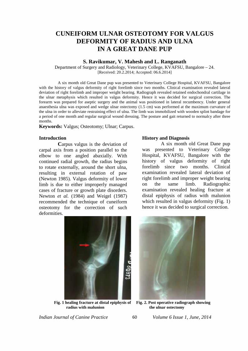
Indian Journal of Canine Practice Volume 6 Issue 1, June, 201460
CUNEIFORM ULNAR OSTEOTOMY FOR VALGUSDEFORMITY OF RADIUS AND ULNA
IN A GREAT DANE PUP
S. Ravikumar, V. Mahesh and L. RanganathDepartment of Surgery and Radiology, Veterinary College, KVAFSU, Bangalore – 24.
[Received: 20.2.2014; Accepted: 06.6.2014]
A six month old Great Dane pup was presented to Veterinary College Hospital, KVAFSU, Bangalorewith the history of valgus deformity of right forelimb since two months. Clinical examination revealed lateraldeviation of right forelimb and improper weight bearing. Radiograph revealed retained endochondral cartilage inthe ulnar metaphysis which resulted in valgus deformity. Hence it was decided for surgical correction. Theforearm was prepared for aseptic surgery and the animal was positioned in lateral recumbency. Under generalanaesthesia ulna was exposed and wedge ulnar ostectomy (1.5 cm) was performed at the maximum curvature ofthe ulna in order to alleviate restraining effect of ulna. The limb was immobilized with wooden splint bandage fora period of one month and regular surgical wound dressing. The posture and gait returned to normalcy after threemonths.Keywords: Valgus; Osteotomy; Ulnar; Carpus.
IntroductionCarpus valgus is the deviation of
carpal axis from a position parallel to theelbow to one angled abaxially. Withcontinued radial growth, the radius beginsto rotate externally, around the short ulna,resulting in external rotation of paw(Newton 1985). Valgus deformity of lowerlimb is due to either improperly managedcases of fracture or growth plate disorders.Newton et al. (1984) and Weigel (1987)recommended the technique of cuneiformosteotomy for the correction of suchdeformities.
History and DiagnosisA six month old Great Dane pup
was presented to Veterinary CollegeHospital, KVAFSU, Bangalore with thehistory of valgus deformity of rightforelimb since two months. Clinicalexamination revealed lateral deviation ofright forelimb and improper weight bearingon the same limb. Radiographicexamination revealed healing fracture atdistal epiphysis of radius with malunionwhich resulted in valgus deformity (Fig. 1)hence it was decided to surgical correction.
Fig. 1 healing fracture at distal epiphysis of Fig. 2. Post operative radiograph showingradius with malunion the ulnar ostectomy

Indian Journal of Canine Practice Volume 6 Issue 1, June, 201461
Surgical treatment The dog was premedicated with
Atropine Sulphate at the rate of 0.04 mgper kg body weight and Diazepam at thedose rate of 0.5 mg per kg body weight.Genaral anaesthesia was induced andmaintained with 2.5% thiopentone sodiumgiven to effect. The region from elbow tocarpal joint was prepared for asepticsurgery and the animal was positioned inlateral recumbency. Ulna was exposedthrough lateral approach by separating allthe underlying soft tissues. Ulnarostectomy (1.5 cm block) was performed atthe maximum curvature of the ulna in orderto alleviate restrainig effect of ulna.Autogenous fat graft has been successfullyused to prevent bony union following ulnarusteotomy. The incision was closed in aroutine manner. Post operatively AC - Veta
(Ampicillin + Cloxacillin) @ 500 mg IVgiven for seven days and Melonexb
(Meloxicam) @ 0.3 mg per kg SC, SID wasadministered for three days. Splint bandagewas applied and regular surgical wounddressing was done for ten days. Surgicalwound healed without any complications.The splint was maintained for about 45days. The posture and gait returned tonormalcy by about three months.
Discussion
Cuneiform (wedge) osteotomyprovided wide flat surfaces for bony unionand therefore significantly contributing tomore fixation stability (Rudy 1971 andEgger 1993). The surgical approachadapted to the ulna was as described byPiemattei and Greedy (1979).
ReferencesEgger, E. L (1993) fractures of radius and
ulna. In : Slatter, D. (ed.) Text book ofsmall animal surgery . vol. II. 2nd edn.,
W.B.Saunders, Philadelphia. P. 1736.Newton , C. D., Nunamaker, D. M. and
Prieur, W. D. (1984). Osteotomy. In:Manual of internal fixation in smallanimals (eds.) Brinker, W. O., Hohn,
R.B. and Prienr, W.D., Newyork,Springer, Verlag, p. 255.
Newton , C. D. (1985). Radial and Ulnarosteotomy. In: Textbook of smallanimal orthopaedics. J. B. Lippincott,p. 533-544.
Piemattei, O. C. and Greedy, R. G. (1979).An atlas of surgical approach to thebone of the dog and cat. 2nd edn., W. B.Saunders. p. 106.
Rudy, R. L. (1971). Vet. Clin. North.America, 1:549.
Weigel, J. P. (1987). Vet. Clin. North Am. :Small Anim. Pract., 17:905.
*****

Indian Journal of Canine Practice Volume 6 Issue 1, June, 201462
BILATERAL HYGROMA IN A GREAT DANE DOG AND ITSSURGICAL MANAGEMENT
I. Nath, Jasmeet Singh, Sidhartha Sanker Behera, Prasant Kumar Sikaand B.K Dwibedy
Department of Veterinary Surgery and Radiology, College of Veterinary Science and AnimalHusbandry, Odisha University of Agriculture and Technology. Bhubaneswar-751003, Odisha.
[Received: 24.2.2014; Accepted: 07.6.2014]
A male Great Dane dog was presented with history of bilateral swelling over the elbow region since 2months. Aspiration with sterile needle revealed it to be a case of hygroma and surgical treatment was plannedaccordingly with the hygroma over left limb to be operated firstly followed by other lesion after healing of firstsurgical wound. On 12th post-operative day, skin sutures were removed which revealed complete healing of thewound. A similar surgical procedure was carried out over the right elbow after 15 days of first surgery and thepatient made an uneventful recovery.Keywords: Hygroma, olecranon bursitis, surgical management.
IntroductionAn elbow hygroma (elbow seroma,
olecranon bursitis) is a fluid-filled cavitysurrounded by dense fibrous connectivetissue arising out of inflammation of anacquired subcutaneous bursa over theolecranon process of ulna (Fossum, 1997).The occurrence of this condition iscommonly reported in horses but rarely incattle (Venugopalan, 2004). It has beenreported to occur most commonly in youngdogs of large breeds before a protectivecallus forms over the bony prominence;however, they may occur in older animalswith neuromuscular disease (Fossum,2002). Repeated trauma to soft tissuesoverlying the bony prominence (olecranonprocess) causing subcutaneousaccumulation of transudative fluid thateventually becomes encapsulated byfibrous tissue lined with a synovialmembrane (Shappel and Little, 1992).Among the different dog breeds giantbreeds like Irish wolfhound, Great Daneand Old English Mastiff appear to beparticularly susceptible to this condition(White, 2003). The present communicationputs on record a successful surgicalmanagement of bilateral elbow hygroma ina Great Dane dog and its surgicalmanagement.
Case History and DiagnosisA nine months old male Great Dane
dog was presented with history of bilateralround swelling over elbow region (Fig.1). Itstarted as small sized swelling over bothelbows and increased gradually to size oftennis ball over a period of 60 days. Theconsultant Vet aspirated it with ahypodermic needle to relive off swellingbut it recurred again and it was referred forexpert opinion and surgical treatment.Palpation of the mass revealed it to be softand fluctuating with no evidence of pain.Clinical examination revealed normal rectaltemperature (102.20F), heart rate (84 beatsper minute) and respiratory rate (26 beatsper minute). Aseptic aspiration revealed atransparent fluid thereby confirming it to becase of hygroma and surgical correctionwas planned accordingly.
Surgical ProcedureThe animal was premedicated with
atropine @ 0.04 mg/kg body wt. SCfollowed 10 minutes later by xylazine @ 1mg/ kg IM respectively. Generalanaesthesia was induced by administrationof ketamine @ 7.5 mg / kg body wt. IV andmaintained by incremental doses ofketamine as and when needed during thesurgical procedure. The dog was restrainedin right lateral recumbency and aseptic

Indian Journal of Canine Practice Volume 6 Issue 1, June, 201463
preparation over the left elbow region wasdone in routine manner. An ellipticalincision was made along the posterolateralaspect of point of elbow and over theswelling. The bursa was gently separatedfrom its soft tissue attachment by bluntdissection taking care not to rupture thebursa or injuring the joint capsule (Fig.2and 3). The excess skin was trimmed offand the subcutaneous tissue was closed bysimple continuous sutures using chromiccatgut no.1 to obliterate the dead spacefollowed by skin closure with horizontalmattress suture using braided silk no. 1(Fig.4). The limb was bandaged properlyfrom elbow region to paw region to restrictmovement and prevent post operativeswelling of the affected limb (Fig.5). Post-
operatively broad antibiotic ceftriaxone @10 mg/ kg body weight for 5 days andanalgesic meloxicam @ 0.2mg/ kg bodyweight for 3 days were administeredintramuscularly. The wound was dressed ondaily basis with povidone iodine solutionand bandaged properly. The owner wasadvised to keep the animal with restrictedmovement to prevent wound dehiscence.Skin sutures were removed on 12th post-operative day and the wound healedcompletely without any complications.
A similar surgical procedure wascarried out over the other limb after 15 daysof first surgery (Fig.6) with similar post-operative treatment and management whichled to uneventful recovery after 14 days ofsurgical procedure.
Fig.1. Bilateral hygroma at elbow region Fig.2. After excision of the left mass
Fig.3. Excised bursa from the left side elbow Fig.4. Closing of the wound
Fig.5. Bandaging of the limb Fig.6. Surgical preparation of right one after healing of left

Indian Journal of Canine Practice Volume 6 Issue 1, June, 201464
Results and DiscussionOlecranon bursitis is characterized by
a movable swelling over the olecranontuberosity and the fore limbs. A commoncause of bursitis is direct trauma whichleads to acute bursitis which swellingcaused by bursitis can be of different sizeand can affect one or both fore limbs(Honnas et al., 1995; Fossum, 1997). Thepresent clinical case was a bilateralcondition affecting both appears suddenlywhile chronic bursitis occurs when thetrauma is mild but repeated in nature(O’Connors, 2005). Chronic bursitis ischaracterized by accumulation of excessivebursal fluid, thickening of bursal wall,extrusions of fibrous bands or septa withinthe bursal cavity and generalizedsubcutaneous thickening (Fathy and Radad,2006). The hygroma in present case waschronic in nature which might be due toconstant irritation on the area of theolecranon owing to its heavy body weight.
Various treatment protocols have beenformulated for management of bursitis inanimal with the acute bursitis being treatedby aspiration of the serous fluid andadministration of hydrocortisone into thebursa to suppress inflammation. However,chronic bursitis can be treated byapplication of counter-irritants or incisionof the bursa with application of an irritantto its interior lining. Aspiration of thecontents and injection of an irritant liketincture iodine or 3-5% carbolic acid leadsto destruction of the bursal lining followedby granulation, cicatrization andobliteration of the cavity (Venugopal,2004).
Surgical treatment is considered to bean effective for treatment of olecranonbursitis particularly for the chronicproliferative and fibrous form (Fathy andRadad, 2006). Rapid and economic healingis also achieved by surgical interventionthan conservative treatment (Arican et al.,2005; Hayat et al., 2009) and it has alsobeen advocated when all other methods oftreatment have failed and the bursa is large
and composed of primarily fibrous tissue(Honnas et al., 1995). In the presentclinical situation, surgical correction wasconsidered because of chronic nature of thelesion. Similarly, two step surgicalprocedure was planned becauseimmobilization of both limbssimultaneously might create problem innormal movement of the patient whichmight increase the chances of wounddehiscence.
ReferencesArican, M., Kocabiyik, A. and Izci, C.
(2005). Treatment of bilateral olecranonbursitis in a horse. Indian Vet. J.,82:325.
Fathy, A. and Radad, K. (2006). Surgicaltreatment and histopathology ofdifferent forms of olecranon andpresternal bursitis in cattle and buffalo.J. Vet. Sci., 7: 287-291.
Fossum, T. W. (1997). Small AnimalSurgery. 2nd Edn. Mosby Publication.Missouri. pp 137-138.
Hayat, A., Cengiz Han, M., Sagliyan, A.and Biricik, H.S. (2009). Differenttreatment of olecranon bursitis in sixhorses. J. Anim. Vet. Adv., 8:1032-1034.
Honnas, C.M., Schumacher, J., McClure,S.R., Crabill, M.R., Carter, G.K.,Schmitz, D.G. and Hoffman, A.G.(1995). Treatment of olecranon bursitisin horses: 10 cases (1986-1993). J. Am.Vet. Med. Assoc., 206: 1022-1026.
O’ Connors, J.J. (2005). Dollar’sVeterinary Surgery. 4th Edn., Tindal andCox, London.
Shappel, K.K. and Little, C.B. (1992). Spec-ial surgical procedure for equine Skin,In: Equine Surgery. Auer, J.A. (Eds.),W.B. Saunders, Philadelphia. pp 272-284.
Venugopalan, A. (2004). Essentials ofVeterinary Surgery. 4th Edn. Oxford &IBH Publishing. New Delhi.
White, R.A.S. (2003). Surgical treatment ofspecific skin disorders. In: Text Book ofSmall Animal Surgery. Slatter, D., (Ed).3rd Edn. W.B. Saunders, Philadelphia,pp 339-355.

Indian Journal of Canine Practice Volume 6 Issue 1, June, 201465
MASSIVELY ENLARGED MAMMARY TUMOUR IN A DOGAND ITS SURGICAL MANAGEMENT
I. Nath, Jasmeet Singh, S.S. Behera, Samir Kumar Das, Sujit Prasad Das andSripati Sethi
Department of Veterinary Surgery and Radiology, College of Veterinary Science and AnimalHusbandry, Odisha University of Agriculture and Technology. Bhubaneswar-751003, Odisha.
[Received: 24.2.2014; Accepted: 04.6.2014]
A 9 years old, female German shepherd dog was presented with history of an unusual oval and flatgrowth in the mid ventral region of the body. The growth started as a small nodule and gradually increased to alarge size over 6 month period. On clinical examination, it was diagnosed as a case of mammary tumour andsurgical excision was planned. Under general anaesthesia using atropine-xylazine-ketamine a circular skinincision was given and the mass was separated from underlying tissues by blunt dissection. Bleeding was checkedby application of haemostatic forceps and ligatures wherever needed. The operative wound was closed in standardmanner. Post-operatively antibiotics for 5 days and analgesics for 3 days were administered. On 10th post-operative day sutures were removed and the recovery was uneventful. Histopathologically it was diagnosed as amixed benign adenoma.Keywords: German shepherd; mammary tumour; surgical management.
IntroductionCanine mammary gland tumors
(CMGTs) are one of the most commonneoplasms of bitches (Vail and Ewen,2000). Mammary gland tumours are mostfrequently found in bitches aged 5 yearsand above (Murphy, 2008). Dachshunds,cocker spaniels, toy poodles, Germanshepherds, mixed–breed dogs have beenreported to have an increased incidence ofmammary neoplasia (Rutteman, 1990).Mammary gland tumours can occuranywhere along the mammary chain incanine, mainly in females, and are thesecond most common group of neoplasmsin dogs, following skin tumours (Moulton,1999). The caudal abdominal and inguinalmammary glands are affected with higherfrequency than thoracic glands (Cassali etal., 2009). The present paper describessuccessful surgical management ofmammary tumour in a dog.
Case History and ObservationA 9 years old, female German
shepherd dog weighing about 30 kg bodyweight was presented with history of anunusual growth in the mid ventral region ofthe body. The growth started as a smallnodule and gradually increased to a large
size over 6 month period. The growth wasoval and flat in gross appearance and theleft 1st and 2nd thoracic teats were affected(Fig.1). Radiograhic examination of chestrevealed clear lung fields without anynodules or tumors. Tentatively it wasdiagnosed as a case of mammary tumourand surgical excision was planned.
Surgical TreatmentThe animal was premedicated
with atropine sulphate @ 0.04mg/kgfollowed 10 minutes later by xylazine @1mg/kg body weight intramuscularly. Theinduction of anaesthesia was carried outwith ketamine @ 10mg/kg intravenouslyfollowed by administration of incrementaldoses as and when needed during surgery.The animal was restrained in lateralrecumbency keeping the tumour massoutward. The site was shaved, cleaned andprepared aseptically for surgery. A circularskin incision was given around the massand the mass was separated fromunderlying tissues by blunt dissection.Bleeding was checked by application ofhaemostatic forceps and ligatures whereverneeded. The subcutaneous tissues wereapposed in continuous pattern using catgutno. 2 for obliteration of dead space (Fig.2).

Indian Journal of Canine Practice Volume 6 Issue 1, June, 201466
Fig.1. Oval and flat mammary tumour Fig.2. Obliteration of dead space after removalof mammary tumour
Fig.3. Skin suturing Fig.4. Excised tumour mass
The skin wound was closed by verticalmattress using silk no. 2 (Fig.3). Post-operatively antibiotic Ceftriaxone @ 20mg/kg body weight twice daily for 5 daysand analgesic Meloxicam @ 0.2 mg/ kgbody weight once daily for 3 days wereadministered. Wound dressing was done onalternate days with povidine iodine. Theowner was advised to muzzle the dog andcover the abdomen by clean cotton cloth toavoid licking and scratching. On 10th post-operative day sutures were removed and therecovery was uneventful.
Results and DiscussionAnamnesis and physical
examination coupled with epidemiologicalfindings have an important role in thediagnosis and prognosis of mammarytumours. Radiographic screening, surgicalbiopsy or aspiration biopsy are beneficialtools for early diagnosis of tumours. (Baker
and Lumsden, 2000). The present case waspresented late and the diagnosis was madeby its location and physical examination.The excised mass was 7.5 X 6 inches indimension and weighed 620 gm. On grosssection, there were numerous cystic lesionswith multiple cauliflowers like masseswhich were hard in consistency (Fig.4, 5).Histologically it was diagnosed as a mixedbenign adenoma with solid lobules ofhyperplastic, well differentiated luminaland myoepitheleal cells embedded inextremely fibrous stroma.Increased incidence of mammary tumourswas found in many large as well as smallerbreeds of dogs. The lowest occurrence wasfound in mongrels, Boxers and Chihuahuas(Brodey et al., 1983). Spayed dogs had a 3to 7 times lower incidence of mammarytumours than the intact ones (Alenza et al.,2000). The effect of spaying on thedecreased incidence of mammary tumour,

Indian Journal of Canine Practice Volume 6 Issue 1, June, 201467
however, depended on the age of the dog atthe time of surgery. The most significantdecrease of incidence was found in femalesspayed prior to their first oestrus (0.5%incidence as compared to intact femaledogs) and between the first and secondoestrus (8%). Advanced age at the time ofspaying increases the incidence of tumors(26% between the second and fourthoestrus) and no positive effect of spaying
was found after the fourth oestrus(Misdorp, 1988). A higher incidence ofmammary tumours is found in dogsreceiving a “home-made” diet and “redmeat” compared to commercial diets.Obesity in young animals (up to 1 years ofage) is another risk factor for developingtumours even at a later age (Alenza et al.,2000). Here as the dog was already old andspaying was not done.
Fig.5. Showing cystic and cauliflower like growths
Surgery remains the mainstay of treatmentfor dogs and cats with most type ofmammary gland tumors. The exceptions areinoperable disease (e.g., highly malignantinflammatory carcinoma of the dogs thataccount for less than 5% of mammarytumours) and distant organ metastasis(Tavasoly et al., 2013). Approximately50% of all mammary tumours aremalignant and almost half of them havespread at the time of diagnosis.Chemotherapy may be recommended,when surgery alone is unlikely to besuccessful, e.g. for tumours that cannot beremoved surgically, tumours that havemetastasized, and tumours that have a highprobability of spreading. Here a case ofbenign mammary tumour involving theanterior glands of a GSD was put onrecord. The dog was followed for next 6months and there was no recurrence ormetastasis.
References:
Alenza, D.P., Pena, L., Del Castillo, N.,Nieto, A.I. (2000). Factors influencingthe incidence and prognosis of caninemammary tumours. J Small AnimPract. 41: 287-291.
Baker, R. and Lumsden, J.H. (2000). Themammary gland. In Color Atlas ofCytology of the Dog and Cat. Editedby Baker R, Lumsden JH. St Louis:Mosby: 253–262.
Brodey, R.S., Goldschmidt, M.H. andRoszel, J.R. (1983). Canine mammarygland neoplasms. J Am Anim HospAssoc. 19: 61-90
Cassali, G.D., Bertagnolli, A.C., Lavalle,G.E., Tavares, W.L.F., Ferreira, E.,Silva, A.E., Campos, C.B. (2009).Perpectives for diagnosis, prognosisand treatment of mammary neoplasmsin dogs. 34th World Small AnimalVeterinary Congress - WSAVA 2009.
Misdorp, W. (1988). Canine mammarytumours: Protective effect of lateovariectomy and stimulating effect of

Indian Journal of Canine Practice Volume 6 Issue 1, June, 201468
progestins. Vet Q. 10: 26-33.Moulton, J.E. (1999). Tumours in Domestic
Animals. 3rd edn, University ofCalifornia Press, Berkley, pp. 518-543.
Murphy, S. (2008). Mammary tumors indogs and cats. In Practice; 30(6):334-339.
Rutteman, G.R. (1990). Hormones andmammary tumour disease in the femaledog: an update. In Vivo, 4(1):433-440.
Tavasoly, A., Golshahi, H., Rezaie, A. andFarhadi, M. (2013). Classification andgrading of canine malignant mammarytumors. Veterinary Research Forum.2013; 4 (1) 25 – 30.
Vail, D.M., Mac Ewen, E.G. (2000).Spontaneously occurring tumors ofcompanion animals as models forhuman cancer. Cancer Invest;18(8):781-792.
*****

Indian Journal of Canine Practice Volume 6 Issue 1, June, 201469
MEGAESOPHAGUS AND ITS MANAGEMENT IN THREEDIFFERENT LARGE BREED ADULT BITCHES
A.K. Maji and Arnab Kumar MajieDeptt. of Veterinary Surgery & Radiology, West Bengal University of Animal & Fishery Sciences,
37, K.B. Sarani, Kolkata-700037.[Received: 24.2.2014; Accepted: 07.6.2014]
Acquired Secondary Megaesophagusin adult dogs can result from numerousneuromuscular, endocrine or inflammatorydisorders as well as from obstructivelessions (Strornbeck and Guilford, 1990;Twedt, 1995), Gastroesophageal sphinter(GES) achalasia during secondaryperistalsis (Boria et al., 2003), hyper andhypoadrenocorticism, esophagealdismotility, paraesophageal hiatal hernia,delayed transit disorders or hypomotility,hypertrophic osteopathy, tigersnakeenvenomation, dysantonomia, acquiredmyasthenia gravis and canine distemper(Mossallanejad et al., 2010). This reportputs on record the successful surgical andnon-surgical management ofmegaesophagus in three adult large breedbitches.
Case History and ObservationsThree bitches – one Labrador
Retriever, 4 years of age, 24 kg body wt.,whelped once, one GSD, 5 years of age, 17kg of body wt., whelped 5 pups once andone Golden Retriever, 11 years of age,respectively were presented to theUniversity Clinics with a history of chronicregurgitation having no prodormal phase ofvomition, dysphasia, repeated swallowingattempts, ptyalism, halitosis and weremaintained by life-saving fluid infusion andsupportive therapy since last 15 days to 1.5months. Regurgitated materials were notbile-stained . All three bitches wererefractory to medicinal treatments done byreferring veterinarians. On physicalexamination the bitches were 2-3 out of 5body condition score and cachectic butbright, alert and responsive with normalrectal temperature, heart and respiratory
rate without any respiratory tract problemas all the cases were receiving courses ofantibiotic therapy. The average result ofbiochemical parameters like blood glucosefasting (78 mg/dl), Serum bilirubin (total0.56 mg/dl, conjugated – 0.20 mg/dl,Unconjugated 0.36 mg/dl), Protein (total –6.3 gm/dl, albumin – 2.5 gm/dl, Globulin –3.8 gm/dl), Alkaline phosphatase – (146nµ/ml), SGPT (24 µ/l), SGOT (36 µ/l),BUN – (11mg/dl) and Serum Creatinine(0.84 mg/dl) were within normal rangesincluding T4(21.0 n mol/l), TSH (0.09ng/ml) and Na+ : K+ (140/5 or 28). The 1st
bitch was examined through bariumesophagogram after administration of 50mlof microbar (Barium Sulphate Paste) and30 ml of distilled water under 4 ml of I/Vdiazepam. The second bitch received lesserdose for this examination. In both the casesdilated thorasic esophagus with stasis ofbarium sulphate suspension and normalthickness of GES with failure to open fortransport were noticed (Fig.1 & Fig.2). In3rd bitch dilated esophagus was sufficientfor diagnosis of megaesophagus in digitalsurvey radiography (Fig.3). X-ray upto 2nd
day post-barium administration revealed noother abnormality of G.I. tract. Bylaboratory findings, radiography andphysical examination hypothyroid-relatedneuropathy, hypoadrenocorticism ormyasthesia gravis or presence ofobstructive lesions were eliminated and
Treatment and DiscussionLower sphincter achalasia was
treated surgically in first case (Fig.4).Distal esophagomyotomy (ModifiedHeller’s method) was performed throughmidventral incision at supine position under

Indian Journal of Canine Practice Volume 6 Issue 1, June, 201470
general anaesthesia. The abdominalincision was from umbilicus to xiphoidprocess. The stomach was retracted byBabcock forceps. The distal part of theesophagus, the lower esophageal sphincterregion and the cardia was visualized. Alongitudinal incision was made on ventralaspect of gastro esophageal junctionthrough tunica serosa and tunica muscularisextending from distal 2 cm of theesophagus to proximal 2 cm of stomach.The esophageal and gastric mucosa bulgedthrough the complete incision. Routineclosure and standard follow up were made(Fig.5). The owner of the 2nd and third
bitches afraid of the operation but all theowners strictly followed for elevatedfeeding at 45º on Baily chair and walking at90º on hind limb for 10 min. after food. Onthe basis of telephone message and visit ofthe local veterinarian quick result wasachieved with the first bitch as after twoyears it became 32 kg of body wt. from thatof 24 kg pre-operation and needs noelevated feeding or extra-care. One and halfyears after post-management the G.S.D.bitch also gained body wt. being 30 kgfrom emaciated pre-treatment 17kg as didwell the golden retriever withcomparatively prolonged elevated feeding.
Fig. 1 : Megaesophagus in the Labrador Retriever Bitch Fig. 2 : Megaesophagus in the GSD bitch
Fig.3: Digital X-ray depicting the megaesophagus Fig. 4: Contrast radiograph suggestive of GES achalasiaof the 3rd bitch (Golden Retriever)
Fig. 5 : Modified Heller’s esophagomyotomy in the 1st bitch

Indian Journal of Canine Practice Volume 6 Issue 1, June, 201471
Congenital idiopathic megaesophagusin pups were reported by Gahlot et al.(2003) and Punnusuamy et al. (2008).Cases of Acquired megaesophagus havebeen reported by Boria et al. (2003), AnilKumar et al (2009) and Massallanejad et al.(2010). Since 1970’s a lot of works hasbeen made in this field. Surgical attemptwas made here following Kipperman andStraw (1988) and Boria et al. (2003).Thediazepam was given to stop theregurgitation during contrast radiography.Unfortunately the definitive cause for thelast two dogs’s acquired megaesophaguswas not determined. Cricopharyngealachalasia (upper achalasia) is a congenitalcondition in dogs. From clinical signs, ageof onset, biochemical and radiographicevaluation and complete response toesophago-myotomy it can be concludedthat the first dog in this report had acquiredlower esophageal achalasia.
SummaryThree adult bitches-one Labrador
Retriever, one GSD and one GoldenRetriever with the history of chronicregurgitation were radiologically evaluatedfor Megaesophagus. From biochemicalparameters, clinical symptoms and historyit was diagnosed as Gastro esophagealSphinter achalesia. In first bitch distalesophagomyotomy( modified Heller’smethod) through midventral incision wasdone. All the bitches were kept on elevatedfeeding at 45 degree on Baily Chair andwalking at 90 degree on hind limb for 10
minutes. All did well up to one and halfyears post operation.
Reference :Anil Kumar, M.C., Kamran, C.A. and Bhat,
M.N., 2009. Megaesophagus in a GreatDane dog and its management – A casereport, Intas polivet. 10(1):80-81.
Boria, P.A., Webster, R.L.C. and Berg, J.2003. Esophageal achalasia andsecondary megaesophagus in a dog.Can. Vet .J. 44(3): 232-234.
Gahlot, T.K., Ahuja, A, Purohit, S. andKumar, A. 2003. Megaesophagus in apup – a case report. J. Canine Develop.Res.3 : 55-56.
Kipperman, B.S. and Straw, R.C. 1988. Sur-gical management of megaesophagus
in a dog.Modern Vet.Practice, 69:8-11.Mossallanejad, B., Ghadiri, A. and Avizeh,
R. 2010. Iranian Jr. Vet. Res. ShirazUniversity. Vol.11, No.4 Ser.No. 33:379- 382
Punnuswamy, K.K., Jeyaraja, K., Unny,N.M., Napolean, R.E. and Subramania-n, M. 2008. Congenital Idiopathicmegnesophagus in a daschund puppy.Indian Vet. J. 85: 989 – 991.
Strombeck, D.R. and Guilford, W.G. 1990. In Strombeck’s Small Animal Gastroen
-terology. Guilford, W.G. Center, S.A.Strombeck, D.R.etal.Edn. 3rd Edn. WBSaunders Co., Philadelphia pp.1129.
Twedt, D.C. 1995. Diseases of the esophag-us. In Textbook of Veterinary InternalMedicine. Ettinger, S.J. and FeldmanE.C. Edn.4th , WB Saunders Co.Philadelphia, pp.1124-1142.
*****

Indian Journal of Canine Practice Volume 6 Issue 1, June, 201472
URETHROCYSTOSCOPIC DIAGNOSIS AND THERAPEUTICMANAGEMENT OF URINARY TRACT DISORDER IN
FEMALE DOGS
V.P. Chandrapuria1, Dinesh Gupta2, Apra Shahi3, Dharmendra Kumar4 and Somil Rai5
1Professor & Head, 2PG Scholar, 3Associate Professor, 4Assistant Professor, 5PhD ScholarDepartment of Surgery and Radiology, College of Veterinary Science and A.H., NDVSU, Jabalpur.
[Received: 27.2.2014; Accepted: 07.6.2014]
The lower urinary tract disorders are common in small animal practice with multiple factors whichcontribute to complicate recurrent urinary infection. The modern diagnostic imaging urethro-cystoscopy hasproven to be a highly effective, minimally invasive technique for diagnosis of lower urinary tract diseases in dogsand cats. In many aspects, urethro-cystoscopy provides enhanced examination of the urinary system compared toother diagnostic procedures such as ultrasonography. Urethro-cystoscopy allows direct visualization of thevestibule, vagina, urethral opening, urethra, urinary bladder and ureteral openings. The present research work wascarried out in eighteen clinical cases of adult female dogs with urinary disorders.
IntroductionUrethro-cystoscopy allows direct
visualization of the vestibule, vagina,urethral opening, urethra, urinary bladderand ureteral openings. The chronicinflammation can be defined with itsextent. Apart, it also visualizes lesions oflower urinary tract for assessment ofrecurrent urinary tract infections, chroniccystitis, tumours of lower urinary tract andhematuria. Biopsy could be obtained forhistopathological and mucosal culturestudy. The calculi can be collected foranalysis of its composition. It is theprocedure of choice for assessment ofurinary trauma (Adams et al., 2008 andDefarges and Dunn, 2008). It can also beused for therapeutic purpose. Operativeindications include performing and judgingcoaptation during periurethral injections,assess elevation of urethro-vaginal junctionduring needle urethropexy and suburethralsling procedures, facilitate surgical repairof urinary tract fistula and urethraldiverticulum and evaluate the ureters andbladder mucosa for inadvertent damageduring surgery (Cundiff and Bent, 1996).
Material and methodThe present research work was
carried out in the Department of VeterinarySurgery and Radiology, TeachingVeterinary Clinical Complex (TVCC),
College of Veterinary Science and AnimalHusbandry, Nanaji Deshmukh VeterinaryScience University, Jabalpur, MadhyaPradesh.
Eighteen clinical cases of adultfemale dogs were taken for the studieswhich were divided into two groups. GroupI (control) comprised of 6 apparentlyhealthy adult animals irrespective of ageand breed and Group II - Comprised of 12adult animals with lower urinary tractaffections. Animals of both the groups weresubjected to ultrasonographic examinationbefore treatment (0th day) and group IIwere also examined on 10th and 20th daypost treatment. Animal was restrained indorsal recumbancy and site preparedaseptically. For ultrasonographicexamination of urinary bladder and urethraanimal was restrained in dorsal recumbencyand Transducer was placed at the midlinecranial to the pubis and parallel to the longaxis. Ultrasonographic examination wasdone by using real time B mode, gray scaledual frequency (3.7/5 M Hz) ultrasoundmachine of Famio 5 (SSA 510 A, ToshibaJapan). Coupling media was applied on theskin surface. The urinary bladder wasscanned for its shape, anatomical position,contents, bladder wall thickness and otherabnormalities.
Animals of both the groups weresubjected to urethro-cystoscopic

Indian Journal of Canine Practice Volume 6 Issue 1, June, 201473
examination before treatment (0th day) andGroup - II were also examined on 10th and20th day post-treatment. The animals werepre-medicated with Atropine Sulphate @0.04 mg/kg b. wt., I.M. and after fiveminutes administration of XylazineHydrochloride @ 1-2 mg/kg b. wt., I.M.and then after ten minutes administration ofKetamine @ 5-7 mg/kg body weight, I.V.Animal was restrained in ventro-dorsal orlateral position followed by insertion of 2.7mm diameter, 18 cm long and 30° anglerigid endoscope enclosed in a sheaththrough urethra along with the infusion ofnormal saline through working channel ofendoscope. The lesions in the urethra andmucosal layer of bladder was recorded andinterpreted.
Cases were treated medicinally and/or surgically as per the clinical conditiondiagnosed. The response of the therapy wasevaluated by ultrasonography and urethro-cystoscopy on 10th and 20th post-treatmentday.
Results and discussionCystitisUltrasonography
In group II (a) at day '0' (pre-treatment) Ultrasonography of the urinarybladder revealed slightly thick bladder walland several hyperechoic fine crystals in theurinary bladder, which represents bladdersludge.
Nyland and Mattoon (1995),Leveille (1998), Kundu and Ghosh (2006)and Sutherland (2008) detected cystitisultrasonographically and found that bladderwall thickness was more cranio-ventrally,became generalized in severe cases ofcystitis. Green (1996) reported presence ofblood, pus and cellular debris in the urine,which visualized as small echoic inanechoic urine. In several cases bladderwall became thick, hyperechoic andirregular. The findings were the result ofcystitis in animals of group II (a). Durmasand Han (2005) also reported that changes
in bladder echogenesity depend upon theconcentration of urine.On 10th post-treatment dayultrasonographic study of the urinarybladder revealed reduced thickening of thebladder wall in all the cases andhyperechoic bladder sludge and cellulardebris reduced when compared to day 0.
On 20thpost-treatment dayultrasonographic detail of the urinarybladder showed almost normal thickeningof the bladder wall in all the cases. Thebladder sludge and cellular debris wasabsent. The mucosa of urinary bladder wasalso smooth and appeared like normalurinary bladder. These findings support theresponse of treatment given for cystitis.
Urethro-cystoscopyIn group II (a) on 0th day urethro-
cystoscopic examination of urinary bladdershowed mucosal hyperemia, corrugation inmucosa and sloughing of bladderepithelium. Hyperemia and erythema arethe usual features of chronic cystitis, butthe urothelium also had general, ragged,fimbriated appearance. Hyperemia inbladder mucosa may be due toinflammation and corrugation of mucosaoccurs due to increased chronicity whichcauses increased mucosal opacity anddecreased elasticity when these changesoccurs, the bladder mucosa becomecorrugated. In present cases there might bechronic cystitis. Such findings were alsoreported by McCarthy (2005), Henderson(2007) and Moore and Ragni (2012).
On 10thpost-treatment day hyperemiaof bladder mucosa and epithelial cast werepresent in the urinary bladder and on 20th
day bladder mucosa was observed almostnormal and corrugation on bladder mucosaand epithelial cast were absent. Thesefindings showed that the treatment given inthe present study was effective in cystitis ofanimals of the present study.
Fimbriated growth in the urethraUltrasonography

Indian Journal of Canine Practice Volume 6 Issue 1, June, 201474
In group II (b) on 0th day (pre-treatment) ultrasonographic view of theurinary bladder revealed anechoic structuresurrounded by a thin hyperechoic wall withnormal thickness. No abnormal pathologywas observed in bladder on sonography. Onsonography neck of urinary bladder andproximal urethra could be visualized butdistal urethra remains unassessable. Thesefindings are similar to Leveille (1998).
The ultrasonographic findingsremained same on 10thand 20th post-treatment day of this group.
Urethro-cystoscopyTransurethral cystoscopic study on
0thday (pre-treatment) revealedgranulomatous (proliferative) lesions inurethra, it showed fimbriation similar asdescribed for the urethral transitional cellcarcinoma. The lumen of the urethra waspartially obliterated with finger likeprojection of the growth. Such papillaryfingers are described as urethral transitioncell carcinoma. In present case there mightbe initiation of the carcinoma in urethra.Similar findings were also reported byMcCarthy (2005), Henderson (2007), Iqballet al. (2011) and Moore and Ragni (2012).
The urethro-cystoscopic findingsremained same on 10thand 20th post-treatment day of this group. Because ownerof dog was not convinced for surgicaltreatment or anticancerous therapy, henceon request simple treatment of cystitis wasinitiated for symptomatic relief.
Polyp in urinary bladderUltrasonography
In group II (c) at day '0' (pre-treatment) ultrasonography of the urinarybladder revealed a hyperechoic structureattached with the dorsal aspect of thebladder near the neck of urinary bladderand urethra. It was measuring 7.5 mm. Thishyperechoic masses did not form anyacoustic shadow and attached with bladderwall, suspected for polyp. The findingswere similar to Green (1996) and Olausson
et al. (2005). Owner of dog was notconvinced for surgical removal or anticancerous therapy so at 10thand 20th post-treatment day, ultrasonographic detail ofthe urinary bladder was found hyperechoicmass similar to shape and size ascompared to day '0' (pre-treatment).
Urethro-cystoscopyThe urethro-cystoscopic findings
revealed one club shaped mass appeareddark red in colour and seen lying across therelatively normal urothelium and wasindicative of polyp in urinary bladder.Polyp is a proliferative condition of thebladder that is usually induced by chronicirritation. Similar findings were describedby McCarthy (2005) and Moore and Ragni(2012) for polyp in bladder of canine.On 10th and 20thpost-treatment day urethro-cystoscopic detail of the urinary bladdershowed similar finding as compared to day'0' (pre-treatment). Because owner of dogwas not agreed with surgical removal oranticancerous therapy hence only medicinaltreatment was given for symptomatic relief.
Ulceration and petechial hemorrhage inurinary bladderUltrasonography
In group II (d) at day '0' (pre-treatment) ultrasonography of the urinarybladder showed large size hyperechoicirregular patch at the apex of bladder whichwas measuring approximately 17 mm. Themucosa of this area was not clear. Thebladder also contains several hyperechoiccrystals present in the lumen of bladder.Similar findings were also observed byGreen (1996).
On 10th post-treatment dayultrasonography of the urinary bladderrevealed ulceration of bladder wall whichwas reduced slightly as compared to day '0'(pre-treatment) and hyperechoic crystals inthe anechoic urine were also reducedslightly.On 20th post-treatment day ultrasonographyof the urinary bladder showed slight

Indian Journal of Canine Practice Volume 6 Issue 1, June, 201475
irregular mucosa of bladder wall,suggestive of erosion of bladder mucosa.
Urethro-cystoscopyIn group II (d) at day '0' (pre-
treatment), urethro-cystoscopic studyshowed swelling, roughening and petechialhemorrhages on bladder wall and urethralmucosa, which were the confirmatorydiagnostic signs of ulceration andhemorrhage on mucosal surface of bladder.It may be due to chronicity andinflammation of mucosa. Similar findings
were also observed by McCarthy (2005)and Moore and Ragni (2012).
On 10th post-treatment day urethro-cystoscopy of the urinary bladder revealedulceration of bladder wall which wasreduced in size as compared to 0thday (pre-treatment) and hyperechoic crystals in theanechoic urine were also seen slightly.
On 20th post-treatment day theurethro-cystoscopy showed slight irregularmucosa of bladder wall and erosion ofbladder mucosa which was the indication ofhealing. These findings reflect the positiveresponse of the treatment given.
Figure 1 -Ultrasonogram group II (d) showing ulceration Figure 2 -Ultrasonogram group II (e) showingin urinary bladder calculi in urinary bladder
Figure 3 -Uretherocystoscopy group II (d) showing Figure 4 -Uretherocystoscopy group II (e)corrugation and ulceration in urinary bladder showing calculi in urinary bladder
CystolithsUltrasonography
In group II (e)at day '0' (pre-treatment) ultrasonography of the urinarybladder revealed three hyperechoic areaswith distal acoustic shadow, visualized in
the lumen of urinary bladder, suggestive ofcalculi. These calculi were measuring 11.9,7.5 and 7.5 mm in diameter. Feeny andJohnson (1986) stated that variation ofechogenesity and amount of acousticshadow, with the composition and

Indian Journal of Canine Practice Volume 6 Issue 1, June, 201476
compactness of calculi. At higherfrequency the calculi may be hyperechoic.Similarly Nyland and Mattoon (1995) alsoobserved changes in the images ofhyperechoic calculi with the position ofanimal and always tend to move todependent portion.Bumin and Temizsoylu (2000) found thatradiography and ultrasonography are veryhelpful for diagnosis of cystic calculi indogs.
Urethro-cystoscopyThe urethro-cystoscopy of urinary
bladder showed white, round or ovalmasses of varying size of bladdercystoliths, resting at apex of bladder.Spiculated surface was looking likecalcium ammonium phosphate calculus.Bladder wall revealed cicatricial pillarsalong with thickening because of chronicinflammation. This was the confirmatorydiagnosis of cystoliths. In the present studycalculi may be struvite calculus. Similarfindings were observed by McCarthy(2005) stated that cystic and urethral stonescan be easily seen with cystoscopy. Theirappearance varies widely depending theircomposition.Findings in the present study coincide withthe observations of McCarthy (2005) andHanderson (2007).
Therapeutic managementThe six animals of group II (a) were
treated medicinally with Nitrofurantoin,Oxybutynin Chloride, Ranitidine andSodium Acid Citrate. Retrograde flushingof bladder was also being done with amixture of normal saline, Povidone Iodineand Metronidazole for twice a week. Thecombination of Povidone Iodine andMetronidazole might have acted on aerobicand anaerobic bacteria, resulted to absenceof infection as shown animal of presentstudy.
Guardabassi et al. (2011) advisedNitrofurantoin for treatment of urinary tract
infection caused by multidrug-resistantbacteria.
Colgan and Williams (2011) andOlson and Haith (2012) reported thatNitrofurantoin is the first drug of choice fortreatment of acute uncomplicated cystitis inhuman being.On the basis of findings of present studyNitrofurantoin was found effective forcystitis in dogs.
De Castro et al. (1984) reportedOxybutynin Chloride as the first drug ofchoice for neurogenic bladder dysfunction.
Three animals of the treated groupwere not responding to treatment at 10th
post-treatment day thus the treatmentcontinued for next 10 days. Theexamination on day 20th revealeddisappearance of clinical signs, urineanalysis showed normal urine colourwithout sediments macroscopically,microscopically absence of erythrocytes,leukocytes and cast and multistripexamination showed normal pH andspecific gravity and negative for glucose,ketone bodies, bilirubin, urobilinogen,protein and nitrites. Ultrasonographicdetails of the urinary bladder showedalmost normal thickening of the bladderwall in all the cases. The mucosal borderwas smooth and a hypoechoic to isoechoicbladder sludge and cellular debris wereabsent. Urethro-cystoscopic study showedalmost normal bladder mucosa.Corrugation on bladder mucosa andepithelial cast were also absent.
In group II (b) and group II (c)owners of dogs weOn the basis of findingsof present study Nitrofurantoin was foundeffective for cystitis in dogs.De Castro et al. (1984) reportedOxybutynin Chloride as the first drug ofchoice for neurogenic bladder dysfunction.
Three animals of the treated groupwere not responding to treatment at 10th
post-treatment day thus the treatmentcontinued for next 10 days. Theexamination on day 20th revealeddisappearance of clinical signs, urine

Indian Journal of Canine Practice Volume 6 Issue 1, June, 201477
analysis showed normal urine colourwithout sediments macroscopically,microscopically absence of erythrocytes,leukocytes and cast and multistripexamination showed normal pH andspecific gravity and negative for glucose,ketone bodies, bilirubin, urobilinogen,protein and nitrites. Ultrasonographicdetails of the urinary bladder showedalmost normal thickening of the bladderwall in all the cases. The mucosal borderwas smooth and a hypoechoic to isoechoicbladder sludge and cellular debris wereabsent. Urethro-cystoscopic study showedalmost normal bladder mucosa.Corrugation on bladder mucosa andepithelial cast were also absent.
In group II (b) and group II (c)owners of dogs were not convinced for anysurgical treatment and anticanceroustherapy, hence on request simple treatmentof cystitis was initiated for symptomaticrelief. On 10th and 20thdays of observationthe fimbriated growth in urethra and polypin the urinary bladder were present same asseen in pre treatment day. Animals had gotsymptomatic relief it indicate the goodresponse of treatment.
The group II (d) was subjected toantibiotic Amoxicillin+ Sulbactum,Ranitidine and Sucralfate. The animalshowed symptomatic relief while theultrasonographic and urethro-cystoscopicexamination revealed decrease in thediameter of ulcer and none of the petechialhemorrhage was noticed on 10th posttreatment day and looking to the responseof treatment for next ten days.
On 20th post treatment day clinicalsigns were disappeared and urineexamination found normal urine colourwithout sediments macroscopically.Absence of erythrocytes, leukocytes andcast microscopically and multistripexamination showed normal pH andspecific gravity and absence of glucose,ketone bodies, bilirubin and urobilinogen,protein and nitrites. Ultrasonographic andurethro-cystoscopic examinations revealed
only irregular mucosa of bladder wall anderosion of bladder mucosa. These findingsobserved good response of treatment. Thehealing of ulcer occurs due to effect ofantiulcer drug which produce an ulceradherent complex with proteinaceousexudates, inhibiting the pepsin activity.Similar findigs reported by Papich (1993)in canine.
The group II (e) was treated withInj. DNS, Inj. Amoxicillin+ Sulbactum,Ranitidine and Sucralfate, Neeri andCystone. Being animal died on 8th post-treatment day so the response of treatmentremains obscured.
Singh et al. (2013) found that use offluid, antibiotics, analgesics, anti-inflammatory drugs, B-Complex, oralurinary antiseptics i.e. Cystone tablets aftersurgery were found to be useful for controlof recurrence of calculus in urinary systemin canines.
ReferencesAdams, L., Berent, A. and Moore, A.
(2008). Laser lithotripsy for theremoval of uroliths in 73 dogs.Journal of the American VeterinaryMedical Association, 232:1026 -1034.
Bumin, A. and Temizsoylu, D. (2000).Radiographic and ultrasonographic diagnosis and surgical removal of cysticcalculi. Ankara UniversitesiVeterinerFakultesi Dergisi, 47: 213-221.
Colgan, R. and Williams, M. (2011).Diagnosis and treatment of acuteuncomplicated cystitis. AmericanFamily Physician, 84: 771-776.
Cundiff, G.W. and Bent, A.E. (1996). Thecontribution of urethrocystoscopy toevaluation of lower urinary tractdysfunction in women. InternationalUrogynecology Journal 7: 307-311.
De Castro, R., Casolari, E. and Ricci, S.(1984). Combination of oxybutyninchloride (Ditropan) with intermittentcatheterization in the treatment ofneurogenic bladder in childhood:results on continence. La Pediatria

Indian Journal of Canine Practice Volume 6 Issue 1, June, 201478
medica e chirurgica, 6:795-803.Defarges, A. and Dunn, M. (2008). Use of
electrohydraulic lithotripsy in 28 dogswith bladder and urethral calculi.Journal of Veterinary InternalMedicine, 22: 1267-1273.
Durmus, A.S. and Han, M.C. (2005).Sonography of renal and urinarybladder in horses. Indian VeterinaryJournal, 82: 784-785.
Feeney, D.A., and Johnston, G.R. (1986).The kidney and ureters. In: Thrall D.E.(ed.) Text Book of VeterinaryDiagnostic Radiology. W. B. SaundersPublishing Co., Philadelphia, 408-423.
Green, R.W. (1996). Small AnimalUltrasound. 2nd Edn., Lippincott R.William and Wilkins Publishing Co.,Philadelphia, 367 p.
Guardabassi, L., Maaland and Marit.(2011). In vitro antimicrobial activity ofnitrofurantoin against Escherichia coliand Staphylococcus pseudintermediusisolated from dogs and cats. Publishingand the Ecology of European ResearchElsevier.
Henderson, A. (2007). Advances in veteran-ary Cystoscopy highlight: collageninjection for urinary incontinence.Indian Pet Journal, 12: 20-22.
Iqball, A., Wazir, V.S., Tripathi, A.K. andSingh, R. (2011). Cystoscopy - Apromising technique in canines. IndianPet Journal, 12: 20-22.
Kundu, P. and Ghosh, D. (2006).Ultrasono-graphic study of urinary bladderdiseases in dogs. Indian Journal ofVeterinary Surgery, 27: 33-34.
Leveille, R. (1998). Ultrasonography ofurinary bladder disorders. VeterinaryClinics of North America: Small AnimalPractice, 28: 799-821.
McCarthy, T.C. (2005). Veterinary endosc-opy for the small animal practitioner. 1st
Edn., Elsevier Saunders Publishing Co.,Elsevier (U.S.A.).
Moore, A.H. and Ragni, R.A. (2012).Clinical manual of small animalendosurgery. 1st Edn., Blackwell
Publishing Co., Ames U.S.A., pp 209-230.
Nyland, T.G. and Mattoon, J.S. (1995).Small Animal Diagnostic Ultrasound.2nd Edn., W. B. Saunders PublishingCo., Philadelphia, 357 p.
Defarges, A. and Dunn, M. (2008). Use ofelectrohydraulic lithotripsy in 28 dogswith bladder and urethral calculi.Journal of Veterinary InternalMedicine, 22: 1267-1273.
Durmus, A.S. and Han, M.C. (2005).Sonography of renal and urinarybladder in horses. Indian VeterinaryJournal, 82: 784-785.
Feeney, D.A., and Johnston, G.R. (1986).The kidney and ureters. In: Thrall D.E.(ed.) Text Book of VeterinaryDiagnostic Radiology. W. B. SaundersPublishing Co., Philadelphia, 408-423.
Green, R.W. (1996). Small AnimalUltrasound. 2nd Edn., Lippincott R.William and Wilkins Publishing Co.,Philadelphia, 367 p.
Guardabassi, L., Maaland and Marit.(2011). In vitro antimicrobial activity ofnitrofurantoin against Escherichia coliand Staphylococcus pseudintermediusisolated from dogs and cats. Publishingand the Ecology of European ResearchElsevier.
Henderson, A. (2007). Advances in veterin-ary Cystoscopy highlight: collageninjection for urinary incontinence.Indian Pet Journal, 12: 20-22.
Iqball, A., Wazir, V.S., Tripathi, A.K. andSingh, R. (2011). Cystoscopy - Apromising technique in canines. IndianPet Journal, 12: 20-22.
Kundu, P. and Ghosh, D. (2006). Ultrason-nographic study of urinary bladderdiseases in dogs. Indian Journal ofVeterinary Surgery, 27: 33-34.
Leveille, R. (1998). Ultrasonography ofurinary bladder disorders. VeterinaryClinics of North America: Small AnimalPractice, 28: 799-821.
McCarthy, T.C. (2005). Veterinaryendoscopy for the small animal

Indian Journal of Canine Practice Volume 6 Issue 1, June, 201479
practitioner. 1st Edn., Elsevier SaundersPublishing Co., Elsevier (U.S.A.).
Moore, A.H. and Ragni, R.A. (2012).Clinical manual of small animal endosu-rgery. 1st Edn., Blackwell PublishingCo., Ames U.S.A., pp 209-230.
Nyland, T.G. and Mattoon, J.S. (1995).Small Animal Diagnostic Ultrasound.2nd Edn., W. B. Saunders PublishingCo., Philadelphia, 357 p.
Olson, R.P. and Haith, K. (2012).Antibioticresistance in urinary tract infections incollege students. Journal of AmericanCollege Health, 60: 471-474.
Olausson, A., Stieger, S.M., Loefgren, S.and Gillingstam, M. (2005). A urinarybladder fibrosarcoma in a young dog.
Journal of Veterinary Radiology andUltrasound, 46: 135.
Papich, M.G. (1993). Antiulcer therapy.Veterinary Clinics of North America:Small Animal Practice, 23: 497-512.
Singh, P., Chawla, S.K., Subash, C., Behl,S.M., Chandolia, R.K. and Tayal, R.(2013). Obstructive urolithiasis incanine - Ultrasonographic andRadiographic Observations.International Journal of MolecularVeterinary Research, 3: 9-12.
Sutherland, S.S. (2008). Bladder andUrethra. In: Atlus of small animalultrasonography. Blackwell PublishingCo., Ames U.S.A., pp 365-383.
*****

Indian Journal of Canine Practice Volume 6 Issue 1, June, 201480
COMPARATIVE GROWTH PERFORMANCE OF PUPPIESON HOMEMADE AND COMMERCIAL
NON – VEGETARIAN FOOD
G.M. Gadegaonkar1, S.A. Kale2, M.B. Patil3 and V.D. Kank4
1Assistant Professor, 2P.G.student, 3Ex-Professor & H.O.D. and 4Ex-ProfessorDepartment of Animal Nutrition, Bombay Veterinary College, Parel, Mumbai- 400 012.
[Received: 08.1.2014; Accepted: 31.5.2014]
The present experiment was undertaken to study the comparative performance of non-vegetarianhomemade food and non-vegetarian commercial dry pet food on growth of growing puppies was studied. Thestudy involved 12 growing puppies. The selected growing puppies were divided into two groups of six each.Group I was fed with homemade food, as per the practice of pet owner. The group II was fed with commercial drypet food (PLB, Pedigree). The trial lasted for 90 days. The average weekly body weights of both the experimentalgroups were comparable. However, total gain in weight of the puppies receiving homemade food weresignificantly (P<0.05) higher than those receiving commercial pet food. The average DM, ME and ME per W0.75
Kg intakes of puppies from groups I were significantly (P<0.05) higher than that of group II, however, the averageDCP intake of both the experimental groups reveled non significant difference. The efficiency of feed utilizationin terms of DM, DCP and ME required per kg gain in weight revealed non-significant diffrence between thegroups. The digestibility of all the nutrients except DM, NFE and CF was higher for group II receivingcommercial dry pet food than homemade group.The TDN and DCP contents of foods for control and treatmentgroup were 82.69 and 24.42 and 88.33 and 29.09%, respectively. The corresponding values for ME were 2.98 and3.19 Mcal per kg. The blood profiles studied revealed that hemoglobin concentration, total protein, albumin,serum calcium and phosphorus improvement was more in case of dry pet food group as compared to thehomemade group. The blood lymphocyte count reduced by 6 units in group II where as in homemade group therewas increase in lymphocyte count by 2 units. The serum carnitine level was increased by 0.29 nmol per mgprotein in group II where as in homemade group there was increase in serum carnitine level by 0.31 nmol per mgprotein. The body condition of puppies receiving homemade food was indicative of obese condition where as incase of commercial pet food it was ideal to their body size. Economics of the study revealed that average dailyfeeding cost of the puppies were Rs. 51.25 and 103.19 per puppy for groups I and II, respectively. Although, costof feeding was higher for group II than group I, body condition achieved was ideal. Thus, it is seen that use ofhomemade food for growing puppies may lead to obesity in the puppies. The use of this food achieved the idealabsolute growth performance of growing puppies. Thus, it can be concluded that feeding of commercial pet foodlead to more uniform and healthy body development with the beneficial effect on blood parameters.
IntroductionFor efficient reproduction and
maintenance of normal health in pets, it isessential to provide balance diet with macroand micro-minerals in appropriate quantity.It can be achieved by providing balanced orcomplete diet to the animals. All nutrientsare required in some specific amounts toplay an important role in various metabolicactivities of animal body. It is beingrecognized now that food is associated withnormal health, skin condition and diseaseprocess. The total disease preventionrequires lifelong dedication to propernutrition. Food needs vary with each lifestage, mental, physical or environmentalstress and with diseases. Therefore it is
essential to provide balanced food as perthe requirement of the pet. To prevent ordecrease the disease occurrence and tomaintain normal health status it isnecessary to provide proper nutrition.
Basically in India traditionalpractice of pet feeding is followed whichincludes feeding on left over or feeding thesame food which is cooked for humanwhich include rice, chapatti, bhakari, milk,pulses, chicken or meat and somevegetables. However, it becomes verydifficult to provide balanced diet formhomemade food. Practically it is essentialto provide some vitamin and mineralsupplements in addition to homemade dietto make it balanced one. Most of

Indian Journal of Canine Practice Volume 6 Issue 1, June, 201481
homemade vegetarian diets are unbalancedespecially for protein and minerals due todeficiency of one or more essential aminoacids and minerals in vegetarian food stuff.Vegetarian diets are also deficient innutrients like carnitine and taurine whichare present in very high amounts in non-vegetarian foodstuffs. In case of westerncountries dog feeding practices are mostlydependent on commercial pet foods as theyare easy to store at room temperature, easyto handle and most convenient way toprovide balance nutrition to dog. Also,there is no effort required to cook it, as it isready to eat food.
Now a day’s variouscommercial pet foods are available in themarket which is well balanced with allnutrients. These commercial foods are alsoavailable for different life stages of theanimals. Large number of Indians prefershomemade diet rather than commercial petfood. However, it is necessary to comparethe performance of pets fed on homemadeand commercial non vegetarian pet food.
It was therefore decided to studyand compare the performance of pets fedwith homemade and commercial nonvegetarian foods,in terms of growth, foodintake & digestibility characteristic ofvarious nutrients.
Materials and MethodsThe study involved 12 growing
puppies of Labrador retriever breed(average age of 3-6 months, average bodyweight was around 15.37 Kg) which weredivided in to two equal groups ( Group I
and II) of six each, on the basis of sex, ageand body weight.The selected growingpuppies were divided into two groups of sixeach containing two male and four femalein each group. During the experimentalperiod puppies were present at educated petowners place located at various places inMumbai.
Experimental foods and feeding scheduleThe experimental groups were
randomly allotted to food treatments. Thedetails of food treatments allotted are givenbelow.Group-I: Received non-vegetarian homem-de food consist of chapatti, rice, chicken,pulses, egg and milk, as per practice ofownerGroup-II: Received commercial non-vegetarian dry pet food as per manufacturerrecommendation (PLB, Pedigree).
Two times feeding waspracticed for both the groups. The foodswere weighed and then offered to both thegroups to arrive at actual daily foodconsumption. The food intake data on dailybasis was converted to weekly basis for thestatistical analysis and drawing ofconclusions.
Composition of foodNon-vegetarian Homemade food
Conventional practice offeeding homemade food was followedthroughout the experiment. The ingredientcomposition (%) of homemade food isgiven in Table 1.
Table 1. Average composition (%) of homemade food per day on as served basisIngredients Quantity (%)Wheat ( Cahpati) 31.37Rice, cooked 32.68Chicken 26.14Pulse 6.54Egg 3.27

Indian Journal of Canine Practice Volume 6 Issue 1, June, 201482
In addition to above diet each puppyreceived 600 ml of milk and 50 g ofbiscuits daily. Once in a week the foodintake was recorded for each puppies ofboth groups. The representative samples of
food were collected and analyzed forchemical composition. The chemicalcomposition of home-made food ispresented in Table 2
Table 2. Percent chemical composition (DMB) of home-made foodNutrients %Dry matter 36.46Organic matter 93.40Crude protein 27.18Ether extract 3.90Crude fibre 2.37Nitrogen free extract 58.95Total ash 7.60
The experimental animals werekept in pet owners home. The normalstandards of hygiene, management, feedingpractices, vaccination and dewormingprograms were followed for all theexperimental puppies throughout theexperimental period.
During the trial period,observations pertaining to the body weightand feed intake were recorded at weeklyinterval for both the groups. Blood samplesof two puppies from each group werecollected before the start and at the end ofthe experiment to analyze bloodlymphocyte count, percentagehaemoglobin, serum calcium, phosphorus,carnitine and taurine, plasma total protein,albumin and globulin concentrations. Theexperiment lasted for 12 weeks. During thelast week, a digestibility trial of seven daysduration was conducted with totalcollection method to study the digestibilityof various nutrients from different feedingtreatments. At the end of experiment, bodycondition score of two puppies from eachgroup was recorded.The economics of bodyweight gain in both the groups was alsostudied over the feed cost.
The energy requirements ofcanines are given in terms of ME, thereforethe TDN values were converted in toME values with the help of followingconversion factor given by Reddy (2006).
1 kg TDN = 3.6 Mcal.
Analysis of homemade food, pet food andblood
Representative samples of foodsused in experiments and fecal samples fromdigestible trial were analyzed for chemicalcomposition as per A.O.A.C. (1990) in theLaboratory of Department of AnimalNutrition, Bombay Veterinary College,Parel, Mumbai - 400012. Blood sampleswere analyzed for CBC, calcium,phosphorus, protein, albumin, carnitine andtaurine in Unique Laboratory, located atParel, Mumbai.
At the end of experiment twopuppies from each group were evaluatedfor body condition. Body condition wasanalyzed by adopting 1-9 score scale toknow the obese, optimum or sub-optimalcondition of experimental puppies.Evaluation of body condition was done byusing score protocol suggested byLaflamme (1993) under the guidance andhelp of panel of three scientists fromDepartment of Animal Nutrition, BombayVeterinary College, Parel, Mumbai -400012.
Economics of dog feeding wasworked out on basis of food cost per dayonly and compared. Other overheads wereconsidered as constant in both the groups.

Indian Journal of Canine Practice Volume 6 Issue 1, June, 201483
Statistical AnalysisObservations of various
parameters recorded during experimentalperiod were tabulated and data werestatistically analyzed as per Snedecor andCochran (1994).
ResultsThe effect of non-vegetarian
homemade food and non-vegetariancommercial dry pet food on theperformance growing puppies is presentedin Table 3.
Table 3. Overall performance of growing puppies from two experimental groupsParameters Group-I Group-II Results
of testInitial body weight (Kg) 15.2 15.55 -Final body weight (Kg) 23.45 21.7 -Total gain in weight (Kg) 8.26 5.29 -Weekly gain in weight (Kg) 0.69 0.53 *Average daily DM consumption (Kg) 0.62 0.47 *Average daily ME intake (Mcal) 1.86 1.50 *
Average daily ME intake (Mcal)/kg W0.75kg 0.20 0.17 *
Average daily DCP consumption (Kg) 0.15 0.14 NSFood efficiencya) DM consumption kg / gain (Kg)b) ME intake Mcal / Kg gainc) DCP intake kg / Kg gain
6.7420.231.65
6.1419.541.79
NSNSNS
Body condition score in 1-9 scale 6.75 5.25 -Biochemical ProfileBlood hemoglobin initial 12.55 11.45 -Blood hemoglobin final 13.00 12.95 -Serum Ca - 1st week 11.30 10.65 -Serum Ca - 12th week 11.40 11.65 -Plasma total protein-1st week 5.90 4.90 -Plasma total protein 12th week 5.8 7.0 -
Plasma albumin 1st week 2.5 1.95 -Plasma albumin 12th week 2.6 2.9 -
Plasma globulin 1st week 3.05 2.75 -Plasma globulin 12th week 3.00 4.1 -Blood lymphocyte at 1st week 30.5 39.5 -Blood lymphocyte at 12th week 32.5 33.5 -
Serum carnitine(nmolper mg protein) at 1st week 2.31 2.39 -
Serum carnitine(nmolper mg protein) at 12th
week2.62 2.68 -
Serum taurine(μmol per litre) at 1st week 4.20 4.00 -
Serum taurine(μmol per litre) at 12th week 4.40 4.18 -
NS: Non Significant *: Significant at 5% **: Significant at 1%

Indian Journal of Canine Practice Volume 6 Issue 1, June, 201484
Table 4. Average percent digestibility coefficients, TDN and DCP contents andME (Mcal per kg) food for experimental foods
Parameters Digestibility coefficients %
Group I Group IIDry matter 86.41 82.51Organic matter 88.02 89.20Crude protein 89.93 90.90Ether extract 83.25 92.75Crude fibre 73.66 62.83Nitrogen Free Extract 83.49 69.26TDN % 82.69 88.33DCP % 24.42 29.09ME(Mcal/ kg food) 2..98 3.19
The average body weights of puppies at thestart of experiment were 15.20 and 15.55kg for groups I and II, respectively. Thecorresponding weights at the end of trialwere 23.58 and 21.70 kg for groups I andII, respectively. The average weekly bodyweights of the puppies from both thegroups were comparable although it wasnumerically higher in puppies from group Ithan that of group II. In contrary to ourfindings of this experiment, Adalinge et al.(2001) and Gujar et al. (2001) reportedsignificantly higher (P<0.05) body weightsin puppies receiving homemade food thanthose on commercial dry pet food.
The total gain in weight ofpuppies during experimental period of 12weeks was 8.38 and 6.15 kg for groups Iand II, respectively. The total gain inweights during experimental period weresignificantly (P<0.05) higher for thepuppies receiving homemade food. Highergain in weights in homemade food mightbe due to higher food intake, due touncontrolled eating habits resulting inhigher energy and fat consumption. Thefindings from present study are inagreement with Adalinge et al. (2001) whoobserved significantly higher (P<0.05) gainin weights in puppies receiving homemadefood than those on commercial dry petfood. Gujar et al. (2001) reportedsignificantly higher (P<0.05) gain in
weights in puppies receiving homemadefood containing fish meal than those on drypet food containing fish meal.
Average daily DM, ME intakeand ME intake (Mcal)/kg W0.75kg ofpuppies from group I receiving home madefood was significantly (P<0.05) higher thanthat of puppies from group II receivingcommercial pet food. The higher DM, MEintake, ME intake (Mcal)/kg W0.75kg bygroup I was also reflected in higher gain inweight by this group as compared to thecommercial pet food. The findings of thepresent experiment corroborated with Gujaret al. (2001) who observed significantlyhigher DM consumption in puppies fed onhomemade food than those on dry pet foodcontaining fish meal.
The average daily DCP intakeof both the experimental groups revealednon-significant difference, although it wasnumerically higher for group I than groupII. Contrary to the findings of presentstudy, Gujar et al. (2001) observedsignificantly (P<0.05) higher DCP intake inpuppies fed on homemade food than thosereceiving dry pet food containing fish meal.Nap et al. (1991) observed significantdifference in average body weightsbetween three experimental groups due todifference in protein intake.
The efficiency of feedutilization by growing puppies from both

Indian Journal of Canine Practice Volume 6 Issue 1, June, 201485
groups was calculated in terms of drymatter, meatbolizable energy and digestiblecrude potein intake per kg gain in weightduring the experimental period. Theaverage DM, ME and DCP required per kggain in weight by puppies of group II wasnumerically higher than group I, however,the difference was statistically non-significant. In contrary to above findingsGujar et al. (2001) observed 2.32 and 0.81kg DM/kg gain in weight in puppies fed onhomemade diet and dry pet food containingfish meal, respectively which indicatedsignificantly higher food efficiency ingroup fed on dry pet food. Adalinge et al.(2001) reported significantly higher(P<0.05) food efficiency in puppiesreceiving commercial dry pet food thanthose on homemade food. Debnath (2000)reported that food efficiency in terms ofDM (kg)/kg gain was 18.58 and 14.78 andin terms of DCP intake kg/kg gain, it was2.55 and 2.20 in growing puppies.
The average percentdigestibility coefficients, TDN and DCPcontents and ME (Mcal per kg) forexperimental foods is given in Table 4. Thedigestibility trial conducted during lastweek of trial revealed that the digestibilityof DM, CF and NFE were higher forhomemade group than the commercial petfood group but the digestibility of OM, CPand EE was higher for commercial pet foodgroup than homemade group. Gujar et al.(2001) found higher digestibilites for all thenutrients except CF in the puppies fed withhomemade diet containing fish meal thanthat of puppies fed with dry pet foodcontaining fish meal. The TDN and DCPcontents of foods for control and treatmentgroup were 82.69 and 24.42 and 88.33 and29.09%, respectively. The correspondingvalues for ME were 2.98 and 3.19 Mcal perkg. This may be the result of overall higherdigestibility of the OM, CP and EE forgroup II as compared to that of controlgroup.
The average body conditionscore of puppies receiving homemade diet
was 6.25 which is indicative of obesecondition and thus showing overweight totheir body size. However, body conditionscore of puppies receiving commercial drypet food was 5.25, which is indicative ofideal condition and thus showing ideal totheir body size.
The mean blood hemoglobinconcentration, blood serum calcium andphosphorus values, plasma total protein,albumin and globulin concentrations, bloodlymphocyte count (%), serum carnitine andtaurine of puppies from group I and groupII in the first and twelve week ofexperiment is presented in Table 3. Fromthese results, it is observed that there wasimprovement in hemoglobin concentration,total protein, albumin, serum calcium andphosphorus in both the groups, however,improvement was more in case of dry petfood group as compared to the homemadegroup. The blood lymphocyte countreduced by 6 units in group II where as inhomemade group there was increase inlymphocyte count by 2 units. The serumcarnitine level was increased by 0.29 nmolper mg protein in group II where as inhomemade group there was increase inserum carnitine level by 0.31 nmol per mgprotein.Economics of the study revealed thataverage daily feeding cost of the puppieswere Rs. 51.25 and 103.19 per puppy forgroups I and II, respectively. Although,cost of feeding was higher for group II thangroup I, body condition achieved was ideal.
ABBREVIATIONS:DM : Dry matter; DCP: Digestible crude protein;ME: Metabolizable energy; W0.75 Kg : Metabolicbody weight; OM: Organic matter; CP: Crudeprotein; EE: Ether extract; CF: Crude fibre; NFE:Nitrogen free extract; TDN: Total digestiblenutrients; DCP: Digestible crude protein; KG: Kilogram.
ConclusionThus, it is seen that use of
homemade food for growing puppies maylead to obesity in the puppies. The use of

Indian Journal of Canine Practice Volume 6 Issue 1, June, 201486
commercial pet food achieved the idealabsolute growth performance of growingpuppies. Thus, it can be concluded fromoverall study that feeding of commercialpet food lead to more uniform and healthybody development with the beneficialeffect on blood parameters.
ReferencesAdlinge, P. S. (2001) M.V.Sc. Thesis
submitted to MAFSU, Maharashtrastate.
A.O.A.C. (1990) Association of OfficialAnalytical Chemists, Official Methodof analysis,15th ed. Collegiate Press,Washington, D. C. – 2004 (4) : 957.
Debnath, B. C. (2000) M. V. Sc. Thesissubmitted to Kokan Krishi Vidyapeet,Dapoli. Maharashtra state.
Gujar, M. G. (2001) M.V.Sc. Thesis
submitted to MAFSU, Maharashtrastate.
Laflamme, D. P. (1993) Proc. N. Am. Vet.Conf., Orlando, PP : 290 - 291.
Nap R.C., H.A.W. Hazewinkle, G. Voorho-utj, W.E.V.D. Brom, S.A. Goedegebur-e and A.Th.V.T. Kloosters (1991)Growth and skeletal development inGreat dane pups fed different levels ofprotein intake. J. Nutr. 121 : 107 - 113.
Reddy, D. V. (2006) Textbook of AppliedNutrition Livestock, poultry, Human,pet, rabbit, and Laboratory AnimalNutrition. PP : 47.
Snedecor, G. M. and G. W. Cochran (1994)Statistical methods, (8th edition),Affiliated East West Press Iowa StateUniversity press, USA. Published byOxford and IBN Publishing Co.Kolkatta-16.

Indian Journal of Canine Practice Volume 6 Issue 1, June, 201487
PREVALENCE OF SPONTANEOUSLY OCCURRINGNEOPLASMS AMONGST CANINES IN JAMMU
Asma Hamid, Shagufta Azmi, Shafiqur Rahman and Maneesh SharmaDivision of Veterinary Pathology, Faculty of Veterinary Sciences and Animal Husbandry,
SKUAST-J, R .S Pura, Jammu 181102.[Received: 13.1.2014; Accepted: 30.5.2014]
The present study was conducted to know the incidence of ber 2010 to November 2011. Out of 55samples, 15 samples were collected from Divineoplasms in dogs. A total of 55 dogs presented in VeterinaryTillo and private clinics from Decemsion of Veterinary Pathology, F.V.Sc & A.H after P.M examination, 31samples from VCTH, FVSC & AH and 9 samples from CVH, Talab Tilloo and Clinics and Teaching Hospital,F.V.S.c & A.H, R.S Pura, Jammu for treatment and also from the dogs brought for P.M examination in thedivision, and from Central Veterinary Hospital (C.V.H), Talab other Private clinics, Jammu. The incidence ofneoplastic conditions was higher in females then males. Highest risk of development of various tumours wasobserved at the age group of 10-12 years, followed by 13-15 and 7-9 years. The frequency of occurrence ofneoplasm were highest in Labrador breed (32.72%) followed by Pomeranian (20%), Alsatian (12.72%),Doberman (9.09%) and Mongrel (7.07%). The season wise prevalence of canine neoplasm was found to bemaximum during April-May (27.27%), followed by June-July, (25.45%), August–September and February-March (18.18%) and December-January (10.90%). Tumour of the skin, mammary gland and genital organs werethe most common sites for the neoplasm. The tumour of the muscles and gastrointestinal tract showed relativelylow incidence, the prevalence of skin tumour was second highest tumour occurring in canines.Key words: Dogs, Neoplasm, Prevalence.
IntroductionCommon feature of cancer is
uncontrolled cell growth and proliferation(multiplication). This loss of cell growthcontrol results from an accumulation ofmutations in genes that control celldivision, inhibit or promote cell divisionand survival. Genetic and environmentalfactors (air pollution, water pollution,infectious agents, and ionizing and nonionizing radiation and more specificallyhormones and medications, occupationalexposures, and lifestyle-associated riskfactors such as diet) have major effects onthe occurrence of cancer (Moulton, 1978).Neoplastic diseases affecting varioustissues, such as mammary gland, skin andcomponents of lympho-haematopoieticand musculoskeletal systems, are commonin the dogs (Vachhani et al., 2004) and oneof the major problems in companionanimals. Considering the pathologicallesions, the highest relative risk ofdysplasia/hyperplasia, benign tumours andmalignant tumours is at the age of 8, 10and 13 years, respectively with highestincidence after 10 years of age. Poodles,English Cocker Spaniels and Dachshunds
had a statistically significant relative riskdeveloping benign and malignant tumours(Zatloukal et al., 2005). The shorter lifespan of dogs (both in terms of life-yearsand generations) and the availability ofextended pedigrees with detailed familyhistories, provide a unique opportunity toaddress causative issues of cancer that willbe important for cancer research.
Materials and MethodsThe present study was conducted
on 55 dogs of different breeds, sex and agegroup presented in Veterinary Clinics andTeaching Hospital, F.V.S.c & A.H, R.SPura, Jammu (31) for treatment and alsofrom the dogs brought for P.Mexamination in the division (15), and fromCentral Veterinary Hospital (C.V.H),Talab Tillo and private clinics (9) fromDecember 2010 to November 2011. Theincidence of neoplastic conditions wasstudied on the basis of age, sex, breed,month and season as well as type oftumour. The data collected was expressedin per centage and mean (Snedecor andCochran, 1994).

Indian Journal of Canine Practice Volume 6 Issue 1, June, 201488
Results and DiscussionSex wise incidence
The prevalence of neoplasticconditions (benign and malignant) wasfound to be more in female 32/55(58.15%) than male 23/55 (41.81%).Similar to this Vachhani e t al. (2004) alsoobserved frequency of occurrence ofneoplasms more in females (60%) thanmale (40%). The same was also reportedby Dorn et al. (1968); Schafer et al.(1998) and Das and Parhi (2003). Thismight be due to reason that mammarytumour in dog is more prevalent amongstcanine which could be a deciding factorfor incidence of tumour more in femaledogs than male dogs. Besides, hormonalimbalance in female dogs could be one ofthe factors responsible for prevalence ofhigher degree of neoplasm in female dogs.
Age wise incidenceHighest risk of development of
various tumours was observed at the agegroup of 10-12 years, followed by 13-15and 7-9 years. At age group of 0-3years,the incidences of neoplasms were lowest
in both sexes (Table 1). The presentobservation are comparable with that ofDorn et al.(1968) who reported peakincidence of canine neoplasms between 6-14 years of age. Similar observationswere also reported by Das and Parhi(2003). Schafer et al. (1998) also reportedhigher tumour incidence at 13.6 years ofage which coincides with findings ofpresent study. Mulligan (1975) during hisstudy of 120 cases of mammary cancerobserved nearly 90% of them between 8 to15 years of age with a rare incidence ofmammary gland tumours in female dogsless than 2 years of age. Boldizsar et al.(1992) reported that incidence of caninemammary tumours was highest in 10 yearsold dogs and remained increasing with ageuntil the 14th years of age. Adak (2005)also found highest incidence of caninemammary tumours (65%) between 8 to 12years of age. The reason for preponderantoccurrence of neoplasia during the old agecould be attributed to long latent periodnecessary for tumour development anddecreases immunological system withincrease age.
Table 1: Age and sex wise prevalence of different neoplasms in canines (N=55)Male FemaleAge group
(years) Benign Malignant Total Benign Malignant TotalTotal
0-3 1 0 1 1 0 1 23-6 1 0 1 1 1 2 37-9 1 3 4 4 3 7 11
10-12 3 5 8 5 7 12 2013-15 3 6 9 5 5 10 19Total 23 32 55
Percentage 41.81% 58.18%
Season wise incidenceThe season wise prevalence of
canine neoplasm was found to bemaximum during April-May (27.27%),followed by June-July, (25.45%), August–September and February-March (18.18%)and December-January (10.90%). Tumourhad no strong relation with month andseason wise prevalence. This mightbe due to reason that long latentperiod is required for thedevelopment of tumour.
Breed wise incidenceBreed wise prevalence of
different neoplasms of canine has beendepicted in Table. 2. The frequency ofoccurrence of neoplasm were highest inLabrador breed (32.72%) followed byPomeranian (20%), Alsatian (12.72%),Doberman (9.09%) and Mongrel(7.07%). Great Dane and Crossbreed dogsshowed lowest (1.81%) prevalence ofdifferent neoplasm. Similar findings inrespect of breed wise incidence were also

Indian Journal of Canine Practice Volume 6 Issue 1, June, 201489
reported by earlier workers (Howard andNielsen, 1965; Priester, 1967). Theincidence of tumour was highest in Boxerbreed. The variation in the breed wiseincidence might be due to highpopulation of certain breeds in theparticular area. Else and Hannant (1979)and Zatloukal et al. (2005) found Poodlesas most susceptible breed followed byLabrador and Cocker Spaniel formammary tumours. Mahopatra et al.(2005) reported Tibetan breed as mostsusceptible followed by Cross-bred,
German shepherd, Spitz, Nondescript,Labrador and Doberman for developmentof canine mammary tumours. Adak (2005)reported that Pomeranian was mostsusceptible followed by Doberman,Mongrels, Alsatian, Daschounds andCocker spaniel for development ofmammary tumours. The variationregarding breed predisposition to tumourin canines might be due to the fact that thepet owners have specific choice forparticular breed depending on popularityof particular breed in that region.
Table 2: Breed wise prevalence (%) of different neoplasms in canines (N=55)Benign (%) (N=25) Malignant(%) (N=30) Total (N=55)Breedn Prevalence (%) n Prevalence (%) n Prevalence (%)
Labrador 10 55.55 8 44.44 18 32.72Pomeranian 6 54.54 5 45.45 11 20
Alsatian 4 57.14 3 42.85 7 12.72Doberman 1 20 4 80 5 9.09Mongrel 1 25 3 7.5 4 7.27
Spitz 1 33.33 2 66.66 3 5.45Boxer 1 33.33 2 66.66 3 5.45Lhasa 0 0 2 66.66 2 3.63
Great Dane 0 0 1 100 1 1.81Cross 1 100 0 0 1.81
25 45.45 30 54.54 55 99.95N=Total number of benign/malignant neoplasm examined in all breedsn=Total number benign/malignant neoplasms observed in each breed.P(prevalence)=n/Nx100
Organ wise incidenceTumour of the skin, mammary
gland and genital organs were the mostcommon sites for the neoplasm. Thetumour of the muscles and gastrointestinaltract showed relatively low incidence, theprevalence of skin tumour was secondhighest tumour occurring in canines(Table 3). In the present study skintumour is the second highest type oftumour amongst canines. This might bedue to the fact that the agents responsiblefor skin tumour are virus, and Aftosmis(1964) reported that perianal glandtumours rank third in frequency of allcanine skin tumours. In the present studyout of 3 cases, 2 (66.66%) cases ofLeiomyoma and 1(33.34%) case ofLeiomyosarcoma were observed. Brodey
and Roszel (1967) reported leiomyoma asthe most frequent tumour in female dog.Eckerlin, (1974) found one case in whichkidney carcinogenic agent, irradiation andtrauma. Head (1953) and Brodey (1970)found that the tumours arising fromperianal glands are the most commontypes of skin tumour in dogs. Nielsen wasextensively affected by a leiomyosarcoma.During the course of study, 4 cases ofgastrointestinal tract tumours wereexamined. Of these, 2 (50%) cases were ofepulis and rest of the 2 (50%) cases werethat of Hard palate. The reason might bedue to viral or parasitic infection.Bodingbauer (1954) found epulides in thegum of dog particularly in the premolarand molar regions. Watrach et al. (1971)studied the natural case of papillomatosis

Indian Journal of Canine Practice Volume 6 Issue 1, June, 201490
of the oral and pharyngeal cavities in 18months old dog with a squamous cellcarcinoma developing in the posteriorportion of the right side of the oral cavity.Genital system showed 80% of thetumours were transmissible veneraltumour and only 20% was affected by theadenoma. Ajello (1949) and Higgens(1966) suggested that many of the skinsites where the tumours are found werecaused by biting and scratching commonin stray dogs. Vachhani et al. (2004)reported that reproductive organ and skinwere the most common sites for theneoplasm. The prevalence ofadenocarcinoma was the highest 10(45.45%), followed by mixed mammarytumour 7 (31.81%) cases, fibroadenoma 3(13.63%) cases and then adenoma 2(9.09%) cases. This might be due to thehormonal changes in the body. Bitchesneutralised prior to any oestrus had 200fold less risk of developing mammary
tumours than entire bitches, thoseneutralised after one cycle had 12 foldless risk and those neutralised aftermaturity had no reduction in risk. Similarfindings were reported by Vachhani et al.(2004) in which mammary gland,reproductive organ and skin were themost common sites for the neoplasm.Moulton (1978) reported that mammarytumours were the second most commongroup of neoplasms in dogs, followingskin tumours. Mukhopadhayay et al.(1992) recorded high incidence of skinneoplasms (32) followed by fibroma (19),venereal granuloma (17) and squamouscell carcinoma (12). It was concluded thatthe prevalence of tumour was older agegroup, in females than males, was m o r ei n L a b r a d o r breed f o l l o w e d byAlsatian, Pomerinian, and Doberman.Skin, mammary gland and genital organswere the most common sites for theneoplasms.
Table 3: General classification of different neoplasms occurring in canines (N=55)S.No. Tissue/Organ affected Type of tumour
No ofcases
Percentage(%)
Fibroma 9 16.36Papilloma 3 5.45
Squamous cell carcinoma 2 3.63Fibrosarcoma 2 3.63
1. Tumour of skin
Myxoma 1 1.81Leiomyoma 2 3.632. Tumour of muscles
Leiomyosarcoma 1 1.81Epulis 2 3.633. Tumour of G.I.T
Hard palate 2 3.63Transmissible veneral tumour 4 7.274. Tumour of genital system
Adenoma 1 1.81Mixed mammary tumours 7 12.72
Adenocarcinoma 10 12.72Fibroadenoma 3 5.45
5. Tumour of mammary gland
Adenoma 2 3.636. Tumour of Lymphoid organ Nodular hyperplasia of spleen 2 3.637. Tumour of eye Corneal 2 3.63
ReferencesAdak, A. (2005). Immunopathological
study of canine mammary tumors.M.V.Sc. Thesis, Bombay VeterinaryCollege, MAFSU Nagpur.
Ajello, P. (1949). II Cosidetto sarcoma didSticker in sede extra genital. NuovaVet., 25: 405-410.
Bodingbauer, J. (1954). Beitrag zur Klinik,Histopathologie, Theraapie andProphylaxe der Epuliden des Hundes.Schweizer. Arch. Tireheilk., 96: 89-102.
Brodey, R.S and Roszel, J.F. (1967).Neoplasm of canine uterus, Vagina andVulva: A Clinicopathologic survey of90 cases. J. Amer. Vet. Med. Ass., 151:

Indian Journal of Canine Practice Volume 6 Issue 1, June, 201491
1294-1307.Brodey, R. S. (1970). Mammary tumors in
Bitches. J. Am. Anim. Hosp. Assoc., 19:61-90.
Das, B.C and Parhi, N.K. (2003).Incidence of canine tumors in Orissa(2000-2002). National Symposium onBasic Pathology and Animal Diseasesand XX Annual Conference of IAVP.November 2003. Pp: 135 (Abstracts).
Boldizsar, H., Szenci, O., Muray, T andCsenki, J. (1992). Studies on caninemammary tumors. Age, seasonal andbreed distribution. Acta. Vet. Hung.,40:75- 87.
Dorn, C. R., Taylor, D. N., Frye, F.L andHibbard, H.H. (1968). Survey of animalneoplasms in alameda and Contra CostaCountries, California I. Methodologyand Description of cases. J. Nat.Cancer Inst., 40: 295-305.
Eckerlin, R.H. (1974). Perforatedduodenum associated with nonobstructive leiomyosarcoma in a dog. J.Amer. Vet.Med.Ass., 165: 449-450.
Else, R. and Hannant, W. (1979). Someepidemiological aspect of mammaryneoplasia in the bitches. Vet. Rec.,104:296-304.
Head, K.W. (1953). Skin Disease: Neoplas-tic diseases. Vet. Rec., 65: 926-929.
Higgens, D.A. (1966). Observations on thecanine transmissible veneral tumor asseen in the Bahamas.Vet.Rec.,79:67-71.
Howard, E.B and Nielsen, S.W. (1965).Neoplasia of the Boxer dog. Amer. J.Vet. Res., 26:112-115.
Mahopatra, H. K., Panda, S. K., Nath, I.,Bose, V.S.C. and Patanayak, D.K.(2005). Occurrence of tumors in dogs.Indian Vet. J., 82: 134-136.
Moulton, J.E. (1978). Tumors in Domesticanimals. 2nd edn. University of Californ
-ia Press Berkeley and Los Angels,California.
Mukhopadhayay, S., Bhattacharyya, S andSom, T.L. (1992). Pathology of skinneoplastic conditions in dogs.International Symposium on Advancesin Animal Cancer Research. Pp: 48(Abstracts).
Mulligan, R. M. (1975). Mammary cancerin the dog: a study of 120 cases. Am. J.Vet. Res., 36(9):1391-1396.
Neilsen,S.W and Aftosmis, J.(1964).Canine perianal gland tumors. J. Amer.Vet. Med. Ass., 144: 127-135.
Priester, W.A. (1967). Canine lymphoma:Relative risks in Boxer breed. J. Nat.Cancer Inst., 39: 833-845.
Schafer, K.A., Kelly, G., Schrader, R.,Griffith, W.C., Muggenburg, B.A.,Tierney, L.A., Lechner, J.E., Janovitz,E.B. and Hahn, E.E. (1998). A caninemodel of familial mammary glandneoplasia. Vet. Pathol., 35: 168-177.
Snedecor, W.G. and Cochran, G.W.(1994).Statistical Methods. 8th eds. IOWAState University Press, Ames, IOWA.
Vachhani K.V., Ghodasara D.J., Parmar,H.C., Jani, P.B and Prajapati, K.S.(2004). Incidence of neoplasticconditions in canines. In: ‘Nationalsymposium on advances inpathological techniques in diagnosis ofanimal, bird and fish diseases and XXIannual conference of IAVP’.pp: 29.
Watrach, A.M., Small, E and Case , M.T.(1971). Canine papilloma: Progressionof oral papilloma to carcinoma. J. Nat.Canc. Inst., 45: 915-920.
Zatloukal, J., Lorenzova, J., Tiche, F.,Neaas, A., Kecova, H. and Kohout, P.(2005). Breed and age as risk factorsfor canine mammary tumors. ActaVeterinaria Brunensis., 74: 103-109.

Indian Journal of Canine Practice Volume 6 Issue 1, June, 201492
A STUDY ON CLINICAL AND HAEMATOBIOCHEMICALPARAMETERS IN CANINE DEMODICOSIS
A. Janus, P.V. Tresamol, K.A. Mercey, Biju P. Habeeb and H. ShameemDeptt. Veterinary Epidemiology and Preventive Medicine, College of Veterinary and Animal Sciences,
Mannuthy, Thrissur, Kerala.[Received: 24.2.2014; Accepted: 07.6.2014]
Clinical and haematobiochemical changes were studied in 20 dogs which were presented at UniversityVeterinary Hospital, Mannuthy with skin lesions. Etiology of the dermatitis was confirmed by directmicroscopical examination of deep skin scrapings as Demodex canis. Measurement of different stages of theparasite was made with a calibrated microscope. Detailed haematological and blood biochemical analysis andestimations were done in the dogs with demodicosis as per standard techniques. The history , distribution of thelesions, nature of the lesions, predilection to age, sex, breed, health condition and clinical remission wererecorded. Haematological and serum biochemical analysis revealed a reduction in RBC count, Hb value, plateletcount and serum albumin level and an increase in neutrophil count, serum total protein and globulin.
IntroductionDemodex mites are considered to
be normal inhabitants of the cutaneousmicrofauna in dogs. Immunosuppressionor a defect in the skin immune systemallows mites to proliferate in the hairfollicles, resulting in clinical signs.Dermatological Changes include erythema,alopecia, follicular hyperkeratosis, pustules,crusts and seborrhea. Often secondarypyoderma further complicates the disease.Demodicosis in dogs occur either aslocalized form or generalized form.Localized demodicosis has a goodprognosis, with the majority of casesresolving without miticidal treatment.Generalized demodicosis may be a severeand potentially life threatening disease.Demodicosis produces haematobiochemicalchanges due to the impact of stress causedby mites. In this paper, clinical andhaematobiochemical changes in dogsaffected with demodicosis are discussed.
Materials and methodsThe dogs which were brought to the
University Veterinary Hospital, Mannuthy,with skin lesions were selected andexamined clinically. Their skin scrapingswere taken and examined directly and bysedimentation method with 10 per centKOH. The dogs which were positive fordemodicosis were selected for detailedstudy. Six apparently healthy dogs brought
to the clinic for routine vaccination wereselected as control animals to record thenormal parameters.
Measurements of different lifestages were taken using a caliberatedmicroscope .The history, distribution of thelesions, nature of the lesions, predilectionto age, sex, breed, health condition,clinical remission were recorded. All thedogs with generalizd demodicosis weretreated with iverrmectin@400 mcg/kgasdaily oral dose, benzyl peroxide shampoo(Petben) and amitraz(RIDD)@4ml/l ofwater for external application and oralimmunostimlants (Immunol syrup). Eightdogs which were having secondarybacterial infections were treated withantibiotics. Dogs with localizeddemodicosis were treated with externalmedications and immunostimulants.Detailed haematological and bloodbiochemical analysis and estimations weredone in the dogs as per standard techniques.The data were analysed using Student’s ‘t’test.
Results and conclusionsSkinscraping examination revealed
cigar shaped Demodex spp mites in 20dogs. Measurements of different lifestageswere taken using a calibrated microscopeand the total body length and breadth ofthe adult parasite ranged from250µm×40µm to 300µm×40µm which was

Indian Journal of Canine Practice Volume 6 Issue 1, June, 201493
in accordance with Muller and Kirk (2012)and confirmed the presence of Demodexcanis. Grossly two forms of demodicosiswere identified. ie. Localized or squamousform and generalized form. Localized formwas identified in six dogs (30%). In thelocalized form few small, circumscribed ,erythematous, hyperpigmented, scalynonpruritic alopecia facially or on theforelegs were obtained. In generalizedform, which was identified in the rest,large multifocal areas of alopecia, erythemaand hyperpigmentation appeared on thehead, trunk and legs were recorded.Secondary bacterial infection were presentin eight dogs.(40%). Interdigital dermatitiswas present in three adult dogs (15%). Agegroup of three months to one year werefound to more prone to this disease(75 %).Disease was invariably found in both thesexes. Of the twenty cases diagnosed as
demodicosis, the highest occurrence wasobserved in Labradors and pugs (five each-25 %). The dogs diagnosed withdemodicosis were not dewormed regularly.The dogs showing erythema, papules,pruritus and alopecia showed gradualimprovements and the skin regained it’snormal texture within 30- 60 days oftreatment. Clinical remission could beidentified in cases (40%) which were notfollowed up regularly.
The type and distribution of thelesions recorded were in accordance withGupta and Prasad (2001). Highestoccurrence of demodicosis in dogs of theage group of three months to one year wasreported by Thushara (2005). In youngones endoparasitism and debility maypredispose them to the disease. In dogsattaining puberty the increased sebumproduction may be a contributing factor.
Table. 1 Haematological parameters of demodicosis affected dogs and controlHaematological parameters Mean +( SE)(n-20) Control(n-6)
Mean+SEtvalue
Haemoglobin (g/dl) 12.137+.25 13.78+.1 4.06 **PCV(%) 38.473+.68 40+1.39 1.2 NS
Totalleukocyte count(103/mm3 15.578+1.008 10.87+3.38 2.06 NS
Neutrophils (103/mm3) 72.42+2.77 71.67+.76 0.17 NS
Lymphocytes(103/mm3) 24.89+2.13 24.83+.6 0.02 NS
Eosinophils(103/mm3) 2.68+.81 1+.26 1.29 NS
Total RBC count 5.77+.19 7.02+.46 3.33**
** indicates significant at 1% level, NS - Not significant
Haematological analysis (Table -1) revealed that the mean RBC count wasless than the control. A statisticallysignificant difference was present betweenthe mean RBC values and haemoglobinvalues . Mean haemoglobin value andmean platelet count were less than thecontrol values.The mean neutrophil countwas higher than the control value. Meantotal protein and mean globulin valueswere higher than the control. Mean albuminwas less than the control. Mean A:G ratiowas less than the control.
The reduced haemoglobin (Hb)concentration in dogs with demodicosis,
indicated by decrease in Hb content andpacked cell volume may be due to thedeteriorated condition of the affected dogsowing to reduced food intake, systemicillness, toxaemia and septicaemia caused bythe mites as well as by secondary bacterialinfection. This is in agreement with Pathakand Bhatia(1986). There was neutrophiliaand eosinophilia. The generalizedinflammation and response of leucocytes toprolonged antigenic stimulus in the form ofchronic demodex mite infection may beresponsible for the leukocytosis andEosinophilia may be due to hypersensitivity

Indian Journal of Canine Practice Volume 6 Issue 1, June, 201494
to persistent Demodex mites in tissues.(Dhume et al, 2002).
The decreased albumin (Table-2)may be due to the excessive breakdown ofproteins due to trauma to skin andproliferation of mites (Jyotsna and Gupta,2005)). The significant decrease observed
in albumin: globulin ratio is due to thedecrease in plasma albumin and relativeincrease in plasma globulin concentration.The present study revealed that dogsaffected with demodicosis needs prolongedcare for uneventful recovery.
Table. 2 Biochemical parameters of demodicosis affected dogs and controlBiochemical parameters Mean +SE(n-20) Control (n-6)
Mean+SEt value
Albumin 3.053+0.05 3.22+0.07 1.92 NS
Globulin 3.55+0.17 3.11+0.15 1.56 NS
A/G ratio 0.896+0.05 1.05+0.06 1.81 NS
Total Protein 6.663+0.17 6.33+0.16 1.17 NS
** indicates significant at 1% level, NS - Not significant
ReferrencesDhume. G. V., Shardode, D. B., Dakshinka
-r, N. P., Shrikhande, G. B. (2002).Haematobiochemical investigation incanine demodecosis. The Blue crossbook., 19:16-17.
Gupta, N and Prasad , B.(2001).Clinicodiag-nosis and therapeutic management ofacariosis in dogs.Ind. J. Vet. Med.21(2):73-75
Jyotsna, S and Gupta, S. K. (2005).Serumprotein profile in demodectic mange.Ind. Vet. Med. J., 28: 35-37.
Miller, W. H., Griffin, C. E and Campbell,
K. L. (2012).Muller and Kirk’s smallanimal dermatology. 7thEd. W. B.Saunders, Philadelphia. p 304
Pathak, K. M. L. and Bhatia, B. B.(1986).Haematobiochemical andpathological changes in a dog withgeneralized demodecosis..Ind. J. Vet.Med. 6(1): 26-28.
Thushara, M. R. (2003). Epidemiologicaland clinicotherapeutic studies oncanine demodicosis.M.VSc thesissubmitted to Kerala AgriculturalUniversity.
*****
ATTENTIONHereby attention of all the Life Members of the I.S.A.C.P. is drawn forupdating their addresses. If any body is not getting the News Letters andother Communications regularly, is requested to update the ISACP withLatest Address, E-mail I.D., Telephone No. and Cell No. to the Society’sRegd. Office – Indian Society for Advancement of Canine Practice, 21/5,Sector – 21, Indiranagar, Lucknow – 226 016; India.
E-mail: [email protected]; [email protected]

Indian Journal of Canine Practice Volume 6 Issue 1, June, 201495
CANINE METASTATIC MAMMARY CARCINOMA INLUNGS: A CASE REPORT
S. Roshini, G.N. Patil, A.K. Mhase, G.K. Sawale, S.D. Moregaonkarand D.P. Kadam
Department of Veterinary Pathology, Bombay Veterinary College, Parel, MAFSU, Mumbai-12.[Received: 26.2.2014; Accepted: 08.6.2014]
IntroductionMammary gland tumors are one of the
most frequently diagnosed neoplasms inbitches, exhibiting a high incidence ofmalignancy and a high degree of metastasisaffecting several other tissues (Morris etal., 2002). In addition, they account foralmost 50% of neoplasm in bitches. Theaverage age at the first clinical evidence is10 years old, with no predilection for thebreed of the bitch (Munson and Moresco,2007). Canine mammary tumors have acomplex pathogenesis, includingsusceptibility of bitches to their ownendogenous sexual steroids (German AJ,2006), environmental contaminants towhich dogs are commonly exposed such asallethrin, cyhalothrin, cypermethrin,deltamethrin and tetramethrin (Andrade etal., 2010), and diet and hormonalcomponents related to obesity. Malignant canine mammary tumorsare characterized for presenting anaggressive inflammatory pattern,particularly in the case of carcinoma, itbehave as highly aggressive and have a lowsurvival rate. They exhibit unlimitedgrowing capability, and are capable toinfiltrate regional lymph drainage andnodes and to cause metastases to othertissues (8). Several studies have reportedthe incidence of canine mammary tumors:In a report in which 672 Beagle bitcheswere evaluated, authors found that 71%frequency of mammary tumors, 19% ofthese were carcinoma and most of the dogsshowed a high frequency of metastasis tolungs (Welch, 2007). Lymph drainage is considered as oneof the most important route for
dissemination of mammary tumormetastasis in dogs. Canine tumors thatinduce formation of new lymph vessel havea higher metastasis potential (Patsikas etal., 2006). Because lung metastasis areresponsible for 25% to 50% of total MCMTmetastasis, it is important to performradiographic evaluation of bitches sufferingfrom MCMT on a routine basis, as well ascytologic evaluation by fine needlepuncture and evaluation of hepato-renalfunction. When a non-cavitary interstitiallung pattern at radiography is observed inbitches suffering from MCMT, it wouldprobably reflect the presence of lungmetastasis which originated from mammarytumors. Accordingly, the clinical prognosisfor bitches affected by mammary tumorswill depend on the histological pattern ofthe tumor they suffers, as well as itsinfiltrating and metastatic capability,characteristics that must be determined byan accurate clinical exam combined with aradiological and histopathologicaldiagnosis. It is also important to considerassessment of the estrogen andprogesterone receptor expression (Chang etal., 2009). This paper reports the case ofcanine mammary carcinoma metastasis tolungs in a dog.
Materials and methods A 16 year old female Cocker Spanieldog was presented for postmortemexamination to the Department with ahistory of recurrent mammary tumor andpartial paralysis. Kidney function testrevealed elevated levels of creatinine andBUN (Creatinine: 8.48 mg/dl, BUN:150.38 mg/dl). Dog showed cachexia and

Indian Journal of Canine Practice Volume 6 Issue 1, June, 201496
anorexia before death. Detailed necropsywas conducted and gross lesions wererecorded. The affected tissue sample werecollected in 10% formalin, processed andembedded in paraffin blocks. Section of 5µm were taken on slides and stained withhaematoxylin and eosin (Culling, 1963).
Result and discussion
Clinical signs observed before deathwas emaciation and labored breathing.
On necropsy, external examinationof dog revealed pale mucous membranesand multiple wart like growths werenoticed on forelimbs, hindlimbs, ventralabdomen, commissures of mouth andaround the eyes. Multiple growths werenoticed on caudal pair of mammary glandmeasuring approx. 4 x 3 cm in size (Fig.1).
Fig.1: showing mammary galnd tumor Fig.2: Spleen- multiple growth
Fig.3 &4: Lung- multiple white circumscribed growth
Grossly, there were areas ofemphysema and atelectosis in lungs. Inaddition to this, Multiple, White, metastaticnodules of mammary carcinoma (Fig. 3 &4) were noticed on both the lobes of lungsmeasuring approx. 0.5 x 1cm in size.Multiple growths were noticed on thesurface of the spleen (Fig 2).
The present findings are inaccordance with the observation of Gomezet al., 2012 in which they reported that lungmetastases is characterized by a structuredinterstitial pattern, non-cavitary,
characterized by the presence of multiplenodules of approximately 5-6 mm diameter.
Stomach showed hemorrhages on themucosa and there were streaks ofhemorrhages noticed on the intestinalmucosa. Mild myocarditis. Liver wasenlarged and congested; cut section showedblood tinged exudates. Kidneys were pale,contracted, puffy and capsule peeled offwith difficulty. On section appeared empty.White focal areas were noticed on thecortical surface of the kidney; multifocaldepressed areas were noticed on the surface
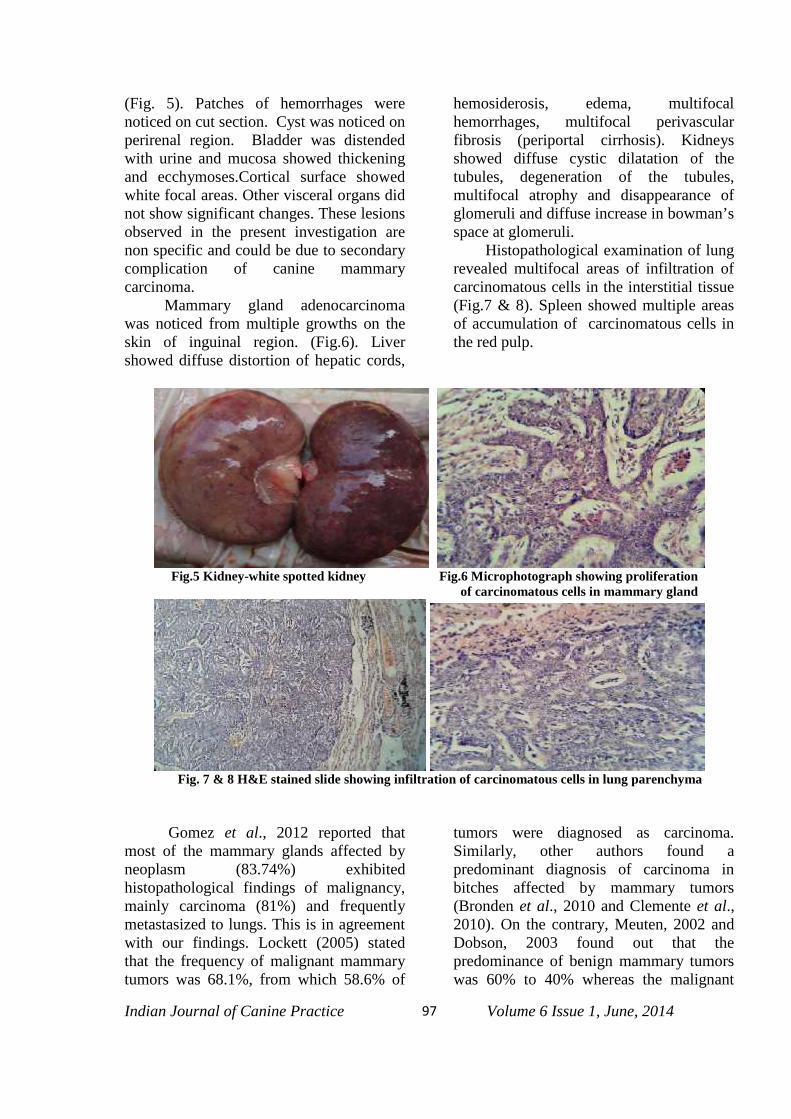
Indian Journal of Canine Practice Volume 6 Issue 1, June, 201497
(Fig. 5). Patches of hemorrhages werenoticed on cut section. Cyst was noticed onperirenal region. Bladder was distendedwith urine and mucosa showed thickeningand ecchymoses.Cortical surface showedwhite focal areas. Other visceral organs didnot show significant changes. These lesionsobserved in the present investigation arenon specific and could be due to secondarycomplication of canine mammarycarcinoma. Mammary gland adenocarcinomawas noticed from multiple growths on theskin of inguinal region. (Fig.6). Livershowed diffuse distortion of hepatic cords,
hemosiderosis, edema, multifocalhemorrhages, multifocal perivascularfibrosis (periportal cirrhosis). Kidneysshowed diffuse cystic dilatation of thetubules, degeneration of the tubules,multifocal atrophy and disappearance ofglomeruli and diffuse increase in bowman’sspace at glomeruli. Histopathological examination of lungrevealed multifocal areas of infiltration ofcarcinomatous cells in the interstitial tissue(Fig.7 & 8). Spleen showed multiple areasof accumulation of carcinomatous cells inthe red pulp.
Fig.5 Kidney-white spotted kidney Fig.6 Microphotograph showing proliferationof carcinomatous cells in mammary gland
Fig. 7 & 8 H&E stained slide showing infiltration of carcinomatous cells in lung parenchyma
Gomez et al., 2012 reported thatmost of the mammary glands affected byneoplasm (83.74%) exhibitedhistopathological findings of malignancy,mainly carcinoma (81%) and frequentlymetastasized to lungs. This is in agreementwith our findings. Lockett (2005) statedthat the frequency of malignant mammarytumors was 68.1%, from which 58.6% of
tumors were diagnosed as carcinoma.Similarly, other authors found apredominant diagnosis of carcinoma inbitches affected by mammary tumors(Bronden et al., 2010 and Clemente et al.,2010). On the contrary, Meuten, 2002 andDobson, 2003 found out that thepredominance of benign mammary tumorswas 60% to 40% whereas the malignant

Indian Journal of Canine Practice Volume 6 Issue 1, June, 201498
mammary tumors were 30%-40%. This isin contrast with the present findings.
SummaryMammary tumors are the second
most common group of neoplasms in dogs,following skin tumors. They are the mostcommon tumors in female dogs andcommon cause for the death of old agebitch. Even though there are treatmentsoptions available for mammary glandtumors, still there is recurrence of tumorsand metastasis to other vital organs are themost common complications. Thiscommunication is an attempt to record theunique case of metastatic carcinoma inlungs and its histopathological features indogs.
ReferencesAndrade, F.H., Figueiroa, F.C., Bersano,
P.R., Bissacot, D.Z., Rocha, NS.(2010).Malignant mammary tumor in femaledogs: environmental contaminants.Diagn Pathol : 5:45.
Brønden, L.B., Nielsen, S.S., Toft, N., Kris-tensen, A.T.,(2010).Data from the Danish veterinary cancer registry on the occ-urrence and distribution of neoplasmsin dogs in Denmark. Vet Rec.166:586-590.
Chang, C.C., Tsai, M.H., Liao, J.W., Chan,J.P., Wong, M.L., Chang, S.C.(2009).Evaluation of hormone receptorexpression for use in predicting survivalof female dogs with malignantmammary gland tumours. J Am VetMed Assoc: 235(4):391-396.
Clemente, M., Pérez-Alenza, M.D., Peña L(2010). Metastasis of canineinflammatory versus non-inflammatory
mammary tumours. J Comp Pathol.143:157-163.
Dobson J, Lascelles D. (2003) BSAVAmanual of canine and feline oncology.2nd ed. London: Blackwell publishing;pp: 345-348.
Culling, C. F. A.1963: Hand book of histop-pathological and histochemical techniq-ues. 3rd Edn. Butter Worths and Co.(Publishers), Ltd. Landon. Pp.402-403.
German, A.J., (2006) The growing problemof obesity in dogs and cats. JNutr:136(7 Suppl):1940S-1946S.
Lockett, M., Merlo, W., Rosciana, A.,Maccio, O., Guaimás L. (2010).Evaluación radiológica en caninos paradetección de metástasis de tumoresmamarios malignos en tórax y abdomen Comunicaciones científicas y technolog-gicas [en línea] 2005 [fecha de acceso15 de febrero de 2010]. URL disponibleen: http://www.unne.edu.ar/Web/cyt/com2005/4-Veterinaria/V-025.pdf
Morris P and Dobson J., (2002) Oncologíaen pequeños animales. Buenos Aires:Intermédica; pp: 123-125.
Meuten D.(2002) Tumors in domesticanimals. 4th ed. New York: Blackwell;pp: 213-215.
Munson L and Moresco A. (2007)Comparative pathology of mammarygland cancers in domestic and wildanimals. Breast Dis: 28:7-21.
Patsikas, M., Karayannopoulou, M.,Kaldrymidoy, E., Papazoglou, L.,Papadopoulou, P. (2006) The lymphdrainage of the neoplastic mammaryglands in the bitch: A lymphographicstudy. Anat Histol Embryol;35:228-234
Welch T. 2007: Small animal surgery. 3rded. New York, Mosby, pp: 553-556 .
*****