Acute vagal stimulation attenuates cardiac metabolic response to -adrenergic stress
Upload
khangminh22Category
view
0download
0
�����������������
Citation: Filippa, M.; Nardelli, M.;
Della Casa, E.; Berardi, A.;
Picciolini, O.; Meloni, S.; Lunardi, C.;
Cecchi, A.; Sansavini, A.;
Corvaglia, L.; et al. Maternal Singing
but Not Speech Enhances Vagal
Activity in Preterm Infants during
Hospitalization: Preliminary Results.
Children 2022, 9, 140. https://doi.org/
10.3390/children9020140
Academic Editor: Mark Dzietko
Received: 15 November 2021
Accepted: 12 January 2022
Published: 21 January 2022
Publisher’s Note: MDPI stays neutral
with regard to jurisdictional claims in
published maps and institutional affil-
iations.
Copyright: © 2022 by the authors.
Licensee MDPI, Basel, Switzerland.
This article is an open access article
distributed under the terms and
conditions of the Creative Commons
Attribution (CC BY) license (https://
creativecommons.org/licenses/by/
4.0/).
children
Article
Maternal Singing but Not Speech Enhances Vagal Activity inPreterm Infants during Hospitalization: Preliminary ResultsManuela Filippa 1,2,*,† , Mimma Nardelli 3,4,† , Elisa Della Casa 5, Alberto Berardi 6 , Odoardo Picciolini 7 ,Sara Meloni 7, Clara Lunardi 8, Alessandra Cecchi 9, Alessandra Sansavini 10 , Luigi Corvaglia 11,12,Enzo Pasquale Scilingo 3,4 , Fabrizio Ferrari 6 and EVC Group ‡
1 Department of Psychology and Educational Sciences, University of Geneva, 24 Rue General Dufour,1211 Geneva, Switzerland
2 Department of Social Sciences, University of Valle d’Aosta, Str. Cappuccini 2, 11100 Aosta, Italy3 Bioengineering and Robotics Research Centre E. Piaggio, 56122 Pisa, Italy;
[email protected] (M.N.); [email protected] (E.P.S.)4 Dipartimento di Ingegneria dell’Informazione, University of Pisa, 56122 Pisa, Italy5 Neonatal Intensive Care Unit, Women’s and Children’s Health Department, University Hospital of Modena,
41124 Modena, Italy; [email protected] Department of Medical and Surgical Sciences of Mother, Children and Adults, University Hospital of Modena,
41124 Modena, Italy; [email protected] (A.B.); [email protected] (F.F.)7 Pediatric Physical Medicine & Rehabilitation Unit, IRCCS Ca’ Granda Ospedale Maggiore Policlinico,
Via Francesco Sforza 35, 20122 Milan, Italy; [email protected] (O.P.);[email protected] (S.M.)
8 Department of Neurosciences, Psychology, Drug Research and Child Health,Careggi University Hospital of Florence, 50139 Florence, Italy; [email protected]
9 Division of Neonatology, Careggi University Hospital, School of Medicine, University of Florence,50134 Florence, Italy; [email protected]
10 Department of Psychology “Renzo Canestrari”, University of Bologna, Viale Berti Pichat 5,40127 Bologna, Italy; [email protected]
11 Neonatal Intensive Care Unit, IRCCS AOU Bologna, Via Massarenti 9, 40138 Bologna, Italy;[email protected]
12 Department of Medical and Surgical Sciences, University of Bologna, Via Massarenti 9, 40138 Bologna, Italy* Correspondence: [email protected]† These authors contributed equally to this work.‡ EVC Group members are listed in Acknowledgments.
Abstract: Background: Early parental interventions in the Neonatal Intensive Care Units (NICUs)have beneficial effects on preterm infants’ short and long-term outcomes. The aim of this study wasto investigate the effects of Early Vocal Contact (EVC)—singing and speaking—on preterm infants’vagal activity and autonomic nervous system (ANS) maturation. Methods: In this multi-centerrandomized clinical trial, twenty-four stable preterm infants, born at 25–32 weeks gestational age,were randomized to either the EVC group or control group, where mothers did not interact with thebabies but observed their behavior. Heart Rate Variability (HRV) was acquired before intervention(pre-condition), during vocal contact, and after it (post condition). Results: No significant effect of thevocal contact, singing and speaking, was found in HRV when the intervention group was comparedto the control group. However, a significant difference between the singing and the pre and postconditions, respectively, preceding and following the singing intervention, was found in the Lowand High Frequency power nu, and in the low/high frequency features (p = 0.037). By contrast,no significant effect of the speaking was found. Conclusions: Maternal singing, but not speaking,enhances preterm infants’ vagal activity in the short-term, thus improving the ANS stability. Futureanalyses will investigate the effect of enhanced vagal activity on short and long-term developmentaloutcomes of preterm infants in the NICU.
Keywords: early vocal contact; heart rate variability; maternal voice; preterm infants
Children 2022, 9, 140. https://doi.org/10.3390/children9020140 https://www.mdpi.com/journal/children

Children 2022, 9, 140 2 of 10
1. Introduction
Several studies have reported an association between prematurity and infants’ ANSmaturation during development [1,2]. One of the best predictors of ANS maturation in theneonatal period is the heart rate variability (HRV), measured as the variation in beat-to-beat(R-R) intervals between two consecutive heart beats [3]. Even if heart rate levels are similar,a healthy full-term neonate has a better beat-to-beat variability than a high-risk pretermneonate [4]. In neonates, improved maturation of the ANS promotes survival duringcritical periods of hospitalization. The ANS continuously matures from the last half ofgestation through the neonatal period [5], and it reflects, through HRV measurements, thegradual activation of the parasympathetic tone, which increases near the term age [6]. Thedeveloping brain in the perinatal period assures that higher cortical processes can integrateautonomic control, with a consequently increased influence of the parasympathetic systemon infants’ regulation activity [7].
In a relational perspective, HRV provides a physiological stand for developing theinfant’s abilities to interact with caregivers and objects during development [8].
In low-risk samples of premature infants, such as the population involved in thepresent study, the association between duration of preterm extrauterine maturation andANS development was not significant before the term-corrected age [9]. Thus, in stablepopulations of preterm infants, with no additional comorbidities other than prematurity,we can presume that fluctuations in the HR could be mainly determined by environmentalintervening factors.
Indeed, the relational environment is fundamental for developing the infants’ reac-tions to social and environmental stimuli, which are mediated by a correct equilibriumbetween the sympathetic and parasympathetic systems [10]. This equilibrium allows forthe emerging of a contingent response to external salient sensory stimuli, which, in turn,constitutes the basis for later co-regulatory experiences [11].
Early kangaroo-care enhanced preterm infants’ autonomic nervous system (ANS) de-velopment, as measured by its vagal activity [12]: infants displayed an improved vagal tonematuration between 32 and 37 weeks of gestational age, with a concomitant improvementin state organization. Moreover, autonomic regulation of preterm infants is enhanced byFamily Nurture Intervention [13]. Finally, exposing preterm infants to their mothers’ voicesin the NICU affected infants’ physiological regulation by increasing oxygen saturation,decreasing episodic apnea and bradycardia, and positively impacting their stability [14–18].More specifically, live maternal singing during skin-to-skin contact induced significantchange in low frequencies (LF) and high frequencies (HF) in the HR. Lower LF/HF ratiowas observed during the vocal intervention and recovery phases, compared with standardkangaroo-care and baseline [19].
Early Vocal Contact (EVC) in the NICU
EVC between parent and premature infant has positive short-term effects on theinfant’s physiological stability and behavior [20,21]. When the mother speaks, the newbornopens its eyes and tends to transition towards a quiet wakefulness; when the mothersings, the initial infants’ behavioral state tends to remain stable [20]. During maternalspeech and singing, compared to the absence of vocal contact, the preterm infants increaseself-touch gestures and eye opening [21]. Finally, during routine, painful, procedures,maternal speech decreases pain scores in preterm infants and increases their endogenousoxytocin release, with potential analgesic effects [22]. The same tendency is evidencedduring singing, but the results are only marginally significant [22]. The results mentioned sofar suggest that maternal singing and speech could have differential effects on hospitalizedpremature infants.
In the present study, we hypothesized that the live maternal voice, both speaking andsinging, would increase HRV during EVC.
This study is part of a larger study investigating the short and long-term effects ofEVC on preterm infants [23].

Children 2022, 9, 140 3 of 10
2. Materials and Methods2.1. Design
A four-site randomized controlled trial has been conducted to investigate the short-termphysiological effects of EVC on preterm infants’ vagal activity, using Heart Rate Variabilityanalysis. Infants were recruited from four university hospitals: Careggi University Hospital(Florence), NICU Fondazione IRCCS Ca’ Granda Ospedale Maggiore Policlinico (Milan),Modena University Hospital (Modena), and Bologna University IRCCS Hospital (Bologna).
2.2. Participants
Twenty-four preterm infants, born at 25–32 weeks and 6 days GA, were recruitedfrom the four centers. Twelve subjects were assigned to the control group and 12 to theexperimental group, randomizing infants by gender and GA.
Due to centralized randomization, children were included in all centers involved,both for the experimental and control group. Descriptions of the involved population aresummarized in Table 1.
Table 1. Infants and mothers’ demographics.
CharacteristicsIntervention Group
(n = 12)Mean (SD)
Control Group (n = 12)Mean (SD)
Gestational age at birth (weeks) 29.1 (2.4) 29.3 (1.5)Birthweight (g) 1226 (367) 1204 (235)
Apgar score at 5 min 7.1 (1.3) 6.4 (2.3)Apgar score at 7 min 8.1 (1.1) 7.9 (1.0)
Gestational age at test (weeks) 34.1 (1.3) 34.2 (1.7)Post-natal age at test (days) 33.8 (19.2) 34.7 (15.8)
Weight at test (g) 1767.8 (258) 1603.8 (308)Maternal citizenship Italian (80%) Italian (70%)
Primiparous Yes (72%) Yes (60%)Mother’s age 35.6 (3.2) 37.7 (6.5)
After obtaining informed consent, families were enrolled. The inclusion criteria forthe newborns were: GA between 25 + 0 and 32 + 6 weeks at birth; Apgar score >7 at 10 min;birth weight between 3rd and 97th percentiles; birth cranial circumference greater than10th percentile; periventricular leukomalacia grade 1; and intraventricular hemorrhagegrade 1–2. Preterm newborn infants were excluded in presence of sepsis, congenitalmalformations and/or genetic abnormalities. Mothers were excluded if they had cleardepressive symptoms, drug abuse, and/or age below 18 years.
2.3. Intervention
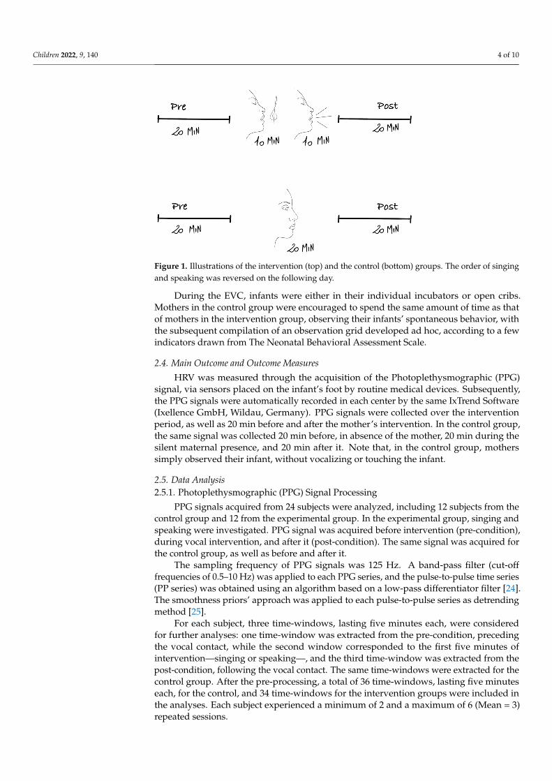
Mothers were asked to speak and sing to their infants over a 10 min period for eachtype of intervention (20 min in total), three times per week, for 2 weeks, midway betweenthe feeding cycles, more than 4 h after the last medical exam. EVC began when thenewborns were in an active sleep state, in calm awake state, or in active awake state, butnot in deep sleep or crying.
The order of the two vocalizations was counterbalanced for the same subject. If thepatient on day 1 started with singing then, on day 2, the first vocalization was speech (SeeFigure 1). The mothers were free to choose which vocalization to start with on day 1.
Children 2022, 9, 140 4 of 10
Figure 1. Illustrations of the intervention (top) and the control (bottom) groups. The order of singingand speaking was reversed on the following day.
During the EVC, infants were either in their individual incubators or open cribs.Mothers in the control group were encouraged to spend the same amount of time as thatof mothers in the intervention group, observing their infants’ spontaneous behavior, withthe subsequent compilation of an observation grid developed ad hoc, according to a fewindicators drawn from The Neonatal Behavioral Assessment Scale.
2.4. Main Outcome and Outcome Measures
HRV was measured through the acquisition of the Photoplethysmographic (PPG)signal, via sensors placed on the infant’s foot by routine medical devices. Subsequently,the PPG signals were automatically recorded in each center by the same IxTrend Software(Ixellence GmbH, Wildau, Germany). PPG signals were collected over the interventionperiod, as well as 20 min before and after the mother’s intervention. In the control group,the same signal was collected 20 min before, in absence of the mother, 20 min during thesilent maternal presence, and 20 min after it. Note that, in the control group, motherssimply observed their infant, without vocalizing or touching the infant.
2.5. Data Analysis2.5.1. Photoplethysmographic (PPG) Signal Processing
PPG signals acquired from 24 subjects were analyzed, including 12 subjects from thecontrol group and 12 from the experimental group. In the experimental group, singing andspeaking were investigated. PPG signal was acquired before intervention (pre-condition),during vocal intervention, and after it (post-condition). The same signal was acquired forthe control group, as well as before and after it.
The sampling frequency of PPG signals was 125 Hz. A band-pass filter (cut-offfrequencies of 0.5–10 Hz) was applied to each PPG series, and the pulse-to-pulse time series(PP series) was obtained using an algorithm based on a low-pass differentiator filter [24].The smoothness priors’ approach was applied to each pulse-to-pulse series as detrendingmethod [25].
For each subject, three time-windows, lasting five minutes each, were consideredfor further analyses: one time-window was extracted from the pre-condition, precedingthe vocal contact, while the second window corresponded to the first five minutes ofintervention—singing or speaking—, and the third time-window was extracted from thepost-condition, following the vocal contact. The same time-windows were extracted for thecontrol group. After the pre-processing, a total of 36 time-windows, lasting five minuteseach, for the control, and 34 time-windows for the intervention groups were included inthe analyses. Each subject experienced a minimum of 2 and a maximum of 6 (Mean = 3)repeated sessions.

Children 2022, 9, 140 5 of 10
2.5.2. Heart Rate Variability Feature Extraction
PP series were converted into HRV signals, using a cubic spline interpolation toresample each series at a rate of 4 Hz. In the next step, the power spectral density of eachof the HRV signals was analyzed using a time-varying parametric spectrum estimationbased on an autoregressive model [26]. Two main bandwidths were investigated, i.e., a lowfrequency band (between 0.02 and 0.2 Hz) and a high frequency band (between 0.2 and1.5 Hz) [27]. Considering each bandwidth, we computed the values of five features:
- Low Frequency (LF) power and High Frequency (HF) power: the absolute power values;- LF power nu and HF power nu: the related values normalized to the sum of the
LF power and HF power (LF power nu = LF power/(LF power + HF power) andHF power nu = HF power/(LF power + HF power));
- LF/HF: the ratio between LF power and HF power.
Considering that more than one recording session was performed for each subject, themedian value of the features calculated on all available sessions was used in the statisticalanalyses (see Section 2.5.3).
2.5.3. Statistical Analysis
Two different statistical analyses were applied to HRV data. An intra-group statisticalanalysis aimed to study the differences in HRV dynamics among the three Time Conditions(pre, during, and post) in each group (intervention and control). Given the non-Gaussianityof data distributions, we applied a non-parametric Friedman statistical test to compare thevalues of each HRV feature in the three conditions. After this, we compared the featurevalues found for each pair of time conditions using a two-tailed Wilcoxon signed-rank.Bonferroni’s test for post hoc analysis was applied in order to test the statistical differencesbetween each pair of conditions [28].
The Kruskal–Wallis non-parametric statistical test was used for the inter-group statisti-cal analysis. We subtracted the values of the HRV features during the pre-condition fromthe values of the HRV features during singing, speaking, and the control group. In the nextstep, we applied the Kruskal–Wallis test to investigate the differences among the groups.The Mann–Whitney test was used to compare each pair of vocal interventions (singingvs. speaking). Likewise, the same test was used to compare singing and speaking in thecontrol group. Additionally, in this case, Bonferroni’s test was used for post hoc analysis ofmultiple comparisons.
3. Results
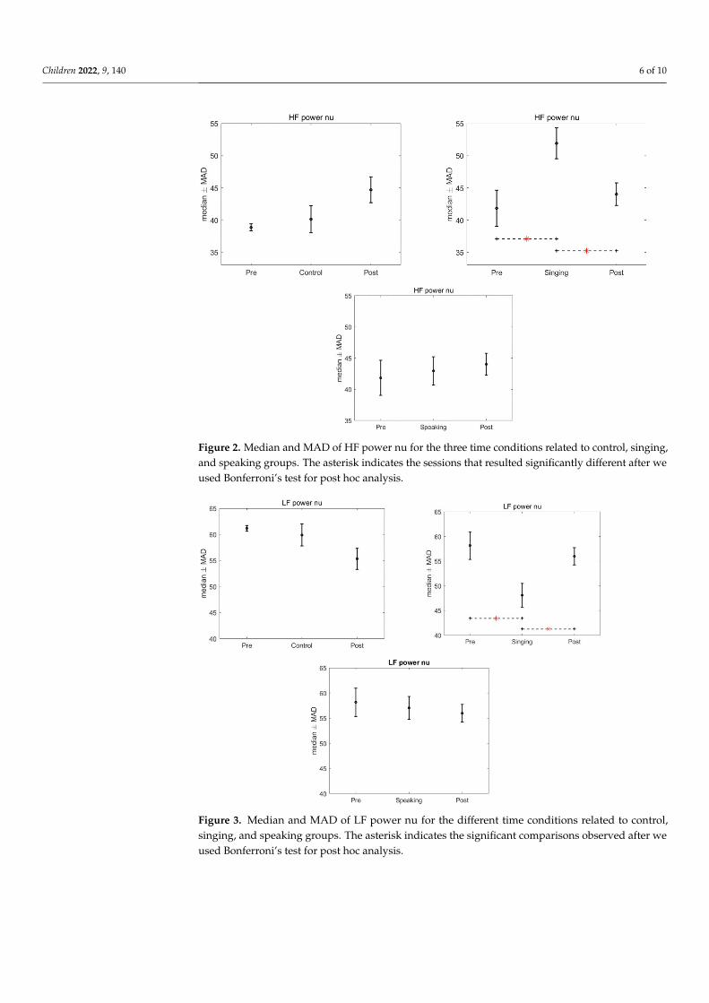
There was no significant effect of the intervention, singing and speaking, found in theHRV when compared to the control group, after the application of Kruskal–Wallis test (LFpower: p = 0.977; HF power: p = 0.893; LF nu: p = 0.224; HF nu: p = 0.224; LF/HF: p = 0.3436).However, significant differences were found in the HRV features in the intra-group analysis,for the time conditions, when we compared the pre, singing and post conditions (p = 0.013).Specifically, after the Friedman test a p-value < 0.05 was assigned to three features: LF powernu, HF power nu, and LF/HF. Bonferroni’s test for post hoc analysis revealed a significantdifference between the singing condition and the pre and post conditions (LF power nuand HF power nu: p = 0.015 and 0.037, LF/HF: p = 0.021 and p = 0.028). Figures 2–4 showthe median and median absolute values (MAD) of LF power nu, HF power nu, and LF/HFfeatures, for each of the three time conditions for control, singing, and speaking groups. Nosignificant differences were found in the three time conditions—pre, during, and post—forthe speaking group. Likewise, no significant differences were found in the three timeconditions—pre, during, and post—for the control group.
Children 2022, 9, 140 6 of 10
Figure 2. Median and MAD of HF power nu for the three time conditions related to control, singing,and speaking groups. The asterisk indicates the sessions that resulted significantly different after weused Bonferroni’s test for post hoc analysis.
Figure 3. Median and MAD of LF power nu for the different time conditions related to control,singing, and speaking groups. The asterisk indicates the significant comparisons observed after weused Bonferroni’s test for post hoc analysis.

Children 2022, 9, 140 7 of 10
Figure 4. Median and MAD of LF/HF for the three time conditions related to the control, singingand speaking groups. The asterisk indicates the significant comparisons observed after we usedBonferroni’s test for post hoc analysis.
4. Discussion
The preliminary results showed that preterm infants’ HRV is modulated in the short-term by the maternal live singing. An assessment of the short-term HRV, as in the presentwork, offers important evidence about maturation and dynamical equilibrium of the ANSin newborns. More specifically, the median and median absolute values of HF were signifi-cantly higher during singing than in the pre and post condition, in absence of the mother’svoice, indicating an improved stability of the ANS during the singing interaction. HF com-ponent of the HRV is correlated to parasympathetic activity, and the related spectral activityin preterm infants is lower than in term infants; this is mainly due to their ANS immatu-rity [29]. Thus, higher values of HF indicate an increase in maturation of parasympatheticactivity, and, indirectly, can be an index of the ANS maturation in preterm infants [30].During maternal singing, both LF and LF/HF features of the HRV were significantly lowerthan in the pre and post condition: these two components reflect both sympathetic andvagal dynamics [31,32], and the ratio of LF to HF can be interpreted as an index of physio-logical arousal. The dominant activity of the LF is a typical consequence of preterm birth,demonstrating a dominance of the sympathetic system over the parasympathetic. In ourstudy, preterm infants, exposed to maternal singing, show a decrease of the LF spectralcomponents, with a concomitant increase of the HF. This concomitant fluctuation expresses,again, an improved balance between these two components and an increase in the infant’svagal activity.
We did not find the same effect with speech. As previously mentioned, maternalspeech has activating effects on the premature infant, and it induces active waking states, asopposed to singing, which tends to stabilize the infant’s state [20]. Furthermore, in a recentstudy, we have shown that maternal speech has a significant effect on the oxytocin releaseof the premature infant during a heel prick procedure, whereas these effects are marginallysignificant for singing. Finally, only maternal speech significantly modulates the infant’spain, reducing its facial and physiological manifestations in infants, whereas this effectwas not found for maternal singing [22]. We can therefore conclude that, during early

Children 2022, 9, 140 8 of 10
vocal contact, maternal singing and speech seem to create different effects on the behavior,physiology and, more generally, on the ANS of premature infants. Maternal singing, infact, with its repetitive structure, homogeneous with respect to a predefined melodic lineand rhythmicity, has a significant impact on HRV and therefore on the vagal activity ofthe newborn [19]. Speech, on the other hand, contains the peculiarity of modulating onthe behavioral signs of the newborn [33]. It does not have a predefined structure likesinging, but it responds to the immediate changes in the infant’s state, and it acts onits endocrine system, with an increase in oxytocin. Thus, maternal singing and speechseem to have different functions in the early vocal communication between parents andinfants. Both forms of communication have evolved with humans over millennia, bothhave characteristics that distinguish them from adult-directed speech and singing, bothmodulate to the age and development of the child, and both convey emotional contentsto which infants are sensitive [34]. However, they also show significant differences. Ina naturalistic context, infant-directed singing is more predictable on the basis of melodicand rhythm repetitions [35], as opposed to speech, and its motivational and structuringproperties facilitate learning. Most importantly, singing is used by adults to modulate theinfants’ state of arousal, sometimes to calm them with lullabies, and sometimes to activatethem with play songs [36].
Moreover, the same differences between the pre, during, and post maternal singingwere not found in the control group, in which the mothers were present but merely observedthe infant’s behavior. Therefore, the silent presence of the mothers next to the incubatoris not sufficient by itself to modulate the infant’s cardiac responses and stimulate itsvagal activity.
Finally, no significant differences were found between the intervention and controlgroups. As a non-significant trend is evidenced in the group comparisons, it is possiblethat an increase in the number of subjects will contribute to deeper statistical analyses con-cerning the differences between the groups under examination, thus, raising the statisticalpower of the study.
5. Conclusions
Preterm infant-directed singing has modulatory effects on infants’ HRV, increasing thevagal activity in newborns. An enhanced autonomic regulation facilitates early nurturinginteractions between preterm infants and their parents [13]. These preliminary results, evenif assessed in a small population, are encouraging, and they provide a foundation for futureanalyses. The primary outcome, HRV, will therefore be analyzed on a larger sample ofinfants during early vocal contact. In addition, the physiological data will be correlatedwith the degree of maternal stress and, above all, with the neuromotor maturation assessedin general movements [37]. As a final step of our future analyses, neonatal HRV data, inresponse to contingent vocal contact, will be correlated with the language skills of infantsinvolved in early intervention.
6. Limitations and Future Works
HRV series of preterm infants were extracted from PPG signals. Choosing to acquirePPG signals instead of ECG, which is the gold standard for HRV extraction, has severaladvantages. Specifically, PPG sensors are less obtrusive than ECG acquisition systems, andthey can be easily integrated into wearable devices.
However, we are aware of the less accurate peak detection on PPG signals comparedto ECG signals [38]. Future studies will be addressed towards the comparison of the resultsobtained through the analyses of PPG and ECG signals, and, additionally, towards theapplication of multivariate indexes for a comprehensive assessment of cardiovasculardynamics [39]. The number of subjects involved in the study will be increased in order toraise the statistical power of the intra-group and inter-group analyses.
Children 2022, 9, 140 9 of 10
Author Contributions: M.F. and M.N. wrote the first draft of the paper. M.F., E.D.C. and F.F. preparedthe protocol. O.P., C.L. and A.S. contributed to protocol design and development. E.D.C., A.B., O.P.,S.M., C.L., A.C., A.S., L.C. and the EVC Group contributed to the data collection. M.N. and E.P.S. didthe analyses. All authors have read and agreed to the published version of the manuscript.
Funding: The present research has been funded by Fondazione Mariani Research Grant 2017, and byPollicino Association, Modena.
Institutional Review Board Statement: The study was approved by Modena-Health Authority’sIndependent Ethics Committee of Area Vasta Emilia Nord (P.0006292/18); Bologna, Bologna HealthAuthority’s Independent Ethics Committee (P. 348/2018/Sper/AOUBo); Milan Mangiagalli, CE,(P. 205); Firenze Careggi Hospital, CE (P. 77/2017).
Informed Consent Statement: Written informed consent has been obtained from the patients.
Data Availability Statement: All data are available upon request.
Acknowledgments: The EVC Group is composed of: Arianna Aceti, Luca Bedetti, Natascia Bertoncelli,Giovanna Lucco, Michele Luzzati, Luca Ori, Chiara Petrolini, and Mariagrazia Zuccarini. We thankGiovanna Talucci, from the Modena University Hospital.Concerning the Neonatal Intensive Care Unit,IRCCS AOU Bologna, and the University of Bologna, Italy, we would like to thank: Giacomo Faldella,neonatologist, for making the implementation of the project possible; Simona Cordella, nurse, andChiara Rizzoli, graduating student, for collaborating in participants’ recruitment and implementationof the intervention. A special acknowledgement to Fabio Mosca, Fondazione IRCCS Ca’ GrandaOspedale Maggiore Policlinico, NICU, Milan, Italy and University of Milan, Department of ClinicalSciences and Community Health, Milan, who made this study possible, making his departmentavailable to researchers. We would also like to acknowledge Carlo Dani Director of the Careggi NICUfor making possible the study. Finally, special credits to Roberto Vicini who assisted in generation ofthe randomisation list.
Conflicts of Interest: The authors declare no conflict of interest.
References1. Mulkey, S.B.; Kota, S.; Swisher, C.B.; Hitchings, L.; Metzler, M.; Wang, Y.; Maxwell, G.L.; Baker, R.; du Plessis, A.J.; Govindan, R.
Autonomic nervous system depression at term in neurologically normal premature infants. Early Hum. Dev. 2018, 123, 11–16.[CrossRef] [PubMed]
2. Yiallourou, S.R.; Witcombe, N.B.; Sands, S.A.; Walker, A.M.; Horne, R.S. The development of autonomic cardiovascular control isaltered by preterm birth. Early Hum. Dev. 2013, 89, 145–152. [CrossRef] [PubMed]
3. Sahni, R.; Schulze, K.F.; Kashyap, S.; Ohira-Kist, K.; Fifer, W.P.; Myers, M.M. Maturational changes in heart rate and heart ratevariability in low birth weight infants. Dev. Psychobiol. J. Int. Soc. Dev. Psychobiol. 2000, 37, 73–81. [CrossRef]
4. Porges, S.W. The Polyvagal Theory: Neurophysiological Foundations of Emotions, Attachment, Communication, and Self-Regulation(Norton Series on Interpersonal Neurobiology); WW Norton & Company: New York, NY, USA, 2011.
5. Mulkey, S.B.; du Plessis, A.J. Autonomic nervous system development and its impact on neuropsychiatric outcome. Pediatric Res.2019, 85, 120–126. [CrossRef]
6. Chiera, M.; Cerritelli, F.; Casini, A.; Barsotti, N.; Boschiero, D.; Cavigioli, F.; Corti, C.G.; Manzotti, A. Heart Rate Variability in thePerinatal Period: A Critical and Conceptual Review. Front. Neurosci. 2020, 14, 561186. [CrossRef]
7. DiPietro, J.A.; Costigan, K.A.; Voegtline, K.M. Studies in fetal behavior: Revisited, renewed, and reimagined. Monogr. Soc. Res.Child Dev. 2015, 80, vii-vii94.
8. Porges, S.W.; Furman, S.A. The early development of the autonomic nervous system provides a neural platform for socialbehaviour: A polyvagal perspective. Infant Child Dev. 2011, 20, 106–118. [CrossRef]
9. Mulkey, S.B.; Govindan, R.B.; Hitchings, L.; Al-Shargabi, T.; Herrera, N.; Swisher, C.B.; Eze, A., Jr.; Russo, S.; Schlatterer, S.D.;Jacobs, M.B.; et al. Autonomic nervous system maturation in the premature extrauterine milieu. Pediatric Res. 2021, 89, 863–868.[CrossRef]
10. Thayer, J.F.; Lane, R.D. A model of neurovisceral integration in emotion regulation and dysregulation. J. Affect. Disord. 2000,61, 201–216. [CrossRef]
11. Porges, S.W. Autonomic regulation and attention. In Attention and Information Processing in Infants and Adults; Campbell, B.A.,Hayne, H., Richardson, R., Eds.; Lawrence Erlbaum Associates: Hillsdale, NJ, USA, 1992; pp. 201–223.
12. Feldman, R.; Eidelman, A.I. Skin-to-skin contact (Kangaroo Care) accelerates autonomic and neurobehavioural maturation inpreterm infants. Dev. Med. Child Neurol. 2003, 45, 274–281. [CrossRef]

Children 2022, 9, 140 10 of 10
13. Porges, S.W.; Davila, M.I.; Lewis, G.F.; Kolacz, J.; Okonmah-Obazee, S.; Hane, A.A.; Kwon, K.Y.; Ludwig, R.J.; Myers, M.M.;Welch, M.G. Autonomic regulation of preterm infants is enhanced by Family Nurture Intervention. Dev. Psychobiol. 2019,61, 942–952. [CrossRef]
14. Best, K.; Bogossian, F.; New, K. Language exposure of preterm infants in the neonatal unit: A systematic review. Neonatology 2018,114, 261–276. [CrossRef]
15. Bieleninik, Ł.; Ghetti, C.; Gold, C. Music therapy for preterm infants and their parents: A meta-analysis. Pediatrics 2016, 138, e20160971.[CrossRef]
16. Filippa, M.; Panza, C.; Ferrari, F.; Frassoldati, R.; Kuhn, P.; Balduzzi, S.; D’Amico, R. Systematic review of maternal voiceinterventions demonstrates increased stability in preterm infants. Acta Paediatr. 2017, 106, 1220–1229. [CrossRef]
17. Provenzi, L.; Broso, S.; Montirosso, R. Do mothers sound good? A systematic review of the effects of maternal voice exposure onpreterm infants’ development. Neurosci. Biobehav. Rev. 2018, 88, 42–50. [CrossRef]
18. Yue, W.; Han, X.; Luo, J.; Zeng, Z.; Yang, M. Effect of music therapy on preterm infants in neonatal intensive care unit: Systematicreview and meta-analysis of randomized controlled trials. J. Adv. Nurs. 2021, 77, 635–652. [CrossRef]
19. Arnon, S.; Diamant, C.; Bauer, S.; Regev, R.; Sirota, G.; Litmanovitz, I. Maternal singing during kangaroo care led to autonomicstability in preterm infants and reduced maternal anxiety. Acta Paediatr. 2014, 103, 1039–1044. [CrossRef]
20. Filippa, M.; Devouche, E.; Arioni, C.; Imberty, M.; Gratier, M. Live maternal speech and singing have beneficial effects onhospitalized preterm infants. Acta Paediatr. 2013, 102, 1017–1020. [CrossRef]
21. Filippa, M.; Menin, D.; Panebianco, R.; Monaci, M.G.; Dondi, M.; Grandjean, D. Live maternal speech and singing increaseself-touch and eye-opening in preterm newborns: A preliminary study. J. Nonverbal. Behav. 2020, 44, 453–473. [CrossRef]
22. Filippa, M.; Monaci, M.G.; Spagnuolo, C.; Serravalle, P.; Daniele, R.; Grandjean, D. Maternal speech decreases pain scores andincreases oxytocin levels in preterm infants during painful procedures. Sci. Rep. 2021, 11, 17301. [CrossRef]
23. Filippa, M.; Della Casa, E.; D’amico, R.; Picciolini, O.; Lunardi, C.; Sansavini, A.; Ferrari, F. Effects of Early Vocal Contact in theNeonatal Intensive Care Unit: Study Protocol for a Multi-Centre, Randomised Clinical Trial. Int. J. Environ. Res. Public Health2021, 18, 3915. [CrossRef] [PubMed]
24. Lázaro, J.; Gil, E.; Vergara, J.M.; Laguna, P. Pulse rate variability analysis for discrimination of sleep-apnea-related decreases inthe amplitude fluctuations of pulse photoplethysmographic signal in children. IEEE J. Biomed. Health Inform. 2013, 18, 240–246.[CrossRef] [PubMed]
25. Tarvainen, M.P.; Ranta-Aho, P.O.; Karjalainen, P.A. An advanced detrending method with application to HRV analysis. IEEE Trans.Biomed. Eng. 2002, 49, 172–175. [CrossRef] [PubMed]
26. Tarvainen, M.P.; Georgiadis, S.D.; Ranta-Aho, P.O.; Karjalainen, P.A. Time-varying analysis of heart rate variability signals witha Kalman smoother algorithm. Physiol. Meas. 2006, 27, 225. [CrossRef]
27. Rosenstock, E.G.; Cassuto, Y.; Zmora, E. Heart rate variability in the neonate and infant: Analytical methods, physiological andclinical observations. Acta Paediatr. 1999, 88, 477–482. [CrossRef]
28. Hochberg, Y.; Tamhane, A.C. Multiple Comparison Procedures; John Wiley & Sons, Inc.: Hoboken, NJ, USA, 1987.29. Javorka, K.; Lehotska, Z.; Kozar, M.; Uhrikova, Z.; Kolarovszki, B.; Javorka, M.; Zibolen, M. Heart rate variability in newborns.
Physiol. Res. 2017, 66, S203–S214. [CrossRef]30. Longin, E.; Gerstner, T.; Schaible, T.; Lenz, T.; König, S. Maturation of the autonomic nervous system: Differences in heart rate
variability in premature vs. term infants. J. Perinat. Med. 2006, 34, 303–308. [CrossRef]31. Malik, M. Heart rate variability: Standards of measurement, physiological interpretation, and clinical use: Task force of the
European Society of Cardiology and the North American Society for Pacing and Electrophysiology. Ann. Noninvasive Electrocardiol.1996, 1, 151–181. [CrossRef]
32. Acharya, U.R.; Joseph, K.P.; Kannathal, N.; Lim, C.M.; Suri, J.S. Heart rate variability: A review. Med. Biol. Eng. Comput. 2006,44, 1031–1051. [CrossRef]
33. Filippa, M.; Gratier, M.; Devouche, E.; Grandjean, D. Changes in infant-directed speech and song are related to preterm infantfacial expression in the neonatal intensive care unit. Interact. Stud. 2018, 19, 427–444. [CrossRef]
34. Corbeil, M.; Trehub, S.E.; Peretz, I. Speech vs. singing: Infants choose happier sounds. Front. Psychol. 2013, 4, 372. [CrossRef]35. Schön, D.; Boyer, M.; Moreno, S.; Besson, M.; Peretz, I.; Kolinsky, R. Songs as an aid for language acquisition. Cognition 2008,
106, 975–983. [CrossRef]36. Cirelli, L.K.; Jurewicz, Z.B.; Trehub, S.E. Effects of maternal singing style on mother–infant arousal and behavior. J. Cogn. Neurosci.
2020, 32, 1213–1220. [CrossRef]37. Ferrari, F.; Cioni, G.; Prechtl, H.F.R. Qualitative changes of general movements in preterm infants with brain lesions. Early Hum.
Dev. 1990, 23, 193–231. [CrossRef]38. Nardelli, M.; Vanello, N.; Galperti, G.; Greco, A.; Scilingo, E.P. Assessing the Quality of Heart Rate Variability Estimated from
Wrist and Finger PPG: A Novel Approach Based on Cross-Mapping Method. Sensors 2020, 20, 3156. [CrossRef]39. Nardelli, M.; Scilingo, E.P.; Valenza, G. Multichannel Complexity Index (MCI) for a multi-organ physiological complexity
assessment. Phys. A Stat. Mech. Its Appl. 2019, 530, 121543. [CrossRef]
Copyright © 2022 FDOKUMEN