UNIVERSITY OF CALIFORNIA Los Angeles Cardiac Vagal Tone ...
177
UNIVERSITY OF CALIFORNIA Los Angeles Cardiac Vagal Tone: Regulator of Inflammation and Clinical Symptoms in Insomnia and Schizophrenia A dissertation submitted in partial satisfaction of the requirements for the degree Doctor of Philosophy in Psychology by Alexandra Claire Reed 2021
-
Upload
khangminh22 -
Category
Documents
-
view
0 -
download
0
Transcript of UNIVERSITY OF CALIFORNIA Los Angeles Cardiac Vagal Tone ...
UNIVERSITY OF CALIFORNIA
Los Angeles
Cardiac Vagal Tone: Regulator of Inflammation and Clinical Symptoms in Insomnia and
Schizophrenia
A dissertation submitted in partial satisfaction of the requirements for the degree
Doctor of Philosophy in Psychology
by
Alexandra Claire Reed
2021

© Copyright by
Alexandra Claire Reed
2021
ii
ABSTRACT OF THE DISSERTATION
Cardiac Vagal Tone: Regulator of Inflammation and Clinical Symptoms in Insomnia and
Schizophrenia
by
Alexandra Claire Reed
Doctor of Philosophy in Clinical Psychology
University of California, Los Angeles, 2021
Professor Cindy M. Yee-Bradbury, Co-Chair
Professor Julienne E. Bower, Co-Chair
Cardiac vagal tone (CVT) reflects the contribution of the parasympathetic nervous
system to cardiac regulation and is hypothesized to contribute to the regulation of inflammation
and expression of psychopathology. CVT may play an important role in sleep disturbance and
insomnia treatment. Through a series of four studies, this dissertation interrogated the
associations between CVT, inflammation, and clinical symptoms in sleep disturbed samples and
evaluated possible roles for CVT in insomnia intervention. Studies 1 and 2 proposed a specific
model: chronic sleep disturbance reduces CVT with downstream effects on inflammation and
clinical symptoms. Studies 1 and 2 then evaluated cross-sectional associations between these
variables and tested exploratory mediation models in two samples with poor sleep quality: 106
older adults with insomnia and 39 patients with first episode schizophrenia. Overall, results did

iii
not support the mediation models in either sample. However, findings revealed novel
associations between CVT and cellular inflammatory processes in older adults and extended a
negative association between CVT and systemic inflammation to schizophrenia, consistent with
the cholinergic anti-inflammatory pathway. Studies 3 and 4 then evaluated possible roles for
CVT in a randomized controlled trial which compared active behavioral treatments (Cognitive
Behavioral Therapy for Insomnia and Tai Chi Chih) to sleep education in older adults with
insomnia. Study 3 revealed that active behavioral interventions led to improvements in insomnia
and clinical symptoms without up-regulating CVT, indicating that CVT is unlikely to be a
mechanism supporting insomnia recovery. Study 4 then determined whether individual
differences in CVT at pre-treatment moderated the effects of active behavioral interventions on
key trial outcomes. Results showed that CVT did not moderate treatment effects. Null findings
from Studies 3 and 4 underscore the need for further identification and evaluation of candidate
mechanisms and moderators to maximize existing behavioral treatments for insomnia. Taken
together, present results add to the mixed literature on CVT. Findings simultaneously challenge
the assumption that CVT is a reliable correlate of clinical state and extend prior research on the
role of the anti-inflammatory pathway in two unique samples with poor sleep quality.

iv
The dissertation of Alexandra Claire Reed is approved.
Michael R. Irwin
Gregory A. Miller
Keith H. Nuechterlein
Cindy M. Yee-Bradbury, Committee Co-Chair
Julienne E. Bower, Committee Co-Chair
University of California, Los Angeles
2021

v
TABLE OF CONTENTS
General Introduction
Project overview
1
9
Study 1: Sleep Quality, Cardiac Vagal Tone, Inflammation, and Clinical Symptoms in
Late Life Insomnia
Introduction
Method
Results
Discussion
13
21
27
30
Study 2: Sleep Quality, Cardiac Vagal Tone, Inflammation, and Clinical Symptoms in
Schizophrenia and Older Adulthood
Introduction
Method
Results
Discussion
41
46
51
57
Study 3: Changes in Cardiac Vagal Tone During Behavioral Interventions for Late Life
Insomnia
Introduction
Method
Results
Discussion
69
73
79
81
Study 4: Cardiac Vagal Tone as a Moderator of Treatment Response to Behavioral
Interventions in Older Adults with Insomnia
Introduction
Method
Results
Discussion
88
94
97
99
General Discussion 106
Appendix
Table 1A. Baseline characteristics of older adult participants, by treatment group (Study 1).
127

vi
References 128
LIST OF TABLES
Table 1. Baseline characteristics of participants in the sample with valid
electrocardiogram data (Study 1).
114
Table 2. Correlations between RSA, inflammation, sleep, and clinical symptoms in
older adult participants at pre-intervention (Study 1).
115
Table 3. Demographic and clinical characteristics of participants (Study 2)
116
Table 4. Associations between sleep quality, RSA, inflammatory markers, clinical
symptoms, and relevant covariates in patients with first-episode
schizophrenia (Study 2).
118
Table 5. Summary of path comparisons for the serial and simple mediation models
between the schizophrenia and older adult samples (Study 2).
119
Table 6. Characteristics of participants by treatment group at pre-intervention
(Study 3).
120
Table 7. Means and standard deviations of RSA by treatment group at pre and post
intervention (Study 3).
121
Table 8. Hierarchical regressions predicting the percent of group treatment
sessions attended from relevant covariates and pre-treatment RSA (Study
4).
122
LIST OF FIGURES
Figure 1. General model of the proposed directional relationships between sleep
quality, CVT, inflammation, and clinical symptoms.
122
Figure 2. Screening, randomization, and completion of post-intervention, 7-month,
and 16-month evaluations (from Irwin et al., 2014).
123
Figure 3.
Statistical models of the serial and single mediation models (Study 2).
124
Figure 4.
Figure 5.
Modified CONSORT diagram showing randomization and completion of
post-intervention, 7-month, and 16-month evaluations for the parent trial
and Study 3. Pre to post change in RSA by treatment group (Study 3).
125
126
vii
ACKNOWLEDGEMENTS
This work was supported by the National Science Foundation (NSF) Graduate Research
Fellowship awarded to Alexandra Reed and National Institute of Mental Health (NIMH) Grants
R01 MH110544 (MPIs: Cindy Yee-Bradbury, PhD, Gregory A. Miller, PhD, Keith Nuechterlein,
PhD) and R01 MH110544 (PI: Keith Nuechterlein, PhD). Data collection for the older adult
studies was previously funded by National Institute on Aging (NIA) R01 AG026364 (PI:
Michael Irwin, MD). I also gratefully acknowledge the staff of the Cousins Center for
Psychoneuroimmunology and the patients and staff of the UCLA Aftercare Research Program
for their important contributions.

viii
VITA
EDUCATION
2020-2021 Clinical Psychology Predoctoral Intern
Greater Hartford Clinical Psychology Consortium, Connecticut VA Health Care
System & University of Connecticut Health
2019 Candidate of Philosophy, Psychology
University of California, Los Angeles, Los Angeles, CA
2017 Master of Arts, Psychology
University of California, Los Angeles, Los Angeles, CA
2007 Bachelor of Arts, Psychology
Tufts University, Medford, MA
FUNDING AWARDS AND HONORS 2016-2019 National Science Foundation Graduate Research Fellowship Award
2016-2019 Letters of Clinical Excellence, UCLA Psychology Clinic
2016 UCLA Graduate Summer Research Award, UCLA
2015-2016 Distinguished University Fellowship, UCLA Graduate Division
2012 Phi Beta Kappa Society, Tufts University
2008-2012 Dean’s List, all undergraduate semesters, Tufts University
2008-2012 Louise C. Vanderhout Memorial Scholarship, Nauset Regional School District
$40,000 academic merit award to attend Tufts University
PUBLICATIONS
Reed, A. C., Lee, J., Green, M. F., Hamilton, H. K., Miller, G. A., Subotnik, K. L., Ventura, J.,
Nuechterlein, K. H., & Yee, C. M. (2020). Associations between physiological responses to
social-evaluative stress and daily functioning in first-episode schizophrenia. Schizophrenia Research. Reed, A. C., Harris, J., & Olincy, A. (2016). Schizophrenia, smoking status, and performance on
the MATRICS Cognitive Consensus Battery. Psychiatry Research. 9(246), 1-8.
http://dx.doi.org/10.1016/j.psychres.2016.08.062.
Reed, A. C. (2014). Integration isn’t enough—mindful practice needed in SPMI health care. The National Psychologist, 23, (4), 13. Available online at:
http://nationalpsychologist.com/2014/07/integration-isnt-enough-mindful-practice-needed-in-
spmi-healthcare/102579.html

1
General Introduction
In the last few decades, there has been a growing interest in cardiac vagal tone (CVT)
prompted by findings that suggest CVT is a strong predictor of health. CVT reflects the
continuous contribution of the parasympathetic nervous system (PNS) to cardiac regulation
through the activity of the vagus nerve (Porges, 1995; 2007). CVT at rest (i.e., tonic CVT) is
widely theorized to be an indicator of autonomic flexibility and self-regulatory capacity
(Appelhans & Luecken, 2006; Balzarotti, Biassoni, Colombo, & Ciceri, 2017). Generally, high
tonic CVT has been linked to optimal physical and psychological functioning; low CVT is
associated with physical and psychological morbidity and is a known predictor of mortality
(Thayer & Lane, 2007; Tsuji et al., 1994; Wulsin, Horn, Perry, Massaro, & D'Agostino, 2015).
As the PNS vagal activity underlying CVT is involved in the regulation of inflammation (Tracey
2002; 2009), inflammation may partially mediate the association between CVT and health. Sleep
is another key regulator of health (Irwin, 2014; 2015). Although chronically poor sleep may alter
the interplay between the PNS and immune system with implications for physical and
psychological health, relatively little is known about whether or how this occurs. The present
dissertation is informed by contemporary theory about CVT and PNS-immune interactions and
was designed to clarify the pathways associated with these relationships.
Cardiac Vagal Tone
The PNS monitors and responds to changes in the body and environment primarily
through tonic and phasic activity of the vagus nerve. The vagus nerve is the longest cranial nerve
(Ulloa, 2005). It is composed of approximately 80% afferent (sensory) fibers and 20% efferent
(motor) fibers and innervates most essential organs (e.g., heart, lungs, stomach, pancreas, liver)
2
enabling fast, bidirectional communication between the brain and viscera (Laborde, Mosley, &
Mertgen, 2018b).
Heart rate is continuously determined by both sympathetic and parasympathetic
innervation of the heart at the sinoatrial node (Berntson et al., 1997). The PNS vagal efferent
fibers that innervate the heart originate from somata primarily in the nucleus ambiguus (NA)
(McCraty & Shaffer, 2015). Three brainstem regions, the dorsal motor nucleus of the vagus
(DMNX), the NA, and nucleus of the solitary tract (NTS) (Berntson et al., 1997), are
hypothesized to support the receipt of visceral information from the heart and regulation of vagal
efferent signaling. PNS signaling through the myelinated vagal efferent pathways triggers the
release of acetylcholine (ACh) by postganglionic terminals at the sinoatrial node; ACh then
binds to muscarinic ACh receptors, decreasing spontaneous depolarization and slowing heart rate
(Shaffer, McCraty, & Zerr, 2014). In contrast, sympathetic terminals on the sinoatrial node
release norepinephrine, initiating a ß1 receptor-mediated second messenger cascade of
intracellular events that increase heart rate (Berntson et al., 1997).
At rest, vagal efferent activity tonically inhibits sympathetic influences to the heart,
slowing the heart rate (Shaffer et al., 2014). Respiration also has a predictable effect on heart rate
and CVT. During inhalation, vagal outflow is inhibited and leads to increases in heart rate.
During exhalation, vagal outflow resumes and slows heart rate (McCraty & Shaffer, 2015). The
variation in the time interval between successive heart beats, heart rate variability (HRV),
fluctuates as a result of these dynamic processes. Research using pharmacological blockade has
reported that the vagus nerve is particularly responsible for HRV within the respiratory or high-
frequency band (Allen, Chambers, & Towers, 2007). A number of time and frequency domain
HRV metrics can be used to estimate CVT including the root mean square of successive

3
differences (RMSSD), percentage of the absolute differences between consecutive interbeat
intervals that are greater than 50 ms (pNN50), high-frequency HRV (HF-HRV), respiratory sinus
arrhythmia (RSA), and others. As RSA is sensitive to subtle changes in psychological and
behavioral variables, highly correlated with these other measures of CVT, and relatively simple
to derive (Berntson, Cacioppo, & Quigley, 1993), the present dissertation operationalized CVT
by evaluating RSA as the natural log of the .12-.40 Hz band-limited time-sampled interbeat
interval series.
Two foundational theories about CVT, polyvagal theory and the neurovisceral integration
model, propose that CVT is an indicator of adaptability and well-being. Polyvagal theory
discusses the evolution of the systems involved in cardiac regulation and proposes that tonic and
phasic changes in CVT enable appropriate physiological and behavioral states that facilitate
prosocial behavior (McCraty & Shaffer, 2015; Porges, 1995; 2007). Specifically, Porges asserts
that the vagus functions as a “brake,” tonically inhibiting sympathetic control of the heart rate at
rest (Porges, 1995; 2007). According to polyvagal theory, high CVT at rest and rapid withdrawal
of this “brake” (i.e., suppression of CVT) during stress enables an organism to self-regulate more
effectively in response to changes in the environment (McCraty & Shaffer, 2015; Porges, 2007).
Specifically, efficient control of the vagal “brake” may facilitate adaptive engagement (i.e.,
socialization) in safe contexts and rapid disengagement (i.e., increased metabolic output for
fight-or-flight responses) in unsafe situations.
Porges also acknowledges that tonic CVT supports survival and health. One of his early
studies demonstrated that associations between resting CVT and health status are apparent from
birth; in comparison to healthy full-term infants, high-risk preterm infants show reduced CVT at
rest (Porges, 1992). From this perspective, depressed CVT at rest and diminished modulation of

4
CVT during challenge are hypothesized to be indicators of increased susceptibility to the
negative effects of stress (Porges, 1992).
Building upon polyvagal theory, the neurovisceral integration model proposes that CVT
is an index of the integration of the central and autonomic nervous systems. More specifically, it
posits that the prefrontal cortex exerts flexible, inhibitory control on cardiac regulation through
the central autonomic network (Gillie & Thayer, 2014; Thayer, Hansen, Saus-Rose, & Johnsen,
2009). Within this framework, CVT is seen as a “meter of system integrity,” which can be used
to index the body’s capacity for regulating attention, executive functioning, emotion, and
allostatic systems such as the HPA axis and immune system (Thayer & Lane, 2000; 2009;
Thayer & Sternberg, 2006). According to this model, previously reported associations between
low CVT, clinical symptoms (e.g., worry, rumination), and indicators of poor physical health
(e.g., poor glucose regulation, elevated peripheral inflammation) may be accounted for by
dysregulation of this neurovisceral regulatory system (Thayer & Sternberg, 2006).
Cardiac Vagal Tone and Inflammation
The PNS vagal activity underlying CVT is also theorized to play an important role in the
regulation of the innate immune system’s response to acute infection or injury through the
inflammatory reflex (Tracey, 2002; 2009). The inflammatory reflex is comprised of an afferent
arc, which senses proinflammatory cytokines in the periphery, and an efferent reflexive arc,
which suppresses inflammation (Rosas-Ballina & Tracey, 2009). This efferent arc has been
described as the “cholinergic anti-inflammatory pathway” and is characterized by a multi-
component sequence. Specifically, vagal efferent signaling activates splenic nerve endings that
release noradrenaline, which activates T cells in the spleen. These T cells release ACh, which
interacts with the alpha-7 nicotinic ACh receptor (α7nAChR) subunit expressed on cytokine-

5
producing cells (e.g., monocytes, macrophages), leading to the suppression of nuclear factor
kappa B (NF-κB) activation and the inhibition of other innate immune responses (e.g.,
downregulation of mitogen-activated protein kinases (MAPKs)) (Huston & Tracey, 2011).
Cumulatively, these signaling cascades inhibit the production and release of proinflammatory
cytokines such as tumor necrosis factor-alpha (TNFα) in innervated tissue and organs including
the spleen, liver, gastrointestinal tract, and heart (Huston & Tracey, 2011; Tracey, 2009).
Tracey’s model suggests that the anti-inflammatory effects of the vagus are rapid,
discrete, and prompted by the presence of acute inflammation (Tracey, 2002; 2009). However,
vagus nerve activity can also be relayed to the hypothalamus and dorsal vagal complex,
stimulating increased release of ACh from the anterior pituitary, activating systemic, humoral
anti-inflammatory effects (Tracey, 2002). From this perspective, PNS activity may exert a tonic
inhibitory effect on innate immune responses. While high PNS activity is expected to result in
tight regulation of these responses, low PNS activity may lead to prolonged inflammatory
responses, gradually contributing to increases in circulating cytokines and the development of
chronic low-grade inflammation (Huston & Tracey, 2011; Pavlov & Tracey, 2012; Tracey,
2002). In line with this hypothesis, inverse associations between tonic CVT and markers of
systemic inflammation have been documented in healthy adults (Cooper et al., 2014; Hu et al.,
2018; Sajadieh et al., 2004; Singh, Hawkley, McDade, Cacioppo, & Masi, 2009; Sloan et al.,
2007). Further animal and human evidence in support of this mechanism is reviewed in Study 1.
Current Conceptualizations of CVT: Trait versus State?
The present dissertation’s approach to interrogating the role of CVT in the context of
chronic sleep disturbance and behavioral interventions for insomnia is informed by an ongoing
debate in the literature about the conceptualization of CVT. In general, some researchers posit
6
that CVT is a trait-like individual difference factor, which predicts behavior and health outcomes
(e.g., Berntson et al., 1997), while others argue that CVT is a malleable, state correlate of health
and well-being (e.g., Bylsma, Salomon, Taylor-Clift, Morris, & Rottenberg, 2013; Laborde,
Mosley, & Mertgen, 2018a). Researchers who claim that CVT is an endophenotype for broad
dysregulation of physiological, cognitive, and affective regulatory processes (e.g., Golosheykin
et al., 2016; Thayer & Lane, 2009), argue that CVT is heritable, stable, state-invariant, and non-
responsive to clinical interventions. In support of this perspective, genetic studies report that
CVT is highly heritable; heritability estimates for a variety of vagally-mediated HRV metrics
range from 47 to 64% (Golosheykin et al., 2016). Longitudinal studies also demonstrate that
within-person estimates of CVT are often stable over periods of weeks or years (Bertsch,
Hagemann, Naumann, Schachinger, & Schulz, 2012; Hu, Lamers, Penninx, & de Geus, 2017).
Further, CVT appears to be state invariant in some clinical samples. For example, individuals
with panic disorder show low CVT, even in the absence of acute panic symptoms (Thayer &
Lane, 2009). As further evidence, proponents of the trait conceptualization cite a number of
studies which suggest that the diminished CVT seen in samples with psychopathology does not
improve when clinical symptoms (e.g., depression) are effectively treated (e.g., Kemp et al.,
2010). Brunoni and colleagues (2013) concluded that CVT is likely a trait marker of major
depressive disorder as they observed that low CVT in depressed individuals was not enhanced by
pharmacological (sertraline) or non-pharmacological (transcranial direct current stimulation)
interventions nor did CVT change with reductions in depressive symptoms.
However, other studies highlight the malleability of CVT and its correspondence with
changes in clinical symptomatology, challenging the trait conceptualization of CVT. It is well
established that normative aging is associated with reductions in tonic CVT over time
7
(Bonnemeier et al., 2003; Umetani, Singer, McCraty, & Atkinson, 1998). Regular aerobic
exercise interventions or use of mind-body practices (e.g., yoga, Tai Chi) can produce within-
person increases in tonic CVT (Sandercock, Bromley, & Brodie, 2005; Zou et al., 2018). Sleep
may also affect CVT. The evidence for associations between acute and chronic sleep
disturbances (e.g., insomnia) and diminished CVT (e.g., Bonnet & Arand, 1998; Palesh et al.,
2008; Tobaldini et al., 2013) is reviewed in detail in subsequent studies within this dissertation.
Other chronic stressors, including social isolation, have also been shown to modulate CVT. One
experimental study in prairie voles demonstrated that chronic isolation led to simultaneous
reductions in CVT and increases in depression and anxiety-like behaviors (Grippo et al., 2007).
Results of human studies additionally challenge the claim of state invariance. In the context of a
cross-sectional study, Blysma and colleagues (2013) reported that CVT is reduced in individuals
with current depression and poor sleep, yet is normalized (i.e., increased) in individuals with
remitted depression. Even more compelling, a handful of treatment studies demonstrate that
increases in CVT can co-occur with improvement in clinical symptoms. For example,
pharmacological (Balogh, Fitzpatrick, Hendricks, & Paige, 1993; Hartmann, Schmidt, Sander, &
Hegerl, 2019) and non-pharmacological intervention studies (Carney et al., 2000; Karavidas et
al., 2007; Liu et al., 2018; Nahshoni et al., 2001) have reported that improvement in CVT co-
occurred with a significant reduction or full remission of depressive symptoms. Collectively,
these studies suggest that within-person changes in tonic CVT may mirror changes in that
individual’s health.
Given empirical support for both state and trait conceptualizations of CVT, it is apparent
that these conceptualizations are not mutually exclusive. For example, trait-like individual
differences in CVT may function as “set points” that limit the malleability of tonic CVT during

8
health behavior interventions. The present dissertation was designed to interrogate possible trait
and state-like properties of CVT in the context of chronic sleep disturbance and recovery.
Sleep, Cardiac Vagal Tone, and Inflammation
It is well established that samples with persistent sleep disruption, such as insomnia, are
at increased risk for chronic diseases and psychiatric comorbidity (e.g., depression) relative to
healthy sleepers (Irwin et al., 2015; Riemann, 2007). As sleep disruption has reliable effects on
inflammatory processes (Irwin, Olmstead, & Carroll, 2016), immune dysfunction is hypothesized
to partially mediate the relationship between poor sleep and suboptimal health. Chronic sleep
disruption may also affect tonic PNS activity, as indexed by CVT. Acute sleep loss is associated
with reduced CVT the next day (Malmberg, Persson, Flisberg, & Ørbaek, 2011; Palesh et al.,
2008; Tobaldini et al., 2013) and samples with chronic insomnia tend to show lower CVT at rest
than samples without sleep complaints (Bonnet & Arand, 1998; Castro-Diehl et al., 2016). If
chronic sleep disruption reduces CVT, this may foster the development of low-grade systemic
inflammation and morbidity by diminishing the PNS’ ability to suppress inflammation through
the cholinergic anti-inflammatory pathway. Further, if CVT is indeed malleable and affected by
changes in sleep quality, behavioral interventions that improve sleep, inflammatory markers, and
clinical symptoms, may do so in part by enhancing tonic CVT.
At the same time, trait-like individual differences in CVT have been linked to sleep
quality and a wide array of inhibitory control processes, which may influence an individual’s
response to behavioral interventions that target sleep restoration. For example, individuals who
showed high CVT during wakefulness before sleep subsequently experienced deeper sleep (i.e.,
longer delta sleep) and rated their sleep quality as better than individuals with low CVT before
sleep (Irwin, Valladares, Motivala, Thayer, & Ehlers, 2006). If, as proposed by the neurovisceral

9
integration model, stable individual differences in CVT index differences in an individual’s
capacity to regulate cognition and emotion, CVT may affect one’s ability to engage with and
benefit from behavioral interventions that restore sleep and improve health.
Project Overview
The present dissertation addressed three gaps in existing research. Although many studies
provide insights about the pairwise relationships between specific clinical symptoms (i.e., either
depression or anxiety or fatigue) and either CVT or inflammatory biomarkers, only a handful of
studies have begun to evaluate PNS-immune contributions to specific disease processes (e.g.,
depression in coronary heart disease: Frasure-Smith, Lespérance, Irwin, Talajic, & Pollock,
2009; fatigue in cancer patients and chronic fatigue syndrome: Crosswell, Lockwood, Ganz, &
Bower, 2014; Park, Jeon, Bang, & Yoon, 2019). Despite the fact that PNS-immune mechanisms
may support the constellation of depression, anxiety, and fatigue often observed in samples with
insomnia, no studies have assessed these relationships in the context of chronic sleep disturbance
or treatment of insomnia. The present dissertation interrogated cross-sectional relationships
between these constructs and determined how they changed over time during different behavioral
interventions for insomnia. This research aimed to inform our understanding of the mechanisms
support insomnia-related impairment and to reveal common pathways to recovery during
insomnia treatment.
To continue improving the efficacy of interventions for insomnia, it is also essential to
consider patient characteristics and the match between the individual and the treatment approach.
A small number of studies have evaluated potential moderators of behavioral interventions for
insomnia including patient age (Irwin, Cole, & Nicassio, 2006), physical activity level (Yeung et
al., 2018), and circadian preferences (Asarnow et al., 2019) with mixed results. Although CVT

10
has shown promise as a moderator of behavioral treatments for depression (e.g., Shapiro et al.,
2007), no studies have yet determined whether individual differences in CVT moderate the
effects of behavioral interventions on insomnia recovery. By evaluating this hypothesis, the
present research could lead to enhanced matching of individuals with insomnia to the most
appropriate interventions.
This dissertation leveraged current theory and knowledge about CVT to address two
overarching aims:
1. Illuminate the role of CVT in clinical populations with sleep disturbance by
interrogating the associations between CVT, inflammation, and clinical symptoms.
Characterize these correlations as a step toward identifying mechanisms supporting the
deleterious effects of poor sleep on physical and psychological health.
2. Evaluate the role(s) of CVT in the context of behavioral interventions for insomnia.
Specifically, determine whether CVT is responsive to and associated with improvement
in clinical state during insomnia treatment and whether individual differences in CVT
moderate the effects of treatment.
These aims were addressed through a series of four studies in two samples: older adults with
insomnia and younger adults with first-episode schizophrenia (SZ).
Study 1 proposed that chronic sleep disturbance may reduce tonic CVT with downstream
effects on inflammation and psychiatric morbidity in older adults with insomnia. To address Aim
1, Study 1 characterized the cross-sectional relationships between sleep quality, CVT,
inflammation (at cellular and systemic levels of analysis), and insomnia-related clinical
symptoms (i.e., depression, anxiety, fatigue, and the negative and positive symptoms of
psychosis) in a sample of older adults with untreated insomnia. On an exploratory basis, we

11
evaluated the hypothesis that the effects of poor sleep quality on clinical symptoms would
be serially mediated by CVT and inflammation (Figure 1).
Study 2 built upon Study 1 in addressing Aim 1 by characterizing the same pairwise
relationships between sleep quality, CVT, peripheral inflammation, and clinical symptoms in a
different population with high rates of chronic sleep disturbance and more severe
psychopathology, young adults who were clinically stable and diagnosed with first episode SZ.
Study 2 also afforded opportunities to examine relationships with potentially common (e.g.,
depression) and distinct clinical symptoms (e.g., positive and negative symptoms of psychosis)
in SZ and older adults with insomnia. Exploratory analyses were pursued to determine whether
the paths between these variables were similar in these distinct samples. This approach aimed to
clarify whether a common mechanism supports the effects of poor sleep on physical and
psychological morbidity in SZ and older adults with insomnia.
Study 3 built upon Studies 1 and 2 and addressed Aim 2 by evaluating the malleability of
CVT in the context of behavioral interventions for older adults with insomnia. Specifically,
Study 3 determined whether active behavioral treatments, which promote a remission of
insomnia and restore normative sleep, led to within-person increases in CVT from pre-to-
post treatment. Furthermore, Study 3 determined the extent to which any within-person
improvements in CVT were associated with improvement in sleep quality, inflammation, and
clinical symptoms (i.e., depression, anxiety, fatigue).
Study 4 addressed Aim 2 by determining whether trait-like individual differences in CVT
affect how older adults respond to active behavioral interventions for insomnia. Specifically, we
evaluated the hypothesis that individual differences in pre-treatment CVT moderated the
effects of established behavioral treatments on improvement in sleep quality, inflammatory

12
markers, and clinical symptoms. Knowledge gained from this research could help to
distinguish individuals who are likely to benefit from current behavioral treatments from those at
greater risk of non-recovery who require a higher level of care.

13
Study 1: Sleep Quality, Cardiac Vagal Tone, Inflammation, and Clinical Symptoms in Late
Life Insomnia
Introduction
Insomnia is characterized by difficulties with sleep initiation, sleep maintenance, and
non-restorative sleep, which impair daytime functioning (American Psychiatric Association,
2013). Risk for insomnia increases with age; older adults (>55 years) experience insomnia at
nearly double the rate of younger adults (30-50 years) (Irwin, Olmstead, Carrillo et al., 2014;
Ohayon, 2002). Relative to older adults without sleep disturbance, those with insomnia are at
increased risk for physical and mental health comorbidities (Bonnet & Arand, 2010; Vgontzas et
al., 2013). Short sleep duration is also an independent predictor of mortality (Grandner, et al.,
2009). Inflammation has been proposed as a key mechanism contributing to insomnia-related
health risks (Irwin, Olmstead, Breen et al., 2014; Irwin, Olmstead, & Carroll, 2016). Although
acute sleep deprivation is known to elicit increases in circulating cytokines (e.g., Shearer et al.,
2001), the mechanisms by which chronic insomnia leads to elevations in systemic inflammation
have not been elucidated (Mullington, Simpson, Meier-Ewert, & Haack, 2010). One possibility
is that chronic sleep disturbance attenuates PNS activity, which then drives increases in
inflammation over time. PNS-mediated efferent vagal activity, which can be assessed by
evaluating measures of CVT, is a known regulator of inflammatory responses through the
cholinergic anti-inflammatory pathway (Tracey, 2002; 2009). Further, inflammation is known to
influence clinical symptoms that are common among individuals with insomnia, including
depression, anxiety, and fatigue (e.g., Cho, Kivimäki, Bower, & Irwin, 2012; Gimeno et al.,
2008; van den Biggelaar et al., 2007; Wium-Andersen, Ørsted, Nielsen, & Nordestgaard, 2013).
The present study proposes a general model to account for the negative effects of sleep

14
disturbance on health: poor sleep quality reduces tonic PNS activity (CVT), driving increases in
inflammation, which then influence clinical symptoms (see Figure 1).
In samples without insomnia, prior studies indicate that CVT is positively associated with
sleep quality and negatively associated with circulating cytokines as well as clinical symptoms
(e.g., Chalmers, Quintana, Abbott, & Kemp, 2014; Cooper et al., 2014; Gouin, Wenzel,
Deschenes, & Dang-Vu, 2013; Meeus et al., 2013; Rottenberg, Clift, Bolden, & Salomon; 2007;
Sajadieh et al., 2004; Werner et al., 2015). However, it is unclear if CVT plays an important role
in inflammation and clinical symptoms in samples with insomnia. It is particularly important to
interrogate the associations among CVT, inflammation, and clinical symptoms in older adults
with insomnia because normative aging is associated with decreases in tonic CVT (Bonnemeier
et al., 2003) and increases in inflammation (Franceschi & Campisi, 2014), which may heighten
these associations and their consequences in this population.
Sleep and Inflammation
Acute sleep loss affects immune function at cellular and molecular levels. Partial sleep
deprivation activates immune signaling pathways (Irwin, 2019), whose sustained activation may
lead to chronic, systemic inflammation. As monocytes are a key source of proinflammatory
cytokines in peripheral blood, prior studies have interrogated stimulated monocyte production of
proinflammatory cytokines following ligation of toll-like receptor (TLR)-4 with LPS (Irwin,
2015) in the context of sleep deprivation. In healthy adults, partial sleep restriction increases
morning levels of stimulated monocyte intracellular production of IL-6 and TNFα (Irwin, Wang,
Campomayor, Collado-Hidalgo, & Cole, 2006). Further, sleep loss led to a significant increase in
transcription of IL-6 and TNFα messenger RNA in this study.

15
Elevations in circulating cytokines are also frequently reported in samples with chronic
sleep disruption, including insomnia (e.g., Burgos et al., 2006; Irwin, 2015). Specifically, IL-6
and CRP are elevated in samples identified for sleep disturbances by questionnaire (Irwin,
Olmstead, & Carroll, 2016). Population studies also indicate that adults with persistent insomnia
show both greater CRP and steeper increases in CRP over two decades, compared to individuals
without insomnia (Parthasarathy et al., 2015). Yet, the mechanisms supporting sustained
increases in systemic inflammation in samples with insomnia are unclear. The present study
hypothesizes that tonic PSN activity, as assessed by CVT, partially accounts for the relationship
between poor sleep and proinflammatory state in older adults with insomnia.
Cardiac Vagal Tone and Sleep
As previously described in the general introduction, CVT has been conceptualized as a
state indicator of health and wellbeing (Bylsma et al., 2013; Kemp & Quintana, 2013).
Specifically, CVT is typically reduced in samples with clinically significant symptoms of
depression, anxiety, and fatigue, relative to healthy individuals (Meeus et al., 2013; Rottenberg,
Chambers, Allen, & Manber, 2007; Thayer, Friedman & Borkovec, 1996) and is negatively
associated with the severity of these symptoms (Alvares et al., 2013; Bassett, 2016; Crosswell et
al., 2014). Importantly, CVT also appears to fluctuate with sleep quality (Jackowska, Dockray,
Endrighi, Hendrickx, & Steptoe, 2012; Malmberg et al., 2011; Tobaldini et al., 2013).
A handful of studies have examined associations between CVT and sleep and
demonstrate that reductions in sleep quality and duration prospectively predict decreases in CVT.
In healthy college students and young children, CVT is positively correlated with sleep
efficiency and quality (Gouin et al., 2013; Michels et al., 2013; Werner et al., 2015). Positive
associations between aspects of sleep quality and CVT have also been observed in clinical

16
populations, including individuals with anxiety (Hovland et al., 2013), depression (Bylsma et al.,
2013), alcohol abuse (Irwin, Valladares, et al., 2006), and physical disease (breast cancer: Palesh
et al., 2008).
There is also some evidence that disruptions in sleep can alter CVT. For instance, a single
night of poor sleep or no sleep predicts reduced CVT on the following day (Malmberg et al.,
2011; Palesh et al., 2008; Tobaldini et al., 2013). Sleep patterns may also influence CVT over
extended periods of time. In typically developing children, poor sleep quality (i.e., long sleep
latency) prospectively predicted low CVT one year later (Michels et al., 2013).
In the context of insomnia, cross-sectional studies often report that patients have lower
CVT at rest than healthy sleepers (Bonnet & Arand, 1998; Cellini, De Zambotti, Covassin, Sarlo,
& Stegagno, 2014; Spiegelhalder et al., 2011; Yang et al., 2011), though some studies have
reported trend level effects (Fang, Huang, Yang, & Tsai, 2008) or failed to replicate these group
differences (Jurysta et al., 2009). These mixed findings may be due to differences in selection of
insomnia patients and controls and HRV measurement and processing techniques (Dodds,
Miller, Kyle, Marshall, & Gordon, 2017). No studies have determined whether poor sleep quality
is associated with reduced CVT in older adults with insomnia. It is important to interrogate this
relationship because insomnia-related reductions in CVT may have downstream effects on
immune regulation and clinical symptoms in older adults.
Cardiac Vagal Tone and the Regulation of Inflammation
Prior research has articulated a mechanism called the cholinergic anti-inflammatory
pathway through which PNS vagal activity suppresses acute inflammatory responses (Huston &
Tracey, 2011; Tracey, 2002; 2009). Animal models of the cholinergic anti-inflammatory
pathway demonstrate that the enhancement of PNS efferent activity through vagal stimulation
17
produces anti-inflammatory effects (Borovikova et al., 2000; Huston et al., 2007), while
inhibition of this efferent activity through vagotomy yields exaggerated inflammatory responses
(van Westerloo et al., 2005; van Westerloo et al., 2006). Low PNS activity, as indexed by low
CVT, is hypothesized to be a risk factor for gradual increases in systemic inflammation because
diminished PNS signaling may fail to resolve acute inflammatory responses and contribute to
increases in circulating cytokines (Huston & Tracey, 2011; Tracey, 2002).
Correlational studies in human samples are generally consistent with this hypothesis.
CVT and markers of systemic inflammation tend to be inversely associated in adults (Cooper et
al., 2014; Sajadieh et al., 2004; Singh et al., 2009), such that individuals with higher CVT have
lower levels of circulating inflammatory markers. Similarly, lipopolysaccharide (LPS)-
stimulated production of IL-6 and TNFα in whole blood was found to be reduced in healthy
middle-aged adults with high CVT relative to those with low CVT (Marsland et al., 2007).
However, so far, only two studies have evaluated associations between CVT and levels of resting
(i.e., unstimulated) intracellular monocyte production of cytokines or LPS-stimulated monocyte
production of cytokines in non-insomnia samples (Irwin, Olmos, Wang et al., 2007; O’Connor et
al., 2007). Results from these studies have diverged from the negative associations between CVT
and systemic inflammatory markers found in previous studies and do not provide much
information about the relationships we might expect to find in older adults with chronic
insomnia. Specifically, in the context of study evaluating the chronic and acute effects of cocaine
on cellular inflammation, Irwin and colleagues reported positive associations between CVT and
unstimulated and stimulated monocyte production of TNFα in a small sample of cocaine
dependent men. In comparison to healthy controls, cocaine dependent men showed lower levels
of unstimulated and stimulated monocyte production of TNFα, which were further reduced by

18
acute administration of cocaine. O’Connor et al. (2007) also reported a positive association
between CVT and stimulated production of IL-6 in young, healthy sleeping women. It remains
unknown how CVT is related to resting levels of intracellular production of cytokines and the
magnitude of the cellular, inflammatory response to LPS stimulation in older adults with primary
insomnia. It is important to note that there is some uncertainty about what an ideal or healthy
stimulated response would be for older adults with chronic insomnia. Though a robust stimulated
response could signal over-sensitivity to a bacterial trigger (i.e., a negative health outcome if this
response leads to exaggerated, sustained inflammation), it could also signal a capacity for
enhanced initial innate immune response to infectious challenge (i.e., a positive health outcome
for older adults).
Several experimental studies indicate that vagal stimulation (which increases CVT) can
help to reduce peripheral inflammation and clinical symptoms in the context of chronic
inflammatory diseases, such as diabetes and rheumatoid arthritis (Johnson & Wilson, 2018;
Koopman et al., 2016). Despite the potential importance of the cholinergic anti-inflammatory
pathway in older adults with insomnia, this mechanism has not been thoroughly evaluated in this
context. This is an important oversight as the combination of insomnia and age-related
reductions in PNS activity may contribute to elevations in systemic inflammation and clinical
symptoms observed in this population.
Insomnia, Inflammation, and Clinical Symptoms
Individuals with insomnia often experience symptoms of depression, anxiety, and fatigue,
which impair daily functioning and reduce quality of life (Kyle, Morgan, & Espie, 2010). As
previously mentioned, prior studies in clinical samples without insomnia have reported inverse
associations between CVT and the severity of depression, anxiety, and fatigue (Alvares et al.,
19
2013; Bassett, 2016; Crosswell et al., 2014). The present research proposes that the association
between CVT and the expression of these symptoms may be mediated, at least in part, by
increases in inflammation.
A large number of preclinical studies indicate that experimental induction of peripheral
inflammation, primarily via LPS administration, causes sickness behaviors in animals (Dantzer
& Kelley, 2006; Dantzer, O'Connor, Freund, Johnson, & Kelley, 2008; Hart, 1988). In rodent
models, LPS administration induces behavioral changes that resemble depression (e.g.,
immobility in the tail-suspension test and the forced-swim test) (Frenois et al., 2007), anxiety
(e.g., decreases in time spent in open areas in the open field test), (Bassi et al., 2012) and fatigue
(e.g., reductions in voluntary wheel-running) (Harden et al., 2011). LPS can also induce parallel
increases in anhedonia, state anxiety, and fatigue in healthy humans (Dooley et al., 2018;
Eisenberger et al., 2010). There is even preliminary evidence that pre-existing sleep disturbances
can amplify inflammation-induced depressed mood, particularly in women (Cho, Eisenberger,
Olmstead, Breen, & Irwin, 2016).
Generally, these transient sickness behaviors are conceptualized as adaptive responses to
inflammation, which allow the organism to preserve energy and recover from infection or injury.
However, continued activation of the peripheral immune system, for instance due to chronic
insomnia, is hypothesized to produce enduring, maladaptive clinical symptoms (Bassi et al.,
2012; Danzer et al., 2008). Previous studies indicate that elevated systemic inflammation (i.e.,
high CRP) is a risk factor for the onset of major depressive disorder (Pasco et al., 2010). In a
study of elderly adults, elevated CRP preceded accelerated increases in depressive symptoms
over 5 years (van den Biggelaar et al., 2007). Similarly, in a large-scale longitudinal study of
adults (i.e., Whitehall II), high CRP and IL-6 at initial evaluation predicted new-onset fatigue
20
over approximately 3 years (Cho et al., 2012). Although CVT may contribute to the expression
of clinical symptoms in older adults with insomnia, through effects on inflammatory biology, no
studies have evaluated the basic associations between these variables in this context.
Cardiac Vagal Tone, Inflammation, and Psychological Functioning in Older Adulthood
It is particularly important to evaluate the relationships between sleep quality, CVT,
inflammation, and clinical symptoms in older adults, because changes associated with normal
aging may contribute to more pronounced associations between these variables and greater
impairment from chronic insomnia in older adults relative to younger adults. Aging alters sleep
architecture and reduces overall sleep quality; sleep onset latency and fragmentation are
increased and overall sleep duration is decreased (Mander, Winer, & Walker, 2017). At the same
time, aging decreases CVT (Bonnemeier et al., 2003; Umetani et al., 1998) and alters immune
system functioning (i.e., immunosenescence) (Aw, Silva, & Palmer, 2007). However, it is
unclear if age-related reductions in CVT contribute to immune system dysfunction in normative
aging. Older adulthood is characterized by increases in chronic low-grade inflammation,
“inflammaging,” that are not explained by overt disease or infection (Franceschi & Campisi,
2014; Irwin & Opp, 2017). These increases in systemic inflammation are hypothesized to
accelerate biological aging and physical and mental health comorbidities in older adulthood
(Franceschi & Campisi, 2014).
Clinical insomnia may exacerbate all of these normative changes associated with aging,
potentially increasing the magnitude of associations between sleep quality and CVT, CVT and
inflammation, and inflammation and clinical symptoms for a subgroup of older adults. Although
it is important to assess these relationships empirically, no prior studies have simultaneously

21
evaluated relationships between sleep quality, CVT, inflammation, and clinical symptoms or
tested crucial paths between these variables in older adults with insomnia.
The Present Study
As a first step toward evaluating the hypothesis that sleep disruption reduces CVT,
leading to increases in inflammation and clinical symptoms in insomnia, Study 1 characterized
the relationships between sleep quality, CVT, inflammation, and clinical symptoms in a sample
of older adults with primary insomnia. Inflammation was assessed at systemic and upstream,
cellular levels of analysis through the evaluation of CRP and unstimulated and stimulated
monocyte co-production of IL-6 and TNFα.
Based on prior studies, we hypothesized that self-reported sleep quality would be
positively associated with CVT and negatively associated with inflammation (i.e., CRP,
unstimulated and stimulated monocyte co-production of IL-6 and TNFα) and clinical symptoms
(depression, anxiety, and fatigue) (e.g., Gouin et al., 2013; Irwin, 2015; Kyle et al., 2010).
Further, CVT would be inversely associated with inflammation and clinical symptoms (e.g.,
Alvares et al., 2013; Bassett, 2016; Cooper et al., 2014; Crosswell et al., 2014; Marsland et al.,
2007). Finally, it was expected that inflammation (across indices) and clinical symptoms would
be positively correlated (e.g., DellaGioia, Devine, Pittman, & Hannestad, 2013; Eisenberger et
al., 2010; Lasselin et al., 2016). On an exploratory basis, the present study also evaluated the
possibility that CVT and inflammatory markers serially mediate the relationship between sleep
quality and clinical symptoms. It was expected that inflammation would mediate the associations
between CVT and clinical symptoms.
Method
22
The present study relied on pre-treatment data from a completed RCT that compared Tai
Chi Chih (TCC) and Cognitive Behavioral Therapy (CBT-I) to an active control Sleep Education
Seminar (SS) for older adults with insomnia (R01 AG026364; PI: Michael Irwin, MD). Primary
outcomes of the comparative efficacy trial were published in 2014 (Irwin, Olmstead, Carrillo, et
al., 2014). As part of the pre-treatment assessment, participants completed structured clinical
interviews (e.g., SCID DSM-IV, clinician-rated Inventory of Depressive Symptomatology).
Participants also underwent three nights of polysomnography (PSG) evaluation including one
evening to allow for adaptation to the lab setting and to screen for sleep apnea and two
consecutive, full overnight recordings at the UCLA General Clinical Research Center (GCRC).
At the second and third PSG evaluations, 10-minutes of resting EKG was obtained from awake
participants in the 30 minutes before lights out. Lights out occurred between the hours of 9 pm to
12 am and was determined by each participant’s average bedtime from two-week sleep diary.
Fasting blood draws for the assessment of inflammatory cytokines were scheduled between 8-10
am on the morning after the third evening of PSG. Participants also completed self-report
questionnaires for the assessment of sleep quality and clinical symptoms (e.g., anxiety, fatigue)
on the second evening of pre-treatment PSG assessments. The clinical interviews, EKG
recordings, blood draws, and questionnaires were used to assess the constructs of interest in the
present study.
Participants
Participants were community-dwelling older adults between 55-85 years old (n =123)
who met criteria for primary insomnia in Diagnostic and Statistical Manual (Fourth Edition, Text
Revision) (DSM-IV-TR) (American Psychiatric Association, 2000) and general insomnia
according to the International Classification of Sleep Disorders (Second Edition, American

23
Academy of Sleep Medicine, 2005). Participants were recruited by advertisement from the Los
Angeles metro area. Exclusion criteria were consistent with the clinical trial and included: 1.
Presence of a comorbid DSM-IV-TR sleep disorder (e.g., sleep apnea) as determined by a single
night of PSG; 2. Night-shift work or irregular sleep pattern; 3. Regular ( ≥ 2 times per week) use
of alcohol or hypnotic medications for sleep; individuals using prescription sleep medications <
3 times per week were enrolled after withdrawing from medications; 4. Current diagnosis of
major depression, unless treated and in remission; 5. Cognitive impairment as assessed by score
< 23 on Mini-Mental State Examination; 6. Abnormal screening of laboratory tests (i.e.,
complete blood count, liver function tests, thyroid function); 7: Tobacco use; 8. Body Mass
Index > 35 kg/m2; 9. Unavailability during the study period; 10. Debilitating condition, which
impeded full participation in the study (e.g., physical disability). Participant characteristics for
the full sample are included in Table 1 of the appendix.
Assessment of Cardiac Vagal Tone
Prior to evaluation of resting EKG, participants completed seated measures (i.e.,
questionnaires) for at least an hour to foster adaptation to the sleep lab environment. During
EKG recordings, participants were in the supine position and awake with their eyes closed. They
were instructed to minimize movement and breathe normally. EKG was acquired using the
EMBLA Sleep System and Somnological Studio software version 5.1 (Somnologica, Flaga hf,
Medical Devices, Iceland), with a bandpass of 0.05 to 200 Hz and sampled at 2000 Hz. Offline,
recordings were exported from Somnologica software as European data format (.edf) and then
converted into text files (.txt) using custom Matlab code. These files were imported into
QRSTool (Allen et al., 2007, 16-bit format), which was used to inspect data, classify R waves,
and edit the time series for missed beats and artifact. To maximize the quality of the EKG

24
recordings selected for analysis, trained raters blind to group membership ranked the quality of
recordings obtained on the second and third (post-adaptation) evenings of PSG. By consensus,
the best quality file was selected for analysis; when files from nights 2 and 3 were of equivalent
quality, the file from night 2 was automatically selected. High quality EKG data were available
from 79 participants at PSG session 2 and 27 participants at PSG session 3. EKG was excluded if
the participant declined PSG (n = 4) or if data was unusable due to technical difficulties (e.g.,
EKG lead detached) (n = 7), or for atypical sinus rhythm (e.g., cardiac arrhythmia, more than one
consecutive ectopic beat) (n = 6). Single ectopic or missed beats were addressed using mid-beat
corrections, which divide the inter-beat interval in half. In accordance with the recommendations
for short-term EKG by the Task Force of the European Society of Cardiology and the North
American Society of Pacing and Electrophysiology (Camm et al., 1996), each inter-beat-interval
(IBI) series was derived from a 5-minute epoch of artifact-free EKG, excluding the first minute
of data. RSA was then estimated in the CMetX program as the natural log of the band-limited
(.12-.40 Hz) variance of each IBI series (Allen et al., 2007). CMetX estimates of RSA have been
shown to correlate > .99 with RSA derived from Porges’ MXEdit program (Allen, 2002;
Rottenberg, Chambers et al., 2007).
Perceived Sleep Quality
The Pittsburgh Sleep Quality Index (PSQI) is a brief self-report measure of perceived
sleep quality, which provides estimates of sleep quality, latency, duration, efficiency,
disturbance, medication use, and the severity of daytime impairment related to sleep problems in
the last month (Buysse, Reynolds, Monk, Berman, & Kupfer, 1989). PSQI total scores range
from 0-21, such that higher scores are indicative of worse sleep quality. Total scores > 5 suggest
clinically significant sleep problems (Buysse et al., 1989).

25
Depression
Depressive symptoms in the past week were assessed using the Inventory of Depressive
Symptomatology, a 28-item semi-structured interview, with good psychometric properties in
adults (IDS-C: Rush et al., 1996). Individual items are scored 0-3; higher total scores from 0-84
reflect greater symptom severity (Rush et al., 2003, Trivedi et al., 2004).
Anxiety
Anxiety in past two weeks was evaluated using the 21-item Beck Anxiety Inventory
(BAI) (Beck, Epstein, Brown, & Steer, 1988). The BAI has acceptable psychometric properties
for use with older adults (Therrien & Hunsley, 2012). Individual items are rated from 0-3,
yielding total scores between 0-63. Higher total scores indicate greater levels of anxiety.
Fatigue
Participants rated symptoms of fatigue in the past week on the 30-item Multidimensional
Fatigue Symptom Inventory Short Form (MFSI-SF) (Stein, Jacobsen, Blanchard & Thors, 2004;
Smets, Garssen, Bonke, & De Haew, 1995). The MFSI is a valid and reliable measure of fatigue
in medically-ill adults (Donovan et al., 2015) that yields a total score and five subscale scores,
which reflect several dimensions of fatigue (general, physical, mental, and emotional fatigue)
and vigor. Individual items are rated 0-4 and subscale scores range from 0-24. With the
exception of the vigor subscale, higher total and subscale scores reflect more severe fatigue. To
obtain a total score between -24-96, the vigor subscale was subtracted from the sum of the
fatigue subscales.
Assessment of Inflammation
To assess CRP, plasma samples were diluted 1:20 and evaluated via high-sensitivity
immunoassay using BN-II system (Dade-Behring, Newark, DE). The lower limit of detection
26
was .175 mg/L and coefficients of variation were < 4% for both intra- and inter-assay reliability.
Samples were assayed in duplicate. As previously reported, unstimulated and stimulated toll-like
receptor (TLR)-4 activation of monocytic co-production of proinflammatory cytokines (IL-6 and
TNFα) were assessed via flow cytometry (Irwin, Wang, et al., 2006). To evaluate stimulated
monocytic co-production of IL-6 and TNFα, blood from heparin-treated tubes was combined
with 100 pg/mL LPS (Sigma, St Louis, MO) and 10 μg/mL brefeldin A (Sigma) and incubated at
37°C for 4 hours. Once cells were permeabilized in fluorescence-activated cell sorting
permeabilizing buffer (BD Biosciences, San Jose, CA), and fluorescence-conjugated antibodies
were added, approximately 12,000 events were counted to determine the net stimulated
percentage of cytokine-producing monocytes. Quadrant coordinates were set based on
unstimulated cells, which were run in parallel.
As body mass index (BMI) and physical activity levels are known to be associated with
inflammatory biomarkers, body weight and height were measured and estimates of weekly
physical activity (metabolic equivalents) were derived from self-report on the Yale Physical
Activity Survey, a validated measure of physical activity in older adults (Harada, Chiu, King, &
Stewart, 2001).
Power Analysis
In order to determine the minimal detectable effect for the planned correlational analyses,
a sensitivity power analysis was conducted in G*Power 3.1.9.3 (Faul, Erdfelder, Buchner, &
Lang, 2008). Given n =106 participants in the sample with usable data and power of 80% (α =
.05), the a priori power analysis revealed that the analyses would be able to detect medium-sized
effects (r = .27, d = .56).
Statistical Analysis

27
After examining the distributions of key variables, zero-order Pearson correlations were
used to examine relationships between sleep quality, resting RSA, cellular and systemic indices
of inflammation, and clinical symptoms including depression, anxiety, and fatigue. Given that
age, BMI, gender, beta blocker use, and physical activity may affect RSA and inflammatory
markers (e.g., Kuch et al., 2001; Navarro et al., 2016; Vella et al., 2017), significant associations
were followed up by hierarchical linear regressions to assess the impact of these covariates. To
address the possibility that observed associations were driven by a subsample of older adults
with cardiac complications, analyses were also conducted excluding older adults with known
heart disease and cardiac medication use.
Further analyses to evaluate key assumptions of the full path model were also pursued on
an exploratory basis. In order to constrain the number of models tested to evaluate relationships
with clinical symptoms (depression, anxiety, fatigue), tests of mediation models were limited to
those predicting depressive symptoms on the IDS-C. PROCESS for SPSS (Hayes, 2018) was
used to evaluate the possibility that RSA and inflammation serially mediate the effects of poor
sleep quality on depressive symptoms (Hayes, 2018; model 6). A simple mediation model was
also evaluated to determine whether inflammation mediates the association between RSA and
depression (Hayes, 2018; model 4). The significance of indirect effects was probed using 95%
percentile bootstrap confidence intervals.
Results
Demographic and clinical characteristics of the sample with usable EKG data (n = 106) at
pre-intervention are presented in Table 1. On average, participants were in their mid-sixties (M=
65.30, SD=6.83), female, Caucasian, and college educated. About half of all participants
endorsed medical comorbidities, the most common of which were cardiac conditions. A total of

28
21 participants reported a current cardiac condition and 34 participants reported regular use of
cardiac medications (e.g., beta blockers). As expected for a sample of older adults with insomnia,
participants endorsed poor sleep quality as evidenced by PSQI total scores above the clinical
cutoff of 5 (M=10.76, SD=2.86). Participants also reported minimal to mild levels of depression
(IDS-C total score: M=9.31 SD= 5.22; Rush et al., 2003, Trivedi et al., 2004) and anxiety (BAI
total score: M= 6.79, SD=6.86; Carney et al., 2011). Regarding fatigue, MFSI-SF total scores
(M= 10.05, SD=17.56) in this sample were higher than those reported in healthy middle-aged
adults (e.g., Ross et al., 2016: M = 3.63, SD =17.95; Shochat & Dagan, 2010: M =7.00, SD
=17.40) and comparable to those observed in elderly adults with chronic health conditions (e.g.,
osteoarthritis, Hawker et al., 2011: M= 10.23 SD=13.68; stroke, Mead et al., 2007: M =10.5 SD
=6.46).
To evaluate the hypothesized relationships between sleep quality, resting RSA, cellular
and systemic indices of inflammation, and clinical symptoms including depression, anxiety, and
fatigue, Pearson correlations were completed (Table 2). First, as expected for a measure of CVT,
RSA was negatively associated with heart rate (r =-.20, p =.04) and had a trend-level negative
association with participant age (r =-.19, p =.05) (Masi et al., 2007; Shaffer & Ginsberg, 2017).
Contrary to expectation, RSA was not correlated with sleep quality and clinical outcomes (i.e.,
depression, anxiety, fatigue; all ps > .05). As hypothesized, greater RSA at rest was associated
with a lower percentage of unstimulated monocytes co-producing IL-6 and TNFα (r =-.23, p
=.02). However, greater RSA was also associated with a greater LPS-stimulated inflammatory
response (a larger difference between stimulated and unstimulated monocyte co-production of
IL-6 and TNFα), r =.25, p =.01. RSA was not related to CRP.

29
The significant associations between RSA and cellular inflammatory markers were
further evaluated using a regression approach to add consideration of key covariates. In step one,
all five covariates (age, BMI, gender, beta blocker use, and weekly metabolic equivalency) were
added to regressions predicting each index of cellular inflammation. In step two, resting RSA
was added. For unstimulated monocyte co-production of IL-6 and TNFα, the full model with all
five covariates and RSA accounted for 7% of the variance (F(6, 93) = 1.18, p =.32). Importantly,
the addition of resting RSA in step two led to significant R-square change over and above all
covariates such that RSA accounted for 5% of the total variance in unstimulated co-production of
IL-6 and TNFα (∆R2 = .05, β= -.23, p =.03). For stimulated monocyte production, the full model
was significant and accounted for 14% of the variance in stimulated inflammatory response (F(6,
94) = 2.55, p = .03). Further, the addition of resting RSA in step two predicted a larger R-square
change in stimulated intracellular inflammatory response over and above all five covariates (∆R2
= .06, β= .25, p =.01).
To evaluate the possibility that a subset of older adults with cardiac conditions were
driving these relationships in the present sample, the zero-order Pearson correlations between
RSA and cellular inflammation were also repeated after excluding individuals who endorsed
heart conditions and/or cardiac medication use at the pre-intervention assessment. In this
restricted sample (n = 63), the negative correlation between RSA and unstimulated monocyte co-
production of IL-6 and TNFα remained significant and was of a similar magnitude (r = -.30, p =
.02). Similarly, the positive correlation between RSA and stimulated monocyte co-production of
IL-6 and TNFα retained its size, but no longer reached significance (r = .19, p = .13) in this
subpopulation. Given these results, it seems unlikely that the observed correlations between

30
resting RSA and indices of cellular inflammation were driven by the subgroup of older adults
with comorbid cardiac conditions or medication use.
As mediation can occur in the absence of a total effect (i.e., no effect of RSA on sleep
quality or RSA on depressive symptoms) (Hayes, 2018), exploratory mediation analyses were
completed. However, analyses did not yield support for serial or single mediation. Briefly, there
was no evidence that RSA in combination with any measure of inflammation (i.e., CRP,
unstimulated and stimulated monocyte co-production IL-6 and TNFα) serially mediated the
effects of poor sleep quality on depressive symptoms. Sample sizes for these serial mediation
analyses varied by inflammatory marker (CRP: n = 84; percentage of unstimulated monocytes
co-producing IL-6 and TNFα: n =98; percentage of stimulated monocytes co-producing IL-6 and
TNFα: n =99). Similarly, none of the assessed inflammatory markers mediated the association
between RSA and depression. Again, sample sizes for the simple mediation analyses varied by
inflammatory marker (CRP: n = 85; unstimulated monocyte co-production: n = 99; stimulated
monocyte co-production: n = 100). For all mediation analyses, all 95% bootstrap confidence
intervals for indirect effects contained zero.
Discussion
The present study tested a conceptual, empirically based model of sleep disturbance by
characterizing the cross-sectional relationships between sleep quality, CVT, inflammation, and
clinical symptoms in a sample of older adults with primary insomnia. Contrary to hypotheses, no
significant associations between CVT and sleep quality or CVT and clinical symptoms were
observed. However, the present results provide a novel, nuanced characterization of the
associations between RSA and inflammation at cellular and systemic levels of analysis in late
life insomnia. Specifically, in line with study hypotheses and the literature on the cholinergic

31
anti-inflammatory pathway, we report that resting RSA was negatively associated with a
measure of cellular inflammation at rest (i.e., the percentage of unstimulated monocytes co-
producing IL-6 and TNFα). At the same time, greater RSA was also associated with a more
robust stimulated inflammatory response at the cellular level (i.e., greater increase in the
percentage of monocytes co-producing IL-6 and TNFα from rest to stimulation with LPS).
Additionally, RSA was not associated with systemic inflammation as indexed by CRP.
Exploratory hypotheses regarding serial mediation, (i.e., RSA and inflammation mediating the
relationship between sleep quality and clinical symptoms) and single mediation (i.e., RSA
mediating the relationship between sleep quality and inflammation) were not supported. Overall,
the present results challenge basic assumptions about the correspondence between CVT, sleep,
and psychological wellbeing over the lifespan. Findings also provide novel insights into the links
between resting CVT and cellular inflammatory processes in a high health-risk sample of older
adults with insomnia.
CVT, Sleep, and Clinical Symptoms
By reporting null associations between both CVT and sleep quality and CVT and clinical
symptoms (i.e., depression, anxiety, fatigue) in a large sample of older adults with primary
insomnia, the present study contributes to the mixed literature on the correspondence between
CVT and wellbeing across the lifespan. Specifically, prior studies have suggested that CVT is
positively correlated with sleep quality in generally healthy, young, non-sleep disturbed samples
(e.g., Gouin et al., 2013, Werner et al., 2015: college students; Jackowska et al., 2012: healthy
women in their 30s; Michels et al., 2013: children). A handful of studies have extended positive
associations between CVT and sleep quality to various clinical samples that experience moderate
levels of sleep disturbance (e.g., Bylsma et al., 2013: adults in their 30s with depression;

32
Hovland et al., 2013: adults in their 40s with panic disorder; Irwin, Valladares, et al., 2006; male
alcoholics in their 40s; Palesh et al., 2008: women in their 50s with metastatic breast cancer).
Although we expected to further extend this association to the context of older adults with
insomnia, there was no significant correlation between pre-sleep RSA and sleep quality in the
present sample. As prior investigations have focused on younger and middle aged adults almost
exclusively, we speculate that the lack of correspondence between CVT and sleep quality in the
present sample may be related to the advanced age of the participants (M = 65.30, SD = 6.83). In
fact, one recent study that evaluated the association between pre-sleep CVT and difficulties
falling asleep (i.e., sleep onset latency) suggested that age affects the strength of the association
between CVT and sleep quality. Specifically, Nano et al., (2020) reported that lower pre-sleep
CVT was associated with more difficulty falling asleep (i.e., longer sleep onset latency) in
younger adults (i.e., ages 20-40), but was not associated with this sleep difficulty in older adults
(>50-100) in their sample. As CVT decreases with age (Bonnemeier et al., 2003; Umetani et al.,
1998), the coupling of CVT and sleep quality may also diminish over the lifespan, as variance in
CVT decreases. Given the restricted age range of the present sample (55-85), age did not
moderate the association between sleep quality and RSA in the present study (analyses not
reported). However, in light of the strong associations between CVT and sleep quality reported
in younger adults it will be important to evaluate this possibility in future studies.
An alternative hypothesis for the lack of correspondence between CVT and sleep quality
in this sample may be that chronic insomnia itself, gradually leads to the uncoupling of CVT and
sleep quality. If this is the case, it will be important for future studies to determine whether
behavioral interventions that improve sleep quality, also restore CVT and the positive association
between CVT and sleep quality that has been observed in healthy sleepers.

33
Lastly, it is also conceivable that some combination of variance in un-assessed insomnia
features and the present methodological approach prevented the observation of an association
between CVT and sleep quality in this sample. Specifically, worry about falling asleep is a
common feature of insomnia (Riemann et al., 2010) and prior research indicates that worry and
rumination can decrease CVT (Brosschot, Gerin, & Thayer, 2006). It is possible that variance in
worry in the period before bedtime, during the assessment of CVT in the sleep lab, obscured an
association between CVT and sleep quality in the present study.
It was also surprising that CVT was not associated with the severity of clinical symptoms
including depression, anxiety, and fatigue, in this sample of older adults with insomnia. Previous
studies have linked low CVT to greater expression of these symptoms in a wide variety of young
and middle-aged clinical populations (Agelink, Boz, Ullrich, & Andrich, 2002; Alvares et al.,
2013; Crosswell et al., 2014; Escorihuela et al., 2020). However, the present study is not the first
to fail to extend these negative associations between CVT and depression and anxiety severity to
older adults. In fact, An et al., (2020) and Brown et al., (2018) reported null associations between
CVT and depressive symptoms in adults older than 60. Chen et al. (2012) even reported a
positive association between CVT and the anxiety symptoms in adults >65. It is important to
note that the older adults with insomnia in the present sample endorsed relatively low levels of
clinical symptoms. The majority reported minimal-to-mild symptoms of depression (IDS-C:
M=9.31 SD= 5.22) and anxiety (BAI: M= 6.79, SD=6.86) and mild-to-moderate levels of fatigue
(M= 10.05, SD=17.56). Although CVT was not associated with any clinical symptoms or sleep
quality in the present sample, poorer sleep quality was related to worse clinical symptoms across
all of these measures. Additional studies are needed to determine whether null associations
between CVT and sleep quality and CVT and clinical symptoms in the present sample reflect

34
normative aging (i.e., age-related decreases in CVT) or the combination of the effects of aging
and untreated insomnia.
CVT and Inflammation
By evaluating the relationships between CVT and inflammation at the systemic level, and
CVT and the potential for inflammatory responses at the cellular level of analysis in the context
of late life insomnia, the present investigation both challenges and extends the theory of a
vagally-mediated anti-inflammatory pathway. First, challenging this theory, we found that
resting RSA was unrelated to systemic inflammation as indexed by CRP. This null finding
contrasts with previously reported negative associations between CVT and CRP from studies in
young and middle-aged adult samples (Cooper et al., 2014; Jarczok et al., 2014; Soares‐Miranda
et al., 2012; Sloan et al., 2007). However, it is worth noting that the few studies that have been
conducted in older adult samples, with and without cardiac complications, have produced more
mixed results with some papers replicating negative associations (Frasure-Smith et al., 2009) and
others reporting null (Sajadieh et al., 2004) or even positive associations between CVT and CRP
(Schäfer et al., 2015; see Williams et al, 2019 for review). Some of the heterogeneity in these
findings may be due to the use of different techniques for the assessment of CVT and CRP and
the inclusion of elderly participants with a wide variety of co-morbid health conditions.
Normative aging may also reduce the strength of the relationship between CVT and CRP. In fact,
a recent meta-analysis reported that the association between a measure of CVT (RMSSD) and
CRP was moderated by age such that the negative correlation became weaker with increasing
participant age (Williams et al., 2019). Although age was not a significant moderator of the
relationship between RSA and CRP within this age restricted sample (age 55-85), the present
study could not assess how this association differs in young and middle-aged adults with primary

35
insomnia. Chronic insomnia itself may also disrupt the normative, anti-inflammatory effects of
vagal activity, attenuating the negative associations between CVT and systemic markers of
inflammation found in healthy sleeping adults.
The present study was also the first to evaluate the associations between CVT and
markers of cellular inflammation in a sample of older adults with primary insomnia. Results
revealed a nuanced pattern of associations between CVT and unstimulated and LPS-stimulated
co-production of cytokines that extend current knowledge about vagal-immune connections and
warrant further study. Overall, resting RSA was negatively associated with unstimulated
monocyte co-production of IL-6 and TNFα and positively associated with LPS stimulated
monocyte co-production of IL-6 and TNFα. The negative association observed between RSA and
unstimulated monocyte co-production of cytokines is consistent with the theory of a vagally-
mediated anti-inflammatory reflex (i.e., that resting vagal activity tonically inhibits the
spontaneous production of pro-inflammatory cytokines) (Tracey et al., 2002). However, it is
important to interpret this negative association cautiously as the measure of unstimulated co-
production of IL-6 and TNFα, the percent of monocytes positive for intracellular production of
both cytokines, was not optimized to detect individual differences (the range was from 0.2-3.2%
and the mean was 1.0%). It is not yet known if this variance has biological or clinical
significance. However, prior investigations which have relied on the same unstimulated assay
have shown that there are group differences between samples with and without inflammatory
disease (i.e., patients with rheumatoid arthritis show greater unstimulated co-production of IL-6
and TNFα than age-matched controls (Bjurström, Olmstead, & Irwin, 2017, unpublished data)
and demonstrate that unstimulated co-production increases after partial sleep deprivation (Irwin,
Wang, et al., 2006).

36
Only one prior study in a small sample of cocaine-dependent middle-aged men and
matched healthy controls, conducted by Irwin et al. (2007), evaluated the association between
unstimulated monocyte intracellular cytokine production and CVT. This study reported that CVT
was positively associated with unstimulated production of TNFα, but not associated with
unstimulated production of IL-6. The present divergent finding of a negative association between
CVT and unstimulated co-production of these cytokines may be attributable to the low variance
of the unstimulated assay in the current sample or due to important differences between the study
samples (e.g., gender, age, drug-dependence, insomnia status). To confidently interpret these
different results, additional investigations reporting on the associations between CVT and
unstimulated monoctye production of cytokines in identified clinical samples and aged-matched
control samples are needed.
The present finding of a positive association between CVT and LPS-stimulated cytokine
production does not immediately fit with Tracey’s model of a vagally-mediated anti-
inflammatory reflex (Tracey et al., 2002). Instead, this association links greater CVT to greater
sensitivity to bacterial challenge in older adults with insomnia. With regard to prior
investigations of LPS-stimulated response, the aforementioned study of cocaine-dependent and
healthy men found that evening CVT was positively associated with evening levels of stimulated
monocyte production of TNFα (Irwin, Olmos, Wang et al., 2007). Additionally, one small study
in healthy young-middle-aged men and women reported that greater morning CVT was
positively associated with morning levels of stimulated intracellular monocyte production of IL-
6 in women, but not men (O’Connor et al., 2007). Of note, this study did not evaluate stimulated
production of TNFα. Although both of these previous studies were conducted in younger adult
samples without insomnia, they reported correlations similar in direction and magnitude between
37
CVT and LPS-stimulated responses to what we observed in a large sample of older adults with
insomnia.
It is worth acknowledging that several factors increase the complexity of directly
comparing the present results to those from the few previous studies that have included cellular
inflammatory markers. One important consideration is that divergent findings may be partially
attributable to normative age-related changes in monocyte populations and functions (Guilliams,
Mildner, & Yona, 2018). Specifically, even though the unstimulated and stimulated assays used
across these studies control for the total number of monocytes, peripheral blood samples from
older adults may contain a larger proportion of senescent cells, particularly non-classical
senescent monocytes, than samples from younger adult populations (Ong et al., 2018). These
senescent monocytes are primed for greater expression and release of pro-inflammatory
cytokines, and greater adherence to endothelial cells, than healthy monocytes (Merino et al.,
2011). The former could impact spontaneous, unstimulated cytokine production and acute
responses to bacterial stimulation by these cells in peripheral blood samples, while the latter
could influence the extent to which these cells are present in peripheral blood vs. tissue. Future
studies should more carefully characterize the monocyte populations (e.g., using cell surface
markers) to determine how age- and potentially insomnia-related changes in monocyte
characteristics might influence inflammatory processes.
In light of the different associations found between CVT and unstimulated and LPS-
stimulated responses in the present study, it is also essential to examine assumptions about
optimal immune responses. It is generally assumed that having low/near zero or undetectable
levels of resting, unstimulated intracellular production of cytokines is beneficial (Walker et al.,
2002). However, assumptions regarding whether it is healthy to have a higher or lower
38
percentage of total monocytes producing cytokines in response to LPS vary between studies. For
instance, a report from the present study’s parent RCT (Irwin, Olmstead, Breen et al., 2015)
suggested that treatment-related reductions in the magnitude of the TLR-4 stimulated response
demonstrated less cellular sensitivity to inflammatory signals, which in turn could be beneficial
to immune health by reducing chronic systemic levels of inflammation. By this logic, older
adults with insomnia showing relatively lower stimulated responses, before any insomnia
interventions, may be less prone to inappropriate or exaggerated (and potentially harmful)
inflammatory response than older adults with insomnia with more robust stimulated responses.
However, since there are no data available from an age-matched population without insomnia, an
absolute determination of “better” or “worse” immune health cannot be made.
It has also been suggested that a lack of increase in TLR-4 stimulated responses by older
normal sleepers following partial sleep deprivation may actually be a reflection of immune
senescence and increased susceptibility to infectious diseases in older adulthood (Carroll et al.,
2015). Carroll and colleagues (2015) compared the effects of partial sleep deprivation on TLR-4
stimulated monocyte production of cytokines in healthy sleeping younger (25-39) vs. older
adults (60+) and reported no increases in stimulated responses in older adults after partial sleep
restriction. The authors suggested that their finding of stable stimulated responses to
bacterial/LPS stimulation in older adults following sleep deprivation signaled no additional
inflammatory risk related to sleep but could also be an indicator of impaired innate immune
function as might be expected in aging adults (Carroll et al., 2015). The implications of these
findings from this acute sleep deprivation paradigm are not clear for older adults with chronically
disrupted sleep. It is possible that conserved sensitivity to bacterial challenge in older adults with
insomnia could support better innate immune resistance to invading pathogens with implications
39
for risk of infectious diseases and vaccine response. Additional studies in older adults with and
without chronic insomnia that incorporate other markers of immune health (e.g., response to
vaccination) are needed to clearly define optimal response patterns and determine the clinical
implications of the present findings.
Overall, the present results provide preliminary evidence that greater CVT is associated
with lower levels of cellular inflammation at rest and a tendency to mount a greater, pro-
inflammatory response to bacterial pathogens in older adults with untreated primary insomnia.
Limitations
The present study is limited by a cross-sectional design that precludes causal inferences
about the observed relationships between sleep, autonomic function, inflammation, and clinical
symptoms in insomnia. Additionally, due to the fact that EKG was collected from participants in
the supine position immediately prior to sleep, when PNS activity is at its highest and the
contributions of the sympathetic nervous system (SNS) are lowest (Jarczok, Guendel, McGrath,
& Balint, 2019; Shaffer et al., 2014), the present study was unable to adequately assess the
contributions of the sympathetic nervous system (SNS) or SNS/PNS balance to heart rate
variability and outcomes of interest, including inflammation. Future studies should consider
evaluating 24-hour EKG or EKG from multiple time points during the day to account for PNS
and SNS contributions to sleep, inflammation, and the expression of clinical symptoms in
insomnia. Lastly, the present study did not include a comparison group of older adults without
primary insomnia. It will also be important for future investigations to determine if the
relationships reported in the present study between RSA, sleep quality, inflammatory markers,
and clinical symptoms are consistent with those observed in healthy sleeping older adults (i.e.,
attributable to normative aging) or specific to older adults with chronic sleep disturbances.

40
Conclusion
The present study provides novel contributions that both challenge and extend prior
assumptions about the associations between PNS, immune function, and clinical presentation in
late life insomnia. Unexpectedly, CVT was not related to sleep quality, systemic inflammation,
or clinical symptoms in this sample. However, greater CVT was associated with lower levels of
unstimulated monocyte production of pro-inflammatory cytokines and greater stimulated pro-
inflammatory responses to LPS. The major contribution of the present study is this nuanced
characterization of the associations between CVT and two upstream measures of cellular
inflammation. These initial data provide a rationale for further investigation of CVT and CVT-
immune pathways in the context of insomnia treatment and recovery.

41
Study 2: Sleep Quality, Cardiac Vagal Tone, Inflammation, and Psychological Symptoms in Schizophrenia and Older Adulthood
Introduction
Study 1 proposed that, in the context of late life insomnia, poor sleep quality reduces
tonic PNS activity with downstream effects on inflammation that, in turn, impact psychological
functioning (see Figure 1). In order to evaluate the general feasibility and utility of this model,
Study 2 characterizes the relationships between sleep quality, PNS activity, inflammation, and
clinical symptoms in another clinical sample: young adults with a significant psychiatric illness,
first-episode (FE) SZ. Although older adults with insomnia and young adults with FE SZ have
unique concerns due to divergence in age and clinical symptoms (e.g., positive and negative
symptoms of psychosis in SZ), they also have common problems. At the population level, both
groups experience high rates of sleep disturbance (Ohayon, 2002; Royuela et al., 2002) and
impairing symptoms of depression and fatigue (Hedlund, Gyllensten, & Hanssonet, 2015; Kyle
et al., 2010; Reeve, Sheaves, & Freeman, 2019; Waters, Naik, & Rock, 2013). Like samples with
insomnia (Bonnet & Arand, 1998; Irwin, Olmstead, Carrillo et al., 2014), patients with SZ show
diminished tonic PNS activity, as assessed by measures of CVT, and elevations across several
indices of inflammation in comparison to healthy peers (Miller & Goldsmith, 2017; Montaquila,
Trachik, & Bedwell, 2015). In light of these common features, the present study sought to
determine whether the direction and magnitude of associations between sleep quality, CVT,
inflammation, and clinical symptoms in FE SZ are similar to those observed in the sample of
older adults with insomnia (Study 1). Further, exploratory evaluation of the crucial paths of the
model linking sleep quality to psychological distress, were pursued to determine whether aspects
of model fit are similar between patients with SZ and older adults with insomnia. The present
study was designed to increase basic knowledge about sleep and PNS functioning in SZ and may

42
help to clarify whether a common mechanism accounts for variability in depression and fatigue
severity in SZ and in older adults with insomnia.
Why Interrogate Sleep Quality in SZ?
High rates of general sleep disruption (52%), insomnia (48%-50%), and hypersomnia
(30%) are frequently reported in patients contending with SZ (Laskemoen et al., 2019; Palmese
et al., 2011; Reeve et al., 2019; Royuela et al., 2002) and appear to be independent of
antipsychotic treatment (Robillard, Rogers, Whitwell, & Lambert, 2012). Poor sleep quality in
SZ, as determined by polysomnography (PSG) and self-report, has been linked to poorer clinical
outcomes, including more severe depression, fatigue, positive and negative symptoms of
psychosis, poorer cognitive functioning, and lower quality of life (Davies, Haddock, Yung,
Mulligan, & Kyle, 2017; Hofstetter, Lysaker, & Mayeda, 2005; Palmese et al., 2011; Reeve,
Sheaves, & Freeman, 2015; Reeve et al., 2019; Ritsner, Kurs, Ponizovsky, & Hadjez, 2004;
Taylor, Goldman, Tandon, & Shipley, 1992). Further, poor sleep is a predictor of psychiatric
relapse in previously stabilized SZ patients (Kaskie, Graziano, & Ferrarelli, 2017). Chronic sleep
problems in individuals with SZ are also linked to physical health conditions, such as obesity, in
SZ samples (e.g., Palmese et al., 2011). Furthermore, sleep-wake disturbances are believed to
contribute to early mortality in SZ by increasing physical health comorbidities and suicidal
behavior (Kamath, Virdi, & Winokur, 2015; Malik et al., 2014). Despite these associations, the
mechanisms by which poor sleep leads to increased physical and clinical symptoms in SZ are
unclear.
Poor Sleep Quality, Cardiac Vagal Tone, and Inflammation in SZ
In parallel to Study 1, the present study assumes that CVT is a state correlate of well-
being and posits that poor sleep quality may contribute to reduced CVT, elevated inflammatory

43
markers, and exacerbation of clinical symptoms in SZ. Study 1 reviewed the evidence supporting
specific relationships between poor sleep and inflammation, sleep quality and CVT, CVT and
inflammation (through the cholinergic anti-inflammatory pathway), and inflammation and
insomnia-related clinical symptoms (e.g., depression, fatigue). The expected directions of these
associations are illustrated in Figure 1. To understand why similar associations are expected in
SZ, a brief review of existing research on CVT and inflammation in SZ is needed.
As previously mentioned, patients with SZ tend to show diminished CVT at rest
compared to healthy samples (Bär et al., 2005; Boettger et al., 2006; Clamor, Lincoln, Thayer, &
Koenig, 2016; Montaquila et al., 2015). Among patients with SZ, reduced CVT predicts greater
total psychotic symptoms and negative symptoms, as assessed by the Positive and Negative
Syndrome Scale (PANSS) or Scale for the Assessment of Negative Symptoms (SANS) and Scale
for the Assessment of Positive Symptoms (SAPS) (Boettger et al., 2006; Chung et al., 2013;
Mathewson, Jetha, Goldberg, & Schmidt, 2012; Okada, Toichi, & Sakihama, 2003). Patients
with SZ also endorse high rates of depression and fatigue (Siris, 2000; Waters et al., 2013).
Although inverse associations between CVT and the severity of depression and fatigue have
been reported in samples with major depression (Agelink et al., 2002; Kemp et al., 2010) and
pathological fatigue (e.g., breast cancer survivors) (Crosswell et al., 2014; Fagundes et al., 2011),
it remains unclear if CVT accounts for variability in depression and fatigue in SZ.
As reviewed in Study 1, diminished CVT is also theorized to be a risk factor for the
gradual development of chronic low-grade inflammation (Tracey et al., 2002). Although sleep-
related reductions in CVT may contribute to proinflammatory state and morbidity in SZ, we are
not aware of any studies that have evaluated associations between CVT and inflammatory
markers in SZ. A number of studies report that patients with SZ show elevated inflammatory
44
markers, including cytokines such as IL-6, TNFɑ, interferon gamma (IFNγ), and CRP in
peripheral blood in comparison with healthy individuals (Corsi-Zuelli et al., 2017; Goldsmith,
Rapaport, & Miller, 2016; Lee et al., 2019; Miller, Buckley, Seabolt, Mellor, & Kirkpatrick,
2011; Miller & Goldsmith, 2017). Acute increases in specific circulating cytokines have also
been linked to greater positive and negative symptoms. For instance, increases in IL-6 in
peripheral blood coincide with the FE of SZ and periods of acute exacerbation of total
psychiatric symptoms (Frommberger et al., 1997; Miller et al., 2011; Pae et al., 2006). In light of
these associations, several clinical trials have aimed to improve psychotic symptoms in SZ by
reducing systemic inflammation. They determined that adjunctive treatment of SZ with
nonsteroidal anti-inflammatory drugs successfully reduced total psychotic symptoms, in a subset
of patients (for review see Sommer et al., 2013).
Elevated markers of systemic inflammation are additionally associated with depressive
symptoms in SZ; patients with elevated CRP (CRP < 3.0mg/L) tend to report more severe
depression than those with normal levels (Faugere et al., 2018), consistent with the broader
literature on inflammation and depression (e.g., Haapakoski et al., 2015). Considering that
fatigue and depression are highly comorbid (Corfield, Martin, & Nyholt, 2016), and that positive
associations between inflammatory mediators and fatigue are frequently reported in other clinical
samples (e.g., Bower, 2008), it is conceivable that fatigue and indicators of peripheral
inflammation are positively correlated in samples with SZ, though this has yet to be tested.
The Present Study
Study 2 evaluated concurrent sleep quality, CVT, inflammation, and clinical symptoms in
a sample with FE SZ as a step toward discovering mechanisms that may account for the observed
relationships between poor sleep quality and clinical symptoms in SZ and their relationship to

45
potentially similar processes in other samples with chronic sleep disturbance (i.e., older adults
with insomnia). Overall, it was hypothesized that sleep quality would be positively associated
with CVT and negatively associated with inflammation and clinical symptoms including
depression, fatigue, and positive and negative symptoms of psychosis (e.g., Gouin et al., 2013;
Lee et al., 2019; Reeve et al., 2019). Consistent with the available literature on the cholinergic
anti-inflammatory pathway in healthy individuals, it was expected that CVT would be inversely
associated with CRP and circulating proinflammatory cytokines (IL-6, IL-8, interferon gamma
(IFNγ), and TNFa) (e.g., Cooper et al., 2014; Sajadieh et al., 2004). It was also hypothesized that
CVT would be associated with clinical symptoms including depression, fatigue, and psychotic
symptoms (e.g., Chung et al., 2013). Finally, a positive correlation was expected between the
inflammatory markers and clinical symptoms of SZ (Faugere et al., 2018; Miller et al., 2011).
As we posit that the same mechanism may account for the effects of poor sleep quality on
psychological functioning in older adults with insomnia (Study 1) and in patients with FE SZ, the
present study also compared crucial components of model fit across the two samples. On an
exploratory basis, we evaluated the possibility that CVT and inflammatory markers serially
mediate the relationship between sleep quality and clinical symptoms and that inflammation
mediates the associations between CVT and clinical symptoms similarly for both groups.
Finally, as it is essential to determine whether sleep quality, CVT, and non-psychotic
clinical symptoms are aberrant in FE SZ relative to healthy peers, the present study included a
healthy comparison (HC) sample to evaluate group differences between FE SZ and HC on these
measures. Based on prior studies, it was hypothesized that patients would endorse worse sleep
quality (including complaints consistent with insomnia), greater levels of depression and fatigue,

46
and show lower CVT at rest than HC (e.g., Frommberger et al., 1997; Laskemoen et al., 2019;
Montaquila et al., 2015; Waters et al., 2013).
Method
The present study relied on data collected from individuals with FE SZ and aged-matched
healthy individuals as part of a competitive supplement (R01 MH110544: MPIs: Cindy Yee-
Bradbury PhD, Gregory A. Miller PhD, Keith Nuechterlein PhD) to an ongoing RCT targeting
cognition in SZ (R01 MH110544: PI: Keith Nuechterlein PhD). Once clinically stabilized on
antipsychotic medications at the UCLA Aftercare Research Program, FE SZ participants
completed comprehensive clinical assessments, venipuncture for the assessment of inflammatory
markers, and a psychophysiology study visit at the Laboratory for Clinical and Affective
Psychophysiology (LCAP). HC were assessed at a single study visit, which included a
comprehensive diagnostic interview (SCID V) and all psychophysiology visit procedures. The
psychophysiology study visit included recording of resting EKG and administration of self-
report assessments of sleep quality, fatigue, and depression. Study visits occurred between the
hours of 10:30 am to 3:30 pm. Resting EKG was recorded for 5 minutes after at least 45 minutes
of acclimatization to the lab. During EKG, participants were seated in a sound-attenuated
chamber and instructed to breathe normally and minimize movement. Patients’ psychotic
symptoms were assessed after psychiatric stabilization and every 3 months by the primary
clinician; data from the closest symptom assessment in time to the study visit was used for the
primary analyses.
Participants
Participants were 39 outpatients diagnosed with FE SZ and 30 HC participants recruited
through the UCLA Aftercare Research Program and LCAP, respectively. All participants
47
provided written informed consent. Inclusion criteria for the FE SZ sample were: 1) diagnosis of
schizophrenia, schizoaffective disorder, mainly depressed type, or schizophreniform disorder, as
assessed by the Structured Clinical Interview for the Diagnostic and Statistical Manual of Mental
Disorders, Fifth Edition (SCID V; First, Williams, Karg, & Spitzer, 2015); 2) initial diagnosis
made ≤ 2 years ago; and 3) stabilized on antipsychotic medication. Exclusion criteria for patients
included: 1) history of alcohol or drug abuse in the past month or dependence in the past 6
months; 2) neurological disorder; 3) traumatic brain injury/major head trauma; and 4) mental
retardation. Exclusion criteria for HC included 1) personal history of a DSM-V psychotic
disorder, bipolar disorder, obsessive-compulsive disorder, or post-traumatic stress disorder; 2)
first-degree family history of any psychotic disorder; 3) personal or family history of psychiatric
hospitalization; 4) current major depression; 5) history of alcohol or drug abuse in the past month
or dependence in the past 6 months; 6) neurological disorder; 7) traumatic brain injury/major
head trauma; and 8) mental retardation.
Assessment of Psychotic Symptoms
Patient’s clinical symptoms were assessed via clinician ratings on the SAPS (Andreasen,
1984a), and the SANS (Andreasen, 1984b). The SAPS global positive symptom summary score
reflects the sum of hallucinations, delusions, bizarre behavior, and positive formal thought
disorder global subscale scores while the SANS global negative symptoms summary score
reflects the sum of the affective flattening, alogia, avolition/apathy, and anhedonia/asociality,
global subscale scores.
Assessment of Depressive Symptoms
All participants completed the Beck Depression Inventory (BDI-II; Beck, Steer, &
Brown, 1996). The BDI-II is a self-report assessment, which is often used to screen for

48
depression in the context of primary care (Smarr & Keefer, 2011). Each of the 21 items on the
BDI is rated 0-3; scores range from 0-63 such that higher scores reflect greater depressive
symptoms over the past two weeks. Previous studies indicate that the BDI-II is highly correlated
with other valid measures of depression (e.g., CDSS, HAM-D, PANSS-D) in SZ samples and
can be used to accurately distinguish individuals with comorbid SZ and clinical depression from
patients without depression (Kim et al., 2006).
Fatigue Assessment
Symptoms of fatigue in the past week were self-rated by all participants on the 30-item
Multidimensional Fatigue Symptom Inventory Short Form (MFSI-SF; Smets et al., 1995; Stein
et al., 2004), as described in Study 1. The MFSI-SF is a reliable and valid measure of fatigue in
the context of cancer research (Donovan et al., 2015) and has been used in samples with SZ (e.g.,
Reeve et al., 2019).
Sleep Quality Assessment
Perceived sleep quality was assessed using the Pittsburgh Sleep Quality Index (PSQI) as
described in Study 1. The PSQI has been used in samples with SZ (e.g., Hofstetter et al., 2005;
Ritsner et al., 2004) and has strong psychometric properties in psychiatric samples that include
patients with SZ (e.g., Doi et al., 2000). All participants completed the PSQI in collaboration
with a graduate-student researcher who provided clarification and support when participants had
questions or provided incomplete responses.
Assessment of Cardiac Vagal Tone
Resting EKG was collected using two 8 mm electrodes placed symmetrically on the right
and left lower ribs. Signals were recorded with a bandpass of 0.05 to 200 Hz and sampled at
2000 Hz. Offline, electroencephalogram (.eeg) files were converted into text (.txt) files using
49
Brainvision Analyzer 2.0.4 software (Brain Products, 2017). Text files were then imported into
QRSTool (Allen et al., 2007). At this stage, all subsequent EKG editing and processing steps to
derive RSA were consistent with the methods reported in Study 1.
Assessment of Inflammation
Inflammatory markers were assessed in SZ patients only. Fasting blood draws occurred
between 8-11 am. Samples were drawn into EDTA tubes for determination of plasma levels of
inflammatory markers. Samples were centrifuged, aliquoted, and stored at −80 °C until batch
analysis. CRP levels were evaluated using the Human CRP Quantikine ELISA (R & D Systems,
Minneapolis, MN) according to the manufacturer’s protocol. The lower limit of detection for this
immunoassay was 0.20 mg/L.
Plasma concentrations of IL-6, IL-8, IL-10, IFNγ, and TNFα were evaluated using a
custom V-PLEX Human Pro-inflammatory panel by MesoScale Discovery (Rockville, MD). The
lower limits of detection were 0.20 pg/mL for IL-6, 0.20 pg/mL for IL-8, 0.10 pg/mL for IL-10,
0.60 pg/mL for IFN-γ, and 0.10 pg/mL for TNF-α. All samples were assayed in duplicate.
Statistical Analysis
To determine whether SZ and HC groups differed on key demographic characteristics,
sleep quality, fatigue, depression, and resting RSA, independent samples t-tests and chi-square
tests were conducted. As the goal was to assess the same model in SZ and older adults with
insomnia, (Study 1 sample), a similar approach was then used to compare SZ and older adult
samples.
Based on evaluation of the distributions of continuous variables in SZ, all inflammatory
variables were log-transformed prior to correlational analyses. Pearson correlations were then
used to examine the associations between resting RSA, inflammatory markers, continuous

50
measures of sleep quality (i.e., total PSQI score, total sleep duration), fatigue, depression, and
negative and positive symptoms of schizophrenia (i.e., SANS and SAPS global scores).
Due to established associations between age, BMI, RSA (Kuch et al., 2001), and
inflammation (Navarro et al., 2016), and known effects of gender (Lee et al., 2019) and
antipsychotic medications on inflammatory processes (e.g., Meyer et al., 2009), covariate
analyses were also completed. Given the small sample size, partial correlations or simple linear
regressions were evaluated for each covariate: age, BMI, gender, and chlorpromazine (CPZ)
equivalencies (Andreasen et al., 2010; Rothe, Heres, & Leucht, 2018). Correlations were also re-
assessed excluding participants reporting regular use of cardiac medications.
On an exploratory basis, we then evaluated key components of the model in FE SZ
including the possibility of serial mediation (i.e., that the effects of sleep quality on clinical
symptoms are serially mediated by CVT and CRP) and single mediation (i.e., that the effects of
CVT on depression and fatigue are mediated by CRP).
In order to compare model fit between FE SZ and older adults, there were two alternative
a priori analysis plans based on specific contingencies. If the simple mediation model was
supported in either or both samples, a set of multiple group analyses in Mplus version 8 would
then be completed. First, a configural model with all paths free to vary between groups would be
fit to the data. Then, an invariant model with each corresponding path fixed between the groups
will be evaluated. Using the chi square output from both of these models, a chi square difference
test would then be used to determine whether the configural or invariant model is a better fit for
the data. A non-significant chi square difference test would indicate that the invariant model is
preferable to the configural model, suggesting that the hypothesized relationships between sleep
quality, CVT, inflammation, and clinical symptoms are similar across both samples. For these

51
exploratory analyses, the dependent variables (i.e., overlapping clinical symptoms) would be
evaluated in the order of importance (i.e., depression and then fatigue). The significance of
mediated effects would be probed using 95% bias corrected bootstrap confidence intervals.
If instead the simple mediation model was not supported in either sample, these full
model multigroup comparisons would not be completed as, by default, the configural model
would be a better fit for the multigroup data than the invariant fixed-paths model. If this was the
case, a set of cross-group comparisons would be conducted in MPlus to determine if the path
coefficients for specific paths within the serial and simple mediation models (e.g., the
relationship between RSA and CRP) differed between the groups. A significant wald test would
indicate that the path coefficient differed for FE SZ and older adults.
Results
Characterizing the Samples
Demographic and clinical characteristics of the FE SZ, HC, and older adult samples are
presented in Table 3. On average, FE SZ participants were generally in their early 20s (M=22.23,
SD=4.17) and had completed approximately two years of college (years of education: M=13.94,
SD=2.00). About a third of FE patients were female (30.77%, 12/39). The diagnostic distribution
was (n=33) schizophrenia, (n=5) schizophreniform, and (n=1) schizoaffective, depressed type.
All FE SZ patients reported use of antipsychotic medications and a total of 3 FE patients
endorsed use of cardiac medications (e.g., beta blockers). Psychotic symptoms, as reflected by
SAPS and SANS scores, were generally in the mild to moderate range and consistent with levels
reported in other FE SZ samples (e.g., Reed et al., 2020). FE SZ patients also endorsed minimal
to mild depressive symptoms and mild to moderate levels of fatigue. As expected, average scores

52
on the PSQI (M=5.55, SD=3.19) were indicative of poor sleep quality in FE SZ; 47.37% of
patients (18/38) had scores above the clinical cutoff (> 5).
The mean and standard deviation for resting RSA in this FE SZ sample (M =5.37,
SD=1.83) were similar to previous studies that used the same methodology in FE SZ (Reed et al.,
2020: M=6.26, SD=1.13) and chronic SZ (Hamilton et al., 2014: M=4.73, SD=1.83). For the
subsample of FE SZ patients (n = 31) for whom plasma was available, the mean time between
RSA assessment and blood sampling was 21 days (SD= 39.33). Means and SD for inflammatory
markers, including CRP, IL-6, IL-8, IL-10, IFN-γ, and TNF-α, are summarized in Table 3. Of
note, 38.71% (12/31) of SZ patients had clinically elevated CRP values (> 3 mg/L). Although no
patients reported acute illness at the time of blood draw, 19.35% (6/31) participants had CRP
values >10 mg/L.
Comparing FE SZ to HC
As expected, chi squared tests and independent samples t-tests showed that SZ and HC
were well matched on gender, ethnicity, and smoking status and had similar levels of personal
and parental education. An independent samples t-test showed that patients were on average
approximately 2 years older than HC, t(67) = 2.45, p =.02. As hypothesized, SZ patients
endorsed more sleep disturbance than HC as indicated by higher average PSQI scores, however
this difference did not reach statistical significance, (t(65) = 1.06, p =.29). Although a greater
percentage of SZ patients (47.37%, 18/38) endorsed PSQI scores above the clinical cutoff than
HC (37.93%, 11/29), the distribution of participants with elevated scores was not different
between the groups. Overall, patients reported spending significantly more time asleep (t(57.02)
=6.81 , p <.001) and more time in bed (t(65) = 4.09 , p <.001) than did HC. Surprisingly, short
sleep duration (< 6 hours) was only endorsed by 2.63% of SZ (1/38) and 10% of HC (3/30).
53
Notably, 28.95% of SZ participants (11/38) endorsed long sleep duration (> 10 hours), while no
HC participants endorsed sleep duration above 8.5 hours. SZ participants and HC did not differ
with regard to complaints about difficulties falling asleep (i.e., sleep latency, PSQI component 2,
t(65) = -.43, p =.63), sleep efficiency (PSQI component 4, t(65) = -.29, p =.78), or sleep
disturbances (PSQI component 5, t(66) = 1.37 p =.17). FE SZ patients endorsed greater use of
sleep medications (PSQI component 6, t(47.66) = 3.00 p =.004) and more daytime dysfunction
(PSQI component 7, t(66) = 3.30 p =.002) than HC.
As anticipated, SZ participants reported higher levels of depression (t(56.75) = 5.45 , p
<.001) and fatigue (t(67) = 2.91 , p =.005) than HC. There was a trend level effect for SZ to have
faster resting heart rates than HC (t(65.65) = 1.96, p = .05). As hypothesized, patients had lower
resting RSA than HC (t(55.87) = -4.10, p <.001). The group difference in RSA remained
significant when adjusting for covariates (age: F(1, 66)= 12.67, p=.001; BMI: F(1, 54)= 9.92,
p=.003; gender: F(1, 66)= 13.74, p <.001) and excluding the participants reporting regular use of
cardiac medications (t(53.11) = -3.93, p <.001). Although 11 HC participants and 1 SZ
participants were missing BMI data, available data indicated that patients had greater BMIs than
HC (t(55) = 2.10, p =.04).
Comparing FE SZ to Older Adults with Insomnia
The SZ sample was then compared to the older adult sample from Study 1 (Table 3). As
expected, FE SZ participants were significantly younger (t(110.92) = -45.76, p <.001) and less
educated than older adults (t(53.97) = -4.84, p <.001). The gender distribution also differed
between groups such that there were more female participants in the older adult sample than the
SZ sample (X2 (1, n = 145) = 20.02, p < .001). While four SZ patients identified as smokers,

54
smoking was an exclusionary criterion for older adults in Study 1 (X2 (1, n = 145) = 11.18, p =
.001).
FE SZ patients endorsed lower levels of sleep disturbance on the PSQI (t(141) = -9.32, p
<.001) than older adults and similar levels of fatigue on the MFSI-SF (t(142) = 1.24, p =. 22).
Depression ratings were similarly in the minimal to mild range on the BDI-II in SZ and on the
IDS-C in older adults.
Additionally, FE SZ patients had higher resting heart rates (t(47.55) = 4.85, p <.001) and
a tendency toward greater RSA (t(46.52) = 2.05, p =.05) than older adults with insomnia.
Differences in RSA between older adults and SZ remained significant with adjustment for BMI
(F(1, 141)= 7.14, p=.008) and gender (F(1, 142)= 9.87, p=.002), but were not significant with
adjustment for age (F(1, 142)= .20, p=.66). Group differences in RSA persisted when individuals
reporting regular use of cardiac medications were excluded from both samples, (t(106) = 3.05, p
=.003).
CRP values were elevated in FE SZ patients relative to older adults, (t(36.63) = 2.55, p
=.02). Group differences in CRP remained significant with adjustment for BMI (F(1, 112)= 9.77,
p=.002), and gender (F(1, 113)= 10.90, p=.001) and when those on cardiac medications were
excluded (t(33.33) = 2.46, p =.02), but were not significant when adjusting for participant age
(F(1, 113)= 1.48, p=.23).
Test of the Primary Hypotheses
To test the primary study hypotheses, the pairwise associations between continuous
measures of sleep quality (i.e., total PSQI score, total sleep time), resting RSA, inflammatory
markers, and clinical symptoms were evaluated within the FE SZ sample. Pearson correlations
are summarized in Table 4. Briefly, sleep quality as assessed by total PSQI score was not

55
associated with RSA. However, there were negative correlations between total sleep time and
resting RSA (r(36)= -.48, p =.002) and total time in bed and RSA (r(36)= -.48, p =.002). These
associations remained significant when controlling for age, BMI, gender, or CPZ, and when
excluding participants on cardiac medications (total sleep time and RSA: age: r(35)= -.49, p
=.002; BMI: r(34) =-.54, p = .001; gender: β = -.49, p =.002; CPZ: r(35) =-.48, p = .003;
exclusion of participants on cardiac medications: r(33) =-.41, p = .02; total time in bed and RSA:
age: r(35)= -.50, p =.002; BMI: r(34)= -.51, p=.001; gender: β = -.49, p =.002; CPZ: r(35) =-.49,
p = .002; exclusion of participants on cardiac medications: r(33) =-.50, p = .003).
PSQI scores were not significantly associated with any inflammatory marker. As
expected, higher PSQI scores were associated with more severe fatigue (r(36)= .33, p =.04). The
association between PSQI and MFSI remained significant when controlling for BMI, CPZ, or
excluding patients on cardiac medications but became a trend when adjusting for age or gender
(age: r(35)= .32, p =.05; BMI: r(34)= .36, p=.03; gender: β = .33, p =.05; CPZ: r(35)= .37, p
=.02; exclusion of participants on cardiac medications: r(33)= .42, p=.01). A similar positive
association between PSQI and BDI-II scores did not reach statistical significance. PSQI was
unrelated to total SANS and SAPS scores.
RSA was negatively associated with CRP (r(29)= -.37, p =.04), but not significantly
associated with other inflammatory markers. The negative association between RSA and CRP
remained significant when adjusting for age and was in the same direction but no longer
significant when controlling for BMI, gender, CPZ, or excluding the participants on cardiac
medications (age: r(28)= -.37, p =.04; BMI: r(27)= -.34, p =.07; gender: β = -.37, p =.05; CPZ:
r(28)= -.36, p =.05; exclusion of participants on cardiac medications: r(26)= -.28, p =.15.)

56
Contrary to study hypotheses RSA was not associated with clinical symptoms
(depression, fatigue, SANS, and SAPS). Similarly, CRP was not associated with clinical
symptoms (depression, fatigue, SANS, and SAPS). However, there were a few significant
associations between individual cytokines and clinical symptoms. Specifically, IL-6 and IFN-γ
were both positively correlated with SAPS (IL-6: r(26)= .41, p =.03; IFN-γ: r(26)= .40, p =.03).
The positive association between IL-6 and SAPS remained significant with adjustment for
covariates and when participants on cardiac medications were excluded (age: r(25)= .40, p =.04;
BMI: r(25)= .51, p =.006; gender: β = .41, p =.04; CPZ: r(25)= .43, p =.03; exclusion of
participants on cardiac medications: r(23)= .45, p =.02.) Similarly, the association between IFN-
γ and SAPS remained significant with adjustment for age, BMI, or CPZ, but not gender or the
exclusion of the participants on cardiac medications (age: r(25)= .40, p =.04; BMI: r(25)= .41, p
=.04; gender: β = .39, p =.05; CPZ: r(25)= .43, p =.03; exclusion of participants on cardiac
medications: r(23)= .40, p =.05.)
There was also a trend-level positive association between IL-10 and BDI scores
(r(29)=.35, p =.05). This association became significant with adjustment for BMI, remained a
trend with adjustment for age, gender, or CPZ, and was not significant when the participants on
cardiac medications were excluded (age: r(28)= .33, p =.07; BMI: r(27)= .38, p =.04; gender: β =
.35, p =.06; CPZ: r(28)= .34, p =.07; exclusion of participants on cardiac medications: r(26)=
.31, p =.11).
Exploratory Mediation Analyses
Exploratory mediation analyses were then completed in the FE SZ sample (see Figure 3).
However, analyses did not yield support for serial or single mediation. Briefly, there was no
evidence that RSA in combination with CRP serially mediated the effects of poor sleep quality

57
on depressive symptoms. Similarly, CRP did not mediate the association between RSA and
depression. Results were also null when fatigue was assessed as the outcome of interest. For all
mediation analyses, all 95% bootstrap confidence intervals for indirect effects contained zero.
Given that we did not find support for serial or single mediation in FE SZ or in the older
adult sample (Study 1), the planned full model multigroup comparisons were not completed as,
by default, the configural model would be a better fit for the multigroup data than the invariant
fixed-paths model. Instead, Wald tests were used to determine if specific paths from the serial or
simple mediation models differed between the groups. Results of these analyses are summarized
in Table 5. Overall, there was only one path coefficient that had a tendency to differ between the
groups across the models (p = .04 or .05); the association between RSA to CRP, which was near
zero in older adults, differed from the significant negative coefficient in FE SZ.
Discussion
The present study evaluated sleep quality, CVT, inflammation, and clinical symptoms in a
sample with FE SZ as a step toward discovering mechanisms that may account for the previously
observed relationships between poor sleep quality and clinical symptoms in SZ and their
relationship to potentially similar processes in other samples with chronic sleep disturbance (i.e.,
older adults with insomnia). The present study also included comparisons between FE SZ and
HC and FE SZ and older adults with insomnia to better characterize the extent of sleep
complaints, autonomic dysfunction, and pro-inflammatory state in these samples. Overall, cross-
sectional tests of the primary hypotheses showed limited support for the full model. Specifically,
hypothesized associations between sleep quality and CVT, sleep quality and inflammatory
markers, and CVT and clinical symptoms (depression, fatigue and positive and negative
symptoms of psychosis) were not supported. However, findings revealed a novel negative

58
association between sleep duration and CVT in FE SZ. As predicted and extending the literature
on the cholinergic anti-inflammatory pathway (Tracey, 2002; 2009) to early psychosis, CVT was
negatively associated with CRP. Results also revealed positive associations between both IL-6
and IFN-γ levels and the severity of positive symptoms of psychosis, as assessed by the SAPS, in
FE SZ that warrant further investigation. Exploratory mediation hypotheses were not supported
in FE SZ or older adults with insomnia. Overall, findings provide new characterization of sleep
quality, CVT, and inflammation in FE SZ and enhance knowledge about PNS-immune
coordination.
Subjective Sleep Quality in FE SZ
Present findings increase knowledge about subjective sleep quality in FE SZ. Consistent with
previous studies that have reported high levels of sleep disturbance in SZ, about half of FE
participants (47.37%) endorsed clinically significant sleep problems on the PSQI as indicated by
total scores > 5. However, the average PSQI score in the present sample (5.55 ± 3.19) was lower
than previously reported scores from SZ patients in the chronic phase of illness (e.g., Hofstetter
et al., 2005: 11.6 ± 4.57; Royuela et al., 2002: 6.63 ± 3.79; Waters et al., 2013: 6.62 ± 3.80).
Based on prior studies (e.g., Laskemoen et al., 2019), it was hypothesized that short sleep
duration would be the dominant sleep complaint among FE SZ participants. However, only one
patient reported sleeping less than six hours per night. Instead, long sleep duration was the most
common sleep concern in FE SZ; nearly one-third (28.95%) of SZ patients reported sleeping
longer than 10 hours per night.
Comparisons with HC and older adults provided helpful contrasts to further contextualize
sleep complaints in FE SZ. Overall, it was surprising that the average PSQI score in FE SZ (5.55
± 3.19) did not differ significantly from HC (4.76 ± 2.82). Nor did FE SZ or HC differ with

59
regard to difficulties falling asleep (sleep latency), sleep efficiency, or sleep disturbances.
However, on average, FE SZ patients reported spending an additional 2.38 hours asleep and 2.48
hours in bed than the young adults in the HC group. As anticipated, FE SZ participants also
reported greater use of sleep medications and more daytime dysfunction than HC. Although it
was expected that subjective sleep quality would be similar in FE SZ and older adult insomnia
samples, PSQI scores in older adults (10.76 ± 2.86) were approximately twice those in FE SZ.
Overall, this characterization of subjective sleep complaints in a FE sample suggests that
sleep quality may not be as “damaged” in early psychosis as previously described (Royuela et
al., 2002). Specifically, we were expecting to observe greater prevalence of classic insomnia
symptoms (e.g., short sleep duration, long sleep latency) in FE SZ and this was the rationale for
proposing cross-sample comparisons with older adults with insomnia. Instead, the present
findings indicate that long sleep duration and excessive time in bed are major contributors to
dissatisfaction with sleep and poor sleep quality in FE SZ.
CVT in FE SZ
As a result of planned comparisons between FE SZ and HC and FE SZ and older adults, the
present study confirms and provides further contextualization of previous reports of autonomic
dysfunction in SZ. Specifically, results replicated prior studies that have reported diminished
CVT in samples with psychotic disorders relative to HC (Bär et al., 2005; Boettger et al., 2006;
Clamor, Lincoln, Thayer, & Koenig, 2016; Montaquila et al., 2015) and extended these findings
to a FE sample. Covariate analyses confirmed that the group difference in RSA observed in the
present study between FE SZ and HC was robust to group differences in age, BMI, gender
composition, and not attributable to the subgroup of participants using cardiac medications.
There was also a trend-level effect for resting RSA to be higher in FE SZ than in older adults
60
with insomnia. However, this difference could be attributable to established age-related
decreases in RSA (Bonnemeier et al., 2003; Umetani et al., 1998) or unique clinical features
associated with early psychosis or chronic insomnia. Although it is unclear why CVT is reduced
in FE SZ relative to HC, diminished CVT may contribute mechanistically to or be a predictor of
poor health outcomes in SZ (Bär et al., 2005).
Inflammation in FE SZ
With regard to inflammation in FE SZ, the present study also replicated established findings
of elevated CRP in SZ (Faugere et al., 2018; Lee et al., 2019; Miller & Goldsmith, 2017). In fact,
the rate of elevated CRP (i.e., CRP > 3 mg/L) in the present sample, approximately 39%, was
remarkably consistent with previous reports from samples of patients in the chronic phase of SZ
(40%, Faugere et al., 2018). Additionally, FE SZ patients had higher average CRP levels than
older adults with insomnia. This difference in CRP persisted when controlling for BMI, gender
composition, and did not appear to be driven by the subset of patients on cardiac medications.
However, as there are many other differences between these samples (e.g., sleep quality, age,
antipsychotic use, other clinical symptoms and comorbidities), the drivers of this group
difference in CRP are unclear. It was particularly remarkable that nearly 20% of the FE SZ
sample had CRP levels greater than 10 mg/L. CRP values between 3-10 mg/L may be
attributable to inflammatory conditions, including periodontal disease, infection, lung disease,
obesity, and metabolic syndrome (Miller, Culpepper, & Rapaport, 2014), which can occur at
high rates in SZ. CRP levels > 10 mg/L often indicate the presence of a chronic inflammatory
disease, cancer, or infection (Miller, Culpepper, & Rapaport, 2014). However, CRP levels > 10
mg/L have also been linked to genetic polymorphisms and observed in apparently healthy
populations (Kushner, Rzewnicki, & Samols, 2006). Although the cause of elevated CRP in the

61
present sample of young adults with FE SZ is unclear, the prevalence of this issue is concerning;
CRP is an independent risk factor for cardiovascular disease (Windgassen, et al., 2011), the
leading cause of death in SZ (Lin et al., 2018).
Tests of the Primary Hypotheses in FE SZ
The main purpose of the present study was to test a specific set of predictions about the
relationships among sleep quality, CVT, inflammatory markers, and clinical symptoms in FE SZ,
as a step toward evaluating a transdiagnostic model to account for the effects of sleep
disturbance on clinical symptoms.
On an exploratory basis, the present study evaluated serial and simple mediation models in
FE SZ and compared these results with those from older adults with insomnia (Study 1).
Sleep Quality and Duration: Associations with CVT, Inflammation, and Clinical Symptoms
First, within the FE SZ sample, it was hypothesized that sleep quality would be positively
associated with CVT and negatively associated with inflammation and clinical symptoms
including depression, fatigue, and positive and negative symptoms of psychosis (e.g., Gouin et
al., 2013; Lee et al., 2019; Reeve et al., 2019). Results revealed no associations between overall
sleep quality and CVT. However, specific aspects of sleep behavior, sleep duration and total time
in bed, were negatively associated with CVT. Sleep quality was also not associated with
inflammatory markers (CRP, IL-6, IL-8, IL-10, TNF-α and IFN-γ). As predicted, poorer sleep
quality (higher PSQI score) was associated with greater fatigue. A similar positive correlation
between PSQI and depression failed to reach statistical significance. Unexpectedly, PSQI scores
were not associated with positive or negative symptoms of psychosis.
The negative associations observed in this study between sleep duration and total time in bed
and CVT are a novel contribution to the literature. Few previous studies have interrogated the

62
relationship between sleep duration and CVT. One study in a small sample of apparently healthy
hospital employees found no relationship between CVT and sleep duration (Sajjadieh et al.,
2020); another, in a large population-based sample of US adults, showed that CVT was lower in
individuals who slept between 6 to 6.9 hours per night relative to those who slept 7 or more
hours per night (Castro-Diehl et al., 2016). Given that both extremes, short sleep duration and
long sleep duration, have been associated with negative health outcomes and early mortality
(Cappuccio et al., 2010), one possibility is that the relationship between CVT and sleep duration
may be best approximated by an inverted U-shaped curve, such that CVT is maximal in
individuals with optimal sleep duration and reduced in populations with extreme (i.e., very short
and very long) sleep duration. Given that the FE SZ sample reported long average sleep duration
and excessive time in bed, the negative associations observed between CVT and these aspects of
sleep appear to be consistent with the proposal that CVT corresponds with health status and
health behaviors (Gidron et al., 2018).
The lack of significant associations between PQSI scores or sleep duration and inflammatory
markers in the present FE SZ sample was somewhat surprising. One prior study in a large sample
with chronic SZ (n=144) reported that worse general sleep quality on a 4-item Likert scale was
associated with higher levels of CRP and IL-6 (Lee et al., 2018). However, the authors did not
find differences in inflammatory profiles among subgroups of patients with varying sleep
duration when sleep duration categories were defined as short (<7 hours/night), optimal (7–8
hours/night) and long (>8 hours/night). Though prior large-scale studies and meta-analytic
findings provide strong evidence that long sleep duration is generally associated with increased
CRP across a variety of adult samples (Grandner et al., 2013; Irwin, Olmstead, & Carroll, 2016;
Irwin & Opp, 2017), the present study did not extend this finding to FE SZ.

63
The finding that worse sleep quality was associated with greater fatigue in SZ was expected
and intuitive. Although a similar correlation between PSQI and BDI score emerged in the
predicted direction, it did not reach statistical significance. With regard to psychotic symptoms,
null results are consistent with two previous studies that did not find associations between
insomnia symptoms and positive symptoms (Palmese et al., 2011) and PSQI scores and Positive
and Negative Syndrome Scale (Ritsner et al., 2004).
CVT and Inflammation
Based upon the literature on the cholinergic anti-inflammatory pathway in healthy
individuals (Tracey, 2002; 2009), it was hypothesized that CVT would be inversely associated
with CRP and circulating proinflammatory cytokines. This hypothesis was partially supported.
The present study replicated and extended a negative association between CVT and CRP (e.g.,
Cooper et al., 2014; Sajadieh et al., 2004), to a FE SZ sample. However, no other associations
between CVT and individual cytokines emerged. Although causation cannot be inferred from the
negative correlation between CVT and CRP in FE SZ, this finding is consistent with prior
research and theory about vagal involvement in, and regulation of, inflammatory responses (e.g.,
Borovikova et al., 2000; Huston et al., 2007; van Westerloo et al., 2005; van Westerloo et al.,
2006). We propose that high resting levels of PNS-activity (high CVT) constrain inflammatory
responses, while PNS-hypoactivity (low CVT) contributes to sustained inflammatory responses
and elevated CRP in FE SZ. Though others have suggested that autonomic dysfunction may
contribute mechanistically to proinflammatory state in SZ (Corsi-Zuelli et al., 2017), the present
study is the first to document the expected association between CVT and CRP in SZ. This
association remained significant when adjusting for age but was not robust to other covariates
(i.e., BMI, gender, or exclusion of participants on cardiac medications), possibly due to the small
64
sample. It may be that the negative association between CVT and CRP is partially due to the
joint effects of a third variable on autonomic and inflammatory processes (e.g., adiposity,
metabolic syndrome). Additional prospective and intervention studies will be key in determining
causality.
Presently, there are few investigations of vagal-immune coordination in clinical samples.
Only one prior pilot study that attempted to directly evaluate the effects of vagal stimulation as a
treatment intervention for SZ. However, this trial was plagued by poor treatment adherence and
inconclusive about the effects of vagal stimulation on proinflammatory cytokines, psychotic
symptoms, and cognitive impairment in SZ (Hasan et al., 2015). Given the present finding of an
inverse association between CVT and CRP in FE SZ, additional studies are warranted to
determine if low efferent vagal activity contributes to the development of systemic inflammation
in SZ. Future investigations should also determine if the combination of low CVT and high CRP
increases risk for poorer physical and mental health prognoses in SZ.
CVT and Clinical Symptoms
It was also hypothesized that CVT would be negatively associated with clinical symptoms in
FE SZ. In contrast to prior studies (Boettger et al., 2006; Chung et al., 2013; Mathewson et al,
2012; Okada et al., 2003), there were no significant associations between CVT and the severity
of depression, fatigue, or psychotic symptoms. These null results in FE SZ were consistent with
findings from Study 1 in which CVT was also unrelated to severity of depression, anxiety, and
fatigue in older adults with insomnia. Taken together, these results challenge the theory that CVT
is a reliable correlate of emotion regulation abilities or subjective wellbeing in clinical samples
(Balzarotti et al., 2017).
Inflammation and Clinical Symptoms

65
Lastly, we expected to replicate previously reported positive associations between
inflammatory markers and clinical symptoms in SZ (Faugere et al., 2018; Miller et al., 2011).
Results revealed positive associations between both IL-6 and IFN-γ levels and the severity of
positive symptoms of psychosis (SAPS) in FE SZ. There was also a trend-level positive
association between IL-10 and depressive symptoms. There were no other significant
associations between other inflammatory markers and clinical symptoms as assessed with the
SAPS, SANS, BDI, and MFSI. The finding of an association between IL-6 and positive
symptoms of psychosis is consistent with the theory that IL-6 is an acute marker that fluctuates
with psychosis (Goldsmith et al., 2016). It may be particularly meaningful that both IL-6 and
IFN-γ levels were correlated with SAPS in the present samples of stabilized, medicated FE SZ
participants. A prior study reported that initial, pre-treatment elevations in IL-6 and IFN-γ were
associated with poor treatment response to antipsychotics in early psychosis (Mondelli et al.,
2015). Elevated IL-6 in FE psychosis has also been linked to lower levels of brain-derived
neurotrophic factor, a known mediator of neurogenesis and neuroplasticity that may impact
recovery trajectories in SZ (Mondelli et al., 2011).
Exploratory Mediation Analyses
The present study also included exploratory mediation analyses to test the full model in
FE SZ and older adults with insomnia. It is important to note that the primary correlational
analyses in FE SZ revealed a lack of direct effects for the proposed simple mediation model
(CVT on depressive symptoms/fatigue) and limited direct effects for the proposed serial
mediation model (n.s. association between PSQI and depression and a small positive association
between PSQI and fatigue). In light of this and the small sample size (n = 31) for inflammatory
measures, it was not surprising that exploratory mediation hypotheses for the serial and simple

66
mediation models were not supported. As there was no evidence of serial or simple mediation in
older adults (Study 1) or the present FE SZ sample, full model multigroup comparisons were not
pursued. Comparisons of the mediation model paths between groups revealed no significant
differences, except that the negative correlation between RSA and CRP in FE SZ was different
from the near zero association between these variables in older adults. Overall, null findings for
the proposed model in both groups indicates that further research is needed to understand the
transdiagnostic pathways that lead from sleep disturbance to poor physical and mental health
outcomes.
Limitations and Future Directions
The present study had several limitations. First, the cross-sectional design precludes
causal inferences about the observed relationships between sleep, autonomic function,
inflammation, and clinical symptoms in FE SZ. COVID-19 related disruptions to data collection
limited the sample size and ability to recruit a larger number of FE SZ patients and a full, aged-
matched cohort of HC participants. It is possible that between group comparisons (i.e., t-tests)
were affected by unequal sample sizes. With regard to group differences in CVT reported in the
present study, it is also important to note that the procedures for the assessment of RSA were
consistent for FE SZ and HC (EKG was assessed in the afternoon from seated participants) but
differed for older adults with insomnia (EKG was assessed in the evening before bedtime from
supine participants). Additionally, the measure of depression differed between study samples
(BDI-II in FE SZ and HC and IDS-C in older adults). There are no prior studies which have
directly compared correlations between the self-reported BDI-II and clinician rated IDS-C to
guide further evaluation of the impact of this methodological difference on the present findings.

67
Finally, as previously acknowledged, due to small sample sizes the serial and simple mediation
analyses in the present study were underpowered, and planned multiple group comparisons to
evaluate the full model in FE SZ and older adults were not completed. Future studies would
benefit from recruiting larger samples and including age-matched healthy sleeping comparison
groups for all clinical samples studied (e.g., FE SZ and older adults with insomnia).
It is concerning that FE SZ patients in this study primarily endorsed long sleep duration
and excessive time in bed, given that long sleep duration is a well-established risk factor for
mortality (Cappuccio et al., 2010). Long sleep duration may contribute to the mortality gap for
populations with serious mental illness (De Mooij et al., 2019). Future studies should determine
whether early interventions for psychosis that reduce time in bed and increase daytime activity
levels (e.g., treatment programs that include aerobic exercise), have a positive impact on health
trajectories for this population. Prior research suggests that a 6-month cognitive training and
aerobic exercise intervention can lead to reductions in IL-6 which correlate with improvement in
depressive symptoms in FE SZ (Ventura et al. 2021). However, it is unclear if aerobic exercise
impacts sleep quality, efficiency, or CVT in FE SZ. In general, further descriptive, longitudinal,
and intervention-focused studies are needed to improve knowledge about the prevalence of
various sleep concerns (i.e., insomnia, hypersomnolence, obstructive sleep apnea) and their
course in early SZ.
Additionally, as the present study was the first to investigate and report a negative
association between CVT and CRP in FE SZ consistent with a vagally-mediated anti-
inflammatory pathway, future studies are warranted to replicate this finding and assess its
potential relevance for clinical practice.

68
Conclusion
Despite the aforementioned limitations, the present results characterize subjective sleep
complaints in FE SZ and extend findings of diminished CVT and elevated CRP to a FE sample.
Findings also provide valuable information about the associations between sleep quality, CVT,
peripheral markers of inflammation, and clinical symptoms (depression, fatigue, and psychotic
symptoms) in FE SZ. Specifically, results extend the previously reported inverse association
between CVT and CRP to this novel clinical context, offering preliminary evidence of intact
PNS-immune coordination in SZ. Overall, findings provide a rationale for further study of sleep
concerns and investigation of PNS-immune pathways in SZ.

69
Study 3: Changes in Cardiac Vagal Tone during Behavioral Interventions for Late
Life Insomnia
Introduction
Insomnia is particularly prevalent among older adults and is a known cause of morbidity
and mortality (Dew et al., 2003; Kripke, Garfinkel, Wingard, Klauber, & Marler, 2002; Ohayon,
2002; Parthasarathy et al., 2015). A number of behavioral interventions have demonstrated
beneficial effects on insomnia and associated physiological and psychological processes,
including inflammation and clinical symptoms of depression, anxiety, and fatigue (Friedrich,
Claßen, & Schlarb, 2018; Irwin, Olmstead, Carrillo et al., 2014; Irwin, Olmstead, & Motivala,
2008; Li et al., 2004; Taylor et al., 2014). Of note, these effects have been observed across
different types of interventions that target different processes. However, the mechanisms through
which these interventions reduce sleep disturbance have not been determined. One plausible
candidate is CVT, which has been proposed as a key regulator of inflammation and clinical
symptoms (e.g., Pavlov & Tracey, 2012; Thayer & Lane, 2009; Thayer & Sternberg, 2006).
Study 3 sought to understand if CVT is malleable in older adults with insomnia in response to
two behavioral interventions (CBT-I and TCC) and to determine to what extent any intervention-
related changes are associated with improved clinical state (Irwin, Olmstead, Carrillo et al.,
2014). This study built upon the cross-sectional approach used in Study 1 by evaluating how
behavioral interventions targeting sleep influence CVT and associated symptoms in older adults.
The goal of the present study was to inform current knowledge about CVT and determine how
behavioral treatments affect CVT and clinical state in this population.
Cardiac Vagal Tone and Behavioral Interventions for Insomnia

70
Evidence-based behavioral interventions for older adults with insomnia include mind-
body interventions such as Tai Chi Chih (i.e., TCC) and CBT-I, the gold standard behavioral
treatment for insomnia (Irwin et al., 2008; Irwin et al., 2014; Li et al., 2004). Though these
interventions are theorized to impact different targets (i.e., TCC: stress and arousal mechanisms;
CBT-I: sleep-related cognition and behavior; Irwin, Olmstead, Carrillo et al., 2014), the present
study evaluates the possibility that these distinctive interventions, which restore sleep and
improve clinical symptoms, have common effects on CVT.
Preliminary evidence, primarily from the depression-treatment literature, suggests that
regular use of mind-body interventions, including Tai Chi and yoga, can simultaneously increase
CVT (Zou et al., 2018) and decrease stress in samples without insomnia. For instance, in non-
depressed and depressed older adults, 12 and 24-week programs of Tai Chi were associated with
increases in CVT (Audette et al., 2006; Liu et al., 2018) and decreases in depressive symptoms
(Liu et al., 2018). Similarly, in middle-aged women with fibromyalgia, 12 weeks of Tai Chi
produced increases in CVT, along with reductions in pain and fatigue (Wong et al., 2018).
A few studies suggest that CBT-based interventions can produce similar effects in
samples without insomnia. For instance, 8 weeks of group CBT resulted in increases in CVT in
young women with irritable bowel syndrome (Jang, Hwang, Padhye, & Meininger, 2017).
Further, this same study reported that treatment-related increases in CVT were positively
correlated with improvement in gastrointestinal symptoms, anxiety, depression, and stress at 16-
weeks after CBT completion. Similarly, in adults with depression and poor self-rated sleep
quality (i.e., PSQI > 5), 12 sessions of a CBT-based group intervention that integrated core
elements of CBT with breathing exercises, led to increased CVT and clinically significant
improvement in sleep quality (Chien, Chung, Yeh, & Lee, 2015).
71
Only one study has specifically examined the impact of CBT-I on CVT (Chung, An,
Park, & Kim, 2011). This study, which was conducted with a mixed sample of middle-aged and
older adults with insomnia, reported that patients who showed clinically significant reductions in
insomnia severity at post-treatment (i.e., treatment responders) experienced significant increases
in time domain and trend-level increases in frequency domain measures of CVT, whereas CVT
did not change in non-responders (Chung et al., 2011).
Together, these studies provide preliminary support for the hypothesis that behavioral
interventions similar to TCC and CBT-I can produce within-person increases in CVT (CBT:
Chein et al., 2015; Chung et al., 2011; Jang et al., 2017; Tai Chi: Audette et al., 2006; Liu et al.,
2018; Wong et al., 2018; Zou et al., 2018). However, several methodological issues limit
conclusions. In particular, these studies have not been well controlled. In fact, out of the
aforementioned studies, only Audette and colleagues (2006) employed an active comparison
group (brisk walking) to demonstrate that treatment-related increases in CVT in elderly women
were specific to their Tai Chi intervention. Comparison samples that receive no treatment
interventions at all, as employed by Liu et al. (2018), Wong et al., (2018) and others, are
problematic as non-specific treatment factors such as increased social support could potentially
contribute to the improvement in CVT and clinical symptoms observed in the active treatment
conditions.
Prior studies have also generally focused on one treatment at a time, either Tai Chi or
CBT, and have not compared across different interventions. It is essential to compare treatment
effects across interventions to determine whether improvement in CVT is restricted to certain
types of interventions or is a common signal of improving clinical state regardless of type of
intervention. In addition, most previous studies have focused exclusively on the connection

72
between CVT and mood symptoms; they have not thoroughly assessed relationships with sleep
quality or inflammation, despite the fact that sleep complaints and inflammation are important
indicators of current health and predictors of health trajectory during older adulthood (Grossman,
2016; Kritchevsky, Cesari, & Pahor, 2005). Most importantly, these previous studies contribute
little to knowledge about unique or common mechanisms (e.g., inflammation) supporting the
link between intervention-related increases in CVT and improvements in clinical state in
insomnia.
The Present Study
Study 3 aimed to address these gaps by means of secondary analysis of data obtained
from a completed RCT that compared 4 months of CBT-I and TCC to an education-only control
condition, Sleep Seminar (SS), for the treatment of insomnia in older adults (Irwin, Olmstead,
Carrillo et al., 2014). Based on the available literature, it was hypothesized that both active
behavioral treatments, CBT-I and TCC, would lead to increases in CVT as compared to SS
(Audette et al., 2006; Chung et al., 2011; Liu et al., 2018). Given that TCC and CBT-I are
hypothesized to lead to improvements in sleep by different mechanisms, differences in
treatment-related change in CVT between these interventions were also probed on an exploratory
basis. It was further expected that treatment-related improvement in CVT during the intervention
period would be associated with increases in sleep quality and reductions in inflammation at
systemic and cellular levels of analysis (CRP, unstimulated and TLR-4 stimulated monocyte
production of proinflammatory cytokines) and clinical symptoms (depression, anxiety, fatigue) at
post treatment and follow-up (3 months and 12 months post-treatment). As a previous insomnia
treatment study by Chung and colleagues (2011) reported that CBT-I-related improvements in
CVT were only observed within treatment-responders in their sample, the present study also

73
determined whether change in CVT differed between treatment responders and non-responders
across treatment groups as indexed by insomnia remission at post-treatment.
The goal of this study was to learn about the role of CVT in insomnia treatment and
recovery. If results indicated that CVT was malleable in older adults with insomnia and the
extent of treatment-related improvement in CVT was related to global improvements in clinical
state, studies targeting the enhancement of CVT during insomnia treatment would be warranted
to determine whether change in CVT contributes mechanistically to sleep restoration. If instead
the present research revealed that CVT was unchanged by behavioral treatment or was
disassociated from improvements in clinical state, this would suggest that CVT is unlikely to be
a mechanism supporting insomnia recovery in older adulthood.
Method
All pre-treatment procedures for the present study, including collection of baseline
measures of CVT, inflammation, sleep, and clinical symptoms, were consistent with those
described for Study 1. After pre-intervention assessments, participants were randomized to TCC,
CBT-I, and SS with a 2:2:1 randomization schedule. Participants then attended 2-hour weekly
group classes with 7-10 participants (CBT-I, TCC, SS) for 4 months, with assessments at 2
and/or 3 months (mid-intervention), 4 months (post-intervention), and 7 and 16 months (follow-
up). At post intervention, participants completed two consecutive nights of PSG with
reassessment of EKG on both evenings and assessment of inflammation on the morning after the
second evening of post-treatment PSG. After post-treatment PSG, participants completed follow-
up assessments at 7 and 16 months, which included morning assessment of cytokine activity and
self-report assessment of sleep and daytime symptoms of insomnia. Figure 2 is the CONSORT
diagram from Irwin, Olmstead, Carrillo et al. (2014) that details the flow of participants though
74
the RCT. The present study relies on EKG data collected at pre and post-intervention, behavioral
data collected at pre-intervention, 4, 7, and 16 months, CRP collected at pre-intervention, 4
months, and 16 months, and two measures of cellular inflammation (unstimulated and TLR-4
activation of monocyte co-production of IL-6 and TNFα) assessed at pre-intervention, 4 months,
7 months, and 16 months. Figure 4 is a modified CONSORT diagram which shows the number
of participants at each time point in the parent trial against those with RSA data included in the
present study.
Participants
Participants and exclusion criteria were fully consistent with Study 1.
Interventions
TCC
TCC is a manualized form of movement meditation based upon the Chinese art of Tai
Chi (Stone, 1996). The TCC intervention used in this study emphasized mindful performance of
slow-paced, non-strenuous movement, relaxation, and control over the body and its physical
functions including arousal-related responsiveness. The first 2 months of TCC focused on
increasing mastery of single forms (20 unique exercises) through repetition in class and at home.
Months 3-4 targeted consolidation of daily practice routines and integrated natural breathing into
group practice. Participants were encouraged to practice TCC between groups and completed
diary assessments of the frequency and duration of independent practice.
CBT-I
As previously described (Irwin, Olmstead, Carrillo et al., 2014), traditional CBT-I (Morin
et al., 2006) was augmented to teach behavioral strategies for mood and day-time energy
enhancement, because individuals with insomnia often report poor day-time functioning
75
including symptoms of fatigue and mood disturbance. To change sleep-interfering thoughts and
behaviors and re-establish restorative sleep, CBT-I included psychoeducation, cognitive
restructuring, stimulus control, mood enhancement, and skill consolidation (Irwin, Olmstead,
Carrillo et al., 2014). Although sleep restriction is traditionally a core component of CBT-I, it
was not emphasized during this intervention as shortened sleep may increase inflammation
(Irwin, Wang, et al., 2006).
Sleep Seminar
SS was an active control intervention, which included didactic presentations and
interactive discussions on a variety of topics related to sleep hygiene and aging, without directly
addressing how participants could apply this information to change their sleep behavior.
Participants in SS received similar attention and group support as those randomized to TCC and
CBT-I and reported similar expectations of benefit at pre-treatment and treatment acceptability
post-treatment (Irwin, Olmstead, Carrillo et al., 2014).
Assessment of Cardiac Vagal Tone
EKG acquisition and processing procedures for data from pre- and post intervention are
be consistent with those described in Study 1. To optimize the quality of the EKG data, EKG
recordings from pre-intervention PSG nights 2 and 3 (after full night adaptation) and post-
intervention nights 1 and 2 were evaluated and ranked by trained raters blind to group
membership. By consensus, the best quality files from each time point were selected for analysis.
When pre-treatment EKG from nights 2 and 3 were of equivalent quality, the file from night 2
was automatically selected; similarly, when post-treatment EKG data from nights 1 and 2 were
of equivalent quality the file from night 1 was selected. Ultimately, high quality EKG data were
available from 79 participants at PSG session 2 and 27 participants at PSG session 3 (total pre n

76
= 106), and 57 at post-assessment 1 and 25 post-assessment 2 (total post n =82). Eighty
participants had high quality data at both pre and post assessments. EKG data was excluded if
the participant declined PSG (pre-intervention n= 4, post-intervention: n=10), dropped out of the
intervention before post PSG assessment (n=11), or if collected data were unusable due to
technical difficulties (e.g., EKG lead detached) (pre-intervention: n = 6, post-intervention: n=7),
or had clear signs of cardiac arrhythmia (e.g., more than one consecutive ectopic beat, pre-
intervention: n = 7, post-intervention: n = 13.)
Insomnia Remission
Insomnia remission was assessed by DSM-IV-TR structured interview with the study
psychiatrist (MRI), who was blind to group assignment (Irwin, Olmstead, Carrillo et al., 2014).
Perceived Sleep Quality
As reported in Study 1, sleep quality was assessed using the Pittsburgh Sleep Quality
Index (PSQI) (Buysse et al., 1989).
Depression, Anxiety, and Fatigue
Symptoms of depression, anxiety, and fatigue were assessed using the IDS-C: (Rush et
al., 1996), BAI (Beck et al., 1988), and MFSI-SF (Smets et al., 1995; Stein et al., 2004) as
described in Study 1.
Inflammation
All assays for CRP and TLR-4 activation of monocyte co-production of IL-6 and TNFα
were completed using the same methods described in Study 1.
Power Analysis
The present study relied on data collected as part of an RCT (Irwin, Olmstead, Carrillo et
al., 2014) that was powered to detect treatment-related differences in insomnia remission at 4
months and did not include a priori hypotheses about differential effects of active treatments

77
(CBT-I, TCC) and sleep education (SS) on change in RSA. The limited available literature
comparing the effects of behavioral interventions similar to TCC and CBT-I and control
interventions on RSA generally suggested that the present study should be powered to find a
small to medium effect size (Liu et al., 2018; Zou et al., 2018).
Given the number of treatment groups (2), repeated EKG assessments (2), estimated
number of participants in the sample with usable pre-and-post EKG data (n = 83), expected
correlation among repeated measures (.5), and α = .05, an a priori power analysis to estimate
achieved power over a range of small to medium effect sizes (η2 = .01-.06) was conducted in
G*Power 3.1.9.3 (Faul et al., 2008). This protocol suggested that the expected power of this
analysis would range from .44-.99 for effect sizes between η2: .01-.06 (i.e., f: .10-.25; d = .20-
.50. Upon further evaluation of the data, the total number of participants with usable pre and post
EKG decreased to n=80 and the correlation between repeated measures (RSA at pre and post)
was r= .61. Therefore, the power of this analysis ranged from .52-.99 for effect sizes between η2:
.01-.06 (i.e., f: .10-.25; d = .20-.50).
Statistical Analyses
To verify that the treatment groups were not different at pre-intervention on key variables
(e.g., RSA, cytokines) one-way ANOVAs and Chi Square tests were performed. Chi Square tests
were also used to confirm that intervention effects from the parent study (Irwin, Olmstead,
Carrillo et al., 2014) were consistent in this subsample (e.g., that active treatments CBT-I and
TCC together yielded higher rates of insomnia remission at post-treatment than SS). To evaluate
change in RSA by group from pre-to-post treatment a mixed model approach was employed. The
mixed model approach used all available data to generate unbiased estimates under the
assumption that data are missing at random (MAR), or missing completely at random (MCAR).

78
Given that there are no well-established tests of the MAR assumption, the MCAR assumption
was tested.
To determine whether active treatment (CBT-I and TCC combined) improved RSA more
than education only (SS), a repeated measures mixed model with treatment type as a between
subject factor with two levels (active: TCC and CBT-I vs. control: SS), RSA assessment time
point as a within subject factor with two levels (pre and post-treatment), and a random intercept
was evaluated. To investigate differences between all three treatment groups, this model was
then repeated with treatment type as a between subject factor with three levels (TCC, CBT-I, and
SS). Pairwise comparisons were then used to determine which group differences (e.g., CBT-I vs.
SS) were driving observed effects. This sequence of analyses was then repeated with age,
gender, beta-blocker use, BMI, and weekly metabolic equivalents (METs) as covariates (Kuch et
al., 2001; Guzzetti, Magatelli, Borroni, & Mezzett, 2001; Huikuri, & Mäkikallio, 2001).
To determine whether the extent of treatment-related change in RSA (i.e., delta RSA)
was associated with change in sleep quality, clinical functioning (fatigue, depression, anxiety),
and inflammation (CRP, monocytic co-production of IL-6 and TNFα) at post intervention, or
predicted change on these outcomes at 7 months, and 16 months, difference scores were created
for each of these dependent variables of interest (e.g., delta pre-post depression). After
assessment of the distributions of these change scores, regressions predicting change on these
key trial outcomes from delta RSA were completed. Next, to test the exploratory hypothesis that
treatment group assignment (e.g., TCC vs. CBT-I) would moderate the association between delta
RSA and change on key trial outcomes, hierarchical regressions predicting these change scores
from delta RSA, treatment group, and the interaction between delta RSA and treatment group
were completed. Significant interactions were followed by analyses of simple slopes.

79
Finally, to evaluate the possibility that treatment-related improvement in RSA would be
greater for treatment-responders than non-responders, regardless of treatment intervention type,
t-tests were used to compare delta RSA in treatment-responsive and unresponsive participants.
Treatment responder status was defined by remission of insomnia diagnosis at post-treatment.
Results
To verify that the three treatment groups did not differ from each other at pre-intervention
on key variables (e.g., RSA, cytokines) and demographics (e.g., gender), ANOVAs and Chi
Square tests were used. Results of these analyses are reported in Table 6. Overall, treatment
groups were similar at pre-intervention. However, the groups did differ on the extent of
stimulated monocyte co-production of IL-6 and TNFα at pre-treatment, F(2, 98) = 9.00, p < .001.
Post hoc analyses using the Tukey HSD criterion for significance indicated that the TCC group
had greater stimulated monocyte co-production of proinflammatory cytokines at pre-intervention
than either the CBT-I or SS groups. Therefore, in subsequent regression analyses examining
change in stimulated monocyte production, pre-treatment stimulated monocyte production was
entered as an additional covariate.
To confirm that the intervention effects on insomnia observed in the present subsample
were comparable to those reported in the parent RCT (Irwin, Olmstead, Carrillo et al., 2014), two
Chi Square tests were completed. Results of this analyses verified that active interventions
(CBT-I and TCC together) produced higher rates of insomnia remission at post treatment than SS
(χ2 = 4.51, p = 0.03). As in the parent trial, the remission rate of CBT-I (52.94%) was
significantly greater than that of TCC (29.63%) and SS (15.79%), (χ2 = 8.05, p =0.02).
Tests of the continuous RSA data at pre and post treatment suggested missing values fit
the MCAR assumption (χ2(2) = 2.83; p = 0.24) and that the mixed model approach was
80
appropriate. In order to evaluate the hypothesis that active insomnia treatment (CBT-I and TCC
combined) would improve RSA more than SS, a repeated measures mixed model was completed
with a random intercept, treatment type as a between subject factor with two levels (active: TCC
and CBT-I vs. control: SS), and RSA assessment time point as a within subject factor with two
levels (pre and post-treatment). This analysis revealed no main effect of time (F(1, 88.96) = .78,
p =.38), no main effect of treatment group (F(1, 103.89 = .55, p =.46; combined main effects
model: F(2, 95.86) = .67, p =.51), and most importantly, no group by time interaction (F(1,
85.91) = .38, p =.54).
Similarly, there were no main effects of time or group and no group by time interaction
when intervention group was assessed as between-subjects factor with three levels (main effect
of time: F(1, 88.68) = .78, p =.38; main effect of group: F(2, 104.35) = .34, p =.71; combined
main effects model: F(3, 98.55) = .49, p =.69; group x time interaction: F(2, 86.40) = .28, p
=.76). Means and standard deviations for the three group analyses are presented in Table 7 (See
Figure 5 for a visualization of change in RSA by group from pre to post intervention.) Results
remained similar when these mixed models were repeated with the addition of relevant
covariates (age, gender, beta-blocker use, BMI, weekly METs). However, a main effect of
participant gender emerged for both main effects models, (treatment as two-level factor: F(1,
98.27) = 5.73, p =.02; treatment as a three-level factor: F(1, 97.29) = 5.65, p =.02), such that
female gender was associated with higher RSA across all groups over time.
Next, to evaluate whether change in clinical state (e.g., delta sleep quality, delta
inflammation, delta depression, delta anxiety, delta fatigue) was associated with delta RSA from
pre to post treatment, simple regressions predicting each outcome change score from delta RSA
were completed. These analyses showed that delta RSA was not associated with the extent of pre

81
to post treatment change on any key trial outcome (delta PSQI: p= .46; delta CRP: p= .68; delta
unstimulated co-production of IL-6 and TNFα: p= .82, delta stimulated co-production of IL-6
and TNFα: p= .44; delta IDS-C: p= .54; delta BAI: p= .42; delta MFSI: p= .92). Similarly, delta
RSA did not predict the total change on these outcomes from pre to 7 and 16-month follow-up
assessments (all p values > .14).
On an exploratory basis, we also evaluated the possibility that the associations between
delta RSA and change on these key clinical outcomes (e.g., delta sleep quality, delta
inflammation, delta anxiety, and delta fatigue) would be moderated by treatment group
assignment (i.e., CBT-I vs. TCC vs. SS). A series of hierarchical regressions predicting clinical
change scores from delta RSA, treatment group (CBT-I, TCC, and SS), and the interaction
between delta RSA and treatment group were completed. Given both the narrow range of delta
RSA across all participants (M= -.05, SD =.87) and the lack of significant group differences in
delta RSA, unsurprisingly treatment group did not moderate the associations between change in
RSA and pre-to-post change on key trial outcomes (all p values >.16). Results were consistent
when pre to 7 month and 16-month change scores were evaluated (all p values > .15).
Finally, to evaluate the possibility that treatment-related improvement in RSA would be
greater for treatment-responders than non-responders, regardless of treatment intervention
assignment, a t-test was used to compare delta RSA between participants still meeting criteria for
insomnia (n =51) vs. in remission (n= 29) at post-treatment. This t-test revealed that there was no
difference in the treatment-related change in RSA between responders and non-responders,
t(78)= -.14, p = .89 for the 80 completers with usable RSA data at both time points.
Discussion

82
The present study evaluated the role of CVT in insomnia treatment and recovery in older
adulthood. Specifically, in the context of a previously completed RCT, we assessed whether
different active behavioral interventions for insomnia (CBT-I and TCC) increased CVT more
than sleep education (SS) and whether treatment-related changes in CVT were associated with
the extent of recovery on key trial outcomes including sleep quality, inflammation at multiple
levels of analysis, and clinical symptoms. Results indicated that neither CBT-I nor TCC led to
significant changes in CVT, even though the rate of insomnia remission was higher for active
treatments (CBT-I and TCC together) than SS. In fact, CVT, as indexed by RSA, was
remarkably stable across all three groups over the 4-month intervention period (r(78) =.61 p
<.001). As there was little variance in treatment-related change in CVT, change in CVT did not
predict insomnia remission at post-treatment or the extent of improvement on key trial outcomes
at post-treatment, 7 months (3 month follow-up) and 16 months (1 year follow-up). Overall, this
pattern of results indicates that CVT is unlikely to be a common mechanism of behavioral
interventions supporting insomnia recovery in older adulthood.
The present study bridges two largely separate literatures that that have begun to probe
the mechanisms by which CBT-based treatments or mind-body interventions, such as Tai Chi,
improve wellbeing. Specifically, a handful of studies have evaluated the effects of either CBT or
Tai Chi on CVT to determine if these interventions up-regulate PNS activity. However, prior
studies have typically been limited to one type of active behavioral intervention and have tended
to focus on younger clinical samples without primary insomnia.
With regard to CBT-based interventions, three previous studies have reported positive
effects of CBT on CVT and clinical symptoms in young adults psychiatric samples without
insomnia (Chien et al., 2015; Garakani et al., 2009; Jang et al., 2017). Only one prior study

83
evaluated the impact of CBT-I, specifically, on CVT and sleep in a small (n=26) mixed sample
of older and middle-aged adults with insomnia (Chung et al., 2011). In their single arm
intervention study of CBT-I, Chung et al., found that treatment responders (i.e., individuals
whose sleep quality improved) also showed significant increases in time domain and trend-level
increases in frequency domain measures of CVT, whereas CVT did not change in non-
responders. As the present study’s group CBT-I intervention was longer (4-months) and more
comprehensive than Chung et al.’s, it was anticipated that CBT-I would lead to simultaneous
improvements in CVT and sleep, relative to SS for the older adults in the present sample.
Instead, we found that CBI-I had no effect on CVT, even though CBT-I improved sleep more
than SS and TCC (Irwin, Olmstead, Carrillo et al., 2014). There were also no differences in
treatment-related change in CVT between treatment responders (i.e., individuals from any
intervention group whose insomnia remitted at post-treatment) and non-responders in the present
sample.
The present findings likely differ from those of Chung et al. (2011) for several
methodological reasons. First, Chung et al. implemented CBT-I as an individualized intervention
that included sleep restriction while the present study delivered CBT-I as a group intervention
without a sleep restriction component due to concerns about the negative impact of short sleep
duration on inflammatory markers (Irwin, Wang, Campomayor, et al., 2006; Irwin, Wang,
Ribeiro et al., 2008). Additionally, Chung et al., presumably assessed daytime CVT (no specific
time of day nor recording position were noted in their methods), while the present study assessed
CVT from supine participants in the sleep lab in the hour before their normal bedtime when CVT
was expected to be maximal (Jarczok et al., 2019; Shaffer et al., 2014). It is possible that the
treatment-related differences reported by Chung et al. (2011) reflect poor experimental control

84
for the diurnal variance in CVT. Another difference that may account for the divergent findings
is that the mean age of Chung et al.’s sample was about 7 years younger than the present study
sample. CVT is known to decrease with age, but it remains unknown whether the malleability of
CVT—including its ability to be up-regulated by behavioral interventions such as CBT-I—is
also reduced by increasing age (Bonnemeier et al., 2003). Given the present novel result that
CBT-I improved clinical state without changing CVT in older adults with insomnia and the prior
studies that have reported effects of CBT-based treatments on CVT and clinical state in younger
adults (Chien et al., 2015; Garakani et al., 2009; Jang et al., 2017), further research to evaluate
the malleability of CVT over the lifespan is warranted. As CBT-I is clearly efficacious for older
adults with insomnia, it is also important for future studies to investigate other putative
mechanisms (e.g., down-regulation of sympathetic nervous system (SNS) activity) supporting
CBT’s treatment effects in this population.
Findings from the present study additionally contribute to the growing literature
evaluating the health benefits and mechanisms of various forms of Tai Chi. Prior studies have
demonstrated that TCC can improve sleep, immune functioning, and clinical symptoms in
samples with insomnia (Irwin, Olmstead, Breen et al., 2015; Irwin, Olmstead, Carrillo et al.,
2014; Irwin, Olmstead, Carrillo et al., 2017). Although it is frequently proposed that regular
practice of Tai Chi enhances physical and mental wellbeing by increasing PNS outflow (Yeung,
Chan, Cheung, & Zou, 2018; Zou et al., 2018), the present study was the first to evaluate
whether TCC alters CVT in the context of insomnia.
The handful of existing studies in non-insomnia samples have yielded inconsistent results
with some trials reporting positive effects of Tai Chi on CVT (Audette et al., 2006; Liu et al.,
2018; Wong et al., 2018) and others reporting null effects (Lu & Kuo, 2012; Sato et al., 2010;

85
Zheng et al., 2018). It is not immediately obvious why these prior findings have been so mixed.
A recent meta-analysis of studies evaluating the effects of mind-body interventions (Tai Chi and
yoga) on heart rate variability parameters noted that a high proportion of reviewed studies had
significant design “flaws,” including failure to use blinded assessors and high attrition rates (Zou
et al., 2018). It also seems likely that the use of different Tai Chi programs, varied assessment
strategies for CVT (i.e., time vs. frequency domain measures, recording position and time of
day), and diverse samples with respect to age, gender, and health status, contribute to this pattern
of mixed results.
In the present study, the older adults randomized to TCC completed a once weekly 2-
hour in-person TCC group for 4 months. Participants were encouraged to practice at home
between sessions and reported on their total independent practice time each week. As TCC
participants indicated they practiced an average of approximately 2 hours independently each
week, it is estimated that they completed about 4 hours of TCC per week for 4 months. Based on
the previous studies reporting positive effects of Tai Chi on CVT from interventions of about 3
hours per week (Audette et al., 2006; Liu et al., 2018; Wong et al., 2018), it was expected that a
TCC intervention of this intensity and duration would enhance CVT in comparison to SS in the
present sample of older adults with insomnia. Instead, the present study found that TCC had null
effects on CVT, even though TCC led to greater improvement in sleep quality than SS (see
Irwin, Olmstead, Carrillo et al. (2014). These novel findings in an older adult sample with
insomnia are similar to those reported by Zheng et al., (2018) in a non-insomnia sample. Zheng
and colleagues found that for healthy but stressed young adults, 5 hours of Tai Chi per week
produced significant reductions in anxiety in the absence of effects on CVT. Taken together,
these studies challenge the theory that the up-regulation of CVT is a primary mechanism

86
supporting the positive effects of Tai Chi on wellbeing. In contrast to the literature on CBT-
based interventions, Tai Chi’s effects on CVT do not appear to depend on the age of the sample.
However, in light of the limited number of investigations and previously reported concerns about
the quality of existing studies, additional studies evaluating the malleability of CVT to mind-
body interventions (Tai Chi, yoga, exercise) over the lifespan are needed to clarify the present
findings.
Given consistent null effects of the gold standard insomnia intervention (CBT-I) and
TCC on CVT, the present study contributes strong evidence that the augmentation of CVT is not
required for the remission of insomnia symptoms in older adults. Further, regardless of treatment
group assignment, individual differences in intervention-related change in CVT were not
associated with insomnia remission or the extent of improvement in sleep quality, inflammatory
markers, or clinical symptoms attained at post-treatment and follow-up. These novel findings
challenge the notion that CVT fluctuates with changes in clinical state (Bylsma et al., 2013).
Instead, given that RSA was stable over four-months and across different intervention groups,
the present results are more consistent with the conceptualization of CVT as a trait-like
individual difference (Beauchaine, 2015; Thayer & Lane, 2000; 2009). If this is the case, it will
be important to determine whether individual differences in CVT at pre-treatment moderate the
effects of behavioral treatments for insomnia on key outcomes.
Strengths of the present study include the evaluation of two different active behavioral
treatments (CBT-I and TCC) and an active control comparison condition (SS), a large sample
(n=80) of older adults who completed interventions and EKG assessments, and vertical
assessment of inflammation (i.e., examination of upstream cellular inflammatory processes and
systemic inflammation). Confidence in the observed null effects of these behavioral treatments

87
for insomnia on CVT is bolstered by reliable assessment of RSA in the present study, as
evidenced by a high correlation between pre and post treatment assessments and replication of
established age and gender effects (Masi et al., 2007; Koenig, & Thayer, 2016; Shaffer &
Ginsberg, 2017; Snieder et al., 2007).
The present study also has several limitations. First, as previously noted, CVT was
evaluated by assessing RSA prior to participants’ typical bedtimes in the sleep lab to capture
treatment-related changes in maximal PNS outflow, which could affect sleep quality.
However, as CVT was not assessed at other times of day, it is possible that treatment
interventions led to undetected changes in daytime or 24-hour CVT. Additionally, the present
study exclusively focused on RSA as an estimate of vagally-mediated PNS activity without
evaluating SNS activity. This choice was made due to a strong a priori interest in PNS activity
and because of specific concerns about the validity of estimating SNS activity with traditional
heart rate variability metrics from pre-sleep, resting, supine EKG (e.g., LF HRV; see critique in
Shaffer & Ginsberg, 2017). However, as common reductions in SNS activity may support
treatment-related improvement in sleep, inflammatory markers, and clinical symptoms, future
studies should endeavor to estimate SNS activity from daytime HRV metrics or by using other
available techniques (e.g., pre-ejection period). As TCC has been shown to acutely decrease SNS
activation as indexed by pre-ejection period in older adults (Motivala, Sollers, Thayer, & Irwin,
2006), investigations of SNS-mechanisms of TCC for insomnia are particularly needed.
Overall, results from the present study challenge the assumption that CVT is a common
mechanism contributing to the ameliorative effects of diverse behavioral interventions for late
life insomnia. Present null findings should motivate further investigations to improve knowledge
about how these treatments exert effects on sleep quality, inflammatory markers, and clinical

88
symptoms. Identification of these mechanisms is an essential step toward refining and
maximizing existing treatments for older adults with insomnia.
Study 4: Cardiac Vagal Tone as a Moderator of Treatment Response to Behavioral Interventions in Older Adults with Insomnia
Introduction
Behavioral interventions including CBT-I and mind-body interventions such as Tai Chi
are known to improve sleep quality in older adults with moderate to severe sleep complaints
(Irwin et al., 2008; Irwin, Olmstead, Carrillo et al., 2014; Li et al., 2004). These interventions
have few contraindications and are reasonably effective treatments for insomnia; TCC and CBT-
I are associated with approximately 30% and 54% remission rates in older adults (Irwin,
Olmstead, Carrillo et al., 2014; Kyle & Spiegelhalder, 2014). However, this also means that
between 46-70% of older adults provided with these interventions will continue to experience
clinically significant insomnia and insomnia-related impairment. There is currently no way to
predict who these individuals will be. To maximize behavioral interventions for older adults with
insomnia, it is essential to identify moderators of treatment response. The present study
evaluated CVT, as indexed by RSA at pre-treatment, as a candidate moderator of behavioral
interventions for late life insomnia. CVT is worthy of interrogation as a potential moderator as
individual differences in CVT are theorized to support differences in cognitive control and
emotion regulation (Thayer & Lane, 2000) that may determine whether an individual is able to
effectively engage with and benefit from TCC and CBT-I for insomnia. Furthermore, individual
differences in CVT predict treatment responses in other clinical contexts (Angelovski, Sattel,
Henningsen, & Sack, 2016; Shapiro et al., 2007; Wendt et al., 2018).
Cardiac Vagal Tone as an Individual Difference Factor

89
As reviewed in the general introduction, CVT is frequently characterized as a trait-like
individual difference factor that affects cognitive and psychological functioning (Beauchaine,
2015; Thayer & Lane, 2000; 2009). Although the conceptualization of CVT as a trait is debated
(e.g., Bylsma et al., 2013; Kemp & Quintana, 2013), individual differences in CVT are often
stable within individuals over periods of weeks and years (Bertsch et al., 2012; Hu et al., 2017)
and have prognostic value over time (Jandackova, Britton, Malik, & Steptoe, 2016; Jarczok,
Koenig, Mauss, Fischer, & Thayer, 2014; Thayer & Sternberg, 2006).
According to the neurovisceral integration model, individual differences in CVT reflect
the activity of a dynamic and integrative neural network (i.e. the central autonomic network),
which facilitates effective control over the emotional, cognitive, and behavioral responses
supporting goal-directed behavior (Gillie & Thayer, 2014; Thayer & Lane, 2009). The prefrontal
cortex exerts inhibitory control over subcortical structures through this network, affecting vagal
input to the heart. From this perspective, high tonic CVT (i.e., greater heart rate variability at
rest) is the result of top-down prefrontal cortex inhibitory control over subcortical circuitry,
which facilitates adaptive responses to environmental changes and challenges (Gillie & Thayer,
2014). In general, high CVT is hypothesized to support cognitive and behavioral flexibility,
while low CVT is theoretically linked to poorer attentional control, emotion regulation, and
social skills (Beauchaine, Gatzke-Kopp, & Mead, 2007; Porges, 2007; 1995; Thayer & Lane,
2000). A review of the evidence supporting associations between CVT and flexible control of
cognition and emotion provides a context for understanding how pre-treatment CVT may
moderate the effects of behavioral interventions for insomnia.
Cardiac Vagal Tone and Cognition

90
Many empirical studies have demonstrated that CVT is associated with automatic and
voluntary control of attention and executive functioning in healthy samples (Jennings, Allen,
Gianaros, Thayer, & Manuck, 2015). For instance, children with higher levels of CVT, as
assessed by RSA at rest, show better performance on a measure of sustained attention than
children with lower CVT (Suess, Porges, & Plude, 1994). Similarly, adults with low CVT show
more difficulties disengaging from task-irrelevant negative stimuli (i.e., fearful faces) than
individuals with high CVT (Park, Vasey, Van Bavel, & Thayer, 2013). Tonic CVT also predicts
differences in working memory and executive functioning abilities across non-stressful and
stressful contexts (Hansen, Johnsen, & Thayer, 2003; 2009). In particular, high CVT appears to
be helpful for navigating complex tasks that require action cascading, such as managing multiple
response options and prioritizing when confronted with various task goals (Colzato &
Steenbergen, 2017). High CVT may even affect control over memory retrieval by suppressing
unhelpful memories or associations (Gillie, Vasey, & Thayer, 2014), which can be adaptive
when individuals must inhibit previously learned information to acquire new associations or use
new strategies.
Cardiac Vagal Tone and Emotion Regulation
Theoretically, individual differences in CVT also support differences in emotion
regulation abilities (Thayer & Lane, 2009). Previous studies have shown that individuals with
high tonic CVT, in comparison to those with low CVT, tend to produce more context-appropriate
emotional responses as assessed by behavior (e.g., reflexive startle response) and self-reported
emotional experience (Appelhans, & Luecken, 2006; Fabes & Eisenberg, 1997; Thayer, &
Brosschot, 2005; Williams et al., 2015). There is some evidence that individuals with high CVT
also spontaneously regulate more frequently and select more effective or adaptive strategies for

91
downregulating negative emotions (Balzarotti et al., 2017). And when viewing an unpleasant
film or photos selected to provoke disgust in the absence of instruction about how to regulate
their emotions, individuals with high CVT reported higher use of cognitive reappraisal and lower
use of avoidance than individuals with low CVT (Aldao, Dixon-Gordon, & De Los Reyes, 2016;
Volokhov & Demaree, 2010). Greater use of emotion regulation strategies may enable
individuals with high CVT to tolerate higher levels of distress, discomfort, or uncertainty, persist
longer with difficult tasks (Segerstrom & Nes, 2007), and maintain greater self-confidence after
failure (Geisler & Kubiak, 2009) than individuals with low CVT.
Cardiac Vagal Tone as a Moderator of Behavioral Treatments for Insomnia
Given these associations between CVT cognition and emotion regulation, individual
differences in pre-treatment CVT may reflect differences in tonic inhibitory control that impact
an individual’s response to behavioral treatments for insomnia. In the context of CBT-I, older
adults with low CVT may experience more difficulties attending to the basic content and
acquiring essential cognitive coping skills (e.g., cognitive restructuring, distraction) than high
CVT participants. In contrast, individuals with higher pre-treatment CVT may be more likely to
acquire these skills and use them outside of group, when they are experiencing challenging
emotions related to their insomnia. For instance, high CVT participants may be more likely to
restructure upsetting thoughts in real time or use stimulus control techniques (i.e., leave the bed
after being unable to fall asleep within 20 minutes) than individuals with low CVT. Given that
CVT is associated with persistence and confidence in the wake of failure (Geisler & Kubiak,
2009; Segerstrom & Nes, 2007), older adults with greater CVT at pre-treatment may also be
more persistent and consistent in their use of skills, even in the absence of immediate
improvements in sleep quality. Conversely, low CVT participants may have trouble inhibiting
92
old thought patterns and behaviors (i.e., catastrophizing, napping) or engage in more avoidance
to cope with negative emotions related to insomnia (Aldao et al., 2016). Prior studies suggest that
avoidance predicts decreased benefit from a variety of behavioral therapies for anxiety and
depression (Porter & Chambless, 2015; Renaud, Russell, & Myhr, 2013; Wheaton, Gershkovich,
Gallagher, Foa, & Simpson, 2018).
For similar reasons, pre-treatment CVT may determine the extent to which an individual
benefits from TCC. As TCC is hypothesized to lead to improvement in sleep through increased
control over physical functioning and arousal-related responsiveness (Irwin, Olmstead, Carrillo
et al., 2014), an individual’s ability to pay attention mindfully, “on purpose, in the present
moment, non-judgmentally” (Kabat-Zinn, 1982) may be crucial for TCC to be effective in
restoring sleep. As individuals with high pre-treatment CVT may be more physiologically
equipped at study entry to inhibit irrelevant distractors and sustain attention (Park et al., 2013;
Suess et al., 1994), they may benefit more from TCC practice than those with low CVT. If low
CVT predicts engagement or learning difficulties in group TCC practice, it may also contribute
to reduced willingness or ability to practice TCC independently outside of group (i.e., TCC
homework). TCC is likely to be most effective when implemented as a routine, daily practice.
Due to potential differences between high CVT and low CVT participants in the spontaneous
regulation of emotions, older adults with high pre-treatment CVT may also be more likely to
practice TCC despite challenging emotions or clinical symptoms (i.e., fatigue) related to
insomnia.
Prior Evidence for Cardiac Vagal Tone as a Predictor of Treatment Response
A handful of studies have examined relationships between pre-treatment CVT and
treatment response to behavioral interventions similar to CBT-I and mind-body interventions

93
(e.g. yoga/ Tai Chi) in samples with depression and anxiety (e.g., Davies, Niles, Pittig, Arch, &
Craske, 2015; Shapiro et al., 2007). Wendt and colleagues (2018) reported that individuals who
fully recovered after exposure therapy for anxiety had higher pre-treatment CVT than individuals
with residual symptoms after treatment. Importantly, individuals with low pre-treatment CVT
were more likely to drop out of therapy than those with high pre-treatment CVT. However, the
opposite has also been reported: lower pre-treatment CVT predicted greater treatment-related
improvement from CBT and Acceptance and Commitment Therapy in adults with mixed anxiety
diagnoses (Davies et al., 2015). A single-arm study that employed a mind-body intervention,
Iyengar yoga, for depression reported that individuals whose depression remitted had higher pre-
treatment CVT than non-remitters (Shapiro et al., 2007). Taken together, these studies suggest
that pre-treatment CVT may be an important predictor of treatment response. However, these
studies have focused primarily on younger adults and individuals without insomnia.
Only one previous study evaluated CVT as a predictor of CBT-I related improvement in
sleep quality in a sample of adults with primary insomnia. In their single-arm trial of CBT-I
Gouin and Dang Vu (2015) reported that pre-treatment CVT predicted treatment response to 6-
weeks of treatment such that greater CVT was associated with prospective increases in
actigraphy-derived sleep efficiency, polysomnography-derived total sleep time, and self-reported
sleep quality at post-treatment. Although this study provides preliminary support that pre-
treatment CVT may be an important moderator of sleep outcomes in CBT-I, Gouin and Dang Vu
did not evaluate the impact of CVT on other indicators of health including inflammation or
psychological functioning (i.e., depression). Moreover, this study did not include a control
intervention or comparison treatment group to determine whether CVT is a general moderator

94
across treatment modalities, including mind-body interventions such as TCC, or specifically
informative in the context of CBT-I.
The Present Study
The primary goal of Study 4 was to address these important gaps in the literature by
means of secondary analysis of data from an already completed study comparing CBT-I and
TCC to an active control sleep education seminar (i.e., SS) in older adults with insomnia (Irwin,
Olmstead, Carrillo et al., 2014). In the context of this trial, it was hypothesized that participants
with higher pre-treatment CVT would benefit more than participants with lower pre-treatment
CVT from both active treatments (CBT-I and TCC) compared to SS. As treatment-related
changes in sleep quality are often accompanied by improvements on other outcomes, including
reductions in inflammation and insomnia-related clinical symptoms (e.g., Barr et al., 2015; Irwin
et al., 2017), it was expected that greater pre-treatment CVT, as assessed by RSA, would predict
more treatment-related improvement in sleep quality, cellular and systemic markers of
inflammation, and psychological functioning (depressive symptoms) from both active
interventions. Given that TCC and CBT-I are thought to lead to improvements in sleep by
different mechanisms (TCC: arousal mechanisms, CBT-I: sleep-related behavior and cognition;
Irwin, Olmstead, Carrillo et al., 2014), the present study also examined the possibility that RSA
moderated the effects of one of these active treatments (e.g., TCC but not CBT-I). Finally, as
prior research suggests that low pre-treatment CVT may be a risk factor for reduced treatment
compliance (i.e., low attendance, drop-out) and unresolved symptoms at post-treatment (Wendt
et al., 2018), the relationships between CVT and these outcomes were evaluated. Knowledge
gained from this study could help to help to identify who is likely to benefit from behavioral

95
interventions and may inform the ability to match individuals with insomnia to the most
appropriate and efficacious treatment intervention to improve their health.
Method
This study relied on data collected as part of a RCT for older adults with insomnia
conducted by Irwin, Olmstead, Carrillo et al. (2014), described in Study 1 and Study 3. Briefly,
participants were randomized to a 4-month treatment with CBT-I, TCC, or SS. Out of the 123
participants, this study included data from 106 individuals with pre-intervention RSA data; a
total of 95 individuals had both pre-treatment EKG (for determination of RSA) and post
treatment sleep quality data available for the for the primary analysis. All EKG acquisition and
processing procedures for the evaluation of pre-treatment RSA were consistent with those
described in Study 1. To assess sleep quality and depressive symptoms, the present study
employed the Pittsburgh Sleep Quality Index (PSQI) (Buysse et al., 1989) and the clinician rated
Inventory of Depressive Symptoms IDS-C: (Rush et al., 1996). These measures were assessed at
pre-intervention, 4, 7, and 16 months. Regarding the assessment of inflammation, all assays were
completed using the same methods described in Study 1. CRP was assessed at pre-intervention, 4
months, and 16 months and TLR-4 activation of monocyte co-production of IL-6 and TNFα
assessed at pre-intervention, 4 months, 7 months, and 16 months.
Power Analysis The parent study (Irwin, Olmstead, Carrillo et al., 2014) was designed to be adequately
powered to detect significant treatment-related remission of insomnia diagnosis, but did not
include hypotheses about moderators of treatment. In their study of CBT-I, Gouin and Dang Vu
(2015) reported that pre-treatment CVT was correlated with treatment-related improvement in
sleep quality on the PSQI with a large effect size (r = .52). However, no studies have evaluated
96
how pre-treatment CVT and different insomnia interventions interact to predict improvement in
sleep quality. Although it is likely the effect size for an interaction would be smaller than the
effect reported by Gouin and Dang Vu (2015), it is unclear how small of an effect the present
study should have been powered to find (Miller & Yee, 2015). In order to determine the minimal
detectable effect for the moderation analysis, a sensitivity power analysis was conducted in
G*Power 3.1.9.3 (Faul et al., 2008). Given the number of predictors (3) and interaction terms (2)
in the regression, the estimated number of participants in the sample with usable data on all
measures (n = 95) and power of 80% (α = .05), the a priori power analysis revealed that the
analyses would be able to detect small to medium effects (f2 = .084).
Statistical Analyses
Moderation hypotheses were tested using PROCESS for SPSS (Hayes, 2018; model 1)
via multiple linear regression with effects for intervention type (active intervention: TCC and
CBT-I vs. control: SS), a single continuous moderator (i.e., pre-treatment RSA), and the
interaction between intervention type and pre-treatment RSA on pre-to-post treatment change
scores on the primary outcome (i.e., sleep quality on the PSQI). Similar moderation models were
evaluated for secondary outcomes including pre-to-post treatment change in depression (IDS-C),
and inflammation (CRP, monocytic co-production of IL-6 and TNFα). Pre-to-follow-up (7
month and 16 month) changes in sleep quality, depression, and monocytic co-production of IL-6
and TNFα, and pre-to-16 month changes in CRP were also examined. On an exploratory basis,
these analyses were repeated with intervention type as a 3-level factor (TCC vs. CBT-I vs. SS).
These moderation analyses were completed with and without age, gender, BMI, beta-blocker
use, and weekly metabolic equivalents as covariates in the model. Significant interactions were
followed by analyses of simple slopes. Missing data was addressed by listwise deletion.
97
To evaluate the hypothesis that low pre-treatment RSA would predict lingering insomnia
at post-treatment, regardless of intervention group, a logistic regression predicting responder
status (responder vs. non-responder) from pre-treatment RSA with and without relevant
covariates (gender, age, BMI, beta-blocker use, and weekly metabolic equivalents) was
completed. Treatment responder status was defined by remission of insomnia diagnosis at post-
treatment. To evaluate the possibility that pre-treatment RSA would predict individual
differences in treatment engagement, pre-treatment RSA and relevant covariates were regressed
on a measure of treatment participation (i.e., session attendance). Finally, to explore the
relationship between pre-treatment RSA and treatment dropout, a t-test was used to determine
whether RSA differed between treatment completers and non-completers.
Results
To evaluate the hypothesis that pre-treatment RSA would moderate the effects of active
vs. control treatment on the primary outcome of sleep quality, a regression predicting pre-to-post
change on the PSQI from pre-treatment RSA, treatment group, and their interaction was
completed. This analysis revealed that pre-treatment RSA was not a significant moderator of
intervention-related change in sleep quality (n = 95, β= -.75, t(91) = -1.41, p =.16). The full
model explained 6% of the variance in delta sleep quality, with the simple effects accounting for
4% of the variance. The interaction effect size was small (f2 = .02). Similarly, pre-treatment RSA
did not moderate intervention-related changes from pre-to-post treatment on secondary outcomes
including depression (IDS-C: n = 96, full model R2 =.05, β=.76, t(92) = 1.43, p =.16) and
inflammation (CRP: n = 78, full model R2 =.07, β=-.20 , t(74) =-.34 , p =.74; unstimulated
monocyte co-production of IL-6 and TNFα: n = 79, full model R2 =.02, β=.41, t(75) = .73, p

98
=.47; stimulated monocyte co-production of IL-6 and TNFα: n = 80, full model R2 =.02, β= -.10,
t(76) = -.18 , p =.86).
To determine whether moderation effects of pre-treatment RSA emerged during the
follow-up period after the interventions ended, similar regressions were completed for key trial
outcomes using pre-to-3 month follow up (7 month) and pre-to-12 month (16 month) change
scores. Pre-treatment RSA did not moderate the effects of treatment on change in sleep quality (n
= 93, full model R2 = .06, β=-.77, t(89) = -1.44, p =.16), depression (n = 92, full model R2 =.03,
β=.67, t(88) = 1.24, p =.22), or inflammation (unstimulated monocyte production: n = 84, total
R2 =.04, β=-.28 , t(80) =-.51, p =.61; stimulated monocyte production: n = 83, full model R2 =.06,
β=-.58, t(79) =-1.03 , p =.31) at 3-month follow up (7 months). Consistent null results were also
found at the one year follow up (sleep quality: n = 93, full model R2 =.03, β=-.54, t(89) =-1.01, p
=.32; depression: n = 93, full model R2 =.005, β=.31, t(89) = .56, p =.58; CRP: n = 74, full model
R2 =.004, β=-.14, t(70) =-.22, p =.83; unstimulated monocyte production: n = 80, full model R2
=.04, β=.10 , t(76) =.17 , p =.87; stimulated monocyte production: n = 81, full model R2 =.009,
β=-.20, t(77) =-.36, p =.72). The inclusion of covariates (gender, beta blocker use, age, BMI,
weekly METs) did not alter null findings for moderation by pre-treatment RSA at any time point.
Additionally, in exploratory analyses, when treatment type was evaluated as a three-level
predictor (TCC vs. CBT-I vs. SS), null results remained.
Next, in order to evaluate the hypothesis that low pre-treatment RSA would predict
unresolved insomnia at post-treatment, a logistic regression was performed to ascertain the
effects of pre-treatment RSA on the likelihood of still meeting criteria for a diagnosis of
insomnia at post treatment. Diagnostic status at post-treatment was available for 97 out of 98
treatment completers (remitters: n = 37, non-remitters: n =60). The logistic regression model was

99
not significant, χ2(1) =.14 , p =.71. Adding relevant covariates (gender, beta-blocker use, age,
BMI, and weekly metabolic equivalents) to this model did not alter the results. Further, results
remained similar when all non-completers (n= 8) were coded as non-remitters (total n = 105,
χ2(1) =.02, p =.89).
To determine whether pre-treatment RSA was associated with important variability in
treatment participation, RSA was regressed on a measure of treatment group attendance.
Complete treatment attendance data were available for n =101 participants who enrolled in
treatment interventions and also had pre-treatment RSA data. Across all treatment groups, there
was a tendency for lower RSA to predict higher attendance (n= 101, β = -.19, F(1, 99)=3.72,
p=.06). However, this tendency did not account for attendance behavior over and above relevant
covariates (age, BMI, gender, beta blocker use, weekly METs) (Table 8).
Finally, on an exploratory basis we also evaluated whether pre-treatment RSA was
different between treatment completers (n=98) and non-completers (i.e., individuals who
dropped out of treatment during the 4-month treatment intervention, n=8). Result revealed that
the treatment dropout group (M= 5.54, SD= .72) had higher pre-treatment RSA than the
treatment completers group (M= 4.69, SD= .99), t(104) = 2.42, p = .02.
Discussion
The present study evaluated pre-treatment CVT as a candidate moderator of the effects of
behavioral interventions for late life insomnia, using data from a three-arm RCT evaluating
CBT-I, TCC, and SS. Contrary to hypotheses, pre-treatment CVT did not moderate intervention
effects on key trial outcomes including sleep quality, inflammation at cellular and systemic
levels of analysis, or clinical symptoms. Nor was pre-treatment CVT a strong predictor of
remission of insomnia diagnosis at post-treatment. Contrary to study hypotheses, the present

100
study did find a trend-level relationship between lower pre-treatment CVT and greater treatment
engagement throughout the intervention period as indexed by treatment group attendance.
Similarly, individuals who dropped out of treatment had greater pre-treatment RSA than
treatment completers. Overall, the present investigation provides compelling evidence that pre-
treatment CVT is not a crucial moderator of these behavioral treatments for older adults with
insomnia and shows that further research is needed to identify other individual differences that
impact the efficacy of these interventions for this high health-risk population.
The present findings add complexity to a developing literature that evaluates the utility of
CVT as a predictor or moderator of behavioral treatments. Prior studies conducted in non-
insomnia clinical samples have generally examined either CBT-based or mind-body
interventions, focused on middle-aged or young adults, and have not assessed treatment-related
changes in inflammation. Existing studies have typically reported positive relationships between
pre-treatment CVT and clinical outcomes. In three different investigations, participants with high
pre-treatment CVT had better outcomes than those with low CVT including lower drop out rates
and more complete remission of anxiety symptoms in response to exposure therapy for panic
disorder (Wendt et al., 2018), greater remission of depressive symptoms after Iyengar yoga for
depression (Shapiro et al., 2007), and greater improvement in PTSD symptoms after CBT-based
interventions for comorbid PTSD and substance abuse disorders (Soder et al., 2019). However,
one investigation found an effect in the opposite direction. In their trial of CBT and Acceptance
and Commitment Therapy for mixed anxiety disorders, Davies et al. (2015) found that
participants with high pre-treatment CVT experienced worse outcomes (i.e., less improvement in
anxiety symptoms) than those with low pre-treatment CVT.

101
Only one prior study evaluated whether pre-treatment CVT had prognostic value in the
context of insomnia treatment. In their single-armed trial of CBT-I for adults with primary
insomnia, Gouin and Dang Vu (2015) found that pre-treatment CVT was a strong predictor of
CBT-I related improvement in sleep quality as assessed by actigraphy-derived sleep efficiency,
polysomnography-derived total sleep time, and self-report on the PSQI (Gouin & Dang Vu,
2015). Building on this study, the present investigation was the first to interrogate pre-treatment
CVT as a moderator of multiple behavioral interventions for insomnia in older adulthood. As
Gouin and Dang Vu implemented a short (6 week) group CBT-I protocol, we expected to extend
the relationships they found between pre-treatment CVT and change in sleep quality to the
present sample of older adults randomized to a longer (4-month) CBT-I intervention. The present
study also probed whether CVT was a general or specific moderator of active behavioral
treatments for insomnia by first comparing two active interventions (TCC and CBT-I) to a sleep
education control group (SS) and then evaluating differences between the active treatments on an
exploratory basis. Like Gouin and Dang Vu, change in sleep quality was the primary outcome of
interest in the present study. However, we also evaluated change on systemic and cellular
markers of inflammation and clinical symptoms associated with insomnia (i.e., depression). The
present study’s primary result, that pre-treatment CVT did not moderate the effects of active
behavioral interventions vs. SS on any of these outcomes, was a surprising departure from
previous findings. Contrary to study hypotheses, CVT did not predict remission status at post
treatment. Additionally, findings regarding relationships between pre-treatment RSA and
treatment compliance (attendance and dropout) were in the opposite direction of the exploratory
hypotheses. There was a trend-level negative association pre-treatment RSA and treatment
engagement as indexed by group attendance and, in contrast to the findings of Wendt et al.

102
(2018), the small number of participants who dropped out of treatment interventions actually had
higher pre-treatment CVT than treatment completers.
It is unclear why individual differences in pre-treatment CVT did not moderate the
effects of active behavioral interventions for insomnia on treatment outcomes or relate to
treatment engagement in expected ways in this sample. The advanced age of the present
participants (M = 65.30, SD = 6.83) and their chronic insomnia are two important considerations.
Although it is well established that CVT decreases with increasing age (Bonnemeier et al., 2003;
Umetani et al., 1998), the implications of age-related reductions in CVT for cognitive, emotional,
and behavioral flexibility in older adulthood are unclear. The neurovisceral integration model
postulates that CVT is a relatively stable index of the efficiency of central–peripheral neural
feedback mechanisms that support self-regulation and behavioral flexibility (Thayer & Lane,
2000). The present study assumed that the links between CVT and these adaptive response
tendencies, which could impact treatment engagement and success, would be robust to age-
related changes in CVT. However, several studies have failed to extend positive associations
between CVT and cognition (Dalise et al., 2020; de Oliveira Matos et al., 2020), emotion
regulation (Batselé et al., 2019), and effective daily behaviors (e.g., health behaviors, Kluttig et
al., 2010) to older adult samples. One possibility is that aging attenuates relationships between
CVT and self-regulatory capacity. Chronic insomnia, which has been linked to accelerated
biological aging (Carroll et al., 2017; Carroll et al., 2016) and cognitive decline in elderly adults
(Cricco, Simonsick, & Foley, 2001; Yaffe, Falvey, & Hoang, 2014), further complicates the
picture for this sample. Additional studies testing the basic assumptions of the neurovisceral
integration model in elderly adults with and without insomnia would help to clarify and
contextualize the present findings.
103
It is important to note that the statistical properties of the measure of CVT in this study,
RSA, do not account for the present null results. RSA was normally distributed and, as expected
for an elderly sample with chronic insomnia, mean RSA was significantly lower in the present
participants (M= 4.73, SE=.10) than in young adults without insomnia (n=96, M =6.7, SE=.10;
Allen et al., 2007). Further, standard errors around the group means were identical. Crucially,
null effects for CVT as a moderator also cannot be attributed to a general lack of treatment
effects in this trial. In particular, with regards to the primary outcome of sleep quality, CBT-I and
TCC together led to greater improvement in sleep quality from pre-to-post treatment than SS in
both the parent RCT and the present subsample.
The present investigation of CVT must also be considered in the context of the limited
literature on moderators of behavioral treatments for insomnia, in which elderly adults with
insomnia are particularly underrepresented (Irwin, Cole, & Nicassio, 2006). A few studies have
assessed individual differences in clinical and demographic factors as candidate moderators, but
have had limited success identifying reliable moderators of in-person and web-based behavioral
treatments for insomnia. Null results have been reported for variables such as gender, age,
education, treatment credibility, insomnia duration, physical activity levels, psychological
comorbidities, circadian preference, and pre-treatment PSG-derived sleep duration (Ström et al.,
2004; Yeung et al., 2018; Vincent, Walsh, & Lewycky, 2013; Manber et al., 2014; Asarnow et
al., 2019; Rochefort et al., 2019). Mixed results have been found for insomnia severity, such that
studies have linked lower-levels of pre-treatment insomnia to both greater odds of remission
(Savard, Savard, & Ivers, 2018) and treatment dropout (Hebert, Vincent, Lewycky, & Walsh
2010; Ström et al., 2004). Out of the aforementioned studies on treatment moderators, only one,
Yeung et al., (2018), was conducted in a sample of older adults with insomnia. Taken together,

104
these previous studies and the present null results suggest further investigations are needed to
identify reliable moderators of behavioral interventions for late life insomnia.
The present study has several notable strengths. Moderation analyses were conducted in a
large sample of older adults who completed pre-treatment EKG, 4-months of treatment
interventions, post-treatment and follow-up assessments; participants were randomly assigned to
treatment groups; additionally, the parent RCT design afforded evaluation of CVT as a general
moderator of active treatments (i.e., CBT-I and TCC vs. SS) and as a specific moderator of
different interventions with unique putative mechanisms (i.e., CBT-I: sleep related-thoughts and
behaviors vs. TCC: arousal mechanisms, Irwin, Olmstead, Carrillo et al., 2014). This study was
also the first to evaluate pre-treatment CVT as a potential moderator of treatment-related changes
in inflammation, which may be crucial for reducing morbidly and mortality in elderly, sleep-
disturbed adults.
An important limitation of the present study was the exclusive focus on CVT. The
present investigation did not measure other individual differences in pre-treatment autonomic
function that could moderate the effects of behavioral interventions for insomnia. Due to a
specific interest in CVT and primary outcome of treatment-related change in sleep quality, CVT
was assessed at bedtime when PNS activity would be near maximal. However, this approach
does not allow for assessment of reliable estimates of SNS activity (Shaffer et al., 2014). As
aging decreases PNS but also increases SNS outflow (Irwin, 1993), future studies should
consider assessing EKG at multiple times of day or employing 24-hour EKG to more fully assess
autonomic profiles that capture PNS, SNS activity and the coordination of PNS and SNS activity
over a full day at pre-treatment. It is possible that older adults with the combination of low CVT
and high SNS activity benefit less from active behavioral treatments for insomnia than their
105
same-aged peers with high CVT and low SNS activity at pre-treatment. In addition to assessing
profiles of autonomic activity, to further test the neurovisceral integration model in older adults,
future studies may also benefit from including assessment of individual differences in cognitive
control, emotion regulation abilities, and behavioral flexibility at pre-treatment.
Overall, results from the present study indicated that individual differences in pre-
treatment CVT did not moderate the efficacy of active behavioral interventions, CBT-I and TCC,
for late life insomnia. In the present study’s parent trial, active treatments together yielded a 42%
remission rate, still leaving approximately 58% of participants with clinically significant
insomnia after treatment (Irwin, Olmstead, Carrillo et al., 2014; Kyle & Spiegelhalder, 2014).
Given this large percentage of individuals who fail to respond to the best available treatments
and limited knowledge about the patient-characteristics that enable treatment success, including
the present null findings for CVT, the identification of reliable moderators should be a top
priority for future insomnia research.
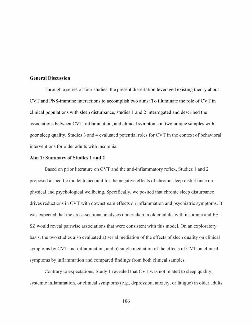
106
General Discussion
Through a series of four studies, the present dissertation leveraged existing theory about
CVT and PNS-immune interactions to accomplish two aims: To illuminate the role of CVT in
clinical populations with sleep disturbance, studies 1 and 2 interrogated and described the
associations between CVT, inflammation, and clinical symptoms in two unique samples with
poor sleep quality. Studies 3 and 4 evaluated potential roles for CVT in the context of behavioral
interventions for older adults with insomnia.
Aim 1: Summary of Studies 1 and 2
Based on prior literature on CVT and the anti-inflammatory reflex, Studies 1 and 2
proposed a specific model to account for the negative effects of chronic sleep disturbance on
physical and psychological wellbeing. Specifically, we posited that chronic sleep disturbance
drives reductions in CVT with downstream effects on inflammation and psychiatric symptoms. It
was expected that the cross-sectional analyses undertaken in older adults with insomnia and FE
SZ would reveal pairwise associations that were consistent with this model. On an exploratory
basis, the two studies also evaluated a) serial mediation of the effects of sleep quality on clinical
symptoms by CVT and inflammation, and b) single mediation of the effects of CVT on clinical
symptoms by inflammation and compared findings from both clinical samples.
Contrary to expectations, Study 1 revealed that CVT was not related to sleep quality,
systemic inflammation, or clinical symptoms (e.g., depression, anxiety, or fatigue) in older adults

107
with insomnia. Nor was either exploratory mediation model supported. Instead, the primary
finding was novel characterization of the associations between CVT and two upstream measures
of cellular inflammation. In older adults with current insomnia, greater CVT was associated with
lower levels of unstimulated monocyte coproduction of proinflammatory cytokines and greater
stimulated proinflammatory responses to LPS. These initial results suggest PNS-immune
coordination at an intracellular level and require further replication and investigation to articulate
clear clinical implications for older adults with and without insomnia.
Similarly, Study 2 found that CVT in FE SZ was not related to sleep quality as reflected
on the PSQI, levels of individual proinflammatory cytokines, or clinical symptoms (e.g.,
depression, fatigue, positive and negative symptoms of psychosis). However, sleep duration and
total time in bed were negatively associated with CVT. As predicted, and in line with prior
research on a vagally-mediated anti-inflammatory pathway (Tracey et al., 2002; 2009), we
extended a previously reported negative association between CVT and CRP in healthy
individuals to patients with FE SZ (e.g., Cooper et al., 2014; Sajadieh et al., 2004). This inverse
association between CVT and systemic inflammation provides preliminary evidence of intact
PNS-immune coordination in FE SZ. As hypothesized, results also revealed positive associations
between specific circulating cytokines, IL-6 and IFNγ, and the severity of positive symptoms of
psychosis.
Neither exploratory mediation model was supported in FE SZ. Given null mediation
results in both older adults and FE SZ and major differences in the drivers of poor sleep quality
in these clinical samples (i.e., insomnia vs. long sleep duration), comparisons of model paths
between groups were of limited consequence.
Aim 1: Implications of Studies 1 and 2

108
Across both studies, the lack of associations between CVT and clinical symptoms is
inconsistent with influential theories about CVT and argues against an essential role for CVT in
the symptoms of insomnia and SZ. Specifically, polyvagal theory, the neurovisceral integration
model, and other recent adaptations (e.g., vagal tank theory: Laborde, Mosley, & Mertgen,
2018b) commonly propose a link between CVT and a broad capacity for self-regulation (e.g.,
context appropriate physiological responses, attentional regulation, social behavior, emotion
regulation, and executive functioning). CVT is theorized to reflect prefrontal inhibitory control,
and disinhibition (i.e., low tonic CVT) has been associated with increased risk for
psychopathology (e.g., Beauchaine, 2015; Thayer & Brosschot, 2005). Though there is relatively
little research directly examining the contributions of CVT to clinical symptoms of insomnia,
prior findings have linked low CVT to a variety of psychiatric disorders in which cardinal
symptoms and sleep disturbance co-occur, such as major depressive disorder (Agelink et al.,
2002) and generalized anxiety disorder (Thayer, Friedman, & Borkovec, 1996). However, the
lack of correspondence between CVT and levels of depression, anxiety, fatigue, or positive and
negative symptoms of psychosis across Studies 1 and 2 challenges the proposition that CVT is a
reliable index of emotion dysregulation in insomnia and FE SZ.
Findings also provide mixed evidence for a vagally-mediated anti-inflammatory pathway
in sleep disturbed samples. Although the negative association between CVT and CRP in FE SZ
is consistent with Tracey’s model of the cholinergic anti-inflammatory pathway (2002; 2009),
results obtained in older adults were not. It is unclear why the relationship between CVT and
systemic inflammation differed between the two samples. One possibility is that age moderates
the negative association between CVT and CRP (Williams et al., 2019), such that the integrity of
this mechanism for PNS inhibition of inflammation diminishes with increasing age. Age-related
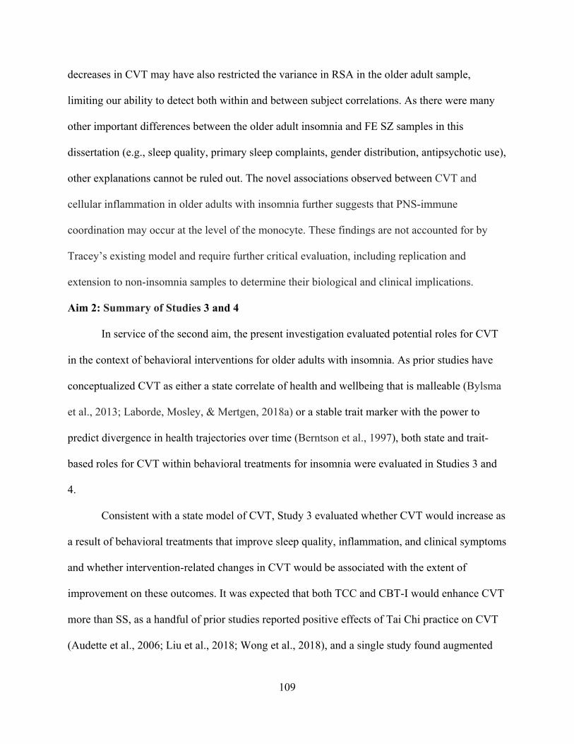
109
decreases in CVT may have also restricted the variance in RSA in the older adult sample,
limiting our ability to detect both within and between subject correlations. As there were many
other important differences between the older adult insomnia and FE SZ samples in this
dissertation (e.g., sleep quality, primary sleep complaints, gender distribution, antipsychotic use),
other explanations cannot be ruled out. The novel associations observed between CVT and
cellular inflammation in older adults with insomnia further suggests that PNS-immune
coordination may occur at the level of the monocyte. These findings are not accounted for by
Tracey’s existing model and require further critical evaluation, including replication and
extension to non-insomnia samples to determine their biological and clinical implications.
Aim 2: Summary of Studies 3 and 4
In service of the second aim, the present investigation evaluated potential roles for CVT
in the context of behavioral interventions for older adults with insomnia. As prior studies have
conceptualized CVT as either a state correlate of health and wellbeing that is malleable (Bylsma
et al., 2013; Laborde, Mosley, & Mertgen, 2018a) or a stable trait marker with the power to
predict divergence in health trajectories over time (Berntson et al., 1997), both state and trait-
based roles for CVT within behavioral treatments for insomnia were evaluated in Studies 3 and
4.
Consistent with a state model of CVT, Study 3 evaluated whether CVT would increase as
a result of behavioral treatments that improve sleep quality, inflammation, and clinical symptoms
and whether intervention-related changes in CVT would be associated with the extent of
improvement on these outcomes. It was expected that both TCC and CBT-I would enhance CVT
more than SS, as a handful of prior studies reported positive effects of Tai Chi practice on CVT
(Audette et al., 2006; Liu et al., 2018; Wong et al., 2018), and a single study found augmented

110
CVT in CBT-I treatment responders (Chung et al., 2011). Contrary to expectations, the results of
Study 3 indicated that CVT did not change significantly in response to 4 months of CBT-I, TCC,
or SS in older adults. Instead, CVT showed remarkable trait-like stability from pre-to-post
treatment. Additionally, intervention-related change in CVT did not predict the extent of
improvement on key trial outcomes, such as sleep quality, inflammation, or clinical symptoms.
Instead, behavioral treatments led to recovery from insomnia without significantly altering CVT.
Based on a trait conceptualization of CVT, and consistent with findings from Study 3 that
CVT was state invariant over 4 months of treatment, Study 4 evaluated whether pre-treatment
CVT moderated the effects of active treatment (CBT-I and TCC) vs. sleep education on key trial
outcomes (i.e., change in sleep quality, cellular and systemic markers of inflammation, and
depressive symptoms). As a previous study in younger adults with insomnia reported that pre-
treatment CVT predicted the extent of sleep recovery in response to CBT-I (Gouin & Dang Vu,
2015), it was hypothesized that participants with greater CVT at pre-treatment would benefit
more from active behavioral treatments than those with lower CVT at pre-treatment. Instead,
results from Study 4 revealed that pre-treatment CVT was not a moderator of response to these
behavioral interventions in late life insomnia. In fact, beyond failing to predict change on key
trial outcomes, pre-treatment CVT was found to be counterintuitively associated with treatment
engagement and drop out.
Aim 1: Implications of Studies 3 and 4
Overall, results obtained in Studies 3 and 4 challenge the idea that CVT plays a central
role in insomnia recovery. Novel findings from Study 3 extend treatment research by indicating
that change in CVT is unlikely to be a mechanism supporting improvement in insomnia,
inflammation, and clinical state in late life insomnia. This contribution is particularly informative

111
to the literature on mind-body interventions (e.g., Tai Chi, yoga), as it is often assumed that
beneficial effects are at least partially attributable to the upregulation of PNS activity (e.g., Liu et
al., 2018; Zou et al., 2018). Results of Study 3 echo those of Studies 1 and 2 and provide similar
challenges to contemporary theories about CVT by demonstrating very little correspondence
between CVT and symptoms. Results also raise an important set of new questions that require
further interrogation. For example, does the malleability of CVT change over the lifespan? And
is the stability of CVT observed in the present study attributable to normative aging processes or
a combination of aging and a chronic illness, such as insomnia? Additionally, given that the
results of Study 3 provide strong evidence that the upregulation of CVT is not a common
mechanism of these behavioral interventions in older adults, it is essential to identify and
investigate other candidate mechanisms to understand how CBT-I and TCC exert their effects on
sleep quality, inflammation, and clinical symptoms in this population.
Results of Study 4 further challenge current conceptualizations of CVT by providing
limited support for its utility as an individual, trait-like predictor of treatment response to
behavioral interventions for insomnia. As some of the results of Study 4 were in the opposite
direction of predictions based upon the neurovisceral integration model, they reveal a need for
careful empirical evaluation of the model’s assumptions and generalizability to clinical samples
across the lifespan. CVT is often heralded to be a stable, trait-like indicator of self-regulatory
capacity, with broad implications for physiological, emotional, and cognitive regulation, from
infancy to older adulthood (Thayer & Lane, 2009). However, present findings linking lower
CVT to greater treatment engagement in older adults with insomnia are inconsistent with such
assumptions. One possibility is that effect sizes for the associations between CVT and self-
regulatory capacity may be overestimated due to publication bias (e.g., Zahn et al., 2016) and

112
limited evaluation in older and chronically ill samples. Additional lifespan research is needed to
determine whether normative aging processes or the presence of chronic insomnia attenuate
relationships between CVT and self-regulatory capacity that were established earlier in life. Null
findings for CVT emphasize the need for further identification and evaluation of candidate
moderators to improve current knowledge about the patient characteristics that determine
treatment responses to behavioral interventions for insomnia.
Limitations and Future Directions
The present investigations had some important limitations. First, all four studies
exclusively focused on CVT, without evaluating other aspects of autonomic function (e.g., SNS
activity) that could more broadly inform knowledge about the interplay between chronic sleep
disturbance, variability in autonomic function, the regulation of inflammation, and the expression
of clinical symptoms. Although there were strong a priori reasons for the exclusive focus on
CVT, future studies should consider assessing profiles of autonomic activity using measures
from multiple times of day or 24-hour EKG to capture the stability and malleability of the
coordination of PNS and SNS activity in clinical samples with sleep disturbance. Another
limitation of the current research is that the interpretation of the reported associations between
CVT and cellular inflammatory processes in Study 1 (i.e., the negative correlation between CVT
and unstimulated monocyte production of pro-inflammatory cytokines and the positive
association between CVT and stimulated monocyte production of pro-inflammatory cytokines in
older adults) is constrained by a lack of prior studies evaluating these inflammatory measures in
healthy sleeping older adults. Additionally, COVID-19 related disruptions to data collection
impacted recruitment of the intended larger sample of FE SZ patients and aged-matched HCs for
Study 2. Finally, EKG acquisition methods (e.g., recording setting, time of day, and position)

113
differed between older adults with insomnia, FE SZ, and HC, potentially impacting between
group comparisons for RSA in Study 2.
Conclusions
Despite the noted limitations, present results contribute novel descriptions of the associations
between CVT and clinical state in two unique clinical samples with sleep disturbance and
demonstrate that CVT is neither a key mechanism nor moderator of behavioral interventions for
insomnia in older adults. As CVT appears to be less integral to sleep disturbance and insomnia
recovery than expected, results draw attention to a need for empirical assessment and critical
evaluation of theories about CVT in complex samples contending with a chronic illness across
the lifespan. Further studies are particularly needed to evaluate the clinical implications of the
correlations reported between CVT and cellular inflammation in older adults and CRP in FE SZ.
Null findings for CVT as a mechanism and moderator of treatment effects are a reminder that we
do not know, and must conduct additional research to determine, how the best behavioral
interventions work and for whom they will be most effective. Identification and evaluation of
mechanisms and moderators should be a top research priority to refine and maximize existing
treatments for insomnia.

114
Tables
Table 1. Baseline characteristics of participants in the sample with valid electrocardiogram data
(Study 1). Variable Participants
n = 106
Age 65.30 (6.83)
Female, n (%) 76 (71.70)
Ethnicity, Hispanic, n (%) 8 (7.55)
Race, Non-White, n (%)a 14 (13.59)
Marital Status, Married, n (%) 47 (44.34)
Education (years) 15.64 (1.48)
Body Mass Index (kg/m2) 25.63 (3.53)
Medical Comorbidity, n (%) 49 (46.23)
Cardiac Condition or
Medication use, n (%)
39 (36.79)
Beta blocker use, n (%) 8 (7.55)
Heart Rate 63.56 (8.74)
RSA 4.73 (1.00)
PSQI, total score 10.76 (2.86)
IDS-C, total score 9.31 (5.22)
BAI, total score 6.79 (6.86)
MFSI, total score 10.05 (17.56)
CRP (mg/L) 1.85 (1.93)
% Unstimulated monocytes
co-producing IL-6 and TNFα
1.02 (.54)
% Stimulated monocytes co-
producing IL-6 and TNFα
49.53 (18.47)
Note: All values are mean (SD) unless noted with percent. aTotal n=103 with self-reported race
(Non-white). RSA, Respiratory Sinus Arrhythmia; PSQI, Pittsburgh Sleep Quality Index; IDS-C,
Inventory of Depressive Symptoms-Clinician Rating Scale; BAI, Beck Anxiety Inventory;
MFSI, Multidimensional Fatigue Severity Index Short Form; CRP, C-reactive Protein.

115
Table 2. Correlations between RSA, inflammation, sleep, and clinical symptoms in older adult participants at pre-intervention (Study 1).
Variable RSA Heart Rate
CRP (mg/L)
% Unstim. IL-6/TNFα
% Stim. IL-6/TNFα
PSQI, total score
IDS-C, total score
BAI, total score
MFSI, total score
Age (years)
BMI (kg/m2)
Weekly METs
RSA 1 Heart Rate -.20* 1 CRP (mg/L) .03 -.02 1 % Unstim. IL-6/TNFα -.23* .08 .06 1
% Stim. IL-6/TNFα .25* -.20* -.02 .11 1
PSQI, total score -.06 -.03 -.05 .08 .15 1
IDS-C total score -.08 .21* -.17 .07 .01 .25** 1
BAI, total score -.03 .13 -.08 .07 -.03 .20* .29** 1
MFSI, total score .00 .23* -.06 .00 -.05 .37** .51** .53** 1
Age (years) -.19+ -.27** .13 -.02 .17 -.02 -.13 -.27** -.18+ 1 BMI (kg/m2) -.08 .21* .26* .06 -.09 .08 .08 .07 .09 -.08 1 Weekly METs .16 .04 -.20* -.11 .10 -.01 -.01 .01 -.11 -.28** -.08 1
Note: +p = .05-.07, *p < .05, **p < .01, ***p < .001. RSA, Respiratory Sinus Arrhythmia; CRP, C-reactive Protein; % Unstim. IL-6/TNFα, the percentage of unstimulated monocytes co-producing IL-6 and TNFα; % Stim. IL-6/TNFα, the percentage of stimulated monocytes co-producing IL-6 and TNFα; PSQI, Pittsburgh Sleep Quality Index; IDS-C, Inventory of Depressive Symptoms-Clinician Rating Scale; BAI, Beck Anxiety Inventory; MFSI, Multidimensional Fatigue Severity Index Short Form; BMI, body mass index; Weekly METs, metabolic equivalents.
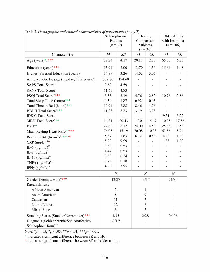
116
Table 3. Demographic and clinical characteristics of participants (Study 2)
Note: +p = .05, *p < .05, **p < .01, ***p < .001. * indicates significant difference between SZ and HC. * indicates significant difference between SZ and older adults.
Schizophrenia Patients (n = 39)
Healthy Comparison
Subjects (n = 30)
Older Adults with Insomnia
(n = 106)
Characteristic M SD M SD M SD Age (years)*/*** 22.23 4.17 20.17 2.25 65.30 6.83
Education (years)*** 13.94 2.00 13.70 1.30 15.64 1.48
Highest Parental Education (years)1 14.89 3.26 14.52 3.05 - -
Antipsychotic Dosage (mg/day, CPZ equiv.2) 332.86 194.60 - - - -
SAPS Total Score3 7.69 4.59 - - - -
SANS Total Score4 PSQI Total Score5*** Total Sleep Time (hours)*** Total Time in Bed (hours)*** BDI-II Total Score6*** IDS-C Total Score7 MFSI Total Score8** BMI9*
Mean Resting Heart Rate/+/***
Resting RSA (ln ms2)10***/* CRP (mg/L)11*
IL-6 (pg/mL)12 IL-8 (pg/mL)13 IL-10 (pg/mL)14 TNFa (pg/mL)15 IFNγ (pg/mL)16
11.59 5.55 9.30 10.94 11.28
- 14.31 27.62 76.05 5.37 5.90 0.60 1.44 0.30 0.79 4.86
4.83 3.19 1.87 2.88 8.23
- 20.43 6.77 15.19 1.83 9.59 0.53 0.53 0.24 0.18 3.95
- 4.76 6.92 8.46 3.19
- 1.30 24.00 70.08 6.72
- - - - - -
- 2.82 0.93 1.76 3.78
- 15.47 4.53 10.03 0.83
- - - - - -
- 10.76
- - -
9.31 10.05 25.63 63.56 4.73 1.85
- - - - -
- 2.86
- - -
5.22 17.56 3.53 8.74 1.00 1.93
- - - - -
N N N Gender (Female/Male)*** Race/Ethnicity African American Asian American Caucasian Latino/Latina Mixed Race
12/27 5 8 11 12 3
13/17 1 9 7 8 5
76/30 - - - - -
Smoking Status (Smoker/Nonsmoker)*** Diagnosis (Schizophrenia/Schizoaffective/ Schizophreniform)17
4/35 33/1/5
2/28 -
0/106 -

117
1Highest parental education (SZ n =37, HC n = 30). 2CPZ equiv. = chlorpromazine equivalents (SZ n =39). 3SAPS = Scale for the Assessment of Positive Symptoms (SZ n =35). 4 SANS = Scale for the Assessment of Negative Symptoms (SZ n =39). 5PSQI = Pittsburgh Sleep Quality Index (SZ n =38, HC n = 29, older adults n =105). 6BDI = Beck Depression Inventory II (SZ n =39, HC n = 28). 7IDS-C= Inventory of Depressive Symptoms (older adults, n = 105). 8MFSI = Multidimensional Fatigue Symptom Inventory Short Form (SZ n =39, HC n =30, older adults n = 105). 9BMI = Body Mass Index(SZ n =38, HC n = 19, older adults n =106). 10RSA = Respiratory Sinus Arrhythmia. 11CRP = C-reactive Protein (SZ n =31, older adults n =85), mg/L = milligrams per liter 12IL-6 = Interleukin-6 (SZ n =31), pg/mL = picograms per milliliter 13IL-8 = Interleukin-8 (SZ n =31) 14IL-10 = Interleukin-10 (SZ n =31) 15TNFa = Tumor necrosis factor alpha (SZ n =31) 16IFNγ = Interferon gamma (SZ n =31) 17Diagnoses (SZ n=36)

118
Table 4. Associations between sleep quality, RSA, inflammatory markers, clinical symptoms, and relevant covariates in patients with first-episode schizophrenia (Study 2).
PSQI Total Sleep Time
Total Time in Bed
Mean Heart Rate
RSA Log CRP
Log IL-6
Log IL-8
Log IL-10
Log TNFa
Log IFNγ
BDI Total Score
MFSI Total Score
SANS SAPS Age BMI CPZ
PSQI 1 -.21 .30+ .02 .08 -.03 .06 .04 .08 .07 .27 .25 .33* .05 .02 -.08 .29+ .02
Total Sleep Time
- 1 .73*** .37* -.48** .23 .10 .23 .36+ .14 -.04 .01 .08 .14 .07 -.26 -.21 .01
Total Time in Bed
- - 1 .41* -.48** -.07 -.12 .43* .24 .15 -.14 .18 .22 .10 -.08 -.29+ -.11 -.06
Mean Heart Rate
- - - 1 -.56*** .29 .31+ .25 .12 .22 .17 -.08 .08 .18 .12 -.09 .04 .09
RSA - - - - 1 -.37* -.17 -.06 -.01 -.10 .09 -.15 -.10 -.09 .01 .03 -.17 -.11 Log CRP
- - - - - 1 .82*** -.10 .27 .21 .39* .07 -.13 .18 .30 -.14 .65*** .11
Log IL-6 - - - - - - 1 -.19 .33+ .09 .48**
.00 -.19 .16 .41* -.09 .60*** .19
Log IL-8 - - - - - - - 1 .09 .37* -.09 -.02 .00 .29 -.13 -.24 .00 -.01 Log IL-10 - - - - - - - - 1 .20 .39* .35+ .27 .16 .28 -.22 -.08 -.12
Log TNFa
- - - - - - - - - 1 .13 .06 .19 .27 .27 -.48** .17 -.15
Log IFNγ - - - - - - - - - - 1 -.18 -.10 .14 .40* .00 .13 .20 BDI Total Score
- - - - - - - - - - - 1 .75*** -.04 .07 -.15 .23 -.45**
MFSI Total Score
- - - - - - - - - - - - 1 .02 .02 -.10 -.05 -.44**
SANS - - - - - - - - - - - - - 1 .55** -.21 .04 .09 SAPS - - - - - - - - - - - - - - 1 -.05 -.01 -.08 Age - - - - - - - - - - - - - - - 1 -.10 .18 BMI - - - - - - - - - - - - - - - - 1 -.02 CPZ - - - - - - - - - - - - - - - - - 1
Note: + <.09; p < .05, **p < .01, ***p < .001.

119
Table 5. Summary of path comparisons for the serial and simple mediation models between the schizophrenia and older adult samples (Study 2).
Model Outcome Path Older adults β
SZ β
Wald test
Wald test p-value
Serial mediation
Depression a1, PSQI-RSA -.06 .08 .45 .50 a2, PSQI-CRP -.07 -.01 .02 .90 d21, RSA-CRP .04 -.34* 3.90+ .05 b1, RSA-Depression -.06 -.17 .24 .63 b2, CRP-Depression -.08 -.02 .13 .72 c’, PSQI-Depression, direct effect .24** .26t .27 .60 c, PSQI-Depression, total effect .25** .25 - - m, PSQI-Depression, indirect effect .01 -.01 - -
Serial mediation
Fatigue a1, PSQI-RSA -.06 .08 .45 .50 a2, PSQI-CRP -.08 -.04 .00 .97 d21, RSA-CRP .04 -.35* 4.01+ .05 b1, RSA-Fatigue .03 -.19 1.13 .29 b2, CRP-Fatigue .02 -.22 1.08 .30 c’, PSQI-Fatigue, direct effect .37*** .33* .04 .85 c, PSQI-Fatigue, total effect .37*** .33* - - m, PSQI-Fatigue, indirect effect .00 .00 - -
Simple mediation
Depression a, RSA-CRP 03 -.34* 3.86+ .05 b, CRP-Depression -.10 -.01 .19 .66 c’, RSA-Depression, direct effect -.08 -.15 .09 .76 c, RSA-Depression, total effect -.08 -.15 - - m, RSA-Depression, indirect effect .00 .00 - -
Simple mediation
Fatigue a, RSA-CRP .03 -.35* 4.10* .04 b, CRP-Fatigue .00 -.26 1.03 .31 c’, RSA-Fatigue, direct effect .00 -.18 .61 .43 c, RSA-Fatigue, total effect .00 -.10 - - m, RSA-Fatigue, indirect effect .00 .09 - -
Note: +<.09; *p < .05, **p < .01, ***p < .001. Wald tests compare the relative strength of the path between groups (all df = 1). All results are standardized coefficients. CRP data were log transformed for both samples for these analyses.

120
Table 6. Characteristics of participants by treatment group at pre-intervention (Study 3).
TCC
n = 38
CBT-I
n = 45
SS
n = 23
Age (years) 66.39 (7.32) 64.20 (5.98) 65.65 (7.51)
Gender, Female, n (%) 26 (68.42) 34 (75.56) 16 (69.57)
Ethnicity, Hispanic, n (%) 2 (5.26) 3 (6.67) 3 (13.04)
Race, Non-White, n (%)a 5 (13.51) 5 (11.36) 4 (18.18)
Marital Status, Married, n (%) 17 (44.74) 22 (48.89) 8 (34.78)
Education (years) 15.76 (1.48) 15.78 (1.44) 15.17 (1.48)
Body Mass Index (kg/m2) 26.03 (3.64) 25.36 (3.33) 25.48 (3.80)
Medical comorbidity, n (%)b 19 (50.00) 18 (40.00) 12 (52.17)
Depression history, n (%) 9 (23.68) 12 (26.67) 8 (34.78)
Other psychiatric history, n (%) 5 (13.16) 8 (17.78) 2 (8.70)
Cardiac condition, n (%) 10 (26.32) 7 (15.56) 4 (17.39)
Cardiac medication use, n (%) 13 (34.21) 13 (28.89) 8 (34.78)
Heart Rate 62.94 (9.71) 64.10 (7.86) 63.51 (8.99)
RSA 4.76 (.92) 4.80 (1.05) 4.57 (1.03)
PSQI total scorec 11.01 (2.96) 10.32 (2.72) 11.24 (2.98)
IDS-C, total scorec 9.51 (4.77) 8.68 (4.71) 10.23 (6.73)
BAI, total score 7.26 (6.19) 7.02 (7.93) 5.57 (5.66)
MFSI, total scored 9.87 (16.56) 10.11 (18.68) 10.22 (17.74)
CRPe 1.83 (1.95) 1.85 (1.97) 1.87 (1.92)
% Unstimulated monocytes
co-producing IL-6 and TNFαf .99 (.41) 1.02 (.61) 1.10 (.55)
% Stimulated monocytes
co-producing IL-6 and TNFαg*** 59.23 (19.94) 44.59 (15.43) 43.24 (15.27) Note: All values are mean (SD) unless noted with percent. Between-group differences were tested with a chi-square test for categorical variables and one-way analysis of variance for continuous variables. *p < .05, **p < .01, ***p < .001. aTotal n=103 (Non-white) bTotal n=102 (Medical Comorbidity) cTCC n=37, CBT-I n=45, SS n=23. (PSQI and IDS-C) dTCC n=38, CBT-I n=44, SS n=23. (MFSI) eTCC n=28, CBT-I n=36, SS n=21. (CRP)

121
fTCC n=35, CBT-I n=44, SS n=21. (Unstimulated Monocytes) gTCC n=36, CBT-I n=44, SS n=21. (Stimulated Monocytes) TCC, Tai Chi Chih; CBT-I, cognitive behavioral therapy for insomnia; SS, Sleep Seminar; RSA, Respiratory Sinus Arrhythmia; PSQI, Pittsburgh Sleep Quality Index; IDS-C, Inventory of Depressive Symptoms-Clinician Rating Scale; BAI, Beck Anxiety Inventory; MFSI, Multidimensional Fatigue Severity Index Short Form; CRP, C-reactive Protein
Table 7. Means and standard deviations of RSA by treatment group at pre and post intervention
(Study 3).
Group Pre-Treatment RSA
Post-Treatment RSA
Active Treatments (TCC + CBT-I)a 4.78 (.99) 4.61 (.92)
TCCb 4.76 (.92) 4.48 (.80) CBT-Ic 4.80 (1.05) 4.71 (1.01)
SSd 4.57 (1.03) 4.59 (1.12) Note: All values are mean (SD). RSA, Respiratory Sinus Arrhythmia; TCC, Tai Chi Chih; CBT-I, cognitive behavioral therapy for insomnia; SS, Sleep Seminar. All p >.05. Total n at pre n=106, post n=82. aFor Active Treatments: pre n=83, post n=63. bFor TCC: pre n=38, post n=29. cFor CBT-I: pre n=45, post n=34. dFor SS: pre n=23, post n=19.

122
Table 8. Hierarchical regressions predicting the percent of group treatment sessions attended from relevant covariates and pre-treatment RSA (Study 4).
Predictors
Percent of treatment sessions attended
DR2
b
Step 1: Age BMI Gender Beta blocker use Weekly METs
.02
.14 -.03 .05 -.04 -.03
Step 2: Pre-treatment RSA
.03
-.17
Step 1: Total R2 .03 Step 2: Total R2 .05
Note: Total n = 101. All p > .05. BMI: body mass index; METs: metabolic equivalents; RSA: Respiratory Sinus Arrhythmia.
Figures
Figure 1. General model of the proposed directional relationships between sleep quality, CVT, inflammation, and clinical symptoms.
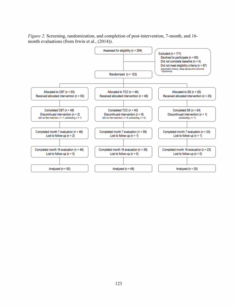
123
Figure 2. Screening, randomization, and completion of post-intervention, 7-month, and 16-month evaluations (from Irwin et al., (2014)).

124
Figure 3. Statistical models of the serial and single mediation models (Study 2).

125
Figure 4. Modified CONSORT diagram showing randomization and completion of post-intervention, 7-month, and 16-month evaluations for the parent trial and Study 3.
Note: Ns from the parent trial are listed in black. Ns below, in blue, represent the number of participants with complete RSA data included in Study 3.

126
Figure 5. Pre to post change in RSA by treatment group (Study 3).
Note: Total n= 80.

127
Appendix Table 1A. Baseline characteristics of older adult participants, by treatment group (Study 1).

128
References Agelink, M. W., Boz, C., Ullrich, H., & Andrich, J. (2002). Relationship between major
depression and heart rate variability. Clinical consequences and implications for
antidepressive treatment. Psychiatry research, 113(1-2), 139-149.
Aldao, A., Dixon-Gordon, K. L., & De Los Reyes, A. (2016). Individual differences in
physiological flexibility predict spontaneous avoidance. Cognition and emotion, 30(5),
985-998.
Allen, J. J. B. (2002). Calculating metrics of cardiac chronotropy: A pragmatic overview.
Psychophysiology, 39, S18.
Allen, J. J. B., Chambers, A. S., & Towers, D. N. (2007). The many metrics of cardiac
chronotropy: A pragmatic primer and a brief comparison of metrics. Biological
psychology, 74, 243–262.
Alvares, G. A., Quintana, D. S., Kemp, A. H., Van Zwieten, A., Balleine, B. W., Hickie, I. B., &
Guastella, A. J. (2013). Reduced heart rate variability in social anxiety disorder:
Associations with gender and symptom severity. PloS one, 8(7), e70468.
doi:10.1371/journal.pone.0070468.
American Academy of Sleep Medicine. (2005). International Classification of Sleep Disorders:
Diagnostic and Coding Manual (2nd ed.). Westchester, IL: American Academy of Sleep
Medicine.
American Psychiatric Association. (2000). Diagnostic and Statistical Manual of Mental
Disorders (DSM-IV-TR) (4th ed). Text Revision. Washington, DC: American Psychiatric
Association.

129
American Psychiatric Association (2013). DSM-5 Task Force: Diagnostic and Statistical Manual
of Mental Disorders (5th ed.). Washington, DC, American Psychiatric Association.
An, H., Han, J. W., Jeong, H. G., Kim, T. H., Lee, J. J., Lee, S. B., ... & Kim, K. W. (2020).
Parasympathetic predominance is a risk factor for future depression: A prospective cohort
study. Journal of affective disorders, 260, 232-237.
Andreasen, N.C. (1984a). The Scale for the Assessment of Positive Symptoms (SAPS).
University of Iowa, Iowa City, IA.
Andreasen, N.C., (1984b). The Scale for the Assessment of Negative Symptoms (SANS).
University of Iowa, Iowa City, IA.
Andreasen, N.C., Pressler, M., Nopoulos, P., Miller, D., & Ho, B.C. (2010). Antipsychotic dose
equivalents and dose-years: A standardized method for comparing exposure to different
drugs. Biological psychiatry, 67, 255-62. doi: 10.1016/j.biopsych.2009.08.040.
Angelovski, A., Sattel, H., Henningsen, P., & Sack, M. (2016). Heart rate variability predicts
therapy outcome in pain-predominant multisomatoform disorder. Journal of
psychosomatic research, 83, 16-21.
Appelhans, B. M., & Luecken, L. J. (2006). Heart rate variability as an index of regulated
emotional responding. Review of general psychology, 10(3), 229-240.
Asarnow, L. D., Bei, B., Krystal, A., Buysse, D. J., Thase, M. E., Edinger, J. D., & Manber, R.
(2019). Circadian preference as a moderator of depression outcome following cognitive
behavioral therapy for insomnia plus antidepressant medications: A report from the
TRIAD study. Journal of clinical sleep medicine, 15(4), 573-580.
Audette, J. F., Jin, Y. S., Newcomer, R., Stein, L., Duncan, G., & Frontera, W. R. (2006). Tai
Chi versus brisk walking in elderly women. Age and ageing, 35(4), 388-393.

130
Aw, D., Silva, A. B., & Palmer, D. B. (2007). Immunosenescence: Emerging challenges for an
ageing population. Immunology, 120(4), 435–446. doi:10.1111/j.1365-
2567.2007.02555.x.
Balogh, S., Fitzpatrick, D. F., Hendricks, S. E., & Paige, S. R. (1993). Increases in heart rate
variability with successful treatment in patients with major depressive disorder.
Psychopharmacology bulletin, 29(2), 201-206.
Balzarotti, S., Biassoni, F., Colombo, B., & Ciceri, M. R. (2017). Cardiac vagal control as a
marker of emotion regulation in healthy adults: A review. Biological psychology, 130,
54-66.
Bär, K. J., Letzsch, A., Jochum, T., Wagner, G., Greiner, W., & Sauer, H. (2005). Loss of
efferent vagal activity in acute schizophrenia. Journal of psychiatric research, 39, 519–
527.
Barr, T., Livingston, W., Guardado, P., Baxter, T., Mysliwiec, V., & Gill, J. (2015). Military
personnel with traumatic brain injuries and insomnia have reductions in PTSD and
improved perceived health following sleep restoration. Annual review of nursing
research, 33, 249-266.
Bassett, D. (2016). A literature review of heart rate variability in depressive and bipolar
disorders. The Australian and New Zealand journal of psychiatry, 50, 511–519.
doi:10.1177/0004867415622689.
Bassi, G. S., Kanashiro, A., Santin, F. M., de Souza, G. E., Nobre, M. J., & Coimbra, N. C.
(2012). Lipopolysaccharide‐induced sickness behaviour evaluated in different models of
anxiety and innate fear in rats. Basic and clinical pharmacology and toxicology, 110(4),
359-369.

131
Batselé, E., Eynde, S. V., Egée, N., Lamotte, M., Borne, P.V., & Fantini-Hauwel, C. (2019).
Difficulties on emotion regulation predict mental health of coronary heart disease patients
in cardiac rehabilitation but heart rate variability does not. Online journal of
cardiovascular research. 2(4), doi:10.33552/OJCR.2019.02.000543.
Beauchaine, T. P. (2015). Respiratory sinus arrhythmia: A transdiagnostic biomarker of emotion
dysregulation and psychopathology. Current opinion in psychology, 3, 43-47.
Beauchaine, T.P., Gatzke-Kopp, L., & Mead, H.K. (2007). Polyvagal theory and developmental
psychopathology: Emotion dysregulation and conduct problems from preschool to
adolescence. Biological psychology, 74, 174-184. doi:10.1016/j.biopsycho.2005.08.008.
Beck, A. T., Epstein, N., Brown, G., & Steer, R. A. (1988). An inventory for measuring clinical
anxiety: Psychometric properties. Journal of consultation in clinical psychology, 56(6),
893–897.
Beck, A. T., Steer, R. A., & Brown, G. K. (1996). Manual for the Beck Depression Inventory-II.
San Antonio, TX: Psychological Corporation.
Berntson, G. G., Bigger, J. T. Jr., Eckberg, D. L., Grossman, P., Kaufmann, P. G., Malik, M., …
& van der Molen, M. W. (1997). Heart rate variability: origins, methods, and interpretive
caveats. Psychophysiology, 34(6), 623-648. doi:10.1111/j.1469-8986.1997.tb02140.x.
Berntson, G. G., Cacioppo, J. T., & Quigley, K. S. (1993). Respiratory sinus arrhythmia:
Autonomic origins, physiological mechanisms, and psychophysiological implications.
Psychophysiology, 30(2), 183-196.
Bertsch, K., Hagemann, D., Naumann, E., Schachinger, H., & Schulz A. (2012). Stability of
heart rate variability indices reflecting parasympathetic activity. Psychophysiology, 49,
672–682. doi:10.1111/j.1469-8986.2011.01341.x.

132
Bjurström, M. F., Olmstead, R., & Irwin, M. R. (2017). Reciprocal relationship between sleep
macrostructure and evening and morning cellular inflammation in rheumatoid arthritis.
Psychosomatic medicine, 79(1), 24–33. doi:10.1097/PSY.0000000000000363.
Boettger, S., Hoyer, D., Falkenhahn, K., Kaatz, M., Yeragani, V. K., & Bär, K. J. (2006). Altered
diurnal autonomic variation and reduced vagal information flow in acute schizophrenia.
Clinical neurophysiology, 117(12), 2715-2722.
Bonnemeier, H., Richardt, G., Potratz, J., Wiegand, U. K., Brandes, A., Kluge, N., & Katus H.
A. (2003). Circadian profile of cardiac autonomic nervous modulation in healthy
subjects: Differing effects of aging and gender on heart rate variability. Journal of
cardiovascular electrophysiology, 14, 791–799. doi:10.1046/j.1540-8167.2003.03078.x.
Bonnet, M. H., & Arand, D. L. (1998). Heart rate variability in insomniacs and matched normal
sleepers. Psychosomatic medicine, 60, 610-615.
Bonnet, M. H., & Arand, D. L. (2010). Hyperarousal and insomnia: State of the science. Sleep
medicine reviews, 14(1), 9-15. doi:10.1016/j.smrv.2009.05.002.
Borovikova, L. V., Ivanova, S., Zhang, M., Yang, H., Botchkina, G. I., Watkins, L. R., ... &
Tracey K. J. (2000). Vagus nerve stimulation attenuates the systemic inflammatory
response to endotoxin. Nature, 405, 458–461.
Bower, J. E. (2008). Behavioral symptoms in breast cancer patients and survivors: Fatigue,
insomnia, depression, and cognitive disturbance. Journal of clinical oncology, 26(5),
768–777. http://doi.org/10.1200/JCO.2007.14.3248.
Brosschot, J. F., Gerin, W., & Thayer, J. F. (2006). The perseverative cognition hypothesis: A
review of worry, prolonged stress-related physiological activation, and health. Journal of
psychosomatic research, 60(2), 113-124.

133
Brown, L., Karmakar, C., Gray, R., Jindal, R., Lim, T., & Bryant, C. (2018). Heart rate
variability alterations in late life depression: a meta-analysis. Journal of affective
disorders, 235, 456-466.
Burgos, I., Richter, L., Klein, T., Fiebich, B., Feige, B., Lieb, K., ... & Riemann, D. (2006).
Increased nocturnal interleukin-6 excretion in patients with primary insomnia: A pilot
study. Brain, behavior, and immunity, 20(3), 246-253.
Buysse, D. J., Reynolds, C. F., Monk, T. H., Berman, S. R., & Kupfer, D. J. (1989). The
Pittsburgh Sleep Quality Index: A new instrument for psychiatric practice and research.
Psychiatry research, 28(2), 193-213.
Bylsma, L. M., Salomon, K., Taylor-Clift, A., Morris, B. H., & Rottenberg, J. (2013).
Respiratory sinus arrhythmia reactivity in current and remitted major depressive disorder.
Psychosomatic medicine, 76(1), 66-73.
Camm, A. J., Malik, M., Bigger, J. T., Breithardt, G., Cerutti, S., Cohen, R. J., ... & Lombardi, F.
(1996). Heart rate variability: Standards of measurement, physiological interpretation and
clinical use. Task Force of the European Society of Cardiology and the North American
Society of Pacing and Electrophysiology. Circulation, 93, 1043–1065.
Cappuccio, F. P., D'Elia, L., Strazzullo, P., & Miller, M. A. (2010). Sleep duration and all-cause
mortality: a systematic review and meta-analysis of prospective studies. Sleep, 33(5),
585-592.
Carney, C. E., Moss, T. G., Harris, A. L., Edinger, J. D., & Krystal, A. D. (2011). Should we be
anxious when assessing anxiety using the Beck Anxiety Inventory in clinical insomnia
patients?. Journal of psychiatric research, 45(9), 1243–1249.
https://doi.org/10.1016/j.jpsychires.2011.03.011.

134
Carney, R. M., Freedland, K. E., Stein, P. K., Skala, J. A., Hoffman, P., & Jaffe, A. S. (2000).
Change in heart rate and heart rate variability during treatment for depression in patients
with coronary heart disease. Psychosomatic medicine, 62(5), 639-647.
Carroll, J. E., Carrillo, C., Olmstead, R., Witarama, T., Breen, E. C., Yokomizo, M., … Irwin, M.
R. (2015). Sleep deprivation and divergent toll-like receptor-4 activation of cellular
inflammation in aging. Sleep, 38(2), 205-211. doi:10.5665/sleep.4398.
Carroll, J. E., Esquivel, S., Goldberg, A., Seeman, T. E., Effros, R. B., Dock, J., Olmstead, R.,
Breen, E. C., & Irwin, M. R. (2016). Insomnia and telomere length in older dults. Sleep,
39(3), 559–564. https://doi.org/10.5665/sleep.5526.
Carroll, J. E., Irwin, M. R., Levine, M., Seeman, T. E., Absher, D., Assimes, T., & Horvath, S.
(2017). Epigenetic aging and immune senescence in women with insomnia symptoms:
Findings from the women's health initiative study. Biological psychiatry, 81(2), 136–144.
https://doi.org/10.1016/j.biopsych.2016.07.008.
Castro-Diehl, C., Diez Roux, A. V., Redline, S., Seeman, T., McKinley, P., Sloan, R., & Shea, S.
(2016). Sleep duration and quality in relation to autonomic nervous system measures:
The multi-ethnic study of atherosclerosis (MESA). Sleep, 39(11), 1927-1940.
doi:10.5665/sleep.6218.
Cellini, N., De Zambotti, M., Covassin, N., Sarlo, M., & Stegagno, L. (2014). Working memory
impairment and cardiovascular hyperarousal in young primary insomniacs.
Psychophysiology, 51(2), 206–214. doi:10.1111/psyp.12167.
Chalmers, J. A., Quintana, D. S., Abbott, M. J., & Kemp, A. H. (2014). Anxiety disorders are
associated with reduced heart rate variability: A meta-analysis. Frontiers in psychiatry, 5,
80. doi:10.3389/fpsyt.2014.00080.
135
Chen, H. C., Yang, C. C., Kuo, T. B., Su, T. P., & Chou, P. (2012). Cardiac vagal control and
theoretical models of co-occurring depression and anxiety: A cross-sectional
psychophysiological study of community elderly. BMC psychiatry, 12(1), 93.
Chien, H. C., Chung, Y. C., Yeh, M. L., & Lee J. F. (2015). Breathing exercise combined with
cognitive behavioural intervention improves sleep quality and heart rate variability in
major depression. Journal of clinical nursing, 24(21-22), 3206-3214.
doi:10.1111/jocn.12972.
Cho, H. J., Eisenberger, N. I., Olmstead, R., Breen, E. C., & Irwin, M. R. (2016). Preexisting
mild sleep disturbance as a vulnerability factor for inflammation-induced depressed
mood: A human experimental study. Translational psychiatry, 6(3), e750.
doi:10.1038/tp.2016.23.
Cho, H. J., Kivimäki, M., Bower, J. E., & Irwin, M. R. (2012). Association of C-reactive protein
and interleukin-6 with new-onset fatigue in the Whitehall II prospective cohort study.
Psychological medicine, 43(8), 1773–1783. doi:10.1017/S0033291712002437.
Chung, M. S., Yang, A. C., Lin, Y. C., Lin, C. N., Chang, F. R., Shen, S. H., ... & Chiu, H. J.
(2013). Association of altered cardiac autonomic function with psychopathology and
metabolic profiles in schizophrenia. Psychiatry research, 210(3), 710-715.
https://doi.org/10.1016/j.psychres.2013.07.034.
Chung, S., An, H., Park, J., & Kim, H. (2011). The effect of non-pharmacological treatment for
psychophysiological insomnia on cardiovascular autonomic regulation assessed using
heart rate variability. Sleep medicine research, 2(1), 10–15.
doi:10.17241/smr.2011.2.1.10.
136
Clamor, A., Lincoln, T., Thayer, J., & Koenig, J. (2016). Resting vagal activity in schizophrenia:
Meta-analysis of heart rate variability as a potential endophenotype. British journal of
psychiatry, 208(1), 9-16. doi:10.1192/bjp.bp.114.160762.
Colzato, L. S., & Steenbergen, L. (2017). High vagally mediated resting-state heart rate
variability is associated with superior action cascading. Neuropsychologia, 106, 1-6.
Cooper, T. M., McKinley, P. S., Seeman, T. E., Choo, T. H., Lee, S., & Sloan, R. P. (2014).
Heart rate variability predicts levels of inflammatory markers: Evidence for the vagal
anti-inflammatory pathway. Brain, behavior, and immunity, 49, 94-100.
Corfield, E. C., Martin, N. G., & Nyholt, D. R. (2016). Co-occurrence and symptomatology of
fatigue and depression. Comprehensive psychiatry, 71, 1-10.
Corsi-Zuelli, F., Brognara, F., Quirino, G., Hiroki, C. H., Fais, R. S., Del-Ben, C. M., … &
Loureiro, C. M. (2017). Neuroimmune interactions in schizophrenia: Focus on vagus
nerve stimulation and activation of the alpha-7 nicotinic acetylcholine receptor.
Frontiers in immunology, 8, 618. doi:10.3389/fimmu.2017.00618.
Cricco, M., Simonsick, E. M., & Foley, D. J. (2001). The impact of insomnia on cognitive
functioning in older adults. Journal of the American Geriatrics Society, 49(9), 1185-
1189.
Crosswell, A., D., Lockwood, K. G., Ganz, P. A., & Bower, J. E. (2014). Low heart rate
variability and cancer-related fatigue in breast cancer survivors.
Psychoneuroendocrinology, 45, 58-66. doi:10.1016/j.psyneuen.2014.03.011.
Dalise, A. M., Prestano, R., Fasano, R., Gambardella, A., Barbieri, M., & Rizzo, M. R. (2020).
Autonomic nervous system and cognitive impairment in older patients: Evidence from
long-term heart rate variability in real-life setting. Frontiers in aging neuroscience, 12.

137
Dantzer, R., & Kelley, K. W. (2006). Twenty years of research on cytokine-induced sickness
behavior. Brain, behavior, and immunity, 21(2), 153–160. doi:10.1016/j.bbi.2006.09.006.
Dantzer, R., O'Connor, J. C., Freund, G. G., Johnson, R. W., & Kelley, K. W. (2008). From
inflammation to sickness and depression: When the immune system subjugates the brain.
Nature reviews. Neuroscience, 9(1), 46-56.
Davies, C. D., Niles, A. N., Pittig, A., Arch, J. J., & Craske, M. G. (2015). Physiological and
behavioral indices of emotion dysregulation as predictors of outcome from cognitive
behavioral therapy and acceptance and commitment therapy for anxiety. Journal of
behavioral therapy and experimental psychiatry, 46, 35-43.
doi:10.1016/j.jbtep.2014.08.002.
Davies, G., Haddock, G., Yung, A. R., Mulligan, L. D., & Kyle, S. D. (2017). A systematic
review of the nature and correlates of sleep disturbance in early psychosis. Sleep
medicine reviews, 31, 25-38. doi:10.1016/j.smrv.2016.01.001.
DellaGioia, N., Devine, L., Pittman, B., & Hannestad, J. (2013). Bupropion pre-treatment of
endotoxin-induced depressive symptoms. Brain, behavior, and immunity, 31, 197-204.
De Mooij, L. D., Kikkert, M., Theunissen, J., Beekman, A. T., de Haan, L., Duurkoop, P. W., ...
& Dekker, J. J. (2019). Dying too soon: excess mortality in severe mental
illness. Frontiers in psychiatry, 10, 855.
De Oliveira Matos, F., Vido, A., Garcia, W. F., Lopes, W. A., & Pereira, A. (2020). A
neurovisceral integrative study on cognition, heart rate variability, and fitness in the
elderly. Frontiers in aging neuroscience, 12, 51. doi:10.3389/fnagi.2020.00051.

138
Dew, M. A., Hoch, C. C., Buysse, D. J., Monk, T. H., Begley, A. E., Houck, P. R., … &
Reynolds, C. F. III. (2003). Healthy older adults' sleep predicts all-cause mortality at 4 to
19 years of follow-up. Psychosomatic medicine, 65, 63–73.
Dodds, K. L., Miller, C. B., Kyle, S. D., Marshall, N. S., & Gordon, C. J. (2017). Heart rate
variability in insomnia patients: A critical review of the literature. Sleep medicine
reviews, 33, 88-100.
Doi, Y., Minowa, M., Uchiyama, M., Okawa, M., Kim, K., Shibui, K., & Kamei, Y. (2000).
Psychometric assessment of subjective sleep quality using the Japanese version of the
Pittsburgh Sleep Quality Index (PSQI-J) in psychiatric disordered and control subjects.
Psychiatry research, 97(2-3), 165-172.
Donovan, K. A., Stein, K. D., Lee, M., Leach, C. R., Ilozumba, O., & Jacobsen, P. B. (2015).
Systematic review of the multidimensional fatigue symptom inventory-short form.
Supportive care in cancer, 23(1), 191-212. doi:10.1007/s00520-014-2389-7.
Dooley, L. N., Kuhlman, K. R., Robles, T. F., Eisenberger, N. I., Craske, M. G., & Bower, J. E.
(2018). The role of inflammation in core features of depression: Insights from paradigms
using exogenously-induced inflammation. Neuroscience and biobehavioral reviews, 94,
219-237. doi:10.1016/j.neubiorev.2018.09.006.
Eisenberger, N. I., Berkman, E. T., Inagaki, T. K., Rameson, L. T., Mashal, N. M., & Irwin, M.
R. (2010). Inflammation-induced anhedonia: Endotoxin reduces ventral striatum
responses to reward. Biological psychiatry, 68(8), 748-754.
Escorihuela, R. M., Capdevila, L., Castro, J. R., Zaragozà, M. C., Maurel, S., Alegre, J., &
Castro-Marrero, J. (2020). Reduced heart rate variability predicts fatigue severity in
139
individuals with chronic fatigue syndrome/myalgic encephalomyelitis. Journal of
translational medicine, 18(1), 1-12.
Fabes, R. A., & Eisenberg N. (1997). Regulatory control and adults’ stress-related response to
daily life events. Journal of personality and social psychology, 73, 1107–1117.
doi:10.1037/0022-3514.73.5.1107.
Fagundes, C. P., Murray, D. M., Hwang, B. S., Gouin, J.P., Thayer, J. F., Sollers, J. J., … &
Kiecolt-Glaser, J. K. (2011). Sympathetic and parasympathetic activity in cancer-related
fatigue: More evidence for a physiological substrate in cancer survivors.
Psychoneuroendocrinology, 36(8), 1137-1147.
Fang, S. C., Huang, C. J., Yang, T. T., & Tsai, P. S. (2008). Heart rate variability and daytime
functioning in insomniacs and normal sleepers: Preliminary results. Journal of
psychosomatic research, 65, 23–30.
Faugere, M., Micoulaud-Franchi, J. A., Faget-Agius, C., Lançon, C., Cermolacce, M., &
Richieri, R. (2018). High C-reactive protein levels are associated with depressive
symptoms in schizophrenia. Journal of affective disorders, 225, 671-675.
doi:10.1016/j.jad.2017.09.004.
Faul, F., Erdfelder, E., Buchner, A., & Lang, A. G. (2008). G*Power Version 3.1.2 [computer
software]. Uiversität Kiel, Germany. Retrieved from http://www.psycho.uni-
duesseldorf.de/abteilungen/aap/gpower3/download-and-register.
First, M. B., Williams, J. B. W., Karg, R. S., & Spitzer, R. L. (2015). Structured Clinical
Interview for DSM-5, Research Version (SCID-5 for DSM-5, Research Version; SCID-5-
RV). Arlington, VA, American Psychiatric Association.

140
Franceschi, C., & Campisi, J. (2014). Chronic inflammation (inflammaging) and its potential
contribution to age-associated diseases. Journals of gerontology series A: Biomedical
sciences and medical sciences, 69(Suppl_1), S4-S9.
Frasure-Smith, N., Lespérance, F., Irwin, M. R., Talajic, M., & Pollock, B. G. (2009). The
relationships among heart rate variability, inflammatory markers and depression in
coronary heart disease patients. Brain, behavior, and immunity, 23(8), 1140-1147.
Frenois, F., Moreau, M., O'Connor, J., Lawson, M., Micon, C., Lestage, J., … & Castanon, N.
(2007). Lipopolysaccharide induces delayed FosB/DeltaFosB immunostaining within the
mouse extended amygdala, hippocampus and hypothalamus, that parallel the expression
of depressive-like behavior. Psychoneuroendocrinology, 32(5), 516–531.
doi:10.1016/j.psyneuen.2007.03.005.
Friedrich, A., Claßen, M., & Schlarb, A. A. (2018). Sleep better, feel better? Effects of a CBT-I
and HT-I sleep training on mental health, quality of life and stress coping in university
students: A randomized pilot controlled trial. BMC psychiatry, 18(1), 268.
doi:10.1186/s12888-018-1860-2.
Frommberger, U. H., Bauer, J., Haselbauer, P., Fräulin, A., Riemann, D., & Berger, M. (1997).
Interleukin-6-(IL-6) plasma levels in depression and schizophrenia: Comparison between
the acute state and after remission. European archives of psychiatry and clinical
neuroscience, 247, 228–233.
Garakani, A., Martinez, J. M., Aaronson, C. J., Voustianiouk, A., Kaufmann, H., & Gorman, J.
M. (2009). Effect of medication and psychotherapy on heart rate variability in panic
disorder. Depression and Anxiety, 26(3), 251-258.

141
Geisler, F. C., & Kubiak, T. (2009). Heart rate variability predicts self‐control in goal pursuit.
European journal of personality, 23(8), 623-633.
Gidron, Y., Deschepper, R., De Couck, M., Thayer, J. F., & Velkeniers, B. (2018). The vagus
nerve can predict and possibly modulate non-communicable chronic diseases:
introducing a neuroimmunological paradigm to public health. Journal of clinical
medicine, 7(10), 371.
Gillie, B. L., & Thayer, J. F. (2014). Individual differences in resting heart rate variability and
cognitive control in posttraumatic stress disorder. Frontiers in psychology, 5, 758.
doi:10.3389/fpsyg.2014.00758.
Gillie, B. L., Vasey, M. W., & Thayer, J. F. (2014). Heart rate variability predicts control over
memory retrieval. Psychological science, 25(2), 458-465.
http://dx.doi.org/10.1177/0956797613508789.
Gimeno, D., Kivimäki, M., Brunner, E. J., Elovainio, M., De Vogli, R., Steptoe, A., … & Ferrie,
J. E. (2008). Associations of c-reactive protein and interleukin-6 with cognitive
symptoms of depression: 12-year follow-up of the Whitehall II study. Psychological
medicine, 39(3), 413–423. doi:10.1017/S0033291708003723.
Goldsmith, D. R., Rapaport, M. H., & Miller, B. J. (2016). A meta-analysis of blood cytokine
network alterations in psychiatric patients: Comparisons between schizophrenia, bipolar
disorder and depression. Molecular psychiatry, 21(12), 1696–1709.
doi:10.1038/mp.2016.3.
Golosheykin, S., Grant, J. D., Novak, O. V., Heath, A. C., & Anokhin, A. P. (2016). Genetic
influences on heart rate variability. International journal of psychophysiology, 115, 65–
73. doi:10.1016/j.ijpsycho.2016.04.008.

142
Gouin, J., & Dang Vu, T. (2015). High frequency heart rate variability as predictor of treatment
response to cognitive-behavioral therapy for insomnia. Sleep medicine, 16, 1, S10.
https://doi.org/10.1016/j.sleep.2015.02.022.
Gouin, J., Wenzel, K., Deschenes, S., & Dang-Vu, T. (2013). Heart rate variability predicts sleep
efficiency. Sleep medicine, 14(1), e142. https://doi.org/10.1016/j.sleep.2013.11.321.
Grandner, M. A., Buxton, O. M., Jackson, N., Sands-Lincoln, M., Pandey, A., & Jean-Louis, G.
(2013). Extreme sleep durations and increased C-reactive protein: effects of sex and
ethnoracial group. Sleep, 36(5), 769-779.
Grandner, M. A., Hale, L., Moore, M., & Patel, N. P. (2009). Mortality associated with short
sleep duration: The evidence, the possible mechanisms, and the future. Sleep medicine
reviews, 14(3), 191-203.
Grossman, E. S. (2016). Enduring sleep complaints predict health problems: A six-year follow-
up of the survey of health and retirement in Europe. Aging and mental health, 21(11),
1155-1163.
Guilliams, M., Mildner, A., & Yona, S. (2018). Developmental and functional heterogeneity of
monocytes. Immunity, 49, 4, 595-613.
Guzzetti, S., Magatelli, R., Borroni, E., & Mezzetti S. (2001). Heart rate variability in chronic
heart failure. Autonomic neuroscience: Basic and clinical, 90(1-2), 102-105.
Haapakoski, R., Mathieu, J., Ebmeier, K. P., Alenius, H., & Kivimäki, M. (2015). Cumulative
meta-analysis of interleukins 6 and 1β, tumour necrosis factor α and C-reactive protein in
patients with major depressive disorder. Brain, behavior, and immunity, 49, 206–215.
doi:10.1016/j.bbi.2015.06.001.
143
Hamilton, H. K., Sun, J. C., Green, M. F., Kee, K. S., Lee, J., Sergi, M., ... & Yee, C. M. (2014).
Social cognition and functional outcome in schizophrenia: The moderating role of cardiac
vagal tone. Journal of abnormal psychology, 123(4), 764.
Hansen, A. L., Johnsen, B. H., & Thayer, J. F. (2003). Vagal influence on working memory and
attention. International journal of psychophysiology, 48(3), 263-274.
Hansen, A. L., Johnsen, B. H., & Thayer, J. F. (2009). Relationship between heart rate variability
and cognitive function during threat of shock. Anxiety, stress, & coping, 22(1), 77-89.
Harada, N. D., Chiu, V., King, A. C., Stewart, A. L. (2001). An evaluation of three self- report
physical activity instruments for older adults. Medicine and science in sports and
exercise, 33, 962-970.
Harden, L. M., du Plessis, I., Roth, J., Loram, L. C., Poole, S., & Laburn, H. P. (2011).
Differences in the relative involvement of peripherally released interleukin (IL)-6, brain
IL-1β and prostanoids in mediating lipopolysaccharide-induced fever and sickness
behavior. Psychoneuroendocrinology, 36(5), 608-622.
Hart, B. L. (1988). Biological basis of the behavior of sick animals. Neuroscience and
Biobehavioral reviews, 2, 123–137.
Hartmann, R., Schmidt, F. M., Sander, C., & Hegerl, U. (2019). Heart rate variability as indicator
of clinical state in depression. Frontiers of psychiatry, 9, 735.
doi:10.3389/fpsyt.2018.00735.
Hasan, A., Wolff-Menzler, C., Pfeiffer, S., Falkai, P., Weidinger, E., Jobst, A., ... & Wobrock, T.
(2015). Transcutaneous noninvasive vagus nerve stimulation (tVNS) in the treatment of
schizophrenia: a bicentric randomized controlled pilot study. European archives of
psychiatry and clinical neuroscience, 265(7), 589-600.

144
Hayes, A. F. (2018). Introduction to mediation, moderation, and conditional process analysis: A
regression-based approach. (2nd ed.). New York, NY: The Guilford Press.
Hebert, E. A., Vincent, N., Lewycky, S., & Walsh, K. (2010). Attrition and adherence in the
online treatment of chronic insomnia. Behavioral sleep medicine, 8(3), 141-150.
Hedlund, L., Gyllensten, A. L., Hansson, L. (2015). A psychometric study of the
multidimensional fatigue inventory to assess fatigue in patients with schizophrenia
spectrum disorders. Community mental health journal, 51(3):377-82.
doi:10.1007/s10597-014-9746-3.
Hofstetter, J. R., Lysaker, P. H., & Mayeda, A. R. (2005). Quality of sleep in patients with
schizophrenia is associated with quality of life and coping. BMC psychiatry, 3, 5–13.
Hovland, A., Pallesen, S., Hammar, Å., Hansen, A. L., Thayer, J. F., Sivertsen, B., … &
Nordhus, I. H. (2013). Subjective sleep quality in relation to inhibition and heart rate
variability in patients with panic disorder, Journal of affective disorders, 150(1), 152-
155. https://doi.org/10.1016/j.jad.2012.12.017.
Hu, M. X., Lamers, F., Neijts, M., Willemsen, G., de Geus, E. J. C., & Penninx, B. W. J. H.
(2018). Bidirectional prospective associations between cardiac autonomic activity and
inflammatory markers. Psychosomatic medicine, 80(5), 475-482.
doi:10.1097/PSY.0000000000000589.
Hu, M. X., Lamers, F., Penninx, B. W. J. H., & de Geus, E. J. C. (2017). Temporal stability and
drivers of change in cardiac autonomic nervous system activity. Autonomic neuroscience,
208, 117-125. doi:10.1016/j.autneu.2017.07.005.
Huikuri, H. V., & Mäkikallio, T. H. (2001). Heart rate variability in ischemic heart disease.
Autonomic neuroscience, 90(1-2), 95-101.

145
Huston, J. M., Gallowitsche-Puerta, M., Ochani, M., Ochani, K., Yuan, R., Rosas-Ballina, M., …
& Tracey, K. J. (2007). Transcutaneous vagus nerve stimulation reduces serum high
mobility group box 1 levels and improves survival in murine sepsis. Critical care
medicine, 35, 2762–2768.
Huston, J. M., & Tracey, K. J. (2011). The pulse of inflammation: Heart rate variability, the
cholinergic anti-inflammatory pathway and implications for therapy. Journal of internal
medicine, 269(1), 45-53.
Irwin, M. (1993). Stress‐induced immune suppression role of the autonomic nervous system.
Annals of the New York Academy of Sciences, 697(1), 203-218.
Irwin, M. R. (2014). Why sleep is important for health: A psychoneuroimmunology perspective.
Annual review of psychology, 66, 143-172. doi: 10.1146/annurev-psych-010213-115205.
Irwin, M. R., Cole, J. C., & Nicassio, P. M. (2006). Comparative meta-analysis of behavioral
interventions for insomnia and their efficacy in middle-aged adults and in older adults
55+ years of age. Health psychology, 25(1), 3-14.
Irwin, M. R., Olmstead R., Breen E. C., Witarama T., Carrillo C., Sadeghi N., … & Cole, S.
(2014). Tai chi, cellular inflammation, and transcriptome dynamics in breast cancer
survivors with insomnia: A randomized controlled trial. Journal of the National Cancer
Institute. Monographs, 50, 295–301. doi: 10.1093/jncimonographs/lgu028.
Irwin, M. R., Olmstead, R., Breen, E. C., Witarama, T., Carrillo, C., Sadeghi, N., … & Cole, S.
(2015). Cognitive behavioral therapy and tai chi reverse cellular and genomic markers of
inflammation in late life insomnia: A randomized controlled trial. Biological
psychiatry, 78(10), 721-729.

146
Irwin, M. R., Olmstead, R., Carrillo, C., Sadeghi, N., Breen, E. C., Witarama, T., … & Nicassio,
P. (2014). Cognitive behavioral therapy vs. tai chi for late life insomnia and inflammatory
risk: A randomized controlled comparative efficacy trial. Sleep, 37(9), 1543-1552.
Irwin, M. R., Olmstead, R., Carrillo, C., Sadeghi, N., Nicassio, P., Ganz, P. A., & Bower, J. E.
(2017). Tai Chi Chih compared with cognitive behavioral therapy for the treatment of
insomnia in survivors of breast cancer: A randomized, partially blinded, noninferiority
trial. Journal of clinical oncology, 35(23), 2656-2665.
Irwin, M. R., Olmstead, R., & Carroll, J. E. (2016). Sleep disturbance, sleep duration, and
inflammation: A systematic review and meta-analysis of cohort studies and experimental
sleep deprivation. Biological psychiatry, 80(1), 40-52.
Irwin, M. R., Olmstead, R., & Motivala, S. J. (2008). Improving sleep quality in older adults
with moderate sleep complaints: A randomized controlled trial of Tai Chi Chih. Sleep,
31(7), 1001-1008.
Irwin, M. R., & Opp, M. R. (2017). Sleep health: Reciprocal regulation of sleep and innate
immunity. Neuropsychopharmacology, 42(1), 129-155.
Irwin, M. R., Valladares E. M., Motivala, S., Thayer, J. F., & Ehlers, C. L. (2006). Association
between nocturnal vagal tone and sleep depth, sleep quality, and fatigue in alcohol
dependence. Psychosomatic medicine, 68, 159–66.
Irwin, M. R., Wang, M., Campomayor, C. O., Collado-Hidalgo, A., & Cole, S. (2006). Sleep
deprivation and activation of morning levels of cellular and genomic markers of
inflammation. Archives of internal medicine, 66, 1756–62.

147
Irwin, M. R., Wang, M., Ribeiro, D., Cho, H. J., Olmstead, R., Breen, E. C., ... & Cole, S.
(2008). Sleep loss activates cellular inflammatory signaling. Biological psychiatry, 64, 6,
538-540.
Jackowska, M., Dockray, S., Endrighi, R., Hendrickx, H., & Steptoe, A. (2012). Sleep problems
and heart rate variability over the working day. Journal of sleep research, 21(4), 434-
440.
Jandackova, V. K., Britton, A., Malik, M., & Steptoe, A. (2016). Heart rate variability and
depressive symptoms: A cross-lagged analysis over a 10-year period in the Whitehall II
study. Psychological medicine, 46(10), 2121-2131. doi: 10.1017/S003329171600060X.
Jang, A., Hwang, S. K., Padhye, N. S., & Meininger, J. C. (2017). Effects of cognitive behavior
therapy on heart rate variability in young females with constipation-predominant irritable
bowel syndrome: A parallel-group trial. Journal of neurogastroenterology and motility,
23(3), 435-445. doi: 10.5056/jnm17017.
Jarczok, M. N., Guendel, H., McGrath, J. J., & Balint, E. M. (2019). Circadian rhythms of the
autonomic nervous system: Scientific implication and practical implementation. In
chronobiology: The science of biological time structure. IntechOpen.
Jarczok, M. N., Koenig, J., Mauss, D., Fischer, J. E., & Thayer, J. F. (2014). Lower heart rate
variability predicts increased level of C-reactive protein 4 years later in healthy,
nonsmoking adults. Journal of internal medicine, 276(6), 667-671. doi:
10.1111/joim.12295.
Jennings, J. R., Allen, B., Gianaros, P. J., Thayer, J. F., & Manuck, S. B. (2015). Focusing
neurovisceral integration: cognition, heart rate variability, and cerebral blood flow.
Psychophysiology, 52(2), 214-224. doi: 10.1111/psyp.12319.

148
Johnson, R. L., & Wilson, C. G. (2018). A review of vagus nerve stimulation as a therapeutic
intervention. Journal of inflammation research, 11, 203-213. doi: 10.2147/JIR.S163248.
Jurysta, F., Lanquart, J. P., Sputaels, V., Dumont, M., Migeotte, P. F., Leistedt, S., … & van de
Borne, P. (2009). The impact of chronic primary insomnia on the heart rate--EEG
variability link. Clinical neurophysiology, 120(6), 1054-1060. doi:
10.1016/j.clinph.2009.03.019.
Kabat-Zinn, J. (1982). An outpatient program in behavioral medicine for chronic pain patients
based on the practice of mindfulness meditation: Theoretical considerations and
preliminary results. General hospital psychiatry, 4, 33-47. doi: 10.1016/0163-
8343(82)90026-3.
Kamath, J., Virdi, S., & Winokur, A. (2015). Sleep disturbances in schizophrenia. Psychiatric
clinics, 38(4), 777-792. doi: 10.1016/j.psc.2015.07.007.
Karavidas, M. K., Lehrer, P. M., Vaschillo, E., Vaschillo, B., Marin, H., Buyske, S., ... &
Hassett, A. (2007). Preliminary results of an open label study of heart rate variability
biofeedback for the treatment of major depression. Applied psychophysiology and
biofeedback, 32(1), 19-30.
Kaskie, R. E., Graziano, B., & Ferrarelli, F. (2017). Schizophrenia and sleep disorders: Links,
risks, and management challenges. Nature and science of sleep, 9, 227-239. doi:
10.2147/NSS.S121076.
Kemp, A. H., & Quintana, D. S. (2013). The relationship between mental and physical health:
Insights from the study of heart rate variability. International journal of
psychophysiology, 89, 288-296. doi:10.1016/j.ijpsycho.2013.06.018.
149
Kemp, A. H., Quintana, D. S., Gray, M. A., Felmingham, K. L., Brown, K., & Gatt, J. M. (2010).
Impact of depression and antidepressant treatment on heart rate variability: A review and
meta-analysis. Biological psychiatry, 67(11), 1067-1074.
Kim, S. W., Kim, S. J., Yoon, B. H., Kim, J. M., Shin, I. S., Hwang, M. Y., & Yoon, J. S.
(2006). Diagnostic validity of assessment scales for depression in patients with
schizophrenia. Psychiatry research, 144(1), 57-63.
Kluttig, A., Schumann, B., Swenne, C. A., Kors, J. A., Kuss, O., Schmidt, H., Werdan, K.,
Haerting, J., & Greiser, K. H. (2010). Association of health behaviour with heart rate
variability: a population-based study. BMC cardiovascular disorders, 10(1), 58.
Koenig, J., & Thayer, J. F. (2016). Sex differences in healthy human heart rate variability: A
meta-analysis. Neuroscience & biobehavioral reviews, 64, 288-310.
Koopman, F. A., Chavan, S. S., Miljko, S., Grazio, S., Sokolovic, S., Schuurman, P. R., … &
Tak, P. P. (2016). Vagus nerve stimulation inhibits cytokine production and attenuates
disease severity in rheumatoid arthritis. Proceedings of the National Academy of
Sciences, 113(29), 8284-8289. doi: 10.1073/pnas.1605635113.
Kripke, D. F., Garfinkel, L., Wingard, D. L., Klauber, M. R., & Marler, M. R. (2002). Mortality
associated with sleep duration and insomnia. Archives of general psychiatry, 59(2), 131-
136.
Kritchevsky, S. B., Cesari, M., & Pahor, M. (2005). Inflammatory markers and cardiovascular
health in older adults. Cardiovascular research, 66(2), 265-275.
Kuch, B., Hense, H. W., Sinnreich, R., Kark, J. D., von Eckardstein, A., Sapoznikov, D., &
Bolte, H. D. (2001). Determinants of short-period heart rate variability in the general
population. Cardiology, 95(3), 131-138.

150
Kushner, I., Rzewnicki, D., & Samols, D. (2006). What does minor elevation of C-reactive
protein signify?. The American journal of medicine, 119(2), 166-e17.
Kyle, S. D., Morgan, K., & Espie, C. A. (2010). Insomnia and health-related quality of life.
Sleep medicine reviews, 14(1), 69-82. doi: 10.1016/j.smrv.2009.07.004.
Kyle, S. D., & Spiegelhalder, K. (2014). The "anti-inflammatory" properties of CBT-I. Sleep,
37(9), 1407-1409. doi: 10.5665/sleep.3982.
Laborde, S., Mosley, E., & Mertgen, A. (2018a). A unifying conceptual framework of factors
associated to cardiac vagal control. Heliyon, 4(12), e01002. doi:
10.1016/j.heliyon.2018.e01002.
Laborde, S., Mosley, E., & Mertgen, A. (2018b). Vagal tank theory: The three rs of cardiac vagal
control functioning - resting, reactivity, and recovery. Frontiers in neuroscience, 12, 458.
doi: 10.3389/fnins.2018.00458.
Laskemoen, J. F., Simonsen, C., Büchmann, C., Barrett, E. A., Bjella, T., Lagerberg, T. V., … &
Aas, M. (2019). Sleep disturbances in schizophrenia spectrum and bipolar disorders - a
transdiagnostic perspective. Comprehensive psychiatry, 91, 6-12. doi:
10.1016/j.comppsych.2019.02.006.
Lasselin, J., Elsenbruch, S., Lekander, M., Axelsson, J., Karshikoff, B., Grigoleit, J. S., ... &
Benson, S. (2016). Mood disturbance during experimental endotoxemia: Predictors of
state anxiety as a psychological component of sickness behavior. Brain, behavior, and
immunity, 57, 30-37.
Lee, E. E., Ancoli-Israel, S., Eyler, L. T., Tu, X. M., Palmer, B. W., Irwin, M. R., & Jeste, D. V.
(2019). Sleep disturbances and inflammatory biomarkers in schizophrenia: Focus on sex
differences. The American journal of geriatric psychiatry, 27(1), 21-31.

151
Li, F., Fisher, K. J., Harmer, P., Irbe, D., Tearse, R. G., & Weimer, C. (2004). Tai chi and self-
rated quality of sleep and daytime sleepiness in older adults: A randomized controlled
trial. Journal of American geriatrics society, 52(6), 892-900.
Lin, J. J., Liang, F. W., Li, C. Y., & Lu, T. H. (2018). Leading causes of death among decedents
with mention of schizophrenia on the death certificates in the United
States. Schizophrenia research, 197, 116-123.
Liu, J., Xie, H., Liu, M., Wang, Z., Zou, L., Yeung, A. S., … & Yang, Q. (2018). The effects of
tai chi on heart rate variability in older Chinese individuals with depression. International
journal of environmental research and public health, 15(12), 2771. doi:
10.3390/ijerph15122771.
Lu, W. A., & Kuo, C. D. (2012). Effect of 3-month Tai Chi Chuan on heart rate variability,
blood lipid and cytokine profiles in middle-aged and elderly individuals. International
Journal of Gerontology, 6(4), 267-272.
Malik, S. Kanwar, A., Sim, L. A., Prokop L. J., Wang, Z., Benkhadra, K., & Murad, M. H.
(2014). The association between sleep disturbances and suicidal behaviors in patients
with psychiatric diagnoses: A systematic review and meta-analysis. Systematic reviews,
3(1), 18.
Malmberg, B. Persson, R., Flisberg,, P., & Ørbaek P. (2011). Heart rate variability changes in
physicians working on night call. International archives of occupational and
environmental health, 84, 293-301.
Manber, R., Bernert, R. A., Suh, S., Nowakowski, S., Siebern, A. T., & Ong, J. C. (2014). CBT
for insomnia in patients with high and low depressive symptom severity: Adherence and
clinical outcomes. Focus, 12(1), 90-98.
152
Mander, B. A., Winer, J. R., & Walker, M. P. (2017). Sleep and human aging. Neuron, 94(1), 19-
36. doi: 10.1016/j.neuron.2017.02.004
Marsland, A. L., Gianaros, P. J., Prather, A. A., Jennings, J. R., Neumann, S. A., & Manuck, S.
B. (2007). Stimulated production of proinflammatory cytokines covaries inversely with
heart rate variability. Psychosomatic medicine, 69(8), 709-716.
Masi, C. M., Hawkley, L. C., Rickett, E. M., & Cacioppo, J. T. (2007). Respiratory sinus
arrhythmia and diseases of aging: obesity, diabetes mellitus, and hypertension. Biological
psychology, 74(2), 212–223. https://doi.org/10.1016/j.biopsycho.2006.07.006
Mathewson, K. J., Jetha, M. K., Goldberg, J. O., & Schmidt, L. A. (2012). Autonomic regulation
predicts performance on Wisconsin Card Sorting Test (WCST) in adults with
schizophrenia. Biological psychology, 91(3), 389-399.
doi:10.1016/j.biopsycho.2012.09.002.
McCraty, R., & Shaffer, F. (2015). Heart rate variability: New perspectives on physiological
mechanisms, assessment of self-regulatory capacity, and health risk. Global advances in
health and medicine, 4(1), 46-61.
Mead, G., Lynch, J., Greig, C., Young, A., Lewis, S., & Sharpe, M. (2007). Evaluation of fatigue
scales in stroke patients. Stroke, 38(7), 2090-2095.
Meeus, M., Goubert, D., De Backer, F., Struyf, F., Hermans, L., Coppieters, I., … & Calders, P.
(2013). Heart rate variability in patients with fibromyalgia and patients with chronic
fatigue syndrome: A systematic review. Seminars in arthritis rheumatism, 43(2), 279-
287. doi: 10.1016/j.semarthrit.2013.03.004.

153
Merino, A., Buendia, P., Martin-Malo, A., Aljama, P., Ramirez, R., & Carracedo, J. (2011).
Senescent CD14+ CD16+ monocytes exhibit proinflammatory and proatherosclerotic
activity. The Journal of immunology, 186(3), 1809-1815.
Meyer, J. M., McEvoy, J. P., Davis, V. G., Goff, D. C., Nasrallah, H. A., Davis, S. M., … &
Lieberman, J. A. (2009). Inflammatory markers in schizophrenia: Comparing
antipsychotic effects in phase 1 of the clinical antipsychotic trials of intervention
effectiveness study. Biological psychiatry, 66(11), 1013-1022.
Michels, N., Clays, E., De Buyzere, M., Vanaelst, B., De Henauw, S., & Sioen, I. (2013).
Children's sleep and autonomic function: Low sleep quality has an impact on heart rate
variability. Sleep, 36(12), 1939–1946. doi: 10.5665/sleep.3234.
Miller, B. J., Buckley, P., Seabolt, W., Mellor, A., & Kirkpatrick, B. (2011). Meta-analysis of
cytokine alterations in schizophrenia: Clinical status and antipsychotic effects. Biological
psychiatry, 70(7), 663-671.
Miller, B. J., Culpepper, N., & Rapaport, M. H. (2014). C-reactive protein levels in
schizophrenia: a review and meta-analysis. Clinical schizophrenia and related
psychoses, 7(4), 223-230.
Miller, B. J., & Goldsmith, D. R. (2017). Towards an immunophenotype of schizophrenia:
Progress, potential mechanisms, and future directions. Neuropsychopharmacology, 42,
299-317. doi: 10.1038/npp.2016.211.
Miller, G. A., & Yee, C. M. (2015). Moving psychopathology forward. Psychological inquiry,
26, 263-267. doi: 10.1080/1047840X.2015.1037818.

154
Mondelli, V., Ciufolini, S., Belvederi Murri, M., Bonaccorso, S., Di Forti, M., Giordano, A., ...
& Dazzan, P. (2015). Cortisol and inflammatory biomarkers predict poor treatment
response in first episode psychosis. Schizophrenia bulletin, 41(5), 1162-1170.
Mondelli, V., Cattaneo, A., Murri, M. B., Di Forti, M., Handley, R., Hepgul, N., ... & Aitchison,
K. J. (2011). Stress and inflammation reduce brain-derived neurotrophic factor
expression in first-episode psychosis: a pathway to smaller hippocampal volume. The
Journal of clinical psychiatry, 72(12), 1677-1684.
Montaquila, J. M., Trachik, B. J., & Bedwell, J. S. (2015). Heart rate variability and vagal tone in
schizophrenia: A review. Journal of psychiatric research, 69, 57-66.
Morin, C. M., Bootzin, R. R., Buysse, D. J., Edinger, J. D., Espie, C. A., & Lichstein, K. L.
(2006). Psychological and behavioral treatment of insomnia: Update of the recent
evidence (1998-2004), Sleep, 29, 1398-1414.
Motivala, S. J., Sollers, J., Thayer, J., & Irwin, M. R. (2006). Tai Chi Chih acutely decreases
sympathetic nervous system activity in older adults. The journals of gerontology series A:
Biological sciences and medical sciences, 61(11), 1177-1180.
Mullington, J. M., Simpson, N. S., Meier-Ewert, H. K., & Haack, M. (2010). Sleep loss and
inflammation. Best practice and research. Clinical endocrinology and metabolism, 24(5),
775-784.
Nahshoni, E., Aizenberg, D., Sigler, M., Zalsman, G., Strasberg, B., Imbar, S., & Weizman, A.
(2001). Heart rate variability in elderly patients before and after electroconvulsive
therapy. The American journal of geriatric psychiatry, 9(3), 255-260.
155
Nano, M. M., Fonseca, P., Vullings, R., & Aarts, R. M. (2017). Measures of cardiovascular
autonomic activity in insomnia disorder: A systematic review. PloS one, 12(10),
e0186716.
Navarro, S. L., Kantor, E. D., Song, X., Milne, G. L., Lampe, J. W., Kratz, M., & White, E.
(2016). Factors associated with multiple biomarkers of systemic inflammation. Cancer,
epidemiology, biomarkers and prevention, 25(3), 521-531. doi: 10.1158/1055-9965.EPI-
15-0956.
O'Connor, M. F., Motivala, S. J., Valladares, E. M., Olmstead, R., & Irwin, M. R. (2007). Sex
differences in monocyte expression of IL-6: role of autonomic mechanisms. American
Journal of physiology-regulatory, integrative and comparative physiology, 293(1), R145-
R151.
Ohayon, M. M. (2002). Epidemiology of insomnia: What we know and what we still need to
learn. Sleep medicine reviews, 6(2), 97-111.
Okada, T., Toichi, M., & Sakihama, M. (2003). Influences of an anticholinergic antiparkinsonian
drug, parkinsonism, and psychotic symptoms on cardiac autonomic function in
schizophrenia. Journal of clinical psychopharmacology, 23(5), 441-447.
Ong, S. M., Hadadi, E., Dang, T. M., Yeap, W. H., Tan, C. T. Y., Ng, T. P., ... & Wong, S. C.
(2018). The pro-inflammatory phenotype of the human non-classical monocyte subset is
attributed to senescence. Cell death and disease, 9(3), 1-12.
Pae, C. U., Yoon, C. H., Kim, T. S., Kim, J. J., Park, S. H., Lee, C. U., … & Paik, I. H. (2006).
Antipsychotic treatment may alter T-helper (TH) 2 arm cytokines. International
immunopharmacology, 6(4), 666-671.
156
Palesh, O., Zeitzer, J. M., Conrad, A., Giese-Davis, J., Mustian, K. M., Popek, V., … & Spiegel,
D. (2008). Vagal regulation, cortisol, and sleep disruption in women with metastatic
breast cancer. Journal of clinical sleep medicine, 4(5), 441-449.
Palmese, L. B., DeGeorge, P. C., Ratliff, J. C., Srihari, V. H., Wexler, B. E., Krystal, A. D., &
Tek, C. (2011). Insomnia is frequent in schizophrenia and associated with night eating
and obesity. Schizophrenia research, 133(1-3), 238-243.
Park, G., Vasey, M. W., Van Bavel, J. J., & Thayer, J. F. (2013). Cardiac vagal tone is correlated
with selective attention to neutral distractors under load. Psychophysiology, 50(4), 398-
406 10.1111/psyp.12029.
Park, H. Y., Jeon, H. J., Bang, Y. R., & Yoon, I. (2019). Multidimensional comparison of
cancer-related fatigue and chronic fatigue syndrome: The role of psychophysiological
markers. Psychiatry investigation, 16(1), 71-79. doi: 10.30773/pi.2018.10.26.
Parthasarathy, S., Vasquez, M. M., Halonen, M., Bootzin, R., Quan, S. F., Martinez, F. D., &
Guerra, S. (2015). Persistent insomnia is associated with mortality risk. The American
journal of medicine, 128(3), 268-275.
Pasco, J. A., Nicholson, G. C., Williams, L. J., Jacka, F. N., Henry, M. J., Kotowicz, M. A., ... &
Berk, M. (2010). Association of high-sensitivity C-reactive protein with de novo major
depression. The British journal of psychiatry, 197(5), 372-377.
Pavlov, V. A., & Tracey, K. J. (2012). The vagus nerve and the inflammatory reflex--linking
immunity and metabolism. Nature reviews. Endocrinology, 8(12), 743-754.
Porges, S. W. (1992). Vagal tone: A physiologic marker of stress vulnerability. Pediatrics, 90,
498-504.

157
Porges, S. W. (1995). Orienting in a defensive world: Mammalian modifications of our
evolutionary heritage—a polyvagal theory. Psychophysiology, 32, 301-318.
Porges, S. W. (2007). The Polyvagal Perspective. Biological psychology, 74(2), 116-143. doi:
10.1016/j.biopsycho.2006.06.009.
Porter, E., & Chambless, D. L. (2015). A systematic review of predictors and moderators of
improvement in cognitive-behavioral therapy for panic disorder and agoraphobia.
Clinical psychology review, 42, 179-192.
Reed, A. C., Lee, J., Green, M. F., Hamilton, H. K., Miller, G. A., Subotnik, K. L., ... & Yee, C.
M. (2020). Associations between physiological responses to social-evaluative stress and
daily functioning in first-episode schizophrenia. Schizophrenia research, 218, 233-239.
Reeve, S., Sheaves, B., & Freeman, D. (2015). The role of sleep dysfunction in the occurrence of
delusions and hallucinations: A systematic review. Clinical psychology review, 42, 96-
115.
Reeve, S., Sheaves, B., & Freeman, D. (2019). Sleep disorders in early psychosis: incidence,
severity, and association with clinical symptoms. Schizophrenia bulletin, 45(2), 287-295.
doi: 10.1093/schbul/sby129.
Renaud, J., Russell, J. J., & Myhr, G. (2013). The association between positive outcome
expectancies and avoidance in predicting the outcome of cognitive behavioural therapy
for major depressive disorder. British journal of clinical psychology, 52(1), 42-52.
Riemann, D. (2007). Insomnia and comorbid psychiatric disorders. Sleep medicine, 8, Suppl 4,
S15-20. doi: 10.1016/S1389-9457(08)70004-2.

158
Riemann, D., Spiegelhalder, K., Feige, B., Voderholzer, U., Berger, M., Perlis, M., & Nissen, C.
(2010). The hyperarousal model of insomnia: a review of the concept and its evidence.
Sleep medicine reviews, 14(1), 19-31.
Ritsner, M., Kurs, R., Ponizovsky, A., & Hadjez, J. (2004). Perceived quality of life in
schizophrenia: Relationships to sleep quality. Quality of life research, 13, 783-791.
Robillard, R., Rogers, N. L., Whitwell, B. G., & Lambert, T. (2012). Are cardiometabolic and
endocrine abnormalities linked to sleep difficulties in schizophrenia? A hypothesis driven
review. Clinical psychopharmacology and neuroscience, 10(1), 1-12. doi:
10.9758/cpn.2012.10.1.1.
Rochefort, A., Jarrin, D. C., Bélanger, L., Ivers, H., & Morin, C. M. (2019). Insomnia treatment
response as a function of objectively measured sleep duration. Sleep medicine, 56, 135-
144.
Rosas-Ballina, M., & Tracey, K. J. (2009). The neurology of the immune system: Neural reflexes
regulate immunity. Neuron, 64(1), 28-32. doi: 10.1016/j.neuron.2009.09.039.
Rothe, P. H., Heres, S., & Leucht, S. (2018). Dose equivalents for second generation long-acting
injectable antipsychotics: The minimum effective dose method. Schizophrenia
research, 193, 23–28. https://doi.org/10.1016/j.schres.2017.07.033.
Rottenberg, J., Chambers, A. S., Allen, J. J., & Manber, R. (2007). Cardiac vagal control in the
severity and course of depression: The importance of symptomatic heterogeneity. Journal
of affective disorders, 103(1-3), 173–179. doi:10.1016/j.jad.2007.01.028.
Rottenberg, J., Clift, A., Bolden, S., & Salomon, K. (2007). RSA fluctuation in major depressive
disorder. Psychophysiology, 44, 450-458.

159
Royuela, A., Macias, J. A., Gil-Verona, J. A., Pastou, J. F., Maniega, M. A., Alonso, J.,… &
Boget, T. (2002). Sleep in schizophrenia: A preliminary study using the Pittsburgh Sleep
Quality Index. Neurobiology of sleep-wakefulness cycle, 2(2), 37-39.
Rush, A. J., Gullion, C. M., Basco, M. R., Jarrett, R. B., & Trivedi, M. H. (1996). The Inventory
of Depressive Symptomatology (IDS): Psychometric properties. Psychological medicine,
26, 477-486.
Rush, A. J., Trivedi, M. H., Ibrahim, H. M., Carmody, T. J., Arnow, B., Klein, D. N., … &
Keller, M. B. (2003). The 16-item Quick Inventory of Depressive Symptomatology
(QIDS), Clinician Rating (QIDS-C), and Self-report (QIDS-SR): A psychometric
evaluation in patients with chronic major depression. Biological psychiatry, 54, 573-583.
Sajadieh, A., Nielsen, O. W., Rasmussen, V., Hein, H. O., Abedini, S., & Hansen, J. F. (2004).
Increased heart rate and reduced heart‐rate variability are associated with subclinical
inflammation in middle‐aged and elderly subjects with no apparent heart disease.
European heart journal, 25(5), 363-370.
Sajjadieh, A., Shahsavari, A., Safaei, A., Penzel, T., Schoebel, C., Fietze, I., ... & Kelishadi, R.
(2020). The Association of Sleep Duration and Quality with Heart Rate Variability and
Blood Pressure. Tanaffos, 19(2), 135.
Sandercock, G. R., Bromley, P. D., & Brodie, D. A. (2005), Effects of exercise on heart rate
variability: Inferences from meta-analysis. Medicine and science in sports and exercise,
37(3), 433-439.
Sato, S., Makita, S., Uchida, R., Ishihara, S., & Masuda, M. (2010). Effect of Tai Chi training on
baroreflex sensitivity and heart rate variability in patients with coronary heart disease.
International heart journal, 51(4), 238-241.

160
Savard, J., Savard, M. H., & Ivers, H. (2018). Moderators of treatment effects of a video-based
cognitive-behavioral therapy for insomnia comorbid with cancer. Behavioral sleep
medicine, 16(3), 294-309.
Schäfer, D., Lambrecht, J., Radtke, T., Wilhelm, M., & Saner, H. (2015). Effect of a Tibetan
herbal mixture on microvascular endothelial function, heart rate variability and
biomarkers of inflammation, clotting and coagulation. European journal of preventive
cardiology, 22(8), 1043-1045.
Segerstrom, S. C., & Nes, L. S. (2007). Heart rate variability reflects self‐regulatory strength,
effort, and fatigue. Psychological science, 18(3), 275-281.
Shaffer, F., & Ginsberg, J. P. (2017). An overview of heart rate variability metrics and
norms. Frontiers in public health, 5, 258.
Shaffer, F., McCraty, R., & Zerr, C. L. (2014). A healthy heart is not a metronome: An
integrative review of the heart's anatomy and heart rate variability. Frontiers in
psychology, 5, 1040. doi: 10.3389/fpsyg.2014.01040.
Shapiro, D., Cook, I. A., Davydov, D. M., Ottaviani, C., Leuchter, A. F., & Abrams, M. (2007).
Yoga as a complementary treatment of depression: Effects of traits and moods on
treatment outcome. Evidence-based complementary and alternative medicine, 4(4), 493-
502.
Shearer, W. T., Reuben, J. M., Mullington, J. M., Price, N. J., Lee, B. N., Smith, E. B., ... &
Dinges, D. F. (2001). Soluble TNF-α receptor 1 and IL-6 plasma levels in humans
subjected to the sleep deprivation model of spaceflight. Journal of allergy and clinical
immunology, 107(1), 165-170.

161
Shochat, T., & Dagan, E. (2010). Sleep disturbances in asymptomatic BRCA1/2 mutation
carriers: women at high risk for breast–ovarian cancer. Journal of sleep research, 19(2),
333-340.
Singh, P., Hawkley, L. C., McDade, T. W., Cacioppo, J. T., & Masi, C. M. (2009). Autonomic
tone and C-reactive protein: a prospective population-based study. Clinical autonomic
research, 19(6), 367-74.
Siris, S. G. (2000). Depression in schizophrenia: Perspective in the era of “atypical”
antipsychotic agents. American journal of psychiatry, 157(9), 1379-1389.
Sloan, R. P., McCreath, H., Tracey, K. J., Sidney, S., Liu, K., & Seeman, T. (2007). RR interval
variability is inversely related to inflammatory markers: The CARDIA study. Molecular
medicine, 13(3-4), 178-184. doi: 10.2119/2006–00112.
Smarr, K. L., & Keefer, A. L. (2011). Measures of depression and depressive symptoms: Beck
Depression Inventory-II (BDI-II), Center for Epidemiologic Studies Depression Scale
(CES-D), Geriatric Depression Scale (GDS), Hospital Anxiety and Depression Scale
(HADS), and Patient Health Questionnaire-9 (PHQ-9). Arthritis care research
(Hoboken), 63, Suppl 11, S454-66. doi: 10.1002/acr.20556.
Smets, E. M., Garssen, B., Bonke, B., & De Haew, J.C. (1995). The Multidimensional Fatigue
Inventory (MFI) psychometric qualities of an instrument to assess fatigue. Journal of
psychosomatic research, 39, 315-325.
Snieder, H., Van Doornen, L. J., Boomsma, D. I., & Thayer, J. F. (2007). Sex differences and
heritability of two indices of heart rate dynamics: A twin study. Twin research and
human genetics, 10(2), 364-372.
162
Soares‐Miranda, L., Negrao, C. E., Antunes‐Correa, L. M., Nobre, T. S., Silva, P., Santos, R., ...
& Mota, J. (2012). High levels of C‐reactive protein are associated with reduced vagal
modulation and low physical activity in young adults. Scandinavian journal of medicine
& science in sports, 22(2), 278-284.
Sommer, I. E., van Westrhenen, R., Begemann, M. J., de Witte, L. D., Leucht, S., & Kahn, R. S.
(2013). Efficacy of anti-inflammatory agents to improve symptoms in patients with
schizophrenia: an update. Schizophrenia bulletin, 40(1), 181-191.
doi:10.1093/schbul/sbt139.
Spiegelhalder, K., Fuchs, L., Ladwig, J., Kyle, S. D., Nissen, C., Voderholzer. U., … &
Riemann, D. (2011). Heart rate and heart rate variability in subjectively reported
insomnia. Journal of sleep research, 20(1 Pt 2), 137-145.
Stein, K. D., Jacobsen, P. B., Blanchard, C. M., & Thors, C. T. (2004). Further validation of
the Multidimensional Fatigue Symptom Inventory-Short Form (MFSI-SF). Journal of pain and
symptom management, 27, 14-23.
Stone, J. F. (1996). Tai Chi Chih, Joy Through Movement. Albuquerque, NM: Good Karma
Publishing.
Suess, P. E., Porges, S. W., & Plude, D. J. (1994). Cardiac vagal tone and sustained attention in
school‐age children. Psychophysiology, 31(1), 17-22.
Task Force of the European Society of Cardiology and the North American Society of Pacing
and Electrophysiology. (1996). Heart rate variability: Standards of measurement,
physiological interpretation, and clinical use. European heart journal, 17(3), 354-81.

163
Taylor, D. J., Zimmerman, M. R., Gardner, C. E., Williams, J. M., Grieser, E. A., Tatum, J. I., ...
& Ruggero, C. (2014). A pilot randomized controlled trial of the effects of cognitive-
behavioral therapy for insomnia on sleep and daytime functioning in college students.
Behavior therapy, 45(3), 376-389.
Taylor, S. F., Goldman, R. S., Tandon, R., & Shipley, J. E., (1992). Neuropsychological function
and REM sleep in schizophrenic patients. Biological psychiatry, 32(6), 529-538.
Thayer, J. F., & Brosschot, J. F. (2005). Psychosomatics and psychopathology: Looking up and
down from the brain. Psychoneuroendocrinology, 30(10), 1050-1058.
Thayer, J. F., Friedman, B. H., & Borkovec, T. D. (1996). Autonomic characteristics of
generalized anxiety disorder and worry. Biological psychiatry, 39(4) 255-266. doi:
10.1016/0006-3223(95)00136-0.
Thayer, J. F., Hansen, A. L., Saus-Rose, E., & Johnsen, B. H. (2009). Heart rate variability,
prefrontal neural function, and cognitive performance: The neurovisceral integration
perspective on self-regulation, adaptation, and health. Annals of behavioral medicine, 37,
141-153. doi: 10.1007/s12160-009-9101-z.
Thayer, J. F., & Lane, R. D. (2000). A model of neurovisceral integration in emotion regulation
and dysregulation. Journal of affective disorders, 61, 201-216.
Thayer, J. F., & Lane, R. D. (2007). The role of vagal function in the risk for cardiovascular
disease and mortality. Biological psychology, 74(2), 224-242.
Thayer, J.F., & Lane, R.D. (2009). Claude Bernard and the heart–brain connection: Further
elaboration of a model of neurovisceral integration. Neuroscience and biobehavioral
reviews, 33(2), 81-88.

164
Thayer, J. F., & Sternberg, E. (2006). Beyond heart rate variability: Vagal regulation of allostatic
systems. Annals of New York Academy of Sciences, 1088, 361-372.
Therrien, Z., & Hunsley, J. (2012). Assessment of anxiety in older adults: A systematic review of
commonly used measures. Aging and mental health, 16(1), 1-16. doi:
10.1080/13607863.2011.602960.
Tobaldini, E., Cogliati, C., Fiorelli, E. M., Nunziata, V., Wu, M. A., Prado, M., … & Montano,
N. (2013). One night on-call: Sleep deprivation affects cardiac autonomic control and
inflammation in physicians. European journal of internal medicine, 24(7), 664-670. doi:
10.1016/j.ejim.2013.03.011.
Tracey, K. J., (2002). The inflammatory reflex. Nature, 420 (2002), 853-859.
Tracey, K. J. (2009). Reflex control of immunity. Nature reviews immunology, 9, 418-428.
Trivedi, M. H, Rush, A. J., Ibrahim, H. M., Carmody, T. J., Biggs, M. M., Suppes, T., … &
Kashner, T. M. (2004). The Inventory of Depressive Symptomatology, Clinician Rating
(IDS-C) and Self-report (IDS-SR), and the Quick Inventory of Depressive
Symptomatology, Clinician Rating (QIDS-C) and Self-report (QIDS-SR) in public sector
patients with mood disorders: A psychometric evaluation. Psychological medicine, 34,
73–82.
Tsuji, H., Venditti, F. J. Jr., Manders, E. S., Evans, J. C., Larson, M. G., Feldman, C.L., & Levy,
D. (1994). Reduced heart rate variability and mortality risk in an elderly cohort: The
Framingham heart study. Circulation, 90(2), 878–83.
Ulloa, L. (2005). The vagus nerve and the nicotinic anti-inflammatory pathway. Nature reviews
drug discovery, 4(8), 673-684.

165
Umetani, K., Singer, D. H., McCraty, R., & Atkinson, M. (1998). Twenty-four hour time
domain heart rate variability and heart rate: Relations to age and gender over nine
decades. Journal of the American College of Cardiology, 31(3), 593-601.
van den Biggelaar, A. H., Gussekloo, J., de Craen, A. J., Frölich, M., Stek, M. L., van der Mast,
R. C., & Westendorp, R. G. (2007). Inflammation and interleukin-1 signaling network
contribute to depressive symptoms but not cognitive decline in old age. Experimental
gerontology, 42(7), 693-701.
van Westerloo, D. J., Giebelen, I. A., Florquin, S., Bruno, M. J., Larosa, G. J., Ulloa, L., … &
van der, P. T. (2006). The vagus nerve and nicotinic receptors modulate experimental
pancreatitis severity in mice. Gastroenterology, 130, 1822-1830.
van Westerloo, D. J., Giebelen, I. A., Florquin, S., Daalhuisen, J., Bruno, M. J., de Vos, A. F., …
& van der Poll, T. (2005) The cholinergic anti-inflammatory pathway regulates the host
response during septic peritonitis. Journal of infectious diseases, 191(12), 2138-2148.
Vella, C. A., Allison, M. A., Cushman, M., Jenny, N. S., Miles, M. P., Larsen, B., … & Blaha,
M. J. (2017). Physical activity and adiposity-related inflammation: The MESA. Medicine
and science in sports and exercise, 49(5), 915-921. doi:
10.1249/MSS.0000000000001179.
Ventura, J., McEwen, S., Subotnik, K. L., Hellemann, G. S., Ghadiali, M., Rahimdel, A., ... &
Nuechterlein, K. H. (2021). Changes in inflammation are related to depression and
amount of aerobic exercise in first episode schizophrenia. Early intervention in
psychiatry, 15(1), 213-216.
166
Vgontzas, A. N., Fernandez-Mendoza, J., Liao, D., & Bixler, E. O. (2013). Insomnia with
objective short sleep duration: The most biologically severe phenotype of the disorder.
Sleep medicine reviews, 17(4), 241-254. doi: 10.1016/j.smrv.2012.09.005.
Vincent, N., Walsh, K., & Lewycky, S. (2013). Determinants of success for computerized
cognitive behavior therapy: Examination of an insomnia program. Behavioral sleep
medicine, 11(5), 328-342.
Volokhov, R. N., & Demaree, H. A. (2010). Spontaneous emotion regulation to positive and
negative stimuli. Brain and cognition, 73(1), 1-6.
Walker, D., Jason, J., Wallace, K., Slaughter, J., Whatley, V., Han, A., ... & Jarvis, W. R. (2002).
Spontaneous cytokine production and its effect on induced production. Clinical and
vaccine immunology, 9(5), 1049-1056.
Waters, F., Naik, N., & Rock, D. (2013). Sleep, fatigue, and functional health in psychotic
patients. Schizophrenia research and treatment, 2013, 1-7. doi: 10.1155/2013/425826.
Wendt, J., Hamm, A. O., Pane-Farre, C. A., Thayer, J. F., Gerlach, A., Gloster, A. T., … &
Richter, J. (2018). Pretreatment cardiac vagal tone predicts dropout from and residual
symptoms after exposure therapy in patients with panic disorder and agoraphobia.
Psychotherapy and psychosomatics, 87, 187-189.
Werner, G. G., Ford, B. Q., Mauss, I. B., Schabus, M., Blechert, J., & Wilhelm, F. H. (2015).
High cardiac vagal control is related to better subjective and objective sleep quality.
Biological psychology, 106, 79-85. doi: 10.1016/j.biopsycho.2015.02.004.
Wheaton, M. G., Gershkovich, M., Gallagher, T., Foa, E. B., & Simpson, H. B. (2018).
Behavioral avoidance predicts treatment outcome with exposure and response prevention
for obsessive–compulsive disorder. Depression and anxiety, 35(3), 256-263.

167
Williams, D. P., Cash, C., Rankin, C., Bernardi, A., Koenig, J., & Thayer, J. F. (2015). Resting
heart rate variability predicts self-reported difficulties in emotion regulation: A focus on
different facets of emotion regulation. Frontiers in psychology, 6, 261. doi:
10.3389/fpsyg.2015.00261.
Williams, D. P., Koenig, J., Carnevali, L., Sgoifo, A., Jarczok, M. N., Sternberg, E. M., &
Thayer, J. F. (2019). Heart rate variability and inflammation: a meta-analysis of human
studies. Brain, behavior, and immunity, 80, 219-226.
Windgassen, E. B., Funtowicz, L., Lunsford, T. N., Harris, L. A., & Mulvagh, S. L. (2011). C-
reactive protein and high-sensitivity C-reactive protein: an update for
clinicians. Postgraduate medicine, 123(1), 114-119.
Wium-Andersen, M. K., Ørsted, D. D., Nielsen, S. F., & Nordestgaard, B. G. (2013). Elevated
C-reactive protein levels, psychological distress, and depression in 73 131 Individuals.
JAMA psychiatry, 70(2), 176-184. doi:10.1001/2013.jamapsychiatry.102.
Wong, A., Figueroa, A., Sanchez-Gonzalez, M. A., Son, W. M., Chernykh, O., & Park, S. Y.
(2018). Effectiveness of tai chi on cardiac autonomic function and symptomatology in
women with fibromyalgia: A randomized controlled trial. Journal of aging and physical
activity, 28, 1-26. doi: 10.1123/japa.2017-0038.
Wulsin, L. R., Horn, P. S., Perry, J. L., Massaro, J. M., D'Agostino, R. B. (2015). Autonomic
imbalance as a predictor of metabolic risks, cardiovascular disease, diabetes, and
mortality. Journal of clinical endocrinology and metabolism. 100(6), 2443-2448. doi:
10.1210/jc.2015-1748.
Yaffe, K., Falvey, C. M., & Hoang, T. (2014). Connections between sleep and cognition in older
adults. The lancet neurology, 13(10), 1017-1028.

168
Yang, A. C., Tsai, S. J., Yang, C. H., Kuo, C. H., Chen, T. J., & Hong, C. J., (2011). Reduced
physiologic complexity is associated with poor sleep in patients with major depression
and primary insomnia. Journal of affective disorders, 131(1-3), 179-85. doi:
10.1016/j.jad.2010.11.030.
Yeung, A., Chan, J. S., Cheung, J. C., & Zou, L. (2018). Qigong and Tai-Chi for mood
regulation. Focus, 16(1), 40-47.
Yeung, T., Martin, J. L., Fung, C. H., Fiorentino, L., Dzierzewski, J. M., Tapia, J. C. R., ... &
Alessi, C. (2018). Sleep outcomes with cognitive behavioral therapy for insomnia are
similar between older adults with low vs. high self-reported physical activity. Frontiers in
aging neuroscience, 10, 274. doi: 10.3389/fnagi.2018.00274.
Zahn, D., Adams, J., Krohn, J., Wenzel, M., Mann, C. G., Gomille, L. K., Jacobi-Scherbening,
V., & Kubiak, T. (2016). Heart rate variability and self-control--A meta-analysis.
Biological psychology, 115, 9–26. https://doi.org/10.1016/j.biopsycho.2015.12.007.
Zheng, S., Kim, C., Lal, S., Meier, P., Sibbritt, D., & Zaslawski, C. (2018). The effects of twelve
weeks of Tai Chi practice on anxiety in stressed but healthy people compared to exercise
and wait‐list groups–A randomized controlled trial. Journal of clinical psychology,
74(1), 83-92.
Zou, L., Sasaki, J. E., Wei, G. X., Huang, T., Yeung, A. S., Neto, O. B., Chen, K. W., … & Hui,
S. S. (2018). Effects of mind-body exercises (tai chi/yoga) on heart rate variability
parameters and perceived stress: A systematic review with meta-analysis of randomized
controlled trials. Journal of clinical medicine, 7(11), 404. doi:10.3390/jcm7110404.