Neuronal Cellular Responses to Extremely Low Frequency Electromagnetic Field Exposure: Implications...
10
Neuronal Cellular Responses to Extremely Low Frequency Electromagnetic Field Exposure: Implications Regarding Oxidative Stress and Neurodegeneration Marcella Reale 1 *, Mohammad A. Kamal 2 , Antonia Patruno 3 , Erica Costantini 1 , Chiara D’Angelo 1 , Miko Pesce 3 , Nigel H. Greig 4 * 1 Department of Experimental and Clinical Sciences, University ‘‘G. d’Annunzio, Chieti, Italy, 2 King Fahd Medical Research Center, King Abdulaziz University, Jeddah, Kingdom of Saudi Arabia, 3 Department of Medicine and Aging Science, University ’G. d’Annunzio’ of Chieti-Pescara, Chieti, Italy, 4 Drug Design and Development Section, Translational Gerontology Branch, Intramural Research Program, National Institute on Aging, National Institutes of Health, Baltimore, Maryland, United States of America Abstract Neurodegenerative diseases comprise both hereditary and sporadic conditions characterized by an identifying progressive nervous system dysfunction and distinctive neuopathophysiology. The majority are of non-familial etiology and hence environmental factors and lifestyle play key roles in their pathogenesis. The extensive use of and ever increasing worldwide demand for electricity has stimulated societal and scientific interest on the environmental exposure to low frequency electromagnetic fields (EMFs) on human health. Epidemiological studies suggest a positive association between 50/60-Hz power transmission fields and leukemia or lymphoma development. Consequent to the association between EMFs and induction of oxidative stress, concerns relating to development of neurodegenerative diseases, such as Alzheimer disease (AD), have been voiced as the brain consumes the greatest fraction of oxygen and is particularly vulnerable to oxidative stress. Exposure to extremely low frequency (ELF)-EMFs are reported to alter animal behavior and modulate biological variables, including gene expression, regulation of cell survival, promotion of cellular differentiation, and changes in cerebral blood flow in aged AD transgenic mice. Alterations in inflammatory responses have also been reported, but how these actions impact human health remains unknown. We hence evaluated the effects of an electromagnetic wave (magnetic field intensity 1mT; frequency, 50-Hz) on a well-characterized immortalized neuronal cell model, human SH-SY5Y cells. ELF-EMF exposure elevated the expession of NOS and O 2 2 , which were countered by compensatory changes in antioxidant catylase (CAT) activity and enzymatic kinetic parameters related to CYP-450 and CAT activity. Actions of ELF-EMFs on cytokine gene expression were additionally evaluated and found rapidly modified. Confronted with co-exposure to H 2 O 2 -induced oxidative stress, ELF-EMF proved not as well counteracted and resulted in a decline in CAT activity and a rise in O 2 2 levels. Together these studies support the further evaluation of ELF-EMF exposure in cellular and in vivo preclinical models to define mechanisms potentially impacted in humans. Citation: Reale M, Kamal MA, Patruno A, Costantini E, D’Angelo C, et al. (2014) Neuronal Cellular Responses to Extremely Low Frequency Electromagnetic Field Exposure: Implications Regarding Oxidative Stress and Neurodegeneration. PLoS ONE 9(8): e104973. doi:10.1371/journal.pone.0104973 Editor: Brandon K Harvey, National Insttitute on Drug Abuse, United States of America Received May 29, 2014; Accepted July 15, 2014; Published August 15, 2014 This is an open-access article, free of all copyright, and may be freely reproduced, distributed, transmitted, modified, built upon, or otherwise used by anyone for any lawful purpose. The work is made available under the Creative Commons CC0 public domain dedication. Data Availability: The authors confirm that all data underlying the findings are fully available without restriction. All relevant data are within the paper. Funding: This work was supported by grants from the Italian MIUR to M. R. and A. P., the King Fahd Medical Research Center, King Abdulaziz University for M. A. K., and the Intramural Research Program, National Institute on Aging, National Institutes of Health for N. H. G. The funders had no role in study design, data collection and analysis, decision to publish, or preparation of the manuscript. Competing Interests: The authors have declared that no competing interests exist. * Email: [email protected] (MR); [email protected] (NG) Introduction Neurodegenerative diseases are characterized by a slow and progressive loss of neurons within the central nervous system (CNS). They generally occur in later life, are often associated with deficits in brain function (e.g., memory and cognition, or movement – depending on the predominant neuronal population impacted) and are defined as hereditary and sporadic conditions; with the majority of being non-familial [1–3]. There are hundreds of disorders that could be described as neurodegenerative diseases. Many are rare, a few are common and include Alzheimer’s disease (AD), Parkinson’s disease (PD), amyotrophic lateral sclerosis (ALS), multiple sclerosis, Huntington’s disease (HD) and multiple system atrophy (MSA), and these, particularly when combined together, represent one of the most critical health concerns currently impacting developed countries. Neurodegenerative diseases often extend over a decade prior to death, but the actual onset of neurodegeneration may silently precede clinical manifestations by numerous years; for AD by as much as two or three decades [4,5]. Specific environmental factors and lifestyle are considered to play a key role in the pathogenesis of neurodegenerative disorders [6– 8]. These can occur during early life and remain quiescent [9,10]. Previous studies have reported that exposure to extremely low- frequency electromagnetic fields (ELF-EMF) can alter animal behavior, cerebral blood flow in aged AD transgenic mice, and modulate gene expression, cell differentiation and survival of neural cell populations [11–13]. An elevated risk of neurodegen- erative diseases has been reported in some subjects with occupational exposure to ELF-EMF at magnetic field levels PLOS ONE | www.plosone.org 1 August 2014 | Volume 9 | Issue 8 | e104973
-
Upload
independent -
Category
Documents
-
view
1 -
download
0
Transcript of Neuronal Cellular Responses to Extremely Low Frequency Electromagnetic Field Exposure: Implications...
Neuronal Cellular Responses to Extremely LowFrequency Electromagnetic Field Exposure: ImplicationsRegarding Oxidative Stress and NeurodegenerationMarcella Reale1*, Mohammad A. Kamal2, Antonia Patruno3, Erica Costantini1, Chiara D’Angelo1,
Miko Pesce3, Nigel H. Greig4*
1 Department of Experimental and Clinical Sciences, University ‘‘G. d’Annunzio, Chieti, Italy, 2 King Fahd Medical Research Center, King Abdulaziz University, Jeddah,
Kingdom of Saudi Arabia, 3 Department of Medicine and Aging Science, University ’G. d’Annunzio’ of Chieti-Pescara, Chieti, Italy, 4 Drug Design and Development Section,
Translational Gerontology Branch, Intramural Research Program, National Institute on Aging, National Institutes of Health, Baltimore, Maryland, United States of America
Abstract
Neurodegenerative diseases comprise both hereditary and sporadic conditions characterized by an identifying progressivenervous system dysfunction and distinctive neuopathophysiology. The majority are of non-familial etiology and henceenvironmental factors and lifestyle play key roles in their pathogenesis. The extensive use of and ever increasing worldwidedemand for electricity has stimulated societal and scientific interest on the environmental exposure to low frequencyelectromagnetic fields (EMFs) on human health. Epidemiological studies suggest a positive association between 50/60-Hzpower transmission fields and leukemia or lymphoma development. Consequent to the association between EMFs andinduction of oxidative stress, concerns relating to development of neurodegenerative diseases, such as Alzheimer disease(AD), have been voiced as the brain consumes the greatest fraction of oxygen and is particularly vulnerable to oxidativestress. Exposure to extremely low frequency (ELF)-EMFs are reported to alter animal behavior and modulate biologicalvariables, including gene expression, regulation of cell survival, promotion of cellular differentiation, and changes in cerebralblood flow in aged AD transgenic mice. Alterations in inflammatory responses have also been reported, but how theseactions impact human health remains unknown. We hence evaluated the effects of an electromagnetic wave (magnetic fieldintensity 1mT; frequency, 50-Hz) on a well-characterized immortalized neuronal cell model, human SH-SY5Y cells. ELF-EMFexposure elevated the expession of NOS and O2
2, which were countered by compensatory changes in antioxidant catylase(CAT) activity and enzymatic kinetic parameters related to CYP-450 and CAT activity. Actions of ELF-EMFs on cytokine geneexpression were additionally evaluated and found rapidly modified. Confronted with co-exposure to H2O2-inducedoxidative stress, ELF-EMF proved not as well counteracted and resulted in a decline in CAT activity and a rise in O2
2 levels.Together these studies support the further evaluation of ELF-EMF exposure in cellular and in vivo preclinical models todefine mechanisms potentially impacted in humans.
Citation: Reale M, Kamal MA, Patruno A, Costantini E, D’Angelo C, et al. (2014) Neuronal Cellular Responses to Extremely Low Frequency Electromagnetic FieldExposure: Implications Regarding Oxidative Stress and Neurodegeneration. PLoS ONE 9(8): e104973. doi:10.1371/journal.pone.0104973
Editor: Brandon K Harvey, National Insttitute on Drug Abuse, United States of America
Received May 29, 2014; Accepted July 15, 2014; Published August 15, 2014
This is an open-access article, free of all copyright, and may be freely reproduced, distributed, transmitted, modified, built upon, or otherwise used by anyone forany lawful purpose. The work is made available under the Creative Commons CC0 public domain dedication.
Data Availability: The authors confirm that all data underlying the findings are fully available without restriction. All relevant data are within the paper.
Funding: This work was supported by grants from the Italian MIUR to M. R. and A. P., the King Fahd Medical Research Center, King Abdulaziz University for M. A. K.,and the Intramural Research Program, National Institute on Aging, National Institutes of Health for N. H. G. The funders had no role in study design, data collectionand analysis, decision to publish, or preparation of the manuscript.
Competing Interests: The authors have declared that no competing interests exist.
* Email: [email protected] (MR); [email protected] (NG)
Introduction
Neurodegenerative diseases are characterized by a slow and
progressive loss of neurons within the central nervous system
(CNS). They generally occur in later life, are often associated with
deficits in brain function (e.g., memory and cognition, or
movement – depending on the predominant neuronal population
impacted) and are defined as hereditary and sporadic conditions;
with the majority of being non-familial [1–3]. There are hundreds
of disorders that could be described as neurodegenerative diseases.
Many are rare, a few are common and include Alzheimer’s disease
(AD), Parkinson’s disease (PD), amyotrophic lateral sclerosis (ALS),
multiple sclerosis, Huntington’s disease (HD) and multiple system
atrophy (MSA), and these, particularly when combined together,
represent one of the most critical health concerns currently
impacting developed countries. Neurodegenerative diseases often
extend over a decade prior to death, but the actual onset of
neurodegeneration may silently precede clinical manifestations by
numerous years; for AD by as much as two or three decades [4,5].
Specific environmental factors and lifestyle are considered to play
a key role in the pathogenesis of neurodegenerative disorders [6–
8]. These can occur during early life and remain quiescent [9,10].
Previous studies have reported that exposure to extremely low-
frequency electromagnetic fields (ELF-EMF) can alter animal
behavior, cerebral blood flow in aged AD transgenic mice, and
modulate gene expression, cell differentiation and survival of
neural cell populations [11–13]. An elevated risk of neurodegen-
erative diseases has been reported in some subjects with
occupational exposure to ELF-EMF at magnetic field levels
PLOS ONE | www.plosone.org 1 August 2014 | Volume 9 | Issue 8 | e104973

comparable with those present in some residential areas (0.2–
5.0 mT). In general, however, epidemiological studies have largely
failed to find strong positive associations between neurodegener-
ative disease occurrence and EMF exposure [14,15]. This could be
due to multiple issues that include (i) the wide variability of
exposure levels between individuals, (ii) the heterogeneic charac-
teristics and small number of subjects studied, (iii) the intensity and
time of EMF exposure, (iv) the target cell phenotype evaluated
and, in particular, (v) the selection and appropriateness of
endpoints appraised. In light of these considerations, there is
strong rationale to evaluate mechanisms via which EMFs may
impact neuronal processes to focus epidemiological studies and
support the selection of defined future endpoints.
Albeit that human data bears the most direct relevance to
human disease, its interpretation is often difficult. Developing and
testing hypotheses is generally more easily undertaken in cellular
studies. Additionally, their much reduced cost and relative ease of
manipulation can allow evaluation of the role of specific
environmental factors, either alone or in combination with other
agents, on key cellular targets considered central to neurodegen-
erative disease development. In relation to molecular events
potentially impacted by ELF-EMF, live, adult human neurons are
not readily available. However, as a valuable alternative, human
SH-SY5Y cells express a neuronal phenotype and represent a well
characterized immortalized line that, although removed from the
in vivo system, provide the opportunity to study responses in
human rather than rodent neural cells [15].
A large body of evidence supports a direct contribution of
inflammation in the development and progression of neurodegen-
eration [16]. In this regard, a wide range of inflammatory markers,
either absent or minimally expressed in the healthly population,
have been found present in AD, MS, PD, HD, ALS and MSA
[17–22]. Additionally, oxidative stress, marked by lipid peroxida-
tion, nitration, reactive carbonyls, and nucleic acid oxidation, is
perhaps the earliest feature of neurodegeneration [23,24] and
occurs in vulnerable neurons preceding any defining classical
pathology.
Within all aerobic cells, and particularly for highly metabolic
neurons, the processes involved in energy production and
respiration inevitably generate reactive oxiygen and nitrogen
species (ROS and RNS, respectively), which represent a wide
range of small signaling molecules with highly reactive unpaired
valence electrons. Oxidative stress occurs when ROS/RNS
production exceeds the abilities of resident antioxidant defense
mechanisms to sequester free radical intermediates, which
consequently escape to then damage major macromolecules
[23,24]. For example, the presence of the reactive peroxynitrite
has been observed in acute and chronic active MS lesions, and
nitric oxide metabolites, lipid peroxidation products are reported
significantly elevated in the serum of patients with MS [25]. A
pronounced increase in NO levels has been described in ALS [26],
and oxidative stress is a common downstream mechanism by
which nigral dopamine neurons are damaged in PD [27]. ROS is
additionally generated by the activation of several inflammatory
enzymes, for example the expression of inducible nitric oxide
synthase (iNOS) is under the transcriptional control of a variety of
inflammatory cytokines, and the expression of proinflammatory
mediators appears to be redox sensitive [28,29].
The fine control of inflammatory mediator levels appears to be
critical to neuronal homeostasis and health. As an example, a
deficiency in neuronal TGF-b signaling promotes neurodegener-
ation and AD [30], whereas augmented TGF-b can act as an anti-
inflammatory cytokine and has potential neuroprotective action in
AD and following a CNS insult [31,32]. Interleukin 18 (IL-18)
exerts proinflammatory effects by inducing gene expression and
synthesis of cytokines, chemokines and adhesion molecules,
whereas its natural inhibitor, IL-18 binding protein (Il-1BP),
operates as a key negative feedback mechanism to both balance
and limit the impact of inflammation [33]. Likewise, the actions of
microglial cells are regulated at a number of stages, such as at the
level of their movement by the monocyte chemoattractant protein
(CCL2/MCP-1) [34–36].
In the current study, we investigated the impact of an
electromagnetic wave (magnetic field intensity, 1mT; frequency,
50 Hz) on SH-SY5Y cell cultures in relation to oxidative stress,
with a focus on select mechanisms to balance oxidative damage
and inflammation. This well characterized cellular model possesses
a number of physiological systems that have parallels to human
neurons to provide potential insight into cellular cascades that, by
exposure to ELF-EMF, may lead towards neurodegeneration.
Materials And Methods
Cell cultureThe neuroblastoma cell line, SH-SY5Y (Sigma-Aldrich, St
Louis, MO, USA), was grown in Dulbecco’s modified Eagle’s
medium (DMEM) containing 10% foetal bovine serum and a
mixture of streptomycin/penicillin. Cultures were maintained at
37uC in a humidified atmosphere of 5% CO2. On a weekly basis,
SH-SY5Y cells were detached and seeded into dishes or multiwell
plates and, on reaching 60–70% confluency, cells were continu-
ously exposed to a 50-Hz ELF-EMF at a flux density of 1 mT
(r.m.s.) produced by an electromagnetic generator located on a
grid to allow air circulation within the central part of its solenoid.
Control, non-exposed cell cultures were grown simultaneously.
Specifically, cells were placed in a different incubator and cultured
under the same conditions and times as ELF-EMF exposed cells.
At the end of incubation, both exposed and non-exposed cells were
harvested using trypsin-EDTA. Their viability was evaluated by
Trypan blue dye exclusion and they were counted in a Burker
chamber. In preliminary studies responses to ELF-EMF appeared
independent of cell density in challenged cells, and cell density
remained similar between challenged and unchallenged cells.
Magnetic field exposure system and exposure conditionsof cell cultures
The experimental setup and ELF-EMF exposure system have
been previously described [37]. Briefly, an oscillating magnetic
field (AC MF) consisted of: (i) a sinusoidal signal 50 Hz waveform
generator (Agilent Technologies model 33220A, Santa Clara, CA,
USA); (ii) a power amplifier (NAD electronics Ltd., model 216,
London, UK); (iii) an oscilloscope (ISO-TECH model ISR658,
Vicenza, Italy) dedicated to monitoring output signals from a
Gaussmeter (MG-3D, Walker Scientific Inc., Worcester, MA,
USA) and the AC MF generator; (iv) a 160 turn solenoid (22 cm
length, 6 cm radius, copper wire diameter of 1.2561025 cm) that
generated a horizontal magnetic field. This solenoid was placed
inside the exposure incubator. The achieved MF intensity (1 mT
(rms)) was continuously monitored using a Hall-effect probe
connected to the Gaussmeter. The environmental magnetic noise
inside the incubator was related to the geomagnetic field (<40 mT)
and to the 50 Hz disturbance associated with the working
incubator (<7 mT (rms).
Cell cultures were maintained in a 5% CO2 atmosphere and at
a temperature of 3760.3uC. For ELF-EMF challenge, cells were
placed within the central region of the solenoid that was
characterized by the greatest field homogeneity (98%) to thereby
receive continuous exposure to steady-state 50-Hz ELF-EMF for
Electromagnetic Field Exposure and Neuronal Stress
PLOS ONE | www.plosone.org 2 August 2014 | Volume 9 | Issue 8 | e104973
defined times up to 24 hr; thereafter, cell were immediately
harvested for the outcome analyses described below. In addition, a
further digital thermometer (HD 2107.2, Delta OHM, Padova,
Italy) was placed inside the solenoid directly alongside the cell
cultures to record local temperature variations, and the temper-
ature of the cell medium was recorded using a specially designed
thermoresistor (HD 9216; Delta OHM, Padova, Italy). No
significant temperature changes were observed associated with
application of the ELF field (DT = 0.1uC). This lack of a thermal
effect on cells maintained at 37uC is in accord with the known
non-thermal nature of ELF interactions with biological molecules
[15,37]. Any low-level Joule heating was efficiently dissipated by
the incubator’s fan mechanism as Dt was ,0.1308uC in the
medium of exposed cells.
Analyses of NOS activityAt defined times, ELF-EMF exposed and unexposed (control)
cells were immediately analysed for NOS activity. This was
assayed by measuring the conversion of L-[2,3-3H]arginine to L-
[2,3-3H]citrulline in HaCaT cell homogenates. Briefly, 1 ml of
radioactive arginine, L-(2,3,4,5)-[3H]Arginine Monohydrochloride
64 Ci/mM, 1 mCi/ ml (Amersham, Arlington Heigths, IL, USA),
5 ml NADPH 10 mM, and 5 ml CaCl2 6 mM (Calbiochem, San
Diego, CA, USA) were added to each sample, which then were
incubated for 30 min at room temperature. Thereafter, the
reaction was stopped by the addition of 400 ml stop-buffer
(50 mM HEPES, pH 5.5, and 5 mM EDTA). Unreacted arginine
was removed by the addition of equilibrated cationic exchange
resin (Dowex AG50WX-8, Sigma-Aldrich). After centrifugation,
the radioactivity, corresponding to L-[3H]-citrulline in the eluate,
was measured by liquid scintillation spectrometry and expressed as
pmol3H/min/mg.
Determination of O22
Production of O22 was determined spectrophotometrically
(Hewlett Packard 8452 A, Palo Alto, CA, USA) by monitoring
the reduction of ferricytochrome c (Type VI, Sigma-Aldrich) at
550 nm, as described by Pritchard and colleagues [38]. Briefly,
ferricytochrome c (50 mmol/L) was added to cuvettes containing
cells and PBS (final volume 1 mL), either in the presence or
absence of superoxide dismutase (SOD, 350 U/ml) and subse-
quent changes in absorbance were followed for 10 min. Rates of
O22 production were calculated on the basis of the molar
extinction coefficient of the reduced ferricytochrome c
[e = 21000 cm 21 (mol/L) 21]. Cell counts were used to calculate
results as nmol O22/106 cells/min.
Measurement of catalase (CAT) activityCAT activity was measured spectrophotometrically as previ-
ously described [39]. The decomposition of H2O2 was monitored
continuously at 240 nm. The assay mixture, in a final volume of
3 ml, contained 10 mM potassium phosphate buffer, 10 mM
H2O2 and 10 mg of protein enzymatic extract. CAT units were
defined as 1 mmole H2O2 decomposed/min at 25uC.
RNA extraction and RT-PCR analysisTotal RNA was extracted from SH-SY5Y cell cultures using
TRIzol reagent (Invitrogen, Life Technologies, Paisley, UK)
according to the manufacturer’s protocol. The RNA concentration
was estimated by measuring its absorbance at 260 nm using a Bio-
Photometer (Eppendorf, Milano, Italy), and RNA samples were
kept frozen at 280uC until use. Purified RNA was electrophoresed
on a 1% agarose gel to assess the integrity of the purified RNA.
One mg of RNA was reverse transcribed into cDNA using a
Quantitect reverse transcription kit (Qiagen, Milano, Italy),
according to the manufacturer’s instructions. Polymerase chain
reaction (PCR) was performed using the mRNA/cDNA specific
cytokine primer pairs (Table 1). All PCR reactions were
performed in a PCR-thermocycler (Eppendorf, Milano, Italy).
The program utilized for PCR amplification was as follows: an
initial period of 5 min at 95uC, followed by a variable number of
cycles of 30 s denaturation at 95uC, 30 s annealing at 60uC and
finally 30 s of extention at 72uC. The programme was terminated
with a period of 10 min at 72uC. To be within the exponential
phase of the semi-quantitative PCR reaction, the appropriate
number of cycles was newly established for every set of samples.
Products were separated by gel electrophoresis on 2% agarose gels
and visualized by ethidium bromide staining. All gels were
scanned and the percent adjusted volume intensities of all of the
RT-PCR products were determined using a Bio-Rad gel
documentation system (Bio-Rad, Hercules, CA, USA).). Mean 6
SD intensities were calculated for all PCR experiments.
Cell viabilityCell viability was determined by quantitative colorimetric MTT
assay (Sigma-Aldrich) using 96-well microplates immediately
following ELF-EMF and control (no exposure) challenge. Briefly,
50 ul of the MTT-labeling reagent, at a final concentration of
0.5 mg/ml, was added to each well at the end of the
dexamethasone/folic acid period, and the plate was then placed
in a humidified incubator (37uC, and 5% CO2 and 95% air (v/v))
for an additional 2 hr period. Thereafter, the production of
formazan was read at 540 nm l using a standard 96-well plate
reader. The intensity of the color produced was proportional to
the number of living SH-SY5Y cells.
Statistical analysisBand intensities of RT- PCR were quantified by a densitometer
and expressed as relative values to the controls. Data are expressed
as means 6 SD from three or more independent experiments. For
statistical analysis, quantitative data were analyzed by Student t
test, with appropriate corrections in the case of multiple
comparisons. Differences are considered significant at p,0.05.
Results
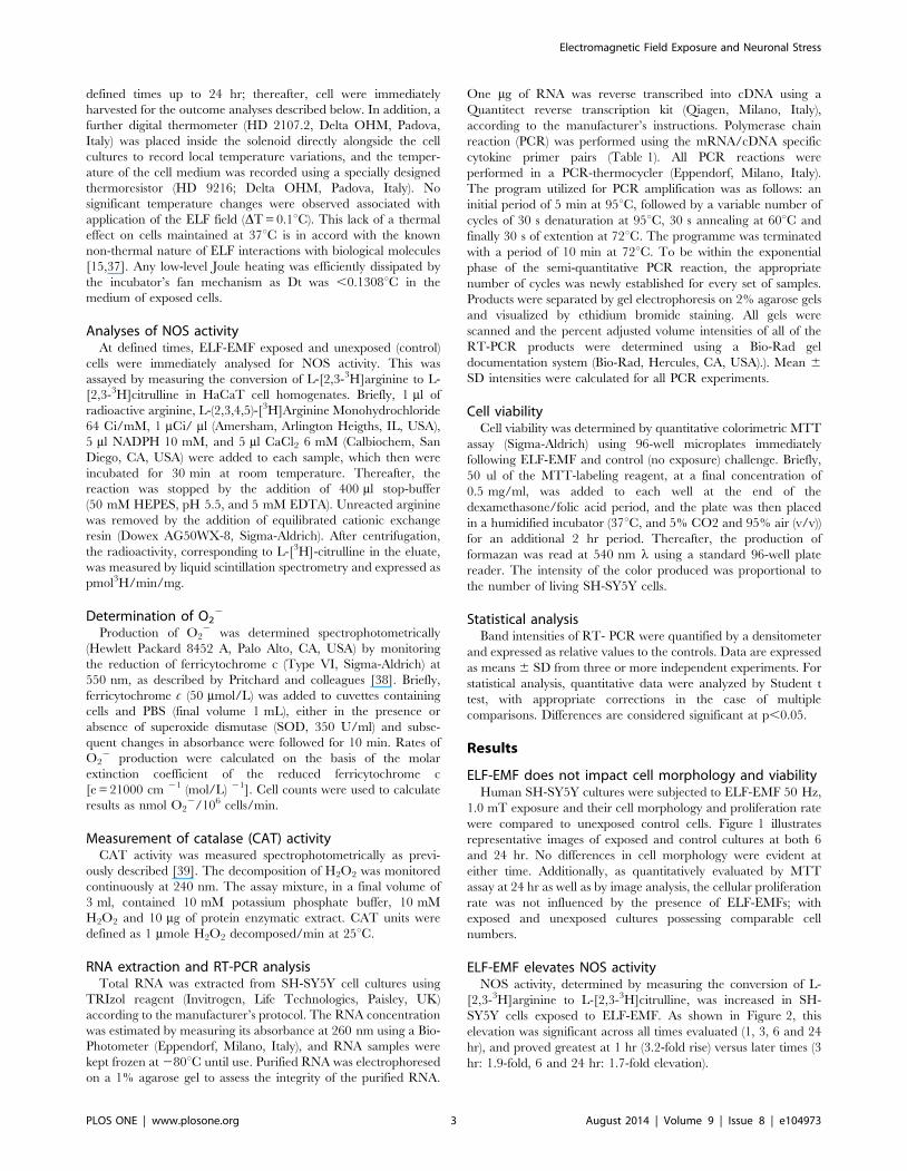
ELF-EMF does not impact cell morphology and viabilityHuman SH-SY5Y cultures were subjected to ELF-EMF 50 Hz,
1.0 mT exposure and their cell morphology and proliferation rate
were compared to unexposed control cells. Figure 1 illustrates
representative images of exposed and control cultures at both 6
and 24 hr. No differences in cell morphology were evident at
either time. Additionally, as quantitatively evaluated by MTT
assay at 24 hr as well as by image analysis, the cellular proliferation
rate was not influenced by the presence of ELF-EMFs; with
exposed and unexposed cultures possessing comparable cell
numbers.
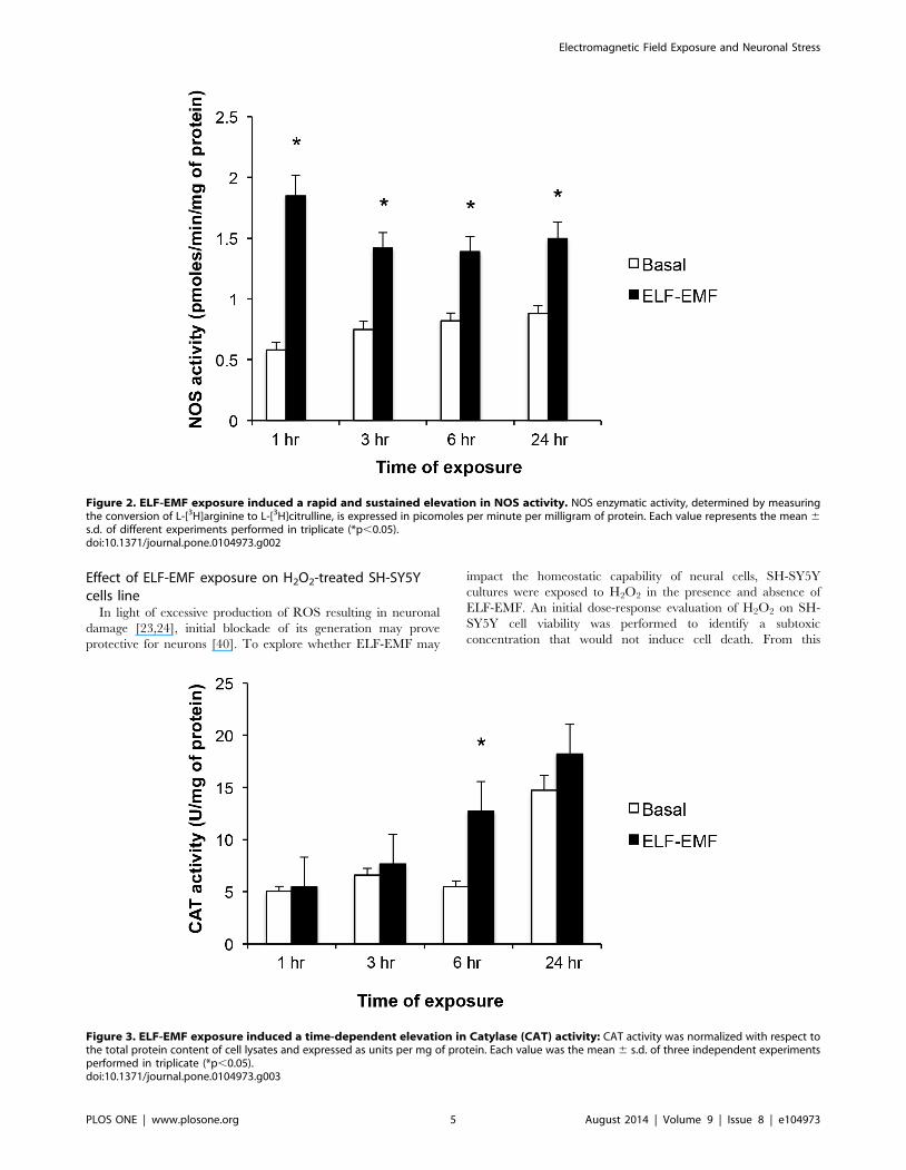
ELF-EMF elevates NOS activityNOS activity, determined by measuring the conversion of L-
[2,3-3H]arginine to L-[2,3-3H]citrulline, was increased in SH-
SY5Y cells exposed to ELF-EMF. As shown in Figure 2, this
elevation was significant across all times evaluated (1, 3, 6 and 24
hr), and proved greatest at 1 hr (3.2-fold rise) versus later times (3
hr: 1.9-fold, 6 and 24 hr: 1.7-fold elevation).
Electromagnetic Field Exposure and Neuronal Stress
PLOS ONE | www.plosone.org 3 August 2014 | Volume 9 | Issue 8 | e104973
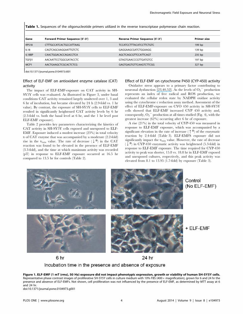
Effect of ELF-EMF on antioxidant enzyme catalase (CAT)activity
The impact of ELF-EMF-exposure on CAT activity in SH-
SY5Y cells was evaluated. As illustrated in Figure 3, under basal
conditions CAT activity remained largely unaltered over 1, 3 and
6 hr of incubation, but became elevated by 24 h (2.9-fold vs. 1 hr
value). By contrast, the exposure of SH-SY5Y cells to ELF-EMF
resulted in significantly heightened CAT activity levels by 6 hr
(2.3-fold vs. both the basal level at 6 hr, and the 1 hr level post
ELF-EMF exposure).
Table 2 provides key parameters characterizing the kinetics of
CAT activity in SH-SY5Y cells exposed and unexposed to ELF-
EMF. Exposure induced a modest increase (23%) in total velocity
u of CAT enzyme that was accompanied by a moderate (2.2-fold)
rise in the umin value. The rate of decrease (QH) in the CAT
reaction was found to be elevated in the presence of ELF-EMF
(3.3-fold), and the time at which maximum activity was recorded
(pT) in response to ELF-EMF exposure occurred at 16.5 hr
compared to 13.5 hr for controls (Table 2).
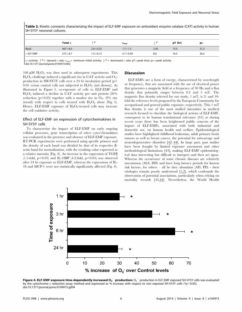
Effect of ELF-EMF on cytochrome P450 (CYP-450) activityOxidative stress appears to a primary factor contributing to
neuronal dysfunction [23,40,52]. As the levels of O22 production
represents an index of free radical and ROS production, we
evaluated the cellular redox state by NADPH oxidase activity
using the cytochrome c reduction assay method. Assessment of the
effect of ELF-EMF-exposure on CYO 450 activity in SH-SY5Y
cells showed that ELF-EMF increased CYP 450 activity and,
consequently, O22 production at all times studied (Fig. 4), with the
greatest increase (62%) occurring after 6 hr of exposure.
A rise (21%) in the total velocity of CYP-450 was measured in
response to ELF-EMF exposure, which was accompanied by a
significant elevation in the rate of increase (qH) of the enzymatic
reaction by 2.4-fold (Table 3). ELF-EMFS exposure did not
significantly impact the umin value. However, the rate of decrease
(QH) in CYP-450 enzymatic activity was heightened (5.3-fold) in
response to ELF-EMF exposure. The time required for CYP-450
activity to peak was shorter, 15.8 vs. 18.8 hr in ELF-EMF exposed
and unexposed cultures, respectively, and this peak activity was
elevated from 8.1 to 13.95 (1.7-fold) by exposure (Table 3).
Table 1. Sequences of the oligonucleotide primers utilized in the reverse transcriptase polymerase chain reaction.
Gene Forward Primer Sequence [5’-3’] Reverse Primer Sequence [5’-3’] Primer size
RPS18 CTTTGCCATCACTGCCATTAAG TCCATCCTTTACATCCTTCTGTC 199 bp
IL18 CAGTCAGCAAGGAATTGTCTC GAGGAAGCGATCTGGAAGG 139 bp
IL18BP CAACTGGACACCAGACCTCA AGCTCAGCGTTCCATTCAGT 235 bp
TGFb1 AACAATTCCTGGCGATACCTC GTAGTGAACCCGTTGATGTCC 197 bp
MCP1 AACTGAAGCTCGCACTCTCG GAGTGAGTGTTCAAGTCTTCGG 327 bp
doi:10.1371/journal.pone.0104973.t001
Figure 1. ELF-EMF (1 mT (rms), 50 Hz) exposure did not impact phenotypic expression, growth or viability of human SH-SY5Y cells.Representative phase contrast images of proliferative SH-SY5Y cells in culture medium with 10% FBS (4006magnification), grown for 6 and 24 hr thepresence and absence of ELF-EMFs. Not shown, cell proliferation was not influenced by the presence of ELF-EMF, as determined by MTT assay at 6and 24 hr.doi:10.1371/journal.pone.0104973.g001
Electromagnetic Field Exposure and Neuronal Stress
PLOS ONE | www.plosone.org 4 August 2014 | Volume 9 | Issue 8 | e104973
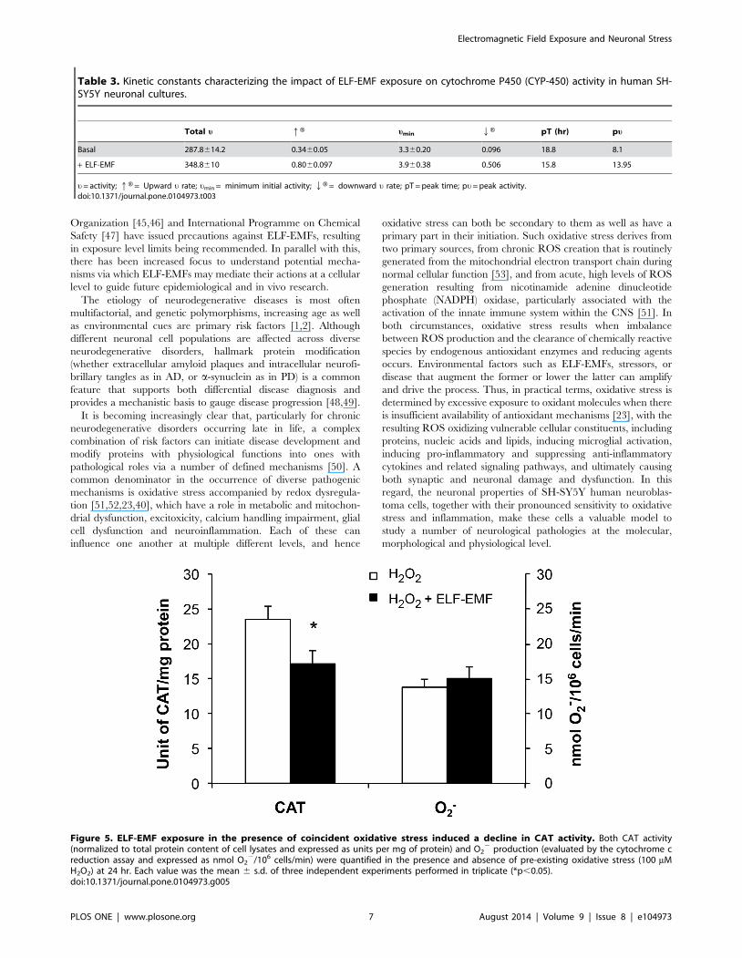
Effect of ELF-EMF exposure on H2O2-treated SH-SY5Ycells line
In light of excessive production of ROS resulting in neuronal
damage [23,24], initial blockade of its generation may prove
protective for neurons [40]. To explore whether ELF-EMF may
impact the homeostatic capability of neural cells, SH-SY5Y
cultures were exposed to H2O2 in the presence and absence of
ELF-EMF. An initial dose-response evaluation of H2O2 on SH-
SY5Y cell viability was performed to identify a subtoxic
concentration that would not induce cell death. From this
Figure 2. ELF-EMF exposure induced a rapid and sustained elevation in NOS activity. NOS enzymatic activity, determined by measuringthe conversion of L-[3H]arginine to L-[3H]citrulline, is expressed in picomoles per minute per milligram of protein. Each value represents the mean 6s.d. of different experiments performed in triplicate (*p,0.05).doi:10.1371/journal.pone.0104973.g002
Figure 3. ELF-EMF exposure induced a time-dependent elevation in Catylase (CAT) activity: CAT activity was normalized with respect tothe total protein content of cell lysates and expressed as units per mg of protein. Each value was the mean 6 s.d. of three independent experimentsperformed in triplicate (*p,0.05).doi:10.1371/journal.pone.0104973.g003
Electromagnetic Field Exposure and Neuronal Stress
PLOS ONE | www.plosone.org 5 August 2014 | Volume 9 | Issue 8 | e104973
100 mM H2O2 was then used in subsequent experiments. This
H2O2 challenge induced a significant rise in CAT activity and O2-
production in SH-SY5Y cells over a 24 hr incubation period (p,
0.05 versus control cells not subjected to H2O2 (not shown)). As
illustrated in Figure 5, co-exposure of cells to ELF-EMF and
H2O2 induced a decline in CAT activity per unit protein (26%
reduction (p,0.05) together with a modest rise in O2- (9% rise
(trend)) with respect to cells treated with H2O2 alone (Fig. 5).
Hence, ELF-EMF exposure of H2O2-treated cells may increase
the cell oxidative activity.
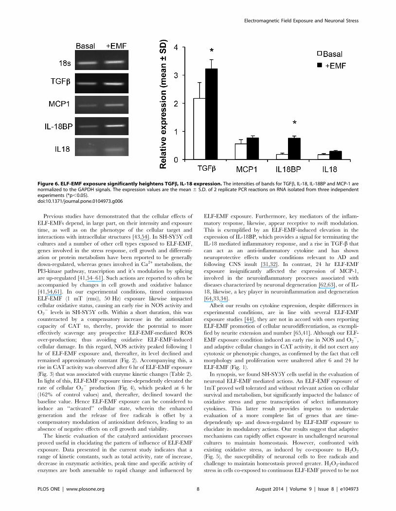
Effect of ELF-EMF on expression of cyto/chemokines inSH-SY5Y cells
To characterize the impact of ELF-EMF on early ongoing
cellular processes, gene transcription of select cyto/chemokines
was evaluated in the presence and absence of ELF-EMF exposure.
RT-PCR experiments were performed using specific primers and
the density of each band was divided by that of its respective b-
actin band for normalization, with the resulting value expressed as
a relative intensity (Fig. 6). An increase in the expression of TGFb(1.5-fold, p,0.05) and IL-18BP (4.2-fold, p,0.05) was observed
after 24 hr exposure to ELF-EMF, whereas the expressions of IL-
18 and MCP-1 were not statistically significantly affected (Fig. 6).
Discussion
ELF-EMFs are a form of energy, characterized by wavelength
or frequency, that are associated with the use of electrical power
that generates a magnetic field at a frequency of 50 Hz and a flux
density that primarily ranges between 0.2 and 5 mT. The
magnetic flux density selected for our study, 1 mT, is 2- and 10-
fold the reference levels proposed by the European Community for
occupational and general public exposure, respectively. This 1 mT
flux density is one of the most studied intensities in medical
research focused to elucidate the biological actions of ELF-EMF,
consequent to its human translational relevance [41] as during
recent years there has been heightened public concern of the
impact of ELF-EMFs, associated with both industrial and
domesitic use, on human health and welfare. Epidemiological
studies have highlighted childhood leukemias, adult primary brain
tumors as well as breast cancer, the potential for miscarrage and
neurodegenerative disorders [42–44]. In large part, past studies
have been fraught by limited exposure assessment and other
methodological limitations [43], making ELF-EMF epidemiolog-
ical data interesting but difficult to interpret and then act upon.
Whereas the occurrence of some chronic diseases are relatively
uncommon (ALS, HD) and have long latency periods for known
risk factors, for others – all be they abundant (AD, PD) - their
etiologies remain poorly understood [1,2], which confounds the
observation of potential associations, particularly when relying on
mortality records [43,44]. Nevertheless, the World Health
Table 2. Kinetic constants characterizing the impact of ELF-EMF exposure on antioxidant enzyme catalase (CAT) activity in humanSH-SY5Y neuronal cultures.
Total u qH umin QH pT (hr) pu
Basal 46766.0 2.060.32 1.761.3 2.45 13.5 31.2
+ ELF-EMF 57568.7 1.560.12 3.760.49 8.0 16.5 26.2
u= activity; qH = Upward u rate; umin = minimum initial activity; QH = downward u rate; pT = peak time; pu= peak activity.doi:10.1371/journal.pone.0104973.t002
Figure 4. ELF-EMF exposure time-dependently increased O22 production: O2
2 production in ELF-EMF exposed SH-SY5Y cells was evaluatedby the cytochrome c reduction assay method and expressed as % increase with respect to non exposed SH-SY5Y cells (*p,0.05).doi:10.1371/journal.pone.0104973.g004
Electromagnetic Field Exposure and Neuronal Stress
PLOS ONE | www.plosone.org 6 August 2014 | Volume 9 | Issue 8 | e104973
Organization [45,46] and International Programme on Chemical
Safety [47] have issued precautions against ELF-EMFs, resulting
in exposure level limits being recommended. In parallel with this,
there has been increased focus to understand potential mecha-
nisms via which ELF-EMFs may mediate their actions at a cellular
level to guide future epidemiological and in vivo research.
The etiology of neurodegenerative diseases is most often
multifactorial, and genetic polymorphisms, increasing age as well
as environmental cues are primary risk factors [1,2]. Although
different neuronal cell populations are affected across diverse
neurodegenerative disorders, hallmark protein modification
(whether extracellular amyloid plaques and intracellular neurofi-
brillary tangles as in AD, or a-synuclein as in PD) is a common
feature that supports both differential disease diagnosis and
provides a mechanistic basis to gauge disease progression [48,49].
It is becoming increasingly clear that, particularly for chronic
neurodegenerative disorders occurring late in life, a complex
combination of risk factors can initiate disease development and
modify proteins with physiological functions into ones with
pathological roles via a number of defined mechanisms [50]. A
common denominator in the occurrence of diverse pathogenic
mechanisms is oxidative stress accompanied by redox dysregula-
tion [51,52,23,40], which have a role in metabolic and mitochon-
drial dysfunction, excitoxicity, calcium handling impairment, glial
cell dysfunction and neuroinflammation. Each of these can
influence one another at multiple different levels, and hence
oxidative stress can both be secondary to them as well as have a
primary part in their initiation. Such oxidative stress derives from
two primary sources, from chronic ROS creation that is routinely
generated from the mitochondrial electron transport chain during
normal cellular function [53], and from acute, high levels of ROS
generation resulting from nicotinamide adenine dinucleotide
phosphate (NADPH) oxidase, particularly associated with the
activation of the innate immune system within the CNS [51]. In
both circumstances, oxidative stress results when imbalance
between ROS production and the clearance of chemically reactive
species by endogenous antioxidant enzymes and reducing agents
occurs. Environmental factors such as ELF-EMFs, stressors, or
disease that augment the former or lower the latter can amplify
and drive the process. Thus, in practical terms, oxidative stress is
determined by excessive exposure to oxidant molecules when there
is insufficient availability of antioxidant mechanisms [23], with the
resulting ROS oxidizing vulnerable cellular constituents, including
proteins, nucleic acids and lipids, inducing microglial activation,
inducing pro-inflammatory and suppressing anti-inflammatory
cytokines and related signaling pathways, and ultimately causing
both synaptic and neuronal damage and dysfunction. In this
regard, the neuronal properties of SH-SY5Y human neuroblas-
toma cells, together with their pronounced sensitivity to oxidative
stress and inflammation, make these cells a valuable model to
study a number of neurological pathologies at the molecular,
morphological and physiological level.
Table 3. Kinetic constants characterizing the impact of ELF-EMF exposure on cytochrome P450 (CYP-450) activity in human SH-SY5Y neuronal cultures.
Total u qH umin QH pT (hr) pu
Basal 287.8614.2 0.3460.05 3.360.20 0.096 18.8 8.1
+ ELF-EMF 348.8610 0.8060.097 3.960.38 0.506 15.8 13.95
u= activity; qH = Upward u rate; umin = minimum initial activity; QH = downward u rate; pT = peak time; pu= peak activity.doi:10.1371/journal.pone.0104973.t003
Figure 5. ELF-EMF exposure in the presence of coincident oxidative stress induced a decline in CAT activity. Both CAT activity(normalized to total protein content of cell lysates and expressed as units per mg of protein) and O2
2 production (evaluated by the cytochrome creduction assay and expressed as nmol O2
2/106 cells/min) were quantified in the presence and absence of pre-existing oxidative stress (100 mMH2O2) at 24 hr. Each value was the mean 6 s.d. of three independent experiments performed in triplicate (*p,0.05).doi:10.1371/journal.pone.0104973.g005
Electromagnetic Field Exposure and Neuronal Stress
PLOS ONE | www.plosone.org 7 August 2014 | Volume 9 | Issue 8 | e104973
Previous studies have demonstrated that the cellular effects of
ELF-EMFs depend, in large part, on their intensity and exposure
time, as well as on the phenotype of the cellular target and
interactions with intracellular structures [43,54]. In SH-SY5Y cell
cultures and a number of other cell types exposed to ELF-EMF,
genes involved in the stress response, cell growth and differenti-
ation or protein metabolism have been reported to be generally
down-regulated, whereas genes involved in Ca2+ metabolism, the
PI3-kinase pathway, trascription and it’s modulation by splicing
are up-regulated [41,54–61]. Such actions are reported to often be
accompanied by changes in cell growth and oxidative balance
[41,54,61]. In our experimental conditions, timed continuous
ELF-EMF (1 mT (rms)), 50 Hz) exposure likewise impacted
cellular oxidative status, causing an early rise in NOS activity and
O22 levels in SH-SY5Y cells. Within a short duration, this was
counteracted by a compensatory increase in the antioxidant
capacity of CAT to, thereby, provide the potential to more
effectively scavenge any prospective ELF-EMF-mediated ROS
over-production; thus avoiding oxidative ELF-EMF-induced
cellular damage. In this regard, NOS activity peaked following 1
hr of ELF-EMF exposure and, thereafter, its level declined and
remained approximately constant (Fig. 2). Accompanying this, a
rise in CAT activity was observed after 6 hr of ELF-EMF exposure
(Fig. 3) that was associated with enzyme kinetic changes (Table 2).
In light of this, ELF-EMF exposure time-dependently elevated the
rate of cellular O22 production (Fig. 4), which peaked at 6 hr
(162% of control values) and, thereafter, declined toward the
baseline value. Hence ELF-EMF exposure can be considered to
induce an ‘‘activated’’ cellular state, wherein the enhanced
generation and the release of free radicals is offset by a
compensatory modulation of antioxidant defences, leading to an
absence of negative effects on cell growth and viability.
The kinetic evaluation of the catalyzed antioxidant processes
proved useful in elucidating the pattern of influence of ELF-EMF
exposure. Data presented in the current study indicates that a
range of kinetic constants, such as total activity, rate of increase,
decrease in enzymatic activities, peak time and specific activity of
enzymes are both amenable to rapid change and influenced by
ELF-EMF exposure. Furthermore, key mediators of the inflam-
matory response, likewise, appear receptive to swift modulation.
This is exemplified by an ELF-EMF-induced elevation in the
expression of IL-18BP, which provides a signal for terminating the
IL-18 mediated inflammatory response, and a rise in TGF-b that
can act as an anti-inflammatory cytokine and has shown
neuroprotective effects under conditions relevant to AD and
following CNS insult [31,32]. In contrast, 24 hr ELF-EMF
exposure insignificantly affected the expression of MCP-1,
involved in the neuroinflammatory processes associated with
diseases characterized by neuronal degeneration [62,63], or of IL-
18, likewise, a key player in neuroinflammation and degeneration
[64,33,34].
Albeit our results on cytokine expression, despite differences in
experimental conditions, are in line with several ELF-EMF
exposure studies [44], they are not in accord with ones reporting
ELF-EMF promotion of cellular neurodifferentiation, as exempli-
fied by neurite extension and number [65,41]. Although our ELF-
EMF exposure condition induced an early rise in NOS and O22,
and adaptive cellular changes in CAT activity, it did not exert any
cytotoxic or phenotypic changes, as confirmed by the fact that cell
morphology and proliferation were unaltered after 6 and 24 hr
ELF-EMF (Fig. 1).
In synopsis, we found SH-SY5Y cells useful in the evaluation of
neuronal ELF-EMF mediated actions. An ELF-EMF exposure of
1mT proved well tolerated and without relevant action on cellular
survival and metabolism, but significantly impacted the balance of
oxidative stress and gene transcription of select inflammatory
cytokines. This latter result provides impetus to undertake
evaluation of a more complete list of genes that are time-
dependently up- and down-regulated by ELF-EMF exposure to
elucidate its modulatory actions. Our results suggest that adaptive
mechanisms can rapidly offset exposure in unchallenged neuronal
cultures to maintain homeostasis. However, confronted with
existing oxidative stress, as induced by co-exposure to H2O2
(Fig. 5), the susceptibility of neuronal cells to free radicals and
challenge to maintain homeostasis proved greater. H2O2-induced
stress in cells co-exposed to continuous ELF-EMF proved to be not
Figure 6. ELF-EMF exposure significantly heightens TGFb, IL-18 expression. The intensities of bands for TGFb, IL-18, IL-18BP and MCP-1 arenormalized to the GAPDH signals. The expression values are the mean 6 S.D. of 2 replicate PCR reactions on RNA isolated from three independentexperiments (*p,0.05).doi:10.1371/journal.pone.0104973.g006
Electromagnetic Field Exposure and Neuronal Stress
PLOS ONE | www.plosone.org 8 August 2014 | Volume 9 | Issue 8 | e104973

well counteracted, resulting in a reduction of CAT activity and a
rise in O22 levels. Hence, in accord with Falone and colleagues
[41], contuinuous ELF-EMF exposure reduced cell tolerance
towards simultaneous oxidative challenges, and may aid our
understanding of the role of concurrent environmental agent
challenges in the development of neurodegenerative diseases.
It is important to note, however, that studies by others [66–68]
have demonstrated that ELF-EMF exposure, in the form of
transcranial magnetic stimulation (60-Hz, 0.7 mT) applied to rats
for 2 hr twice daily, can prove neuroprotective. Administered prior
to and after a toxic insult to the brain, for example systemic
injection of 3-nitropropionic acid to induce an animal model of
HD [69], ELF-EMF can mitigate oxidative damage, elevate
neurotrophic protein levels in brain and ameliorate behavioral
deficits [66–68], as well as potentially augment neurogenesis [70].
Such studies reiterate that the level and timing of exposure are
critical factors impacting outcome measures, and can be poten-
tially scheduled to optimize endogenous compensatory mecha-
nisms following an adverse challenge.
Further studies designed to evaluate the actions of ELF-EMF
under multiple conditions, including chronic or sporadic exposure
in combination with common stressors pertinent to real life,
appear warranted and may both aid our understanding of the true
biological impact of ELF-EMF and scientifically anchor proposed
exposure limits.
Acknowledgments
The authors dedicate this paper to the memory of their friend and
colleague Giovina Vianale.
Author Contributions
Conceived and designed the experiments: MR AP EC CA MP. Performed
the experiments: MR AP EC CA MP. Analyzed the data: MR MK EC CA
NG. Contributed reagents/materials/analysis tools: AP MP. Contributed
to the writing of the manuscript: MR MK NG. Figures: MR NG.
References
1. Bossy-Wetzel E, Schwarzenbacher R, Lipton SA (2004) Molecular pathways to
neurodegeneration. Nat Med. 10 Suppl:S2–9.
2. Bertram L, Tanzi RE (2005) The genetic epidemiology of neurodegenerative
disease. J Clin Invest. 115(6):1449–57.
3. Moreno-Gonzalez I, Soto C (2012) Natural animal models of neurodegenerative
protein misfolding diseases. Curr Pharm Des. 18(8):1148–58.
4. Mullard A (2012) Sting of Alzheimer’s failures offset by upcoming prevention
trials. Nature Rev. Drug Discov. 11: 657–60.
5. Becker RE, Greig NH, Giacobini E, Schneider LS, Ferrucci L (2014) A newroadmap for drug development for Alzheimer’s disease. Nat Rev Drug Discov.
13(2):156.
6. Brown RC, Lockwood AH, Sonawane BR (2005) Neurodegenerative diseases:
An overview of environmental risk factors. Environmental Health Perspectives113: 1250–6.
7. Cannon JR, Greenamyre JT (2011) The Role of environmental exposures inneurodegeneration and neurodegenerative diseases. Toxicological Sciences 124:
225–50.
8. Kraft AD, Harry GJ (2011) Features of microglia and neuroinflammation
relevant to environmental exposure and neurotoxicity. Int. J. Environ. Res.
Public Health 8: 2980–18.
9. Lahiri DK, Maloney B, Basha MR, Ge YW, Zawia NH (2007) How and when
environmental agents and dietary factors affect the course of Alzheimer’s disease:the "LEARn" model (latent early-life associated regulation) may explain the
triggering of AD. Curr Alzheimer Res. 4(2):219–28.
10. Lahiri DK, Maloney B, Zawia NH (2009) The LEARn model: an epigenetic
explanation for idiopathic neurobiological diseases. Mol Psychiatry. 14(11):992–
1003
11. Luukkonen J, Liimatainen A, Hoyto A, Juutilainen J, Naarala J (2011) Pre-
exposure to 50 Hz magnetic fields modifies menadione-induced genotoxic effectsin human SH-SY5Y neuroblastoma cells. PLoS One 6: e18021.
12. Piacentini R, Ripoli C, Mezzogori D, Azzena GB, Grassi C (2008) Extremelylow-frequency electromagnetic fields promote in vitro neurogenesis via
upregulation of Ca(v)1-channel activity. J Cell Physiol 215: 129–39.
13. Arendash GW, Mori T, Dorsey M, Gonzalez R, Tajiri N, et al. (2012)
Electromagnetic treatment to old Alzheimer’s mice reverses beta-amyloiddeposition, modifies cerebral blood flow, and provides selected cognitive benefit.
PLoS One 7: e35751
14. Sonnier H, Kolomytkin O, Marino A (2003) Action potentials from humanneuroblastoma cells in magnetic fields. Neuroscience Letters 337: 163–6.
15. Del Vecchio G, Giuliani A, Fernandez M, Mesirca P, Bersani F, et al. (2009)Effect of Radiofrequency Electromagnetic Field Exposure on In Vitro Models of
Neurodegenerative Disease. Bioelectromagnetics 30: 564–72.
16. Tweedie D, Sambamurti K, Greig NH (2007) TNF-alpha inhibition as a
treatment strategy for neurodegenerative disorders: new drug candidates andtargets. Curr Alzheimer Res. 4(4):378–85.
17. Reale M, Iarlori C, Feliciani C, Gambi D (2008) Peripheral chemokinereceptors, their ligands, cytokines and Alzheimer’s disease. J Alzheimer’s Dis. 14
(2): 147–59.
18. Reale M, De Angelis F, Di Nicola M, Capello E, Di Ioia M, et al. (2012)Relation between Pro-inflammatory Cytokines and Acetylcholine Levels in
Relapsing-Remitting Multiple Sclerosis Patients. Int J Mol Sci. 13(10):12656–64.
19. Reale M, Iarlori C, Thomas A, Gambi D, Perfetti B, et al. (2009) Peripheral
cytokines profile in Parkinson’s Disease. Brain, Behavior Immunity 23(1):55–63.
20. Reale M, Kamal MA, Velluto L, Gambi D, Di Nicola M, et al. (2012)
Relationship between Inflammatory Mediators, Ab Levels and APOE Genotypein Alzheimer’s Disease. Curr Alzheimer Res. 9(4):447–57.
21. Moreau C, Devos D, Brunaud-Danel V, Defebvre L, Perez T, et al. (2005)
Elevated IL-6 and TNF-alpha levels in patients with ALS: inflammation orhypoxia? Neurology 65: 1958–60.
22. Poloni M, Facchetti D, Mai R, Micheli A, Agnoletti L, et al. (2000) Circulating
levels of tumour necrosis factor-alpha and its soluble receptors are increased inthe blood of patients with amyotrophic lateral sclerosis. Neurosci Lett 287: 211–
4.
23. Bonda DJ, Wang X, Perry G, Nunomura A, Tabaton M, et al. (2010) Oxidative
stress in Alzheimer disease: A possibility for prevention. Neuropharmacology 59:290–4.
24. Stefani IC, Wright D, Polizzi KM, Kontoravdi C (2012) The role of ER stress-induced apoptosis in neurodegeneration. Curr Alzheimer Res. 9(3):373–87.
25. Ortiz GG, Macias-Islas MA, Pacheco-Moises FP, Cruz-Ramos JA, Sustersik S,et al. (2009) Oxidative stress is increased in serum from Mexican patients with
relapsing-remitting multiple sclerosis. Dis. Markers 26: 35–9.
26. Taskiran D, Sagduyu A, Yuceyar N, Kutay FZ, Pogun S (2000) Increasedcerebrospinal fluid and serum nitrite and nitrate levels in amyotrophic lateral
sclerosis. Int J Neurosci 101: 65–72
27. Blandini F (2013) Neural and immune mechanisms in the pathogenesis of
Parkinson’s disease. J Neuroimmune Pharmacol. 8(1):189–201.
28. Korhonen R, Lahti A, Kankaanranta H, Moilanen E (2005) Nitric oxide
production and signaling in inflammation. Curr. Drug Targets Inflamm. Allergy4: 471–9.
29. Vaz AR, Silva SL, Barateiro A, Fernandes A, Falcao AS, et al. (2011) Pro-inflammatory cytokines intensify the activation of NO/NOS, JNK1/2 and
caspase cascades in immature neurons exposed to elevated levels of
unconjugated bilirubin. Exp Neurol. 229(2):381–90.
30. Tesseur I, Zou K, Esposito L, Bard F, Berber E, et al. (2006) Deficiency in
neuronal TGF-beta signaling promotes neurodegeneration and Alzheimer’spathology. J Clin Invest. 116: 3060–9.
31. Battista D, Ferrari CC, Gage FH, Pitossi FJ (2006) Neurogenic niche modulationby activated microglia: transforming growth factor beta increases neurogenesis
in the adult dentate gyrus. Eur J Neurosci. 23(1): 83–93.
32. Ren RF, Hawver DB, Kim RS, Flanders KC (1997) Transforming growth
factor-b protects human hNT cells from degeneration induced by b-amyloid
peptide: involvement of the TGF-b type II receptor. Mol Brain Res. 48: 315–22.
33. Dinarello CA, Novick D, Kim S, Kaplanski G (2013) Interleukin-18 and IL-18
Binding Protein. Front Immunol. 4: 289.
34. Sutinen EM, Pirttila T, Anderson G, Salminen A, Ojala JO (2012) Pro-
inflammatory interleukin-18 increases Alzheimer’s disease-associated amyloid-bproduction in human neuron-like cells. J Neuroinflammation. 9: 199.
35. Reale M, Greig NH, Kamal MA (2009) Peripheral chemo-cytokine profiles inAlzheimer’s and Parkinson’s diseases. Mini Rev Med Chem. 9(10):1229–41.
36. Bose S, Cho J (2013) Role of chemokine CCL2 and its receptor CCR2 inneurodegenerative diseases. Arch Pharm Res. 36(9):1039–50.
37. Vianale G, Reale M, Amerio P, Stefanachi M, Di Luzio S, et al. (2008)Extremely Low Frequency Electromagnetic Field (ELF-EMF) enhances human
keratinocyte cell growth and decreases pro-inflammatory chemokines produc-
tion. Brit J Dermatol 158(6):1189–96.
38. Pritchard KA Jr, Groszek L, Smalley DM, Sessar WC, Wu M, et al. (1995)
Native low-density lipoprotein increases endothelial cell nitric oxide synthasegeneration of superoxide anion. Circ Res 77: 510–518.
39. Aebi HE (1974) Methods of Enzymatic Analysis. In: Bergmayer HU, editor.Chemie. 2nd Edition, Vol. 2. Weinheim: F.R.G. 673–84.
40. Su B, Wang X, Nunomura A, Moreira PI, Lee HG, et al. (2008) Oxidative stresssignaling in Alzheimer’s disease. Curr Alzheimer Res. 5(6):525–32.
Electromagnetic Field Exposure and Neuronal Stress
PLOS ONE | www.plosone.org 9 August 2014 | Volume 9 | Issue 8 | e104973
41. Falone S, Grossi MR, Cinque B, D’Angelo B, Tettamanti E, et al. (2007) Fifty
hertz extremely low-frequency electromagnetic field causes changes in redox and
differentiative status in neuroblastoma cells. Int J Biochem Cell Biol.
39(11):2093–106.
42. Lacy-Hulbert A, Metcalfe JC, Hesketh R (1998) Biological responses to
electromagnetic fields. FASEB J 12(6): 395–420.
43. Feychting M, Ahlbom A, Kheifets L (2005) EMF and health. Ann Rev Public
Health 26: 165–189.
44. Consales C, Merla C, Marino C, Benassi B (2012) Electromagnetic fields,
oxidative stress, and neurodegeneration. Int J Cell Biol. 2012: 683897.
45. WHO (2007) ‘‘Electromagnetic fields and public health. Exposure to extremely
low frequency fields,’’ Fact Sheet no. 322, WHO, Geneva, Switzerland. ,
http://www.who.int/peh-emf/publications/facts/fs322/en/. viewed May 16,
2014.
46. WHO (2007) (Environmental Health Criteria 238), Extremely Low Frequency
Fields, vol. 35, WHO, Geneva, Switzerland. ,http://www.who.int/peh-emf/
publications/Complet_DEC_2007.pdf. viewed May 16, 2014.
47. International Programme On Chemical Safety (1984) Environmental Health
Criteria 35. Extremely Low Frequency (ELF) Fields ,http://www.inchem.org/
documents/ehc/ehc/ehc35.htm - PartNumber:9. viewed May 16, 2014.
48. Jack CR Jr, Knopman DS, Jagust WJ, Petersen RC, Weiner MW, et al. (2013)
Tracking pathophysiological processes in Alzheimer’s disease: an updated
hypothetical model of dynamic biomarkers. Lancet Neurol. 12(2):207–16.
49. Saracchi E, Fermi S, Brighina L (2013) Emerging candidate biomarkers for
Parkinson’s disease: a review. Aging Dis. 5(1):27–34.
50. Moreno-Gonzalez I, Soto C (2011) Misfolded protein aggregates: mechanisms,
structures and potential for disease transmission. Semin Cell Dev Biol.
22(5):482–7.
51. von Bernhardi R, Eugenın J (2012) Alzheimer’s disease: redox dysregulation as a
common denominator for diverse pathogenic mechanisms. Antioxid Redox
Signal. 16(9):974–1031.
52. Bonda DJ, Castellani RJ, Zhu X, Nunomura A, Lee HG, et al. (2011) A novel
perspective on tau in Alzheimer’s disease. Curr Alzheimer Res. 8(6):639–42.
53. Barja G (1998) Mitochondrial free radical production and aging in mammals
and birds. Ann N Y Acad Sci 854: 224–238.
54. Eleuteri AM, Amici M, Bonfili L, Cecarini V, Cuccioloni M, et al. (2009) 50 Hz
extremely low frequency electromagnetic fields enhance protein carbonyl groups
content in cancer cells: effects on proteasomal systems. J Biomed Biotechnol.
2009: 834239.
55. Simko M, Mattsson MO (2004) Extremely low frequency electromagnetic fields
as effectors of cellular responses in vitro: possible immune cell activation. J Cell
Biochem. 93(1):83–92.
56. Reale M, De Lutiis MA, Patruno A, Speranza L, Felaco M, et al. (2006)
Modulation of MCP-1 and iNOS by 50-Hz sinusoidal electromagnetic field.
Nitric Oxide. 15(1):50–7.
57. Patruno A, Amerio P, Pesce M, Vianale G, Di Luzio S (2010) Extremely low
frequency electromagnetic field (elf-emf) modulate iNOS, eNOS and Cox-2expressions in the human keratinocyte cell line hacat: potential therapeutical
effects in wound healing. Brit J Dermatol 162: 258–66.
58. Patruno A, Tabrez S, Amerio P, Pesce M, Vianale G, et al. (2011) Kinetic Studyon the Effects of Extremely Low Frequency Electromagnetic Field on Catalase,
Cytochrome P450, Inducible Nitric Oxide Synthase in Human HaCaT andTHP-1 Cell Lines. CNS Neurol Disord Drug Targets. 10(8):936–44.
59. Sunkari VG, Aranovitch B, Portwood N, Nikoshkov A (2011) Effects of a low-
intensity electromagnetic field on fibroblast migration and proliferation.Electromagnet Biol Med 30(2): 80–5.
60. Antonini RA, Benfante R, Gotti C, Moretti M, Kuster N, et al. (2006) Extremelylow-frequency electromagnetic field (ELF-EMF) does not affect the expression of
alpha3, alpha5 and alpha7 nicotinic receptor subunit genes in SH-SY5Yneuroblastoma cell line. Toxicol Lett. 164(3):268–77
61. Martınez-Samano J, Torres-Duran PV, Juarez-Oropeza MA, Verdugo-Dıaz L
(2012) Effect of acute extremely low frequency electromagnetic field exposure onthe antioxidant status and lipid levels in rat brain. Arch Med Res. 43(3):183–9.
62. Gerard C, Rollins BJ (2001) Chemokines and disease. Nature Immunology 2 (2):108–115.
63. Azizi G, Khannazer N, Mirshafiey A (2014) The potential role of chemokines in
Alzheimer’s disease pathogenesis. Am J Alzheimers Dis Other Demen. [Epubahead of print]
64. Bossu P, Ciaramella A, Salani F, Vanni D, Palladino I, et al. (2010) Interleukin-18, from neuroinflammation to Alzheimer’s disease. Curr Pharm Des.
16(38):421365. Lisi A, Ledda M, Rosola E, Pozzi D, D’Emilia E, et al. (2006) Extremely low
frequency electromagnetic field exposure promotes differentiation of pituitary
corticotrope-derived AtT20 D16V cells. Bioelectromagnetics 27: 641–51.66. Tasset I, Medina FJ, Jimena I, Aguera E, Gascon F, et al. (2010)
Neuroprotective effects of extremely low-frequency electromagnetic fields on aHuntington’s disease rat model: effects on neurotrophic factors and neuronal
density. Neuroscience 209: 54–63.
67. Tasset I, Perez-Herrera A, Medina FJ, Arias-Carrion O, Drucker-Colın R, et al.(2013) Extremely low-frequency electromagnetic fields activate the antioxidant
pathway Nrf2 in a Huntington’s disease-like rat model. Brain Stimul. 6: 84–6.68. Tunez I, Drucker-Colın R, Jimena I, Medina FJ, Munoz Mdel C, et al. (2006)
Transcranial magnetic stimulation attenuates cell loss and oxidative damage inthe striatum induced in the 3-nitropropionic model of Huntington’s disease. J
Neurochem. 97: 619–30.
69. Tunez I, Santamarıa A (2009) Model of Huntington’s disease induced with 3-nitropropionic acid. Rev Neurol, 48: 430–434.
70. Arias-Carrion O, Verdugo-Dıaz L, Feria-Velasco A, Millan-Aldaco D,Gutierrez AA, et al. (2004) Neurogenesis in the subventricular zone following
transcranial magnetic field stimulation and nigrostriatal lesions. J Neurosci Res.
78: 16–28.
Electromagnetic Field Exposure and Neuronal Stress
PLOS ONE | www.plosone.org 10 August 2014 | Volume 9 | Issue 8 | e104973