Translational regulation of BACE-1 expression in neuronal and non-neuronal cells
Neuronal synchrony: Peculiarity and generalityThomas Nowotny,1,a� Ramon Huerta,2,b� and Mikhail I. Rabinovich2,c�
1Centre for Computational Neuroscience and Robotics, Informatics, University of Sussex,Falmer, Brighton BN1 9QJ, United Kingdom2Institute for Nonlinear Science, University of California San Diego, 9500 Gilman Drive,La Jolla, California 92093-0402, USA
�Received 5 March 2008; accepted 2 June 2008; published online 22 September 2008�
Synchronization in neuronal systems is a new and intriguing application of dynamical systemstheory. Why are neuronal systems different as a subject for synchronization? �1� Neurons in them-selves are multidimensional nonlinear systems that are able to exhibit a wide variety of differentactivity patterns. Their “dynamical repertoire” includes regular or chaotic spiking, regular or cha-otic bursting, multistability, and complex transient regimes. �2� Usually, neuronal oscillations arethe result of the cooperative activity of many synaptically connected neurons �a neuronal circuit�.Thus, it is necessary to consider synchronization between different neuronal circuits as well. �3�The synapses that implement the coupling between neurons are also dynamical elements and theirintrinsic dynamics influences the process of synchronization or entrainment significantly. In thisreview we will focus on four new problems: �i� the synchronization in minimal neuronal networkswith plastic synapses �synchronization with activity dependent coupling�, �ii� synchronization ofbursts that are generated by a group of nonsymmetrically coupled inhibitory neurons �heteroclinicsynchronization�, �iii� the coordination of activities of two coupled neuronal networks �partialsynchronization of small composite structures�, and �iv� coarse grained synchronization in largersystems �synchronization on a mesoscopic scale�. © 2008 American Institute of Physics.�DOI: 10.1063/1.2949925�
The observation of synchrony (coherence, correlation) inthe brain and the entire nervous system has a long his-tory. Neuronal synchronization has been experimentallyanalyzed on many different levels of system complexity.Even though the functional role of this phenomenon isstill not absolutely clear, neuroscientists agree that thelevel of synchrony is one of the key characteristics of theactivity of the brain/neuronal systems in general. Bothlimit cases, too strong and too weak synchronization arecharacteristic for certain brain disorders, such as epi-lepsy, Parkinson’s disease, Alzheimer’s disease, andschizophrenia (see Refs. 1 and 2). In this short review wediscuss a few mechanisms of neuronal synchronizationthat illustrate the specificity of the corresponding phe-nomena in neuronal circuits, and their similarity to clas-sical examples in physical systems. We also discuss brieflythe influence of the complexity of real neuronal systemson synchronous dynamics.
I. INTRODUCTION: THE FUNCTIONAL ROLEOF NEURONAL SYNCHRONIZATION
The question “Does synchronization in the brain reallyplay a key role in controlling behavior and cognition or is itjust an epiphenomenon that accompanies the cooperative in-formation processing by huge numbers of neurons in the
brain?” has been the focus of numerous intensivediscussions3–7 and still has no convincing answer. However,many fascinating experiments have revealed synchronization�coherence or correlation� within and between different partsof the brain. These experiments have suggested a lot of in-teresting hypotheses about the functional role of synchroni-zation in the brain, but verifying them in vivo has remaineddifficult due to the intricacies of exerting sufficient experi-mental control to change some control parameters whilekeeping others precisely constant. A promising alternativeapproach to solve this dilemma, at least partially, is to resortto dynamical models of how the brain executes specific func-tions depending on environmental conditions.
A. Background
More than 30 years ago the popular binding hypothesisregarding the role of synchronization in sensory brain areaswas formulated.8–11 According to this hypothesis, the oscil-latory synchronization of activity in different brain regionssubserves the perceptual and cognitive function of associat-ing �“binding together”� different sensory or cognitive as-pects of an object �reflected in concurrent activities in differ-ent neuron populations�. The requirement of synchronyarises from the need to prevent illusory conjunctions of re-sponses that are elicited concurrently but are otherwiseunrelated.12 The gamma-band phase synchronization is themost accepted mechanism for informational integration bysynchronization across different brain areas.4–6,13,14
a�Electronic mail: [email protected]; URL: http://www.sussex.ac.uk/informatics/profile206151.html.
b�Electronic mail: [email protected]�Electronic mail: [email protected].
CHAOS 18, 037119 �2008�
1054-1500/2008/18�3�/037119/13/$23.00 © 2008 American Institute of Physics18, 037119-1
The binding hypothesis has been criticized because ofthe difficulties in explaining how downstream brain areascan distinguish meaningful mutual synchronization betweenneurons from accidental co-occurence of spikes.15 Modelingefforts have suggested that this problem may be circum-vented by self-organization through short-term plasticity.16
Nevertheless, the binding hypothesis remains controversialbut there is also no accepted alternative hypotheses on howto avoid the illusory conjunction problem.
Experimental evidence for the involvement of synchro-nization in sensory information processing and coordinationof activities of different brain regions comes from recentobservations in behavioral studies. For example, Fell andcollaborators17 observed, based on the intracranial electroen-cephalograms from human rhinal cortex and hippocampus,that the level of synchronization was greater while subjectslearned a word later remembered than when trying to learnwords later forgotten. The authors hypothesize that increasedgamma phase coupling may reflect a change in the functionalconnectivity between rhinal and hippocampal regions that isimportant for memory formation. Similar effects have beenobserved in rats that are subjected to a hard decision prob-lem. If a rat has to choose between two very similar choices,the gamma frequency band elevates its power dramatically.18
While this is not a direct proof of the relevance of gammaband oscillations for information processing in the brain itcertainly shows their critical involvement. Furthermore, bothobservations are consistent with the evidence linking syn-chronization of brain regions to attention.7,19–24
Long-range synchronization has also been intensively in-vestigated in the neuronal circuits related to the control ofmotor behavior. The main hypothesis here is, however, quitedifferent: Abnormally increased synchronization or abnormalpatterns of synchronization in motor systems may be causingpathologies such as tremors and Parkinson’s disease.3
B. Subject of the review
It is possible to multiply the number of interesting hy-potheses about the functional role of neuronal synchroniza-tion beyond the classic examples mentioned above. Whilethese intriguing ideas certainly motivate us to have a closerlook at neuronal synchronization phenomena, we will leavethe question of functional roles aside for now and focus thisreview on the more concrete question whether and why neu-ronal systems may be unusual as a subject for synchroniza-tion.
Is it just because neuronal systems are very complex;i.e., have huge numbers of oscillators and complex connec-tivities? Or are there other reasons?
To approach this question we have to remind some spe-cific features of neuronal systems, in addition to networkcomplexity:
�i� Neurons themselves are multidimensional nonlinearsystems that are able to exhibit a variety of differentactivities. These encompass tonic spiking, regular orchaotic bursting, multistability, and complex transientregimes.
�ii� Usually, neuronal oscillations are the result of the co-operative activity of many synaptically connected cir-cuits. Thus, it is necessary to consider synchronizationbetween different neuronal circuits in addition to thesynchronization of individual neurons.
�iii� Synapses are dynamical systems in their own rightand influence the process of synchronization or en-trainment significantly.
�iv� In the brain, spatio-temporal information is embeddedin oscillations that exist at many different scales inspace and time. In space it can be synchronization ofindividual neurons, small groups of neurons or smallcortical areas, or, at the other end of the scale, globalbrain synchronization that includes both hemispheres.The same applies with respect to time: brain rhythms�temporal scales� cover a wide range of time scalesfrom 5 ms �200 Hz sharp-wave ripples�25 to 10 s�sleep waves and cognitive rhythms�.26
�v� In neuronal systems a new control parameterappears—the size of the neuronal population that issubject to synchronization. For example, Bibbig andcollaborators27 have shown that the frequency of theentrainment of a neuronal population in the hippoc-ampus can depend on the level of recruitment of neu-rons in the process of synchronization. They observedthat in conditions in which only very few cells arerecruited in the oscillations, the population rhythmhas a frequency in the gamma band �30–80 Hz�.However, if the recruitment of cells was enhanced byapplication of a neurotransmitter, the network gener-ated a slower beta-frequency �12–20 Hz� oscillationas a near-subharmonic of the forcing gamma rhythm.
�vi� In many cases the synchronization in neuronal sys-tems is transient. It is important to remember thatnearly all cognitive processes in the brain are tran-sient; i.e., that most behavioral and cognitive func-tions of the brain correspond to a stimulus-dependentor spontaneous succession of global brain states andrapid transitions between them. Recently, Gervasoniand collaborators28 proposed that transient oscillatorysynchronization is one potential dynamical mecha-nisms for the rapid switching of brain states. Theypresented direct evidence that global state transitionsoccur simultaneously across multiple forebrain areasin form of drastic changes in neuronal synchroniza-tion. They also hypothesize that such transient syn-chronization events may facilitate the exchange of in-formation within and across brain areas.
Any of these additional properties has a potential impact onsynchronization and its dynamical properties. In the follow-ing we will first give a few examples of experiments involv-ing classical nonlinear dynamics ideas of synchronizationand then focus on a subset of four important problems of notso classical nature: �i� the synchronization in minimal neu-ronal networks with plastic synapses �synchronization withactivity dependent coupling�, �ii� synchronization of burststhat are generated by a group of nonsymmetrically coupledinhibitory neurons �heteroclinic synchronization�, �iii� the
037119-2 Nowotny, Huerta, and Rabinovich Chaos 18, 037119 �2008�
coordination of activities of two coupled neuronal networks�partial synchronization of small composite structures�, and�iv� coarse grained synchronization in larger systems �syn-chronization on a mesoscopic scale�.
II. NEW LIFE OF CLASSICAL IDEAS
Classical phenomena such as mutual synchronization,entrainment, and chaotic synchronization are now observedin many biological experiments, not only in vitro but also invivo �see Ref. 29 for a review�. We discuss below just a fewsuch experiments and modeling results to illustrate the diver-sity of “new bottles for the old wine.”
A. Synchrony through common input
Many automatic and voluntary behaviors require thatmotor commands are tightly coordinated between left andright cerebral hemispheres in vertebrates �see, for example,Refs. 30 and 31�. The neuronal mechanism underlying suchcoordination is classical synchronization. It can be either mu-tual synchronization, because of direct synaptic connectionsbetween left and right hemispheres, or synchronization bycommon input �an external field�. For example, song produc-tion in birds is a highly stereotyped, learned motor behaviorthat requires finely tuned coordination between hemispheres.In Ref. 32, neuronal activity was recorded simultaneouslyfrom the song control nucleus HVc in each hemisphere ofsinging adult male zebra finches. It was observed that thepatterns of recorded multiunit activity in the two hemi-spheres were highly correlated during short segments of thesong motor pattern.33–35 Because the anatomic data have notrevealed the presence of interhemispheric connections be-tween the two HVcs, the observed “synchronization” evi-dently is a result of common inputs to both hemispheres.Motor control areas that are involved in this coordination,apparently play a dual role in controlling motor output. Onone hand, output signals instruct downstream motor struc-tures �muscles�, whereas on the other hand, they transmitsignals that serve to synchronize both hemispheres.
B. Mutual neuronal synchronization
Neurons can interact via inhibitory and excitatory syn-apses or gap junctions �ohmic electrical connections� to pro-duce basic forms of neuronal activity, which serve as build-ing blocks for neuronal microcircuit dynamics. The mostclassical example from a nonlinear dynamical systems pointof view, are pairs of neurons coupled with gap junctions.Phase locking of such electrically coupled pairs of inhibitoryinterneurons plays an important role in the dynamics ofneocortex.36–39 Another example is the synchronization ofconditionally bursting neurons with electrotonic coupling inthe mammalian spinal cord.40,41 This form of synchroniza-tion is important for the generation of stable rhythms neces-sary for the locomotor activity of animals. Theoretical andmodeling works predict that electrical synapses promote syn-chronization in both phase and anti-phase modes.42–44 Morecomplex and somewhat nonintuitive modes of dynamical be-havior also emerge that allow a large sensitivity for fre-quency regulation,45,46 which can be controlled by the exter-
nal stimulation.46 It is interesting to note that all thesetheoretical observations originate from studying the three-dimensional �3D� neuron model proposed by Hindmarsh andRose47 �see below� that can account for a large variety ofneuronal behaviors.
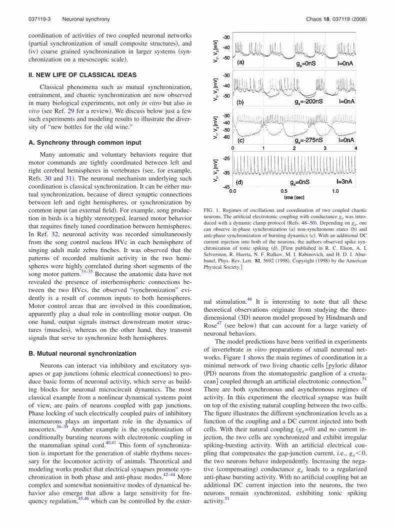
The model predictions have been verified in experimentsof invertebrate in vitro preparations of small neuronal net-works. Figure 1 shows the main regimes of coordination in aminimal network of two living chaotic cells �pyloric dilator�PD� neurons from the stomatogastric ganglion of a crusta-cean� coupled through an artificial electrotonic connection.51
There are both synchronous and asynchronous regimes ofactivity. In this experiment the electrical synapse was builton top of the existing natural coupling between the two cells.The figure illustrates the different synchronization levels as afunction of the coupling and a DC current injected into bothcells. With their natural coupling �ga=0� and no current in-jection, the two cells are synchronized and exhibit irregularspiking-bursting activity. With an artificial electrical cou-pling that compensates the gap-junction current, i.e., ga�0,the two neurons behave independently. Increasing the nega-tive �compensating� conductance ga leads to a regularizedanti-phase bursting activity. With no artificial coupling but anadditional DC current injection into the neurons, the twoneurons remain synchronized, exhibiting tonic spikingactivity.51
FIG. 1. Regimes of oscillations and coordination of two coupled chaoticneurons. The artificial electrotonic coupling with conductance ga was intro-duced with a dynamic clamp protocol �Refs. 48–50�. Depending on ga, onecan observe in-phase synchronization �a� non-synchronous states �b� andanti-phase synchronization of bursting dynamics �c�. With an additional DCcurrent injection into both of the neurons, the authors observed spike syn-chronization of tonic spiking �d�. �First published in R. C. Elson, A. I.Selverston, R. Huerta, N. F. Rulkov, M. I. Rabinovich, and H. D. I. Abar-banel, Phys. Rev. Lett. 81, 5692 �1998�. Copyright �1998� by the AmericanPhysical Society.�
037119-3 Neuronal synchrony Chaos 18, 037119 �2008�
Similar phenomena have been found in experiments withtwo pyloric central pattern generators �CPGs� connectedthrough artificial inhibitory synapses implemented in dy-namic clamp.48,49,52 Reciprocal inhibitory coupling betweenthe pacemaker groups �consisting of the anterior burster�AB� neuron and two pyloric dilator �PD� neurons in eachCPG� leads to anti-phase synchronization while nonrecipro-cal coupling from the lateral pyloric neurons to the PD neu-rons of the other CPG produces in-phase synchronization.53
C. Entrainment
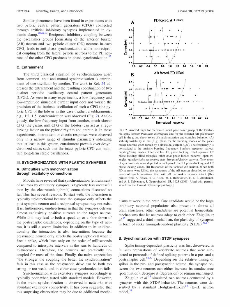
The third classical situation of synchronization apartfrom common input and mutual synchronization is entrain-ment of one oscillator by another. The work in Ref. 54 ad-dresses the entrainment and the resulting coordination of twodistinct periodic oscillatory central pattern generators�CPGs�. As seen in many experiments, a low-frequency andlow-amplitude sinusoidal current input does not worsen theprecision of the intrinsic oscillation of such a CPG �the py-loric CPG of the lobster in this case�; rather, a subharmonic,e.g., 1:2, 1:5, synchronization was observed �Fig. 2�. Analo-gously, the low-frequency input from another, much slowerCPG �the gastric mill CPG of the lobster� can act as a regu-larizing factor on the pyloric rhythm and entrain it. In theseexperiments, intermittent or chaotic responses were observedonly in a narrow range of stimulus parameters indicatingthat, at least in this system, entrainment prevails over desyn-chronized states such that the intact pyloric CPG can main-tain long-term stable oscillations.
III. SYNCHRONIZATION WITH PLASTIC SYNAPSES
A. Difficulties with synchronizationthrough excitatory connections
Models have revealed that synchronization �entrainment�of neurons by excitatory synapses is typically less successfulthan by the electrotonic �ohmic� connections discussed sofar. This has several reasons. To start with, the interaction istypically unidirectional because the synapse only affects thepost-synaptic neuron and a reciprocal synapse may not exist.It is, furthermore, unidirectional in that the synapses deliversalmost exclusively positive currents to the target neuron.While this may lead to both a speed-up or a slow-down ofthe postsynaptic oscillations, depending on the type of neu-ron, it is still a severe limitation. In addition to its unidirec-tionality the interaction is also intermittent because thepresynaptic neuron only affects the postsynaptic cell when itfires a spike, which lasts only on the order of millisecondscompared to interspike intervals in the tens to hundreds ofmilliseconds. Therefore, the neurons are practically un-coupled for most of the time. Finally, the naive expectation“the stronger the coupling the better the synchronization”fails in this case as the presynaptic drive can be both toostrong or too weak, and in either case synchronization fails.
Synchronization with excitatory synapses accordingly istypically poor when tested with standard models. However,in the brain, synchronization is observed in networks withabundant excitatory connectivity. It has been suggested thatthis surprising observation may be due to additional mecha-
nisms at work in the brain. One candidate would be the largeinhibitory neuronal populations also present in almost allbrain structures, other candidates are potential homeostaticmechanisms that let neurons adapt to each other. Zhigulin etal.55 suggested a third mechanism, the plasticity of synapsesin form of spike timing-dependent plasticity �STDP�.56,57
B. Synchronization with STDP synapses
Spike timing-dependent plasticity was first discovered inin vitro preparations of vertebrate neurons that were sub-jected to protocols of defined spiking patterns in a pre- and apostsynaptic cell.56,57 Depending on the relative timing ofspikes in the pre- and postsynaptic neuron, the synapse be-tween the two neurons can either increase its conductance�potentiation�, decrease it �depression� or remain unchanged.
Zhigulin et al.55 simulated two neurons connected by asynapses with this STDP behavior. The neurons were de-scribed by a standard Hodgkin–Huxley58 �H–H� neuronmodel,59
FIG. 2. Arnol’d maps for the forced intact pacemaker group of the Califor-nia spiny lobster Panulirus interruptus and for the isolated AB pacemakercell in the group show zones of synchronization and complex behavior. �A�stability/instability in the �A , f� plane for the bursting modes of the pace-maker neurons when forced by a sinusoidal current Isin�t�. The frequency f isnormalized to the intrinsic bursting frequency. Symbols represent variousbursting/firing modes: filled circles, 1:1 phase locking; filled squares, 1:2phase locking; filled triangles, other n :m phase-locked patterns; open tri-angles, quasiperiodic responses; stars, irregular/chaotic patterns. Two zonesof synchronization are depicted in each panel: the 1:1 phase-locking and 1:2phase-locking zones. �B� Responses of the isolated AB neuron. When bothPD neurons were killed, the responses of the AB neuron alone led to widerzones of synchronizations than with all pacemaker neurons intact. �Re-printed from A. Szücs, R. C. Elson, M. I. Rabinovich, H. D. I. Abarbanel,and A. I. Selverston, J. Neurophysiol. 85, 1623 �2001�. Used with permis-sion from the Journal of Neurophysiology.�
037119-4 Nowotny, Huerta, and Rabinovich Chaos 18, 037119 �2008�
CdVi�t�
dt= − INa�t� − IK�t� − Ileak�t� − Isyn�t� − Istim, �1�
where i=1,2 denotes the number of the pre- and postsynap-tic neuron, respectively, the leak current is given by Ileak�t�=gleak�Vi�t�−Eleak�, and INa�t� and IK�t� were59
INa�t� = gNami�t�3hi�t��Vi�t� − ENa� ,
�2�IK�t� = gKni�t�4�Vi�t� − EK� .
Istim is a constant input current forcing each neuron to spikewith a constant, Istim-dependent frequency, and the secondneuron was driven by the first via the excitatory synapticcurrent Isyn given by Eq. �4� below. The activation and inac-tivation variables yi�t�= �ni�t� ,mi�t� ,hi�t�� satisfied standardfirst-order kinetics �see Refs. 55 and 59�. These neurons wereconnected by a one-directional coupling of a chemical syn-apse described by
Isyn�t� = g�t�S�t�V2�t� , �3�
dS�t�dt
= ��1 − S�t��H�V1�t�� − �S�t� , �4�
with H�V�=1+tanh�10V� /4. The coupling strength g wasthen subject to the inverse STDP learning rule
�g�t� = G��t� = − A sgn��t�exp�− ���t�� , �5�
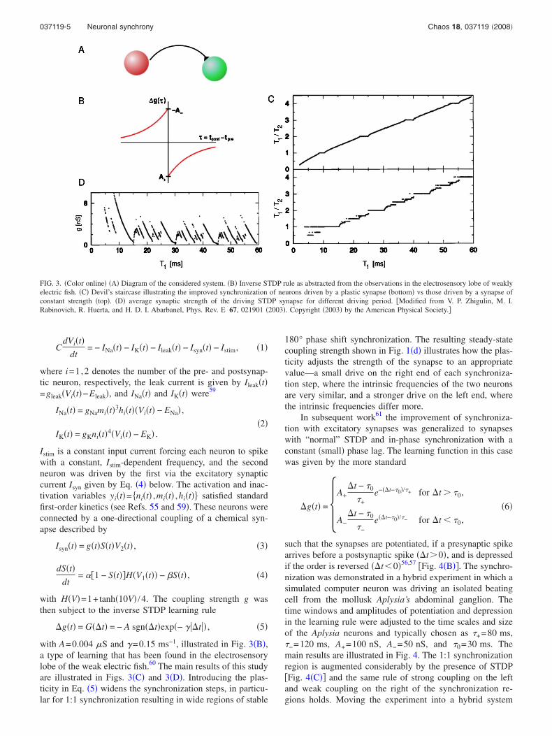
with A=0.004 �S and �=0.15 ms−1, illustrated in Fig. 3�B�,a type of learning that has been found in the electrosensorylobe of the weak electric fish.60 The main results of this studyare illustrated in Figs. 3�C� and 3�D�. Introducing the plas-ticity in Eq. �5� widens the synchronization steps, in particu-lar for 1:1 synchronization resulting in wide regions of stable
180° phase shift synchronization. The resulting steady-statecoupling strength shown in Fig. 1�d� illustrates how the plas-ticity adjusts the strength of the synapse to an appropriatevalue—a small drive on the right end of each synchroniza-tion step, where the intrinsic frequencies of the two neuronsare very similar, and a stronger drive on the left end, wherethe intrinsic frequencies differ more.
In subsequent work61 the improvement of synchroniza-tion with excitatory synapses was generalized to synapseswith “normal” STDP and in-phase synchronization with aconstant �small� phase lag. The learning function in this casewas given by the more standard
�g�t� = �A+�t − �0
�+e−��t−�0�/�+ for �t � �0,
A−�t − �0
�−e��t−�0�/�− for �t � �0,
�6�
such that the synapses are potentiated, if a presynaptic spikearrives before a postsynaptic spike ��t�0�, and is depressedif the order is reversed ��t�0�56,57 �Fig. 4�B��. The synchro-nization was demonstrated in a hybrid experiment in which asimulated computer neuron was driving an isolated beatingcell from the mollusk Aplysia’s abdominal ganglion. Thetime windows and amplitudes of potentiation and depressionin the learning rule were adjusted to the time scales and sizeof the Aplysia neurons and typically chosen as �+=80 ms,�−=120 ms, A+=100 nS, A−=50 nS, and �0=30 ms. Themain results are illustrated in Fig. 4. The 1:1 synchronizationregion is augmented considerably by the presence of STDP�Fig. 4�C�� and the same rule of strong coupling on the leftand weak coupling on the right of the synchronization re-gions holds. Moving the experiment into a hybrid system
FIG. 3. �Color online� �A� Diagram of the considered system. �B� Inverse STDP rule as abstracted from the observations in the electrosensory lobe of weaklyelectric fish. �C� Devil’s staircase illustrating the improved synchronization of neurons driven by a plastic synapse �bottom� vs those driven by a synapse ofconstant strength �top�. �D� average synaptic strength of the driving STDP synapse for different driving period. �Modified from V. P. Zhigulin, M. I.Rabinovich, R. Huerta, and H. D. I. Abarbanel, Phys. Rev. E 67, 021901 �2003�. Copyright �2003� by the American Physical Society.�
037119-5 Neuronal synchrony Chaos 18, 037119 �2008�

automatically introduced considerable amounts of noise andadditional nonstationarity �e.g., short-term plasticity or ho-meostatic effects in the target neuron�. The effect of im-proved synchronization proved immune against these pertur-bations. This work also made a clear prediction for therelationship between the learning rule and the ensuing syn-chronized state: The phase lag in the synchronized stateshould be directly linked to a shift �0 in the STDP learningcurve of similar magnitude. This prediction was recentlyverified in a rather different system, the olfactory system ofthe locust.62 Here, the authors found that neurons in the lobeof the mushroom body of the locust exhibit activity that issynchronized with the activity of the presynaptic intrinsiccells of the mushroom body, the Kenyon cells. This synchro-nization seems to be regulated and improved by STDP of theexcitatory synapses from the Kenyon cells to the lobe neu-rons. Furthermore, they also observed the predicted smallshift in the STDP learning curve, which they extracted fromtheir paired-spike recordings.
The idea of plastic synapses improving synchronizationhas been taken up in several subsequent works including astudy on enhancement of synchronization in neuronalensembles63,64 and the interaction of two neurons with lessregular intrinsic activity.65 It was also used in more appliedwork; e.g., for how to avoid pathological synchronizationstates that may have come about by the STDP of excitatoryconnectivity with deep brain stimulation.66 This idea was
based on the observation that besides the fully synchronizedstate there may be other non-synchronous stable states67 innetworks with excitatory STDP connectivity.
IV. SYNCHRONIZATION OF INHIBITORY MOTIFS
A. Ultrasubharmonic synchronization
According to the traditional view of synchronization, aweak periodic input is able to lock a nonlinear oscillator at afrequency close to that of the input �1:1 zone�. If the forcingincreases, it is possible to achieve synchronization at subhar-monic bands as well. Rabinovich et al.68 demonstrated theinverse phenomenon in a competitive dynamical system:with a weak signal the 1:1 zone is narrow, but the synchro-nization of ultra-subharmonics is dominant. In the system’sphase space, there exists a heteroclinic contour in the autono-mous regime, which is the image of sequential dynamics.Under the action of a weak periodic forcing, in the vicinityof the contour a stable limit cycle with long period appears.This results in very low-frequency oscillations that lock tothe finite frequency of the forcing. It has been hypothesizedthat this phenomenon could be the origin for the synchroni-zation of slow and fast brain rhythms.
1. Rate modelFigure 5 shows the synchronization bands for different
strengths of the forcing in a system of three units describedby
FIG. 4. �Color online� Synchronization �entrainment� of an isolated beating cell of the mollusk Aplysia Californica with a simulated synapse with “normal”STDP. �A� Illustration of the “circuit.” �B� STDP rule used. �C� Devil’s staircase showing the extended 1:1 synchronization plateau. �D� final �stationary�average synaptic strength of the STDP synapse. Note that synchronization does not only occur more often with the STDP synapse but also is more precise ina noisy environment �indicated by the much smaller error bars on the average ISIs in �C�. �Data first published in T. Nowotny, V. P. Zhigulin, A. I. Selverston,H. D. I. Abarbanel, and M. I. Rabinovich, J. Neurosci. 23, 9776 �2003�.�
037119-6 Nowotny, Huerta, and Rabinovich Chaos 18, 037119 �2008�
dai
dt= ai1 − ai + �
j�i
N
ijaj� + �t� + ��i�� ft,ai� , �7�
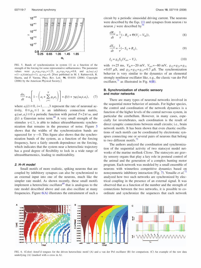
where ai�t� 0, i=1, . . . ,3 represent the rate of neuronal ac-tivity, 0�ij �1 is an inhibitory connection matrix,�i��t ,ai� 0 a periodic function with period T=2� /�, and�t� a Gaussian noise term.68 A very small strength of thestimulus ��1, is able to induce ultrasubharmonic synchro-nization that remains in the presence of noise. Figure 5shows that the widths of the synchronization bands aresqueezed for �→0. This figure also shows that the synchro-nization bands of the system, as a function of the forcingfrequency, have a fairly smooth dependence on the forcing,which indicates that the system near a heteroclinic trajectoryhas a good degree of flexibility to lock in a wide range ofultrasubharmonics, leading to multistability.
2. H–H modelSmall motifs of more realistic, spiking neurons that are
coupled by inhibitory synapses can also be synchronized toan external input into one of the neurons, much like thesimpler rate model. As shown recently, these small motifsimplement a heteroclinic oscillator69 that is analogous to therate model described above and can also oscillate at manyfrequencies. Figure 6�A� illustrates the entrainment of such a
circuit by a periodic sinusoidal driving current. The neuronswere described by the Eqs. �1� and synapses from neuron i toneuron j were described by70
dRij
dt=
1
��− Rij + ��Vj − Vth�� , �8�
dSij
dt=
1
��− Sij + Rij� , �9�
Iij = gijSij�Vrev − Vi� , �10�
with �=25 ms, Vth=−20 mV, Vrev=−80 mV, g12=g23=g31
=0.07 �S, and g21=g32=g13=0.7 �S. The synchronizationbehavior is very similar to the dynamics of an elementalstrongly nonlinear oscillator like, e.g., the classic van der Poloscillator,71 as illustrated in Fig. 6�B�.
B. Synchronization of chaotic sensoryand motor networks
There are many types of neuronal networks involved inthe sequential motor behavior of animals. For higher species,the control and coordination of the network dynamics is afunction of the higher levels of the central nervous system, inparticular the cerebellum. However, in many cases, espe-cially for invertebrates, such coordination is the result ofdirect synaptic connections between small circuits; i.e., brainnetwork motifs. It has been shown that even chaotic oscilla-tions of such motifs can be coordinated by electrotonic syn-apses connecting one or several pairs of neurons that belongto two different motifs.72
The authors analyzed the coordination and synchroniza-tion of the sequential activity of two statocyst model net-works of the marine mollusk Clione. The statocysts are grav-ity sensory organs that play a key role in postural control ofthe animal and the generation of a complex hunting motorprogram. Each network was modeled by a small ensemble ofneurons with winnerless competitive dynamics based onnonsymmetric inhibitory interaction �Fig. 7�. Venaille et al.72
analyzed how two such networks are synchronized by elec-trical coupling in the presence of an external signal. It wasobserved that as a function of the number and the strength ofconnections between the two networks, it is possible to co-ordinate and synchronize the sequences that each network
FIG. 5. Bands of synchronization in system �1� as a function of thestrength of the forcing for some representative subharmonics. The parametervalues were 12=23=31=1.25, 13=21=32=0.8, and �1�� ft ,ai�= �1−ai��sin�� ft�+1�, �2=�3=0. �First published in M. I. Rabinovich, R.Huerta, and P. Varona, Phys. Rev. Lett. 96, 014101 �2006�. Copyright�2006� by the American Physical Society.�
FIG. 6. �Color� Arnol’d tongues for the driven heteroclinic motif �A� and a van der Pol oscillator �B� for comparison. �C� An example of the raw dataunderlying �A� �marked with a cross in A�.
037119-7 Neuronal synchrony Chaos 18, 037119 �2008�
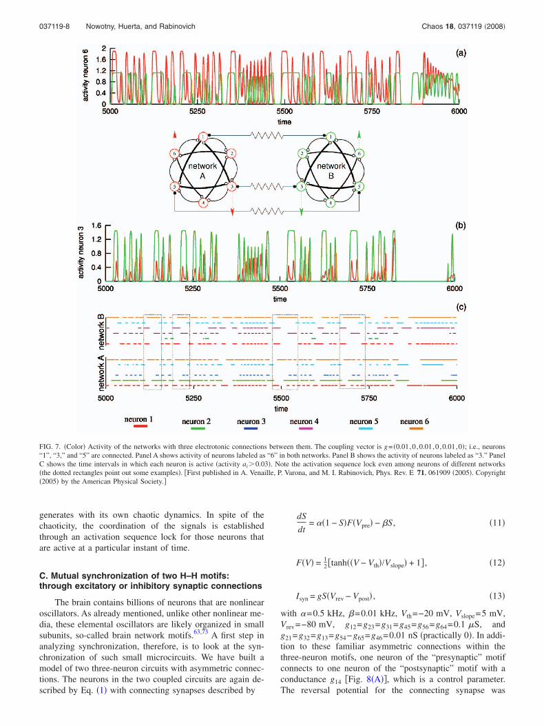
generates with its own chaotic dynamics. In spite of thechaoticity, the coordination of the signals is establishedthrough an activation sequence lock for those neurons thatare active at a particular instant of time.
C. Mutual synchronization of two H–H motifs:through excitatory or inhibitory synaptic connections
The brain contains billions of neurons that are nonlinearoscillators. As already mentioned, unlike other nonlinear me-dia, these elemental oscillators are likely organized in smallsubunits, so-called brain network motifs.63,73 A first step inanalyzing synchronization, therefore, is to look at the syn-chronization of such small microcircuits. We have built amodel of two three-neuron circuits with asymmetric connec-tions. The neurons in the two coupled circuits are again de-scribed by Eq. �1� with connecting synapses described by
dS
dt= ��1 − S�F�Vpre� − �S , �11�
F�V� = 12 �tanh��V − Vth�/Vslope� + 1� , �12�
Isyn = gS�Vrev − Vpost� , �13�
with �=0.5 kHz, �=0.01 kHz, Vth=−20 mV, Vslope=5 mV,Vrev=−80 mV, g12=g23=g31=g45=g56=g64=0.1 �S, andg21=g32=g13=g54−g65=g46=0.01 nS �practically 0�. In addi-tion to these familiar asymmetric connections within thethree-neuron motifs, one neuron of the “presynaptic” motifconnects to one neuron of the “postsynaptic” motif with aconductance g14 �Fig. 8�A��, which is a control parameter.The reversal potential for the connecting synapse was
FIG. 7. �Color� Activity of the networks with three electrotonic connections between them. The coupling vector is g= �0.01,0 ,0.01,0 ,0.01,0�; i.e., neurons“1”, “3,” and “5” are connected. Panel A shows activity of neurons labeled as “6” in both networks. Panel B shows the activity of neurons labeled as “3.” PanelC shows the time intervals in which each neuron is active �activity ai�0.03�. Note the activation sequence lock even among neurons of different networks�the dotted rectangles point out some examples�. �First published in A. Venaille, P. Varona, and M. I. Rabinovich, Phys. Rev. E 71, 061909 �2005�. Copyright�2005� by the American Physical Society.�
037119-8 Nowotny, Huerta, and Rabinovich Chaos 18, 037119 �2008�

Erev,14=0 mV for an excitatory connection �Fig. 8�B�� andErev,14=−80 mV for an inhibitory coupling �Fig. 8�C��. Allneurons in the postsynaptic motif are excited by a constantIstim=0.15 nA, while all neurons of the presynaptic motif re-ceive a stimulus current Istim, which is varied in a range from0.12 to 0.5 nA, corresponding to uncoupled period ratios ofT1 /T2=0.5. . .1.1.
We observe some peculiarities immediately. Due to thepresynaptic motif’s own intrinsic complexity the period ra-tios do not vary smoothly with the injected currents leadingto the white stripes of unattainable uncoupled frequency ra-tios in Figs. 8�B� and 8�C�. In comparison between excita-tory and inhibitory coupling we notice a much larger regionof 1:2 synchronization for the latter. If we compare these
FIG. 8. �Color� Synchronization of two three-neuron motifs of H–H neurons �A, B, C� compared to the synchronization of “typical” bursting neurons �D, E�,here map-based bursting neurons �Ref. 74�. �A� The connectivity with strong interactions within the motifs and a weaker interaction between one of each ofthe motif neurons �neurons 1 and 4�. �B� and �D� show the synchronization regions for excitatory coupling, while �C� and E� were obtained for inhibitorycoupling.
037119-9 Neuronal synchrony Chaos 18, 037119 �2008�
results to the synchronization behavior of two “generic”bursting neurons, the same difference of enhanced 1:2 syn-chronization appears. However, the overall synchronizationregions are quite differently shaped.
The presented results show that the details of spikingactivities of the neurons in the motif, in fact, do not influencethe motif’s interaction with other motifs as it behaves muchlike a more elemental oscillator. This is a very importantmessage that supports one of the basic principles that thebrain uses: the dynamics on different levels of brain com-plexity hierarchy that operate on different time and spacescales are relatively independent �see also Ref. 69�.
V. COARSE-GRAINED SYNCHRONIZATION
A. Patterns of synchronization
On a larger scale beyond neuronal network motifs, neu-ronal media can be seen as nonequilibrium media that gen-erate ordered or coherent spatio-temporal structures due tothe dynamical interaction between their elements, the neu-rons, and the intrinsic properties of these neurons. Pretchl etal.75,76 reported observations of cortical oscillations in theturtle leading to linear and circular waves that are a signatureof the presence of an underlying excitable nonequilibriummedium. In Refs. 77 and 78 the authors could also find theformation of coarse grain dynamics in a slide of the mam-malian cortex by blocking the inhibition of the neuronal me-dia. One of the main features of neuronal media is the exis-tence of different time scales in the individual oscillations.These time scales produce a rich and complicated behaviorthat requires a coarse grained interpretation of the whole sys-tem to comprehend it. A very good system to understand thecoarse grain dynamics of neuronal media is the Hindmarsh–Rose model47 because two time scales are present in it andwhile it has rich dynamics including chaos it can still beexpressed in a simple mathematical form. The system is builtaround a fast variable for the spiking dynamics combinedwith a slow variable that produces modulation in the spikingbehavior of the neuron. The template system of ordinary dif-ferential equations �ODEs� is given by
dxi
dt= yi + axi
3 − zi + ei − g�j
�xi − xj� ,
dyi
dt= b − cxi
2 − yi,1
�
dzi
dt= − rzi + rS�xi + d� ,
where the index j runs over the nearest neighbors of thelattice where usually S=4, a=3, d=1.6, b=1, c=5, and r isset in the chaotic regime with value r=0.0021. The strengthof the dissipative coupling is determined by g. Computersimulations of networks built from heterogeneous elementsdescribed by this set of ODEs and individual values for ei
indicate that cooperative behavior among the elements isable to produce large-scale coherent structures with slowperiodic oscillations despite the presence of chaoticelements.79,80
B. Coarse grained variables
In order to understand the origin of the coherent struc-tures in this system, the dynamics of a group has to betraced. These groups display the following type of behaviordepending on g: �i� chaotic behavior which dimension in-creases with the number of elements for small coupling, �ii�chaotic synchronization of the slow oscillations, and �iii�complete chaotic synchronization for strong coupling.
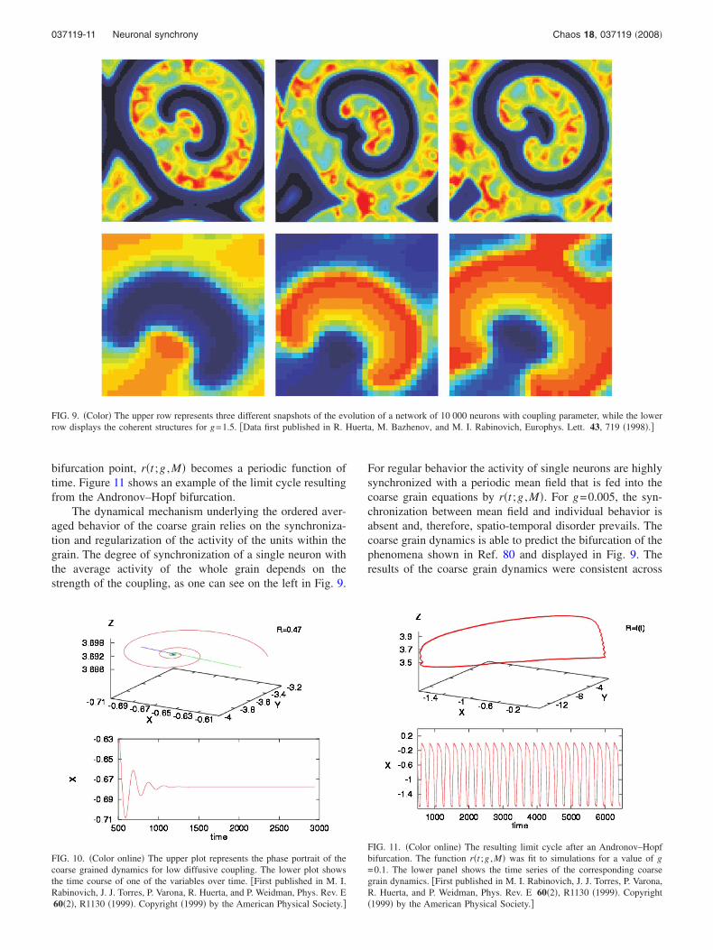
Figure 9 illustrates the effect of the coupling onto thecoarse grain size of the coherent structure that is developingin the system. The stronger the coupling is the larger is thesize of the “grain.” In Ref. 79 a coarse grain variable repre-sentative of this group was proposed. A new set of variablesobtained from an average measure of the grain that dependson g can be built, given by
X�t� =1
M�i=1
M
xi�t� = �xi�CG, �14�
Y�t� = �yi�CG, �15�
Z�t� = �zi�CG, �16�
where M is the number of neurons in the cluster. To build anapproximate ODE for the cluster dynamics, one can use
xi�t� = X�t� + i�t;g,M� , �17�
yi�t� = Y�t� + �i�t;g,M� , �18�
zi�t� = Z�t� + �i�t;g,M� , �19�
which are then introduced into the Hindmarsh–Rose equa-tions to obtain
dX
dt= Y + aX2 + ar�t;g,M� − X3 − 3Xr�t;g,M� − Z + � , �20�
dY
dt= − cX2 − Y − �cr�t;g,M� − b� , �21�
1
�
dZ
dt= − Z + S�X + d� , �22�
where �= �ei�CG, �i�t ;g ,M��= ��i�t ;g ,M��= ��i�t ;g ,M��=0,and the only function that plays a role, ignoring higher orderterms than �i
2, is r�t ;g ,M�= �i2�CG.
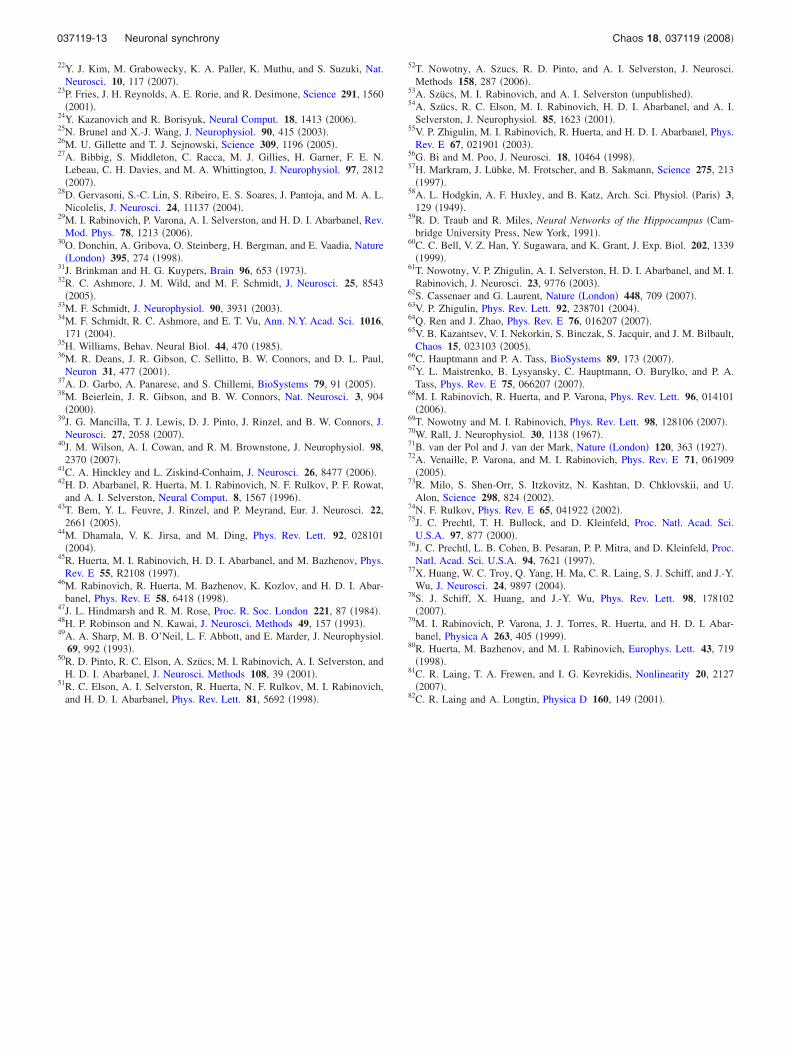
The origin of the periodic average behavior emergingwhen using Eqs. �20�–�22� can be understood by a pitchforkbifurcation, where r�t ;g ,M� is either replaced by a constantor by a periodic function that represents the type of meanfield action of the neighboring elements. For sufficientlysmall g, r�t ;g ,M� is nearly constant with values in the range0.4–0.5 and only a single stable fixed point is present corre-sponding to steady-state behavior of the cluster �Fig. 10�.
For r�t ;g ,M��Rcrit �g�gcrit�, the fixed point becomesunstable and the limit cycle in the 3D phase space of theaverage coarse grained system undergoes a supercriticalAndronov–Hopf bifurcation to a stable limit cycle. At the
037119-10 Nowotny, Huerta, and Rabinovich Chaos 18, 037119 �2008�
bifurcation point, r�t ;g ,M� becomes a periodic function oftime. Figure 11 shows an example of the limit cycle resultingfrom the Andronov–Hopf bifurcation.
The dynamical mechanism underlying the ordered aver-aged behavior of the coarse grain relies on the synchroniza-tion and regularization of the activity of the units within thegrain. The degree of synchronization of a single neuron withthe average activity of the whole grain depends on thestrength of the coupling, as one can see on the left in Fig. 9.
For regular behavior the activity of single neurons are highlysynchronized with a periodic mean field that is fed into thecoarse grain equations by r�t ;g ,M�. For g=0.005, the syn-chronization between mean field and individual behavior isabsent and, therefore, spatio-temporal disorder prevails. Thecoarse grain dynamics is able to predict the bifurcation of thephenomena shown in Ref. 80 and displayed in Fig. 9. Theresults of the coarse grain dynamics were consistent across
FIG. 9. �Color� The upper row represents three different snapshots of the evolution of a network of 10 000 neurons with coupling parameter, while the lowerrow displays the coherent structures for g=1.5. �Data first published in R. Huerta, M. Bazhenov, and M. I. Rabinovich, Europhys. Lett. 43, 719 �1998�.�
FIG. 10. �Color online� The upper plot represents the phase portrait of thecoarse grained dynamics for low diffusive coupling. The lower plot showsthe time course of one of the variables over time. �First published in M. I.Rabinovich, J. J. Torres, P. Varona, R. Huerta, and P. Weidman, Phys. Rev. E60�2�, R1130 �1999�. Copyright �1999� by the American Physical Society.�
FIG. 11. �Color online� The resulting limit cycle after an Andronov–Hopfbifurcation. The function r�t ;g ,M� was fit to simulations for a value of g=0.1. The lower panel shows the time series of the corresponding coarsegrain dynamics. �First published in M. I. Rabinovich, J. J. Torres, P. Varona,R. Huerta, and P. Weidman, Phys. Rev. E 60�2�, R1130 �1999�. Copyright�1999� by the American Physical Society.�
037119-11 Neuronal synchrony Chaos 18, 037119 �2008�

different topologies of network connectivity. The formationof large-scale coherent structures in nonequilibrium mediaconsisting of discrete elements with fast and slow oscilla-tions has two key features. The first is the regularizationphenomena inside clusters of chaotic elements. This regular-ization of the behavior is the result of the action of the av-eraged activity of fast pulsations in the slow coarse graindynamics. The second feature is the instability of the homo-geneous oscillation modes in a media considered to be acoarse grain lattice.
The effectiveness of the coarse grain approach for under-standing the bifurcation of a complex neuronal system hasalso been proven to be useful in neuronal fields.81 A neuronalfield is described by a set of partial integro-differential equa-tions
�u�x,t��t
= − u�x,t� + �−�
�
J�x − y�f�I + u�y,t� − a�y,t��dy ,
�23�
��a�x,t�
�t= Au�x,t� − a�x,t� , �24�
on −��x�� with periodic boundary conditions as ex-plained in Ref. 82, where u�x , t� is the activity of the neuronsat location x and time t and a�x , t� is the intrinsic activity ofthe neurons. The remaining functions are the connectivityfunction J�x−y� and the firing rate of the neurons f�u�,which is not spatially dependent. The approach consists indefining a new coarse scalar variable V�t�, which is the in-stantaneous difference in position between the peak of u�x , t�and a�x , t�. Laing et al. assume that V�t� satisfies an un-known Langevin equation and then numerically obtain theeffective potential for the corresponding Fokker–Planckequation. The zeros of the drift parameter ��V� of theFokker–Planck equation are numerically estimated to obtainFig. 12 by using a non-negligible level of noise. This ap-
proach is successful in predicting a pitchfork bifurcation ofthe neuronal media �Eq. �23�� via a coarse grain description.
VI. CONCLUSION
Reflecting one more time on the neuronal synchroniza-tion phenomena that we discussed above, an expert in non-linear dynamical theory may say “it is very interesting, butwhere is the peculiarity?”. Indeed, despite all the specifics ofthe complex biochemical processes that characterize the ac-tivity of individual elements of neuronal networks, i.e., theneurons and synapses, creating a dynamical model has be-come, after formalizing the problem, just another, albeit im-portant, application of the universal tools and approaches ofnonlinear dynamical theory. The answer to the questionabout peculiarity lies in a different aspect: The phenomenathat we described in this review appear too specific, andsometimes even pathological, for the traditional models ofnonlinear dynamical theory. However, for neuronal systemsthey are generic. This is the peculiarity. We recall here onlyone example: heteroclinic synchronization. It can be de-scribed in the framework of a typical rate model of neuronalinformation processing. Since all variables in this model arepositive or equal zero by construction �ai 0� the phasespace of the system is bounded by manifolds aj =0 and, be-cause of that, the heteroclinic sequence that exists in thissystem can be structurally stable. This may seem peculiar ingeneral nonlinear dynamical theory, but for neuronal systemsit is a generic phenomenon.
ACKNOWLEDGMENTS
This work was partly supported by the NIH, Grant No.R01 NS050945 �R.H., M.R.�, and a RCUK fellowship�T.N.�.
1A. Schnitzler and J. Gross, Nat. Rev. Neurosci. 6, 285 �2005�.2P. J. Uhlhaas and W. Singer, Neuron 52, 155 �2006�.3P. Brown, Mov Disord. 18, 357 �2003�.4A. K. Engel, P. Fries, and W. Singer, Nat. Rev. Neurosci. 2, 704 �2001�.5P. Fries, Trends Cogn. Sci. 9, 474 �2005�.6P. Fries, D. Nikoli, and W. Singer, TINS 30, 309 �2007�.7E. Niebur, S. S. Hsiao, and K. O. Johnson, Curr. Opin. Neurobiol. 12, 190�2002�.
8C. Legendy, in Progress in Cybernetics, edited by J. Rose �Gordon andBreach, New York, 1970�, Vol. 1.
9P. Milner, Psychol. Rev. 81, 521 �1974�.10C. V. der Malsburg, Tech. Rep. 81-2, 1981; reprinted in Models of Neural
Networks II, edited by E. Domany, J. L. van Hemmen, and K. Schukten�Springer, Berlin, 1994�.
11W. Singer, Annu. Rev. Physiol. 55, 349 �1993�.12C. von der Malsburg, in The Neural and Molecular Bases of Learning,
edited by M. K. J. P. Changeux �John Wiley & Sons, Chichester, 1987�.13F. Varela, J. P. Lachaux, E. Rodriguez, and J. Martinerie, Nat. Rev.
Neurosci. 2, 229 �2001�.14N. Axmacher, F. Mormann, G. Fernndez, C. E. Elger, and J. Fell, Brain
Res. Rev. 52, 170 �2006�.15M. N. Shadlen and J. A. Movshon, Neuron 24, 67 �1999�.16A. Raffone and C. van Leeuwen, Chaos 13, 1090 �2003�.17J. Fell, P. Klaver, K. Lehnertz, T. Grunwald, C. Schaller, C. E. Elger, and
G. Fernández, Nat. Neurosci. 4, 1259 �2001�.18J. Beshel, N. Kopell, and L. M. Kay, J. Neurosci. 27, 8358 �2007�.19C. Börgers and N. J. Kopell, Neural Comput. 20, 383 �2008�.20J. Gross, F. Schmitz, I. Schnitzler, K. Kessler, K. Shapiro, B. Hommel,
and A. Schnitzler, Proc. Natl. Acad. Sci. U.S.A. 101, 13050 �2004�.21T. Womelsdorf and P. Fries, Curr. Opin. Neurobiol. 17, 154 �2007�.
FIG. 12. The zeros of the drift term of the Fokker–Planck equation derivedfrom the neuronal field using the coarse variable. The coarse grain variableis plotted against the gain variable of the neuronal field equations. The filledcircles represent stable solutions while the open circles represent unstableones. �Generated from data provided by C. R. Laing, first published in C. R.Laing, T. A. Frewen, and I. G. Kevrekidis, Nonlinearity 20, 2127 �2007�.�
037119-12 Nowotny, Huerta, and Rabinovich Chaos 18, 037119 �2008�
22Y. J. Kim, M. Grabowecky, K. A. Paller, K. Muthu, and S. Suzuki, Nat.Neurosci. 10, 117 �2007�.
23P. Fries, J. H. Reynolds, A. E. Rorie, and R. Desimone, Science 291, 1560�2001�.
24Y. Kazanovich and R. Borisyuk, Neural Comput. 18, 1413 �2006�.25N. Brunel and X.-J. Wang, J. Neurophysiol. 90, 415 �2003�.26M. U. Gillette and T. J. Sejnowski, Science 309, 1196 �2005�.27A. Bibbig, S. Middleton, C. Racca, M. J. Gillies, H. Garner, F. E. N.
Lebeau, C. H. Davies, and M. A. Whittington, J. Neurophysiol. 97, 2812�2007�.
28D. Gervasoni, S.-C. Lin, S. Ribeiro, E. S. Soares, J. Pantoja, and M. A. L.Nicolelis, J. Neurosci. 24, 11137 �2004�.
29M. I. Rabinovich, P. Varona, A. I. Selverston, and H. D. I. Abarbanel, Rev.Mod. Phys. 78, 1213 �2006�.
30O. Donchin, A. Gribova, O. Steinberg, H. Bergman, and E. Vaadia, Nature�London� 395, 274 �1998�.
31J. Brinkman and H. G. Kuypers, Brain 96, 653 �1973�.32R. C. Ashmore, J. M. Wild, and M. F. Schmidt, J. Neurosci. 25, 8543
�2005�.33M. F. Schmidt, J. Neurophysiol. 90, 3931 �2003�.34M. F. Schmidt, R. C. Ashmore, and E. T. Vu, Ann. N.Y. Acad. Sci. 1016,
171 �2004�.35H. Williams, Behav. Neural Biol. 44, 470 �1985�.36M. R. Deans, J. R. Gibson, C. Sellitto, B. W. Connors, and D. L. Paul,
Neuron 31, 477 �2001�.37A. D. Garbo, A. Panarese, and S. Chillemi, BioSystems 79, 91 �2005�.38M. Beierlein, J. R. Gibson, and B. W. Connors, Nat. Neurosci. 3, 904
�2000�.39J. G. Mancilla, T. J. Lewis, D. J. Pinto, J. Rinzel, and B. W. Connors, J.
Neurosci. 27, 2058 �2007�.40J. M. Wilson, A. I. Cowan, and R. M. Brownstone, J. Neurophysiol. 98,
2370 �2007�.41C. A. Hinckley and L. Ziskind-Conhaim, J. Neurosci. 26, 8477 �2006�.42H. D. Abarbanel, R. Huerta, M. I. Rabinovich, N. F. Rulkov, P. F. Rowat,
and A. I. Selverston, Neural Comput. 8, 1567 �1996�.43T. Bem, Y. L. Feuvre, J. Rinzel, and P. Meyrand, Eur. J. Neurosci. 22,
2661 �2005�.44M. Dhamala, V. K. Jirsa, and M. Ding, Phys. Rev. Lett. 92, 028101
�2004�.45R. Huerta, M. I. Rabinovich, H. D. I. Abarbanel, and M. Bazhenov, Phys.
Rev. E 55, R2108 �1997�.46M. Rabinovich, R. Huerta, M. Bazhenov, K. Kozlov, and H. D. I. Abar-
banel, Phys. Rev. E 58, 6418 �1998�.47J. L. Hindmarsh and R. M. Rose, Proc. R. Soc. London 221, 87 �1984�.48H. P. Robinson and N. Kawai, J. Neurosci. Methods 49, 157 �1993�.49A. A. Sharp, M. B. O’Neil, L. F. Abbott, and E. Marder, J. Neurophysiol.
69, 992 �1993�.50R. D. Pinto, R. C. Elson, A. Szücs, M. I. Rabinovich, A. I. Selverston, and
H. D. I. Abarbanel, J. Neurosci. Methods 108, 39 �2001�.51R. C. Elson, A. I. Selverston, R. Huerta, N. F. Rulkov, M. I. Rabinovich,
and H. D. I. Abarbanel, Phys. Rev. Lett. 81, 5692 �1998�.
52T. Nowotny, A. Szucs, R. D. Pinto, and A. I. Selverston, J. Neurosci.Methods 158, 287 �2006�.
53A. Szücs, M. I. Rabinovich, and A. I. Selverston �unpublished�.54A. Szücs, R. C. Elson, M. I. Rabinovich, H. D. I. Abarbanel, and A. I.
Selverston, J. Neurophysiol. 85, 1623 �2001�.55V. P. Zhigulin, M. I. Rabinovich, R. Huerta, and H. D. I. Abarbanel, Phys.
Rev. E 67, 021901 �2003�.56G. Bi and M. Poo, J. Neurosci. 18, 10464 �1998�.57H. Markram, J. Lübke, M. Frotscher, and B. Sakmann, Science 275, 213
�1997�.58A. L. Hodgkin, A. F. Huxley, and B. Katz, Arch. Sci. Physiol. �Paris� 3,
129 �1949�.59R. D. Traub and R. Miles, Neural Networks of the Hippocampus �Cam-
bridge University Press, New York, 1991�.60C. C. Bell, V. Z. Han, Y. Sugawara, and K. Grant, J. Exp. Biol. 202, 1339
�1999�.61T. Nowotny, V. P. Zhigulin, A. I. Selverston, H. D. I. Abarbanel, and M. I.
Rabinovich, J. Neurosci. 23, 9776 �2003�.62S. Cassenaer and G. Laurent, Nature �London� 448, 709 �2007�.63V. P. Zhigulin, Phys. Rev. Lett. 92, 238701 �2004�.64Q. Ren and J. Zhao, Phys. Rev. E 76, 016207 �2007�.65V. B. Kazantsev, V. I. Nekorkin, S. Binczak, S. Jacquir, and J. M. Bilbault,
Chaos 15, 023103 �2005�.66C. Hauptmann and P. A. Tass, BioSystems 89, 173 �2007�.67Y. L. Maistrenko, B. Lysyansky, C. Hauptmann, O. Burylko, and P. A.
Tass, Phys. Rev. E 75, 066207 �2007�.68M. I. Rabinovich, R. Huerta, and P. Varona, Phys. Rev. Lett. 96, 014101
�2006�.69T. Nowotny and M. I. Rabinovich, Phys. Rev. Lett. 98, 128106 �2007�.70W. Rall, J. Neurophysiol. 30, 1138 �1967�.71B. van der Pol and J. van der Mark, Nature �London� 120, 363 �1927�.72A. Venaille, P. Varona, and M. I. Rabinovich, Phys. Rev. E 71, 061909
�2005�.73R. Milo, S. Shen-Orr, S. Itzkovitz, N. Kashtan, D. Chklovskii, and U.
Alon, Science 298, 824 �2002�.74N. F. Rulkov, Phys. Rev. E 65, 041922 �2002�.75J. C. Prechtl, T. H. Bullock, and D. Kleinfeld, Proc. Natl. Acad. Sci.
U.S.A. 97, 877 �2000�.76J. C. Prechtl, L. B. Cohen, B. Pesaran, P. P. Mitra, and D. Kleinfeld, Proc.
Natl. Acad. Sci. U.S.A. 94, 7621 �1997�.77X. Huang, W. C. Troy, Q. Yang, H. Ma, C. R. Laing, S. J. Schiff, and J.-Y.
Wu, J. Neurosci. 24, 9897 �2004�.78S. J. Schiff, X. Huang, and J.-Y. Wu, Phys. Rev. Lett. 98, 178102
�2007�.79M. I. Rabinovich, P. Varona, J. J. Torres, R. Huerta, and H. D. I. Abar-
banel, Physica A 263, 405 �1999�.80R. Huerta, M. Bazhenov, and M. I. Rabinovich, Europhys. Lett. 43, 719
�1998�.81C. R. Laing, T. A. Frewen, and I. G. Kevrekidis, Nonlinearity 20, 2127
�2007�.82C. R. Laing and A. Longtin, Physica D 160, 149 �2001�.
037119-13 Neuronal synchrony Chaos 18, 037119 �2008�
Copyright © 2022 FDOKUMEN