Contact Lens and Anterior Eye
10
Contents lists available at ScienceDirect Contact Lens and Anterior Eye journal homepage: www.elsevier.com/locate/clae Cultivation and characterization of pterygium as an ex vivo study model for disease and therapy Natasha Josifovska a , Dóra Júlia Szabó a , Richárd Nagymihály a , Zoltán Veréb a , Andrea Facskó a , Ketil Eriksen b , Morten C. Moe b , Goran Petrovski a,b, ⁎ a Stem Cells and Eye Research Laboratory, Department of Ophthalmology, Faculty of Medicine, University of Szeged, Koranyi Fasor 10-11, 6720 Szeged, Hungary b Center for Eye Research, Department of Ophthalmology, Oslo University Hospital and University of Oslo, Kirkeveien 166, N-0407 Oslo, Norway ARTICLE INFO Keywords: Pterygium Long-term cultures Tissue engineering Mitomycin C IL-6 IL-8 ABSTRACT Purpose: Development of ex vivo model to study pathogenesis, inflammation and treatment modalities for pterygium. Methods: Pterygium obtained from surgery was cultivated (3 months). Gravitational attachment method using viscoelastic facilitated adherence of graft and outgrowing cells. Medium contained serum as the only growth supplement with no use of scaffolds. Surface profiling of the multi-layered cells for hematopoietic- and mesenchymal stem cell markers was performed. Examination of cells by immunohistochemistry using pluripotency, oxidative stress, stemness, migration and proliferation, epithelial and secretory markers was performed. The effect of anti-proliferative agent Mitomycin C upon secretion of pro-inflammatory cytokines IL-6 and IL-8 was assessed. Results: Cells showed high expression of migration- (CXCR4), secretory- (MUC1, MUC4) and oxidative damage- (8-OHdG) markers, and low expression of hypoxia- (HIF-1α) and proliferation- (Ki-67) markers. Moderate and low expression of the pluripotency markers (Vimentin and ΔNp63) was present, respectively, while the putative markers of stemness (Sox2, Oct4, ABCG-2) and epithelial cell markers- (CK19, CK8-18) were weak. The surface marker profile of the outgrowing cells revealed high expression of the hematopoietic marker CD47, mesenchymal markers CD90 and CD73, minor or less positivity for the hematopoietic marker CD34, mesenchymal marker CD105, progenitor marker CD117 and attachment protein markers while low levels of IL-6 and IL-8 secretion ex vivo, were inhibited upon Mitomycin C treatment. Conclusion: Ex vivo tissue engineered pterygium consists of a mixture of cells of different lineage origin, suitable for use as a disease model for studying pathogenesis ex vivo, while opening possibilities for new treatment and prevention modalities. 1. Introduction Pterygium is a common ocular surface disorder characterized by an active wing-shaped overgrowth of epithelial and fibrovascular tissue growing from the conjunctiva towards the limbus and onto the cornea, thus giving cosmetic alterations to the ocular surface and in advanced cases impairing vision [1–4]. It consists of a head which falls onto the anterior cornea, a neck which covers the superficial limbus, and a body which overlies the sclera. The first sign of pterygium is formation of a cap which appears as halo in front of the head and goes deep into the epithelium without respecting the limbal epithelium stem cell border [5]. It is not fully known whether the stem cell deficiency alone or other factors drive the centripetal migration of the pterygium cells onto the cornea, or how sclera support may play a role in the process. The pathogenesis of pterygium remains uncertain, although it is thought to be mainly caused by ultraviolet (UV) radiation. A wide range of alternative pathogenic factors have been proposed, including viral infections, epigenetic aberrations, epithelial-mesenchymal transition, immunologic and anti-apoptotic mechanisms, angiogenic and lymphan- giogenic stimulation, deregulation of extracellular matrix (ECM) mod- ulators and growth factors, inflammation cascades, recruitment of bone-marrow-derived stem- and progenitor cells, and modifications in the cholesterol metabolism. Most of these factors are rather related to the development and maintenance of the disease than to its origin [6]. In the present study, cells were adherently cultivated and grown out of the pterygium using gravitational force from viscoelastic material for more than three months or until they formed multi-layered structures. Surface marker profiling using hematopoietic- and mesenchymal stem http://dx.doi.org/10.1016/j.clae.2017.04.002 Received 6 October 2016; Received in revised form 30 March 2017; Accepted 19 April 2017 ⁎ Corresponding author at: Department of Ophthalmology, University of Szeged, Korányi fasor 10-11, 6720, Szeged, Hungary. E-mail address: [email protected] (G. Petrovski). Contact Lens and Anterior Eye 40 (2017) 283–292 1367-0484/ © 2017 British Contact Lens Association. Published by Elsevier Ltd. All rights reserved. MARK
-
Upload
khangminh22 -
Category
Documents
-
view
4 -
download
0
Transcript of Contact Lens and Anterior Eye
Contents lists available at ScienceDirect
Contact Lens and Anterior Eye
journal homepage: www.elsevier.com/locate/clae
Cultivation and characterization of pterygium as an ex vivo study model fordisease and therapy
Natasha Josifovskaa, Dóra Júlia Szabóa, Richárd Nagymihálya, Zoltán Veréba, Andrea Facskóa,Ketil Eriksenb, Morten C. Moeb, Goran Petrovskia,b,⁎
a Stem Cells and Eye Research Laboratory, Department of Ophthalmology, Faculty of Medicine, University of Szeged, Koranyi Fasor 10-11, 6720 Szeged, Hungaryb Center for Eye Research, Department of Ophthalmology, Oslo University Hospital and University of Oslo, Kirkeveien 166, N-0407 Oslo, Norway
A R T I C L E I N F O
Keywords:PterygiumLong-term culturesTissue engineeringMitomycin CIL-6IL-8
A B S T R A C T
Purpose: Development of ex vivo model to study pathogenesis, inflammation and treatment modalities forpterygium.Methods: Pterygium obtained from surgery was cultivated (3 months). Gravitational attachment method usingviscoelastic facilitated adherence of graft and outgrowing cells. Medium contained serum as the only growthsupplement with no use of scaffolds. Surface profiling of the multi-layered cells for hematopoietic- andmesenchymal stem cell markers was performed. Examination of cells by immunohistochemistry usingpluripotency, oxidative stress, stemness, migration and proliferation, epithelial and secretory markers wasperformed. The effect of anti-proliferative agent Mitomycin C upon secretion of pro-inflammatory cytokines IL-6and IL-8 was assessed.Results: Cells showed high expression of migration- (CXCR4), secretory- (MUC1, MUC4) and oxidative damage-(8-OHdG) markers, and low expression of hypoxia- (HIF-1α) and proliferation- (Ki-67) markers. Moderate andlow expression of the pluripotency markers (Vimentin and ΔNp63) was present, respectively, while the putativemarkers of stemness (Sox2, Oct4, ABCG-2) and epithelial cell markers- (CK19, CK8-18) were weak. The surfacemarker profile of the outgrowing cells revealed high expression of the hematopoietic marker CD47,mesenchymal markers CD90 and CD73, minor or less positivity for the hematopoietic marker CD34,mesenchymal marker CD105, progenitor marker CD117 and attachment protein markers while low levels ofIL-6 and IL-8 secretion ex vivo, were inhibited upon Mitomycin C treatment.Conclusion: Ex vivo tissue engineered pterygium consists of a mixture of cells of different lineage origin, suitablefor use as a disease model for studying pathogenesis ex vivo, while opening possibilities for new treatment andprevention modalities.
1. Introduction
Pterygium is a common ocular surface disorder characterized by anactive wing-shaped overgrowth of epithelial and fibrovascular tissuegrowing from the conjunctiva towards the limbus and onto the cornea,thus giving cosmetic alterations to the ocular surface and in advancedcases impairing vision [1–4]. It consists of a head which falls onto theanterior cornea, a neck which covers the superficial limbus, and a bodywhich overlies the sclera. The first sign of pterygium is formation of acap which appears as halo in front of the head and goes deep into theepithelium without respecting the limbal epithelium stem cell border[5]. It is not fully known whether the stem cell deficiency alone or otherfactors drive the centripetal migration of the pterygium cells onto thecornea, or how sclera support may play a role in the process.
The pathogenesis of pterygium remains uncertain, although it isthought to be mainly caused by ultraviolet (UV) radiation. A wide rangeof alternative pathogenic factors have been proposed, including viralinfections, epigenetic aberrations, epithelial-mesenchymal transition,immunologic and anti-apoptotic mechanisms, angiogenic and lymphan-giogenic stimulation, deregulation of extracellular matrix (ECM) mod-ulators and growth factors, inflammation cascades, recruitment ofbone-marrow-derived stem- and progenitor cells, and modifications inthe cholesterol metabolism. Most of these factors are rather related tothe development and maintenance of the disease than to its origin [6].
In the present study, cells were adherently cultivated and grown outof the pterygium using gravitational force from viscoelastic material formore than three months or until they formed multi-layered structures.Surface marker profiling using hematopoietic- and mesenchymal stem
http://dx.doi.org/10.1016/j.clae.2017.04.002Received 6 October 2016; Received in revised form 30 March 2017; Accepted 19 April 2017
⁎ Corresponding author at: Department of Ophthalmology, University of Szeged, Korányi fasor 10-11, 6720, Szeged, Hungary.E-mail address: [email protected] (G. Petrovski).
Contact Lens and Anterior Eye 40 (2017) 283–292
1367-0484/ © 2017 British Contact Lens Association. Published by Elsevier Ltd. All rights reserved.
MARK
cell (HSC and MSC) markers to determine the possible phenotype andorigin of these cells was also used. Furthermore, full characterization ofmarkers for pluripotency and stemness: Vimentin (Vim), tumour/transformation-related protein 63 (p63/TP63), Sex DeterminingRegion Y-related HMG-box (Sox2), Octamer-binding transcriptionfactor 4 (Oct4), and ATP-binding cassette sub-family G member 2(ABCG-2), oxidative stress: 8-hydroxy-2' −deoxyguanosine (8-OHdG)and hypoxia-inducible factor 1 alpha (HIF-1α), migration andproliferation:C-X-C chemokine receptor type 4 (CXCR4) and (Ki-67/MKI67), epithelial: Cytokeratin (CK19 and 8–18) and secretory mar-kers: Mucin (MUC1 and MUC4) was done.
Inflammatory processes in the pterygium can cause reactive woundformation, which may induce dysregulated and inappropriate tissueremodelling, fibrotic proliferation, enhanced vascularization and de-position of ECM, leading to formation of hypertrophic scarring andrecurrence. Inflammation can induce the angiogenic pathways, whichcan result in neovascularization and contribute to further pterygiumgrowth. Mitomycin C (MMC), an anti-mitotic agent often used inrecurrent pterygium ablation surgery can induce apoptosis of kerato-cytes and myofibroblasts [6]. Herein, the effect MMC has upon thesecretion of pro-inflammatory cytokines, interleukin 6 (IL-6) and IL-8was checked after repeated pterygium graft cultivation, thus resemblingclosely its application in vivo for recurrent cases.
2. Materials and methods
2.1. Pterygium harvesting and cultivation
All tissue collection complied with the Guidelines of the HelsinkiDeclaration and was obtained from surgery following a patient signedconsent. The tissue harvesting was approved by the Local Committeefor Medical Research Ethics at University of Oslo. The removed graftwas then cultivated in 24-well cell culture plates using Dulbecco-modified Eagle’s medium (DMEM) with 4.5 g glucose/L supplemented,10% fetal bovine serum (FBS) and 200 mM/mL L-glutamine (Sigma-Aldrich, Germany), as well as 1% antibiotic/antimycotic solution (PAA,Pasching, Austria). Before cultivation, viscoelastic (ProVisc, Alcon, FortWorth, TX, USA) [7] was added on top of the explant to allow flatteningand adherence of the tissue onto the surface of the well. Medium waschanged every other day and the tissues were cultivated for more thanthree months or until they formed multi-layered cells which could belifted easily from the cell culture plate by Colibri forceps before fixationand further analysis.
2.2. Cell viability assay
Cell viability was determined using Annexin V-Fluorescein isothio-cyanate (FITC)/Propidium Iodide (PI) assay (MBL International,Woburn, MA, USA) and trypan blue exclusion test (Sigma Aldrich,MO, USA). For both assays, the cells were collected by trypsinization inculturing media, then centrifuged and resuspended in Binding Bufferfor the Annexin V-FITC/PI assay. Furthermore, they were stained withAnnexin V-FITC, PI, and Annexin V-FITC/PI and analysed by FlowingSoftware 2.5 (PerttuTerho, Turku Centre for Biotechnology, Universityof Turku, Finland). 50 μL cell suspension was mixed with equal parts oftrypan blue solution and cells were counted in a Hemocytometer(Burker chamber).
2.3. Immunophenotyping of cells
The immunophenotype of the long-term multi-layered cultures ofpterygium containing outgrowing cells was determined by flow cyto-metry. FITC-, R-phycoerythrin (PE)- and allophycocyanin (APC)- con-
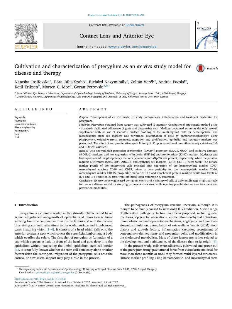
Fig. 1. Cell morphology, viability and phenotyping of the cultivated pterygium. Fibroblastic cells (Aa), pterygium epithelial cells (Ab) and graft with outgrowing cells forming 3D layeredstructure (Ac) (Magnification: 10×, (*) 20× and (**) 40×). Cellular viability of the 3D outgrowing cells from the pterygium using Annexin V-FITC/PI assay and Trypan blue exclusiontest (B). Cell surface markers phenotyping of the 3D outgrowing cells by FACS analysis; a plot of the percent positive cells versus different markers is shown as mean ± SD (C).
Table 1Immunophenotyping of the multi-layered outgrowing pterygium cells. Expression ofdifferent surface markers shown as percent (%).
Multi-layered outgrowing pterygium cells
HSC markers CD34 10.8 ± 12.7%CD47 79.1 ± 17%
Progenitor marker CD117/c-kit 3.3. ± 3.2%
MSC markers CD90 87.2 ± 9.5%CD73 76.4 ± 11%CD105 1.3 ± 1.4%
ECM attachment proteins markers CD146/MCAM 2.6 ± 4.7%CD166/ALCAM 48.1 ± 18.1%CD44/H-CAM 41.5 ± 12.3%CD112/Nectin-2 4.1 ± 4%
N. Josifovska et al. Contact Lens and Anterior Eye 40 (2017) 283–292
284
jugated antibodies were used to measure the expression of CD34, CD44,CD90/Thy-1, CD73, CD105 and CD166/ALCAM (all from Biolegend,San Diego, CA, USA); CD47, CD117/c-kit, CD146/MCAM, CD112 (allfrom R&D Systems, Minneapolis, MN, USA) (for further details refer toS1Table). Samples were measured by FACS Calibur flow cytometer (BDBiosciences Immunocytometry Systems) and data were analysed byFlowing Software 2.5 (PerttuTerho, Turku Centre for Biotechnology,University of Turku, Finland).
2.4. Immunofluorescent staining
Pterygium obtained right after surgery and that cultivated as long-term multi-layered cultures were collected and fixed in 4% parafor-maldehyde at room temperature, later dehydrated in ascending alcoholseries and embedded in paraffin; 3–4 μm thick tissue sections wereprepared using a rotary microtome, then mounted onto histologicalslides. After heat-induced antigen retrieval and blocking, immunofluor-escent labelling was performed. The samples were characterized formarkers of pluripotency and stemness (Vim, ΔNp63, Sox2, Oct4, ABCG-2,), oxidative stress (8-OHdG, HIF-1α), migration and proliferation-(CXCR4 and Ki-67), epithelial- (CK19 and 8–18) and secretory markers(MUC1 and MUC4) (S2 Table summarizes the primary antibodies anddilutions used for immunofluorescent staining). Nuclear staining wasperformed using 4′, 6-diamidino-2-phenylindole (DAPI). Fluorescent
images were taken by a ZEISS Axio Observer.Z1 (ZEISS, Oberkochen,Germany) microscope. The quantification of positive cells was carriedout using standard ImageJ software by three independent individuals.The number of positive cells on the full field of view was taken intoaccount with the help of nuclear (DAPI) staining. Multiple pictureswere taken of each sample and the results averaged out as mean ±standard deviation (SD).
2.5. Secretion of inflammatory cytokines by ELISA
High glucose-containing DMEM medium with 5% FBS was appliedto the multi-layered outgrowing cells and the supernatants werecollected after 24 h. In addition, treatment of the cells with 0.5 mg/mL MMC (Kyowa, Takeda Belgium-Brussels) was applied for 5 min andexchanged by fresh medium for another 15 min prior to the supernatantcollection, thus resembling the clinical use of MMC in surgery. Limbalepithelial stem cell (LESC) cultures cultivated in high glucose-contain-ing medium with 10% FBS were also used as a control. The secretedcytokines, IL-6 and IL-8, were analysed by a commercial ELISA kit(R & D, Germany) according to the manufacturer's protocol. Threeindependent experiments were performed on outgrowing cells fromfour different pterygium donors.
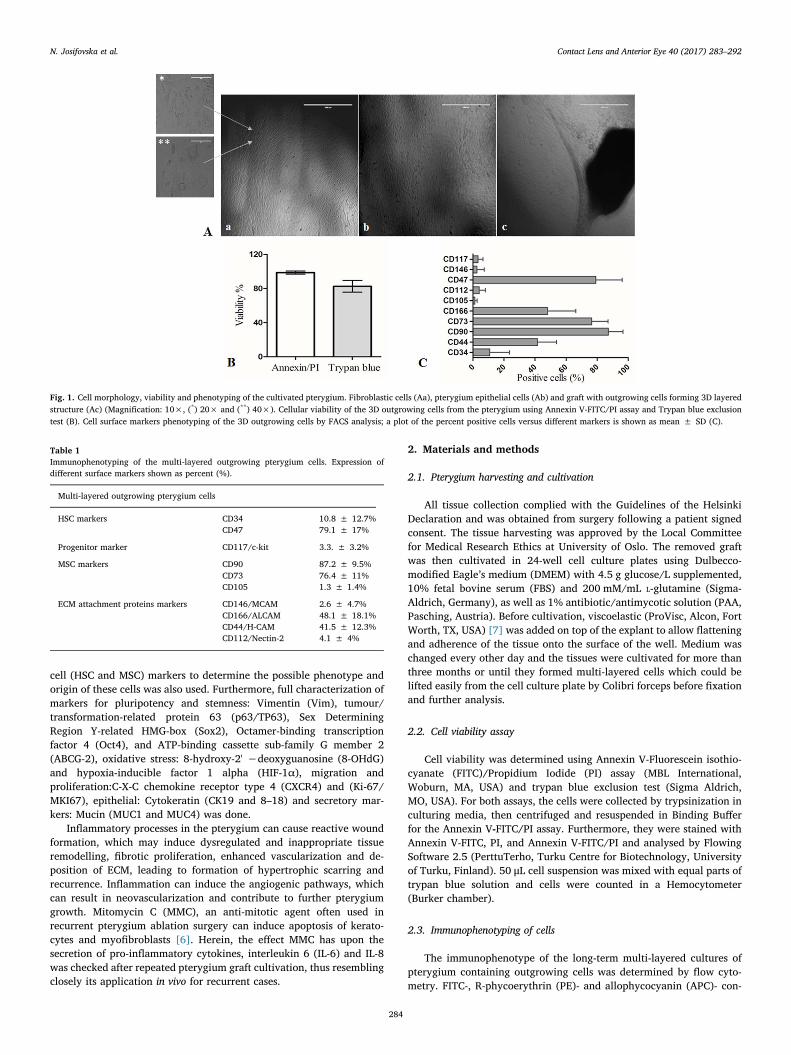
Fig. 2. Pterygium tissue and 3D outgrowing cells stained for pluripotency markers. Pterygium tissue-containing cultures (A, C), and outgrowing cells forming 3D structures (B, D) werestained for Vim and ΔNp63, respectively (Magnification: 40×). White bar represents data from pterygium tissue, while the black bar shows results from independently grown 3Doutgrowing cells. Data are expressed as mean ± SD.
N. Josifovska et al. Contact Lens and Anterior Eye 40 (2017) 283–292
285
2.6. Statistical analysis
Each experiment was performed at least three times and eachsample was tested in triplicates. Data are expressed as mean ± stan-dard deviation (SD). Statistically significant differences were deter-mined by student-t-tests, a p-value ≤ 0.05 was regarded significant.
3. Results
Pterygium morphology depends on two types of cells: fibroblasticwhich are long-shaped (Fig. 1Aa), and squamous epithelial cells tightly
packed together (Fig. 1Ab). After long term cultivation, pterygiumformed spontaneous multi-layered structure that was easily tangibleand removable from the plate for further analysis (Fig. 1Ac). Theviability of the outgrowing cells tested by the Annexin V-FITC/PI assayand Trypan blue method was (98.6 ± 1.7% and 82.6 ± 6.9%),respectively (Fig. 1B). The expression of hematopoietic cell surfacemarkers CD34 and CD47 was (10.8 ± 12.7%and 79.1 ± 17%),respectively. CD117/c-kit, a progenitor cell marker was low(3.3 ± 3.2%), while a very high expression of the MSC markersCD90 (87.2 ± 9.5%) and CD73 (76.4 ± 11%) were measured, incontrast to the MSC marker CD105 which was (1.3 ± 1.4%). The
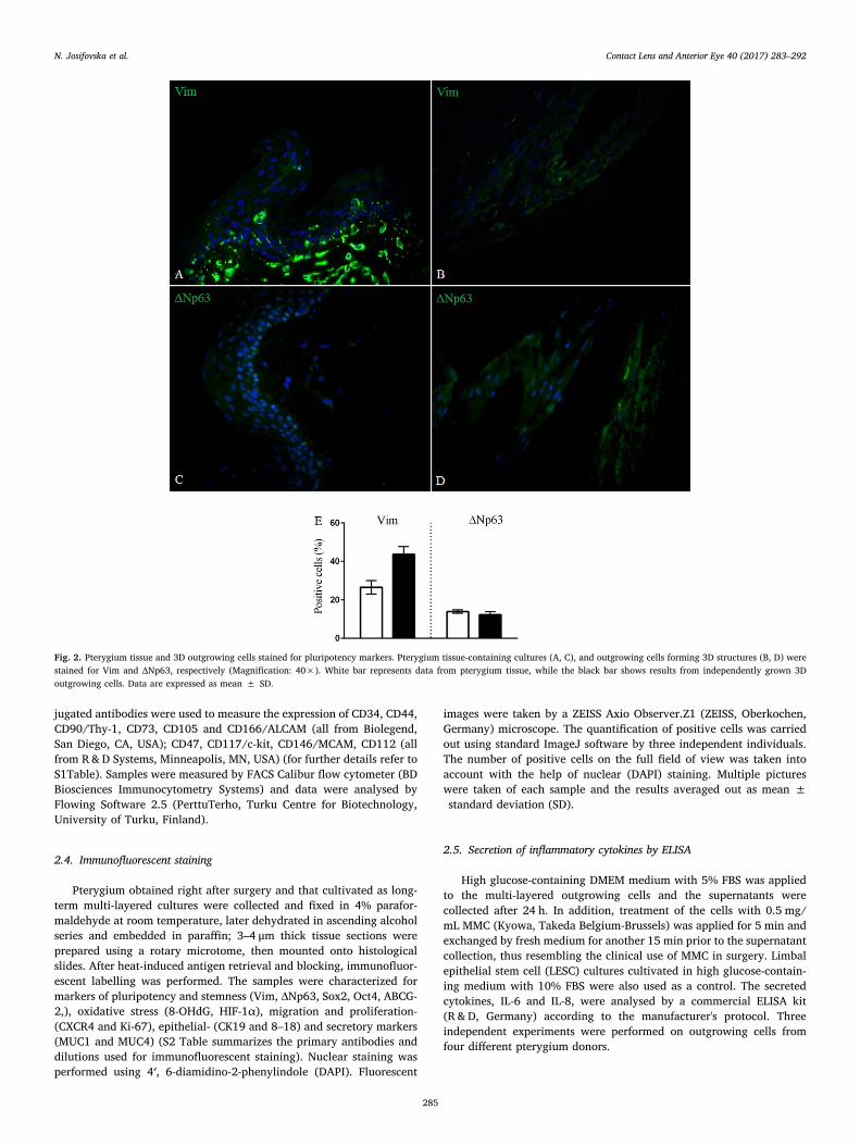
Fig. 3. Pterygium tissue and 3D outgrowing cells stained for markers of stemness. Pterygium tissue-containing cultures (A, C and E), and outgrowing cells forming 3D structures (B, D andF) were stained for Sox2, Oct4 and ABCG-2, respectively (Magnification: 40×). White bar represents data from pterygium tissue, while the black bar shows results from independentlygrown 3D outgrowing cells. Data are expressed as mean ± SD.
N. Josifovska et al. Contact Lens and Anterior Eye 40 (2017) 283–292
286
expression of ECM attachment proteins, which is important for themaintenance of the cellular growth were tested next: CD146/MCAM(2.6 ± 4.7%), CD166/ALCAM (48.1 ± 18.1%), CD112/Nectin-2(4.1 ± 4%) and CD44/homing-associated cell adhesion molecule (H-CAM) (41.5 ± 12.3%) (Fig. 1C). The expression of surface markers (%)is summarised in Table 1.
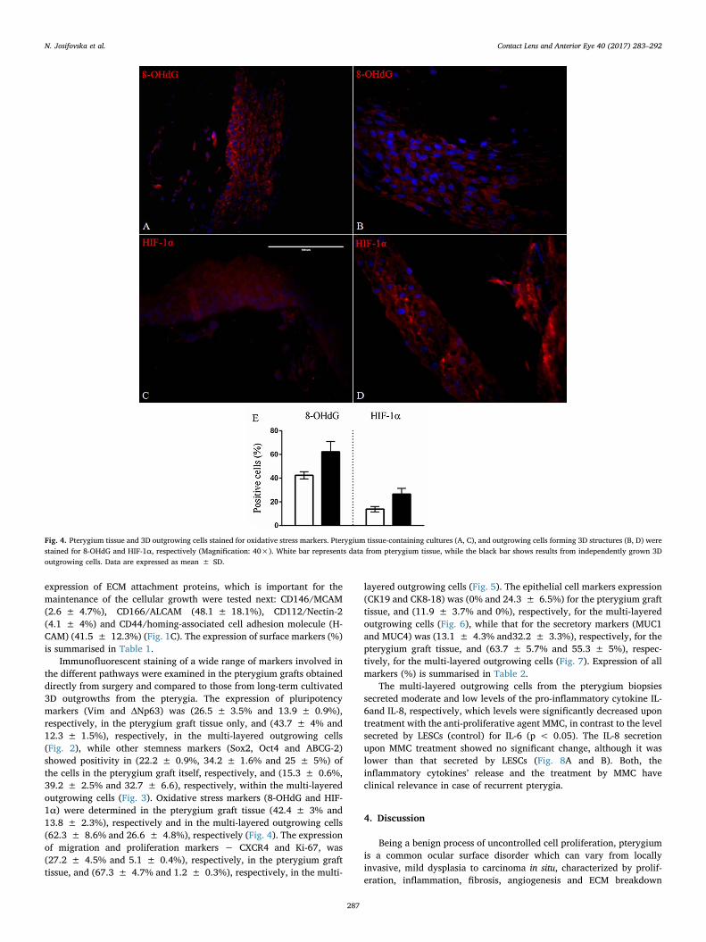
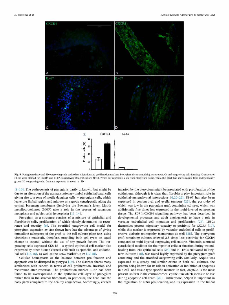
Immunofluorescent staining of a wide range of markers involved inthe different pathways were examined in the pterygium grafts obtaineddirectly from surgery and compared to those from long-term cultivated3D outgrowths from the pterygia. The expression of pluripotencymarkers (Vim and ΔNp63) was (26.5 ± 3.5% and 13.9 ± 0.9%),respectively, in the pterygium graft tissue only, and (43.7 ± 4% and12.3 ± 1.5%), respectively, in the multi-layered outgrowing cells(Fig. 2), while other stemness markers (Sox2, Oct4 and ABCG-2)showed positivity in (22.2 ± 0.9%, 34.2 ± 1.6% and 25 ± 5%) ofthe cells in the pterygium graft itself, respectively, and (15.3 ± 0.6%,39.2 ± 2.5% and 32.7 ± 6.6), respectively, within the multi-layeredoutgrowing cells (Fig. 3). Oxidative stress markers (8-OHdG and HIF-1α) were determined in the pterygium graft tissue (42.4 ± 3% and13.8 ± 2.3%), respectively and in the multi-layered outgrowing cells(62.3 ± 8.6% and 26.6 ± 4.8%), respectively (Fig. 4). The expressionof migration and proliferation markers − CXCR4 and Ki-67, was(27.2 ± 4.5% and 5.1 ± 0.4%), respectively, in the pterygium grafttissue, and (67.3 ± 4.7% and 1.2 ± 0.3%), respectively, in the multi-
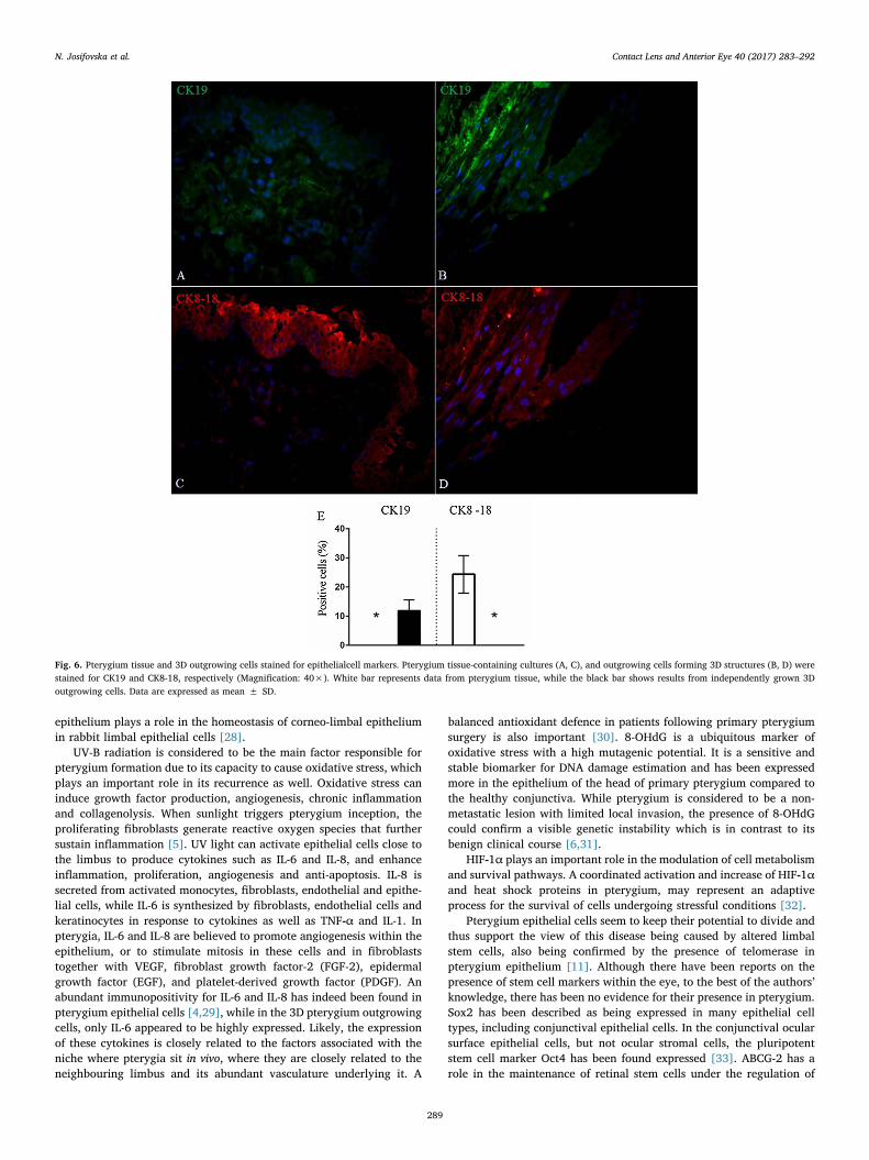
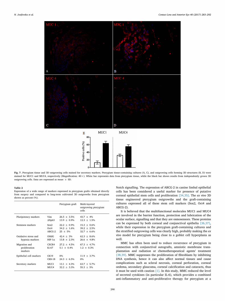
layered outgrowing cells (Fig. 5). The epithelial cell markers expression(CK19 and CK8-18) was (0% and 24.3 ± 6.5%) for the pterygium grafttissue, and (11.9 ± 3.7% and 0%), respectively, for the multi-layeredoutgrowing cells (Fig. 6), while that for the secretory markers (MUC1and MUC4) was (13.1 ± 4.3% and32.2 ± 3.3%), respectively, for thepterygium graft tissue, and (63.7 ± 5.7% and 55.3 ± 5%), respec-tively, for the multi-layered outgrowing cells (Fig. 7). Expression of allmarkers (%) is summarised in Table 2.
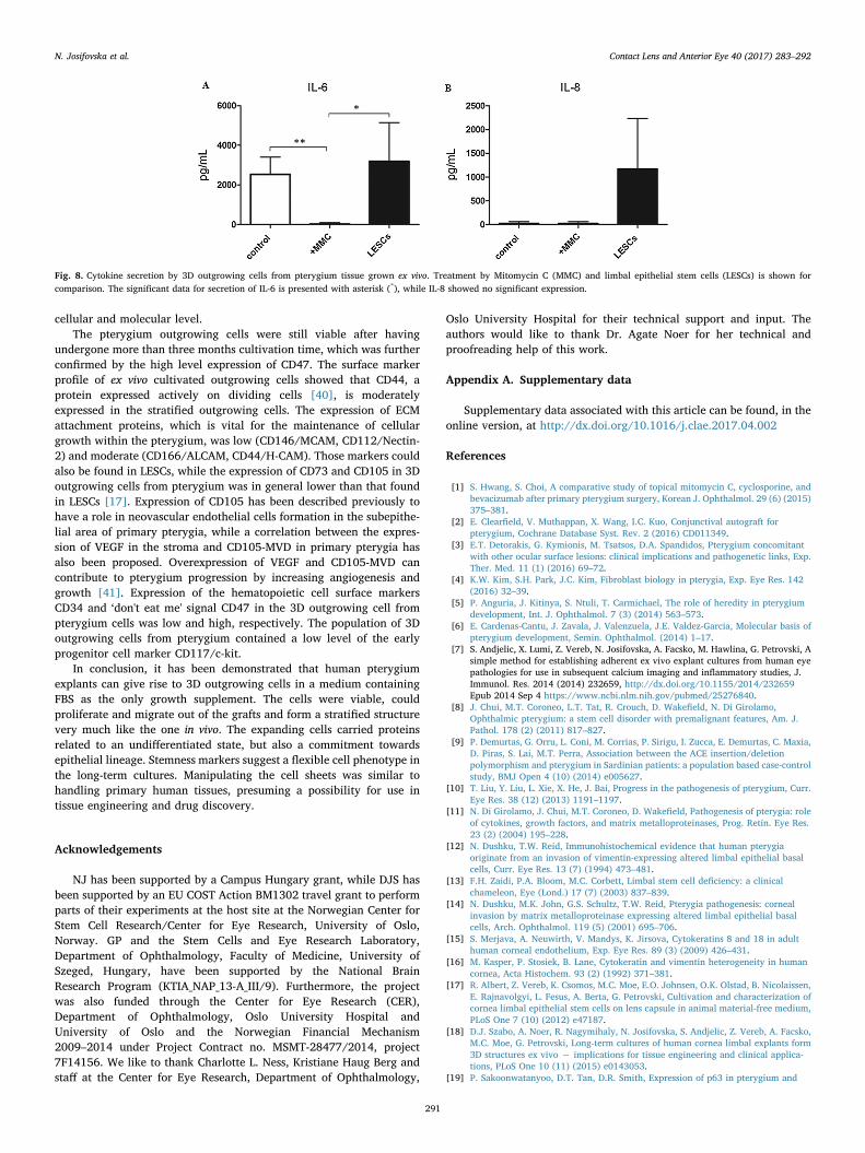
The multi-layered outgrowing cells from the pterygium biopsiessecreted moderate and low levels of the pro-inflammatory cytokine IL-6and IL-8, respectively, which levels were significantly decreased upontreatment with the anti-proliferative agent MMC, in contrast to the levelsecreted by LESCs (control) for IL-6 (p < 0.05). The IL-8 secretionupon MMC treatment showed no significant change, although it waslower than that secreted by LESCs (Fig. 8A and B). Both, theinflammatory cytokines’ release and the treatment by MMC haveclinical relevance in case of recurrent pterygia.
4. Discussion
Being a benign process of uncontrolled cell proliferation, pterygiumis a common ocular surface disorder which can vary from locallyinvasive, mild dysplasia to carcinoma in situ, characterized by prolif-eration, inflammation, fibrosis, angiogenesis and ECM breakdown
Fig. 4. Pterygium tissue and 3D outgrowing cells stained for oxidative stress markers. Pterygium tissue-containing cultures (A, C), and outgrowing cells forming 3D structures (B, D) werestained for 8-OHdG and HIF-1α, respectively (Magnification: 40×). White bar represents data from pterygium tissue, while the black bar shows results from independently grown 3Doutgrowing cells. Data are expressed as mean ± SD.
N. Josifovska et al. Contact Lens and Anterior Eye 40 (2017) 283–292
287
[8–10]. The pathogenesis of pterygia is partly unknown, but might bedue to an alteration of the normal stationary limbal epithelial basal cellsgiving rise to a zone of motile daughter cells − pterygium cells, whichleave the limbal region and migrate as a group centripetally along thecorneal basement membrane dissolving the Bowman’s layer. Matrixmetalloproteinases (MMP) take a role in the process of squamousmetaplasia and goblet cells' hyperplasia [11–14].
Pterygium as a structure consists of a mixture of epithelial andfibroblastic cells, proliferation of which closely determines its recur-rence and severity [6]. The stratified outgrowing cell model forpterygium expansion ex vivo shown here has the advantage of givingimmediate adherence of the graft to the cell culture plate (e.g. usingviscoelastic material), therefore, providing both cell types an equalchance to expand, without the use of any growth factors. The out-growing cells expressed CK8-18 −a typical epithelial cell marker alsoexpressed by other human corneal cells such as epithelial and endothe-lial cells [15,16], as well as the LESC marker CK19 [17,18].
Cellular homeostasis or the balance between proliferation andapoptosis can be disrupted in pterygia [19]. The disorder shares manysimilarities with cancer, in terms of cell proliferation, invasion andrecurrence after resection. The proliferation marker Ki-67 has beenfound to be overexpressed in the epithelial cell layer of pterygiumrather than in the stromal fibroblasts, in particular, the head and thebody parts compared to the healthy conjunctiva. Accordingly, corneal
invasion by the pterygium might be associated with proliferation of theepithelium, although it is clear that fibroblasts play important role inepithelial–mesenchymal interactions [6,20–22]. Ki-67 has also beenexpressed in conjunctival and eyelid tumours [23], the positivity ofwhich was low in the pterygium graft containing cultures, which wasadditionally five times less expressed in the multi-layered outgrowingtissue. The SDF-1/CXCR4 signalling pathway has been described indevelopmental processes and adult angiogenesis to have a role invascular endothelial cell migration and proliferation [24]. LESCsthemselves possess migratory capacity or positivity for CXCR4 [17],while this marker is expressed by vascular endothelial cells in prolif-erative diabetic retinopathy membranes as well [25]. The pterygiumgraft-containing cultures showed 2.5 times less positivity for CXCR4compared to multi-layered outgrowing cell cultures. Vimentin, a crucialcytoskeletal mediator for the repair of cellular function during wound-healing from lens epithelial cells [26] and in LESCs cultivated in long-term cultures [18], was found highly expressed by the pterygium-graftcontaining and the stratified outgrowing cells. Similarly, ΔNp63 wasexpressed at a steady and similar extent in both cell cultures, theprotein being known for its role in activation or inhibition of apoptosisin a cell- and tissue-type specific manner. In fact, ΔNp63α is the mostpresent isoform in the central corneal epithelium which seems to be lostduring apoptotic cell death [27]. Furthermore, ΔNp63 is important inthe regulation of LESC proliferation, and its expression in the limbal
Fig. 5. Pterygium tissue and 3D outgrowing cells stained for migration and proliferation markers. Pterygium tissue-containing cultures (A, C), and outgrowing cells forming 3D structures(B, D) were stained for CXCR4 and Ki-67, respectively (Magnification: 40×). White bar represents data from pterygium tissue, while the black bar shows results from independentlygrown 3D outgrowing cells. Data are expressed as mean ± SD.
N. Josifovska et al. Contact Lens and Anterior Eye 40 (2017) 283–292
288
epithelium plays a role in the homeostasis of corneo-limbal epitheliumin rabbit limbal epithelial cells [28].
UV-B radiation is considered to be the main factor responsible forpterygium formation due to its capacity to cause oxidative stress, whichplays an important role in its recurrence as well. Oxidative stress caninduce growth factor production, angiogenesis, chronic inflammationand collagenolysis. When sunlight triggers pterygium inception, theproliferating fibroblasts generate reactive oxygen species that furthersustain inflammation [5]. UV light can activate epithelial cells close tothe limbus to produce cytokines such as IL-6 and IL-8, and enhanceinflammation, proliferation, angiogenesis and anti-apoptosis. IL-8 issecreted from activated monocytes, fibroblasts, endothelial and epithe-lial cells, while IL-6 is synthesized by fibroblasts, endothelial cells andkeratinocytes in response to cytokines as well as TNF-α and IL-1. Inpterygia, IL-6 and IL-8 are believed to promote angiogenesis within theepithelium, or to stimulate mitosis in these cells and in fibroblaststogether with VEGF, fibroblast growth factor-2 (FGF-2), epidermalgrowth factor (EGF), and platelet-derived growth factor (PDGF). Anabundant immunopositivity for IL-6 and IL-8 has indeed been found inpterygium epithelial cells [4,29], while in the 3D pterygium outgrowingcells, only IL-6 appeared to be highly expressed. Likely, the expressionof these cytokines is closely related to the factors associated with theniche where pterygia sit in vivo, where they are closely related to theneighbouring limbus and its abundant vasculature underlying it. A
balanced antioxidant defence in patients following primary pterygiumsurgery is also important [30]. 8-OHdG is a ubiquitous marker ofoxidative stress with a high mutagenic potential. It is a sensitive andstable biomarker for DNA damage estimation and has been expressedmore in the epithelium of the head of primary pterygium compared tothe healthy conjunctiva. While pterygium is considered to be a non-metastatic lesion with limited local invasion, the presence of 8-OHdGcould confirm a visible genetic instability which is in contrast to itsbenign clinical course [6,31].
HIF-1α plays an important role in the modulation of cell metabolismand survival pathways. A coordinated activation and increase of HIF-1αand heat shock proteins in pterygium, may represent an adaptiveprocess for the survival of cells undergoing stressful conditions [32].
Pterygium epithelial cells seem to keep their potential to divide andthus support the view of this disease being caused by altered limbalstem cells, also being confirmed by the presence of telomerase inpterygium epithelium [11]. Although there have been reports on thepresence of stem cell markers within the eye, to the best of the authors’knowledge, there has been no evidence for their presence in pterygium.Sox2 has been described as being expressed in many epithelial celltypes, including conjunctival epithelial cells. In the conjunctival ocularsurface epithelial cells, but not ocular stromal cells, the pluripotentstem cell marker Oct4 has been found expressed [33]. ABCG-2 has arole in the maintenance of retinal stem cells under the regulation of
Fig. 6. Pterygium tissue and 3D outgrowing cells stained for epithelialcell markers. Pterygium tissue-containing cultures (A, C), and outgrowing cells forming 3D structures (B, D) werestained for CK19 and CK8-18, respectively (Magnification: 40×). White bar represents data from pterygium tissue, while the black bar shows results from independently grown 3Doutgrowing cells. Data are expressed as mean ± SD.
N. Josifovska et al. Contact Lens and Anterior Eye 40 (2017) 283–292
289
Notch signalling. The expression of ABCG-2 in canine limbal epithelialcells has been considered a useful marker for presence of putativecorneal epithelial stem cells and proliferation [34,35]. The ex vivo 3Dtissue engineered pterygium outgrowths and the graft-containingcultures expressed all of these stem cell markers (Sox2, Oct4 andABCG-2).
It is believed that the multifunctional molecules MUC1 and MUC4are involved in the barrier function, protection and lubrication of theocular surface, signalling and that they are osmosensors. These proteinscan be expressed by both corneal and conjunctival epithelia [36,37],while their expression in the pterygium graft-containing cultures andthe stratified outgrowing cells was clearly high, probably making the exvivo model for pterygium being close to a goblet cell hyperplasia aswell.
MMC has often been used to reduce recurrence of pterygium inconnection with conjunctival autografts, amniotic membrane trans-plantation and radiation or chemotherapeutical agents’ treatment[38,39]. MMC suppresses the proliferation of fibroblasts by inhibitingDNA synthesis, hence it can also affect normal tissues and causecomplications such as scleral necrosis, corneal perforation, cornealoedema, secondary glaucoma, corneal calcification and cataracts, thusit must be used with caution [1]. In this study, MMC reduced the levelof secreted cytokines (in particular IL-6), which provides a combinedanti-inflammatory and anti-proliferative therapy for pterygium at a
Fig. 7. Pterygium tissue and 3D outgrowing cells stained for secretory markers. Pterygium tissue-containing cultures (A, C), and outgrowing cells forming 3D structures (B, D) werestained for MUC1 and MUC4, respectively (Magnification: 40×). White bar represents data from pterygium tissue, while the black bar shows results from independently grown 3Doutgrowing cells. Data are expressed as mean ± SD.
Table 2Expression of a wide range of markers expressed in pterygium grafts obtained directlyfrom surgery and compared to long-term cultivated 3D outgrowths from pterygiumshown as percent (%).
Pterygium graft Multi-layeredoutgrowing pterygiumcells
Pluripotency markers Vim 26.5 ± 3.5% 43.7 ± 4%ΔNp63 13.9 ± 0.9% 12.3 ± 1.5%
Stemness markers Sox2 22.2 ± 0.9% 15.3 ± 0.6%Oct4 34.2 ± 1.6% 39.2 ± 2.5%ABCG-2 25 ± 5% 32.7 ± 6.6%
Oxidative stress andhypoxia markers
OHdG 42.4 ± 3% 62.3 ± 8.6%HIF-1α 13.8 ± 2.3% 26.6 ± 4.8%
Migration andproliferationmarkers
CXCR4 27.2 ± 4.5% 67.3 ± 4.7%Ki-67 5.1 ± 0.4% 1.2 ± 0.3%
Epithelial cell markers CK19 0% 11.9 ± 3.7%CK8-18 24.3 ± 6.5% 0%
Secretory markers MUC1 13.1 ± 4.3% 63.7 ± 5.7%MUC4 32.2 ± 3.3% 55.3 ± 5%
N. Josifovska et al. Contact Lens and Anterior Eye 40 (2017) 283–292
290
cellular and molecular level.The pterygium outgrowing cells were still viable after having
undergone more than three months cultivation time, which was furtherconfirmed by the high level expression of CD47. The surface markerprofile of ex vivo cultivated outgrowing cells showed that CD44, aprotein expressed actively on dividing cells [40], is moderatelyexpressed in the stratified outgrowing cells. The expression of ECMattachment proteins, which is vital for the maintenance of cellulargrowth within the pterygium, was low (CD146/MCAM, CD112/Nectin-2) and moderate (CD166/ALCAM, CD44/H-CAM). Those markers couldalso be found in LESCs, while the expression of CD73 and CD105 in 3Doutgrowing cells from pterygium was in general lower than that foundin LESCs [17]. Expression of CD105 has been described previously tohave a role in neovascular endothelial cells formation in the subepithe-lial area of primary pterygia, while a correlation between the expres-sion of VEGF in the stroma and CD105-MVD in primary pterygia hasalso been proposed. Overexpression of VEGF and CD105-MVD cancontribute to pterygium progression by increasing angiogenesis andgrowth [41]. Expression of the hematopoietic cell surface markersCD34 and ‘don't eat me' signal CD47 in the 3D outgrowing cell frompterygium cells was low and high, respectively. The population of 3Doutgrowing cells from pterygium contained a low level of the earlyprogenitor cell marker CD117/c-kit.
In conclusion, it has been demonstrated that human pterygiumexplants can give rise to 3D outgrowing cells in a medium containingFBS as the only growth supplement. The cells were viable, couldproliferate and migrate out of the grafts and form a stratified structurevery much like the one in vivo. The expanding cells carried proteinsrelated to an undifferentiated state, but also a commitment towardsepithelial lineage. Stemness markers suggest a flexible cell phenotype inthe long-term cultures. Manipulating the cell sheets was similar tohandling primary human tissues, presuming a possibility for use intissue engineering and drug discovery.
Acknowledgements
NJ has been supported by a Campus Hungary grant, while DJS hasbeen supported by an EU COST Action BM1302 travel grant to performparts of their experiments at the host site at the Norwegian Center forStem Cell Research/Center for Eye Research, University of Oslo,Norway. GP and the Stem Cells and Eye Research Laboratory,Department of Ophthalmology, Faculty of Medicine, University ofSzeged, Hungary, have been supported by the National BrainResearch Program (KTIA_NAP_13-A_III/9). Furthermore, the projectwas also funded through the Center for Eye Research (CER),Department of Ophthalmology, Oslo University Hospital andUniversity of Oslo and the Norwegian Financial Mechanism2009–2014 under Project Contract no. MSMT-28477/2014, project7F14156. We like to thank Charlotte L. Ness, Kristiane Haug Berg andstaff at the Center for Eye Research, Department of Ophthalmology,
Oslo University Hospital for their technical support and input. Theauthors would like to thank Dr. Agate Noer for her technical andproofreading help of this work.
Appendix A. Supplementary data
Supplementary data associated with this article can be found, in theonline version, at http://dx.doi.org/10.1016/j.clae.2017.04.002
References
[1] S. Hwang, S. Choi, A comparative study of topical mitomycin C, cyclosporine, andbevacizumab after primary pterygium surgery, Korean J. Ophthalmol. 29 (6) (2015)375–381.
[2] E. Clearfield, V. Muthappan, X. Wang, I.C. Kuo, Conjunctival autograft forpterygium, Cochrane Database Syst. Rev. 2 (2016) CD011349.
[3] E.T. Detorakis, G. Kymionis, M. Tsatsos, D.A. Spandidos, Pterygium concomitantwith other ocular surface lesions: clinical implications and pathogenetic links, Exp.Ther. Med. 11 (1) (2016) 69–72.
[4] K.W. Kim, S.H. Park, J.C. Kim, Fibroblast biology in pterygia, Exp. Eye Res. 142(2016) 32–39.
[5] P. Anguria, J. Kitinya, S. Ntuli, T. Carmichael, The role of heredity in pterygiumdevelopment, Int. J. Ophthalmol. 7 (3) (2014) 563–573.
[6] E. Cardenas-Cantu, J. Zavala, J. Valenzuela, J.E. Valdez-Garcia, Molecular basis ofpterygium development, Semin. Ophthalmol. (2014) 1–17.
[7] S. Andjelic, X. Lumi, Z. Vereb, N. Josifovska, A. Facsko, M. Hawlina, G. Petrovski, Asimple method for establishing adherent ex vivo explant cultures from human eyepathologies for use in subsequent calcium imaging and inflammatory studies, J.Immunol. Res. 2014 (2014) 232659, http://dx.doi.org/10.1155/2014/232659Epub 2014 Sep 4 https://www.ncbi.nlm.nih.gov/pubmed/25276840.
[8] J. Chui, M.T. Coroneo, L.T. Tat, R. Crouch, D. Wakefield, N. Di Girolamo,Ophthalmic pterygium: a stem cell disorder with premalignant features, Am. J.Pathol. 178 (2) (2011) 817–827.
[9] P. Demurtas, G. Orru, L. Coni, M. Corrias, P. Sirigu, I. Zucca, E. Demurtas, C. Maxia,D. Piras, S. Lai, M.T. Perra, Association between the ACE insertion/deletionpolymorphism and pterygium in Sardinian patients: a population based case-controlstudy, BMJ Open 4 (10) (2014) e005627.
[10] T. Liu, Y. Liu, L. Xie, X. He, J. Bai, Progress in the pathogenesis of pterygium, Curr.Eye Res. 38 (12) (2013) 1191–1197.
[11] N. Di Girolamo, J. Chui, M.T. Coroneo, D. Wakefield, Pathogenesis of pterygia: roleof cytokines, growth factors, and matrix metalloproteinases, Prog. Retin. Eye Res.23 (2) (2004) 195–228.
[12] N. Dushku, T.W. Reid, Immunohistochemical evidence that human pterygiaoriginate from an invasion of vimentin-expressing altered limbal epithelial basalcells, Curr. Eye Res. 13 (7) (1994) 473–481.
[13] F.H. Zaidi, P.A. Bloom, M.C. Corbett, Limbal stem cell deficiency: a clinicalchameleon, Eye (Lond.) 17 (7) (2003) 837–839.
[14] N. Dushku, M.K. John, G.S. Schultz, T.W. Reid, Pterygia pathogenesis: cornealinvasion by matrix metalloproteinase expressing altered limbal epithelial basalcells, Arch. Ophthalmol. 119 (5) (2001) 695–706.
[15] S. Merjava, A. Neuwirth, V. Mandys, K. Jirsova, Cytokeratins 8 and 18 in adulthuman corneal endothelium, Exp. Eye Res. 89 (3) (2009) 426–431.
[16] M. Kasper, P. Stosiek, B. Lane, Cytokeratin and vimentin heterogeneity in humancornea, Acta Histochem. 93 (2) (1992) 371–381.
[17] R. Albert, Z. Vereb, K. Csomos, M.C. Moe, E.O. Johnsen, O.K. Olstad, B. Nicolaissen,E. Rajnavolgyi, L. Fesus, A. Berta, G. Petrovski, Cultivation and characterization ofcornea limbal epithelial stem cells on lens capsule in animal material-free medium,PLoS One 7 (10) (2012) e47187.
[18] D.J. Szabo, A. Noer, R. Nagymihaly, N. Josifovska, S. Andjelic, Z. Vereb, A. Facsko,M.C. Moe, G. Petrovski, Long-term cultures of human cornea limbal explants form3D structures ex vivo − implications for tissue engineering and clinical applica-tions, PLoS One 10 (11) (2015) e0143053.
[19] P. Sakoonwatanyoo, D.T. Tan, D.R. Smith, Expression of p63 in pterygium and
Fig. 8. Cytokine secretion by 3D outgrowing cells from pterygium tissue grown ex vivo. Treatment by Mitomycin C (MMC) and limbal epithelial stem cells (LESCs) is shown forcomparison. The significant data for secretion of IL-6 is presented with asterisk (*), while IL-8 showed no significant expression.
N. Josifovska et al. Contact Lens and Anterior Eye 40 (2017) 283–292
291
normal conjunctiva, Cornea 23 (1) (2004) 67–70.[20] S. Kase, S. Takahashi, I. Sato, K. Nakanishi, K. Yoshida, S. Ohno, Expression of
p27(KIP1) and cyclin D1, and cell proliferation in human pterygium, Br. J.Ophthalmol. 91 (7) (2007) 958–961.
[21] K. Liang, Z. Jiang, B.Q. Ding, P. Cheng, D.K. Huang, L.M. Tao, Expression of cellproliferation and apoptosis biomarkers in pterygia and normal conjunctiva, Mol.Vis. 17 (2011) 1687–1693.
[22] M. Nubile, C. Curcio, M. Lanzini, R. Calienno, M. Iezzi, A. Mastropasqua, M. DiNicola, L. Mastropasqua, Expression of CREB in primary pterygium and correlationwith cyclin D1, ki-67, MMP7, p53, p63, Survivin and Vimentin, Ophthalmic Res. 50(2) (2013) 99–107.
[23] J. Reszec, L. Kanczuga-Koda, M. Sulkowska, M. Koda, J. Cylwik, M. Barwijuk-Machala, S. Sulkowski, An evaluation of Ki-67 and PCNA expression in conjunctivaland eyelid tumours, Folia Morphol. (Warsz) 63 (1) (2004) 95–98.
[24] G.A. Strasser, J.S. Kaminker, M. Tessier-Lavigne, Microarray analysis of retinalendothelial tip cells identifies CXCR4 as a mediator of tip cell morphology andbranching, Blood 115 (24) (2010) 5102–5110.
[25] A.M. Abu El-Asrar, S. Struyf, G. Opdenakker, J. Van Damme, K. Geboes, Expressionof stem cell factor/c-kit signaling pathway components in diabetic fibrovascularepiretinal membranes, Mol. Vis. 16 (2010) 1098–1107.
[26] A.S. Menko, B.M. Bleaken, A.A. Libowitz, L. Zhang, M.A. Stepp, J.L. Walker, Acentral role for vimentin in regulating repair function during healing of the lensepithelium, Mol. Biol. Cell 25 (6) (2014) 776–790.
[27] D.M. Robertson, S.I. Ho, H.D. Cavanagh, C-terminal cleavage of DeltaNp63alpha isassociated with TSA-induced apoptosis in immortalized corneal epithelial cells,Invest. Ophthalmol. Vis. Sci. 51 (8) (2010) 3977–3985.
[28] Y.J. Hsueh, P.C. Kuo, J.K. Chen, Transcriptional regulators of the DeltaNp63: theirrole in limbal epithelial cell proliferation, J. Cell. Physiol. 228 (3) (2013) 536–546.
[29] N. Di Girolamo, R.K. Kumar, M.T. Coroneo, D. Wakefield, UVB-mediated inductionof interleukin-6 and −8 in pterygia and cultured human pterygium epithelial cells,Invest. Ophthalmol. Vis. Sci. 43 (11) (2002) 3430–3437.
[30] A. Kormanovski, F. Parra, A. Jarillo-Luna, E. Lara-Padilla, J. Pacheco-Yepez,R. Campos-Rodriguez, Oxidant/antioxidant state in tissue of prymary and recurrentpterygium, BMC Ophthalmol. 14 (2014) 149.
[31] M.T. Perra, C. Maxia, A. Corbu, L. Minerba, P. Demurtas, R. Colombari, D. Murtas,S. Bravo, F. Piras, P. Sirigu, Oxidative stress in pterygium: relationship between p53and 8-hydroxydeoxyguanosine, Mol. Vis. 12 (2006) 1136–1142.
[32] D. Pagoulatos, N. Pharmakakis, J. Lakoumentas, M. Assimakopoulou, Etaypoxia-inducible factor-1alpha, von Hippel-Lindau protein, and heat shock proteinexpression in ophthalmic pterygium and normal conjunctiva, Mol. Vis. 20 (2014)441–457.
[33] M.W. Poon, J. He, X. Fang, Z. Zhang, W. Wang, J. Wang, F. Qiu, H.F. Tse, W. Li,Z. Liu, Q. Lian, Human ocular epithelial cells endogenously expressing SOX2 andOCT4 yield high efficiency of pluripotency reprogramming, PLoS One 10 (7) (2015)e0131288.
[34] M. Morita, N. Fujita, A. Takahashi, E.R. Nam, S. Yui, C.S. Chung, N. Kawahara,H.Y. Lin, K. Tsuzuki, T. Nakagawa, R. Nishimura, Evaluation of ABCG2 and p63expression in canine cornea and cultivated corneal epithelial cells, Vet. Ophthalmol.18 (1) (2015) 59–68.
[35] S. Bhattacharya, A. Das, K. Mallya, I. Ahmad, Maintenance of retinal stem cells byAbcg2 is regulated by notch signaling, J. Cell Sci. 120 (Pt. 15) (2007) 2652–2662.
[36] M. Berry, R.B. Ellingham, A.P. Corfield, Membrane-associated mucins in normalhuman conjunctiva, Invest. Ophthalmol. Vis. Sci. 41 (2) (2000) 398–403.
[37] B. Govindarajan, I.K. Gipson, Membrane-tethered mucins have multiple functionson the ocular surface, Exp. Eye Res. 90 (6) (2010) 655–663.
[38] C.J. Jaworski, M. Aryankalayil-John, M.M. Campos, R.N. Fariss, J. Rowsey,N. Agarwalla, T.W. Reid, N. Dushku, C.A. Cox, D. Carper, G. Wistow, Expressionanalysis of human pterygium shows a predominance of conjunctival and limbalmarkers and genes associated with cell migration, Mol. Vis. 15 (2009) 2421–2434.
[39] E. Toker, M. Eraslan, Recurrence after primary pterygium excision: amnioticmembrane transplantation with fibrin glue versus conjunctival autograft with fibringlue, Curr. Eye Res. 41 (1) (2016) 1–8.
[40] N.P. Liu, W.L. Roberts, L.P. Hale, M.C. Levesque, D.D. Patel, C.L. Lu, G.J. Jaffe,Expression of CD44 and variant isoforms in cultured human retinal pigmentepithelial cells, Invest. Ophthalmol. Vis. Sci. 38 (10) (1997) 2027–2037.
[41] J. Zhang, M. Zhang, X. Li, T. Zheng, G. Mu, W. Liu, H. Xie, X. Liu, Correlation ofvascular endothelial growth factor and CD105-microvascular density in primarypterygium, J. Huazhong Univ. Sci. Technol. Med. Sci. 31 (4) (2011) 560–564.
N. Josifovska et al. Contact Lens and Anterior Eye 40 (2017) 283–292
292