The Role of p27(Kip1) in Dasatinib-Enhanced Paclitaxel Cytotoxicity in Human Ovarian Cancer Cells
Upload
independentCategory
view
1download
0
Jab1/CSN5 negatively regulates p27 and plays a role in thepathogenesis of nasopharyngeal carcinoma
Yunbao Pan1,2,Δ, Qingxiu Zhang2,Δ, Ling Tian2, Xin Wang4, Xiaohang Fan1, HuizhongZhang4, Francois X. Claret2,3, and Huiling Yang1
1Department of Pathophysiology, Zhongshan School of Medicine, Sun Yat-Sen University,Guangzhou, Guangdong 510080, P.R. China2Department of Systems Biology, The University of Texas MD Anderson Cancer Center, Houston,TX, 77030 USA3 Cancer Biology Program and Experimental Therapeutic Program, The University of TexasGraduate School of Biomedical Sciences at Houston, Houston, TX, 77030 USA4State Key Laboratory of Oncology in South China, Sun Yat-Sen University Cancer Center,Guangzhou, Guangdong 510080, P.R. China
AbstractNasopharyngeal carcinoma (NPC) is an Epstein-Barr virus-associated malignancy most commonin East Asia and Africa. Aberrant expression of Jab1/CSN5, a negative regulator of the cell cycleinhibitor p27, is correlated with reduced p27 expression and associated with advanced tumor stageand poor prognosis in several human cancers. In this study, we examined the functionalrelationship between Jab1 and p27 protein expression in NPC. Immunohistochemical analysisshowed an inverse association between Jab1 and p27 in NPC tissue samples, and overexpressionof Jab1 correlated with poor survival in NPC patients. Mechanistically, Jab1 and p27 were foundto interact directly in NPC cells, with Jab1 mediating p27 degradation in a proteasome-dependentmanner. Knockdown of Jab1 resulted in a remarkable increase in p27 levels and inhibition of cellproliferation, indicating that Jab1 targets p27 for degradation, thereby controlling its stability. Jab1depletion also enhanced the antitumor effects of cisplatin in NPC cells. Together, our findingssuggest that Jab1 overexpression plays an important role in the pathogenesis of NPC throughJab1-mediated p27 degradation. Jab1 therefore represents a novel diagnostic marker andtherapeutic target in patients with NPC.
Keywordsnasopharyngeal carcinoma; tumorigenesis; Epstein-Barr virus-associated malignancy; p27Kip1;Jab1/CSN5
Address correspondence to: François X. Claret, Department of Systems Biology, The University of Texas MD Anderson CancerCenter, Houston, TX 77030 USA. Phone: 713-563 4204; Fax: 713-563 4205; [email protected]. Huiling Yang, Department ofPathophysiology, Zhongshan School of Medicine, Sun Yat-Sen University, Guangzhou, Guangdong 510080, P.R. China. Phone:86-20-87332268, Fax: 86-20-87331209, [email protected].ΔThese two authors contributed equally to this work and should be considered as co-first author.
Conflict of interest statement: The authors have no conflicts of interest to declare.
NIH Public AccessAuthor ManuscriptCancer Res. Author manuscript; available in PMC 2013 April 01.
Published in final edited form as:Cancer Res. 2012 April 1; 72(7): 1890–1900. doi:10.1158/0008-5472.CAN-11-3472.
NIH
-PA Author Manuscript
NIH
-PA Author Manuscript
NIH
-PA Author Manuscript

IntroductionNasopharyngeal carcinoma (NPC), originating from the epithelial lining of the nasopharynx,is a squamous-cell carcinoma (1). This neoplasm has remarkable ethnic and geographicdistribution, with a high prevalence in southern China, Southeast Asia, northern Africa, andAlaska (2, 3). The annual incidence peaks at 50 cases per 100,000 people in the endemicregions, but is rare in the Western world (1 per 100,000) (2). Etiologic studies haveindicated that Epstein-Barr virus (EBV) infection, environmental factors, and geneticsusceptibility are associated with NPC (1, 4). In China more than 95% of NPCs are non-keratinizing carcinoma, while fewer than 5% are keratinizing carcinoma; thus, in our studywe use CNE1 (keratinizing carcinoma) and CNE2 (non-keratinizing carcinoma) cells torepresent the two mainly histologic types (5).
Radiotherapy and adjuvant cisplatin chemotherapy have become the standard treatments forNPC (6). The 5-year survival rate after treatment is approximately 70% (7). Thirty to fortypercent of patients will develop distant metastasis within 4 years (8), and once metastasisoccurs, the prognosis is very poor.
To develop better treatment approaches, it is important to understand the molecular basis ofthe development and progression of NPC. Genetic alterations have been reported in NPCincluding a frequent reduction of p27 expression (9, 10). Furthermore, low p27 expressionlevels may contribute to the aggressive behavior of NPC (11). Overexpression of cyclin EmRNA has been shown to predict poor prognosis in NPC and has been correlated with anadvanced stage of NPC and a low overall survival rate (12). Recently, it has been reportedthat PIK3CA gene amplification is frequently observed in advanced-stage NPC, whichemphasizes the association between PIK3CA gene amplification and poor prognosis (11). Ithas also been shown that Akt promotes cell proliferation and survival in NPC (4, 13).However, additional molecular abnormalities resulting in the deregulation of cell-cycleprogression may also occur.
Jab1/CSN5 (Jab1 hereafter) as we initially identified as a c-Jun coactivator, is also known asthe fifth component of the COP9 signalosome (CSN) complex (CSN5) (14, 15). Jab1promotes cell proliferation and inactivates p27 by inducing translocation of p27 from thenucleus to the cytoplasm, which accelerates p27 degradation through the ubiquitin-dependent proteasome pathway and promotes cell-cycle progression (16). p27 is a universalcyclin-dependent kinase (Cdk) inhibitor that directly inhibits the enzymatic activity ofcyclin-Cdk complexes, resulting in cell-cycle arrest at G1 (17). In addition, p27 proteinlevels are increased in quiescent cells and rapidly decrease after cells are stimulated withmitogens (18). Although transcriptional regulation is possible, the cellular abundance of p27is primarily regulated at the posttranslational level by the ubiquitin-proteasome pathway(19). Jab1 overexpression is correlated with a loss of p27 and a lower rate of survival inpatients with breast cancer, suggesting a role in breast cancer pathogenesis (20). This inverseassociation between Jab1 and p27 expression has also been observed in anaplastic large celllymphoma (21), ovarian cancer (22), pancreatic adenocarcinomas (23, 24), and other cancertypes (25–27). However, the mechanisms leading to p27 downregulation in NPC remainundefined. Because Jab1 overexpression is correlated with the loss of p27 in several cancers,and low p27 expression is associated with higher tumor grades (28), we hypothesized thatJab1 functions as a negative regulator of p27 and as such may play a role in the pathogenesisof NPC.
To test our hypothesis, we assessed Jab1 and p27 expression in a series of 45 NPC and 30nasopharyngeal inflammation tissue specimens. We found that Jab1 overexpression wasassociated with absent or low expression of p27 in these samples. To further elucidate the
Pan et al. Page 2
Cancer Res. Author manuscript; available in PMC 2013 April 01.
NIH
-PA Author Manuscript
NIH
-PA Author Manuscript
NIH
-PA Author Manuscript

role of Jab1 in p27 degradation in NPC, we infected NPC cell lines with an adenoviralvector overexpressing Jab1 and found that p27 levels were significantly reduced. We alsodetected a direct physical interaction between Jab1 and p27 in NPC cells. Furthermore,inhibition of endogenous Jab1 expression with specific short interfering RNAs (siRNAs)resulted in a substantial increase of p27 levels and inhibition of cell proliferation, indicatingthat Jab1 controls the stability of p27 by targeting it for degradation in NPC. Interestingly,siRNA-mediated depletion of Jab1 inhibited cell proliferation and accelerated apoptotic celldeath in NPC. Moreover, Jab1 depletion enhanced the antitumor effects of cisplatin in NPCcells. This may suggest that Jab1 is a potential target for treating NPC.
Materials and MethodsPatients and tissue samples
All patients were from the Cancer Center of Sun Yat-Sen University in 2003. The studygroup consisted of 36 men and 9 women with NPC who underwent radiotherapy and thecontrol group consisted of 13 men and 17 women with nasopharyngeal inflammation.Patients that had preoperative diagnosis and did not receive preoperative chemo-radiationtreatment were selected for this study based on the availability of archived paraffin-embedded NPC and nasopharyngitis tissue blocks for immunohistochemical analysis.Ethical approval was obtained from the cancer center and fully informed consent from allpatients before sample collection. Surgical staging of tumors had been done according to theAmerican Joint Committee on Cancer tumor-node-metastasis system and tumor grading wasbased on currently used histopathologic criteria.
ReagentsCell culture medium were from Mediatech Inc (Mannassas, VA) and fetal bovine serum(FBS) were obtained from Gibco (Grand Island, NY, USA). The antibodies used were Jab1(Santa Cruz, CA), p27, and PARP (BD Biosciences PharMingen, San Diego, CA);caspase-3, Lamin A/C, and Myc-tag (Cell Signaling Technology, Beverly, MA); and Flagand β-actin (Sigma-Aldrich, St. Louis, MO). The Lipofectamine Plus and Oligofectaminereagents were from Invitrogen (Carlsbad, CA). NE-PER nuclear and cytoplasmic extractionreagents and Western Lightning Chemiluminescence Plus reagent were from ThermoScientific Pierce (Rockford, IL, USA). Annexin V/PI kit was from BD Biosciences (PaloAlto, CA). Cell Proliferation Kit was from Roche (Indianapolis, IN, USA).
Human tissues and immunohistochemical analysisA total of 75 formalin-fixed, paraffin-embedded human primary NPC and noncancerousinflamed nasopharyngeal specimens from 45 NPC and 30 nasopharyngitis patients wereanalyzed. The p27 and Jab1 levels in the formalin-fixed paraffin-embedded tissue sectionswere measured by immunohistochemical analysis, as previously described (24). Briefly,heat-induced retrieval of Jab1 and p27 antigens was performed, and the slides wereincubated with the primary antibodies. An immunoreaction was detected with the LSAB+kit from Dakocytomation. We used 3, 3'-diaminobenzidine as the chromogen andhematoxylin as the counterstain. Expression levels of Jab1 and p27 were evaluated bycounting at least 500 tumor cells in representative high-power fields. Tumor cells wereconsidered positive for Jab1 or p27 when nuclear or cytoplasmic staining was present. Foreach tumor, we determined a proportion score and an intensity score. The proportion scorerepresented the estimated fraction of positively stained cells (0 = less than 5%; 1 = 5% to35%; 2 = 35% to 65%; 3 = greater than 65%). The intensity score represented the estimatedaverage staining intensity of the positive cells (0 = no staining; 1 = weak staining; 2=intermediate staining; 3= strong staining). The overall amount of protein present in eachtumor was then expressed as the average of the proportion and intensity scores for negative
Pan et al. Page 3
Cancer Res. Author manuscript; available in PMC 2013 April 01.
NIH
-PA Author Manuscript
NIH
-PA Author Manuscript
NIH
-PA Author Manuscript

and positive cells (ranges = 0 and 0.5 to 3, respectively). We defined the scores 0, 0.5 to 1.0,1.5 to 2.0, and 2.5 to 3.0 as negative (−), weakly positive (+), and highly positive [(++), (+++)], respectively.
Cell culturesHuman NPC cell lines CNE1 (well-differentiated, from Cancer Center, Sun Yat-SenUniversity, China), CNE2 (poorly differentiated, from Cancer Center, Sun Yat-SenUniversity, China), HONE1 (poorly differentiated, the generous gift of Prof. Ronald Glaser,The Ohio State University Medical Center) and C666.1 (undifferentiated, the generous giftof Prof. Kwok-Wai, Lo, Prince of Wales Hospital, The Chinese University of Hong Kong)were cultured in RPMI medium containing 10% fetal bovine serum and penicillin-streptomycin sulfate. 293T human embryonic kidney cells were cultured in Dulbecco'smodified Eagle medium (Invitrogen) with 10% fetal bovine serum and penicillin-streptomycin. HOK16B (Normal keratinocyte cells, the generous gift of Prof. Jeffrey N.Myers, MD. Anderson Cancer Center) were cultured in keratinocyte-SFM media containing30 mg/ml bovine pituitary extract, 0.2 ng/ml epidermal growth factor (EGF), 5% fetalbovine serum, and penicillin-streptomycin sulfate. All cell lines were incubated at 37 °C inan atmosphere of 5% CO2.
Cell extracts and immunoblottingCells in the log-phase of growth were collected, washed twice in cold phosphate-bufferedsaline (PBS), and lysed as described previously (29). Nuclear fractions of the indicated celllysates were separated using NE-PER nuclear and cytoplasmic extraction reagents accordingto the manufacturer's directions. Proteins were separated by 10% sodium dodecyl sulfate-polyacrylamide gel electrophoresis (SDS-PAGE), transferred to nitrocellulose membranes,and probed with anti-Jab1, anti-p27, anti-Myc, anti-PARP and anti-caspase-3. β-actin wasused as the internal positive control for all immunoblots. Immunoreactive bands weredetected using HRP-conjugated secondary antibodies with the Western LightningChemiluminescence Plus reagent. The protein levels were quantified using ImageJ software(National Institute of Health, Bethesda, MD, USA. http://rsb.info.nih.gov/ij). Activity ofPARP and Caspase-3 were measure as percentage of cleavage bands intensity to the totalbands and calculated as follows: % PARP or Caspase-3 = 100% × Tc/Tt, where Tc is theintensity value of the cleavage bands, and Tt is the intensity value of the total bands.
Adenoviral vectors and gene transductionA recombinant adenoviral vector expressing a doxycycline-regulated (Tet-Off) form ofhuman Jab1 (Ad-Myc-Jab1) was constructed, as previously described (24). NPC cells weretransduced for 48 hours with a regulatory virus (adeno-X Tet-Off, Clontech, Palo Alto, CA)and Ad-Myc-Jab1 at a multiplicity of infection of 50 in a growth medium containing 10%FBS in the presence or absence of 1 μg/mL doxycycline, a tetracycline analogue.
Proteasome inhibition assaysNPC cells were treated with the proteasome inhibitors LLnL (35 μM) or MG132 (30 μM),or LLM (25 μM), and DMSO as a control. After 16 hours of treatment, the cells wereharvested, whole-cell lysates were prepared, and immunoblotting was done with antibodiesagainst p27. β-actin antibodies were used as a loading control. NPC cells were transducedwith Ad-Myc-Jab1 as described above. Thirty-two hours after infection, proteasomeinhibitors (LLnL or MG132 or LLM), or DMSO (control) were added to the medium for 16hours. Forty-eight hours after infection, whole-cell lysates were prepared, and proteins wereanalyzed by immunoblotting.
Pan et al. Page 4
Cancer Res. Author manuscript; available in PMC 2013 April 01.
NIH
-PA Author Manuscript
NIH
-PA Author Manuscript
NIH
-PA Author Manuscript

SiRNA, DNA transfection and co-immunoprecipitation assaysFor the siRNA analysis, Jab1 siRNA and control siRNA oligonucleotides were cloned intoan RNAi vector (BD Biosciences PharMingen) according to the manufacturer's instructionsand as described by Kouvaraki (24). The Myc-Jab1 and Flag-p27, GFP-Jab1 and Cherry-p27plasmids were transfected into either 293T or CNE1 cells using the Lipofectamine Plusreagent, and cells were either lysed for co-immunoprecipitation assays or examined with afluorescence microscope.
Co-immunoprecipitation assays were done with the whole cell lysates from NPC cells orDNA transfected 293T cells, as described earlier (24). Briefly, cell lysates were incubated inRIPA buffer for 4 hours at 4 °C with either anti-Myc or anti-Jab1 antibodies or non-immunemouse serum as a control. Proteins were separated by 12% SDS-PAGE, transferred tonitrocellulose membranes, and analyzed by immunoblotting.
Cell proliferation assayThe 3-(4,5-dimethylthiazol-2-yl)-2,5-diphenyltetrazolium bromide (MTT) assay was used toevaluate cell viability, as described previously (29). Briefly, 48 hours after transfection,NPC cells were seeded in 96-well plates (500 cells/well for growth curves or 2000 cells/wellfor cisplatin treatment) in 100 μl of RPMI-1640 medium with 8% fetal bovine serum. Afterthe indicated incubation period, the MTT labeling reagent (final concentration 0.5 mg/ml)was added, and the spectrophotometric absorbance of the samples was read using amicroplate (ELISA) reader at 570 nm. The inhibition ratios (%) of cell survival or colonyformation were calculated as the ratio of the indicated treatment group to the control groupas follows: % inhibition ratio = 100% × Nt/Nc, where Nt is the OD value (colony number)of the treatment group and Nc is the OD value (colony number) of control group.
Colony formation assayForty-eight hours after transfection, NPC cells (200 cells/well) were plated in 6-well platesfor growth analysis in RPMI-1640 medium with 8% FBS. The following day, the cells weretreated with 0 or 0.5μM cisplatin for 48 hours. After 10 days, the cells were fixed withmethanol, stained with 0.1% crystal violet, and scored by counting with an invertedmicroscope, using the standard definition that a colony consists of 50 or more cells.
Flow-cytometry analysis of the cell cycle and apoptosisForty-eight hours after transfection, the cells were collected and fixed overnight in 75% coldethanol at −20 °C. Cells were washed twice in cold PBS and labeled with propidium iodide(PI) and analyzed immediately after staining using a FACScan flow cytometer (BDBiosciences) and FlowJo software.
Flow cytometric analysis was used to differentiate between living, early apoptotic, lateapoptotic/necrotic, and necrotic cells by staining with Annexin V-FITC and PI. Briefly, aftertreating cells with 5 μM cisplatin for 48 hours, the transfected cells were labeled withAnnexin V-FITC and PI according to the manufacturer's recommendations. Quantificationof Annexin V-FITC and PI binding was performed using a FACScan flow cytometer.
Statistical analysisFisher's exact test was used to compare the expression and localization of Jab1 and p27 withvarious clinicopathologic variables. The Spearman test was used to analyze the associationbetween cytoplasmic Jab1 and p27. Overall survival was defined as the time from diagnosisto death. Kaplan-Meier analysis was used to examine the association of Jab1 and p27expression and survival. Statistical analysis for the results was done using Student t test
Pan et al. Page 5
Cancer Res. Author manuscript; available in PMC 2013 April 01.
NIH
-PA Author Manuscript
NIH
-PA Author Manuscript
NIH
-PA Author Manuscript

when only two groups, or one-way analysis of variance when more than two groups.Differences between groups were stated to be statistically significant when P<0.05. Allcomputations were carried out with SPSS 16.0 (SPSS, Chicago, IL, USA)
ResultsPatient characteristics and demographics
To analyze the Jab1 and p27 expression patterns, we used 45 NPC tissue samples (medianage, 41; range, 20–73 years) and 30 nasopharyngeal inflammation tissue samples (medianage, 36; range, 14–65 years). The clinicopathologic characteristics of the NPC patients aresummarized in Table 1.
Jab1 and p27 expression patterns in tissue samples from patients with noncancerousinflamed and nasopharyngeal carcinoma
Immunohistochemical analysis of nasopharyngitis and NPC tissue samples showed that Jab1was localized to the cytoplasm and nucleus of NPC cells. 74% had positive cytoplasmicstaining and 56% had positive nuclear staining for Jab1 in NPC. The staining in these caseswas higher than that in the noncancerous inflamed nasopharyngeal tissue, which was 10% incytoplasmic and 16% in nuclear, respectively, p < 0.01 (Fig. 1A). In contrast, nuclear p27level was higher in noncancerous inflamed nasopharyngeal tissues (67%) than that in theNPC tissues (58%). However, 64% cytoplasmic p27 staining was observed in NPC cases,which was higher than that in noncancerous tissue (37%), P < 0.05 (Fig. 1B). Furthermore,cytoplasmic Jab1 expression in NPC tissues was inversely associated with nuclear p27patterns (R=−0.389, P = 0.008), and correlated with cytoplasm p27 (R=0.328, P = 0.028)(Fig. 1C).
Correlation of Jab1/p27 expression with clinical outcomeSurvival analysis by the Kaplan-Meier method showed that high expression of Jab1 tendedto correlate with a poor prognosis, p < 0.01 (Fig. 1D). Similar results were obtained from thesubsets of patients with Non-keratinizing carcinoma (Supplementary Fig S1A) andKeratinizing squamous carcinoma (Supplementary Fig S1B). The medians for survival timein patients with negative and weakly positive Jab1 tumors was 34 months, compared with 14months in patients with highly positive Jab1 tumors, (p<0.01). Meanwhile, cytoplasmic p27positive expression correlate with a poor prognosis, the medians for survival time was 19months in positive group whereas 36 months in negative group, (p<0.05).
Jab1 promotes p27 degradation through proteolysis and is sensitive to proteasomeinhibitors
Immunoblotting also showed strong Jab1 expression in NPC cells but not normalkeratinocyte cells, where weak Jab1 expression was detected (Fig. 2A). However, there wasweaker p27 expression in CNE1 and CNE2 cells. Treatment NPC cells with specificproteasome inhibitors resulted in a significant increase in total p27 levels (Fig. 2B). Toinvestigate whether Jab1 overexpression in NPC cells downregulates p27 levels, wetransduced those two cell lines with Jab1 adenovirus in the absence or presence ofdoxycycline and measured Myc-JAB and p27 levels 48 hours after infection. Using thisinducible (Tet-Off) system, Jab1 overexpression led to a significant decrease in p27 levels inall cell lines tested (Fig. 2C). Downregulation of p27 mediated by Jab1 overexpression wasinhibited in cells that had been treated with proteasome inhibitors (LLnL, MG132 and LLM)but not in those treated with DMSO (Fig. 2D), indicating that Jab1 promotes p27degradation through proteolysis and is sensitive to proteasome inhibitors. These data providedirect evidence of a functional relationship between Jab1 and p27 in NPC cells.
Pan et al. Page 6
Cancer Res. Author manuscript; available in PMC 2013 April 01.
NIH
-PA Author Manuscript
NIH
-PA Author Manuscript
NIH
-PA Author Manuscript

Jab1 specifically interacts with p27To determine whether Jab1 and p27 directly interact, we conducted co-immunoprecipitationanalyses. Lysates were immunoprecipitated with anti-Jab1 antibodies and immunoblottedwith anti-p27 antibodies (Fig. 3A). We found that endogenous Jab1 indeed interacts withp27 (Fig. 3A). This interaction was specific, because the Jab1-containing lysates were notimmunoprecipitated with non-immune rabbit serum. To confirm these results, we examinedthe ectopic Jab1 and p27 interactions by transfecting Myc-Jab1 and Flag-p27 plasmids into293T cells. As expected, Myc-Jab1 binds to Flag-p27 (Fig. 3B). Moreover, data showedmost p27 in a predominantly nuclear location when Cherry-p27 was transfected into CNE1cells, but both nuclear and cytoplasmic staining were seen when GFP-Jab1 was induced(Fig. 3C and S3). These data strongly indicate that Jab1 associates with p27 in NPC cells.
Depletion of Jab1 by siRNA increases accumulation of p27 and induces cell-cycle arrestand inhibits cell proliferation in NPC cell lines
To assess the effect of silencing Jab1 in human NPC cells, we transfected CNE1 and CNE2cells with Jab1 siRNA oligonucleotides or control siRNA oligonucleotides cloned into theRNAi-vector system. Forty-eight hours after transfection, Jab1 protein levels weresubstantially decreased (by > 97%) in both Jab1 siRNA-transfected cell lines compared withcells transfected with the control siRNA oligonucleotides (Fig. 4A). The decrease inendogenous Jab1 protein expression was associated with a significant increase in p27 levelsand nuclear accumulation of p27 in nucleus (Fig. 4A), suggesting the biological importanceof Jab1 in regulating p27 proteolysis in NPC.
Next, to determine whether knockdown of Jab1 would lead to an increase in p27 activity, asindicated by inhibition of the G1-to-S phase progression, we used PI staining to analyze theproportion of cells that were in the G1 and S phases of the cell cycle. In CNE1 cells, 24% ofthe control siRNA-transfected cells were in the S phase compared with 37% of cells in theG1 phase, and 14% of the Jab1 siRNA-transfected cells were in the S phase compared with47% of cells in the G1 phase (Fig. 4B). Similar results were obtained from CNE2 cells (25%in S phase and 37% in G1 phase for control cells versus 18% in S phase and 42% in G1phase for Jab1 siRNA-transfected cells) and HONE1 cells (33% in S phase and 28% in G1phase for control cells versus 22% in S phase and 42% in G1 phase for Jab1 siRNA-transfected cells). To confirm the above conclusion, we next conducted a co-immunoprecipitation experiment measure the amount of p27/cyclin E/Cdk2 inhibitorycomplex formation. As shown in Fig. 4C, p27 levels in Jab1 knockdown cells were higherwhen immunoprecipitated with Cdk2, indicating more complex formation. These datasuggest that downregulation of Jab1 induces stabilization of p27, therefore increasing p27levels and activity.
To investigate whether downregulation of Jab1 was involved in growth suppression, we firstanalyzed whether the siRNA-mediated Jab1 inhibition could recapitulate the tumor-suppressor effect in NPC cell lines. The expression levels of Jab1 protein in Jab1 siRNA-transfected NPC cells were significantly less than those in the control siRNA-transfectedcells (Fig. 4A). Moreover, the siRNA-mediated Jab1 knockdown also significantly inhibitedthe in vitro growth and colony formation of NPC cells (Figs. 5A and B). Notably, the growthinhibitory effects of the Jab1-siRNA knockdown suggest that Jab1 targeting is a mechanismof tumor suppression in NPC cells. In addition, we tested the effect of knocking down Jab1on apoptosis. Forty-eight hours after the siRNA was transfected, we used Annexin V and PIstaining (Fig. 5C) or analysis of cleaved PARP and caspase-3 (Fig. 5D). However, there wasno significant difference between the control and Jab1-siRNA treated groups; thus, wecannot conclude that Jab1 knockdown initiates apoptosis under these conditions.
Pan et al. Page 7
Cancer Res. Author manuscript; available in PMC 2013 April 01.
NIH
-PA Author Manuscript
NIH
-PA Author Manuscript
NIH
-PA Author Manuscript

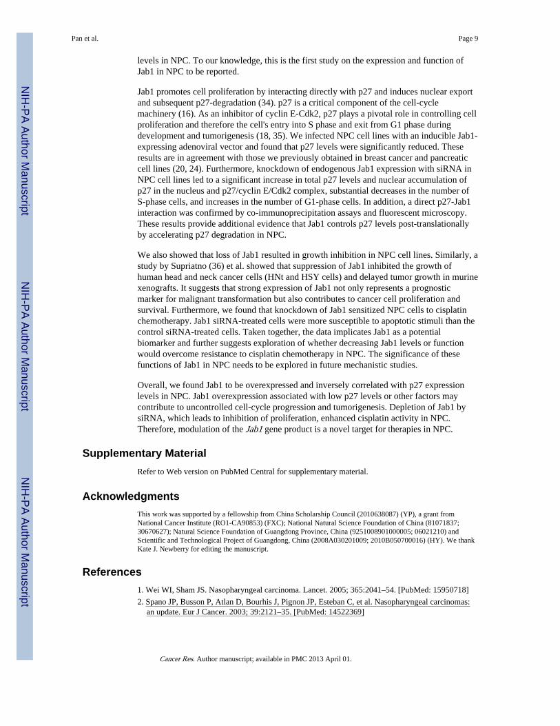
Jab1 depletion enhances the antitumor effects of cisplatin in NPCSince cisplatin is the main treatment for NPC, we proposed to investigate Jab1 may involvein the antitumor effects of cisplatin. We firstly tested whether inhibition of Jab1 enhancesthe antitumor effects of cisplatin by MTT assay. Data showed that 21%, 18%, and 7% ofCNE1, CNE2 and HONE1 cells treated with control siRNA and cisplatin were inhibited,whereas, 38%, 38% and 26% cells were inhibited by Jab1 siRNA and cisplatin, suggestingJab1-knockdown NPC cells were more sensitive to cisplatin than cells treated with controlsiRNA (Fig. 6A). To test whether Jab1 plays a role in anchorage-independent growth inresponse to cisplatin, which may reflect in vivo tumorigenicity, we performed a colonyformation assay in NPC cells treated with Jab1 siRNA and cisplatin. We found that the Jab1knockdown cells had significant inhibition of colony formation (73% in CNE1, 90% inCNE2, and 66% in HONE1) compared with the control siRNA-treated cells (22% in CNE1,60% in CNE2, and 14% in HONE1) in response to cisplatin (Fig. 6B).
We further examined whether Jab1 knockdown could enhance cisplatin-induced apoptosis inNPC cells. We found that cisplatin induced more apoptosis in Jab1 knockdown cells thanthat in control siRNA-treated cells, which is 1.4 folds in CNE1 and 4.7 folds in CNE2respectively (Fig. 6C) when using Annexin V and PI staining, and 1.6 in CNE1 and 3.2 inCNE2 when using Hochst33342 staining (Fig. 2S). However, there were no significantdifferences in apoptosis induced by control and Jab1 siRNA. Proteolytic cleavage of PARPand cleaved caspase-3 are the hallmarks of apoptosis. Thus, we also examined the effect ofJab1 siRNA on the proteolytic cleavage of PARP and cleaved caspase-3 in response tocisplatin. Compared with the control siRNA-treated cells, cisplatin consistently inducedmore proteolytic cleavage of PARP (12% change in CNE1, 30% change in CNE2, and 6%change in HONE1) and cleaved caspase-3 (11% change in CNE1, 50% change in CNE2,and 31% change in HONE1) in Jab1 knockdown cells (Fig. 6D).
DiscussionThe molecular basis of NPC pathogenesis remains poorly defined, and this has hindered thedevelopment of new treatments. Therefore, identification of novel mechanisms involved inNPC oncogenesis is needed.
In this study, we identified the role of Jab1-mediated p27 degradation in NPC oncogenesis.First, we found Jab1 was aberrantly expressed in either cytoplasm or nucleus NPC tissuescompare to noncancerous inflamed nasopharyngeal tissue. All (14/14) strong Jab1-positivepatients died within 41 months whereas 11 of 31 weak or Jab1-negative patients were aliveafter 60 months. We also showed that Jab1 protein is overexpressed in NPC cell lines butnot in paired normal keratinocyte cells. Our findings on p27 expression also confirm thoseof previous reports. For instance, Hwang et al (11) reported that in NPC patients 47 of 69cases expressed low levels of p27. In our study, 29 of 45 NPC cases (64%) expressed low orno nuclear p27, and 28 cases (62%) expressed low or no cytoplasmic p27. In addition, p27'snuclear-cytoplasmic translocation has been observed in human tumors and is associated withpoor survival (30–33), it was agreement with our studies that NPC patients with cytoplasmicpositive p27 had a poor survival.
Furthermore, cytoplasmic Jab1 expression in NPC tissues was inversely associated withnuclear p27 patterns (R=−0.389, P = 0.008), and correlated with cytoplasm p27 (R=0.328, P= 0.028). Our finding of an inverse correlation between Jab1 and p27 expression levels inNPC is in agreement with our previously published results on breast cancer and pancreaticcarcinomas (20, 24) and similar associations reported in other epithelial or lymphoidmalignancies (21, 22). This suggesting that Jab1 has a physiologic role in controlling p27
Pan et al. Page 8
Cancer Res. Author manuscript; available in PMC 2013 April 01.
NIH
-PA Author Manuscript
NIH
-PA Author Manuscript
NIH
-PA Author Manuscript
levels in NPC. To our knowledge, this is the first study on the expression and function ofJab1 in NPC to be reported.
Jab1 promotes cell proliferation by interacting directly with p27 and induces nuclear exportand subsequent p27-degradation (34). p27 is a critical component of the cell-cyclemachinery (16). As an inhibitor of cyclin E-Cdk2, p27 plays a pivotal role in controlling cellproliferation and therefore the cell's entry into S phase and exit from G1 phase duringdevelopment and tumorigenesis (18, 35). We infected NPC cell lines with an inducible Jab1-expressing adenoviral vector and found that p27 levels were significantly reduced. Theseresults are in agreement with those we previously obtained in breast cancer and pancreaticcell lines (20, 24). Furthermore, knockdown of endogenous Jab1 expression with siRNA inNPC cell lines led to a significant increase in total p27 levels and nuclear accumulation ofp27 in the nucleus and p27/cyclin E/Cdk2 complex, substantial decreases in the number ofS-phase cells, and increases in the number of G1-phase cells. In addition, a direct p27-Jab1interaction was confirmed by co-immunoprecipitation assays and fluorescent microscopy.These results provide additional evidence that Jab1 controls p27 levels post-translationallyby accelerating p27 degradation in NPC.
We also showed that loss of Jab1 resulted in growth inhibition in NPC cell lines. Similarly, astudy by Supriatno (36) et al. showed that suppression of Jab1 inhibited the growth ofhuman head and neck cancer cells (HNt and HSY cells) and delayed tumor growth in murinexenografts. It suggests that strong expression of Jab1 not only represents a prognosticmarker for malignant transformation but also contributes to cancer cell proliferation andsurvival. Furthermore, we found that knockdown of Jab1 sensitized NPC cells to cisplatinchemotherapy. Jab1 siRNA-treated cells were more susceptible to apoptotic stimuli than thecontrol siRNA-treated cells. Taken together, the data implicates Jab1 as a potentialbiomarker and further suggests exploration of whether decreasing Jab1 levels or functionwould overcome resistance to cisplatin chemotherapy in NPC. The significance of thesefunctions of Jab1 in NPC needs to be explored in future mechanistic studies.
Overall, we found Jab1 to be overexpressed and inversely correlated with p27 expressionlevels in NPC. Jab1 overexpression associated with low p27 levels or other factors maycontribute to uncontrolled cell-cycle progression and tumorigenesis. Depletion of Jab1 bysiRNA, which leads to inhibition of proliferation, enhanced cisplatin activity in NPC.Therefore, modulation of the Jab1 gene product is a novel target for therapies in NPC.
Supplementary MaterialRefer to Web version on PubMed Central for supplementary material.
AcknowledgmentsThis work was supported by a fellowship from China Scholarship Council (2010638087) (YP), a grant fromNational Cancer Institute (RO1-CA90853) (FXC); National Natural Science Foundation of China (81071837;30670627); Natural Science Foundation of Guangdong Province, China (9251008901000005; 06021210) andScientific and Technological Project of Guangdong, China (2008A030201009; 2010B050700016) (HY). We thankKate J. Newberry for editing the manuscript.
References1. Wei WI, Sham JS. Nasopharyngeal carcinoma. Lancet. 2005; 365:2041–54. [PubMed: 15950718]
2. Spano JP, Busson P, Atlan D, Bourhis J, Pignon JP, Esteban C, et al. Nasopharyngeal carcinomas:an update. Eur J Cancer. 2003; 39:2121–35. [PubMed: 14522369]
Pan et al. Page 9
Cancer Res. Author manuscript; available in PMC 2013 April 01.
NIH
-PA Author Manuscript
NIH
-PA Author Manuscript
NIH
-PA Author Manuscript

3. Ong YK, Heng DM, Chung B, Leong SS, Wee J, Fong KW, et al. Design of a prognostic indexscore for metastatic nasopharyngeal carcinoma. Eur J Cancer. 2003; 39:1535–41. [PubMed:12855259]
4. Yip WK, Leong VC, Abdullah MA, Yusoff S, Seow HF. Overexpression of phospho-Akt correlateswith phosphorylation of EGF receptor, FKHR and BAD in nasopharyngeal carcinoma. Oncol Rep.2008; 19:319–28. [PubMed: 18202777]
5. Zheng XK, Chen LH, Wang WJ, Ye F, Liu JB, Li QS, et al. Impact of prolonged fraction deliverytimes simulating IMRT on cultured nasopharyngeal carcinoma cell killing. International journal ofradiation oncology, biology, physics. 2010; 78:1541–7.
6. Al-Sarraf M, LeBlanc M, Giri PG, Fu KK, Cooper J, Vuong T, et al. Chemoradiotherapy versusradiotherapy in patients with advanced nasopharyngeal cancer: phase III randomized Intergroupstudy 0099. J Clin Oncol. 1998; 16:1310–7. [PubMed: 9552031]
7. Lee AW, Yau TK, Wong DH, Chan EW, Yeung RM, Ng WT, et al. Treatment of stage IV(A–B)nasopharyngeal carcinoma by induction-concurrent chemoradiotherapy and acceleratedfractionation. Int J Radiat Oncol Biol Phys. 2005; 63:1331–8. [PubMed: 16169677]
8. Le QT, Tate D, Koong A, Gibbs IC, Chang SD, Adler JR, et al. Improved local control withstereotactic radiosurgical boost in patients with nasopharyngeal carcinoma. Int J Radiat Oncol BiolPhys. 2003; 56:1046–54. [PubMed: 12829140]
9. Bei JX, Li Y, Jia WH, Feng BJ, Zhou G, Chen LZ, et al. A genome-wide association study ofnasopharyngeal identifies three new susceptibility loci. Nat Genet. 2010; 42:599–603. [PubMed:20512145]
10. Natasya Naili MN, Hasnita CH, Shamim AK, Hasnan J, Fauziah MI, Narazah MY, et al.Chromosomal alterations in Malaysian patients with nasopharyngeal carcinoma analyzed bycomparative genomic hybridization. Cancer Genet Cytogenet. 2010; 203:309–12. [PubMed:21156250]
11. Hwang CF, Su CY, Huang SC, Huang CC, Fang FM, Lui CC, et al. Low expression levels of p27correlate with loco-regional recurrence in nasopharyngeal carcinoma. Cancer Lett. 2003; 189:231–6. [PubMed: 12490316]
12. Ko MT, Su CY, Huang SC, Chen CH, Hwang CF. Overexpression of cyclin E messengerribonucleic acid in nasopharyngeal carcinoma correlates with poor prognosis. J Laryngol Otol.2009; 123:1021–6. [PubMed: 19275777]
13. Mei YP, Zhou JM, Wang Y, Huang H, Deng R, Feng GK, et al. Silencing of LMP1 induces cellcycle arrest and enhances chemosensitivity through inhibition of AKT signaling pathway in EBV-positive nasopharyngeal carcinoma cells. Cell Cycle. 2007; 6:1379–85. [PubMed: 17507800]
14. Wei N, Deng XW. The COP9 signalosome. Annu Rev Cell Dev Biol. 2003; 19:261–86. [PubMed:14570571]
15. Claret FX, Hibi M, Dhut S, Toda T, Karin M. A new group of conserved coactivators that increasethe specificity of AP-1 transcription factors. Nature. 1996; 383:453–7. [PubMed: 8837781]
16. Tomoda K, Kubota Y, Kato J. Degradation of the cyclin-dependent-kinase inhibitor p27Kip1 isinstigated by Jab1. Nature. 1999; 398:160–5. [PubMed: 10086358]
17. Sherr CJ, Roberts JM. CDK inhibitors: positive and negative regulators of G1-phase progression.Genes Dev. 1999; 13:1501–12. [PubMed: 10385618]
18. Polyak K, Lee MH, Erdjument-Bromage H, Koff A, Roberts JM, Tempst P, et al. Cloning ofp27Kip1, a cyclin-dependent kinase inhibitor and a potential mediator of extracellularantimitogenic signals. Cell. 1994; 78:59–66. [PubMed: 8033212]
19. Pagano M, Tam SW, Theodoras AM, Beer-Romero P, Del Sal G, Chau V, et al. Role of theubiquitin-proteasome pathway in regulating abundance of the cyclin-dependent kinase inhibitorp27. Science. 1995; 269:682–5. [PubMed: 7624798]
20. Kouvaraki MA, Rassidakis GZ, Tian L, Kumar R, Kittas C, Claret FX. Jun activation domain-binding protein 1 expression in breast cancer inversely correlates with the cell cycle inhibitorp27(Kip1). Cancer Res. 2003; 63:2977–81. [PubMed: 12782606]
21. Rassidakis GZ, Claret FX, Lai R, Zhang Q, Sarris AH, McDonnell TJ, et al. Expression ofp27(Kip1) and c-Jun activation binding protein 1 are inversely correlated in systemic anaplasticlarge cell lymphoma. Clin Cancer Res. 2003; 9:1121–8. [PubMed: 12631617]
Pan et al. Page 10
Cancer Res. Author manuscript; available in PMC 2013 April 01.
NIH
-PA Author Manuscript
NIH
-PA Author Manuscript
NIH
-PA Author Manuscript

22. Sui L, Dong Y, Ohno M, Watanabe Y, Sugimoto K, Tai Y, et al. Jab1 expression is associated withinverse expression of p27(kip1) and poor prognosis in epithelial ovarian tumors. Clin Cancer Res.2001; 7:4130–5. [PubMed: 11751512]
23. Fukumoto A, Ikeda N, Sho M, Tomoda K, Kanehiro H, Hisanaga M, et al. Prognostic significanceof localized p27Kip1 and potential role of Jab1/CSN5 in pancreatic cancer. Oncol Rep. 2004;11:277–84. [PubMed: 14719054]
24. Kouvaraki MA, Korapati AL, Rassidakis GZ, Tian L, Zhang Q, Chiao P, et al. Potential role of Junactivation domain-binding protein 1 as a negative regulator of p27kip1 in pancreaticadenocarcinoma. Cancer Res. 2006; 66:8581–9. [PubMed: 16951171]
25. Berg JP, Zhou Q, Breuhahn K, Schirmacher P, Patil MA, Chen X, et al. Inverse expression of Junactivation domain binding protein 1 and cell cycle inhibitor p27Kip1: influence on proliferation inhepatocellular carcinoma. Hum Pathol. 2007; 38:1621–7. [PubMed: 17651785]
26. Ito Y, Yoshida H, Matsuzuka F, Matsuura N, Nakamura Y, Nakamine H, et al. Jun activationdomain-binding protein 1 expression in malignant lymphoma of the thyroid: its linkage to degreeof malignancy and p27 expression. Anticancer Res. 2003; 23:4121–5. [PubMed: 14666612]
27. Shen L, Tsuchida R, Miyauchi J, Saeki M, Honna T, Tsunematsu Y, et al. Differentiation-associated expression and intracellular localization of cyclin-dependent kinase inhibitor p27KIP1and c-Jun co-activator JAB1 in neuroblastoma. Int J Oncol. 2000; 17:749–54. [PubMed:10995887]
28. Dahinden C, Ingold B, Wild P, Boysen G, Luu VD, Montani M, et al. Mining tissue microarraydata to uncover combinations of biomarker expression patterns that improve intermediate stagingand grading of clear cell renal cell cancer. Clin Cancer Res. 2010; 16:88–98. [PubMed: 20028743]
29. Tian L, Peng G, Parant JM, Leventaki V, Drakos E, Zhang Q, et al. Essential roles of Jab1 in cellsurvival, spontaneous DNA damage and DNA repair. Oncogene. 2010; 29:6125–37. [PubMed:20802511]
30. Loda M, Cukor B, Tam SW, Lavin P, Fiorentino M, Draetta GF, et al. Increased proteasome-dependent degradation of the cyclin-dependent kinase inhibitor p27 in aggressive colorectalcarcinomas. Nat Med. 1997; 3:231–4. [PubMed: 9018245]
31. Masciullo V, Sgambato A, Pacilio C, Pucci B, Ferrandina G, Palazzo J, et al. Frequent loss ofexpression of the cyclin-dependent kinase inhibitor p27 in epithelial ovarian cancer. Cancer Res.1999; 59:3790–4. [PubMed: 10446997]
32. Singh SP, Lipman J, Goldman H, Ellis FH Jr. Aizenman L, Cangi MG, et al. Loss or alteredsubcellular localization of p27 in Barrett's associated adenocarcinoma. Cancer Res. 1998;58:1730–5. [PubMed: 9563491]
33. Ciaparrone M, Yamamoto H, Yao Y, Sgambato A, Cattoretti G, Tomita N, et al. Localization andexpression of p27KIP1 in multistage colorectal carcinogenesis. Cancer Res. 1998; 58:114–22.[PubMed: 9426067]
34. Shackleford TJ, Claret FX. JAB1/CSN5: a new player in cell cycle control and cancer. Cell Div.2010; 5:26. [PubMed: 20955608]
35. Toyoshima H, Hunter T. p27, a novel inhibitor of G1 cyclin-Cdk protein kinase activity, is relatedto p21. Cell. 1994; 78:67–74. [PubMed: 8033213]
36. Supriatno, Harada K.; Yoshida, H.; Sato, M. Basic investigation on the development of moleculartargeting therapy against cyclin-dependent kinase inhibitor p27Kip1 in head and neck cancer cells.Int J Oncol. 2005; 27:627–35. [PubMed: 16077910]
Pan et al. Page 11
Cancer Res. Author manuscript; available in PMC 2013 April 01.
NIH
-PA Author Manuscript
NIH
-PA Author Manuscript
NIH
-PA Author Manuscript

Figure 1. Expression patterns of Jab1 and p27 in non-neoplastic tissues and NPC tissues(A) Overall Jab1 immunoreactivity was low in non-neoplastic tissue (left) compared to NPC(middle). (Right) Percentage of cases with Jab1 expression in nasopharyngeal tissues andNPC. (B) Nuclear p27 was strongly expressed in non-neoplastic tissue (left). A case of NPC,with the predominantly cytoplasmic staining is shown (middle). (Right) Percentage of caseswith p27 expression in non-neoplastic tissues and NPC. Original magnification, ×200; insets×400. (C) Tumors with cytoplasmic Jab1 expression were associated with nuclear andcytoplasmic p27. R-values and p-values from Spearman's test. (D) Kaplan-Meier analysis ofthe association of Jab1 expression (left) and survival as well as association of cytoplasmicp27 (right) and survival.
Pan et al. Page 12
Cancer Res. Author manuscript; available in PMC 2013 April 01.
NIH
-PA Author Manuscript
NIH
-PA Author Manuscript
NIH
-PA Author Manuscript

Figure 2. Jab1 is overexpressed and regulates p27 levels in NPC cells(A) Whole-cell lysates were prepared from the cells as indicated. β-actin was used as acontrol for protein loading and integrity. The relative Jab1 and p27 intensity from 5 samplesis shown. (B) Proteolysis of p27 in NPC cells is sensitive to proteasome inhibitors. (C)Inducible expression of Ad-Myc-Jab1 (Tet-off) downregulates endogenous p27 levels inNPC cell lines. Lysates were prepared from Ad-Myc-Jab1-infected cells in the absence (−)or presence (+) of doxycycline (Dox). (D) NPC cells were transduced with the Ad-Myc-Jab1virus, and treated as indicated with proteasome inhibitor. The protein levels were quantifiedusing ImageJ software.
Pan et al. Page 13
Cancer Res. Author manuscript; available in PMC 2013 April 01.
NIH
-PA Author Manuscript
NIH
-PA Author Manuscript
NIH
-PA Author Manuscript

Figure 3. Jab1 specifically interacts with p27(A) Cell lysates were immunoprecipitated with either non-immune mouse serum or Jab1 andimmunoblotted with anti-p27. Immunoglobulin G heavy chain (HC) and light chain (LC)were indicated. (B) 293T cells were transfected with either Myc-Jab1 or Flag-p27 or bothfor 48 hours, and then cell lysates were immunoprecipitated with Myc and immunoblottedwith Flag. Cell lysates immunoblotted with the indicated antibodies are shown in the bottompanel. (C) CNE1 cells were transfected with a GFP vector and Cherry-p27 or GFP-Jab1 andCherry-p27 for 48 hours, and then examined under a fluorescence microscope. Originalmagnification ×200.
Pan et al. Page 14
Cancer Res. Author manuscript; available in PMC 2013 April 01.
NIH
-PA Author Manuscript
NIH
-PA Author Manuscript
NIH
-PA Author Manuscript

Figure 4. Downregulation of Jab1 increases p27 levels and induces cell-cycle arrest in NPC(A) Cells were transfected with either a siRNA targeting Jab1 or a scrambled sequence(control); 48 hours later, the total cellular proteins and nuclear proteins were isolated andimmunoblotted with anti-Jab1 and anti-p27 antibodies. Anti-β-actin or Lamin A/Cantibodies were used as loading controls. (B) NPC cells were transfected as described in(A); 48 hours later, cells were stained with PI and then analyzed by flow cytometry. (Left)Representative results of cell cycle assays with NPC cells. (Right) The percentage of G1 andS phase cells are shown data represent three independent experiments, mean±s.d. * P <0.05,* *P <0.01. (C) Cells were transfected with either a siRNA targeting Jab1 or a scrambledsequence (control); 48 hours later, cell lysates were immunoprecipitated with Cdk2 andimmunoblotted with anti-p27.
Pan et al. Page 15
Cancer Res. Author manuscript; available in PMC 2013 April 01.
NIH
-PA Author Manuscript
NIH
-PA Author Manuscript
NIH
-PA Author Manuscript

Figure 5. Knockdown of Jab1 inhibits cell proliferation(A) NPC cells were transfected with Jab1 siRNA for 48 hours, and then cell growth wasdetermined by an MTT assay. Control-siRNA (Cont-si); Jab1-siRNA (Jab1-si). (B)Representative results of colony formation assays with NPC cells treated with controlsiRNA or different doses of 1 nM (+) or 5 nM (++) of Jab1 siRNA. Jab1 siRNA. (Bottom)Quantification of the relative number of colonies is shown. Data represent three independentexperiments, mean±s.d. *P <0.05, **P <0.01. (C) Cells were treated with control siRNA orJab1 siRNA for 48 hours, and then stained with Annexin V and PI and measured by flowcytometry. (Bottom) Quantification of the percentage of living cells. Data represent threeindependent experiments, mean±s.d. (D) Cells were transfected with siRNA for 48 hours,and then cell lysates were immunoblotted with the indicated antibodies for apoptosisanalysis.
Pan et al. Page 16
Cancer Res. Author manuscript; available in PMC 2013 April 01.
NIH
-PA Author Manuscript
NIH
-PA Author Manuscript
NIH
-PA Author Manuscript
Figure 6. Jab1 depletion enhances the antitumor effects of cisplatin in NPC cells(A) Forty-eight hours after the cells were treated with control (Cont-si) or Jab1 siRNA(Jab1-si), NPC cells were treated with the 2 μM of cisplatin (CP) for another 48 hours, andthe growth-inhibitory effects of cisplatin were quantified by an MTT assay. The inhibitionratio (%) is marked on the graphs. Data represent three independent experiments, mean±s.d.**P <0.01. (B) 1 nM (+) or 5 nM (++) of Jab1 siRNA-treated NPC cells were seeded into 6-well plates and exposed to cisplatin (CP) for 48 hours, and 10 days later the number ofcolonies formed were counted. (Left) Representative results of colony formation assays.(Right) Quantification of the relative number of colonies. The inhibition ratios (%) aremarked on the graphs. Data represent three independent experiments, mean ±s.d. *P <0.05,**P <0.01. (C) siRNA-transfected NPC cells were stained with Annexin V and PI aftercisplatin treatment for 48 hours. (Left) Representative results of apoptosis assays in NPCcells. (Right) Quantification of the percentage of apoptotic cells. Data represent threeindependent experiments, mean±s.d. *P <0.05, **P <0.01. (D) siRNA-transfected NPC cellwere exposed to 10 μM of cisplatin for 48h; apoptotic cells were measured by the westernblot analysis of cleaved PARP and cleaved caspase-3. Protein levels were quantified usingImageJ software.
Pan et al. Page 17
Cancer Res. Author manuscript; available in PMC 2013 April 01.
NIH
-PA Author Manuscript
NIH
-PA Author Manuscript
NIH
-PA Author Manuscript

NIH
-PA Author Manuscript
NIH
-PA Author Manuscript
NIH
-PA Author Manuscript
Pan et al. Page 18
Table 1
Characteristics of 45 NPC patients included in the study
Characteristics No. patients (n=45)
Gender (male/female) 36/9
Median age 41
Tumor histologic type
Squamous cell carcinoma 32 (71%)
Non-keratinizing carcinomas 13 (29%)
Differentiation
Poor 32 (71%)
No differentiation 13 (29%)
Lymph node metastasis
Positive 40 (89%)
Negative 5 (11%)
Stage
I 0
II 12 (27%)
III 20 (44%)
IV 13 (29%)
Cancer Res. Author manuscript; available in PMC 2013 April 01.
Copyright © 2022 FDOKUMEN