Subcellular trafficking of phytochemicals explored using auto-fluorescent compounds in maize cells
Upload
univ-lille1Category
view
2download
0
ORIGINAL ARTICLE
Ets-1 p27: a novel Ets-1 isoform with dominant-negative effects on the
transcriptional properties and the subcellular localization of Ets-1 p51
C Laitem1,4, G Leprivier1,3,4, S Choul-Li1, A Begue1, D Monte1, D Larsimont2, P Dumont1,M Duterque-Coquillaud1 and M Aumercier1
1CNRS Unite Mixte de Recherche 8161, Institut de Biologie de Lille, Institut Pasteur de Lille, Universites de Lille 1 and Lille 2,IFR 142, Lille, France and 2Department of Pathology, Institut Jules Bordet, Brussels, Belgium
The transcription factor Ets-1 is implicated in variousphysiological processes and invasive pathologies. Weidentified a novel variant of ets-1, ets-1D(III–VI),resulting from the alternative splicing of exons III to VI.This variant encodes a 27 kDa isoform, named Ets-1 p27.Ets-1 p27 lacks the threonine-38 residue, the Pointeddomain and the transactivation domain, all of which arerequired for the transactivation of Ets-1 target genes.Both inhibitory domains surrounding the DNA-bindingdomain are conserved, suggesting that Ets-1 p27, like thefull-length Ets-1 p51 isoform, is autoinhibited for DNAbinding. We showed that Ets-1 p27 binds DNA in thesame way as Ets-1 p51 does and that it acts both at atranscriptional and a subcellular localization level, therebyconstituting a dual-acting dominant negative of Ets-1 p51.Ets-1 p27 blocks Ets-1 p51-mediated transactivation oftarget genes and induces the translocation of Ets-1 p51from the nucleus to the cytoplasm. Furthermore, Ets-1p27 overexpression represses the tumor properties ofMDA-MB-231 mammary carcinoma cells in correlationwith the known implication of Ets-1 in various cellularmechanisms. Thus the dual-acting dominant-negativefunction of Ets-1 p27 gives to the Ets-1 p27/Ets-1 p51ratio a determining effect on cell fate.Oncogene (2009) 28, 2087–2099; doi:10.1038/onc.2009.72;published online 20 April 2009
Keywords: Ets-1; transcriptional regulation; subcellularlocalization; dominant-negative isoform; alternativesplicing
Introduction
Splicing of intronic sequences is a physiological cellularmechanism implicated in RNA maturation. During this
process, exonic sequences can be alternatively spliced,generating either nonsense mRNAs, which will bedegraded, or mature mRNAs, which can encodeproteins with different structures and properties, calledisoforms (Smith and Valcarcel, 2000; Stamm et al.,2005). Alternative splicing of mRNA precursors can bemodulated in a spatiotemporal manner and canparticipate in the regulation of gene functions (Shinand Manley, 2004). Alternative splicing occurs innumerous members of the Ets family of transcriptionfactors such as GABPa (O’Leary et al., 2005), Tel(Sasaki et al., 2004), Spi-B (Ray-Gallet et al., 1996), Ets-1 (Baillat et al., 2002), Fli-1 (Sarrazin et al., 2000), ER81(Coutte et al., 1999), Erg (Hewett et al., 2001), ESE-1(Lei et al., 2007), ELF-1 (Cho et al., 2004), Elk-1 (Araudet al., 2007) and ER-71 (De Haro and Janknecht, 2005).The Ets family is characterized by a well-conserved
DNA-binding domain (DBD), called the ETS domain,and recognizes specific DNA elements, called Ets-binding sites (EBS), found in the promoters of targetgenes. The founding member of this family, Ets-1, isinvolved in numerous cellular mechanisms such asproliferation (de Nigris et al., 2001; Higuchi et al.,2007), differentiation (Lulli et al., 2006; John et al.,2007), migration (Kita et al., 2001), invasion (Taniguchiet al., 2007) and apoptosis (Teruyama et al., 2001).However, when overexpressed, Ets-1 is partly respon-sible for the development of invasive pathologies such asrheumatoid arthritis (Redlich et al., 2001), glomerulone-phritis (Raffetseder et al., 2004) or cancers (Dittmer,2003). Ets-1 is now considered as a marker of poorprognosis in numerous cancers (Nakayama et al., 2001;Alipov et al., 2005; Katayama et al., 2005).The activity of Ets-1 is tightly controlled, especially at
the DNA-binding level, to ensure a specific biologicalaction. Indeed, Ets-1 is autoinhibited for DNA bindingdue to the presence of two inhibitory domains surround-ing and interacting with its DBD (Lee et al., 2005). Tocounteract autoinhibition and improve DNA binding,Ets-1 interacts with partners, enabling their cooperativebinding to adjacent DNA elements (Pufall and Graves,2002). Ets-1 can also counteract its autoinhibition onits own by binding to a particular organization ofpalindromic EBS spaced by 4bp, as in the stromelysin-1matrix metalloproteinase and the p53 oncosuppressorpromoters (Wasylyk et al., 1991; Baillat et al., 2002,2009). Two molecules of Ets-1 cooperatively bind to the
Received 21 October 2008; revised 4 February 2009; accepted 10 March2009; published online 20 April 2009
Correspondence: Dr M Aumercier, CNRS UMR 8161, Institut deBiologie de Lille, 1 rue du Prof Calmette, BP 447, 59021, Lille Cedex,France.E-mail: [email protected] address: Department of Molecular Oncology, BritishColumbia Cancer Research Centre, Vancouver, British Columbia,Canada.4These authors contributed equally to this work.
Oncogene (2009) 28, 2087–2099& 2009 Macmillan Publishers Limited All rights reserved 0950-9232/09 $32.00
www.nature.com/onc
EBS palindrome, thereby forming an Ets-1/DNA/Ets-1ternary complex, which is critical for full transactivation(Baillat et al., 2002).Until now, only two isoforms of Ets-1 have been
described: the full-length protein Ets-1 p51 (usuallycalled Ets-1) and the isoform encoded by the ets-1DVIIalternative splicing variant Ets-1 p42 (Koizumi et al.,1990). This latter variant lacks exon VII, which encodes,in particular, the N-terminal inhibitory domain. There-fore, Ets-1 p42 is not autoinhibited and presentsdifferent DNA-binding and transcriptional propertiesfrom the full-length Ets-1 p51 protein (Fisher et al.,1994; Baillat et al., 2002). Thus, Ets-1 p51 and Ets-1 p42regulate different target genes, illustrating regulation ofEts-1 function through alternative splicing (Li et al.,1999; de Nigris et al., 2001; Taniguchi et al., 2007).In this study, we identified a novel variant of ets-1,
named ets-1D(III–VI), which results from the alter-native splicing of exons III to VI. This variant encodes a27 kDa isoform, named Ets-1 p27. By characterizing themolecular and cellular properties of Ets-1 p27, we showthat Ets-1 p27 is the first natural Ets-1 dominantnegative that acts both at a transcriptional and asubcellular localization level and which represses thetumoral properties of invasive mammary carcinomacells.
Results
ets-1D(III–VI) is a novel ets-1 variant expressed in atissue-specific mannerA novel ets-1 variant was identified by reverse transcrip-tion (RT)–PCR using flanking primers in HIG-82 rabbitsynovial fibroblastic cells in addition to transcriptscorresponding to full-length ets-1 and exon-VII-deletedets-1DVII (data not shown). The complete codingsequence of this variant, which is available in GenBankunder accession number EU939724, demonstrates that itresults from the alternative splicing of exons III to VI(Figure 1a, left). Therefore, this novel ets-1 variant wascalled ets-1D(III–VI). Its expression was confirmed inHIG-82 cells by RT–PCR using two other primer pairs(Figure 1a): the pair 1, composed of primers designed inexon A and exon VII and the pair 2, composed ofprimers located at the ets-1D(III–VI) splicing junctionand in exon VII.Using this two primer pairs, we showed by RT–PCR
that both ets-1 and ets-1D(III–VI) transcripts weresynthesized in HIG-82 cells, MG-63 human osteosarco-ma cells and MDA-MB-231 invasive human mammaryadenocarcinoma cells (Figure 1b). The complete codingsequence of the human ets-1D(III–VI) variant in MDA-MB-231 cells was determined and is available inGenBank under accession number AY943926.Reverse transcription–PCR performed on RNAs
from human mammary adenocarcinomas (Figure 1c,lanes 1–3) and PCR on cDNA from a human mammarytissue library (Figure 1c, lane 4) revealed that the twotranscripts were expressed in tumor mammary tissues,but not in normal tissues. Next, cDNA libraries of
human fetal and adult tissues were analysed by PCR(Figure 1d). Both ets-1 and ets-1D(III–VI) weredetected in all fetal tissues tested, but varied with tissuetype in adult tissues. Indeed, both ets-1 and ets-1D(III–VI)were expressed in spleen, thymus, placental and ovariantissues, but neither in brain nor kidney tissues. Thus, ets-1D(III–VI) was expressed along with full-length ets-1 insome tissue and their ratio depended on cell andtissue types, suggesting a tissue-dependent effect ofets-1D(III–VI) on ets-1 properties.
ets-1D(III–VI) variant encodes Ets-1 p27, a novel Ets-1isoformThe novel ets-1D(III–VI) variant gives rise to a 225-amino-acid Ets-1 protein, which lacks the threonine-38(Thr38) residue as well as the Pointed (PNT) andtransactivation (TAD) domains, all of which arerequired for the transactivation of Ets-1 target genes(Jayaraman et al., 1999; Seidel and Graves, 2002; Fouldset al., 2004). However, it conserves the DBD flanked bythe two inhibitory domains. Given its apparent mole-cular mass of 27 kDa, this isoform was called Ets-1 p27,in contrast to full-length Ets-1 that has a mass of 51 kDa(Figure 2a). Western blot analysis of native andets-1D(III–VI) cDNA-transfected HIG-82 cells, usingthe C-20 anti-Ets-1 antibody directed against Ets-1DBD, showed the presence of a 27 kDa endogenousprotein (Figure 2b, lane 1), which migrates exactly thesame way as exogenous Ets-1 p27 does (Figure 2b, lane2). Thus, Ets-1 p27 was endogenously expressed in HIG-82 cells. Furthermore, its intensity was roughly equiva-lent to that of Ets-1 p42. A higher molecular massprotein was also detected in transfected HIG-82 cells(Figure 2b, lane 2). This protein probably correspondsto post-translationally modified Ets-1 p27, which wasonly weakly observed in native cells due to its lowexpression level.Expression of Ets-1 p27 was then assessed in various
cell lines by western blot using either the C-20 anti-Ets-1antibody directed against Ets-1 DBD or a specificanti-Ets-1 p27 antibody, directed against the Ets-1 p27alternative splicing junction (Figure 2c). Results showedthat HIG-82 (Figure 2c, lane 2), MDA-MB-231(Figure 2c, lane 3), MG-63 (Figure 2c, lane 4) andJurkat (Figure 2c, lane 5) cells expressed endogenouslythe novel Ets-1 p27 isoform, which was recognizedby both antibodies. The Ets-1 p27 isoform is thusendogenously expressed in numerous cell lines.
Ets-1 p27 binds to the EBS palindrome of thestromelysin-1 promoter in the same way as Ets-1 p51 doesand competes with Ets-1 p51 for bindingWe then determined whether Ets-1 p27 was autoinhib-ited like Ets-1 p51 and whether autoinhibition iscounteracted by cooperative binding to the palindromicEBS of the stromelysin-1 promoter. Electrophoreticmobility shift assays (EMSAs) were carried out usingEts-1 p51 or Ets-1 p27 recombinant proteins and 30-mer32P-labeled DNA probes containing wild-type (WT) ormutant EBS palindromes of the stromelysin-1 promoter
Ets-1 p27, a dual-acting Ets-1 dominant negativeC Laitem et al
2088
Oncogene
(Table 1; Figure 3a). As expected, results showed thatEts-1 p51 and Ets-1 p27 formed ternary complexes withthe wild-type EBS palindrome (indicated by arrows 1and 2 in Figure 3a, lanes 1 and 5), whereas mutationof one (M1, M2) or both (M1M2) EBS preventedboth isoforms from forming DNA/protein complexes(Figure 3a, lanes 2–4 and 6–8). In both cases, thesecomplexes were disrupted by an 800� molar excess of
WT non-labeled DNA probe (Figure 3b, lanes 2 and 5),but not by an 800� molar excess of M1M2 non-labeledDNA probe (Figure 3b, lanes 3 and 6). This confirmedthe complexes’ specificity to the EBS palindrome.Surface plasmon resonance (SPR) measurement
experiments were then carried out using Biacoretechnology (Figure 3c). For both Ets-1 p27 and Ets-1p51, the measured resonance unit (RU) signal corre-
Figure 1 Evidence for a novel ets-1 alternative splicing variant, named ets-1D(III–VI), and its cellular and tissue distribution alongwith ets-1. (a) The ets-1 and ets-1D(III–VI) schematic representation and detection by reverse transcription (RT)–PCR in HIG-82cells. Left: translated exons are represented as boxes and noted from A to IX. Arrow couples (indicated by 1 and 2) represent primerpairs, used in subsequent PCR experiments. Right: single-strand cDNAs were synthesized from HIG-82 total RNAs and ets-1 cDNAswere amplified by PCR using the two primer pairs (pairs 1 and 2) indicated in the left. Amplified PCR products, which correspond toets-1 and ets-1D(III–VI), were resolved on a 2% agarose gel stained with ethidium bromide and are indicated by arrows related to thescheme. (b) RT–PCR analysis of different cell lines. RNAs from HIG-82, MG-63 and MDA-MB-231 cells were retrotranscribed andets-1 cDNAs were amplified by PCR using the two primer pairs described in (a). Amplification of glyceraldehyde-3-phosphatedehydrogenase (gapdh) with specific primers was used as a loading control and amplification without template as a negative control(NC). (c) Analysis of tumor and normal mammary tissues by RT–PCR and PCR. RNAs extracted from human mammaryadenocarcinomas (lanes 1–3) and cDNAs from a normal mammary tissue library (lane 4) were amplified by RT–PCR and PCR,respectively, as described in (b). (d) Analysis of fetal and adult human tissues by PCR. cDNAs from fetal and adult human tissuelibraries were amplified by PCR as described in (b).
Ets-1 p27, a dual-acting Ets-1 dominant negativeC Laitem et al
2089
Oncogene

sponding to binding to the WT probe was higher thanthe sum of those obtained with M1 and M2 probes(Figure 3c, indicated by dotted line M1þM2). Ets-1 p27thus binds cooperatively to the EBS palindrome of thestromelysin-1 promoter in the same way as Ets-1 p51does.Next, we assessed the ability of Ets-1 p27 to compete
with Ets-1 p51 for binding. For this purpose, EMSAexperiments were carried out using the 30-mer32P-labeled WT DNA probe (Table 1) and various Ets-1 p51/Ets-1 p27 ratios (Figure 3d). Results showed thatthe Ets-1 p51/DNA/Ets-1 p51 ternary complex(Figure 3d, lane 1) was disrupted in a dose-dependentmanner in the presence of Ets-1 p27 (Figure 3d, lanes2–4), leading to the formation of an Ets-1 p27/DNA/Ets-1 p27 ternary complex (Figure 3d, lane 5). Thisdisruption was accompanied by the formation of anintermediate migrating complex (indicated by arrow i),composed of one molecule of each isoform. Thus, the
novel Ets-1 p27 isoform competes with Ets-1 p51 forbinding to the EBS palindrome of the stromelysin-1promoter.
Ets-1 p27 is a dominant negative for Ets-1 p51 and otherautoinhibited Ets proteins transcriptional transactivationTo verify if Ets-1 p27 can block Ets-1 p51 transcriptionalproperties, luciferase transactivation assays were per-formed with the stromelysin-1 promoter (WT or M1M2)using increasing concentrations of Ets-1 p27 with orwithout Ets-1 p51 (Figure 4a). HEK-293 cells were usedbecause they lack endogenous Ets-1, allowing us tomonitor the expression of the different Ets-1 isoforms. Itappeared that Ets-1 p27 was unable to activate thestromelysin-1 promoter on its own (Figure 4a, lanes 1–8).Nevertheless, it repressed in a dose-dependent manner theactivation of the WT promoter mediated by Ets-1 p51(Figure 4a, lanes 9–12). This was mediated through theEBS palindrome, because EBS mutations abrogated anyEts-1 p27 effects (Figure 4a, lanes 13–16). Thus, Ets-1p27 constitutes a dominant-negative isoform for Ets-1p51 transactivation of the stomelysin-1 promoter.Because autoinhibited Ets-2 and PEA-3 proteins alsoactivate this promoter through the EBS palindrome(Buttice and Kurkinen, 1993; Higashino et al., 1995),luciferase transactivation assays were also carried out, asdescribed above, using Ets-2 (Figure 4b) and PEA-3(Figure 4c) as transcriptional activators. Results showedthat Ets-1 p27 acts also as a dominant negative for Ets-2and PEA-3 transactivation of the stromelysin-1 promo-ter. Furthermore, we demonstrated that Ets-1 p27repressed in a dose-dependent manner the synergisticactivation of the collagenase-1 promoter induced by Ets-1 p51 and the c-Jun/c-Fos (AP-1) complex, through the12-O-tetradecanoylphorbol-13-acetate (TPA) oncogeneresponse unit, composed of an EBS adjacent to an AP-1site (see Supplementary Information, Figure S1 andTable S1). Thus, Ets-1 p27 is also a dominant negativefor Ets-1 p51 transactivation of target promotersmediated through combinatorial regulation with inter-action partners.
Ets-1 p27 delocalizes Ets-1 p51 from the nucleus to thecytoplasmTo mediate its transcriptional dominant-negative func-tion, Ets-1 p27 must be coexpressed with Ets-1 p51 in
Figure 2 ets-1D(III–VI) encodes a 27 kDa protein, called Ets-1p27. (a) Schematic representation of Ets-1 p51 and Ets-1 p27proteins. Boxes correspond to translated regions and dotted lines tountranslated regions. Threonine-38 (Thr38), Pointed domain(PNT), transactivation domain (TAD), inhibitory domain (I) andDNA-binding domain (DBD) are indicated. (b) Western blotanalysis of Ets-1 expression in HIG-82 cells. pcDNA3 expressionvectors without insert (lane 1) and encoding Ets-1 p27 (lane 2) weretransfected in HIG-82 cells at 50–80% confluence. At 48 h aftertransfection, cell lysates (30mg total proteins) were analysed bywestern blot using the C-20 anti-Ets-1 antibody directed againstDBD. (c) Western blot analysis of Ets-1 p27 expression in severalcell lines. Cell lysates (30mg total proteins) of HEK-293 previouslytransfected with Ets-1 p27 encoding pcDNA3 expression vector(lane 1), HIG-82 (lane 2), MG-63 (lane 3), MDA-MB-231 (lane 4)and Jurkat (lane 5) were analysed by western blot using the C-20anti-Ets-1 antibody and a specific anti-Ets-1 p27 antibody directedagainst Ets-1 p27 alternative splicing junction.
Table 1 Wild-type and mutant sequences of the stromelysin-1promoter
Name Sequences
EBS EBS
WT 50yACCAAGACAGGAAFFF�!
GCACTTCC �FFF
TGGAGATTAy30
M1 50yACCAAGACAAAAAGCACTTCCTGGAGATTAy30
M2 50yACCAAGACAGGAAGCACTTTTTGGAGATTAy30
M1M2 50yACCAAGACAAAAAGCACTTTTTGGAGATTAy30
Sequences of the �223/�193 EBS palindrome of the stromelysin-1promoter and its mutants.The EBS core sequences are represented as boldface letters in thesequence. Mutations are boxed.
Ets-1 p27, a dual-acting Ets-1 dominant negativeC Laitem et al
2090
Oncogene

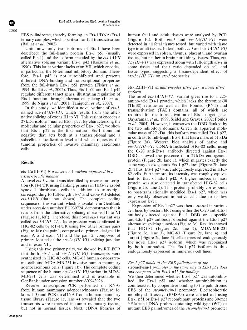
the nucleus. To determine the subcellular localization ofEts-1 p27, immunofluorescence assays were performedon HEK-293 cells exogenously expressing Ets-1 p51 orEts-1 p27 (Figure 5a). Results confirmed the nuclearlocalization of Ets-1 p51 (Figure 5a, lane 2) and showeda nuclear and cytoplasmic expression of the novel Ets-1p27 isoform (Figure 5a, lane 3). Thus, not only is Ets-1p27 present in the nucleus but its presence inthe cytoplasm suggests that Ets-1 p27 may sequestrateEts-1 p51 partners in the cytoplasm or directlydelocalize Ets-1 p51 from the nucleus to the cytoplasm.
To test this hypothesis, we performed colocalizationexperiments using Ets-1 p51 fused to a Flag-tagged andEts-1 p27 fused to the enhanced green fluorescentprotein (EGFP). Immunofluorescence experiments per-formed on HEK-293 cells expressing Flag-Ets-1 p51(Figure 5b) or EGFP-Ets-1 p27 (Figure 5c) confirmedthat tags did not alter protein subcellular localization ofeither isoform. Vector used to encode EGFP-Ets-1 p27intrinsically expressed EGFP detected in control(Figure 5c, lane 1). Colocalization experiments showedthat in the presence of Ets-1 p27, Ets-1 p51 was mainly
Figure 3 Ets-1 p27 binds cooperatively to the Ets-binding sites (EBS) palindrome of the stromelysin-1 promoter and competes withEts-1 p51 for binding. (a) Electrophoretic mobility shift assay. Ets-1 p51 (lanes 1–4) or Ets-1 p27 (lanes 5–8) (4 pmol) was incubatedwith wild-type (WT, lanes 1 and 5), mutant M1 (lanes 2 and 6), mutant M2 (lanes 3 and 7) or mutant M1M2 (lanes 4 and 8) 32P-labeledDNA probes (0.5 ng). (b) Binding specificity of Ets-1 p51 and Ets-1 p27 to the EBS palindrome. Ets-1 p51 (lanes 1–3) or Ets-1 p27(lanes 4–6) (4 pmol) was incubated with WT 32P-labeled DNA probe (0.5 ng) in the absence (lanes 1 and 4) or presence of unlabeledcompetitors (WT 800� , lanes 2 and 5 or M1M2 800� , lanes 3 and 6). For both experiments, DNA/protein complexes were separatedby electrophoresis in a 5% nondenaturing polyacrylamide gel. Free probes were detected at the bottom of the gel. Arrows indicateternary complexes (arrow 1, Ets-1 p51/DNA/Ets-1 p51 and arrow 2, Ets-1 p27/DNA/Ets-1 p27). (c) Sensorgrams illustrating real-timebinding of Ets-1 p51 and Ets-1 p27 (200 nM) to a sensor chip functionalized with 150 RU of WT, M1 and M2 oligonucleotides. Thedotted line (M1þM2) represents the sum of M1 and M2 sensorgrams. A flow cell functionalized with M1M2 oligonucleotide was usedas a reference for nonspecific binding. Data are expressed as relative responses in resonance units (RU) after subtraction of thenonspecific signal. (d) Electrophoretic mobility shift assay. Ets-1 p51 (4 pmol; lane 1), Ets1 p27 (4 pmol; lane 5) or Ets-1 p51 (4 pmol)with an increase amount of Ets-1 p27 (0–15 pmol) (lanes 2–4) was incubated with WT 32P-labeled DNA probe (0.5 ng). DNA/proteincomplexes were separated by electrophoresis in a 5% nondenaturing polyacrylamide gel. Arrows 1 and 2 are described above andarrow i corresponds to Ets-1 p51/DNA/Ets-1 p27 intermediate ternary complex.
Ets-1 p27, a dual-acting Ets-1 dominant negativeC Laitem et al
2091
Oncogene

Figure 4 Ets-1 p27 acts as a dominant negative for transactivation of the stromelysin-1 promoter mediated by autoinhibited Ets-1 p51, Ets-2 and PEA-3 through the Ets-binding sites (EBS) palindrome. (a) Effect of Ets-1 p27 on the basal or Ets-1 p51-induced stromelysin-1promoter activity. pGL3 luciferase reporter constructs (100ng; wild-type (WT), lanes 1–4 and 9–12 or M1M2, lanes 5–8 and 13–16) weretransfected into HEK-293 cells at a 50–80% confluence in the absence (�) or in the presence (þ ) of Ets-1 p51 expression vector (100ng;lanes 9–16) and with increasing amounts of Ets-1 p27 expression vector (0–100ng; lanes 2–4, 6–8, 10–12 and 14–16). (b) Effect of Ets-1 p27on the Ets-2-induced stromelysin-1 promoter activity. pGL3 luciferase reporter constructs (100ng; WT, lanes 1–5 or M1M2, lanes 6–10) weretransfected into HEK-293 cells at a 50–80% confluence with Ets-2 expression vector (100ng; lanes 2–5 and 7–10) and an increasing amountof Ets-1 p27 expression vector (0–100ng; lanes 3–5 and 8–10). (c) Effect of Ets-1 p27 on the PEA3-induced stromelysin-1 promoter activity.pGL3 luciferase reporter constructs (100ng; WT, lanes 1–5 or M1M2, lanes 6–10) were transfected into HEK-293 cells at a 50–80%confluence with PEA3 expression vector (100ng; lanes 2–5 and 7–10) and an increasing amount of Ets-1 p27 expression vector (0–100ng;lanes 3–5 and 8–10). In all experiments, luciferase activity, measured 48h after transfection, is expressed as the percentage of WT constructactivity induced by Ets-1 p51, Ets-2 or PEA3, respectively. Cell lysates (30mg total proteins) were analysed by western blot using differentantibodies (see Materials and methods). Results are the average of three experiments performed in triplicate.
Figure 5 Ets-1 p27 delocalizes Ets-1 p51 from the nucleus to the cytoplasm. (a) Native Ets-1 p51 and Ets-1 p27 subcellular localization. HEK-293cells were transfected with empty (1mg, lane 1), Ets-1 p51 (1mg, lane 2) or Ets-1 p27 (1mg, lane 3) pcDNA3 expression vectors. Cells were fixed withparaformaldehyde and treated with rabbit C-20 anti-Ets-1 antibody followed by goat anti-rabbit antibody coupled to Alexa Fluor 594. (b) Effect ofFlag-tag on Ets-1 p51 localization. HEK-293 cells were transfected with empty (1mg, lane 1) or Flag-Ets-1 p51 (1mg, lane 2) pcDNA3 expressionvectors, fixed with paraformaldehyde and treated, to detect Flag proteins, with mouse anti-Flag M2 antibody followed by goat anti-mouseantibody coupled to Alexa Fluor 594. (c) Effect of enhanced green fluorescent protein (EGFP)-tag on Ets-1 p27 localization. HEK-293 cells weretransfected with EGFP (1mg, lane 1) or EGFP-Ets-1 p27 (1mg, lane 2) pEGFP-C1 expression vectors, fixed with paraformaldehyde and naturalfluorescence of EGFP was examined. The empty pEGFP-C1 vector encodes EGFP. (d) Effect of Ets-1 p27 on Ets-1 p51 subcellular localization.HEK-293 cells were transfected with empty (500ng, lanes 1 and 2) or Flag-Ets-1 p51 (500ng, lanes 3–5) pcDNA3 expression vector, and withEGFP (500ng, lanes 1 and 3) or EGFP-Ets-1 p27 (500ng, lanes 2–5) pEGFP-C1 expression vectors. Proteins were detected as described in (b) and(c). (e) Endogenous effect of Ets-1 p27. MDA-MB-231 cells stably expressing Flag-Ets-1 p27 (lane 2) or not (lane 1) were fixed withparaformaldehyde and treated with rabbit C-20 anti-Ets-1 antibody followed by goat anti-rabbit antibody coupled to Alexa Fluor 594. In allexperiments, nuclei were visualized with Hoechst stain. Cells were examined by fluorescencemicroscopy at � 100magnification. Scale bar¼ 20mm.
Ets-1 p27, a dual-acting Ets-1 dominant negativeC Laitem et al
2092
Oncogene
detected in the cytoplasm instead of the nucleus(Figure 5d, lanes 4–5), but EGFP alone did not havethis effect on Ets-1 p51 (Figure 5d, lane 3). Thus, Ets-1p27 induces Ets-1 p51 translocation from the nucleus tothe cytoplasm. The capacity of Ets-1 p27 to delocalizeendogenous Ets-1 p51 was then tested in MDA-MB-231cells, which express the three Ets-1 isoforms. In control
MDA-MB-231 cells, Ets-1 proteins were mainly de-tected in the nucleus, but weak signals were alsoobserved in the cytoplasm (Figure 5e, lane 1). Thiscytoplasmic signal probably corresponded to endo-genous Ets-1 p27 or Ets-1 p51 delocalized by theendogenous Ets-1 p27 isoform. In MDA-MB-231 cellsstably overexpressing Ets-1 p27, Ets-1 proteins were
Ets-1 p27, a dual-acting Ets-1 dominant negativeC Laitem et al
2093
Oncogene

clearly transferred from the nucleus to the cytoplasm(Figure 5e, lane 2). Taken together, these experimentsunambiguously demonstrate that an increase in theEts-1 p27/Ets-1 p51 ratio induces the delocalization ofendogenous Ets-1 protein from the nucleus to thecytoplasm.
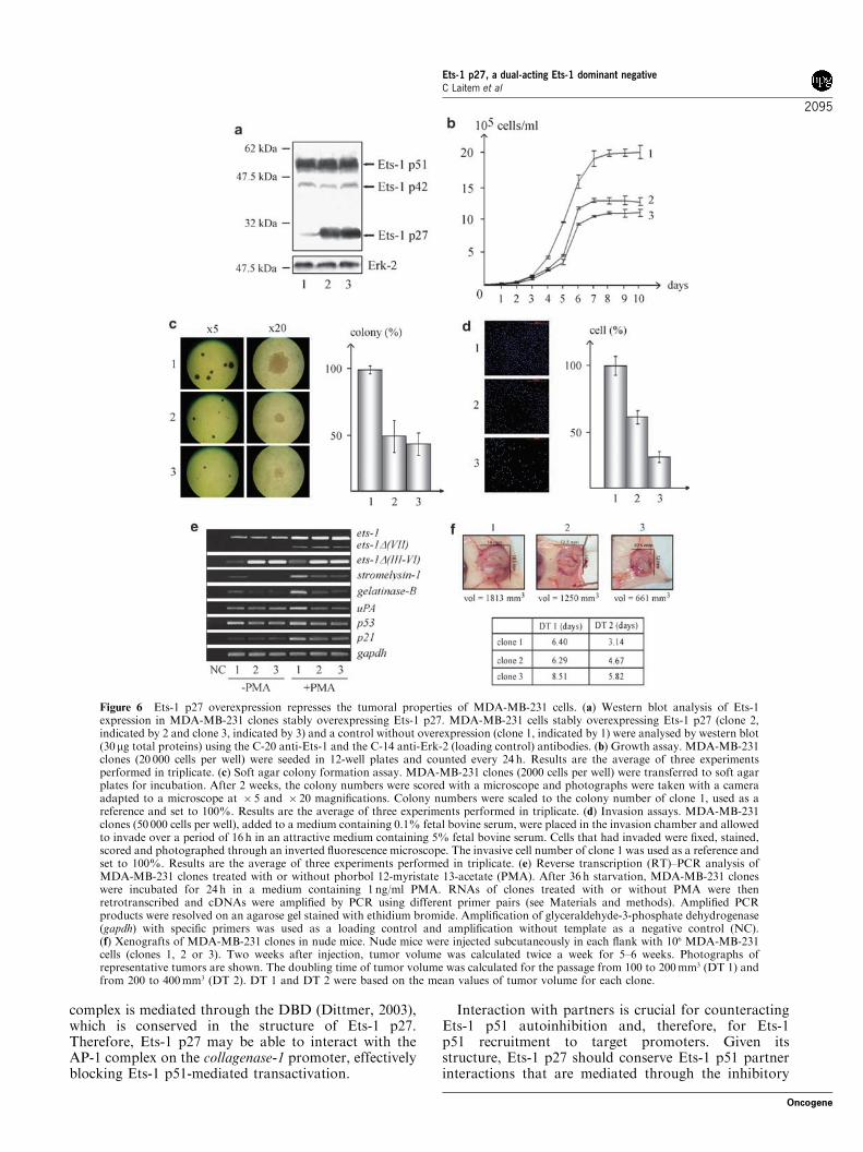
Ets-1 p27 overexpression represses the tumoral propertiesof MDA-MB-231 cellsOwing to the dual-acting dominant-negative function ofEts-1 p27 and the known implication of Ets-1 p51 invarious cellular processes, we explored the effect of theincrease of Ets-1 p27 expression on endogenous Ets-1p51 properties. As Ets-1 isoforms are expressed in cellsthat have invasive properties, Ets-1 p27 was stablyoverexpressed in invasive MDA-MB-231 mammarycarcinoma cells by retroviral infection (Figure 6a, clones2 and 3 indicated by 2 and 3, respectively) and comparedto cells that did not overexpress Ets-1 p27 (Figure 6a,clone 1 indicated by 1). Phenotypic analysis of theseclones showed that Ets-1 p27 overexpression (1) sloweddown anchorage growth of MDA-MB-231 cells andinduced the arrest of their proliferation at a lowerconfluent state (Figure 6b), (2) reduced the number andthe size of the colonies formed in soft agar (Figure 6c)and (3) repressed the cells’ ability to digest thereconstituted basement membrane and migrate throughthe porous membrane of an invasion chamber(Figure 6d). Taken together, these results demonstratethat an increase in the Ets-1 p27/Ets-1 p51 ratiorepresses the properties of proliferation, transformationand invasion of MDA-MB-231 cells. This effectappeared stronger in clone 3, where Ets-1 p27 expressionwas slightly higher, compared to clone 2 (Figure 6a),suggesting a dose-dependent effect of Ets-1 p27. As acontrol of specificity, we also stably overexpressed Ets-1p27 in noninvasive MCF-7 mammary carcinoma cells(data not shown), which do not endogenously expressEts-1 and some of its target genes such as stromelysin-1and collagenase-1. However, we could not detect anyeffect of Ets-1 p27 on the phenotype of these cells (datanot shown), suggesting that the Ets-1 p27 effectdescribed in MDA-MB-231 cells may function mainlyby counteracting endogenous Ets-1 properties.Moreover, RT–PCR analysis of the MDA-MB-231
clones treated with or without phorbol 12-myristate13-acetate (PMA), a tumor inducer that increases ets-1expression, showed that the overexpression of Ets-1 p27induced a decrease in the mRNA levels of Ets-1 targetgenes such as stromelysin-1, gelatinase-B, uPA, p53 andp21, at basal and/or PMA-induced levels (Figure 6e).Furthermore, the phenotypic results were corroboratedby those obtained in vivo (Figure 6f). Xenografts ofMDA-MB-231 clones in nude mice by subcutaneousinjection showed that the growth of the tumor formedby clones 2 and 3 was repressed compared to that ofclone 1, as illustrated by the size of the tumor and thetime it took for the tumor to double in volume. Therepression of tumor growth was higher for clone 3compared to clone 2, suggesting once again a dose-
dependent effect of Ets-1 p27 on MDA-MB-231 proper-ties. Thus, overexpression of Ets-1 p27 in MDA-MB-231 cells reduces tumor growth.
Discussion
Although several variants of ets-1 were detected in human(Jorcyk et al., 1991), only two isoforms have beendescribed up to now: Ets-1 p51, the full-length proteinand Ets-1 p42, an isoform lacking the exon-VII-encodingdomain. In this study, we identified ets-1D(III–VI), anovel ets-1 variant resulting from the alternative splicingof exons III to VI. This variant, first identified in HIG-82rabbit synovial fibroblastic cells, was detected in manyhuman tissues known for expressing Ets-1 (Rowe andPropst, 1992; Maroulakou and Bowe, 2000; Takai et al.,2006; Figure 1). The relative ratio of both ets-1 and ets-1D(III–VI) varied with cell and tissue type, suggestingtissue-specific effects of ets-1D(III–VI).This ets-1D(III–VI) variant encodes a 27 kDa
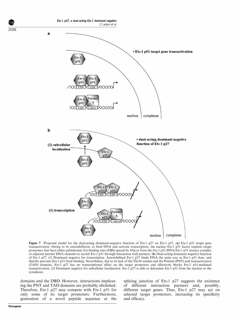
isoform, named Ets-1 p27, the structure of whichsuggested a dominant-negative function (Figure 2).Results of this study enable us to confirm thisassumption and to propose a model in which Ets-1p27 constitutes a dual-acting dominant negative for Ets-1 p51 (Figure 7). In this model, Ets-1 p27 acts both at atranscriptional level in blocking Ets-1 p51-mediatedtransactivation of target genes and at a subcellularlocalization level in inducing the translocation of Ets-1p51 from the nucleus to the cytoplasm (Figure 7b).The DNA-binding and transcriptional properties of
Ets-1 p27 were assessed on the EBS palindrome of thestromelysin-1 promoter, the regulation of which is welldocumented. The stromelysin-1 promoter is stimulatedthrough this EBS palindrome by autoinhibited Ets-1(Wasylyk et al., 1991), Ets-2 (Buttice and Kurkinen,1993) or PEA-3 (Higashino et al., 1995). Here, wedemonstrated that autoinhibited Ets-1 p27 competeswith Ets-1 p51 for binding to the EBS palindrome of thestromelysin-1 promoter (Figure 3), demonstrating itsdominant-negative effect toward Ets-1 p51 binding(Figure 7b). This competition implicates the formationof an intermediate Ets-1 p51/DNA/Ets-1 p27 ternarycomplex, suggesting that both isoforms interact witheach other on the EBS palindrome of the stromelysin-1promoter. Furthermore, Ets-1 p27 blocks, on the onehand, the transactivation of the stromelysin-1 promoterinduced not only by Ets-1 p51 but also by Ets-2 andPEA-3 (Figure 4). This effect most likely arises owingto the formation of the Ets-1 p27 ternary complex,rendering the EBS inaccessible to other Ets activatorfactors. On the other hand, Ets-1 p27 blocks thesynergistic transactivation of the collagenase-1 promoterinduced by Ets-1 p51 and c-Jun/c-Fos complex (seeSupplementary Information, Figure S1). In this context,the dominant-negative effect of Ets-1 p27 may resultfrom its ability to replace Ets-1 p51 in the interactionwith its partners (Figure 7b). This is supported by thefact that the interaction of Ets-1 p51 with the AP-1
Ets-1 p27, a dual-acting Ets-1 dominant negativeC Laitem et al
2094
Oncogene
complex is mediated through the DBD (Dittmer, 2003),which is conserved in the structure of Ets-1 p27.Therefore, Ets-1 p27 may be able to interact with theAP-1 complex on the collagenase-1 promoter, effectivelyblocking Ets-1 p51-mediated transactivation.
Interaction with partners is crucial for counteractingEts-1 p51 autoinhibition and, therefore, for Ets-1p51 recruitment to target promoters. Given itsstructure, Ets-1 p27 should conserve Ets-1 p51 partnerinteractions that are mediated through the inhibitory
Figure 6 Ets-1 p27 overexpression represses the tumoral properties of MDA-MB-231 cells. (a) Western blot analysis of Ets-1expression in MDA-MB-231 clones stably overexpressing Ets-1 p27. MDA-MB-231 cells stably overexpressing Ets-1 p27 (clone 2,indicated by 2 and clone 3, indicated by 3) and a control without overexpression (clone 1, indicated by 1) were analysed by western blot(30mg total proteins) using the C-20 anti-Ets-1 and the C-14 anti-Erk-2 (loading control) antibodies. (b) Growth assay. MDA-MB-231clones (20 000 cells per well) were seeded in 12-well plates and counted every 24 h. Results are the average of three experimentsperformed in triplicate. (c) Soft agar colony formation assay. MDA-MB-231 clones (2000 cells per well) were transferred to soft agarplates for incubation. After 2 weeks, the colony numbers were scored with a microscope and photographs were taken with a cameraadapted to a microscope at � 5 and � 20 magnifications. Colony numbers were scaled to the colony number of clone 1, used as areference and set to 100%. Results are the average of three experiments performed in triplicate. (d) Invasion assays. MDA-MB-231clones (50 000 cells per well), added to a medium containing 0.1% fetal bovine serum, were placed in the invasion chamber and allowedto invade over a period of 16 h in an attractive medium containing 5% fetal bovine serum. Cells that had invaded were fixed, stained,scored and photographed through an inverted fluorescence microscope. The invasive cell number of clone 1 was used as a reference andset to 100%. Results are the average of three experiments performed in triplicate. (e) Reverse transcription (RT)–PCR analysis ofMDA-MB-231 clones treated with or without phorbol 12-myristate 13-acetate (PMA). After 36 h starvation, MDA-MB-231 cloneswere incubated for 24 h in a medium containing 1 ng/ml PMA. RNAs of clones treated with or without PMA were thenretrotranscribed and cDNAs were amplified by PCR using different primer pairs (see Materials and methods). Amplified PCRproducts were resolved on an agarose gel stained with ethidium bromide. Amplification of glyceraldehyde-3-phosphate dehydrogenase(gapdh) with specific primers was used as a loading control and amplification without template as a negative control (NC).(f) Xenografts of MDA-MB-231 clones in nude mice. Nude mice were injected subcutaneously in each flank with 106 MDA-MB-231cells (clones 1, 2 or 3). Two weeks after injection, tumor volume was calculated twice a week for 5–6 weeks. Photographs ofrepresentative tumors are shown. The doubling time of tumor volume was calculated for the passage from 100 to 200mm3 (DT 1) andfrom 200 to 400mm3 (DT 2). DT 1 and DT 2 were based on the mean values of tumor volume for each clone.
Ets-1 p27, a dual-acting Ets-1 dominant negativeC Laitem et al
2095
Oncogene
domains and the DBD. However, interactions implicat-ing the PNT and TAD domains are probably abolished.Therefore, Ets-1 p27 may compete with Ets-1 p51 foronly some of its target promoters. Furthermore,generation of a novel peptide sequence at the
splicing junction of Ets-1 p27 suggests the existenceof different interaction partners and, possibly,different target genes. Thus, Ets-1 p27 may act onselected target promoters, increasing its specificityand efficacy.
Figure 7 Proposed model for the dual-acting dominant-negative function of Ets-1 p27 on Ets-1 p51. (a) Ets-1 p51 target genetransactivation. Owing to its autoinhibition, to bind DNA and activate transcription, the nuclear Ets-1 p51 factor requires targetpromoters that have either palindromic Ets-binding sites (EBS) spaced by 4 bp to form the Ets-1 p51/DNA/Ets-1 p51 ternary complexor adjacent partner DNA elements to recruit Ets-1 p51 through interaction with partners. (b) Dual-acting dominant-negative functionof Ets-1 p27. (1) Dominant negative for transcription. Autoinhibited Ets-1 p27 binds DNA the same way as Ets-1 p51 does, andthereby prevents Ets-1 p51 from binding. Nevertheless, due to its lack of the Thr38 residue and the Pointed (PNT) and transactivation(TAD) domains, Ets-1 p27 has no transcriptional effect on the target promoters and effectively blocks Ets-1 p51-mediatedtransactivation. (2) Dominant negative for subcellular localization. Ets-1 p27 is able to delocalize Ets-1 p51 from the nucleus to thecytoplasm.
Ets-1 p27, a dual-acting Ets-1 dominant negativeC Laitem et al
2096
Oncogene
In addition to its transcriptional dominant-negativeeffect, Ets-1 p27 is also shown to be localized (Figure 5):(1) in the nucleus, required for the mediation of itstranscriptional properties and (2) in the cytoplasm,leaving the possibilities open as to other functions. Asillustrated by our model (Figure 7b), we also demon-strated that Ets-1 p27 has the ability to delocalize Ets-1p51 from the nucleus to the cytoplasm. Translocation ofEts-1 p51 has been described during the terminaldifferentiation of erythroid progenitors (Lulli et al.,2006). When localized in the nucleus in these cells, Ets-1p51 induces the expression of the GATA-2 gene, whicheffectively blocks differentiation. Therefore, the deloca-lization of Ets-1 p51 makes this transcription factorunavailable for the regulation of GATA-2. Transloca-tion of Ets-1 p51 has also been described in a studyperformed on mouse thymocytes (Pognonec et al.,1990). The stimulation of these cells with a calciumionophore induces the phosphorylation of Ets-1 p51on the exon-VII-encoded domain, which is then redis-tributed from the nucleus to the cytoplasm. Thisphosphorylation also reinforces Ets-1 p51 autoinhibi-tion and, therefore, represses DNA binding (Cowley andGraves, 2000). As Ets-1 p27 conserves the exon-VII-encoded domain, it may also be phosphorylated, therebyrepressing DNA binding in response to calcium signal-ing. In this case, dominant-negative function of Ets-1p27 may act preferentially through subcellular localiza-tion rather than through transcriptional effects. Thus,depending on the cellular signaling pathway, Ets-1 p27may favor one or the other of its two dominant-negativefunctions.Even if Ets-1 is mainly described as a nuclear factor
(Kitange et al., 1999; Redlich et al., 2001; Li et al., 2007),other studies have shown a cytoplasmic localization, inparticular in breast (Mylona et al., 2006), ovary(Takai et al., 2002) and endometrial (Takai et al., 2000)carcinomas as well as in quiescent endothelial cells(Valter et al., 1999). In these studies, the different isoformsof Ets-1 could not be discriminated. However, we canspeculate that the cytoplasmic localization of Ets-1 maycorrespond to the detection of either delocalized Ets-1 p51protein or Ets-1 p27 isoform, whose expression may beincreased in these situations. In spite of its weakexpression, Ets-1 p27 may optimize its dominant-negativefunction, suggesting that Ets-1 p27 has a real biologicaleffect. Furthermore, Ets-1 p27 may also increase itsdominant-negative potential toward Ets-1 p51 throughthe sequestration of its partners that participate in Ets-1p51 effects.The dominant-negative function of Ets-1 p27 toward
endogenous Ets-1 p51 is confirmed in the context of theMDA-MB-231 mammary carcinoma cells, because anincrease in the Ets-1 p27/Ets-1 p51 ratio represses theirtumoral properties (Figure 6). The biological relevanceof this effect is supported by the fact that we detectedEts-1 p27 expression in vivo in breast adenocarcinomas(Figure 1c). Surprisingly, the Ets-1 p27-encoded variantis highly expressed in fetal lung but not in adult lung(Figure 1d). Therefore, it would be very interesting toassess the expression of Ets-1 p27 in lung carcinoma
explants to determine if it is synthesized in these tumors.Owing to the implication of Ets-1 in various cellularmechanisms, the existence of this dual-acting dominant-negative isoform and its repressor effect on tumor cellproperties suggests a determinant effect of the Ets-1p27/Ets-1 p51 ratio on cell fate.
Materials and methods
Isolation of rabbit and human ets-1D(III–VI) cDNAWe used the RT–PCR technique to isolate ets-1D(III–VI)cDNA from HIG-82 and MDA-MB-231 cells. A primer pairflanking the ets-1 open-reading frame (50-GCACCATGAAGGCGGCCGTGG-30 and 50-CCCCTTCCATGTCCATCACTC-30; Baillat et al., 2006) was used to amplifyets-1 cDNAs after reverse transcription. PCR productcorresponding to the shorter cDNAs was purified, cloned intopCR Blunt II-TOPO (Invitrogen, Carlsbad, CA, USA) andsequenced. Sequences are available in the GenBank databaseunder accession number EU939724 for the rabbit sequenceand AY943926 for the human sequence.
Retroviral infectionRetrovirus production and infection were performed usingempty pLPCX (clone 1) or Flag-Ets-1 p27 pLPCX (clones 2and 3) as previously described (Kherrouche et al., 2006). Afterinfection, MDA-MB-231 cells were selected with 1 mg/mlpuromycin for 96 h and routinely cultured in the presence of500 ng/ml puromycin.
Western blot analysisWestern blot analysis was performed as previously described(Baillat et al., 2006) using (1) 30mg of total cell lysate proteinsor (2) 25 ml of total cell lysates in the case of reporter geneassays, which were boiled in Laemmli buffer (50mM Tris (pH6.8), 2% sodium dodecyl sulfate, 5% b-mercaptoethanol, 10%glycerol, 0.1% bromophenol blue). Primary antibodies used(Santa Cruz Biotechnology, Santa Cruz, CA, USA) were theC-20 anti-Ets-1, C-20 anti-Ets-2, 16 anti-PEA3, C-14 anti-Erk-2,H-79 anti-cJun and H-125 anti-cFos. The specific anti-Ets-1 p27was produced (Eurogentec, Angers, France) in rabbit againstan immunogen peptide (of the sequence ELFPSPGKLGG)designed at the alternative splicing junction.
Electrophoretic mobility shift assayDouble-stranded synthetic oligonucleotides corresponding tothe WT and mutants (M1, M2 and M1M2) of the humanstromelysin-1 (�223/�193) promoter region were used toperform EMSA experiments as previously described (Laitemet al., 2008). For probe competition, 400 ng of WT or M1M2non-labeled probe (800� ) were added to the reaction mixture.For protein competition, Ets-1 p51 (4 pmol) was incubatedwith increasing quantity of recombinant Ets-1 p27 (0–15 pmol)with 0.5 ng of WT-labeled probe in the same conditions.
SPR-binding assaySurface plasmon resonance measurements were carried outusing a BIAcore 2000 apparatus (GE Healthcare, Waukesha,WI, USA), as previously described (Baillat et al., 2002).See Supplementary Information for Cell culture, RNA
extraction and RT–PCR assays, eukaryotic expression vectorconstructions, bacterial expression vector constructions andrecombinant protein production, reporter vector construc-tions, transient transfection, transfection and reporter gene
Ets-1 p27, a dual-acting Ets-1 dominant negativeC Laitem et al
2097
Oncogene
assay, immunofluorescence, growth assay, soft agar colonyformation assay, invasion assay and xenograft in nude mice.
Conflict of interest
The authors declare no conflict of interest.
Acknowledgements
We thank Isabelle Roland, Anne-Claire Flourens, NathalieTomavo and Arnaud Legrand for technical assistance, and
Dr Yvan de Launoit for stimulating discussions. We thank theMicroscopy Facility of the Institut Pasteur de Lille Campus.This work was supported by the Centre National de laRecherche Scientifique (CNRS) and by grants from la Liguecontre le Cancer-Comite du Pas-de-Calais and from theFondation pour la Recherche Medicale-Comite Nord-Pas-de-Calais (FRM). The Ministere de la Recherche et del’Enseignement Superieur provided a student fellowship toClelia Laitem. La Ligue contre le Cancer provided a studentfellowship to Gabriel Leprivier. The CNRS and the ConseilRegional du Nord-Pas-de-Calais provided a PhD fellowship(BDI) to Souhaila Choul-li.
References
Alipov G, Nakayama T, Ito M, Kawai K, Naito S, Nakashima Met al. (2005). Overexpression of Ets-1 proto-oncogene in latent andclinical prostatic carcinomas. Histopathology 46: 202–208.
Araud T, Genolet R, Jaquier-Gubler P, Curran J. (2007). Alternativelyspliced isoforms of the human elk-1 mRNA within the 50
UTR: implications for ELK-1 expression. Nucleic Acids Res 35:4649–4663.
Baillat D, Begue A, Stehelin D, Aumercier M. (2002). ETS-1transcription factor binds cooperatively to the palindromic headto head ETS-binding sites of the stromelysin-1 promoter bycounteracting autoinhibition. J Biol Chem 277: 29386–29398.
Baillat D, Laitem C, Leprivier G, Margerin C, Aumercier M. (2009).Ets-1 binds cooperatively to the palindromic Ets-binding sites in thep53 promoter. Biochem Biophys Res Commun 378: 213–217.
Baillat D, Leprivier G, Regnier D, Vintonenko N, Begue A, Stehelin Det al. (2006). Stromelysin-1 expression is activated in vivo by Ets-1through palindromic head-to-head Ets binding sites present in thepromoter. Oncogene 25: 5764–5776.
Buttice G, Kurkinen M. (1993). A polyomavirus enhancer A-bindingprotein-3 site and Ets-2 protein have a major role in the12-O-tetradecanoylphorbol-13-acetate response of the human stro-melysin gene. J Biol Chem 268: 7196–7204.
Cho JY, Akbarali Y, Zerbini LF, Gu X, Boltax J, Wang Y et al.(2004). Isoforms of the Ets transcription factor NERF/ELF-2physically interact with AML1 and mediate opposing effects onAML1-mediated transcription of the B cell-specific blk gene. J Biol
Chem 279: 19512–19522.Coutte L, Monte D, Imai K, Pouilly L, Dewitte F, Vidaud M et al.
(1999). Characterization of the human and mouse ETV1/ER81transcription factor genes: role of the two alternatively splicedisoforms in the human. Oncogene 18: 6278–6286.
Cowley DO, Graves BJ. (2000). Phosphorylation represses Ets-1 DNAbinding by reinforcing autoinhibition. Genes Dev 14: 366–376.
De Haro L, Janknecht R. (2005). Cloning of the murine ER71 gene(Etsrp71) and initial characterization of its promoter. Genomics 85:493–502.
de Nigris F, Mega T, Berger N, Barone MV, Santoro M, Viglietto Get al. (2001). Induction of ETS-1 and ETS-2 transcriptionfactors is required for thyroid cell transformation. Cancer Res 61:2267–2275.
Dittmer J. (2003). The biology of the Ets1 proto-oncogene. Mol Cancer
2: 29.Fisher RJ, Fivash M, Casas-Finet J, Erickson JW, Kondoh A, Bladen
SV et al. (1994). Real-time DNA binding measurements of the ETS1recombinant oncoproteins reveal significant kinetic differencesbetween the p42 and p51 isoforms. Protein Sci 3: 257–266.
Foulds CE, Nelson ML, Blaszczak AG, Graves BJ. (2004).Ras/mitogen-activated protein kinase signaling activates Ets-1 andEts-2 by CBP/p300 recruitment. Mol Cell Biol 24: 10954–10964.
Hewett PW, Nishi K, Daft EL, Clifford Murray J. (2001). Selectiveexpression of erg isoforms in human endothelial cells. Int J Biochem
Cell Biol 33: 347–355.
Higashino F, Yoshida K, Noumi T, Seiki M, Fujinaga K. (1995).Ets-related protein E1A-F can activate three different matrixmetalloproteinase gene promoters. Oncogene 10: 1461–1463.
Higuchi T, Bartel FO, Masuya M, Deguchi T, Henderson KW, Li Ret al. (2007). Thymomegaly, microsplenia, and defective homeo-static proliferation of peripheral lymphocytes in p51-Ets1 isoform-specific null mice. Mol Cell Biol 27: 3353–3366.
Jayaraman G, Srinivas R, Duggan C, Ferreira E, Swaminathan S,Somasundaram K et al. (1999). p300/cAMP-responsive element-binding protein interactions with ets-1 and ets-2 in the transcrip-tional activation of the human stromelysin promoter. J Biol Chem
274: 17342–17352.John SA, Clements JL, Russell LM, Garrett-Sinha LA. (2007). Ets-1
regulates plasma cell differentiation by interfering with theactivity of the transcription factor Blimp-1. J Biol Chem 283:951–962.
Jorcyk CL, Watson DK, Mavrothalassitis GJ, Papas TS. (1991). Thehuman ETS1 gene: genomic structure, promoter characterizationand alternative splicing. Oncogene 6: 523–532.
Katayama S, Nakayama T, Ito M, Naito S, Sekine I. (2005).Expression of the ets-1 proto-oncogene in human breast carcinoma:differential expression with histological grading and growth pattern.Histol Histopathol 20: 119–126.
Kherrouche Z, Blais A, Ferreira E, De Launoit Y, Monte D. (2006).ASK-1 (apoptosis signal-regulating kinase 1) is a direct E2F targetgene. Biochem J 396: 547–556.
Kita D, Takino T, Nakada M, Takahashi T, Yamashita J, Sato H.(2001). Expression of dominant-negative form of Ets-1 suppressesfibronectin-stimulated cell adhesion and migration through down-regulation of integrin alpha5 expression in U251 glioma cell line.Cancer Res 61: 7985–7991.
Kitange G, Kishikawa M, Nakayama T, Naito S, Iseki M, Shibata S.(1999). Expression of the Ets-1 proto-oncogene correlates withmalignant potential in human astrocytic tumors. Mod Pathol 12:618–626.
Koizumi S, Fisher RJ, Fujiwara S, Jorcyk C, Bhat NK, Seth A et al.(1990). Isoforms of the human ets-1 protein: generation byalternative splicing and differential phosphorylation. Oncogene 5:675–681.
Laitem C, Choul-Li S, Baillat D, Begue A, Aumercier M. (2008).Efficient system for biotinylated recombinant Ets-1 productionin Escherichia coli: a useful tool for studying interactions betweenEts-1 and its partners. Protein Expr Purif 62: 53–63.
Lee GM, Donaldson LW, Pufall MA, Kang HS, Pot I, Graves BJ et al.(2005). The structural and dynamic basis of Ets-1 DNA bindingautoinhibition. J Biol Chem 280: 7088–7099.
Lei W, Jaramillo RJ, Harrod KS. (2007). Transactivation of lunglysozyme expression by Ets family member ESE-1. Am J Physiol
Lung Cell Mol Physiol 293: L1359–L1368.Li B, Lager J, Wang D, Li D. (2007). Ets-1 participates in and
facilitates developmental expression of hypoxia-induced mitogenicfactor in mouse lung. Front Biosci 12: 2269–2278.
Ets-1 p27, a dual-acting Ets-1 dominant negativeC Laitem et al
2098
Oncogene

Li R, Pei H, Papas T. (1999). The p42 variant of ETS1 protein rescuesdefective Fas-induced apoptosis in colon carcinoma cells. Proc Natl
Acad Sci USA 96: 3876–3881.Lulli V, Romania P, Morsilli O, Gabbianelli M, Pagliuca A, Mazzeo S
et al. (2006). Overexpression of Ets-1 in human hematopoieticprogenitor cells blocks erythroid and promotes megakaryocyticdifferentiation. Cell Death Differ 13: 1064–1074.
Maroulakou IG, Bowe DB. (2000). Expression and function of Etstranscription factors in mammalian development: a regulatorynetwork. Oncogene 19: 6432–6442.
Mylona EE, Alexandrou PT, Giannopoulou IA, Rafailidis PI,Markaki S, Keramopoulos A et al. (2006). Study of the topographicdistribution of ets-1 protein expression in invasive breastcarcinomas in relation to tumor phenotype. Cancer Detect Prev
30: 111–117.Nakayama T, Ito M, Ohtsuru A, Naito S, Sekine I. (2001). Expression
of the ets-1 proto-oncogene in human colorectal carcinoma. Mod
Pathol 14: 415–422.O’Leary DA, Koleski D, Kola I, Hertzog PJ, Ristevski S. (2005).
Identification and expression analysis of alternative transcripts ofthe mouse GA-binding protein (Gabp) subunits alpha and beta1.Gene 344: 79–92.
Pognonec P, Boulukos KE, Bosselut R, Boyer C, Schmitt-VerhulstAM, Ghysdael J. (1990). Identification of a Ets1 variant proteinunaffected in its chromatin and in vitro DNA binding capacities byT cell antigen receptor triggering and intracellular calcium rises.Oncogene 5: 603–610.
Pufall MA, Graves BJ. (2002). Autoinhibitory domains: modulareffectors of cellular regulation. Annu Rev Cell Dev Biol 18: 421–462.
Raffetseder U, Wernert N, Ostendorf T, van Roeyen C, Rauen T,Behrens P et al. (2004). Mesangial cell expression of proto-oncogeneEts-1 during progression of mesangioproliferative glomerulone-phritis. Kidney Int 66: 622–632.
Ray-Gallet D, Tavitian A, Moreau-Gachelin F. (1996). An alterna-tively spliced isoform of the Spi-B transcription factor. Biochem
Biophys Res Commun 223: 257–263.Redlich K, Kiener HP, Schett G, Tohidast-Akrad M, Selzer E, Radda
I et al. (2001). Overexpression of transcription factor Ets-1 inrheumatoid arthritis synovial membrane: regulation of expressionand activation by interleukin-1 and tumor necrosis factor alpha.Arthritis Rheum 44: 266–274.
Rowe A, Propst F. (1992). Ets-1 and Ets-2 protooncogene expressionin theca cells of the adult mouse ovary. Exp Cell Res 202: 199–202.
Sarrazin S, Starck J, Gonnet C, Doubeikovski A, Melet F, Morle F.(2000). Negative and translation termination-dependent positivecontrol of FLI-1 protein synthesis by conserved overlapping 5’upstream open reading frames in Fli-1 mRNA. Mol Cell Biol 20:2959–2969.
Sasaki K, Nakamura Y, Maki K, Waga K, Nakamura F, Arai H et al.(2004). Functional analysis of a dominant-negative DeltaETS TEL/ETV6 isoform. Biochem Biophys Res Commun 317: 1128–1137.
Seidel JJ, Graves BJ. (2002). An ERK2 docking site in the Pointeddomain distinguishes a subset of ETS transcription factors. Genes
Dev 16: 127–137.Shin C, Manley JL. (2004). Cell signalling and the control of pre-
mRNA splicing. Nat Rev Mol Cell Biol 5: 727–738.Smith CW, Valcarcel J. (2000). Alternative pre-mRNA splicing: the
logic of combinatorial control. Trends Biochem Sci 25: 381–388.Stamm S, Ben-Ari S, Rafalska I, Tang Y, Zhang Z, Toiber D et al.
(2005). Function of alternative splicing. Gene 344: 1–20.Takai N, Miyazaki T, Fujisawa K, Nasu K, Miyakawa I. (2000).
Expression of c-Ets1 is associated with malignant potential inendometrial carcinoma. Cancer 89: 2059–2067.
Takai N, Miyazaki T, Nishida M, Nasu K, Miyakawa I. (2002). c-Ets1is a promising marker in epithelial ovarian cancer. Int J Mol Med 9:287–292.
Takai N, Ueda T, Narahara H, Miyakawa I. (2006). Expression ofc-Ets1 protein in normal human placenta. Gynecol Obstet Invest 61:15–20.
Taniguchi H, Fujiwara Y, Doki Y, Sugita Y, Sohma I, Miyata H et al.(2007). Gene therapy using ets-1 transcription factor decoyfor peritoneal dissemination of gastric cancer. Int J Cancer 121:1609–1617.
Teruyama K, Abe M, Nakano T, Iwasaka-Yagi C, Takahashi S,Yamada S et al. (2001). Role of transcription factor Ets-1 in theapoptosis of human vascular endothelial cells. J Cell Physiol 188:243–252.
Valter MM, Hugel A, Huang HJ, Cavenee WK, Wiestler OD, PietschT et al. (1999). Expression of the Ets-1 transcription factor inhuman astrocytomas is associated with Fms-like tyrosine kinase-1(Flt-1)/vascular endothelial growth factor receptor-1 synthesis andneoangiogenesis. Cancer Res 59: 5608–5614.
Wasylyk C, Gutman A, Nicholson R, Wasylyk B. (1991). The c-Etsoncoprotein activates the stromelysin promoter through thesame elements as several non-nuclear oncoproteins. EMBO J 10:1127–1134.
Supplementary Information accompanies the paper on the Oncogene website (http://www.nature.com/onc)
Ets-1 p27, a dual-acting Ets-1 dominant negativeC Laitem et al
2099
Oncogene
Copyright © 2022 FDOKUMEN