S-Phase Kinase-Associated Protein 2 Expression in Non-Hodgkin's Lymphoma Inversely Correlates with...
10
S-Phase Kinase-Associated Protein 2 Expression in Non-Hodgkin’s Lymphoma Inversely Correlates with p27 Expression and Defines Cells in S Phase Roberto Chiarle,* Yan Fan,* Roberto Piva,* Hugo Boggino,* Jeffrey Skolnik,* Domenico Novero, † Giorgio Palestro, † Chris De Wolf-Peeters, ‡ Marco Chilosi, § Michele Pagano,* and Giorgio Inghirami* From the Department of Pathology and Kaplan Comprehensive Cancer Center,* New York University School of Medicine, New York, New York; the Department of Pathology, † University of Torino, Torino, Italy; the Department of Pathology and Experimental Medicine Research Center, § University of Verona, Verona, Italy; and the Division of Morphology and Molecular Pathology, ‡ Catholic University Leuven, Leuven, Belgium The protein expression of the cyclin-dependent ki- nase inhibitor p27 is often deregulated in human tu- mors. In lymphomas the inactivation of p27 is achieved through either increased degradation 1 or sequestration via D cyclins, 2 and p27 protein levels have been shown to have a prognostic significance. 1,3 Recently, S-phase kinase-associated protein 2 (Skp2) has been proved to mediate p27 degradation in nor- mal cells 4–7 and to have oncogenetic properties. 8,9 In this study, B-, T-, and myeloid hematopoietic cell lines and a well-characterized panel of human lym- phomas (n 244) were studied for the expression of Skp2. In human lymphomas, the expression of Skp2 strongly related to the grade of malignancy , being low in indolent tumors and very high in aggressive lym- phomas. Moreover, the percentages of Skp2- and S- phase-positive cells, as measured by DNA content or BrdU labeling, strictly matched and closely parallel that of Ki-67 and cyclin A. An inverse correlation between Skp2 and p27 was found in the majority of lymphoma subtypes. Nonetheless, most mantle cell lymphomas and a subset of diffuse large cell lympho- mas failed to show this correlation, suggesting that alternative pathway(s) for the regulation of p27 might exist. The detection of Skp2 protein either by flow cytometry or by immunohistochemistry represents a simple method to precisely assess the S phase of lym- phomas. The potential diagnostic and prognostic value of Skp2 is discussed. (Am J Pathol 2002, 160:1457–1466) The S-phase kinase-associated protein 2 (Skp2) belongs to a family of proteins called F-box proteins (Fbps) char- acterized by an 40-amino acid long motif called F-box. Skp1, Cul1, and Roc1/Rbx1 associate with different Fbps to form E3 ubiquitin ligase complexes known as SCF. The Fbp subunits of the SCF complex ensure the specific recognition and ubiquitination of a large number of sub- strates that are in turn degraded by the proteasome. 10 Skp2 was first identified to interact with the cyclin A-Cdk2 complex in immortalized fibroblasts and transformed cells and required for G 1 to S transition. 11 In vitro and in vivo studies have demonstrated that the SCF complexes con- taining Skp2, as Fbp (SCF Skp2 ), bind to p27, which is spe- cifically ubiquitinated by the ubiquitin-conjugating enzyme Ubc3. 4–7 Skp2-deficient mice have smaller organs than littermate controls, their cells grow more slowly, and they display centrosome overduplication together with the accu- mulation of p27 and free cyclin E. 7 Importantly, in Skp2 transgenic mice, the forced expression of Skp2 in T cells cooperates with the N-ras oncogene leading to lym- phomagenesis. 8 Finally, the tumorigenic role of Skp2 has been suggested by several investigators. 9,12 The deregulated expression of p27 plays a critical role in the pathogenesis of many human tumors. 13 The protein levels of p27 in normal cells are mainly regulated by an ubiquitin-mediated degradation. 14 Moreover, an enhanced protein degradation via the ubiquitin-proteasome pathway is also responsible for the low levels of p27 protein in ag- gressive human tumors 15–17 and in mantle cell lymphomas (MCLs). 1 We and others have demonstrated that the protein levels of p27 expression are different in specific subsets of human lymphomas, being high in low-grade and low in high-grade lymphomas. 1,3,18 More importantly, the partial or total loss of p27 function defines a group of patients that exhibit low overall survival and poor outcome. 1,3 Supported by National Institutes of Health grants ROI-CA90773 (to G.I.), ROI-CA76584, and ROI-GM57587 (to M.P.). R. C. and Y. F. both contributed equally to this work. Accepted for publication January 18, 2002. R. C. is on leave from the University of Torino, Department of Pathology, Via Santena, 10126, Torino, Italy. Present address of J. S.: Department of Pediatrics, Boston Children’s Hospital, 300 Longwood Avenue, Boston, MA 02115. Address reprint requests to Dr. Giorgio Inghirami, New York University, Department of Pathology and Kaplan Comprehensive Cancer Center, 550 First Ave., New York, NY 10016. E-mail: [email protected]. American Journal of Pathology, Vol. 160, No. 4, April 2002 Copyright © American Society for Investigative Pathology 1457
-
Upload
independent -
Category
Documents
-
view
1 -
download
0
Transcript of S-Phase Kinase-Associated Protein 2 Expression in Non-Hodgkin's Lymphoma Inversely Correlates with...
S-Phase Kinase-Associated Protein 2 Expression inNon-Hodgkin’s Lymphoma Inversely Correlates withp27 Expression and Defines Cells in S Phase
Roberto Chiarle,* Yan Fan,* Roberto Piva,*Hugo Boggino,* Jeffrey Skolnik,*Domenico Novero,† Giorgio Palestro,†
Chris De Wolf-Peeters,‡ Marco Chilosi,§
Michele Pagano,* and Giorgio Inghirami*From the Department of Pathology and Kaplan Comprehensive
Cancer Center,* New York University School of Medicine, New
York, New York; the Department of Pathology,† University of
Torino, Torino, Italy; the Department of Pathology and
Experimental Medicine Research Center,§ University of Verona,
Verona, Italy; and the Division of Morphology and Molecular
Pathology,‡ Catholic University Leuven, Leuven, Belgium
The protein expression of the cyclin-dependent ki-nase inhibitor p27 is often deregulated in human tu-mors. In lymphomas the inactivation of p27 isachieved through either increased degradation1 orsequestration via D cyclins,2 and p27 protein levelshave been shown to have a prognostic significance.1,3
Recently, S-phase kinase-associated protein 2 (Skp2)has been proved to mediate p27 degradation in nor-mal cells4–7 and to have oncogenetic properties.8,9 Inthis study, B-, T-, and myeloid hematopoietic celllines and a well-characterized panel of human lym-phomas (n � 244) were studied for the expression ofSkp2. In human lymphomas, the expression of Skp2strongly related to the grade of malignancy, being lowin indolent tumors and very high in aggressive lym-phomas. Moreover, the percentages of Skp2- and S-phase-positive cells, as measured by DNA content orBrdU labeling, strictly matched and closely parallelthat of Ki-67 and cyclin A. An inverse correlationbetween Skp2 and p27 was found in the majority oflymphoma subtypes. Nonetheless, most mantle celllymphomas and a subset of diffuse large cell lympho-mas failed to show this correlation, suggesting thatalternative pathway(s) for the regulation of p27 mightexist. The detection of Skp2 protein either by flowcytometry or by immunohistochemistry represents asimple method to precisely assess the S phase of lym-phomas. The potential diagnostic and prognosticvalue of Skp2 is discussed. (Am J Pathol 2002,160:1457–1466)
The S-phase kinase-associated protein 2 (Skp2) belongsto a family of proteins called F-box proteins (Fbps) char-acterized by an �40-amino acid long motif called F-box.Skp1, Cul1, and Roc1/Rbx1 associate with different Fbpsto form E3 ubiquitin ligase complexes known as SCF. TheFbp subunits of the SCF complex ensure the specificrecognition and ubiquitination of a large number of sub-strates that are in turn degraded by the proteasome.10
Skp2 was first identified to interact with the cyclin A-Cdk2complex in immortalized fibroblasts and transformed cellsand required for G1 to S transition.11 In vitro and in vivostudies have demonstrated that the SCF complexes con-taining Skp2, as Fbp (SCFSkp2), bind to p27, which is spe-cifically ubiquitinated by the ubiquitin-conjugating enzymeUbc3.4–7 Skp2-deficient mice have smaller organs thanlittermate controls, their cells grow more slowly, and theydisplay centrosome overduplication together with the accu-mulation of p27 and free cyclin E.7 Importantly, in Skp2transgenic mice, the forced expression of Skp2 in T cellscooperates with the N-ras oncogene leading to lym-phomagenesis.8 Finally, the tumorigenic role of Skp2 hasbeen suggested by several investigators.9,12
The deregulated expression of p27 plays a critical role inthe pathogenesis of many human tumors.13 The proteinlevels of p27 in normal cells are mainly regulated by anubiquitin-mediated degradation.14 Moreover, an enhancedprotein degradation via the ubiquitin-proteasome pathwayis also responsible for the low levels of p27 protein in ag-gressive human tumors15–17 and in mantle cell lymphomas(MCLs).1 We and others have demonstrated that the proteinlevels of p27 expression are different in specific subsets ofhuman lymphomas, being high in low-grade and low inhigh-grade lymphomas.1,3,18 More importantly, the partial ortotal loss of p27 function defines a group of patients thatexhibit low overall survival and poor outcome.1,3
Supported by National Institutes of Health grants ROI-CA90773 (to G.I.),ROI-CA76584, and ROI-GM57587 (to M.P.).
R. C. and Y. F. both contributed equally to this work.
Accepted for publication January 18, 2002.
R. C. is on leave from the University of Torino, Department of Pathology,Via Santena, 10126, Torino, Italy.
Present address of J. S.: Department of Pediatrics, Boston Children’sHospital, 300 Longwood Avenue, Boston, MA 02115.
Address reprint requests to Dr. Giorgio Inghirami, New York University,Department of Pathology and Kaplan Comprehensive Cancer Center, 550First Ave., New York, NY 10016. E-mail: [email protected].
American Journal of Pathology, Vol. 160, No. 4, April 2002
Copyright © American Society for Investigative Pathology
1457
In this study we have analyzed the expression of Skp2 incell lines and human lymphomas and compared it to otherproliferation indexes and to the expression of its substratep27. Our findings demonstrate that Skp2 protein levelstightly correlate with the late G1 and S phase in neoplasticcell lines and tumors. We have found that there is a directcorrelation among Skp2 and Ki-67 and cyclin A and aninverse relationship between Skp2 and p27 protein levels inthe majority of human lymphomas. However, in a subset oflymphomas this correlation is lost, suggesting that alterna-tive pathways of p27 inactivation might exist.
Materials and Methods
Pathological Samples
A panel of 244 well-characterized non-Hodgkin’s lympho-mas (NHLs) was retrieved from the archives of the Divi-sion of Hematopathology of the New York UniversitySchool of Medicine, from the Division of Morphology andMolecular Pathology of the Catholic University of Leuvenin Belgium, and the Surgical Pathology Departments ofthe Universities of Torino and Verona in Italy. NHLs wereclassified according to the International LymphomaStudy Group, based on hematoxylin and eosin and im-munoperoxidase stains, and clinical and molecular data,as previously described.1 The NHLs characterized in thisstudy included: 28 chronic lymphocytic leukemia/smalllymphocytic lymphomas (CLL/SLLs), 51 follicular lym-phomas (FL), 34 mucosa-associated lymphoid tissue B-cell lymphomas, 34 MCLs, and 76 diffuse large B-celllymphomas (DLCLs), 15 lymphoblastic lymphomas (6B-lymphoblastic lymphoma and 9 T-lymphoblastic lym-phomas), 25 Burkitt’s lymphomas, and 5 Burkitt-like lym-phomas. FL were classified based on the criteria of therevised World Health Organization classification19 and ofMann and Berard.20 A single case of plasma cell leuke-mia (�90% plasma cells) was also included for the flowcytometry staining. The following cell lines were alsoused: Namalwa, Jurkat, Karpas 299, DHL, JB, KM512,and K562.
Antibodies
The monoclonal antibodies (mAbs) applied for the immu-nohistochemistry in this study included: anti-p27 (KIP-1,1:1000; Transduction Laboratories, Lexington, KY, USA),anti-p53 (DO-1, 1:200; Santa Cruz Biotechnology, SantaCruz, CA, USA), anti-Ki-67 (MIB-1, 1:1000; Immunotech,Marseille, France), anti-cyclin E (1:50; Novocastra, CA,USA) and cyclin A (1:100, Santa Cruz Biotechnology).Mouse mAbs to Skp2 were produced in collaborationwith Zymed Inc., South San Francisco, CA, USA. Immu-nohistochemical staining and Western blotting for Skp2were performed using a cocktail of four different affinity-purified mAbs (clones 2C8D9, 2C2B12, 4A9B2, and2G12E9) or a single mAb (clone 4A9B2),8 as indicated.
Western Blot Analysis and in Vitro ProteinDegradation Assay
For Western Blotting, cells were lysed (50 mmol/L Tris-HCl, pH 7.4, 150 mmol/L NaCl, 0.1% Triton X-100, 5mmol/L ethylenediaminetetraacetic acid, 1 mmol/LNa3VO4, and 1 mmol/L phenylmethyl sulfonyl fluoride andprotease inhibitors) and 20 to 30 �g of proteins wereelectrophoresed in sodium dodecyl sulfate-polyacrylam-ide gel electrophoresis gel and transferred onto nitrocel-lulose membranes. The membranes were first blockedand then incubated with the primary antibody (anti-Skp2,clone 4A9B2, 1:500; anti-p27, KIP-1, 1:2000; and anti-cyclin A, 1:1000) for 1 hour at room temperature. Afterthree washes, filters were incubated with horseradishperoxidase-conjugated goat anti-mouse or anti-rabbit an-tibodies (1:2000; Amersham, Arlington Heights, IL, USA)for 1 hour at room temperature. Detection of immunocom-plexes was performed with an enhanced chemilumines-cence system (ECL, Amersham).1
In vitro degradation assays were performed on cryopre-served tissue samples.1 Frozen tissues of MCLs (express-ing low, intermediate, and high protein levels of p27), with�80% of tumor cells were sectioned and quickly disruptedby nitrogen decompression in 100 �l of lysing buffer (50mmol/L Tris-HCl, pH 8.3, 5 mmol/L MgCl2, and 1 mmol/Ldithiothreitol). Samples were then frozen and thawed threeconsecutive times. Lysates were spun down at 15,000 rpmand supernatants were collected and frozen at �80°C.Histidine-tagged p27 (100 �g) was incubated at 37°C fordifferent intervals in 60 �l of degradation mix containing 30�g of protein tissue homogenates, 50 mmol/L Tris-HCL (pH8.0), 5 mmol/L MgCl2, 1 mmol/L dithiothreitol, 2 mmol/L ATP,10 mmol/L creatine phosphokinase, and 10 mmol/L creat-ine phosphatase. The in vitro degradation of p27 was ana-lyzed by immunoblotting with anti-p27 mAb.
Immunohistochemical Staining
For immunohistochemistry, slides were subjected to micro-waving for 20 minutes in 10 mmol/L of citrate buffer (pH 8.0for Skp2, cyclin E, and cyclin A; pH 6.0 for p27, p53, andKi67). Immunostaining were performed on formalin-fixed,paraffin-embedded tissues using the avidin biotin peroxi-dase complex method and a semiautomated immuno-stainer (DAKO, Carpinteria, CA, USA or Ventana Systems,Tucson, AZ, USA) as described.1 Three independent pa-thologists (RC, YF, and GI) evaluated the immunostaining.At least 10 high-power fields were randomly chosen and atleast 100 cells/field were counted. Tumors were scored aspercentage of positive cells for each antigen. The statisticalsignificance of the means was calculated using a Student’st-test. The statistical significance of Spk2 versus cyclin Aexpression was calculated with the chi-square test. P �0.05 was required for statistical significance.
Flow Cytometry and BrdU Labeling
For flow cytometric staining, untreated cell lines or lovas-tatin-treated (24 hours, 50 �mol/L; Sigma Chemical Co.,
1458 Chiarle et alAJP April 2002, Vol. 160, No. 4
St. Louis, MO, USA) cells were fixed and permeabilizedaccording to the manufacturer’s instruction (Fix andPerm; Caltag, Burlingame, CA). Cells were subsequentlystained using a cocktail of anti-Skp2 mAbs (1:100,Zymed). Anti-Skp2 mAbs were detected using phyco-erythrin-conjugated horse anti-mouse antibody (1:200;Biosource, Camarillo, CA, USA). For DNA content deter-mination, cells were fixed for 1 hour in 70% ethanol at4°C. After washing, cells were treated with RNase (0.25mg/ml) and stained with propidium iodide (50 �g/ml). TheS-phase fraction was calculated using the Modfit pro-gram from Becton-Dickinson, Mountain View, CA.
For BrdU labeling, 1 � 106 cells or 1-mm-thick freshtissues (three normal tonsils and seven randomly se-lected NHLs) were incubated in complete cell-culturemedia (RPMI-1640 with 10% fetal calf serum) in the pres-ence of 10 �mol/L of BrdU for 30 minutes (suspensioncells) or for 2 hours (tissue samples) at 37°C in 5% CO2.For BrdU determination using flow cytometry, cells weresubsequently washed, fixed in 70% alcohol for 30 min-utes, denatured with 2 N HCl, and then incubated with ananti-BrdU fluorescein isothiocyanate-labeled mAb (1:10,Becton-Dickinson) and propidium iodide (50 �g/ml). ForBrdU determination using immunohistochemistry, tissuesamples were rinsed in phosphate-buffered saline andfixed overnight in 10% buffered formalin. The incorpora-tion of BrdU in proliferating cells was then detected byenzymatic digestion (0.05% proteinase K for 20 minutesat 37°C), denaturation (2 N HCl for 30 minutes at 37°C),and incubation with an anti-BrdU fluorescein isothiocya-nate-conjugated antibody (30 minutes at room tempera-ture, Becton-Dickinson). Bound anti-BrdU fluorescein iso-thiocyanate-conjugated antibodies were detected usingan alkaline-conjugated anti-fluorescein isothiocyanateantibody (1:200; Roche, Indianapolis, IN) and NBT/BCIPas a substrate.
Results
Skp2 Expression in Human Lymphoid Tissues
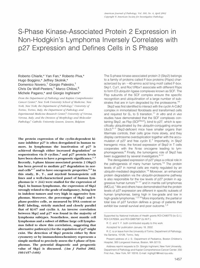
The specificity of our anti-Skp2 antibodies was first eval-uated in cell lines derived from human lymphomas byWestern blotting. Skp2 mAbs detected only a single bandmigrating slightly faster than a tagged Skp2 recombinantprotein (Figure 1A). We subsequently evaluated by im-munohistochemistry Skp2 protein expression in normalprimary and secondary lymphoid tissues. In primary lym-phoid organs, Skp2 staining was restricted to the nucleiof a subset of cortical thymocytes as well as of precursormyeloid and erythroid cells in the bone marrow (Figure 1,B and C). In peripheral lymphoid organs, centroblasts orlarge centrocytes, within the dark zone of the germinalcenters, were preferentially positive (Figure 1, D and E).In contrast, small resting cells within the mantle of theB-cell follicle and interfollicular T lymphocytes were neg-ative. Nonetheless, in the interfollicular areas some of theintermediate large cells were also Skp2-positive. In theoral mucosa as well as in the epidermis, Skp2-positive
cells were confined, similarly to the Ki-67-positive cells,within the basal layer of the epithelium (data not shown).
Skp2 Is Expressed in S Phase in NeoplasticLymphoid Cells
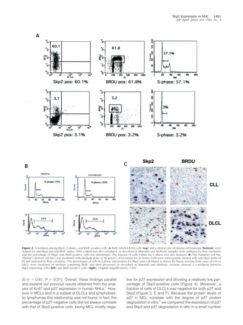
Since in normal cells the levels of Skp2 vary during thecell cycle, with a peak of expression at G1 to S through Sphase,11 we investigated the kinetics of Skp2 duringcell-cycle progression in neoplastic lymphoid cells toseek for a possible aberrant pattern and/or deregulation.First, we compared Skp2 expression to the fraction ofcells in S phase calculated by DNA content and BrdUincorporation in multiple and T-lymphoblastoid cell linesand in a case of plasma cell leukemia. In all of the celllines the percentage of Skp2-positive cells always corre-sponded with the percentage of cells in the S phase(Table 1, Figure 2A). Moreover, flow cytometric analysisdemonstrated two distinct subpopulations of Skp2-posi-tive cells, with intermediate and high intensity of expres-sion, suggesting that different levels of expression ofSkp2 are achieved in different phases of the cell cycle(Figure 2B). To further confirm the relation between Skp2and S phase, we blocked the progression into the Sphase of multiple lymphoid cell lines by inducing an earlyG1 cell-cycle arrest. In cells arrested by lovastatin in G1
phase, both the percentage of cells in S phase and thoseof Skp2-positive cells decreased in a comparable man-
Figure 1. Skp2 expression in lymphoid cell lines and normal tissues. A:Western blot analysis on human lymphoid cell lines. Cells from the indicatedcell lines were lysed and Western blotting was performed as described inMaterial and Methods. Lane 1, Namalwa cell line; lane 2, DHL cell line; lane3, recombinant His-tagged Skp2 protein. B–E: Formalin-fixed, paraffin-em-bedded tissue sections were stained by immunohistochemistry with an anti-Skp2 mAb, as described in Materials and Methods. In the thymus (B) and inthe bone marrow (C), Skp2 stained primarily the immature subcorticalthymocytes and immature hematopoietic cells, respectively. In lymph nodesand tonsils, Skp2 labeled active proliferating cells within germinal centersand scattered immunoblasts in interfollicular areas (D and E).
Skp2 Expression in NHL 1459AJP April 2002, Vol. 160, No. 4
ner (Figure 2B). These results were confirmed in all lym-phoma-derived cell lines, demonstrating that Skp2 ex-pression in neoplastic lymphoid cells as well in normalnonlymphoid cells,11 peaks in late G1 to S phase and ismaintained throughout the S phase. Next, we correlatedthe percentage and distribution of Skp2-positive cellswith BrdU incorporation in fresh lymphoma samples. Thisapproach showed a close correspondence betweenSkp2- and BrdU-positive cells (Figure 2C), confirmingthat the majority of Skp2-positive cells are actively syn-thesizing the DNA in normal as well in neoplastic lym-phoma cells.
Skp2 Expression in Human Lymphomas
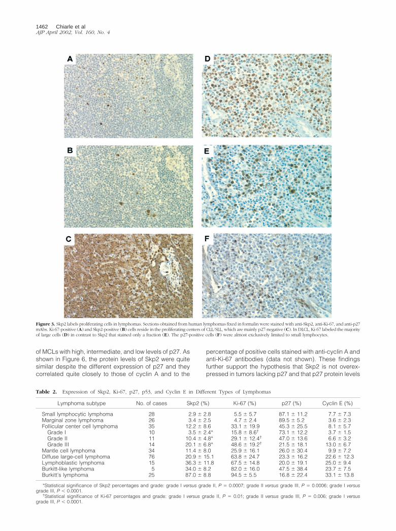
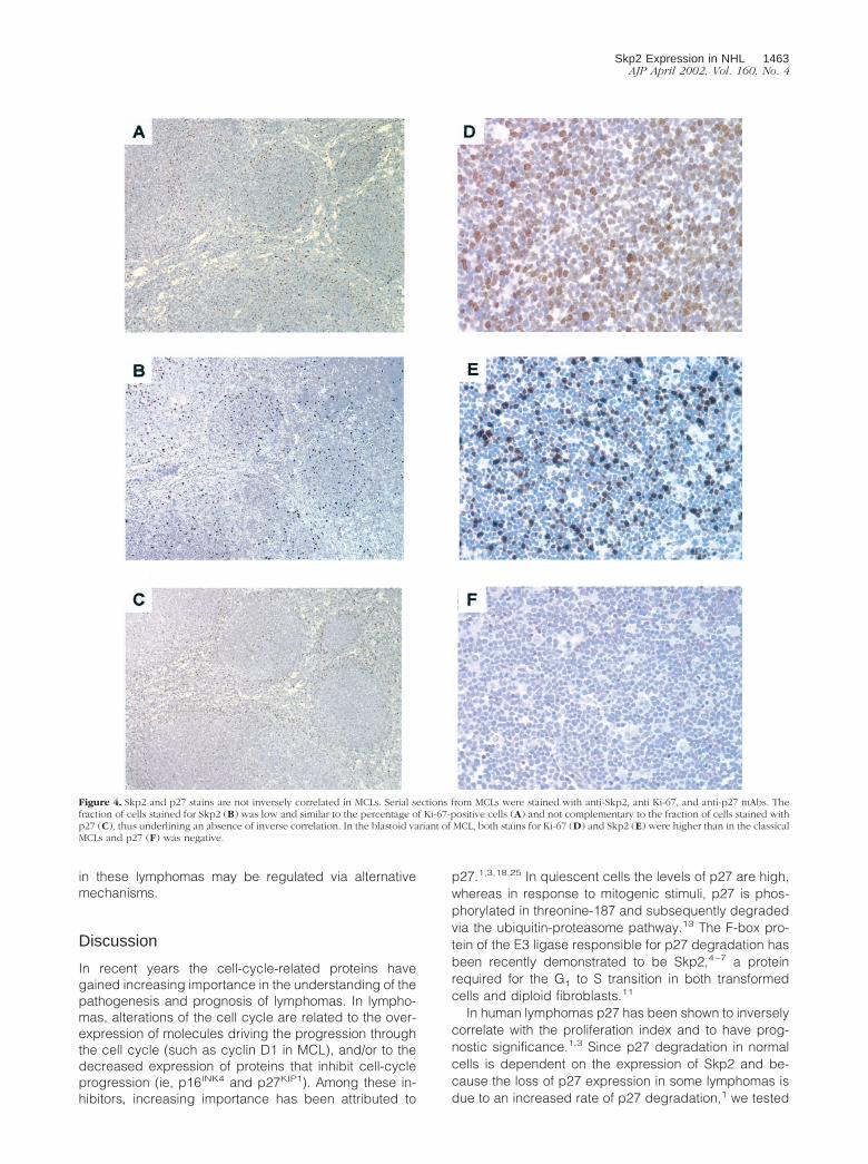
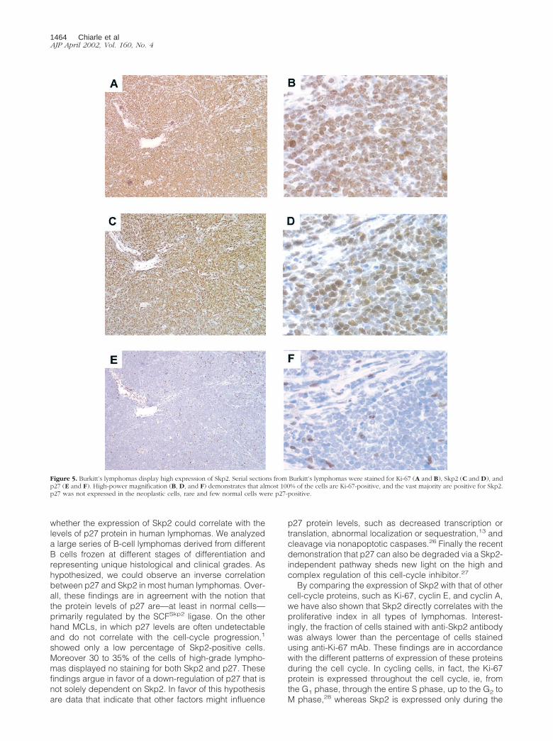
The expression of Skp2 was subsequently evaluated inB-cell NHLs representing all grades of malignancy. InCLLs and Skp2-positive cells were primarily localizedwithin the proliferation centers primarily restricted topara-immunoblasts. (Figure 3B). In mucosa-associatedlymphoid tissue B-cell lymphomas only 5% of the neo-plastic cells were found to be positive for Skp2. Skp2expression in FL was restricted to a fraction of centro-blasts and large centrocytes, but a variable number ofinterfollicular cells were also found positive. More impor-tantly, the percentage of Skp2-positive cells significantlycorrelated with the histological grade of these lymphoma(mean percentage: FL grade I, 3.5%; grade II, 10.4%,and grade III, 20.1%). Notably the sole expression ofSkp2 statistically correlated with the FL grading obtainedusing conventional cytological criteria20 (grade I versusgrade II, P � 0.0007; grade II versus grade III, P �0.0006; grade I versus grade III, P � 0.0001) (Table 2). Apositive correlation was also found when the percentagesof Ki-67-positive cells were correlated to the same groupsof FL. In MCLs, Skp2-positive tumor cells were uniformlydistributed (Figure 4, B and E), with clusters of Skp2-positive cells corresponding to nonneoplastic cells withinresidual normal germinal centers. As expected, the blas-toid and large cell variants of MCL21 showed the highestpercentages of Skp2-positive cells (Figure 4E). Interest-ingly, among high-grade lymphomas, a very high per-centage of Skp2-positive cells was found in Burkitt’s lym-phomas (87 � 8.8%) (Figure 5). By contrast, in DLCL andBurkitt-like and lymphoblastic lymphomas the percent-age of Skp2-positive cells ranged from 10 to 65%. These
values were statistically different and we were able toseparate these tumors from Burkitt’s lymphomas (P �0.0001). Finally, when all types of lymphomas were com-pared, the percentage of Skp2-positive cells strictly cor-related with the distinct groups of NHLs being low inlow-grade lymphomas and higher in high-grade tumors(P � 0.001) (Table 2).
Correlation of Skp2 Expression with OtherCell-Cycle Markers
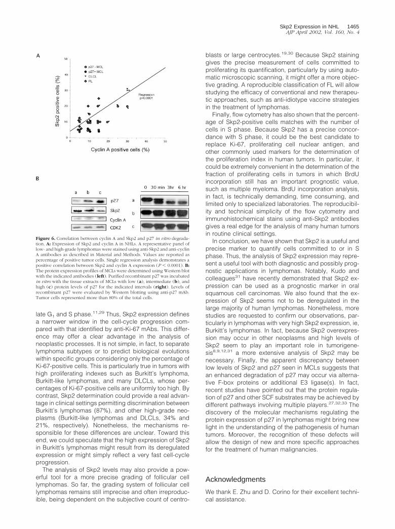
We next compared the expression of Skp2 and Ki-67, themost commonly used proliferation marker, with those ofp27, cyclin E, and cyclin A. Skp2 expression showed astatistical correlation with Ki-67 and cyclin E both in low-grade lymphomas, such as CLLs (r � 0.47, P � 0.01),and in high-grade lymphomas such as DLCL (r � 0.63and r � 0.48, respectively; P � 0.0001). Nevertheless,the percentage of Skp2-positive cells always corre-sponded to a subset of Ki-67-positive cells. As previouslydescribed, in high-grade lymphomas (lymphoblastic lym-phoma, DLCL, Burkitt’s and Burkitt-like lymphomas),Ki-67 was expressed by the very large majority of tumorcells (mean percentage �64%), whereas the percentageof Skp2-positive cells varied among these categories(Table 2). Interestingly, in lymphoblastic lymphomas,DLCL, and Burkitt-like lymphomas anti-Skp2 mAb identi-fied only a fraction (mean percentage �36% of the totaltumors cells) of the Ki-67-positive cells. Conversely, thepercentages of Skp2- and Ki-67-positive cells were verysimilar in Burkitt’s lymphomas (87% versus 92.6%, re-spectively). As shown above, the expression of Skp2 inlymphoid cell lines highly correlates with the percentageof cells within the S phase and BrdU-positive cells. Tocorroborate this finding, we further investigated the rela-tionship between Skp2 expression and the expression ofcyclin A. A total of 38 cases, including FL (n � 10), MCL(n � 18), and DLCL (n � 10) lymphomas, were studied.We decided to study the expression of cyclin A becauseits expression is largely limited to S to G2 phases22,23 andit correlates with the percentages of S to G2 and BrdU innormal and transformed cells.24 To identify whether thep27, and cyclin A and Skp2 expression were correlated,we selected p27-negative (n � 14) and p27-positive (n �4) MCL lymphomas. Using this approach, we demon-strated that Skp2 and cyclin A expression directly corre-lated in all tumors, including p27-negative lymphomas(Figure 6A). Overall, these findings further support thehypothesis that in human lymphomas the percentage ofSkp2-positive cells is closely correlated to the fraction ofproliferating cells.
We and others have recently demonstrated that p27protein expression not only inversely correlates with thegrade of lymphomas, but it also has a prognostic signif-icance.1,3,18 Because in normal cells p27 protein levelsare tightly regulated during the cell cycle via Skp2, weinvestigated whether the protein levels of p27 also in-versely correlated with Skp2 in human lymphomas. Asanticipated, in a very large majority of lymphomas therewas an inverse correlation between p27 and Skp2 (Table
Table 1. Comparison between Skp2-Positive Cells and theFraction of Cells in S Phase
Cell line Skp2* DNA† BrdU*
Karpas 51 51 52Jurkat 39 38 NDNamalwa 49 42 46DHL 55 47 51JB 46 36 45KM512 60 50 56K562 60 62 57Plasma cell leukemia 3 3 3
*, Percent of positive cells.†, Percent of S phase.Representative data of at least two experiments.
1460 Chiarle et alAJP April 2002, Vol. 160, No. 4
2) (r � 0.81, P � 0.01). Overall, these findings paralleland expand our previous results obtained from the anal-ysis of Ki-67 and p27 expression in human NHLs.1 How-ever in MCLs and in a subset of DLCLs and lymphoblas-tic lymphomas this relationship was not found. In fact, thepercentage of p27-negative cells did not always correlatewith that of Skp2-positive cells, being MCL mostly nega-
tive for p27 expression and showing a relatively low per-centage of Skp2-positive cells (Figure 4). Moreover, afraction of cells of DLCLs was negative for both p27 andSkp2 (Figure 3, E and F). Because the protein levels ofp27 in MCL correlate with the degree of p27 proteindegradation in vitro,1 we compared the expression of p27and Skp2 and p27 degradation in vitro in a small number
Figure 2. Correlation among Skp2-, S phase-, and BrdU-positive cells. A: BrdU-labeled K562 cells (top) and a clinical case of plasma cell leukemia (bottom) werestained for anti-Skp2 and anti-BrdU mAbs. DNA content was also calculated, as described in Materials and Methods. Samples were analyzed by flow cytometryand the percentage of Skp2- and BrdU-positive cells was determined. The fraction of cells within the S phase was also itemized. B: The Namalwa cell line(Burkitt’s-derived cell line) was incubated with medium alone or 50 �mol/L of lovastatin for 24 hours. Cells were subsequently stained with anti-Skp2 mAbs ofPI and analyzed by flow cytometry. The percentages of cells in S phase and positive for Skp2 were calculated as above. C: Tissue sections from cases of CLL orDLCL were incubated in medium containing BrdU and then processed as described in Materials and Methods. Sections showed a correlation betweenSkp2-expressing cells (left) and BrdU-positive cells (right). Original magnifications, �200.
Skp2 Expression in NHL 1461AJP April 2002, Vol. 160, No. 4
of MCLs with high, intermediate, and low levels of p27. Asshown in Figure 6, the protein levels of Skp2 were quitesimilar despite the different expression of p27 and theycorrelated quite closely to those of cyclin A and to the
percentage of positive cells stained with anti-cyclin A andanti-Ki-67 antibodies (data not shown). These findingsfurther support the hypothesis that Skp2 is not overex-pressed in tumors lacking p27 and that p27 protein levels
Figure 3. Skp2 labels proliferating cells in lymphomas. Sections obtained from human lymphomas fixed in formalin were stained with anti-Skp2, anti-Ki-67, and anti-p27mAbs. Ki-67-positive (A) and Skp2-positive (B) cells reside in the proliferating centers of CLL/SLL, which are mainly p27-negative (C). In DLCL, Ki-67 labeled the majorityof large cells (D) in contrast to Skp2 that stained only a fraction (E). The p27-positive cells (F) were almost exclusively limited to small lymphocytes.
Table 2. Expression of Skp2, Ki-67, p27, p53, and Cyclin E in Different Types of Lymphomas
Lymphoma subtype No. of cases Skp2 (%) Ki-67 (%) p27 (%) Cyclin E (%)
Small lymphocytic lymphoma 28 2.9 � 2.8 5.5 � 5.7 87.1 � 11.2 7.7 � 7.3Marginal zone lymphoma 26 3.4 � 2.5 4.7 � 2.4 89.5 � 5.2 3.6 � 2.3Follicular center cell lymphoma 35 12.2 � 8.6 33.1 � 19.9 45.3 � 25.5 8.1 � 5.7
Grade I 10 3.5 � 2.4* 15.8 � 8.6† 73.1 � 12.2 3.7 � 1.5Grade II 11 10.4 � 4.8* 29.1 � 12.4† 47.0 � 13.6 6.6 � 3.2Grade III 14 20.1 � 6.8* 48.6 � 19.2† 21.5 � 18.1 13.0 � 6.7
Mantle cell lymphoma 34 11.4 � 8.0 25.9 � 16.1 26.0 � 30.4 9.9 � 7.2Diffuse large-cell lymphoma 76 20.9 � 15.1 63.8 � 24.7 23.3 � 16.2 22.6 � 12.3Lymphoblastic lymphoma 15 36.3 � 11.8 67.5 � 14.8 20.0 � 19.1 25.0 � 9.4Burkitt-like lymphoma 5 34.0 � 8.2 82.0 � 16.0 47.5 � 38.4 23.7 � 7.5Burkitt’s lymphoma 25 87.0 � 8.8 94.5 � 5.5 16.8 � 22.4 33.1 � 13.8
*Statistical significance of Skp2 percentages and grade: grade I versus grade II, P � 0.0007; grade II versus grade III, P � 0.0006; grade I versusgrade III, P � 0.0001.
†Statistical significance of Ki-67 percentages and grade: grade I versus grade II, P � 0.01; grade II versus grade III, P � 0.006; grade I versusgrade III, P � 0.0001.
1462 Chiarle et alAJP April 2002, Vol. 160, No. 4
in these lymphomas may be regulated via alternativemechanisms.
Discussion
In recent years the cell-cycle-related proteins havegained increasing importance in the understanding of thepathogenesis and prognosis of lymphomas. In lympho-mas, alterations of the cell cycle are related to the over-expression of molecules driving the progression throughthe cell cycle (such as cyclin D1 in MCL), and/or to thedecreased expression of proteins that inhibit cell-cycleprogression (ie, p16INK4 and p27KIP1). Among these in-hibitors, increasing importance has been attributed to
p27.1,3,18,25 In quiescent cells the levels of p27 are high,whereas in response to mitogenic stimuli, p27 is phos-phorylated in threonine-187 and subsequently degradedvia the ubiquitin-proteasome pathway.13 The F-box pro-tein of the E3 ligase responsible for p27 degradation hasbeen recently demonstrated to be Skp2,4–7 a proteinrequired for the G1 to S transition in both transformedcells and diploid fibroblasts.11
In human lymphomas p27 has been shown to inverselycorrelate with the proliferation index and to have prog-nostic significance.1,3 Since p27 degradation in normalcells is dependent on the expression of Skp2 and be-cause the loss of p27 expression in some lymphomas isdue to an increased rate of p27 degradation,1 we tested
Figure 4. Skp2 and p27 stains are not inversely correlated in MCLs. Serial sections from MCLs were stained with anti-Skp2, anti Ki-67, and anti-p27 mAbs. Thefraction of cells stained for Skp2 (B) was low and similar to the percentage of Ki-67-positive cells (A) and not complementary to the fraction of cells stained withp27 (C), thus underlining an absence of inverse correlation. In the blastoid variant of MCL, both stains for Ki-67 (D) and Skp2 (E) were higher than in the classicalMCLs and p27 (F) was negative.
Skp2 Expression in NHL 1463AJP April 2002, Vol. 160, No. 4
whether the expression of Skp2 could correlate with thelevels of p27 protein in human lymphomas. We analyzeda large series of B-cell lymphomas derived from differentB cells frozen at different stages of differentiation andrepresenting unique histological and clinical grades. Ashypothesized, we could observe an inverse correlationbetween p27 and Skp2 in most human lymphomas. Over-all, these findings are in agreement with the notion thatthe protein levels of p27 are—at least in normal cells—primarily regulated by the SCFSkp2 ligase. On the otherhand MCLs, in which p27 levels are often undetectableand do not correlate with the cell-cycle progression,1
showed only a low percentage of Skp2-positive cells.Moreover 30 to 35% of the cells of high-grade lympho-mas displayed no staining for both Skp2 and p27. Thesefindings argue in favor of a down-regulation of p27 that isnot solely dependent on Skp2. In favor of this hypothesisare data that indicate that other factors might influence
p27 protein levels, such as decreased transcription ortranslation, abnormal localization or sequestration,13 andcleavage via nonapoptotic caspases.26 Finally the recentdemonstration that p27 can also be degraded via a Skp2-independent pathway sheds new light on the high andcomplex regulation of this cell-cycle inhibitor.27
By comparing the expression of Skp2 with that of othercell-cycle proteins, such as Ki-67, cyclin E, and cyclin A,we have also shown that Skp2 directly correlates with theproliferative index in all types of lymphomas. Interest-ingly, the fraction of cells stained with anti-Skp2 antibodywas always lower than the percentage of cells stainedusing anti-Ki-67 mAb. These findings are in accordancewith the different patterns of expression of these proteinsduring the cell cycle. In cycling cells, in fact, the Ki-67protein is expressed throughout the cell cycle, ie, fromthe G1 phase, through the entire S phase, up to the G2 toM phase,28 whereas Skp2 is expressed only during the
Figure 5. Burkitt’s lymphomas display high expression of Skp2. Serial sections from Burkitt’s lymphomas were stained for Ki-67 (A and B), Skp2 (C and D), andp27 (E and F). High-power magnification (B, D, and F) demonstrates that almost 100% of the cells are Ki-67-positive, and the vast majority are positive for Skp2.p27 was not expressed in the neoplastic cells, rare and few normal cells were p27-positive.
1464 Chiarle et alAJP April 2002, Vol. 160, No. 4
late G1 and S phase.11,29 Thus, Skp2 expression definesa narrower window in the cell-cycle progression com-pared with that identified by anti-Ki-67 mAbs. This differ-ence may offer a clear advantage in the analysis ofneoplastic processes. It is not simple, in fact, to separatelymphoma subtypes or to predict biological evolutionswithin specific groups considering only the percentage ofKi-67-positive cells. This is particularly true in tumors withhigh proliferating indexes such as Burkitt’s lymphoma,Burkitt-like lymphomas, and many DLCLs, whose per-centages of Ki-67-positive cells are uniformly too high. Bycontrast, Skp2 determination could provide a real advan-tage in clinical settings permitting discrimination betweenBurkitt’s lymphomas (87%), and other high-grade neo-plasms (Burkitt-like lymphomas and DLCLs, 34% and21%, respectively). Nonetheless, the mechanisms re-sponsible for these differences are unclear. Toward thisend, we could speculate that the high expression of Skp2in Burkitt’s lymphomas might result from its deregulatedexpression or might simply reflect a very fast cell-cycleprogression.
The analysis of Skp2 levels may also provide a pow-erful tool for a more precise grading of follicular celllymphomas. So far, the grading system of follicular celllymphomas remains still imprecise and often irreproduc-ible, being dependent on the subjective count of centro-
blasts or large centrocytes.19,30 Because Skp2 staininggives the precise measurement of cells committed toproliferating its quantification, particularly by using auto-matic microscopic scanning, it might offer a more objec-tive grading. A reproducible classification of FL will allowstudying the efficacy of conventional and new therapeu-tic approaches, such as anti-idiotype vaccine strategiesin the treatment of lymphomas.
Finally, flow cytometry has also shown that the percent-age of Skp2-positive cells matches with the number ofcells in S phase. Because Skp2 has a precise concor-dance with S phase, it could be the best candidate toreplace Ki-67, proliferating cell nuclear antigen, andother commonly used markers for the determination ofthe proliferation index in human tumors. In particular, itcould be extremely convenient in the determination of thefraction of proliferating cells in tumors in which BrdUincorporation still has an important prognostic value,such as multiple myeloma. BrdU incorporation analysis,in fact, is technically demanding, time consuming, andlimited only to specialized laboratories. The reproducibil-ity and technical simplicity of the flow cytometry andimmunohistochemical stains using anti-Skp2 antibodiesgives a real edge for the analysis of many human tumorsin routine clinical settings.
In conclusion, we have shown that Skp2 is a useful andprecise marker to quantify cells committed to or in Sphase. Thus, the analysis of Skp2 expression may repre-sent a useful tool with both diagnostic and possibly prog-nostic applications in lymphomas. Notably, Kudo andcolleagues31 have recently demonstrated that Skp2 ex-pression can be used as a prognostic marker in oralsquamous cell carcinomas. We also found that the ex-pression of Skp2 seems not to be deregulated in thelarge majority of human lymphomas. Nonetheless, morestudies are requested to confirm our observations, par-ticularly in lymphomas with very high Skp2 expression, ie,Burkitt’s lymphomas. In fact, because Skp2 overexpres-sion may occur in other neoplasms and high levels ofSkp2 seem to play an important role in tumorigene-sis8,9,12,31 a more extensive analysis of Skp2 may benecessary. Finally, the apparent discrepancy betweenlow levels of Skp2 and p27 seen in MCLs suggests thatan enhanced degradation of p27 may occur via alterna-tive F-box proteins or additional E3 ligase(s). In fact,recent studies have pointed out that the protein regula-tion of p27 and other SCF substrates may be achieved bydifferent pathways involving multiple players.27,32,33 Thediscovery of the molecular mechanisms regulating theprotein expression of p27 in lymphomas might bring newlight in the understanding of the pathogenesis of humantumors. Moreover, the recognition of these defects willallow the design of new and more specific approachesfor the treatment of human malignancies.
Acknowledgments
We thank E. Zhu and D. Corino for their excellent techni-cal assistance.
Figure 6. Correlation between cyclin A and Skp2 and p27 in vitro degrada-tion. A: Expression of Skp2 and cyclin A in NHLs. A representative panel oflow- and high-grade lymphomas were stained using anti-Skp2 and anti-cyclinA antibodies as described in Material and Methods. Values are reported aspercentage of positive tumor cells. Single regression analysis demonstrates apositive correlation between Skp2 and cyclin A expression (P � 0.0001). B:The protein expression profiles of MCLs were determined using Western blotwith the indicated antibodies (left). Purified recombinant p27 was incubatedin vitro with the tissue extracts of MCLs with low (a), intermediate (b), andhigh (c) protein levels of p27 for the indicated intervals (right). Levels ofrecombinant p27 were evaluated by Western blotting using anti-p27 mAb.Tumor cells represented more than 80% of the total cells.
Skp2 Expression in NHL 1465AJP April 2002, Vol. 160, No. 4
References
1. Chiarle R, Budel LM, Skolnik J, Firzzera G, Chilosi M, Corato A,Pizzolo G, Magidson J, Montagnoli A, Pagano M, Maes B, De Wolf-Peeters C, Inghirami G: Increased proteasome degradation of cyclin-dependent kinase inhibitor p27 is associated with a decreased over-all survival in mantle cell lymphoma. Blood 2000, 95:619–626
2. Sanchez-Beato M, Camacho FI, Martinez-Montero JC, Saez AI, Vil-luendas R, Sanchez-Verde L, Garcia JF, Piris MA: Anomalous highp27/KIP1 expression in a subset of aggressive B-cell lymphomas isassociated with cyclin D3 overexpression. p27/KIP1-cyclin D3 colo-calization in tumor cells. Blood 1999, 94:765–772
3. Erlanson M, Portin C, Linderholm B, Lindh J, Roos G, Landberg G:Expression of cyclin E and the cyclin-dependent kinase inhibitor p27in malignant lymphomas—prognostic implications. Blood 1998, 92:770–777
4. Carrano AC, Eytan E, Hershko A, Pagano M: SKP2 is required forubiquitin-mediated degradation of the CDK inhibitor p27. Nat Cell Biol1999, 1:193–199
5. Tsvetkov LM, Yeh KH, Lee SJ, Sun H, Zhang H: p27(Kip1) ubiquiti-nation and degradation is regulated by the SCF(Skp2) complexthrough phosphorylated Thr187 in p27. Curr Biol 1999, 9:661–664
6. Sutterluty H, Chatelain E, Marti A, Wirbelauer C, Senften M, Muller U,Krek W: p45SKP2 promotes p27Kip1 degradation and induces Sphase in quiescent cells. Nat Cell Biol 1999, 1:207–214
7. Nakayama K, Nagahama H, Minamishima YA, Matsumoto M, Naka-michi I, Kitagawa K, Shirane M, Tsunematsu R, Tsukiyama T, Ishida N,Kitagawa M, Hatakeyama S: Targeted disruption of Skp2 results inaccumulation of cyclin E and p27(Kip1), polyploidy and centrosomeoverduplication. EMBO J 2000, 19:2069–2081
8. Latres E, Chiarle R, Schulman BA, Pavletich NP, Pellicer A, InghiramiG, Pagano M: Role of the F-box protein Skp2 in lymphomagenesis.Proc Natl Acad Sci USA 2001, 98:2515–2520
9. Gstaiger M, Jordan R, Lim M, Catzavelos C, Mestan J, Slingerland J,Krek W: Skp2 is oncogenic and overexpressed in human cancers.Proc Natl Acad Sci USA 2001, 98:5043–5048
10. Kipreos ET, Pagano M: The F-box protein family. Genome Biol 2000,1:3002.1–3002.7
11. Zhang H, Kobayashi R, Galaktionov K, Beach D: p19Skp1 andp45Skp2 are essential elements of the cyclin A-CDK2 S phase ki-nase. Cell 1995, 82:915–925
12. Hershko D, Bornstein G, Ben-Izhak O, Carrano A, Pagano M, KrauszMM, Hershko A: Inverse relation between levels of p27(Kip1) and ofits ubiquitin ligase subunit Skp2 in colorectal carcinomas. Cancer2001, 91:1745–1751
13. Slingerland J, Pagano M: Regulation of the Cdk inhibitor p27 and itsderegulation in cancer. J Cell Physiol 2000, 183:10–17
14. Pagano M, Tam SW, Theodoras AM, Beer-Romero P, Del Sal G, ChauV, Yew PR, Draetta GF, Rolfe M: Role of the ubiquitin-proteasomepathway in regulating abundance of the cyclin-dependent kinaseinhibitor p27. Science 1995, 269:682–685
15. Loda M, Cukor B, Tam SW, Lavin P, Fiorentino M, Draetta GF, JessupJM, Pagano M: Increased proteasome-dependent degradation of thecyclin-dependent kinase inhibitor p27 in aggressive colorectal carci-nomas. Nat Med 1997, 3:231–234
16. Esposito V, Baldi A, De Luca A, Groger AM, Loda M, Giordano GG,Caputi M, Baldi F, Pagano M, Giordano A: Prognostic role of thecyclin-dependent kinase inhibitor p27 in non-small cell lung cancer.Cancer Res 1997, 57:3381–3385
17. Piva R, Cancelli I, Cavalla P, Bortolotto S, Dominguez J, Draetta GF,
Schiffer D: Proteasome-dependent degradation of p27/kip1 in glio-mas. J Neuropathol Exp Neurol 1999, 58:691–696
18. Sanchez-Beato M, Saez AI, Martinez-Montero JC, Sol Mateo M,Sanchez-Verde L, Villuendas R, Troncone G, Piris MA: Cyclin-depen-dent kinase inhibitor p27KIP1 in lymphoid tissue: p27KIP1 expressionis inversely proportional to the proliferative index. Am J Pathol 1997,151:151–160
19. Harris NL, Jaffe ES, Diebold J, Flandrin G, Muller-Hermelink HK,Vardiman J, Lister TA, Bloomfield CD: The World Health Organizationclassification of hematological malignancies report of the ClinicalAdvisory Committee Meeting, Airlie House, Virginia, November 1997.Mod Pathol 2000, 13:193–207
20. Mann RB, Berard CW: Criteria for the cytologic subclassification offollicular lymphomas: a proposed alternative method. Hematol Oncol1983, 1:187–192
21. Zoldan MC, Inghirami G, Masuda Y, Vandekerckhove F, Raphael B,Amorosi E, Hymes K, Frizzera G: Large-cell variants of mantle celllymphoma: cytologic characteristics and p53 anomalies may predictpoor outcome. Br J Haematol 1996, 93:475–486
22. Pagano M, Pepperkok R, Verde F, Ansorge W, Draetta G: Cyclin A isrequired at two points in the human cell cycle. EMBO J 1992, 11:961–971
23. Henglein B, Chenivesse X, Wang J, Eick D, Brechot C: Structure andcell cycle-regulated transcription of the human cyclin A gene. ProcNatl Acad Sci USA 1994, 91:5490–5494
24. Erlandsson F, Linnman C, Ekholm S, Bengtsson E, Zetterberg A: Adetailed analysis of cyclin A accumulation at the G(1)/S border innormal and transformed cells. Exp Cell Res 2000, 259:86–95
25. Quintanilla-Martinez L, Thieblemont C, Fend F, Kumar S, Pinyol M,Campo E, Jaffe ES, Raffeld M: Mantle cell lymphomas lack expres-sion of p27Kip1, a cyclin-dependent kinase inhibitor. Am J Pathol1998, 153:175–182
26. Frost V, Al-Mehairi S, Sinclair AJ: Exploitation of a non-apoptoticcaspase to regulate the abundance of the cdkI p27(KIP1) in trans-formed lymphoid cells. Oncogene 2001, 20:2737–2748
27. Hara T, Kamura T, Nakayama K, Oshikawa K, Hatakeyama S, Na-kayama KI: Degradation of p27Kip1 at the G0–G1 transition mediatedby a Skp2-independent ubiquitination pathway. J Biol Chem 2001,276:48937–48943
28. Gerdes J, Lemke H, Baisch H, Wacker HH, Schwab U, Stein H: Cellcycle analysis of a cell proliferation-associated human nuclear anti-gen defined by the monoclonal antibody Ki-67. J Immunol 1984,133:1710–1715
29. Carrano AC, Pagano M: Role of the f-box protein skp2 in adhesion-dependent cell cycle progression. J Cell Biol 2001, 153:1381–1390
30. Martin AR, Weisenburger DD, Chan WC, Ruby EI, Anderson JR, VoseJM, Bierman PJ, Bast MA, Daley DT, Armitage JO: Prognostic valueof cellular proliferation and histologic grade in follicular lymphoma.Blood 1995, 85:3671–3678
31. Kudo Y, Kitajima S, Sato S, Miyauchi M, Ogawa I, Takata T: Highexpression of S-phase kinase-interacting protein 2, human F-boxprotein, correlates with poor prognosis in oral squamous cell carci-nomas. Cancer Res 2001, 61:7044–7047
32. Malek NP, Sundberg H, McGrew S, Nakayama K, Kyriakidis TR,Roberts JM: A mouse knock-in model exposes sequential proteolyticpathways that regulate p27Kip1 in G1 and S phase. Nature 2001,413:323–327
33. Koepp DM, Schaefer LK, Ye X, Keyomarsi K, Chu C, Harper JW,Elledge SJ: Phosphorylation-dependent ubiquitination of cyclin E bythe SCFFbw7 ubiquitin ligase. Science 2001, 294:173–177
1466 Chiarle et alAJP April 2002, Vol. 160, No. 4