Melanocortin receptors in rat liver cells: change of gene expression and intracellular localization...
13
Histochem Cell Biol (2012) 137:279–291 DOI 10.1007/s00418-011-0899-7 123 ORIGINAL PAPER Melanocortin receptors in rat liver cells: change of gene expression and intracellular localization during acute-phase response Ihtzaz Ahmed Malik · Jakob Triebel · Jessica Posselt · Sajjad Khan · Pierluigi Ramadori · Dirk Raddatz · Giuliano Ramadori Accepted: 29 November 2011 / Published online: 20 December 2011 © The Author(s) 2011. This article is published with open access at Springerlink.com Abstract MCRs are known to be expressed predomi- nantly in the brain where they mediate metabolic and anti- inXammatory functions. Leptin plays an important role in appetite and energy regulation via signaling through mela- nocortin receptors (MCRs) in the brain. As serum levels of MCR ligands are elevated in a clinical situation [acute- phase response (APR)] to tissue damage, where the liver is responsible for the metabolic changes, we studied hepatic gene expression of MCRs in a model of muscle tissue dam- age induced by turpentine oil (TO) injection in rats. A sig- niWcant increase in gene expression of all Wve MCRs (MC4R was the highest) in liver at the RNA and protein level was detected after TO injection. A similar pattern of increase was also found in the brain. Immunohistology showed MC4R in the cytoplasm, but also in the nucleus of parenchymal and non-parenchymal liver cells, whereas MC3R-positivity was mainly cytoplasmic. A time-depen- dent migration of MC4R protein from the cytoplasm into the nucleus was observed during APR, in parallel with an increase in -MSH and leptin serum levels. An increase of MC4R was detected at the protein level in wild-type mice, while such an increase was not observed in IL-6ko mice during APR. Moreover, treatment of isolated liver cells with melanocortin agonists (-MSH and THIQ) inhibited the endotoxin-induced upregulation of the acute-phase cytokine (IL-6, IL1 and TNF-) gene expression in KupVer cells and of chemokine gene expression in hepato- cytes. MCRs are expressed not only in the brain, but also in liver cells and their gene expression in liver and brain tissue is upregulated during APR. Due to the presence of speciWc ligands in the serum, they may mediate metabolic changes and exert a protective eVect on liver cells. Keywords Melanocortin receptors · Cytokines · Liver · Brain · Acute-phase response Introduction The melanocortin system consists of several agonists, two antagonists and Wve receptors. The agonists, including -melanocyte-stimulating hormone (-MSH), -MSH, -MSH and ACTH, are all derived from tissue-speciWc post- translational processing of a pre-prohormone, pro-opiomela- nocortin (POMC) (Butler 2006; Mountjoy et al. 1992). Five melanocortin receptors (MCRs) mediate the diverse actions of the melanocortins. They are numbered MC1R to MC5R according to the sequence of their cloning (Mountjoy et al. 1992). The melanocortin 4 receptor (MC4R), the fourth of Wve known receptor subtypes, is widely expressed in the central nervous system (Gantz and Fong 2003; Mountjoy et al. 1992; Tao 2010). The MC4R, as a part of the central melanocortin system, is also known for the mediation of I. A. Malik and J. Triebel contributed equally to this work. Electronic supplementary material The online version of this article (doi:10.1007/s00418-011-0899-7) contains supplementary material, which is available to authorized users. I. A. Malik · J. Triebel · J. Posselt · S. Khan · P. Ramadori · D. Raddatz · G. Ramadori (&) Division of Gastroenterology and Endocrinology, Department of Internal Medicine, University Medical Center Göttingen, Robert-Koch-Str. 40, 37075 Göttingen, Germany e-mail: [email protected] Present Address: J. Triebel Instituto de Neurobiología, Universidad Nacional Autónoma de México (UNAM), Campus UNAM-Juriquilla, Blvd. Juriquilla 3001, 76230 Querétaro, QRO, Mexico
-
Upload
independent -
Category
Documents
-
view
0 -
download
0
Transcript of Melanocortin receptors in rat liver cells: change of gene expression and intracellular localization...

Histochem Cell Biol (2012) 137:279–291
DOI 10.1007/s00418-011-0899-7ORIGINAL PAPER
Melanocortin receptors in rat liver cells: change of gene expression and intracellular localization duringacute-phase response
Ihtzaz Ahmed Malik · Jakob Triebel · Jessica Posselt · Sajjad Khan · Pierluigi Ramadori · Dirk Raddatz · Giuliano Ramadori
Accepted: 29 November 2011 / Published online: 20 December 2011© The Author(s) 2011. This article is published with open access at Springerlink.com
Abstract MCRs are known to be expressed predomi-nantly in the brain where they mediate metabolic and anti-inXammatory functions. Leptin plays an important role inappetite and energy regulation via signaling through mela-nocortin receptors (MCRs) in the brain. As serum levels ofMCR ligands are elevated in a clinical situation [acute-phase response (APR)] to tissue damage, where the liver isresponsible for the metabolic changes, we studied hepaticgene expression of MCRs in a model of muscle tissue dam-age induced by turpentine oil (TO) injection in rats. A sig-niWcant increase in gene expression of all Wve MCRs(MC4R was the highest) in liver at the RNA and proteinlevel was detected after TO injection. A similar pattern ofincrease was also found in the brain. Immunohistologyshowed MC4R in the cytoplasm, but also in the nucleus ofparenchymal and non-parenchymal liver cells, whereasMC3R-positivity was mainly cytoplasmic. A time-depen-dent migration of MC4R protein from the cytoplasm into
the nucleus was observed during APR, in parallel with anincrease in �-MSH and leptin serum levels. An increase ofMC4R was detected at the protein level in wild-type mice,while such an increase was not observed in IL-6ko miceduring APR. Moreover, treatment of isolated liver cellswith melanocortin agonists (�-MSH and THIQ) inhibitedthe endotoxin-induced upregulation of the acute-phasecytokine (IL-6, IL1� and TNF-�) gene expression inKupVer cells and of chemokine gene expression in hepato-cytes. MCRs are expressed not only in the brain, but also inliver cells and their gene expression in liver and brain tissueis upregulated during APR. Due to the presence of speciWcligands in the serum, they may mediate metabolic changesand exert a protective eVect on liver cells.
Keywords Melanocortin receptors · Cytokines · Liver · Brain · Acute-phase response
Introduction
The melanocortin system consists of several agonists, twoantagonists and Wve receptors. The agonists, including�-melanocyte-stimulating hormone (�-MSH), �-MSH,�-MSH and ACTH, are all derived from tissue-speciWc post-translational processing of a pre-prohormone, pro-opiomela-nocortin (POMC) (Butler 2006; Mountjoy et al. 1992). Fivemelanocortin receptors (MCRs) mediate the diverse actionsof the melanocortins. They are numbered MC1R to MC5Raccording to the sequence of their cloning (Mountjoy et al.1992). The melanocortin 4 receptor (MC4R), the fourth ofWve known receptor subtypes, is widely expressed in thecentral nervous system (Gantz and Fong 2003; Mountjoyet al. 1992; Tao 2010). The MC4R, as a part of the centralmelanocortin system, is also known for the mediation of
I. A. Malik and J. Triebel contributed equally to this work.
Electronic supplementary material The online version of this article (doi:10.1007/s00418-011-0899-7) contains supplementary material, which is available to authorized users.
I. A. Malik · J. Triebel · J. Posselt · S. Khan · P. Ramadori · D. Raddatz · G. Ramadori (&)Division of Gastroenterology and Endocrinology, Department of Internal Medicine, University Medical Center Göttingen, Robert-Koch-Str. 40, 37075 Göttingen, Germanye-mail: [email protected]
Present Address:J. TriebelInstituto de Neurobiología, Universidad Nacional Autónoma de México (UNAM), Campus UNAM-Juriquilla, Blvd. Juriquilla 3001, 76230 Querétaro, QRO, Mexico
123

280 Histochem Cell Biol (2012) 137:279–291
orexigenic and anorexic signals in the control of appetite,food intake, body weight control and energy homeostasis(Gantz and Fong 2003). In fact, chronic central and periphe-ral administration of non-selective melanocortin receptoragonists reduces food consumption and causes weight loss.Experimental observations on MC4R¡/¡ mice indicate animbalance in fat consumption and oxidation compared to therespective wild-type strain, supporting the notion that thisreceptor plays a key role in the regulation of energy expendi-ture and fatty acid oxidation (Butler 2006).
Besides the melanocortin system, some other circulatinghormones are thought to play an important role as modula-tors of energy Xow within the organism. For example, theadipocyte-derived hormone leptin is involved in regulationof metabolic processes by acting on the brain (Vrang et al.2002). In fact, a number of experiments have documentedthat circulating leptin enters the brain to interact with hypo-thalamic neurones regulating appetite and metabolism(Seeley et al. 1997; Vrang et al. 2002).
It has been reported that leptin is able to induce geneexpression of pro-opiomelanocortin (POMC) in mousebrain (Vrang et al. 2002) and the role of arcuate POMCneurons as downstream mediators of leptin’s eVects uponenergy homeostasis is also well recognized (Seeley et al.1997). Moreover, pharmacological blockade of MC4Rattenuates the ability of leptin to reduce food intake or bodyweight in rats (Seeley et al. 1997).
Leptin and melanocortin receptors are known to be abun-dantly expressed in brain (Seeley et al. 1997). Furthermore,leptin is thought to regulate hepatic and peripheral tissueglucose metabolism through interaction with the MCRs(Shimizu et al. 2007; Toda et al. 2009). Leptin receptors,however, are also found in the liver (Cohen et al. 2007) rais-ing the possibility of a similar role there. Indeed, leptin hasbeen attributed an important role in the regulation of fattyacid uptake in hepatocytes (Ge et al. 2010). In contrast toleptin receptors, MCRs have not been described in the liver.
Acute-phase response (APR) is the systemic reaction totissue injury and inXammation. It is clinically characterizedby systemic symptoms such as fever, weakness, adinamia,somnolence and loss of appetite. In the blood, leucocytosis,increased erythrocyte sedimentation rate, increased serumlevel of corticosteroids and dramatic changes in serumlevels of acute-phase proteins (APP) represent the moststriking features of APR (Ramadori and Christ 1999;Ramadori et al. 2008). Metabolic derangements in APRinclude increased resting energy expenditure and proteolysis,besides hyperglycemia associated with increased glucoseproduction and oxidation, depressed glycogenesis, insulinresistance, and increased glucagon and catecholamineactivities (Mizock 1995).
During acute-phase situations due to tissue damage, theliver is exposed to the acute-phase mediators produced by
inXammatory cells, which induce metabolic changes and adramatic change of the protein synthesis in the hepatocytes.Furthermore, corpusculate materials like tissue debris anddamaged erythrocytes are taken up by liver macrophagesthat stimulate them to synthesize tumor necrosis factor-�(TNF-�), interleukin-6 (IL-6) and interleukin-1� (IL-1�) orinterferon-� (Ramadori and Christ 1999). These cytokinestogether with those synthesized at the injury site can alsoinduce synthesis of other mediators by the KupVer cells andby the hepatocytes such as CXCL8 (IL-8) (Sheikh et al.2006) or CCL2 (MCP-1) (Malik et al. 2010b).
Some of these mediators are also known to exert meta-bolic eVects. In fact, upregulation of CXCL10 is thought tobe linked to increased fatty acid production in the liver ofobese hepatitis C virus (HCV)-infected patients (Palmeret al. 2010). Furthermore, CCL2 is known to be associatedwith insulin resistance (Sartipy and LoskutoV 2003) as wasreported for IL-6 (Wallenius et al. 2002).
Hepatocytes not only express the receptors for diVerentcytokines, but also receptors for several hormones such asglucocorticoids (Engblom et al. 2007), thyroid hormones(Malik et al. 2010a), insulin (Fehlmann et al. 1982) andleptin as mentioned above (Vrang et al. 2002). Moreover, itis also known that the leptin serum level is elevated inacute-phase conditions, caused by the administration of,e.g., lipopolysaccharide (LPS) or turpentine oil (TO)(Faggioni et al. 1998).
According to previous reports, expression of MCRs wasmainly found in the murine central nervous system (CNS)(Gantz and Fong 2003; Mountjoy et al. 1992), from wherethey can exert their eVects on other organs (Toda et al.2009) outside the brain, such as the liver (Bitto et al. 2011).However, the detection or direct inXuence of MCRs in theliver has not been reported in the literature so far.
As mentioned above, the liver is the major metabolicorgan and also the main target organ during APR. The pres-ent study addresses the question of whether the MCRs areexpressed in rat and mouse liver cells. Apart from detectingMC4R in the liver and in liver cells, we also demonstrate thattissue damage induced by the intra-muscular administrationof turpentine oil can induce changes in the hepatic expressionof MCRs at the mRNA and protein level. The results of ourin vitro experiments suggest that these changes could have animportant metabolic and eventually local anti-inXammatoryfunction under normal and pathological conditions.
Methods
Antibodies
A rabbit polyclonal antibody speciWc to MC4R and MC3Rwas obtained from Abcam (Cambridge, USA); a monoclonal
123
Histochem Cell Biol (2012) 137:279–291 281
antibody speciWc to �-actin was purchased from Sigma(Munich, Germany). We also bought mouse anti-rat ED1monoclonal antibody, Serotec (Duesseldorf, Germany),mouse anti-SMA Sigma (Munich, Germany) and mouse anti-CK19 Novocastra (Newcastle upon Tyne, UK) antibodies.
Induction of acute-phase reaction (APR)
APR was induced and organs were removed as describedpreviously (Ramadori et al. 2010). BrieXy, tissue damagewas induced by injecting 5 ml/kg TO to male Wistar rats(about 200 g body weight) and 10 ml/kg to male B6.129S2-Il6tm1Kopf (IL-6 KO) mice and male C57BL/6J (wild-type)mice (about 25–28 g body weight) in both right and left hindlimbs of the animals; and an intraperitoneal injection with50 �g LPS from E. coli serum type was given. Control ani-mals were treated in the same way for each time point, butwith saline injection in both limbs. All animals were caredfor according to the university’s guidelines, German regula-tions for the protection of animals and NIH guidelines.
Isolation of total RNA and real-time-PCR
Total RNA was isolated and converted into cDNA forRT-PCR from rat tissues and cells according to a protocoldescribed already (Malik et al. 2010a). The housekeepinggenes ubiquitin C (UBC) and �-actin were used as normaliz-ers. Primer sequences used are shown in Table 1. All sampleswere assayed in duplicate. The cDNA was ampliWed by run-ning RT-PCR samples in a 1% agarose electrophoresis gel at80 V for 1 h. DNA bands were visualized by intercalatingethidium bromide staining (Sigma, Munich, Germany).
Tissue sections and immunohistochemistry
Peroxidase staining (POD) and immunoXuorescence stain-ing were performed as described before (Malik et al.
2010b) with an antibody directed against MC4R diluted inPBS in the ratio of 1:100.
Isolation of tissue lysates, cytosolic lysates and nuclear protein extracts
Tissue lysates, nuclear protein and cytosolic extracts fromrat liver were prepared as described previously (Malik et al.2010a; Ramadori et al. 2010).
Immunoprecipitation
As much as 40 �l of liver tissue homogenate and 400 �l ofPBS containing protease inhibitors (Roche, Mannheim,Germany) were incubated with 50 �l protein A-agarose(Roche, Mannheim, Germany) for 1 h at 4°C under rota-tion. The sample was centrifuged for 30 s at 6,000 rpm and4 �l of the rabbit polyclonal antibody against MC4R wasadded to the supernatant. After incubation for 6 h at 4°Cunder rotation, the sample was incubated with 50 �l of pro-tein A-agarose for 16 h under the same conditions. Thesample was centrifuged for 5 min at 6,000 rpm and thesupernatant was discharged. The pellet was washed withPBS and then resuspended in 25 �l of ultrapure water, and15 �l of the suspension was analyzed with Western blot.
Western blot analysis
The Western blot was performed as described previously(Malik et al. 2010a) by using 50 �g total protein and 30 �gnuclear extracts. The primary antibody to MC4R andMC3R was used at a 1:100 dilution.
Immunizing peptide
For experiments with the immunizing peptide, we used aconcentration of the rabbit polyclonal anti-MC4R antibody
Table 1 Rat primer sequences used in this study
Primer 5�–3� Forward 5�–3� Reverse
MC1R GGTGTCCAGTCTCTGCTTCC GGGACAGTGTCACAATGCTG
MC2R CGCTACATCACCATCTTCCA CCGCTCCCTGTACAGAACAT
MC3R GGCGTGATGTTCATCGTCTA GCGAAGAGGAACATGTGGAT
MC4R CAC AGT ATC GGG CGT TCT TT GTA ATT GCG CCC TTC ATG TT
MC5R TGTTCGACTCCATGATCTGC CGTCATGATGTGGTGGTAGC
IL-6 GTC AAC TCC ATC TGC CCT TCA G GGC AGT GGC TGT CAA CAA CAT
TNF-� ACA AGG CTG CCC CGA CTA T CTC CTG GTA TGA AGT GGC AAA TC
IL-1� TAC CTA TGT CTT GCC CGT GGA G ATC ATC CCA CGA GTC ACA GAG G
CXCL10 CTGTCGTTCTCTGCCTCGTG GGATCCCTTGAGTCCCACTCA
CCL2 AGGCAGATGCAGTTAATGCCC ACACCTGCTGCTGGTGATTCTC
�-Actin ACCACCATGTACCCAGGCATT CCACACAGAGTACTTGCGCTCA
UBC CACCAAGAAGGTCAAACAGGAA AAGACACCTCCCCATCAAACC
123

282 Histochem Cell Biol (2012) 137:279–291
of 2 �g/�l diluted in TBS-T with 2.5% milk divided equallyinto two tubes. Into the Wrst tube was added 1 �g/ml ofMC4R immunizing peptide (Abcam, Cambridge, USA). Thesecond tube contained the anti-MC4R antibody with noimmunizing peptide. Both tubes were incubated at roomtemperature for 30 min. A membrane containing liver tissuelysate in all lanes was split in half. One piece of the mem-brane was incubated with the solution containing the anti-MC4R antibody that was neutralized with the immunizingpeptide, and the other one with the untreated antibody.
Enzyme-linked immunosorbent assay (ELISA)
�-MSH (ELISA for �-MSH: cat. no: EK-043-01, PhoenixPharmaceuticals, Burlingame, USA) and leptin (cat. no.RL-83K Millipore, Billerica, USA) levels in serum weredetermined using commercially available ELISA kitsaccording to the instructions of the manufacturer.
Isolation of rat liver macrophages (KupVer cells) and hepatocytes
KupVer cells and hepatocytes were isolated according tothe method described previously (Malik et al. 2010b) TheKupVer cells were exposed to 5 �M �-MSH (Tocris Biosci-ence, Missouri, USA) and 5 �M THIQ ([(N-[(3R)-1,2,3,4-tetrahydroisoquinolinium-3-ylcarbonyl]-(1R)-1-(4-chloro-benzyl)-2-[4-cyclohexyl-4-(1H-1,2,4-triazol-1-ylmethyl)piperidin-1-yl]-2-oxoethylamine) (Tocris Bioscience, Missouri,USA) in the presence or absence of 10 �g/ml concentrationof LPS (Sigma, Munich, Germany), whereas hepatocyteswere only exposed to 5 �M �-MSH and 5 �M THIQ in3-ml cell culture medium. The KupVer cells and hepato-cytes were then harvested in all in vitro experiments at 1, 3and 6 h after treatment. Non-treated cells of all studied timepoints served as controls in all experiments.
Statistical analysis
The PCR data were analyzed using Graph Pad Prism 4 soft-ware (San Diego, USA). All experimental errors are shownas SEM. Statistical signiWcance was calculated by one-wayANOVA and Dunnett’s post hoc test. SigniWcance wasaccepted at P < 0.05.
Results
Change in MCRs gene expression during APR in rat
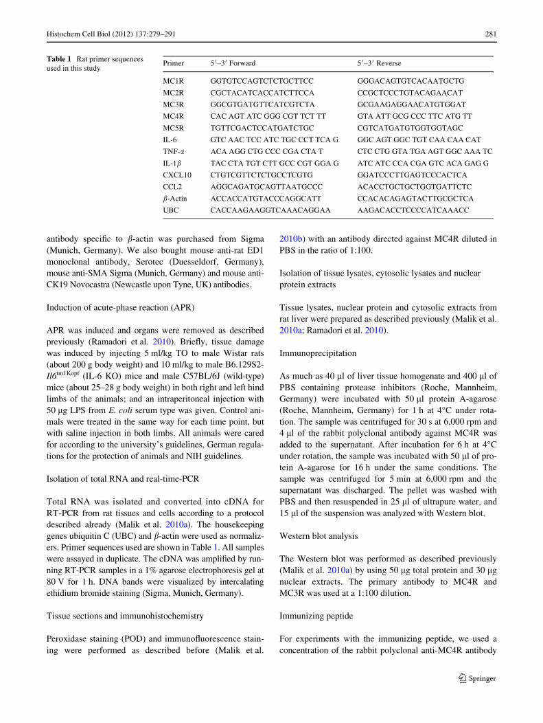
By means of RT-PCR analysis, the presence of all MCRswas detected at the mRNA level in the rat liver tissue ofcontrol animals. An early increase of MC4R gene expres-
sion, starting from 1 h and reaching a maximum (6.3 § 2.3-fold) at 6 h, could be observed after the onset of APR; how-ever, the gene expression of MC4R remained above thecontrol level for 36 h. A signiWcant upregulation in geneexpression of MC2R (3.5 § 0.9-fold) and MC5R(3.8 § 0.9-fold) was detected with a maximum of 12 h,whereas a mild increase in gene expression of MC1R(2.6 § 0.3-fold at 2 h) and MC3R (2.1 § 0.2-fold at 12 h)was also found (Fig. 1a). The detection of MCRs genes inliver tissue was conWrmed by analyzing the PCR productthrough agarose gel-electrophoresis and comparing it tobrain tissue at the RNA level (Fig. 1b).
Immunodetection of MCRs in rat liver and brain tissue sections
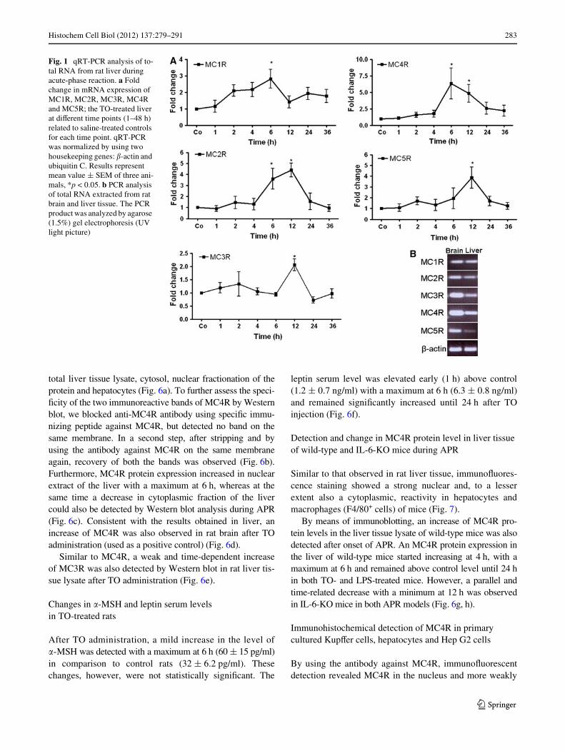
By means POD and immunoXuorescence staining, using arabbit polyclonal antibody against MC4R, a strong nuclearand to a lesser extent also a cytoplasmic reactivity wasdetected mainly in the hepatocytes of the control livers(Fig. 2; Fig. S1). In liver, positivity was also noticed in andaround the cells of vessel walls of portal and central areasand in ED1-, CK-19 and SMA+ cells of the liver (Fig. 3).An increase in the nuclear expression of MC4R with aparallel decrease in the cytoplasmic expression was alsoobserved with onset of APR.
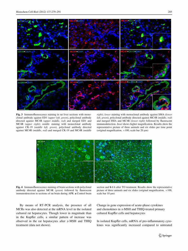
Similar to what was observed in the liver, POD andimmunoXuorescence staining directed against MC4Rrevealed a pattern of strong nuclear and weak cytoplasmicpositivity in brain sections. The reactivity of MC4R wasnot only observed in cells with small nuclei (microglia), butalso in cells with large nuclei (macroglia) in brain sections(Fig. 4a, b; Fig. S2). In contrast to nuclear expression ofMC4R, a strong cytoplasmic expression of MC3R in thehepatocytes was detected in the liver tissue. A similarpattern was also noticed in brain sections (Fig. 5a, b). In braintissue sections, MC3R and MC4R were used as a positivecontrol in this study, as both are known to be constitutivelyexpressed in the brain (Mountjoy et al. 1992; Tao 2010).
The negative control of the indirect immunoXuorescencestaining, a tissue section incubated with unspeciWc rabbitimmunoglobulins instead of the speciWc antibody to MC4R,showed no speciWc signals (Figs. S1, S2).
MC4R protein level in liver tissue during APR
Using antibody against MC4R, immunoprecipitation (IP) ofliver tissue proteins revealed an immunoreactive band at 37and 55 kDa in the liver tissue lysate. However, in the super-natant using the same antibody, only a band at 55 kDa wasvisible, indicating that this band was more abundant thanthe lower molecular weight band (Fig. S3). Similar bandsof MC4R were also detected by Western blot analysis in the
123
Histochem Cell Biol (2012) 137:279–291 283
total liver tissue lysate, cytosol, nuclear fractionation of theprotein and hepatocytes (Fig. 6a). To further assess the speci-Wcity of the two immunoreactive bands of MC4R by Westernblot, we blocked anti-MC4R antibody using speciWc immu-nizing peptide against MC4R, but detected no band on thesame membrane. In a second step, after stripping and byusing the antibody against MC4R on the same membraneagain, recovery of both the bands was observed (Fig. 6b).Furthermore, MC4R protein expression increased in nuclearextract of the liver with a maximum at 6 h, whereas at thesame time a decrease in cytoplasmic fraction of the livercould also be detected by Western blot analysis during APR(Fig. 6c). Consistent with the results obtained in liver, anincrease of MC4R was also observed in rat brain after TOadministration (used as a positive control) (Fig. 6d).
Similar to MC4R, a weak and time-dependent increaseof MC3R was also detected by Western blot in rat liver tis-sue lysate after TO administration (Fig. 6e).
Changes in �-MSH and leptin serum levels in TO-treated rats
After TO administration, a mild increase in the level of�-MSH was detected with a maximum at 6 h (60 § 15 pg/ml)in comparison to control rats (32 § 6.2 pg/ml). Thesechanges, however, were not statistically signiWcant. The
leptin serum level was elevated early (1 h) above control(1.2 § 0.7 ng/ml) with a maximum at 6 h (6.3 § 0.8 ng/ml)and remained signiWcantly increased until 24 h after TOinjection (Fig. 6f).
Detection and change in MC4R protein level in liver tissue of wild-type and IL-6-KO mice during APR
Similar to that observed in rat liver tissue, immunoXuores-cence staining showed a strong nuclear and, to a lesserextent also a cytoplasmic, reactivity in hepatocytes andmacrophages (F4/80+ cells) of mice (Fig. 7).
By means of immunoblotting, an increase of MC4R pro-tein levels in the liver tissue lysate of wild-type mice was alsodetected after onset of APR. An MC4R protein expression inthe liver of wild-type mice started increasing at 4 h, with amaximum at 6 h and remained above control level until 24 hin both TO- and LPS-treated mice. However, a parallel andtime-related decrease with a minimum at 12 h was observedin IL-6-KO mice in both APR models (Fig. 6g, h).
Immunohistochemical detection of MC4R in primary cultured KupVer cells, hepatocytes and Hep G2 cells
By using the antibody against MC4R, immunoXuorescentdetection revealed MC4R in the nucleus and more weakly
Fig. 1 qRT-PCR analysis of to-tal RNA from rat liver during acute-phase reaction. a Fold change in mRNA expression of MC1R, MC2R, MC3R, MC4R and MC5R; the TO-treated liver at diVerent time points (1–48 h) related to saline-treated controls for each time point. qRT-PCR was normalized by using two housekeeping genes: �-actin and ubiquitin C. Results represent mean value § SEM of three ani-mals, *p < 0.05. b PCR analysis of total RNA extracted from rat brain and liver tissue. The PCR product was analyzed by agarose (1.5%) gel electrophoresis (UV light picture)
123

284 Histochem Cell Biol (2012) 137:279–291
in the cytoplasm of cultured KupVer cells, hepatocytes andHep G2 cells, conWrming the in vivo data. However,KupVer cells showed the strongest expression among allother stained cells (Fig. 8a–c).
Change in gene expression of MCRs in �-MSH and THIQ-treated primary cultured rat KupVer cells and hepatocytes
We investigated the gene expression of MCRs at themRNA level in cultured KupVer cells and hepatocytes atdiVerent time points (2, 4 and 6 h) after treatment with�-MSH (MCRs agonist) and THIQ (speciWc MC4R agonist)and compared them to non-treated controls for each time
point. An early and strong change in the gene expression ofMC4R (3.9 § 0.3-fold at 2 h) was observed after treatmentof KupVer cells with THIQ. Similar Wndings were alsoobserved in MC4R gene expression after treatment with�-MSH (MCRs agonist); however, the THIQ eVect wasfaster and stronger. Moreover, stimulation with THIQ alsoupregulated the gene expression of MC2R; this eVect wasstronger than that observed after treatment with �-MSH.Treatment of KupVer cells with �-MSH upregulated geneexpression of MC3R followed by MC5R and MCR1,whereas in comparison to non-treated controls, a mildchange was also detected in MC5R followed by MC3R andMC1R at the mRNA level after treatment with THIQ(Fig. 9).
Fig. 2 ImmunoXuorescence staining of liver sections with polyclonalantibody directed against MC4R (red), followed by Xuorescent immu-nodetection in rat liver sections during acute-phase response after TOtreatment. Left control liver, right 6 h after TO treatment. Upper
MC4R and DAPI staining; middle MC4R staining; lower DAPI stain-ing. Inset shows higher magniWcation. Results show the representativepicture of three animals and six slides per time point (original magni-Wcation, £200, scale bar 20 �m)
123
Histochem Cell Biol (2012) 137:279–291 285
By means of RT-PCR analysis, the presence of allMCRs was also detected at the mRNA level in the isolatedcultured rat hepatocytes. Though lower in magnitude thanin the KupVer cells, a similar pattern of increase wasobserved in the rat hepatocytes after �-MSH and THIQtreatment (data not shown).
Change in gene expression of acute-phase cytokines and chemokines in �-MSH and THIQ-treated primary cultured KupVer cells and hepatocytes
In isolated KupVer cells, mRNA of pro-inXammatory cyto-kines was signiWcantly increased compared to untreated
Fig. 3 ImmunoXuorescence staining in rat liver sections with mono-clonal antibody against ED1 (upper left, green), polyclonal antibodydirected against MC4R (upper middle, red) and merged ED1 andMC4R (upper right); middle staining with monoclonal antibodyagainst CK-19 (middle left, green), polyclonal antibody directedagainst MC4R (middle, red) and merged CK-19 and MC4R (middle
right); lower staining with monoclonal antibody against SMA (lowerleft, green), polyclonal antibody directed against MC4R (middle, red)and merged SMA and MC4R (lower right) followed by Xuorescentimmunodetection. Inset shows higher magniWcation. Results show therepresentative picture of three animals and six slides per time point(original magniWcation, £100, scale bar 20 �m)
Fig. 4 ImmunoXuorescence staining of brain sections with polyclonalantibody directed against MC4R (green) followed by Xuorescentimmunodetection in sections of rat brain during APR. a Control brain
section and b 6 h after TO treatment. Results show the representativepicture of three animals and six slides (original magniWcation, £100,scale bar 10 �m)
123

286 Histochem Cell Biol (2012) 137:279–291
controls upon exposure to LPS. The early (2 h) increase inthe cytokine group was observed with a maximum in geneexpression of TNF-� at 4 h (182 § 15-fold), followed byIL-6 (123 § 4-fold) and IL-1� (52 § 2-fold) at 6 h afterLPS treatment. The upregulating eVect of LPS on TNF-�,IL-6 and IL-1� was signiWcantly reduced by 40, 70 and46%, respectively, by adding �-MSH together with LPS inculture medium. Gene expression of cytokines was also sig-niWcantly reduced by the addition of THIQ together withLPS; however, the reducing eVect of �-MSH was higher onIL-6 and IL1� than on THIQ. The reduction in gene expres-sion of TNF-� was similar with treatment of both moleculesin the presence of LPS (Fig. 10a).
In isolated cultured hepatocytes, gene expression ofCCL2 and CXCL10 was signiWcantly downregulated (up to60%) after 4–6 h of exposure to �-MSH or THIQ, respec-tively (Fig. 10b).
Discussion
In this study, to the best of our knowledge, we demonstratefor the Wrst time the presence of melanocortin receptors(MCRs) in rat and mouse liver cells. An upregulation ingene expression of MCRs in the liver and brain tissue wasdetected in an acute-phase model induced by TO damage toskeletal muscle. In parallel, an increase in �-MSH level anda signiWcant increase in leptin rat serum level were found.After intramuscular TO injection, a “migration” of MC4Rprotein from cytoplasm to the nucleus could be detected byimmunohistochemistry and Western blot analysis of thecell fractions.
A similar pattern of increase in MC4R level was alsoobserved in the liver of two mice models of acute-phasereaction, although the order of magnitude of the changesobserved in the liver of IL-6ko mice was much lower thanthat observed in the wild-type animals.
To demonstrate the distribution and change of MC4Rgene expression in the liver taking place under the experi-mental conditions used, we showed a change of MC4Rgene expression in rat brain tissue similar to that observedin liver tissue. In fact, MC4R gene expression signiWcantlyincreased during the time after tissue damage was inducedin the muscle. Both immunoprecipitation and Western blotanalysis showed a probable non-glycosylated (37 kDa) andglycosylated (55 kDa) band of MC4R in liver tissue lysate.In previous studies, in addition to a 55-kDa band, dimeriza-tion of MC4R has also been reported (Biebermann et al.2003; Elsner et al. 2006).
In contrast to treatments inducing an acute systemicresponse and also liver damage, such as administering bac-terial endotoxin LPS (Caruso et al. 2007), TO is known toinduce aseptic local abscesses without detectable injury toother tissues (Malik et al. 2010a; Sheikh et al. 2006).Therefore, the TO-induced APR model allows us to studythe eVect of cytokines produced at distant sites in the liver.It reproduces the changes observed in human disease statesdue to localized tissue injury (Malik et al. 2010a; Ramadoriand Christ 1999; Sheikh et al. 2006), making it an idealmodel for studying the metabolic consequences of humanAPR.
The melanocortin system consists of several agonists,apart from �-MSH (native speciWc agonist of MCRs); thediscovery of an orally active non-peptide MC4R mimetic,called THIQ (selective agonist of MC4R), has also beenreported (Sebhat et al. 2002). THIQ-stimulated 3�,5�-cyclicmonophosphate (cAMP) generation is reported to act simi-larly to that induced by �-MSH in human as well as in ratMC4R assays (Sebhat et al. 2002). �-MSH exerts its eVectby melanocortin receptors (MCRs) and is known to be pro-duced in the pituitary gland, brain and several peripheraltissues, including immune cells, by posttranslational pro-cessing of the pro-opiomelanocortin precursor protein(Gantz and Fong 2003; Mountjoy et al. 1992; Tao 2010).
Fig. 5 ImmunoXuorescence staining of liver and brain sections duringAPR. ImmunoXuorescence staining with polyclonal antibody directedagainst MC3R a in liver sections (green) and b brain section (red) fol-
lowed by Xuorescent immunodetection during APR. Inset shows high-er magniWcation. Results show the representative picture of threeanimals and six slides (original magniWcation, £100, scale bar 10 �m)
123

Histochem Cell Biol (2012) 137:279–291 287
We showed that MCRs were not only expressed in therat liver cells, but also that their gene expression was upreg-ulated at the mRNA and protein level during acute-phaseresponse. An upregulation in MCRs gene expression isknown to be due to an increased concentration of theiragonist(s) (Gantz and Fong 2003). The binding of the ago-nist to the receptor in the cytoplasm and its migration to thenucleus could explain the increase of the MC4R protein in
the nuclear fraction during APR. In fact, this observedmigration of MC4R could be due to its binding not only to�-MSH, but also to leptin, as leptin is also able to directlyinteract with the melanocortin system in mouse brain(Vrang et al. 2002), and inhibition of MC4R attenuates theleptin signaling in rat brain (Seeley et al. 1997). This couldbe true for liver as well, because an increased serum levelof leptin was also detected in our studied model. Moreover,
Fig. 6 Detection of MC4R and MC3R by Western blot analysis of ratliver and brain. a Western blot analysis revealed two immunoreactivebands: one at 37 kDa and a second at 55 kDa. Lane (L)1 contains ratliver total lysate, L2 liver cytosolic extraction, L3 liver nuclear extractsand L4 total protein of cultured rat hepatocytes. b Top rat liver totallysates with immunoreactive bands at 37 and 55 kDa, middle after neu-tralizing the anti-MC4R antibody with a speciWc immunizing peptide.Bottom reprobing of the same membrane with the antibody againstMC4R without immunizing peptide. c Changes in MC4R protein levelduring APR. Top contains rat liver total lysate, middle liver cytosolicextraction, bottom liver nuclear extracts. d IdentiWcation and changes
in MC4R level in rat brain total lysate during APR, e detection andchanges of MC3R level in rat liver total lysate. f Changes in serumconcentration of �-MSH and leptin during APR. �-MSH (top) and lep-tin (bottom) levels were measured with enzyme-linked immunosorbentassay (ELISA). Results represent mean value § SEM (*P < 0.05 ana-lyzed by one-way ANOVA; n = 3). g Detection of MC4R in the liverof wild-type and IL-6-ko mice during APR. Total protein lysate ofmouse liver after TO administration; h total protein lysate of mice liverafter LPS treatment. �-actin (43 kDa) was used as equal loading con-trol in Western blot analysis. Results show the representative picture ofthree animals
123

288 Histochem Cell Biol (2012) 137:279–291
leptin receptors are known to be expressed in hepatocytes(Cohen et al. 2007). A similar mechanism of cytoplasm tonuclear movement has already been reported in glucocorti-
coid receptors (Picard and Yamamoto 1987) and insulinreceptors (Fehlmann et al. 1982) after binding to theirligand. A link between and leptin and the melanocortin sys-tem has already been established. A previous study on ratsshowed that leptin regulates the glucose Xuxes of hepaticand peripheral tissues through a melanocortin-dependentpathway (Shimizu et al. 2007; Toda et al. 2009). Anotherstudy stated that mice lacking OB-Rb (leptin receptor) inPOMC neurons show hyperinsulinemia (van de Wall et al.2008). Likewise, glucose uptake by the eVect of intra-ven-tromedial nucleus of the hypothalamus (VMH) leptin alsodepends on MC4R (Toda et al. 2009). Together, these stud-ies clearly support a relationship between leptin and themelanocortin system of the brain.
As we previously showed, the serum level of IL-6 is dra-matically elevated in the rat model of TO-induced APR(Sheikh et al. 2006). In this study, we analyze MC4R geneexpression in IL-6ko mice using the same APR model.Strikingly, in the absence of IL-6, the increase of MC4Rhepatic gene expression after TO injection was much lowerthan that observed in wild-type animals, indicating a direct,regulative function of this important acute-phase cytokine.
A contribution of cytokines (IL-6 and TNF-�) to themanifestation of whole-body insulin resistance and to thedirect regulation of glucose metabolism has already beendescribed (Lazar 2005; Wallenius et al. 2002). Further-more, IL-6 given peripherally at high doses can causeincreased levels of blood lipids and blood glucose(Nonogaki et al. 1995). Administration of IL-6 could stimu-late the mobilization and utilization of fatty acids and couldalso increase lipid metabolism (Glund and Krook 2008).Moreover, it can also acutely enhance circulating levels oftriglycerides in rats (Nonogaki et al. 1995), suggesting theimportance of this cytokine in APR-induced metabolic dis-order.
The relation between leptin and melanocortin or leptinand cytokines has been studied partially (Shimizu et al.2007; Vrang et al. 2002; Wallenius et al. 2002); however,
Fig. 8 ImmunoXuorescence detection of MC4R in isolated rat a KupVer cells, b hepatocytes and c human hepatoma cell line (HepG2). Resultsare representative of three experiments for each cell type (original magniWcation £400, scale bar 40 �m)
Fig. 7 Double immunoXuorescence staining with polyclonal antibodydirected against MC4R (red) and F4/80 (green) followed by Xuores-cent immunodetection in sections of mouse liver during APR. Uppercontrol liver, lower 6 h after TO treatment. Inset shows higher magni-Wcation. White arrow indicates the hepatocytes positive for MC4R andwhite arrowhead indicates the KupVer cells positive for MC4R and F4/80. Results show the representative picture of three animals and sixslides (original magniWcation, £200, scale bar 20 �m)
123

Histochem Cell Biol (2012) 137:279–291 289
interaction among leptin, the melanocortin system andcytokines together has not been investigated so far. A pre-vious report stated that despite the absence of IL-6, theserum leptin level was increased in IL-6ko mice duringAPR (Faggioni et al. 1998; Wallenius et al. 2002). Thiscould suggest that although MC4R gene expression is notupregulated in the absence of IL-6, a strong metabolic eVectcould be achieved by the interaction between leptin andMC4R.
Consideration of our data further implies that the mela-nocortin system could have a dual role: on one hand,MC4R is not only the target of acute-phase cytokines, butmay control their production as well.
In this study, we show that the melanocortin systeminXuences energy metabolism at diVerent levels duringAPR. In fact, it not only mediates the eVect of acute-phasecytokines, but may also participate in controlling the syn-thesis of APR cytokines and chemokines in liver macro-
phages (KupVer cells) and hepatocytes respectively. Thelatter have also been shown to have metabolic eVects(Sartipy and LoskutoV 2003; Sell and Eckel 2009).
Indeed, in our current study, treatment of KupVer cellswith endotoxin lipopolysaccharide (LPS) increased geneexpression of acute-phase mediators; this increase was sig-niWcantly inhibited by �-MSH and THIQ, respectively.Moreover, both agonists were able to downregulate thegene expression of potential chemokines in cultured rathepatocytes.
In an additional experiment, IL-6 treatment of hepatocytesincreased the gene expression of two major acute-phaseproteins: �2-macroglobulin (�2M) and heme oxygenase-1(HO-1); however, this upregulation of �2M was transientlyreduced in hepatocytes when �-MSH was also added in theculture medium. In contrast, the eVect of IL-6 on expres-sion of HO-1 was not reduced by �-MSH (manuscript inpreparation). It may suggest that the main role of MC4R is
Fig. 9 RT-PCR analysis of total RNA from rat isolated KupVer cellstreated with �-MSH and THIQ. Data shown as fold changes in mRNAexpression of MC1R, MC2R, MC3R, MC4R and MC5R at diVerenttime points related to untreated controls for each time point. qRT-PCR
was normalized by using two housekeeping genes: �-actin and ubiqui-tin C. Results represent mean value § SEM of three experiments,*p < 0.05
123

290 Histochem Cell Biol (2012) 137:279–291
to reduce possible pro-inXammatory consequences ofacute-phase cytokines, such as production of chemokinesand not the production of anti-inXammatory proteins (e.g.,antiprotease).
Recently, a study on mice reported a protective eVect of�-MSH against experimental multiple organs (includingliver) dysfunction syndrome (MODS) by counteracting thesystemic inXammatory response. The protective eVect issuggested through brain MC4R-triggered activation of thecholinergic anti-inXammatory pathway, which is counter-acted and inhibited by the administration of the MC4Rantagonist (HS024) (Bitto et al. 2011). However, our dataoVer a possible alternative or additional explanation by indi-cating that MC4R plays a more direct role within liver cells.
In conclusion, the presence of MCRs in liver cellsstrongly suggests their role in liver energy metabolism.
Acknowledgments This research was supported by Dr. SabineMihm, Professor at the Department of Gastroenterology and Endocrinol-ogy, University Medical Center, Georg-August University Goettingen.
We thank Dr. Hassan Dihazi, Associate Professor at the Department ofNephrology and Rheumatology, University Medical Center, Georg-August University Goettingen and Dr. Mark Stettner from the Depart-ment of Neurology, Heinrich-Heine University Duesseldorf for theirscientiWc advice.
ConXict of interest The authors show no conXict of interest.
Open Access This article is distributed under the terms of the Crea-tive Commons Attribution Noncommercial License which permits anynoncommercial use, distribution, and reproduction in any medium,provided the original author(s) and source are credited.
References
Biebermann H, Krude H, Elsner A, Chubanov V, Gudermann T,Gruters A (2003) Autosomal-dominant mode of inheritance of amelanocortin-4 receptor mutation in a patient with severe early-onset obesity is due to a dominant-negative eVect caused byreceptor dimerization. Diabetes 52:2984–2988
Bitto A, Polito F, Altavilla D, Irrera N, Giuliani D, Ottani A, MinutoliL, Spaccapelo L, Galantucci M, Lodi R, Guzzo G, Guarini S,
Fig. 10 a RT-PCR analysis of total RNA from rat isolated KupVercells treated with LPS in the presence and absence of �-MSH andTHIQ and b isolated hepatocytes treated only with �-MSH and THIQ.Data shown as fold changes in mRNA expression of TNF-�, IL-6, IL-1�, CCL2 and CXCL1 at diVerent time points related to untreated con-
trols for each time point. qRT-PCR was normalized by using twohousekeeping genes: �-actin and ubiquitin C. Results represent meanvalue § SEM of three experiments, *p < 0.05. L LPS, M �-MSH andT THIQ
123
Histochem Cell Biol (2012) 137:279–291 291
Squadrito F (2011) Melanocortins protect against multiple organdysfunction syndrome in mice. Br J Pharmacol 162:917–928
Butler AA (2006) The melanocortin system and energy balance.Peptides 27:281–290
Caruso C, Durand D, Schioth HB, Rey R, Seilicovich A, Lasaga M(2007) Activation of melanocortin 4 receptors reduces the inXam-matory response and prevents apoptosis induced by lipopolysac-charide and interferon-gamma in astrocytes. Endocrinology148:4918–4926
Cohen SE, Kokkotou E, Biddinger SB, Kondo T, Gebhardt R, KratzschJ, Mantzoros CS, Kahn CR (2007) High circulating leptin recep-tors with normal leptin sensitivity in liver-speciWc insulin receptorknock-out (LIRKO) mice. J Biol Chem 282:23672–23678
Elsner A, Tarnow P, Schaefer M, Ambrugger P, Krude H, Gruters A,Biebermann H (2006) MC4R oligomerizes independently ofextracellular cysteine residues. Peptides 27:372–379
Engblom D, Kornfeld JW, Schwake L, Tronche F, Reimann A, BeugH, Hennighausen L, Moriggl R, Schutz G (2007) Direct glucocor-ticoid receptor-Stat5 interaction in hepatocytes controls body sizeand maturation-related gene expression. Genes Dev 21:1157–1162
Faggioni R, Fantuzzi G, Fuller J, Dinarello CA, Feingold KR, GrunfeldC (1998) IL-1 beta mediates leptin induction during inXamma-tion. Am J Physiol 274:R204–R208
Fehlmann M, Carpentier JL, Le CA, Thamm P, Saunders D, Branden-burg D, Orci L, Freychet P (1982) Biochemical and morphologi-cal evidence that the insulin receptor is internalized with insulinin hepatocytes. J Cell Biol 93:82–87
Gantz I, Fong TM (2003) The melanocortin system. Am J PhysiolEndocrinol Metab 284:E468–E474
Ge F, Zhou S, Hu C, Lobdell H, Berk PD (2010) Insulin- and leptin-regulated fatty acid uptake plays a key causal role in hepatic ste-atosis in mice with intact leptin signaling but not in ob/ob or db/db mice. Am J Physiol Gastrointest Liver Physiol 299:G855–G866
Glund S, Krook A (2008) Role of interleukin-6 signalling in glucoseand lipid metabolism. Acta Physiol (Oxf) 192:37–48
Lazar MA (2005) How obesity causes diabetes: not a tall tale. Science307:373–375
Malik IA, Baumgartner BG, Naz N, Sheikh N, Moriconi F, RamadoriG (2010a) Changes in gene expression of DOR and other thyroidhormone receptors in rat liver during acute-phase response. CellTissue Res 342:261–272
Malik IA, Moriconi F, Sheikh N, Naz N, Khan S, Dudas J, MansurogluT, Hess CF, Rave-Frank M, Christiansen H, Ramadori G (2010b)Single-dose gamma-irradiation induces up-regulation of chemo-kine gene expression and recruitment of granulocytes into theportal area but not into other regions of rat hepatic tissue. Am JPathol 176:1801–1815
Mizock BA (1995) Alterations in carbohydrate metabolism duringstress: a review of the literature. Am J Med 98:75–84
Mountjoy KG, Robbins LS, Mortrud MT, Cone RD (1992) The clon-ing of a family of genes that encode the melanocortin receptors.Science 257:1248–1251
Nonogaki K, Fuller GM, Fuentes NL, Moser AH, Staprans I, GrunfeldC, Feingold KR (1995) Interleukin-6 stimulates hepatic triglycer-ide secretion in rats. Endocrinology 136:2143–2149
Palmer C, Corpuz T, Guirguis M, O’Toole S, Yan K, Bu Y, JorgensonJ, Talbot M, Loi K, Lloyd A, Zekry A (2010) The eVect of obesity
on intrahepatic cytokine and chemokine expression in chronichepatitis C infection. Gut 59:397–404
Picard D, Yamamoto KR (1987) Two signals mediate hormone-depen-dent nuclear localization of the glucocorticoid receptor. EMBO J6:3333–3340
Ramadori G, Christ B (1999) Cytokines and the hepatic acute-phase re-sponse. Semin Liver Dis 19:141–155
Ramadori G, Moriconi F, Malik I, Dudas J (2008) Physiology andpathophysiology of liver inXammation damage and repair.J Physiol Pharmacol 59(Suppl 1):107–117
Ramadori P, Ahmad G, Ramadori G (2010) Cellular and molecularmechanisms regulating the hepatic erythropoietin expression dur-ing acute-phase response: a role for IL-6. Lab Invest 90:1306–1324
Sartipy P, LoskutoV DJ (2003) Monocyte chemoattractant protein 1 inobesity and insulin resistance. Proc Natl Acad Sci USA 100:7265–7270
Sebhat IK, Martin WJ, Ye Z, Barakat K, Mosley RT, Johnston DB,Bakshi R, Palucki B, Weinberg DH, MacNeil T, Kalyani RN,Tang R, Stearns RA, Miller RR, Tamvakopoulos C, Strack AM,McGowan E, Cashen DE, Drisko JE, Hom GJ, Howard AD, Mac-Intyre DE, Van der Ploeg LH, Patchett AA, Nargund RP (2002)Design and pharmacology of N-[(3R)-1,2,3,4-tetrahydroisoquin-olinium-3-ylcarbonyl]-(1R)-1-(4-chlorobenzyl)-2-[4-cyclohexyl-4-(1H–1,2,4-triazol-1-ylmethyl)piperidin-1-yl]-2-oxoethylamine(1), a potent, selective, melanocortin subtype-4 receptor agonist.J Med Chem 45:4589–4593
Seeley RJ, YagaloV KA, Fisher SL, Burn P, Thiele TE, van Dijk G,Baskin DG, Schwartz MW (1997) Melanocortin receptors in lep-tin eVects. Nature 390:349
Sell H, Eckel J (2009) Chemotactic cytokines, obesity and type 2 dia-betes: in vivo and in vitro evidence for a possible causal correla-tion? Proc Nutr Soc 68:378–384
Sheikh N, Tron K, Dudas J, Ramadori G (2006) Cytokine-induced neu-trophil chemoattractant-1 is released by the noninjured liver in arat acute-phase model. Lab Invest 86:800–814
Shimizu H, Inoue K, Mori M (2007) The leptin-dependent and -inde-pendent melanocortin signaling system: regulation of feeding andenergy expenditure. J Endocrinol 193:1–9
Tao YX (2010) The melanocortin-4 receptor: physiology, pharmacol-ogy, and pathophysiology. Endocr Rev 31:506–543
Toda C, Shiuchi T, Lee S, Yamato-Esaki M, Fujino Y, Suzuki A,Okamoto S, Minokoshi Y (2009) Distinct eVects of leptin and amelanocortin receptor agonist injected into medial hypothalamicnuclei on glucose uptake in peripheral tissues. Diabetes 58:2757–2765
van de Wall E, Leshan R, Xu AW, Balthasar N, Coppari R, Liu SM, JoYH, MacKenzie RG, Allison DB, Dun NJ, Elmquist J, LowellBB, Barsh GS, de Luca C, Myers MG Jr, Schwartz GJ, Chua SCJr (2008) Collective and individual functions of leptin receptormodulated neurons controlling metabolism and ingestion. Endo-crinology 149:1773–1785
Vrang N, Kristensen P, Tang-Christensen M, Larsen PJ (2002) EVectsof leptin on arcuate pro-opiomelanocortin and cocaine-amphet-amine-regulated transcript expression are independent of circulat-ing levels of corticosterone. J Neuroendocrinol 14:880–886
Wallenius V, Wallenius K, Ahren B, Rudling M, Carlsten H, DicksonSL, Ohlsson C, Jansson JO (2002) Interleukin-6-deWcient micedevelop mature-onset obesity. Nat Med 8:75–79
123