STAT1 Mutations in Autosomal Dominant Chronic Mucocutaneous Candidiasis
Upload
independentCategory
view
2download
0
www.elsevier.com/locate/issn/10434666
Cytokine 36 (2006) 218–228
STAT1 and STAT3 phosphorylation by porins are independentof JAKs but are dependent on MAPK pathway and plays
a role in U937 cells production of interleukin-6
Marilena Galdiero a, Mariateresa Vitiello a,*, Marina D’Isanto a,Katia Raieta a, Emilia Galdiero b
a Department of Experimental Medicine, Section of Microbiology and Clinical Microbiology, Faculty of Medicine, Second University of Naples, Naples, Italyb Department of General Physiology, Faculty of Science MM. FF. NN., Hygiene and Microbiology Section, University of Naples ‘‘Federico II’’, Naples, Italy
Received 26 September 2006; received in revised form 28 November 2006; accepted 16 December 2006
Abstract
A group of transcription factors, termed signal transducers and activators of transcription (STATs), appears to orchestrate the down-stream events propagated by cytokine/growth factor interactions with their cognate receptors. Similarly, cytoplasmic Janus kinases(JAKs) seem to play a critical role in diverse signal transduction pathways that govern cellular survival, proliferation, differentiationand apoptosis. In this work, we analysed the effects of the Salmonella enterica serovar Typhimurium porins on signaling by the JAK/STAT pathway and IL-6 release in U937 cells. Porins and LPS of membrane from Gram-negative bacteria are factors implicated in septicshock. In our assays porins induce interleukin-6 (IL-6) release (110 ± 2.6 pg/ml) 24 h after stimulation and STAT1/STAT3 tyrosine(Tyr701/Tyr705) and serine (Ser727) phosphorylation after 15 min. By using several selective inhibitors we demonstrate that porins mod-ulate the activation of STAT1/STAT3 through mitogen activated protein kinases (MAPKs) and not JAKs. Furthermore, we demonstrat-ed that STAT1 and STAT3 are not involved in the modulation of IL-6 release in U937 cells stimulated with porins. Inhibition oftyrosine/serine phosphorylation mediated by MAPKs of STAT1 and STAT3 decrease the IL-6 secretion following porin stimulation.Therefore, suggesting a key role of this pathway in phosphorylation of Ser 727 in STAT1 and STAT3. These results are confirmedby porin or LPS-induced nuclear translocation of STAT1 and STAT3 in U937 cells.� 2007 Elsevier Ltd. All rights reserved.
Keywords: Porins; IL-6 release; JAK/STAT pathway
1. Introduction
Signaling and transcriptional activity are importantprocesses induced by several bacterial components whichmodulate many of the pathways involved in the host
1043-4666/$ - see front matter � 2007 Elsevier Ltd. All rights reserved.
doi:10.1016/j.cyto.2006.12.003
* Corresponding author. Fax: +39 081 5667578.E-mail address: [email protected] (M. Vitiello).
1 Abbreviations used: STAT, signal transducers and activators of transcriptionkinases; LPS, lipopolysaccharide; SDS–PAGE, SDS–polyacrylamide gel electrTTBS, Tris-buffered saline Tween 20; BSA, bovine serum albumin; ELISApolymixin B; PTK, protein tyrosine kinase; ERK, extracellular-signal-regulkappa B.
inflammatory response and in the innate and adaptativeimmune functions.
Mycobacterial parasites [1], peptidoglycan [2], lipopoly-saccharide (LPS)1 [3], outer membrane proteins [4,5] andother specific bacterial products besides their activity as
; JAK, Janus protein tyrosine kinase; MAPKs, mitogeni activated proteinophoresis; ECL, enhanced chemiluminescence; TBS, Tris-buffered saline;, enzyme-linked immunoassay; PBS, phosphate-buffered saline; PBM,
ated kinase; AP-1, activate activating protein 1; NF-jB, nuclear factor
M. Galdiero et al. / Cytokine 36 (2006) 218–228 219
immunogens, on coming into contact with cells of theinflammatory and immune responses, induce the produc-tion of peptide and small peptide mediators such as cyto-kines, defensins, etc., which have different structures andfunctions. The molecular mechanisms which regulate theinteractions of the small peptide mediators with host cellsare well known, whereas there is little understanding ofthe mechanisms by which different bacterial productsinduce similar responses, releasing cytokines and defensins[6].
Porins from Salmonella enterica serovar Typhimurium,Mannheimia haemolytica and Haemophilus influenzae
induce tyrosine phosphorylation of several proteins inTHP-1 cells and in macrophages from C3H/HeJ mice [7].
Among the most prominent tyrosine phosphorylatedbands in porin-stimulated cells there are a number of pro-teins with molecular masses similar to those of the tyrosine/serine/threonine protein kinase family. Mitogen-activatedprotein kinases (MAPKs) and their upstream kinasesactivate a number of transcription factors and induce tran-scription from a variety of inflammatory genes in responseto LPS and cytokines [8]. MAPKs are kinases that signalthe intracellular responses to an array of extracellular stim-uli that include mitogens, growth factors, pathogen-derivedproducts, and other physical stressors.
Additionally, porins activate the Raf-1-MEK1/2-MAPKpathway and transcriptional factors AP-1 and NF-jB [9].
The family of transcription factors called STATs (signaltransducers and activators of transcription) have beenfound to be activated by the Janus kinases (JAKs) thatare associated with the cytokine receptor components[10]. In resting cells the STATs, when phosphorylated bythe JAKs, dimerize via their SH2 domains and translocateto the nucleus, where they interact with specific DNAsequences and transactivate the associated genes.
The ability of STATs to homo-or-hetero-dimerizeallows cytokine-specific cellular responses [11]. Severalpolypeptide ligands use the JAK-STAT molecules in signaltransduction [12]. Four JAKs have been identified in mam-mals: JAK1, JAK2, JAK3 and Tyk2. They have molecularmasses ranging from 120 to 140 kDa and a conserved struc-ture with distinct regions. Two JAKs of the same or differ-ent class must be in close proximity for phosphorylation tooccur. Seven STAT proteins have currently been identifiedin mammalian cells, with molecular masses between 75 and95 kDa. Many different models have been used to investi-gate the role of JAK/STAT in response to infection in vivo
[13]. The models range from polymicrobial peritonitis [14]and Klebsiella pneumoniae lung infection [15], to intraperi-toneal injection of pathogens or their components, such asendotoxin [16].
In the present study, we investigated the ability of por-ins to directly activate the JAK/STAT signal transductionpathway and stimulate IL-6 release from U937 cells. Theresults will contribute to our current knowledge on themechanism and function of JAK/STAT signaling insepsis.
2. Materials and methods
2.1. Cell lines
U937 monocytes (ATCC CRL-1593.2) were grown at37 �C in 5% CO2 in RPMI 1640 and differentiated as pre-viously described [17]. Cells were tested every 2 weeks bya PCR-based detection assay for mycoplasma contamina-tion [18].
2.2. Bacterial strain
The bacterial strain used was S. enterica serovarTyphimurium SH5014 grown in nutrient broth for 18–24 h at 37 �C under agitation; cells were harvested at theend of the exponential growth phase.
2.3. Preparation of porins and LPS
Porins were isolated from the lysozyme–EDTA extract-ed envelopes of S. enterica serovar Typhimurium strainSH5014 as previously reported [9]. The protein content ofthe porins preparation was determined by the method ofLowry et al. [19] and checked by SDS–PAGE accordingto Laemmli [20]. SDS–PAGE revealed two bands withmolecular weights of 34 and 36 kDa, confirming the purityof the preparation. LPS was isolated using the phenol/chlo-roform/ether method described previously [9]. All possibletraces of LPS were revealed on SDS–PAGE stained withsilver nitrate as described by Tsai and Frasch [21] and bythe Limulus-amoebocyte-lysate assay (Limulus test, pbiinternational, Milan Italy) according to Yin et al. [22].
2.4. Cell stimulation with S. enterica serovar Typhimurium
porins and LPS for preparation of cell lysates
U937 cells (3 · 106 cells/ml) were stimulated with differ-ent concentrations (1, 5 and 10 lg/ml) of stimuli for differ-ent time periods (15, 30 and 60 min) in 96-wellpolypropylene plates. After incubation, the cells were pre-pared as previously reported [9] and used for enhancedchemiluminescence’s (ECL) Western Blot analysis.
2.5. Kinase phosphorylation
Cell lysates were immunoprecipitated and used for wes-tern blotting. Immunoprecipitation was carried out withthe appropriate antibodies (anti-JAK1, -JAK2, -JAK3,-Tyk2, -STAT1, -STAT2, -STAT3, -STAT4, -STAT5,-STAT6) (Santa Cruz Biotechnology, Inc.). Blots wereblocked for 1 h at room temperature in Tris-buffered saline(TBS [150 mM NaCl, 20 mM Tris–HCl, pH 7.5]) contain-ing 1% bovine serum albumin (BSA; Sigma–AldrichS.r.l., Milano, Italy) plus 1% blotting grade blockernon-fat milk (Bio-Rad Laboratories) and subsequentlymembranes were washed twice with TBS containing0.05% Tween 20 (TTBS) before incubation for 1 h at room

220 M. Galdiero et al. / Cytokine 36 (2006) 218–228
temperature with anti-phosphoaminoacid monoclonalantibodies (anti-phosphotyrosine, anti-phosphoserine oranti-phosphothreonine antibody; Calbiochem, Novabio-chem Corporation, GmbH) diluted in TBS containing 1%BSA. After being washed six times with TTBS for 3 min,PVDF membranes were incubated at room temperaturefor 2 h with anti-mouse or anti-rabbit IgG HRP secondaryantibodies diluted 1:3000. They were then washed six timeswith TTBS and twice with PBS for 5 min. In someexperiments, the same blot was used for detection ofp(Tyr701)STAT1 (Santa Cruz Biotechnology, Inc.),p(Tyr705)STAT3 (Santa Cruz Biotechnology,Inc.), p(Ser727)STAT1 Upstate Biotechnology (LakePlacid, NY), p(Ser727)STAT3 (New England BioLabs),p(Tyr1007/1008)JAK2 Upstate Biotechnology (LakePlacid, NY) and p(TyrTyr1054/1055)Tyk2 (Cell SignalingTechnology Inc., Beverly, MA) after stripping of the previ-ous antibody with Restore Western blot stripping buffer(Pierce).
2.6. Cytokine release
All assays were carried out using 3 · 106/ml U937 cellsstimulated with different concentrations of stimuli (5 lg/ml of porins or 1 lg/ml of LPS) for 24 h at 37 �C in 5%CO2; the time points and concentrations have been deter-mined in preliminary experiments as that giving maximumrelease. After incubation the samples were centrifuged at1800 rpm at 4 �C for 10 min and the supernatants werecollected and stored at �70 �C. IL-6 release was measuredby ELISA using pair-matched monoclonal antibodies,according to the manufacturer’s recommendations (RocheDiagnostic SpA, Milan, Italy).
2.7. Inhibitors of signal transduction
In some experiments, before stimulation, U937 cells werepretreated with PD-098059 (New England Biolabs, Inc.), aselective inhibitor of MEK1 activator [23]; SB203580 (Cal-biochem, Novabiochem Corporation, GmbH), a specificinhibitor of the p38 pathway [24]; AG490 (Calbiochem–Novabiochem Corp., CA), a specific JAK inhibitor [25];H7, a serine/threonine kinase inhibitor [26]; genistein, abroad-range tyrosine kinase inhibitor [27] for 60 min.
2.8. STATs trans-activation analysis
To detect and quantify STATs activation in U937 cells,we used ELISA-based Trans-Am transcription factor kits(Active Motif, Carlsbad, USA) that exploited a patentedtechnology to attach an oligonucleotide containing theSTAT consensus binding site (5 0-TTCCCGGAA-3 0) to a96-well plate according to the transcription factors ana-lysed [28]. The active form of STAT contained in cellextract specifically binds to this oligonucleotide. The pri-mary antibodies used to detect STAT recognize an epitopeon STAT1, STAT3, STAT5A or 5B that accessible only
when the STAT is activated and bound to its targetDNA. Preparation of nuclear cell extracts was doneaccording to the manufacturer’s instructions. The specific-ity of the assays was checked by measuring the ability ofsoluble wild-type or mutated STAT oligonucleotide toinhibit binding. In preliminary assays, the Trans-Am kitsshowed a good correlation with an EMSA in detectingthe DNA binding capacity of STATs. The optimal timeof stimulation (1 h) and amount of porins (5 lg/ml) orLPS (1 lg/ml) used in the STAT ELISA were determinedin preliminary experiments.
2.9. Endotoxin test
All materials and solutions were tested for LPS by aLimulus test as described by Yin et al. [22]. The results ofthis test was compared with a standard LPS solution whichwas Limulus test positive at 0.1 EU/ml.
2.10. Lactate dehydrogenase (LDH) assay
LDH assays were carried out according to manufactur-er’s instructions using a cytotoxicity detection kit (RocheDiagnostic SpA, Milan, Italy).
2.11. Reproducibility
Gels were scanned for densitometry analysis by SigmaGel software (Sigma–Aldrich S.r.l., Milano, Italy). Theresults shown are from a single experiment typical of atleast three giving similar results. The results were expressedas mean values ± standard errors of three independentobservations. Comparisons between cytokine test was doneby Student’s t-test, with statistical significance consideredto P 6 0.01 and P 6 0.05 for STATs trans-activationanalysis and densitometry analysis of band intensity.
3. Results
3.1. Purity of porin preparations
The purification of the porin preparation and the even-tual contamination by LPS have been amply discussed inprevious works [29,30]. Using the Limulus test, the LPScontamination in the porin preparations, used in this study,was estimated to be <0.005% (w/w) compared with a stan-dard S. enterica serovar Typhimurium LPS solution. Theconcentration of porins used in our experimental proce-dures, contains a biologically insufficient percentage ofLPS (about 50 pg/lg of porins), which did not induceany enzyme phosphorylation or cytokine release.
3.2. Porin and LPS induce release of IL-6 in U937 cells
It has been extensively documented that porins have theability to stimulate the synthesis and release of pro- andanti-inflammatory cytokines [31].
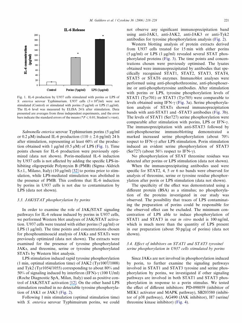
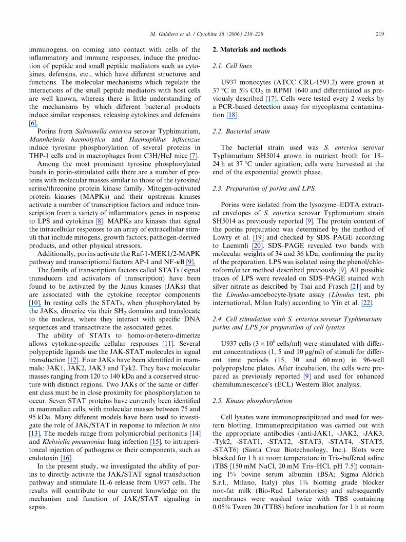
Fig. 1. IL-6 production by U937 cells stimulated with porins or LPS oS. enterica serovar Typhimurium. U937 cells (3 · 106/ml) were nostimulated (Control) or stimulated with porins (5 lg/ml) or LPS (1 lg/ml)The IL-6 level was measured by ELISA 24 h after stimulation. Datapresented are averages from three independent experiments, and the errobars indicate the standard errors of the means (*P 6 0.01; Student’s t-test)
M. Galdiero et al. / Cytokine 36 (2006) 218–228 221
ft.
r.
Salmonella enterica serovar Typhimurium porins (5 lg/mlor 0.2 lM) induced IL-6 production (110 ± 2.6 pg/ml) 24 hafter stimulation, representing at least 60% of the produc-tion obtained with 1 lg/ml (0.5 lM) of LPS (Fig. 1). Timepoints chosen for IL-6 production were previously opti-mized (data not shown). Porin-mediated IL-6 inductionby U937 cells is not affected by adding the specific LPS-in-hibiting oligopeptide Polymyxin B (PMB) (Sigma–AldrichS.r.l., Milano, Italy) (10 lg/ml) [32] to porins prior to stim-ulation, while LPS-mediated stimulation was abolished inthe presence of PMB. This confirms that IL-6 inductionby porins in U937 cells is not due to contamination byLPS (data not shown).
3.3. JAK/STAT phosphorylation by porins
In order to examine the role of JAK/STAT signalingpathways for IL-6 release induced by porins in U937 cells,we performed Western blot analyses of JAK/STAT activa-tion. U937 cells were treated with either porins (5 lg/ml) orLPS (1 lg/ml). The time points and concentrations chosenfor phosphoaminoacid analysis of JAKs and STATs werepreviously optimized (data not shown). The extracts wereexamined for the presence of tyrosine phosphorylatedJAKs, and threonine, serine or tyrosine phosphorylatedSTATs by Western blot analysis.
LPS stimulation induced rapid tyrosine phosphorylation(1 min, optimal stimulation time) of JAK2 (Tyr1007/1008)and Tyk2 (Tyr1054/1055) corresponding to about 80% and50% of signaling induced by interferon (IFN)-c (100 U/ml)(Roche Diagnostic SpA, Milan, Italy) used as positive con-trol of JAK/STAT activation [12]. On the other hand LPSstimulation resulted in no detectable tyrosine phosphoryla-tion of JAK1 or JAK3 (Fig. 2).
Following 1 min stimulation (optimal stimulation time)with S. enterica serovar Typhimuirum porins, we could
not observe any significant immunoprecipitation bandusing anti-JAK1, anti-JAK2, anti-JAK3 or anti-Tyk2antibodies for tyrosine phosphorylation analysis (Fig. 2).
Western blotting analysis of protein extracts derivedfrom U937 cells treated for 15 min with either porins(5 lg/ml) or LPS (1 lg/ml) revealed several STAT phos-phorylated proteins (Fig. 3). The time points and concen-trations chosen were previously optimised. The lysatesobtained were immunoprecipitated by antibodies that spe-cifically recognised STAT1, STAT2, STAT3, STAT4,STAT5 or STAT6 enzymes. Immunoblot analyses wereperformed using anti-phosphothreonine, anti-phosphoser-ine or anti-phosphotyrosine antibodies. After stimulationwith porins or LPS, tyrosine phosphorylation levels ofSTAT1 (Tyr701) or STAT3 (Tyr705) were comparable tolevels obtained using IFN-c (Fig. 3a). Serine phosphoryla-tion analysis of STATs showed immunoprecipitationbands with anti-STAT1 and -STAT3 antibodies (Fig. 3b).The levels of STAT1 (Ser727) serine phosphorylation werecomparable after stimulation with porins, LPS or IFN-c.The immunoprecipitation with anti-STAT3 followed byanti-phosphoserine immunoblotting demonstrated amarked increased serine phosphorylation (about 70%respect to IFN-c) after LPS stimulation. Porin stimulationinduced an evident serine phosphorylation of STAT3(Ser727) (about 56% respect to IFN-c).
No phosphorylation of STAT threonine residues wasdetected after porins or LPS stimulation (data not shown).
When the immunoprecipitating antibodies used werespecific for STAT2, 4, 5 or 6 no bands were observed foranalysis of threonine, serine or tyrosine residue phosphor-ylation after porin or LPS stimulation (data not shown).
The specificity of the effect was demonstrated using adifferent protein (BSA) as a stimulus; no phosphoryla-tion of the proteins investigated in our study wasobserved. The possibility that traces of LPS contaminat-ing the preparation of porins could be responsible forthe observed effect can be excluded. The minimum con-centration of LPS able to induce phosphorylation ofSTAT1 and STAT3 in our in vitro model is 100 ng/ml,which is much more than the quantity of LPS presentin our preparation (about 50 pg/lg of porins) (data notshown).
3.4. Effect of inhibitors on STAT1 and STAT3 tyrosine/
serine phosphorylation in U937 cells stimulated by porins
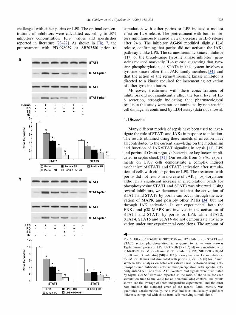
Since JAKs are not involved in phosphorylation inducedby porin, to further examine the signaling pathwaysinvolved in STAT1 and STAT3 tyrosine and serine phos-phorylation by porins, we investigated if other signalingpathways are involved in both STAT1 and STAT3 phos-phorylation in response to a porin stimulus. We testedthe effect of different inhibitors: PD-098059 (inhibitor ofMEK1 activator and MAPK pathway), SB203580 (inhibi-tor of p38 pathway), AG490 (JAK inhibitor), H7 (serine/threonine kinase inhibitor) (Fig. 4).

Fig. 2. Western blot analysis of JAKs activation. U937 cells (3 · 106/ml) were stimulated with either porins, LPS or IFN-c (positive control) for 1 min.The immunoprecipitated proteins for JAK1, JAK2, JAK3 and Tyk2 were separated by electrophoresis, transferred to PVDF membrane and probed withAbs specific for anti-phosphotyrosine. In some experiments, the same blot was used for detection of p(Tyr1007/1008)JAK2 and p(Tyr1054/1055)Tyk2after stripping of the previous antibody. Gels were scanned for densitometry analysis with the Sigma Gel Software, and the ratio of the value for eachstimulation time to the value for an unstimulated control is shown. The data are averages from three different experiments, and the error bars indicate thestandard errors of the means. Band intensity was quantified densitometrically. *P 6 0.05 indicates statistically significant difference compared to IFN-cstimulated cells.
222 M. Galdiero et al. / Cytokine 36 (2006) 218–228
Cells were pretreated for 60 min with PD-098059(25 lM) and then stimulated with either porins or LPS.From cell lysates a decrease in intensity of the immunopre-cipitation bands with anti-STAT1 or anti-STAT3 antibod-ies for tyrosine phosphorylation analysis was observed.Using porins the decrease in intensity of the immunopre-cipitation bands with anti-STAT1 or anti-STAT3 antibod-ies for tyrosine phosphorylation (Tyr701 and Tyr705,respectively) analysis was about 35% (Fig. 4a) and usingLPS it was about 40% (Fig. 4b).
Pretreatment with SB203580 (10 lM for 60 min) prior tostimulation with porins or LPS resulted in a considerabledecrease in intensity of STAT1 and STAT3 immunoprecip-itation bands for tyrosine phosphorylation analysis. Theintensity of the bands was reduced of about 50% witheither porins or LPS.
When using AG490, at a concentration of 10 lM for60 min, we could not observe any change in the immuno-precipitated bands for tyrosine phosphorylated of STAT1or STAT3 after stimulation with porins (Fig. 4a), whileLPS reduced activation of about 20% (Fig. 4b). The cellpretreatment with H7 (25 lM for 60 min) modulated bothporin or LPS-induced STAT1 and STAT3 tyrosine phos-phorylation. For maximal transcriptional activity, STAT1and STAT3 need to be phosphorylated on both tyrosineand serine residues [33]. To verify STAT1 and STAT3 ser-ine phosphorylation, U937 cells were pretreated with PD-098059, SB203580 or H7 prior to the addition of porinsor LPS. The results, presented in Fig. 5, indicate thatPD-098059, reduced the phosphorylation status of STAT1or STAT3 serine (about 30% for porins and 35% for LPS)as well as the pretreatment with SB203580 (about 50% for
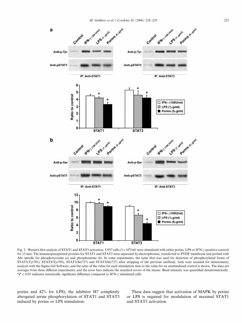
Fig. 3. Western blot analysis of STAT1 and STAT3 activation. U937 cells (3 · 106/ml) were stimulated with either porins, LPS or IFN-c (positive control)for 15 min. The immunoprecipitated proteins for STAT1 and STAT3 were separated by electrophoresis, transferred to PVDF membrane and probed withAbs specific for phosphotyrosine (a) and phosphoserine (b). In some experiments, the same blot was used for detection of phosphorylated forms ofSTAT1(Tyr701), STAT3(Tyr705), STAT1(Ser727) and STAT3(Ser727) after stripping of the previous antibody. Gels were scanned for densitometryanalysis with the Sigma Gel Software, and the ratio of the value for each stimulation time to the value for an unstimulated control is shown. The data areaverages from three different experiments, and the error bars indicate the standard errors of the means. Band intensity was quantified densitometrically.*P 6 0.05 indicates statistically significant difference compared to IFN-c stimulated cells.
M. Galdiero et al. / Cytokine 36 (2006) 218–228 223
porins and 42% for LPS); the inhibitor H7 completelyabrogated serine phosphorylation of STAT1 and STAT3induced by porins or LPS stimulation.
These data suggest that activation of MAPK by porinsor LPS is required for modulation of maximal STAT1and STAT3 activation.
224 M. Galdiero et al. / Cytokine 36 (2006) 218–228
3.5. Porin-induced nuclear translocation of STAT1 and
STAT3
Experiments investigate the dimerization by porin orLPS-activated STAT1 and STAT3 and the concomitant
nuclear translocation of dimers in U937 cells were carriedout. The results obtained showed that increased specificbinding of active STAT1 or STAT3 (Fig. 6) were detectedin U937 lysates 1 h after stimulation with porins (5 lg/ml)or LPS (1 lg/ml).
Further experiments were performed to ascertainwhether MAPKs had a role in the binding of STAT1 andSTAT3 to DNA by porins. Specifically, we looked at theeffects of the PD-098059, SB203580, AG490 and H7 inhib-itors on binding of STAT1 and STAT3 to a consensusbinding site. PD-098059 pretreatment was effective indecreased STAT1 or STAT3 porin or LPS-mediated nucle-ar translocation (about 37% for porins and about 45% forLPS). The pretreatment with the p38 inhibitor SB203580decreased porin or LPS stimulated STAT1 or STAT3 bind-ing to DNA (50%), suggesting that the functionality ofthese proteins requires the activation of p38 pathway. Fur-thermore, the results presented in Fig. 6, indicate thatAG490, had a decreasing effect on STAT1 and STAT3DNA binding induced by LPS (22%). On the contrary,no effect was evidenced on STAT1 or STAT3 nucleartranslocation following porin stimulation.
We finally investigated the effect of H7 on the inductionof porin- or LPS-mediated nuclear translocation of STAT1or STAT3. Treatment of U937 cells with H7 partially sup-pressed porin or LPS induction of STAT1 binding to aconsensus binding site (30%) (Fig. 6). Moreover, H7 pre-treatment caused an evident inhibition of STAT3 LPS-mediated DNA binding (70%), while modulated the porineffect on STAT3 nuclear translocation (Fig. 6).
3.6. MAPK and not JAKs are involved in IL-6 production in
U937 cells stimulated by porins
To further examine the signaling pathways involved inIL-6 release by porins and LPS, U937 cells were incubatedwith specific inhibitors of ERK1/ERK2 (PD-098059,25 lM for 60 min), p38 MAPKs (SB203580, 10lM for60 min), JAKs (AG490, 10 lM for 60 min), a serine/threo-nine kinase inhibitor (H7, 25 lM for 60 min) or a totalPTKs inhibitor (genistein, 50 lM for 60 min) before being
Fig. 4. Effect of PD-098059, SB203580, AG490 and H7 inhibitors onSTAT1 and STAT3 tyrosine phosphorylation in response to S. enterica
serovar Typhimurium porins or LPS. U937 cells (3 · 106/ml) wereincubated with PD-098059 (25 lM for 60 min, MEK1 inhibitor) (PD),SB203580 (10 lM for 60 min, p38 inhibitor) (SB), AG490 (10 lM for60 min, JAK/STAT pathway inhibitor) (AG) or H7 (a serine/threoninekinase inhibitor, 25 lM for 60 min) and stimulated with porins (a) or LPS(b) for 15 min. Western blot analysis on total cell extracts was performedusing anti-phosphotyrosine antibodies after immunoprecipitation withspecific antibody anti-STAT1 or anti-STAT3. Western blot signals werequantitated by Sigma Gel Software and reported as the ratio of the valuefor each stimulation time to the value for an non-stimulated control. Theresults shown are the average of three independent experiments, and theerror bars indicate the standard error of the means. Band intensity wasquantified densitometrically. *P 6 0.05 indicates statistically significantdifference compared with those from cells receiving stimuli alone.
b
M. Galdiero et al. / Cytokine 36 (2006) 218–228 225
challenged with either porins or LPS. The optimal concen-trations of inhibitors were calculated according to 50%inhibitory concentration (IC50) values and specificitiesreported in literature [23–27]. As shown in Fig. 7, thepretreatment with PD-098059 or SB203580 prior to
stimulation with either porins or LPS induced a modesteffect on IL-6 release. The pretreatment with both inhibi-tors simultaneously caused a clear decrease in IL-6 releaseafter 24 h. The inhibitor AG490 modified slightly IL-6release, confirming that porins did not activate the JAKspathway unlike LPS. The serine/threonine kinase inhibitor(H7) or the broad-range tyrosine kinase inhibitor (geni-stein) reduced markedly IL-6 release suggesting that tyro-sine phosphorylation of STATs in this system involves atyrosine kinase other than JAK family members [34], andthat the action of the serine/threonine kinase inhibitor isdirected to a kinase required for incrementing activationof other tyrosine kinases.
Moreover, treatments with these concentrations ofinhibitors did not significantly affect the basal level of IL-6 secretion, strongly indicating that pharmacologicalresults in this study were not contaminated by non-specificcell damage, as confirmed by LDH assay (data not shown).
4. Discussion
Many different models of sepsis have been used to inves-tigate the role of STATs and JAKs in response to infection.The results obtained using these models of infection haveall contributed to the current knowledge on the mechanismand function of JAK/STAT signaling in sepsis [11]. LPSand porins of Gram-negative bacteria are key factors impli-cated in septic shock [31]. Our results from in vitro experi-ments on U937 cells demonstrate a complex indirectmechanism of STAT1 and STAT3 activation after stimula-tion of cells with either porins or LPS. The treatment withporins did not results in increase of JAK phosphorylationalthough a significant increase in precipitation bands forphosphotyrosine STAT1 and STAT3 was observed. Usingseveral inhibitors, we demonstrated that the activation ofSTAT1 and STAT3 by porins can occur through the acti-vation of MAPK and possibly other PTKs [34] but notthrough JAK activation. In our experiments, both theERKs and p38 MAPK are involved in the activation ofSTAT1 and STAT3 by porins or LPS, while STAT2,STAT4, STAT5 and STAT6 did not demonstrate any acti-vation under our experimental conditions. The amount of
Fig. 5. Effect of PD-098059, SB203580 and H7 inhibitors on STAT1 andSTAT3 serine phosphorylation in response to S. enterica serovarTyphimurium porins or LPS. U937 cells (3 · 106/ml) were incubated withPD-098059 (25 lM for 60 min, MEK1 inhibitor) (PD), SB203580 (10 lMfor 60 min, p38 inhibitor) (SB) or H7 (a serine/threonine kinase inhibitor,25 lM for 60 min) and stimulated with porins (a) or LPS (b) for 15 min.Western blot analysis on total cell extracts was performed using anti-phosphoserine antibodies after immunoprecipitation with specific anti-body anti-STAT1 or anti-STAT3. Western blot signals were quantitatedby Sigma Gel Software and reported as the ratio of the value for eachstimulation time to the value for an non-stimulated control. The resultsshown are the average of three independent experiments, and the errorbars indicate the standard error of the means. Band intensity wasquantified densitometrically. *P 6 0.05 indicates statistically significantdifference compared with those from cells receiving stimuli alone.
b
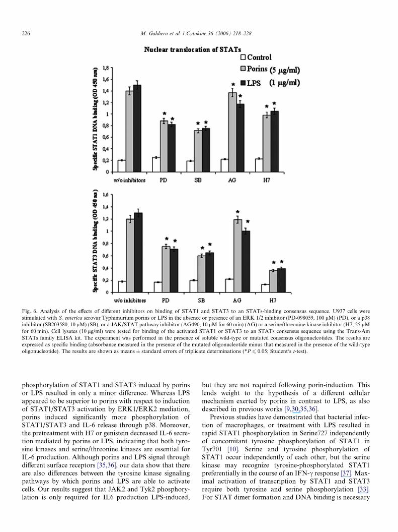
Fig. 6. Analysis of the effects of different inhibitors on binding of STAT1 and STAT3 to an STATs-binding consensus sequence. U937 cells werestimulated with S. enterica serovar Typhimurium porins or LPS in the absence or presence of an ERK 1/2 inhibitor (PD-098059, 100 lM) (PD), or a p38inhibitor (SB203580, 10 lM) (SB), or a JAK/STAT pathway inhibitor (AG490, 10 lM for 60 min) (AG) or a serine/threonine kinase inhibitor (H7, 25 lMfor 60 min). Cell lysates (10 lg/ml) were tested for binding of the activated STAT1 or STAT3 to an STATs consensus sequence using the Trans-AmSTATs family ELISA kit. The experiment was performed in the presence of soluble wild-type or mutated consensus oligonucleotides. The results areexpressed as specific binding (absorbance measured in the presence of the mutated oligonucleotide minus that measured in the presence of the wild-typeoligonucleotide). The results are shown as means ± standard errors of triplicate determinations (*P 6 0.05; Student‘s t-test).
226 M. Galdiero et al. / Cytokine 36 (2006) 218–228
phosphorylation of STAT1 and STAT3 induced by porinsor LPS resulted in only a minor difference. Whereas LPSappeared to be superior to porins with respect to inductionof STAT1/STAT3 activation by ERK1/ERK2 mediation,porins induced significantly more phosphorylation ofSTAT1/STAT3 and IL-6 release through p38. Moreover,the pretreatment with H7 or genistein decreased IL-6 secre-tion mediated by porins or LPS, indicating that both tyro-sine kinases and serine/threonine kinases are essential forIL-6 production. Although porins and LPS signal throughdifferent surface receptors [35,36], our data show that thereare also differences between the tyrosine kinase signalingpathways by which porins and LPS are able to activatecells. Our results suggest that JAK2 and Tyk2 phosphory-lation is only required for IL6 production LPS-induced,
but they are not required following porin-induction. Thislends weight to the hypothesis of a different cellularmechanism exerted by porins in contrast to LPS, as alsodescribed in previous works [9,30,35,36].
Previous studies have demonstrated that bacterial infec-tion of macrophages, or treatment with LPS resulted inrapid STAT1 phosphorylation in Serine727 independentlyof concomitant tyrosine phosphorylation of STAT1 inTyr701 [10]. Serine and tyrosine phosphorylation ofSTAT1 occur independently of each other, but the serinekinase may recognize tyrosine-phosphorylated STAT1preferentially in the course of an IFN-c response [37]. Max-imal activation of transcription by STAT1 and STAT3require both tyrosine and serine phosphorylation [33].For STAT dimer formation and DNA binding is necessary
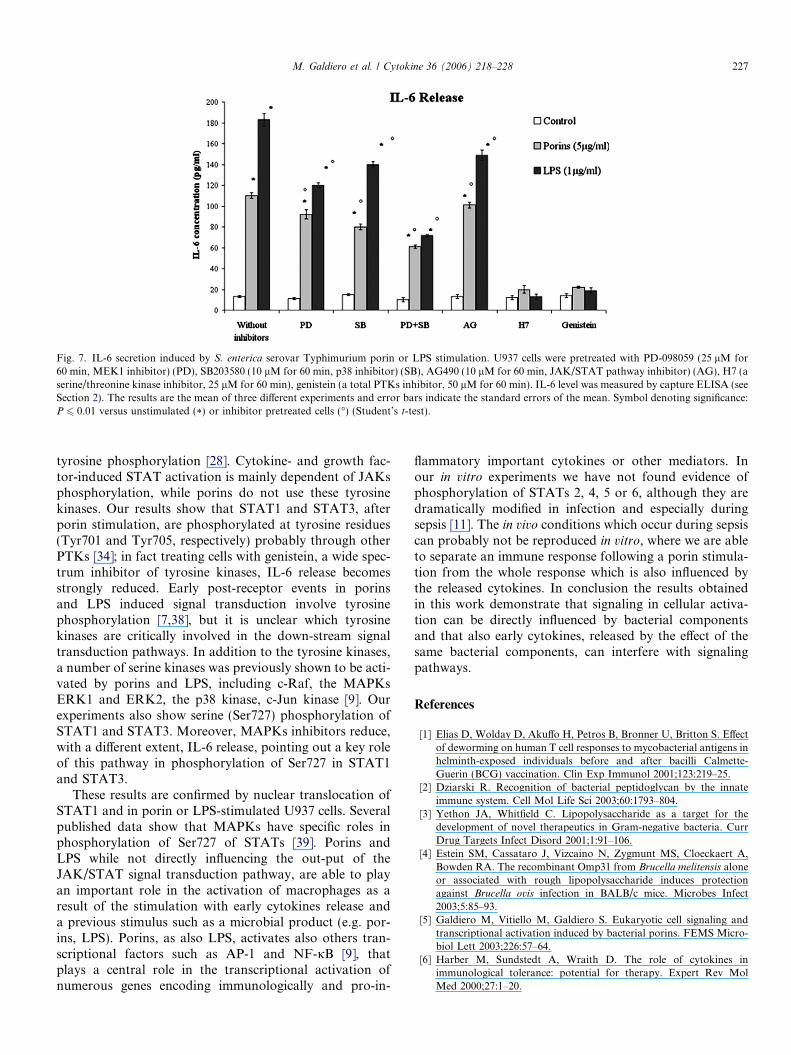
Fig. 7. IL-6 secretion induced by S. enterica serovar Typhimurium porin or LPS stimulation. U937 cells were pretreated with PD-098059 (25 lM for60 min, MEK1 inhibitor) (PD), SB203580 (10 lM for 60 min, p38 inhibitor) (SB), AG490 (10 lM for 60 min, JAK/STAT pathway inhibitor) (AG), H7 (aserine/threonine kinase inhibitor, 25 lM for 60 min), genistein (a total PTKs inhibitor, 50 lM for 60 min). IL-6 level was measured by capture ELISA (seeSection 2). The results are the mean of three different experiments and error bars indicate the standard errors of the mean. Symbol denoting significance:P 6 0.01 versus unstimulated (*) or inhibitor pretreated cells (�) (Student’s t-test).
M. Galdiero et al. / Cytokine 36 (2006) 218–228 227
tyrosine phosphorylation [28]. Cytokine- and growth fac-tor-induced STAT activation is mainly dependent of JAKsphosphorylation, while porins do not use these tyrosinekinases. Our results show that STAT1 and STAT3, afterporin stimulation, are phosphorylated at tyrosine residues(Tyr701 and Tyr705, respectively) probably through otherPTKs [34]; in fact treating cells with genistein, a wide spec-trum inhibitor of tyrosine kinases, IL-6 release becomesstrongly reduced. Early post-receptor events in porinsand LPS induced signal transduction involve tyrosinephosphorylation [7,38], but it is unclear which tyrosinekinases are critically involved in the down-stream signaltransduction pathways. In addition to the tyrosine kinases,a number of serine kinases was previously shown to be acti-vated by porins and LPS, including c-Raf, the MAPKsERK1 and ERK2, the p38 kinase, c-Jun kinase [9]. Ourexperiments also show serine (Ser727) phosphorylation ofSTAT1 and STAT3. Moreover, MAPKs inhibitors reduce,with a different extent, IL-6 release, pointing out a key roleof this pathway in phosphorylation of Ser727 in STAT1and STAT3.
These results are confirmed by nuclear translocation ofSTAT1 and in porin or LPS-stimulated U937 cells. Severalpublished data show that MAPKs have specific roles inphosphorylation of Ser727 of STATs [39]. Porins andLPS while not directly influencing the out-put of theJAK/STAT signal transduction pathway, are able to playan important role in the activation of macrophages as aresult of the stimulation with early cytokines release anda previous stimulus such as a microbial product (e.g. por-ins, LPS). Porins, as also LPS, activates also others tran-scriptional factors such as AP-1 and NF-jB [9], thatplays a central role in the transcriptional activation ofnumerous genes encoding immunologically and pro-in-
flammatory important cytokines or other mediators. Inour in vitro experiments we have not found evidence ofphosphorylation of STATs 2, 4, 5 or 6, although they aredramatically modified in infection and especially duringsepsis [11]. The in vivo conditions which occur during sepsiscan probably not be reproduced in vitro, where we are ableto separate an immune response following a porin stimula-tion from the whole response which is also influenced bythe released cytokines. In conclusion the results obtainedin this work demonstrate that signaling in cellular activa-tion can be directly influenced by bacterial componentsand that also early cytokines, released by the effect of thesame bacterial components, can interfere with signalingpathways.
References
[1] Elias D, Wolday D, Akuffo H, Petros B, Bronner U, Britton S. Effectof deworming on human T cell responses to mycobacterial antigens inhelminth-exposed individuals before and after bacilli Calmette-Guerin (BCG) vaccination. Clin Exp Immunol 2001;123:219–25.
[2] Dziarski R. Recognition of bacterial peptidoglycan by the innateimmune system. Cell Mol Life Sci 2003;60:1793–804.
[3] Yethon JA, Whitfield C. Lipopolysaccharide as a target for thedevelopment of novel therapeutics in Gram-negative bacteria. CurrDrug Targets Infect Disord 2001;1:91–106.
[4] Estein SM, Cassataro J, Vizcaino N, Zygmunt MS, Cloeckaert A,Bowden RA. The recombinant Omp31 from Brucella melitensis aloneor associated with rough lipopolysaccharide induces protectionagainst Brucella ovis infection in BALB/c mice. Microbes Infect2003;5:85–93.
[5] Galdiero M, Vitiello M, Galdiero S. Eukaryotic cell signaling andtranscriptional activation induced by bacterial porins. FEMS Micro-biol Lett 2003;226:57–64.
[6] Harber M, Sundstedt A, Wraith D. The role of cytokines inimmunological tolerance: potential for therapy. Expert Rev MolMed 2000;27:1–20.
228 M. Galdiero et al. / Cytokine 36 (2006) 218–228
[7] Galdiero M, Vitiello M, D’Isanto M, Peluso L, Galdiero M.Induction of tyrosine phosphorylated proteins in THP-1 cells bySalmonella typhimurium, Pasteurella haemolytica and Haemophilus
influenzae porins. FEMS Immunol Med Microbiol 2001;13:121–30.
[8] Bath NR, Zhang P, Lee JC, Hogan EL. Extracellular signal-regulatedkinase and p38 subgroups of mitogen-activated protein kinasesregulate inducible nitric oxide synthase and tumor necrosis factor-alpha gene expression in endotoxin-stimulated primary glial cultures.J Neurosci 1998;18:1633–44.
[9] Galdiero M, Vitiello M, Sanzari E, D’Isanto M, Tortora A,Longanella A, et al. Porins from Salmonella enterica serovarTyphimurium activate the transcription factors activating protein 1and NF-kappaB through the Raf-1-mitogen-activated protein kinasecascade. Infect Immun 2002;70:558–68.
[10] Kisseleva T, Bhattacharya S, Braunstein J, Schindler CW. Signalingthrough the JAK/STAT pathway, recent advances and futurechallenges. Gene 2002;285:1–24.
[11] Scott MJ, Godshall CJ, Cheadle WG. Jaks, STATs, cytokines, andsepsis. Clin Diagn Lab Immunol 2002;9:1153–9.
[12] Darnell Jr JE, Kerr IM, Stark GR. JAK-STAT pathways andtranscriptional activation in response to IFNs and other extracellularsignaling proteins. Science 1994;264:1415–21.
[13] Leonard WJ, O’Shea JJ. JAKs and STATs: biological implications.Annu Rev Immunol 1998;16:293–322.
[14] Shrotri MS, Peyton JC, Cheadle WG. Mouse peritonitis model usingcecal ligation and puncture. In: Sande MA, Carbon C, Fantin B,Kaminsky R, Kern ER, O’Reilly T, editors. Handbook of animalmodels of infection, vol. 173. San Diego: Academic Press; 1999.
[15] Greenberger MJ, Strieter RM, Kunkel SL, Danforth JM, GoodmanRE, Standiford TJ. Neutralization of IL-10 increases survival in amurine model of Klebsiella pneumoniae. J Immunol 1995;155:722–9.
[16] Zisman DA, Kunkel SL, Strieter RM, Gauldie J, Tsai WC, BramsonJ, et al. Anti-interleukin-12 therapy protects mice lethal endotoxemiabut impairs bacteria clearance in murine Escherichia coli peritonealsepsis. Shock 1997;8:349–56.
[17] Galdiero M, Tortora A, Damiano N, Vitiello M, Longanella A, et al.Induction of cytokine mRNA expression in U937 cells by Salmonella
typhimurium porins is regulated by different phosphorylation path-ways. Med Microbiol Immunol 2005;194:13–23.
[18] Rawadi G, Dussurget O. Advances in PCR-based detection ofmycoplasmas contaminating cell cultures. PCR Methods Appl1995;4:199–208.
[19] Lowry OH, Rosebrough NJ, Farr AL, Randall RJ. Proteinmeasurement with the Folin phenol reagent. J Biol Chem1951;193:265–75.
[20] Laemmli UK. Cleavage of structural proteins during the assembly ofthe head of bacteriophage T4. Nature 1970;277:680–5.
[21] Tsai CM, Frasch CE. A sensitive silver stain for detecting lipopoly-saccharides in polyacrylamide gels. Anal Biochem 1982;119:115–9.
[22] Yin ET, Galanos C, Kinsky S, Bradshaw RA, Wessler S, Luderity O,et al. Picogram-sensitive assay for endotoxin: gelation of Limuluspolyphemus blood cell lysate induced by purified lipopolysaccharidesand lipid A from Gram-negative bacteria. Biochem Biophys Acta1972;261:284–9.
[23] Alessi DR, Cuenda A, Cohen P, Dudley DT, Saltiel AR. PD-098059is a specific inhibitor of the activation of mitogen-activated proteinkinase kinase in vitro and in vivo. J Biol Chem 1995;270:27489–94.
[24] Cuenda A, Rouse J, Doza YN, Meier R, Cohen P, Gallagher TF,et al. SB203580 is a specific inhibitor of a MAP kinase homologuewhich is stimulated by cellular stress and interleukin-1. FEBS Lett1995;364:229–33.
[25] Kirken RA, Erwin RA, Taub D, Murphy WJ, Behbod F, Wang L,et al. Tyrphostin AG-490 inhibits cytokine-mediated JAK3/ATA-T5a/signal transduction and cellular proliferation of antigen-activat-ed human T cells. J Leukoc Biol 1999;65:891–9.
[26] Ku WC, Cheng AJ, Wang TC. Inhibition of telomerase activity byPKC inhibitors in human nasopharyngeal cancer cells in culture.Biochem Biophys Res Commun 1997;241:730–6.
[27] Davis JN, Kucuk O, Sarkar FH. Genistein inhibits NF-Kappa Bactivation in prostate cancer cells. Nutr Biochem 1999;35:167–74.
[28] Schindler C, Darnell Jr JE. Transcriptional responses topolypeptide ligands: the JAK-STAT pathway. Annu Rev Biochem1995;64:621–51.
[29] Galdiero F, Tufano MA, Galdiero M, Masiello S, Di Rosa M.Inflammatory effects of Salmonella typhimurium porins. Infect Immun1990;58:3183–8.
[30] Galdiero M, Cipollaro de l’Ero G, Donnarumma G, Marcatili A,Galdiero F. Interleukin-1 and interleukin-6 gene expression in humanmonocytes stimulated with Salmonella typhimurium porins. Immu-nology 1995;86:612–9.
[31] Galdiero M, D’Isanto M, Vitiello M, Galdiero M. Role of bacterialporins in the inflammatory and immunological responses. Recent ResDevel Immunol 2004;6:1–14.
[32] Morrison DC, Jacobs DM. Binding of polymyxin B to thelipid A portion of bacterial lipopolysaccharides. Immunochem1976;13:813–8.
[33] Weng Z, Zhong Z, Darnell Jr JE. Maximal activation of transcriptionby STAT1 and STAT3 requires both tyrosine and serine phosphor-ylation. Cell 1995;82:241–50.
[34] Galdiero M, D’Isanto M, Vitiello M, Finamore E, Peluso L, GaldieroM. Monocytic activation of protein tyrosine kinase, protein kinase Aand protein kinase C induced by porins isolated from Salmonella
enterica serovar Typhimurium. J Infect 2003;46:111–9.[35] Galdiero M, D’Isanto M, Vitiello M, Galdiero M. Porins from
Salmonella enterica serovar Typhimurium induce TNF-alpha, IL-6and IL-8 release by CD14-independent and CD11a/CD18-dependentmechanisms. Microbiology 2004;147:2697–704.
[36] Galdiero M, Galdiero M, Finamore E, Rossano F, Gambuzza M,Catania MR, et al. Haemophilus influenzae porin induces Toll-likereceptor 2-mediated cytokine production in human monocytes andmouse macrophages. Infect Immun 2004;72:1204–9.
[37] Kovarik P, Stoiber D, Novy M, Decker T. Stat 1 combines signalsderived from IFN-c and LPS receptors during macrophages activa-tion. EMBO J 1998;17:3660–8.
[38] Meng F, Lowell CA. Lipopolysaccharide (LPS)-induced macrophageactivation and signal transduction in the absence of Src-familykinases Hck, Fgr, and Lyn. J Exp Med 1997;185:1661–70.
[39] Decker T, Kovarik P. Serine phosphorylation of STATs. Oncogene2000;19:2628–37.
Copyright © 2022 FDOKUMEN