An Application to Ebola Type Infectious Disease - Letters in ...

2006, 80(11):5156. DOI: 10.1128/JVI.02349-05. J. Virol.
BaslerViktor E. Volchkov, Stuart T. Nichol and Christopher F.Osvaldo Martinez, Megan L. Shaw, Caroline Carbonnelle, St. Patrick Reid, Lawrence W. Leung, Amy L. Hartman, and Blocks STAT1 Nuclear Accumulation
1αEbola Virus VP24 Binds Karyopherin
http://jvi.asm.org/content/80/11/5156Updated information and services can be found at:
These include:
REFERENCEShttp://jvi.asm.org/content/80/11/5156#ref-list-1at:
This article cites 62 articles, 31 of which can be accessed free
CONTENT ALERTS more»articles cite this article),
Receive: RSS Feeds, eTOCs, free email alerts (when new
http://journals.asm.org/site/misc/reprints.xhtmlInformation about commercial reprint orders: http://journals.asm.org/site/subscriptions/To subscribe to to another ASM Journal go to:
on June 22, 2014 by guesthttp://jvi.asm
.org/D
ownloaded from
on June 22, 2014 by guest
http://jvi.asm.org/
Dow
nloaded from

JOURNAL OF VIROLOGY, June 2006, p. 5156–5167 Vol. 80, No. 110022-538X/06/$08.00�0 doi:10.1128/JVI.02349-05Copyright © 2006, American Society for Microbiology. All Rights Reserved.
Ebola Virus VP24 Binds Karyopherin �1 and Blocks STAT1Nuclear Accumulation
St. Patrick Reid,1† Lawrence W. Leung,1† Amy L. Hartman,2 Osvaldo Martinez,1 Megan L. Shaw,1Caroline Carbonnelle,3 Viktor E. Volchkov,3 Stuart T. Nichol,2 and Christopher F. Basler1*
Department of Microbiology, Mount Sinai School of Medicine, New York, New York 100291; Special Pathogens Branch,Division of Viral and Rickettsial Diseases, National Center for Infectious Diseases, Centers for Disease Control
and Prevention, 1600 Clifton Road, MS G-14, Atlanta, Georgia 303292; and Filovirus Laboratory,University Claude Bernard Lyon-1, INSERM U412, 69007 Lyon, France3
Received 7 November 2005/Accepted 15 March 2006
Ebola virus (EBOV) infection blocks cellular production of alpha/beta interferon (IFN-�/�) and the abilityof cells to respond to IFN-�/� or IFN-�. The EBOV VP35 protein has previously been identified as anEBOV-encoded inhibitor of IFN-�/� production. However, the mechanism by which EBOV infection inhibitsresponses to IFNs has not previously been defined. Here we demonstrate that the EBOV VP24 protein functionsas an inhibitor of IFN-�/� and IFN-� signaling. Expression of VP24 results in an inhibition of IFN-inducedgene expression and an inability of IFNs to induce an antiviral state. The VP24-mediated inhibition of cellularresponses to IFNs correlates with the impaired nuclear accumulation of tyrosine-phosphorylated STAT1(PY-STAT1), a key step in both IFN-�/� and IFN-� signaling. Consistent with this proposed function for VP24,infection of cells with EBOV also confers a block to the IFN-induced nuclear accumulation of PY-STAT1.Further, VP24 is found to specifically interact with karyopherin �1, the nuclear localization signal receptor forPY-STAT1, but not with karyopherin �2, �3, or �4. Overexpression of VP24 results in a loss of karyopherin�1–PY-STAT1 interaction, indicating that the VP24-karyopherin �1 interaction contributes to the block to IFNsignaling. These data suggest that VP24 is likely to be an important virulence determinant that allows EBOVto evade the antiviral effects of IFNs.
The filoviruses, Ebola virus (EBOV) and Marburg virus,cause periodic outbreaks of severe hemorrhagic fever in hu-mans. In EBOV outbreaks consisting of more than 10 reportedcases, mortality rates have ranged from 40 to 90% (41), andMarburg virus outbreaks have had reported case fatality ratesranging from 25 to 80% (13). This extreme virulence has madeEbola and Marburg viruses of concern both as naturallyemerging pathogens and as potential bioweapons (41).
The molecular mechanisms contributing to the severe patho-genesis of filovirus infection are poorly understood. Severalpotential mechanisms contributing to EBOV virulence havebeen reviewed (41). These include cytotoxicity of the viralglycoprotein, the production of proinflammatory cytokines,and the dysregulation of the coagulation cascade due to theproduction of tissue factor (14, 20, 21, 62, 64). Infection alsoappears to induce a general immune suppression (11, 53).Possible mechanisms contributing to this suppression includeinhibition of dendritic cell activation and an induction of lym-phocyte apoptosis (2, 8, 18, 22, 43). Each of these pathogenicprocesses likely occurs as a result of the active replication ofthe virus. Thus, the ability of the virus to counteract earlyantiviral responses, including those of the host’s interferonsystem, likely plays an important role in EBOV virulence (41).
EBOV encodes mechanisms to counteract the host inter-feron (IFN) response by blocking both production of IFN-�/�
and cellular responses to IFN-�/� or -� treatment (6, 24, 26,27). We previously demonstrated that the EBOV VP35 proteinsuppresses IFN-�/� production by inhibiting the activation ofinterferon regulatory factor 3 (IRF-3) (5, 7, 51), and subse-quent studies confirm that VP35 exerts this function (8, 28).However, the manner in which EBOV blocks signaling fromthe IFN-�/� or -� receptor has remained incompletely defined.
IFN-�/�, a family of structurally related proteins, and IFN-�bind to two distinct receptors but activate similar signalingpathways (reviewed in reference 38). For both pathways, ligandbinding activates receptor-associated Jak family tyrosine ki-nases. These undergo auto- and transphosphorylation andphosphorylate the cytoplasmic domains of the receptor sub-units. The receptor-associated phosphotyrosine residues thenserve as docking sites for the SH2 domains of STAT proteins.The receptor-associated STATs then undergo tyrosine-phos-phorylation and form homo- or heterodimers via reciprocalSH2 domain-phosphotyrosine interactions. Signaling from theIFN-�/� receptor results predominately in the formation ofSTAT1:STAT2 heterodimers which additionally interact withIRF-9. IFN-� signaling results predominately in the formationof STAT1:STAT1 homodimers. Upon dimerization, theSTAT1:STAT2 heterodimer or the STAT1:STAT1 ho-modimer interacts with a specific member of the karyopherin �(also known as importin �) family of nuclear localization signal(NLS) receptors, karyopherin �1 (importin �5) (45, 48, 55).This interaction with karyopherin �1 mediates the nuclearaccumulation of these STAT1-containing complexes (45, 48,55). The consequence of the activation and nuclear accumula-tion of these complexes is the specific transcriptional regula-
* Corresponding author. Mailing address: Department of Microbi-ology, Box 1124, Mount Sinai School of Medicine, 1 Gustave L. LevyPlace, New York, NY 10029. Phone: (212) 241-4847. Fax: (212) 534-1684. E-mail: [email protected].
† S.P.R. and L.W.L. contributed equally to this study.
5156
on June 22, 2014 by guesthttp://jvi.asm
.org/D
ownloaded from

tion of numerous genes, some of which have antiviral proper-ties (39).
In the present study, we demonstrate that the Zaire EBOVVP24 protein, a minor viral structural protein previously im-plicated in viral nucleocapsid formation (31, 63), viral buddingor assembly (3, 25), and host range determination (61), iscapable of blocking both IFN-�/� and IFN-� signaling. Mech-anistically, we find that VP24 prevents the nuclear accumula-tion of tyrosine-phosphorylated STAT1 (PY-STAT1); more-over, we provide evidence that an interaction between VP24and karyopherin �1 mediates this inhibition. The result is asubversion of the antiviral effects of IFNs. Consistent with therole of VP24 as an EBOV-encoded antagonist of IFN signalingpathways, we find that EBOV infection also imposes a block tothe nuclear accumulation of PY-STAT1 following IFN treat-ment. Subversion of the IFN signaling pathways likely pro-motes EBOV propagation in vivo and contributes to viralpathogenesis. Strategies that target this function of VP24 mayenhance the efficacy of IFNs toward EBOV.
MATERIALS AND METHODS
Cells and viruses. 293T cells, 293 cells, and Vero cells were maintained inDulbecco’s modified Eagle’s medium (DMEM) supplemented with 10% fetalbovine serum (FBS). Sendai virus strain Cantell was grown in 10-day-old em-bryonated chicken eggs at 37°C for 48 h. Zaire EBOV was grown and EBOVinfections were performed in Vero E6 cells at the Centers for Disease Controland Prevention under biosafety level 4 (BSL-4) containment. For analysis byimmunofluorescence microscopy, Vero cells were plated onto 12-mm-diameterglass coverslips and infected at a multiplicity of infection (MOI) of 0.5. Foranalysis of tyrosine-phosphorylated STAT1 by Western blotting, cells were cul-tured in a 24-well plate and infected at an MOI of 1.0.
Reporter gene assays. Vero cells were transfected using Lipofectamine 2000(Invitrogen, Carlsbad, CA) as outlined in the manufacturer’s protocol. Cellmonoloayers, 90 to 95% confluent in six-well plates, were transfected with 1.5 �gof an ISG54 promoter chloramphenicol acetyltransferase (CAT) reporter con-struct (pHISG54-CAT) or of an IRF-1 gamma-activated sequence (GAS) ele-ment-driven luciferase reporter construct (35), 1 �g of a constitutively expressingluciferase reporter construct (pCAGGS-luc), and 2.5 �g of the relevant expres-sion plasmid. Twenty-four hours posttransfection, cells were washed and main-tained in Dulbecco’s modified Eagle medium containing 0.3% bovine serumalbumin, with or without (mock-treated control) 1,000 U/ml of human IFN-� or1,000 U/ml of human IFN-� (as indicated) (Calbiochem, San Diego, CA). Twenty-four hours post-IFN treatment, cells were harvested and analyzed for CAT andluciferase activities. The CAT activity was quantified by using a PhosphorImager andnormalized to the luciferase activity.
Western blots and coimmunoprecipitations. To detect STAT1 levels, 293Tcells were transfected as described above with 10 ng of a human IRF-9 expressionplasmid (pCAGGS-hIRF-9) and 2.5 �g of the expression plasmids indicated inthe text. We included pCAGGS-hIRF-9 because these 293T cells do not effi-ciently respond to IFN-� without overexpression of IRF-9 (data not shown).Twenty-four hours posttransfection, the medium was replaced with DMEM,0.3% bovine serum albumin (BSA), 1,000 U/ml human IFN-� (Calbiochem).Eighteen hours post-IFN-� addition, cells were harvested and lysed in extractbuffer (50 mM Tris [pH 7.5], 280 mM NaCl, 0.5% Igepal, 0.2 mM EDTA, 2 mMEGTA, 10% glycerol, 1 mM dithiothreitol, 1 mM sodium vanadate, and proteaseinhibitors [Complete; Roche]). Total cell extracts were analyzed by sodiumdodecyl sulfate-polyacrylamide gel electrophoresis (SDS-PAGE), and the pro-teins were transferred to a polyvinylidene difluoride (PVDF) membrane andprobed with rabbit polyclonal antibodies against human STAT1 (BD Transduc-tion Laboratories) and �-actin (Sigma). The Western blots were developed usingthe Western Lightning ECL kit (Perkin-Elmer, Boston, MA) and Kodak BioMaxfilm (Kodak, Rochester, NY).
To detect MxA in EBOV-infected Vero cells, cell lysates were frozen anddecontaminated by exposure to 5 million rads of gamma radiation. Aliquots ofthe protein lysates were then resolved by SDS-PAGE and transferred to PVDF.The membrane was blocked in 5% milk and 0.2% Tween 20 in PBS (PBS-T) andthen incubated with a mouse antibody raised against MxA diluted 1:500 (theantibody was generously provided by Georg Koch and Otto Haller, Department
of Virology, University of Freiberg, Freiberg, Germany), and a mouse antibodyraised against glyceraldehyde-3-phosphate dehydrogenase (GAPDH) (Abcam,Cambridge, United Kingdom). After rinsing with PBS-T, the blot was incubatedwith an alkaline phosphatase-conjugated goat antibody raised against mouseimmunoglobulin G (IgG) (Rockland, Gilbertsville, PA) and visualized with ECFreagent and a Storm 860 PhosphorImager (Amersham Biosciences, Piscataway,NJ). To detect the presence of the EBOV protein VP24, the membrane wasincubated with a rabbit antibody raised against VP24 (diluted 1:2,000), rinsedwith PBS-T, and incubated with a horseradish peroxidase (HRP)-conjugatedgoat antibody raised against rabbit IgG (Sigma, St. Louis, MO). The VP24Western blot was developed using the Western Lightning ECL kit (Perkin-Elmer, Boston, MA) and Kodak BioMax film (Kodak, Rochester, NY).
For coimmunoprecipitation experiments, 293T cells were transfected with 1 �gof expression plasmids unless otherwise indicated. Following a 24-h incubation,cells were washed in ice-cold PBS and lysed in 500 �l of extract buffer for 10 minon ice. For Fig. 6B and C, prior to lysis, the cells were mock treated or treatedwith 1,000 U/ml human IFN-� (Calbiochem) for 1 h as indicated. Extracts werecentrifuged at 13,000 rpm for 10 min at 4°C in a microcentrifuge, the supernatantwas collected, and 20 �l of 50% slurry of M2 (anti-FLAG) monoclonal antibodycross-linked to agarose beads (Sigma) was added and incubated for at least 1 hat 4°C with rotation. The M2-agarose beads were washed three times with extractbuffer and boiled in SDS-PAGE sample buffer. For VP24 coimmunoprecipita-tion with FLAG-karyopherin �s, the immunoprecipitated material was analyzedby SDS-PAGE and immunoblotting with rabbit polyclonal antibody againstFLAG (Sigma) or against VP24. Separately, blots were probed with an anti-VP35monoclonal antibody. For FLAG-karyopherin �1, STAT1-green fluorescent pro-tein (GFP) and hemagglutinin (HA)-VP24 coimmunoprecipitations, the immu-noprecipitated material was analyzed by SDS-PAGE and immunoblotting withrabbit polyclonal antibody against PY-STAT1 (pY701; Cell Signaling Technol-ogy) or against STAT1 (BD Biosciences) where indicated. Additionally, immu-noblotting was also performed with monoclonal anti-HA and anti-FLAG anti-bodies (Sigma) where indicated. Whole-cell extracts (1% of total material usedfor immunoprecipitation) were immunoblotted with the same antibodies in par-allel. The Western blots were developed using the Western lightning ECL kit(Perkin-Elmer, Boston, MA) and Kodak BioMax film (Kodak, Rochester, NY).
Measuring the antiviral state in cells. Vero cells were transfected by usingLF2000 with 2.5 �g of the indicated expression plasmid. Twenty-four hoursposttransfection, the cells were mock treated or treated with 1,000 U/ml ofhuman IFN-� as described above. Twenty-four hours post-IFN-� treatment, thecells were infected with Newcastle disease virus expressing GFP (NDV-GFP)(6.4 turkey red blood cell hemagglutinating units/ml) (50), and after 24 h ofinfection GFP was visualized by fluorescence microscopy.
STAT1 and GFP–IRF-3 nuclear translocation. Vero cells were plated onto12-mm-diameter glass coverslips and transfected with empty vector (pCAGGS),pCAGGS-FLAG-VP24, or pCAGGS-FLAG-VP35 using Lipofectamine 2000following the manufacturer’s recommended protocol. Twenty-four hours post-transfection, the cells were serum starved for 4 h and then either mock treatedor treated with 1,000 U/ml of human IFN-� or human IFN-� for 30 min at 37°C.After rinsing three times with PBS containing 1 mM each of calcium chloride andmagnesium chloride (PBS-CM), the cells were placed on ice and fixed with�20°C methanol for 10 min. After rehydrating in PBS-CM, the cells wereblocked in PBS-CM containing 4% normal goat serum, 1% BSA, and 0.075%saponin for 45 min at ambient temperature. The coverslips were incubated witha mouse antibody raised against STAT1 (5 �g/ml; BD Bioscience, FranklinLakes, NJ) and a rabbit antibody raised against the FLAG epitope (4 �g/ml;Sigma, St. Louis, MO) in 1% BSA–Tris-buffered saline (TBS) for 1 h, followedby three washes of PBS. The coverslips were incubated with an Alexa 488-conjugated goat antibody raised against mouse IgG and an Alexa 594-conjugatedantibody raised against rabbit IgG (0.5 �g/ml each; Molecular Probes, Eugene,OR) and Hoechst 33342 (0.1 �g/ml; Molecular Probes, Eugene, OR) for 30 min.After rinsing, the coverslips were mounted on slides and imaged on a ZeissAxiophot 2 equipped with a Ratiga charge-coupled device (CCD) camera. Indi-vidual color channels were acquired as 8-bit monochrome images and pseudo-colored using ImageJ (NIH, Bethesda, MD). To quantify the number of cellsexpressing nuclear STAT1, at least three independent fields (at least 36 cells) foreach experimental condition were acquired. Cells that demonstrated colocaliza-tion of Hoechst and STAT1 staining were considered to have nuclear STAT1.
GFP–IRF-3 translocation assays were performed in Vero cells following apreviously described procedure (5).
Detection of tyrosine-phosphorylated STAT1 in IFN-�-treated Vero cells. (i)Immunofluorescence microscopy. Vero cells were plated on 12-mm-diameterglass coverslips and cultured in 10% FBS in DMEM. Before treatment withIFN-�, cells were rinsed twice with Optimem medium (Invitrogen) and incu-
VOL. 80, 2006 VP24 BLOCKS STAT1 NUCLEAR ACCUMULATION 5157
on June 22, 2014 by guesthttp://jvi.asm
.org/D
ownloaded from

bated in the absence of serum for 4 h. Human IFN-� was added (1,000 U/ml inDMEM), and the cells were cultured for 30 min. The cells were then fixed andblocked as described above for STAT1 staining. The cells were rinsed three timeswith 1% BSA in TBS and incubated with a rabbit antibody raised against asynthetic phosphopeptide (keyhole limpet hemocyanin coupled) correspondingto residues surrounding Tyr 701 of human Stat1 (0.2 �g/ml; Cell Signaling,Beverly, MA) and a mouse antibody raised against the FLAG epitope (4 �g/ml;Sigma, St. Louis MO) in 1% BSA–TBS for 1 h, followed by three washes of PBS.For cells infected with EBOV under BSL-4 conditions, coverslips were decon-taminated at this stage by incubation with 4% paraformaldehyde in PBS for 48 hbefore transferring them to a BSL-2 environment.
The coverslips were incubated with Alexa 594-conjugated goat antibodiesraised against mouse IgG (0.5 �g/ml; Molecular Probes, Eugene, OR), Alexa488-conjugated antibodies raised against rabbit IgG (0.5 �g/ml each; MolecularProbes, Eugene, OR) and Hoechst 33342 (0.1 �g/ml; Molecular Probes, Eugene,OR) for 30 min. After rinsing, the coverslips were mounted on slides and imagedon a Zeiss Axiophot 2 equipped with a Ratiga CCD camera. Individual colorchannels were acquired as 8-bit monochrome images and pseudocolored usingImageJ (NIH, Bethesda, MD). To quantify the number of cells expressing nu-clear PY-STAT1, four independent fields of Ebola virus-infected cells wereacquired. Cells that demonstrated colocalization of Hoechst and STAT1 stainingwere considered to have nuclear STAT1. Cells demonstrating staining for theEbola virus VP35 protein were considered to be infected.
(ii) To detect the presence of tyrosine-phosphorylated STAT1 in EBOV-infected Vero cells, extracts were prepared using the Pierce NE-PER kit (Pierce,Rockford, IL) according to the manufacturer’s recommendation. The lysateswere frozen and decontaminated with 5 million rads of gamma radiation. Priorto analysis, the nuclear and cytoplasmic fractions were combined to produce atotal lysate. The protein samples were resolved on a 10% polyacrylamide gel bySDS-PAGE and transferred to PVDF. After blocking in 5% nonfat milk and0.2% Tween 20 in TBS (TBS-T), the membrane was probed by incubation witha rabbit antibody raised against tyrosine-phosphorylated STAT1 (0.02 �g/ml;Cell Signaling, Beverly, MA) and a rabbit antibody raised against GAPDH(Abcam, Cambridge, United Kingdom). After rinsing, the blot was incubatedwith an alkaline phosphatase-conjugated goat antibody raised against rabbit IgG(Rockland, Gilbertsville, PA) and visualized using using ECF reagent and aStorm 860 PhosphorImager (Amersham Biosciences, Piscataway, NJ).
RESULTS
EBOV VP24 inhibits induction of gene expression by IFN-�and IFN-�. In order to identify an EBOV protein(s) capable ofinhibiting cellular responses to IFN treatment, cDNAs encod-ing individual EBOV proteins were screened for their ability toinhibit the IFN-�-induced expression of an IFN-responsive,ISG54 promoter-driven CAT reporter gene. Only the VP24protein was able to efficiently and reproducibly inhibit geneactivation in this assay (data not shown). The ability of VP24 toinhibit IFN-� signaling is demonstrated in Fig. 1A. In thisexperiment, Vero cells were cotransfected with the ISG54-CAT reporter and an empty expression plasmid or expressionplasmids for the Nipah virus W protein, a previously demon-strated inhibitor of IFN signaling (50, 56), FLAG-taggedVP24, an untagged VP24 protein, or VP35, the EBOV proteinpreviously implicated as an inhibitor of IFN-�/� production (5,7) (Fig. 1A). An �30-fold induction in CAT activity was seenin IFN-�-treated samples transfected with empty vector (ascompared with an empty vector-transfected, mock-treatedsample). This activation was inhibited in cells expressing eithertagged or untagged VP24 protein and in cells expressing NipahW, whereas a different EBOV protein, VP35, did not inhibitIFN-�-induced gene expression (Fig. 1A).
The ability of VP24 to inhibit IFN-induced expression of anendogenous gene, STAT1, was also assessed. (STAT1 is a keytranscription factor in the IFN signaling pathways, and itsexpression is upregulated upon IFN-� treatment [15, 37].) Af-
ter 18 h of treatment with IFN-�, a clear increase in STAT1protein expression was observed in cells transfected with emptyvector or with VP35 expression plasmid (Fig. 1B, compareempty vector-IFN-�- to empty vector-transfected, mock-treated cells). In contrast, either an untagged or a FLAG-tagged VP24 plasmid was able to inhibit IFN-�-inducedSTAT1 expression (Fig. 1B).
Given that EBOV infection is reported to block not only
FIG. 1. Ebola virus VP24 inhibits IFN-� and -� induced gene ex-pression. (A) IFN-�-induced reporter gene activation in Vero cells wasmeasured. Cells were cotransfected with the ISG54-CAT reporterplasmid, a constitutively expressed Renilla luciferase reporter plasmidand empty vector (Vec) or with plasmids expressing Nipah virus Wprotein (W), Flag-tagged EBOV VP24 (F-24), untagged VP24 (24), orEBOV VP35 (35). Twenty-four hours posttransfection, the cells eitherwere mock treated or were treated with 1,000 U of human IFN-�/mlfor 24 h, harvested, and then assayed for CAT and luciferase activities.The data are presented as the activation (fold) relative to empty vector,mock-treated controls. Error bars indicate means � the standard de-viation of three experiments. (B) Levels of STAT1 gene expression in293T cells transfected with empty vector (Vec) or plasmids expressingthe indicated protein are shown. Twenty-four hours posttransfection,the cells were treated with 1,000 U of IFN-�/ml, and 18 h posttreat-ment, the cells were lysed and subjected to Western blot analysis withanti-STAT1 (�STAT1) and anti-�-actin (� �-actin) antibodies.(C) Levels of IFN-�-induced reporter gene activation in Vero cellstransfected with empty vector (Vec) or with plasmids expressingEBOV VP35 (35) or FLAG-tagged EBOV VP24 (24). Vero cells weretransfected with the IFN-�-responsive IRF-1-promoter luciferase re-porter and a constitutively expressed Renilla luciferase reporter. Twen-ty-four hours posttransfection, the cells were either mock treated ortreated with 1,000 U/ml of IFN-�, and 24 h posttreatment, reportergene activity was measured. IFN-�-induced reporter values were nor-malized to the Renilla luciferase reporter. Results are presented asinduction (fold) of the IRF-1–luciferase reporter relative to an emptyvector-transfected, mock-treated control. Error bars indicate the mean� the standard deviation of three experiments.
5158 REID ET AL. J. VIROL.
on June 22, 2014 by guesthttp://jvi.asm
.org/D
ownloaded from
IFN-�/� but also IFN-� signaling, we asked whether the an-tagonistic effect of VP24 extended to the IFN-� pathway. Forthis purpose, a luciferase reporter under control of the IFN-�-responsive IRF-1 promoter, which contains GAS elementswas used. IFN-� treatment resulted in reporter activation inempty vector-transfected cells. VP35, which is unable to inhibitIFN-�/� signaling, was also unable to inhibit IFN-�-mediatedsignaling. In contrast, VP24 was able to inhibit reporter geneactivation (Fig. 1C).
Each of the experiments described in Fig. 1 included a con-stitutively expressed reporter plasmid to which the levels ofIFN-induced gene expression were normalized. When the ex-pression of a constitutively-expressed Renilla luciferase genewas analyzed over the course of four independent transfec-tions, luciferase values in IFN-�-treated cells were not statis-tically different in VP24-expressing versus VP35- or VP40-expressing cells (data not shown). This suggests that VP24 isnot exerting a nonspecific, global effect upon gene expression.In addition, when VP24 was transfected at a ratio of 5:1 with aGFP expression plasmid and GFP-expressing cells sorted byfluorescence-activated cell sorting were analyzed for their up-take of 7-AAD, a vital dye, we did not detect any evidence thatVP24 was cytotoxic (data not shown).
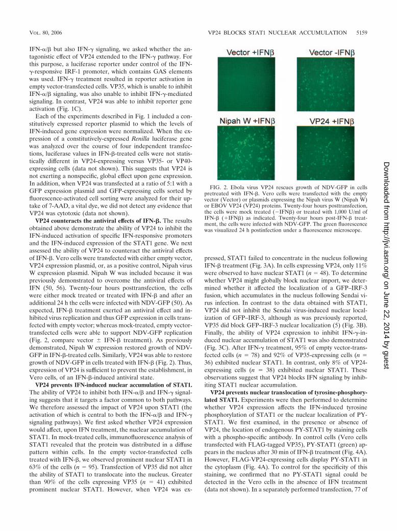
VP24 counteracts the antiviral effects of IFN-�. The resultsobtained above demonstrate the ability of VP24 to inhibit theIFN-induced activation of specific IFN-responsive promotersand the IFN-induced expression of the STAT1 gene. We nextassessed the ability of VP24 to counteract the antiviral effectsof IFN-�. Vero cells were transfected with either empty vector,VP24 expression plasmid, or, as a positive control, Nipah virusW expression plasmid. Nipah W was included because it waspreviously demonstrated to overcome the antiviral effects ofIFN (50, 56). Twenty-four hours posttransfection, the cellswere either mock treated or treated with IFN-� and after anadditional 24 h the cells were infected with NDV-GFP (50). Asexpected, IFN-� treatment exerted an antiviral effect and in-hibited virus replication and thus GFP expression in cells trans-fected with empty vector; whereas mock-treated, empty vector-transfected cells were able to support NDV-GFP replication(Fig. 2, compare vector � IFN-� treatment). As previouslydemonstrated, Nipah W expression restored growth of NDV-GFP in IFN-�-treated cells. Similarly, VP24 was able to restoregrowth of NDV-GFP in cells treated with IFN-� (Fig. 2). Thus,expression of VP24 is sufficient to prevent the establishment, inVero cells, of an IFN-�-induced antiviral state.
VP24 prevents IFN-induced nuclear accumulation of STAT1.The ability of VP24 to inhibit both IFN-�/� and IFN-� signal-ing suggests that it targets a factor common to both pathways.We therefore assessed the impact of VP24 upon STAT1 (theactivation of which is central to both the IFN-�/� and IFN-�signaling pathways). We first asked whether VP24 expressionwould affect, upon IFN treatment, the nuclear accumulation ofSTAT1. In mock-treated cells, immunofluorescence analysis ofSTAT1 revealed that the protein was distributed in a diffusepattern within cells. In the empty vector-transfected cellstreated with IFN-�, we observed prominent nuclear STAT1 in63% of the cells (n 95). Transfection of VP35 did not alterthe ability of STAT1 to translocate into the nucleus. Greaterthan 90% of the cells expressing VP35 (n 41) exhibitedprominent nuclear STAT1. However, when VP24 was ex-
pressed, STAT1 failed to concentrate in the nucleus followingIFN-� treatment (Fig. 3A). In cells expressing VP24, only 11%were observed to have nuclear STAT1 (n 48). To determinewhether VP24 might globally block nuclear import, we deter-mined whether it affected the localization of a GFP–IRF-3fusion, which accumulates in the nucleus following Sendai vi-rus infection. In contrast to the data obtained with STAT1,VP24 did not inhibit the Sendai virus-induced nuclear local-ization of GFP–IRF-3, although as was previously reported,VP35 did block GFP–IRF-3 nuclear localization (5) (Fig. 3B).Finally, the ability of VP24 expression to inhibit IFN-�-in-duced nuclear accumulation of STAT1 was also demonstrated(Fig. 3C). After IFN-� treatment, 95% of empty vector-trans-fected cells (n 78) and 92% of VP35-expressing cells (n 36) exhibited nuclear STAT1. In contrast, only 8% of VP24-expressing cells (n 38) exhibited nuclear STAT1. Theseobservations suggest that VP24 blocks IFN signaling by inhib-iting STAT1 nuclear accumulation.
VP24 prevents nuclear translocation of tyrosine-phosphory-lated STAT1. Experiments were then performed to determinewhether VP24 expression affects the IFN-induced tyrosinephosphorylation of STAT1 or the nuclear localization of PY-STAT1. We first examined, in the presence or absence ofVP24, the location of endogenous PY-STAT1 by staining cellswith a phospho-specific antibody. In control cells (Vero cellstransfected with FLAG-tagged VP35), PY-STAT1 (green) ap-pears in the nucleus after 30 min of IFN-� treatment (Fig. 4A).However, FLAG-VP24-expressing cells display PY-STAT1 inthe cytoplasm (Fig. 4A). To control for the specificity of thisstaining, we confirmed that no PY-STAT1 signal could bedetected in the Vero cells in the absence of IFN treatment(data not shown). In a separately performed transfection, 77 of
FIG. 2. Ebola virus VP24 rescues growth of NDV-GFP in cellspretreated with IFN-�. Vero cells were transfected with the emptyvector (Vector) or plasmids expressing the Nipah virus W (Nipah W)or EBOV VP24 (VP24) proteins. Twenty-four hours posttransfection,the cells were mock treated (�IFN�) or treated with 1,000 U/ml ofIFN-� (�IFN�) as indicated. Twenty-four hours post-IFN-� treat-ment, the cells were infected with NDV-GFP. The green fluorescencewas visualized 24 h postinfection under a fluorescence microscope.
VOL. 80, 2006 VP24 BLOCKS STAT1 NUCLEAR ACCUMULATION 5159
on June 22, 2014 by guesthttp://jvi.asm
.org/D
ownloaded from

FIG. 3. EBOV protein VP24 prevents IFN-mediated nuclear translocation of STAT1. (A) IFN-� treatment (30 min) of Vero cells causesSTAT1 to relocate from the cytoplasm (vector) to the nucleus (vector � IFN-�). In cells expressing FLAG-VP24 (FLAG-VP24 � IFN-�; relevantcells marked with an asterisk), STAT1 fails to relocate to the nucleus after IFN-� treatment. IFN-� treatment of FLAG-VP35-expressing cells(FLAG-VP35 � IFN-�; relevant cells marked with an asterisk) causes STAT1 to relocate to the nucleus. Upper panels show only STAT1 images.Lower panels show the STAT1 images (green) merged with images of FLAG-tagged Ebola virus proteins (red). (B) Vero cells express GFP–IRF-3in the cytoplasm in the absence of viral infection (vector), but translocate GFP–IRF-3 to the nucleus when infected with Sendai virus (vector �
5160 REID ET AL. J. VIROL.
on June 22, 2014 by guesthttp://jvi.asm
.org/D
ownloaded from
SeV). Coexpression of FLAG-VP35 prevents translocation of GFP–IRF-3 (FLAG-VP35 � SeV), but FLAG-VP24-expressing cells are still ableto traffic GFP–IRF-3 to the nucleus (FLAG-VP24 � SeV). Upper panels show the GFP–IRF-3 (green) merged with Hoechst nuclear staining(blue). Lower panels show the FLAG-tagged Ebola virus proteins. (C) In Vero cells cotransfected with empty vector, STAT1 is predominatelycytoplasmic in the absence of IFN-� treatment (vector), but STAT1 concentrates in the nucleus after a 30-min treatment with IFN-� (vector �IFN-�). In the presence of FLAG-VP24, IFN-� treatment fails to relocate STAT1 to the nucleus (FLAG-VP24 � IFN-�).
FIG. 4. VP24 prevents the nuclear translocation of phosphorylated STAT1 (Tyr 701). (A) After treatment with IFN-� for 30 min, cellsexpressing FLAG-VP35 (top row, red channel) are able to translocate PY-STAT1 (P-STAT1; green channel) to the nucleus. In cells expressingFLAG-VP24 (bottom row, red channel) PY-STAT1 is located in the cytoplasm. (B) VP24 expression does not detectably inhibit the tyrosinephosphorylation of STAT1. 293T cells were transfected with expression plasmids encoding: pCAGGS empty vector (vector), luciferase, Nipah virusV (NipV), VP35, VP24, or FLAG-VP24. After treatment with IFN-� (or mock treatment), the cells were lysed and analyzed for the presence ofPY-STAT1 by Western blotting.
VOL. 80, 2006 VP24 BLOCKS STAT1 NUCLEAR ACCUMULATION 5161
on June 22, 2014 by guesthttp://jvi.asm
.org/D
ownloaded from

86 (89%) mock-transfected, IFN-treated cells displayed nu-clear tyrosine-phosphorylated STAT1 30 min post-IFN addi-tion. Similarly, 30 of 42 (71%) FLAG-VP35-transfected, IFN-treated cells displayed nuclear tyrosine-phosphorylatedSTAT1. For these VP35 cells, there was a minority of cellswhich displayed high levels of PY-STAT1 staining that hadeither cytoplasmic PY-STAT1 or at least PY-STAT1 that wasnot in a clearly defined nucleus. In contrast, for the IFN-treated, FLAG-VP24 transfectants, in a total of 107 expressingcells, only 8 (7.5%) had nuclear PY-STAT1 (data not shown).
Since an effect of VP24 expression upon levels of PY-STAT1might be difficult to detect using fluorescence microscopy, wealso examined STAT1 tyrosine phosphorylation by Westernblotting extracts from transfected, IFN-�-treated 293T cells,30 min following IFN-�-treatment. Both FLAG-VP35- andFLAG-VP24-transfected cells contained amounts of PY-STAT1 similar to that seen in cells expressing no recombinantprotein or an irrelevant protein, luciferase (Fig. 4B). In acontrol sample, expression of the Nipah virus V protein, whichhas been shown to prevent STAT1 phosphorylation (52, 56),reduced the amount of PY-STAT1 (Fig. 4B). We conclude thatVP24 inhibits transcriptional responses to IFN, at least in part,by preventing the normal trafficking of PY-STAT1 to the nu-cleus.
Expression of supraphysiological intracellular concentra-tions of VP24 could conceivably lead to effects of VP24 not
seen in EBOV-infected cells. To address this concern, Westernblots were performed on cell lysates prepared from EBOV-infected and VP24-transfected cells. The lysates for this exper-iment were derived from Vero cells infected such that 50% ofcells stained positive for viral antigen or Vero cells transfectedwith FLAG-VP24 such that approximately 40% of cells werestained by anti-FLAG antibody. A significantly stronger VP24signal was detected from the infected cell lysate versus thetransfected cell lysate (data not shown). Thus, the transfectionexperiments described above appear to involve biologicallyrelevant levels of VP24.
EBOV infection inhibits both IFN-�-induced gene expres-sion and nuclear accumulation of PY-STAT1. If VP24 contrib-utes to the inhibition of IFN signaling by EBOV, EBOV in-fection should inhibit the nuclear transport of PY-STAT1. Toaddress this hypothesis, we infected Vero cells with EBOV(MOI 1) and, 24 h postinfection, treated them overnightwith IFN-�. By Western blotting, MxA, a protein whose ex-pression is induced by interferon (1), was found to be up-regu-lated following IFN-� treatment in mock-infected cells, but thisup-regulation was absent in EBOV-infected cells (Fig. 5A). In-fection of the cells was confirmed by examining expression ofthe VP24 protein (Fig. 5A). These data are consistent withprevious reports that EBOV infection blocks IFN signaling(27). In a separate experiment, Vero cells were grown on glasscoverslips, infected with EBOV (MOI 0.5), and, 24 h postin-
FIG. 5. EBOV infection inhibits IFN-�-induced expression of MxA and prevents nuclear translocation of PY-STAT1. (A) Mock-infected Verocells express MxA in response to overnight treatment with IFN-� (lane 2). However, EBOV-infected cells (MOI 1) do not produce MxA inresponse to IFN-� (lane 4). Lanes 1 and 2 were mock infected. Lanes 3 and 4 were infected with EBOV. Lanes 2 and 4 were treated with IFN-�,while lanes 1 and 3 were mock treated. (Top panel) Western blot to detect MxA and GAPDH. (Bottom panel) Western blot to detect VP24.(B) Mock-infected Vero cells translocate PY-STAT1 to the nucleus after 30 min of IFN-� treatment (top panel), but EBOV-infected cells retainPY-STAT1 in the cytoplasm (bottom panel). Red represents viral antigen (VP35), and green represents PY-STAT1.
5162 REID ET AL. J. VIROL.
on June 22, 2014 by guesthttp://jvi.asm
.org/D
ownloaded from

fection, treated with IFN-� for 30 min. After fixation, the cellswere processed for immunofluorescence analysis of PY-STAT1 and of viral antigen (the VP35 protein). Greater than90% of cells lacking significant viral antigen staining (n 74)demonstrated nuclear STAT1 (Fig. 5B, upper panel). Consis-tent with the data obtained when VP24 was expressed by trans-fection, we observed that only 21% of EBOV-infected cells(n 80) accumulated PY-STAT1 in their nucleus (Fig. 5B,lower panel). It should be noted that, in these experiments,cells were infected at an MOI of 0.5 and examined 48 h postin-fection. Under these conditions, it is likely that more thanone round of infection was initiated in these cultures, andthus some of the infected cells were probably expressingonly low levels of VP24.
VP24 interacts with the STAT1 nuclear localization signalreceptor, karyopherin �1, and inhibits VP24–PY-STAT1 inter-action. PY-STAT1 is reported to accumulate in the nucleus viainteraction with a specific nuclear localization signal receptor,karyopherin �1 (45, 48, 55). Given the ability of VP24 toprevent the nuclear accumulation of STAT1, we asked whetherVP24 might interact with this or other nuclear localizationsignal receptors. FLAG-tagged karyopherin �1, �2, �3, or �4expression plasmid was cotransfected with empty plasmid, un-tagged VP24 expression plasmid, or VP35 expression plasmid.One day posttransfection, immunoprecipitations were performedwith an anti-FLAG monoclonal antibody, and the precipitatedmaterial was analyzed by Western blotting with antibodies recog-nizing FLAG, VP24, or VP35. VP24 coprecipitated exclusivelywith karyopherin �1 (Fig. 6A). In contrast, VP35 did not copre-cipitate with any of the karyopherin �s (data not shown).
To determine if the interaction of VP24 with karyopherin �1might account for the loss of STAT1 nuclear accumulation, weasked if VP24 is able to disrupt the STAT1-karyopherin �1interaction. FLAG-tagged karyopherin �1 was coexpressedwith or without STAT1-GFP in the presence and absence ofVP24. In the absence of IFN-� (Fig. 6B, lanes 1 to 3), noSTAT1-GFP was coprecipitated with the karyopherin �1, de-spite the presence of STAT1-GFP in the whole-cell lysates(Fig. 6B, lanes 1 and 3). However, following addition of IFN-�to the cells (Fig. 6B, lanes 4 to 6), STAT1-GFP coprecipitatedwith karyopherin �1 (Fig. 6B, lane 4). VP24 was once againcoprecipitated with karyopherin �1, regardless of whether ornot IFN-� was added (Fig. 6B, lanes 2, 3, 5, and 6). Mostsignificantly, STAT1-GFP did not associate with karyopherin�1 when VP24 was present (Fig. 6B, lane 6). To determinewhether there is a dose-dependent effect of V24 on karyo-pherin �1-STAT1 interaction, we coexpressed STAT1-GFPwith karyopherin �1 (Fig. 6C, lanes 1 to 5) or without karyo-pherin �1 (Fig. 6C, lane 6). VP24 either was not expressed(Fig. 6C, lane 1) or was expressed in increasing amounts (lanes2 to 5). The cells were treated with IFN-�, and immunopre-cipitation of the FLAG-karyopherin �1 was performed. In thisexperiment, detection of STAT1-GFP was performed with an-ti-PY-STAT1 antibody, which, in our hands, is more sensitivethan the Western blot for total STAT1. Western blotting wasalso performed to detect FLAG-karyopherin �1 and VP24. Aswas seen previously, the activated STAT1-GFP coprecipitatedwith karyopherin �1 when VP24 was absent (Fig. 6C, lane 1).In contrast, no STAT1-GFP was precipitated with anti-FLAGantibody when the Flag-karyopherin �1 was omitted (Fig. 6C,
lane 6). When a low level of VP24 was present in the precip-itation, less PY-STAT1–GFP was coprecipitated (Fig. 6C, lane2). When higher levels of VP24 were present, the level ofSTAT-GFP coprecipitated was further decreased (Fig. 6C,lanes 2 to 5). These data suggest that the interaction of VP24with karyopherin �1 inhibits karyopherin �1–PY-STAT1 asso-ciation, thus providing an explanation as to how VP24 mayinhibit STAT1 nuclear accumulation.
DISCUSSION
The data described above provide a molecular explanationfor the impaired cellular responses to IFN-�/� and to IFN-� inEBOV-infected cells and shed further light on the ability ofthis highly lethal pathogen to modulate the host IFN system.Previously, it was demonstrated that EBOV-infected humanumbilical vein endothelial cells did not respond to either IFN-�treatment or IFN-� treatment (27). Specifically, the up-regu-lation of several IFN-�/�- or IFN-�-induced genes was absentor reduced in the infected cells (27). However, IL-1�-inducedexpression of IL-6 and ICAM-1 was intact, demonstrating thatinfection did not globally block transcriptional responses to allcytokines (27). Electrophoretic mobility shift assays on nuclearextracts further demonstrated that in EBOV-infected cells,IFN-�- and IFN-�-induced nuclear transcription factor com-plexes were absent or were present in greatly reduced amounts(27). The present study more clearly defines the EBOV-im-posed block to the IFN-�/� and IFN-� signaling pathways andidentifies a single viral protein, VP24, capable of exerting thisfunction. EBOV infection and VP24 expression appear to haveequivalent effects upon these signaling pathways. In both cases,the tyrosine phosphorylation of STAT1 in response to IFNremains intact. Rather, the critical step blocked by eitherEBOV infection or by VP24 expression, is the nuclear accu-mulation of STAT1 (Fig. 4 and 5). Of note, VP24 does notglobally inhibit nuclear translocation of proteins, as it did notprevent the Sendai virus-induced nuclear accumulation ofGFP–IRF-3 (Fig. 3). The latter observation is consistent withthe view that EBOVs utilize a second protein, VP35, to blockactivation of IRF-3 (5–8, 28, 51).
The nuclear localization of STAT1 (reviewed in references47 and 49) is triggered by its tyrosine phosphorylation-induced,SH2 domain-dependent dimerization (23, 54, 57, 58). In itsinactive state, STAT1 shuttles between the cytoplasm and nu-cleus via an energy-independent mechanism involving directinteractions with nucleoporins (44). Tyrosine phosphorylationresults in STAT1 heterodimerization with STAT2 (followingIFN-�/� treatment) or STAT1 homodimerization (followingIFN-� treatment) (reviewed in reference 38). Dimerization ofSTAT1 results in conformational changes revealing an atypicalNLS. This NLS directly interacts with the carboxy-terminalarmadillo repeats of karyopherin �1 (importin �5). Given thatactivated STAT1 did not detectably interact with other karyo-pherin � proteins, including karyopherin �2 (importin �1,Rch1) and karyopherin �4 (importin �3, Qip1), or importin�7, it may be only karyopherin �1 which mediates nuclearaccumulation of PY-STAT1 (45, 48, 55). Following its nuclearaccumulation, DNA containing STAT1 binding sequences cancompete with karyopherin �1 for binding to STAT1 ho-modimers, allowing STAT1 to interact with its target promot-
VOL. 80, 2006 VP24 BLOCKS STAT1 NUCLEAR ACCUMULATION 5163
on June 22, 2014 by guesthttp://jvi.asm
.org/D
ownloaded from
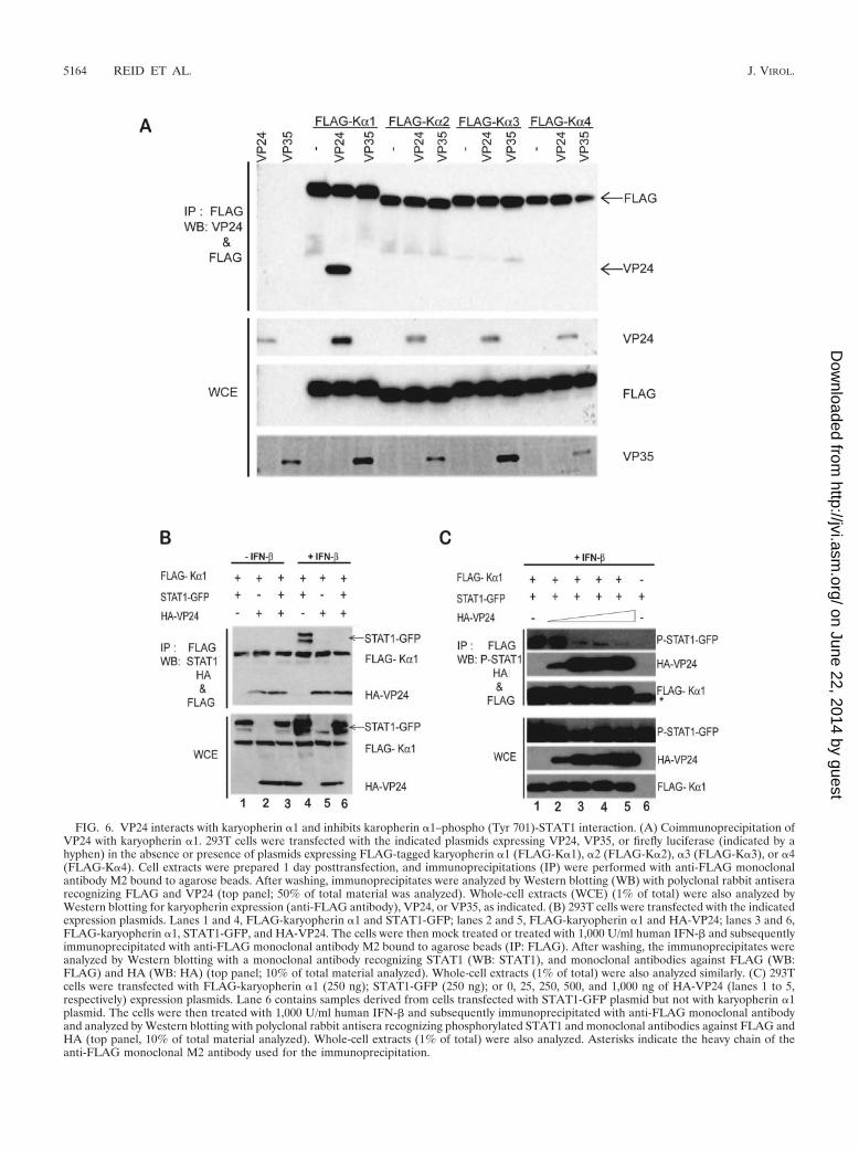
FIG. 6. VP24 interacts with karyopherin �1 and inhibits karopherin �1–phospho (Tyr 701)-STAT1 interaction. (A) Coimmunoprecipitation ofVP24 with karyopherin �1. 293T cells were transfected with the indicated plasmids expressing VP24, VP35, or firefly luciferase (indicated by ahyphen) in the absence or presence of plasmids expressing FLAG-tagged karyopherin �1 (FLAG-K�1), �2 (FLAG-K�2), �3 (FLAG-K�3), or �4(FLAG-K�4). Cell extracts were prepared 1 day posttransfection, and immunoprecipitations (IP) were performed with anti-FLAG monoclonalantibody M2 bound to agarose beads. After washing, immunoprecipitates were analyzed by Western blotting (WB) with polyclonal rabbit antiserarecognizing FLAG and VP24 (top panel; 50% of total material was analyzed). Whole-cell extracts (WCE) (1% of total) were also analyzed byWestern blotting for karyopherin expression (anti-FLAG antibody), VP24, or VP35, as indicated. (B) 293T cells were transfected with the indicatedexpression plasmids. Lanes 1 and 4, FLAG-karyopherin �1 and STAT1-GFP; lanes 2 and 5, FLAG-karyopherin �1 and HA-VP24; lanes 3 and 6,FLAG-karyopherin �1, STAT1-GFP, and HA-VP24. The cells were then mock treated or treated with 1,000 U/ml human IFN-� and subsequentlyimmunoprecipitated with anti-FLAG monoclonal antibody M2 bound to agarose beads (IP: FLAG). After washing, the immunoprecipitates wereanalyzed by Western blotting with a monoclonal antibody recognizing STAT1 (WB: STAT1), and monoclonal antibodies against FLAG (WB:FLAG) and HA (WB: HA) (top panel; 10% of total material analyzed). Whole-cell extracts (1% of total) were also analyzed similarly. (C) 293Tcells were transfected with FLAG-karyopherin �1 (250 ng); STAT1-GFP (250 ng); or 0, 25, 250, 500, and 1,000 ng of HA-VP24 (lanes 1 to 5,respectively) expression plasmids. Lane 6 contains samples derived from cells transfected with STAT1-GFP plasmid but not with karyopherin �1plasmid. The cells were then treated with 1,000 U/ml human IFN-� and subsequently immunoprecipitated with anti-FLAG monoclonal antibodyand analyzed by Western blotting with polyclonal rabbit antisera recognizing phosphorylated STAT1 and monoclonal antibodies against FLAG andHA (top panel, 10% of total material analyzed). Whole-cell extracts (1% of total) were also analyzed. Asterisks indicate the heavy chain of theanti-FLAG monoclonal M2 antibody used for the immunoprecipitation.
5164 REID ET AL. J. VIROL.
on June 22, 2014 by guesthttp://jvi.asm
.org/D
ownloaded from

ers (17, 45). Ultimately, nuclear STAT1 is dephosphorylated,dissociates from DNA, and is exported to the cytoplasm in aCRM1-dependent manner (29, 30, 44, 46, 47). Our data do notprovide any evidence that VP24 prevents the tyrosine phos-phorylation of STAT1, but they do demonstrate an inhibitionof STAT1 nuclear import.
It was interesting to note, given its ability to inhibit nuclearaccumulation of STAT1, that VP24 is reported to exhibit alargely perinuclear localization in EBOV-infected cells (25).Thus, VP24 would seem appropriately positioned to influencenuclear/cytoplasmic transport. We therefore tested VP24 forthe ability to interact with karyopherin � proteins. VP24 se-lectively interacted, in coimmunoprecipitation studies, withkaryopherin �1, the previously identified NLS receptor forSTAT1 (45, 48, 55). That this interaction may mediate theobserved defect in STAT1 nuclear import is supported by theability of VP24 to prevent the association of an activatedSTAT1-GFP protein with karyopherin �1 (Fig. 6). This selec-tive interaction of VP24 with karyopherin �1 likely also ex-plains why VP24 does not inhibit the nuclear import of IRF-3,as the nuclear import of this protein has been attributed tokaryopherin �3 and �4 (36) (Fig. 3). However, further studieswill be required to address the possibility that VP24 may alsoinfluence the nuclear import of other molecules that use karyo-pherin �1.
EBOV is an enveloped, nonsegmented negative-strandRNA virus with a genome of approximately 19 kb, and VP24 isone of eight major EBOV-encoded proteins (53). Althoughseveral functions have previously been attributed to the EBOVVP24 protein, its precise role in viral replication remains am-biguous and somewhat controversial. Early studies suggestedthat VP24 is found in EBOV virions and, as a consequence,VP24 was postulated to be a “minor matrix protein” or tofunction in viral uncoating (16, 53). Although the primaryEBOV matrix protein is VP40, and VP40 appears to be theprincipal force driving the budding of EBOV particles, recentstudies do support a possible role for VP24 in viral budding. Inparticular, VP24 oligomerizes, is hydrophobic, and associateswith cellular membranes, all properties characteristic of viralmatrix proteins (25). In addition, VP24 itself appears to bud atsome level from cells (25). However, coexpression of VP24with VP40 did not appear to enhance budding by VP40 (40). Inaddition, VP24 may play an important role in assembly of viralnucleocapsids as EBOV nucleocapsids could be reconstitutedby coexpression of the EBOV nucleoprotein (NP), VP35, andVP24 (31). VP24 could be coimmunoprecipitated with NPand VP35 (31); however, VP24 did not cosediment with NP andVP35 in density gradients, suggesting that, while it promotesnucleocapsid formation, it may not be an essential componentof the nucleocapsid structure (31). In a different study, VP24was not required for either the formation or the infectivity ofEbola virus-like particles carrying an EBOV minigenome (63).More recently, the use of small interfering RNAs targetingMarburg virus VP24 supports a role for this protein in filovirusassembly and release from infected Vero cells (3). Given thisarray of potential functions, it will be of interest to determinewhich of these is influenced by the ability of VP24 to block IFNsignaling pathways or to interact with karyopherin �1.
Understanding the determinants of EBOV virulence in dif-ferent species may provide insights into EBOV pathogenesis in
humans and may suggest novel therapeutic approaches. TheIFN system has been clearly implicated in the susceptibility ofmice to EBOV disease (9, 12, 42), and the sequence of VP24,along with that of several other genes, changed following theadaptation, by serial passage, of Zaire EBOV to mice (10).(The full sequence of the mouse-adapted EBOV is availableunder GenBank accession no. AF499101.) It is therefore in-triguing that adaptation of EBOV to another host, guinea pigs,is also associated with changes in VP24. Specifically, adapta-tion of a Zaire EBOV from a nonlethal form to a form lethalto guinea pigs was associated with 5 amino acid changes, oneeach in the nucleoprotein and L (polymerase), and 3 aminoacid changes in VP24 (61). Sequence analysis of the VP24 genefrom an independently adapted EBOV also identified anamino acid change in VP24, and it was suggested, based onthese observations, that changes in VP24 are likely to play animportant role in guinea pig adaptation (61). Although STAT1and karyopherin �1 are highly conserved between human andmouse, with each protein displaying greater than 90% aminoacid identity between the two species, it will be of interest todetermine whether these changes in VP24 result in an en-hanced ability to counteract interferon responses in these newhosts and whether the adaptation of EBOV to the new speciescorrelates with an increased affinity of VP24 for karyopherin �molecules of the new host.
At present, no vaccines against EBOV are licensed for use inhumans, although experimental vaccines that are effective innonhuman primates have been described (34, 59, 60). In ad-dition, no antivirals are available to treat these severe infec-tions, although an inhibitor of tissue factor was found to offersome protection to experimentally infected nonhuman pri-mates (19). The ability of VP24 to block IFN signaling path-ways has obvious implications for the efficacy of IFN as ananti-EBOV therapy. Although 200 IU/ml of IFN-�2b wasfound to inhibit EBOV replication 100-fold in Vero cells (33),daily intramuscular treatment of EBOV-infected monkeys witha relatively high dose of IFN-�2b beginning 18 h postinocula-tion merely delayed viremia and death by about 1 day (33).These data suggest that IFNs have limited efficacy againstEBOV and fit with the view that VP24 expression rendersEBOV relatively insensitive to IFN. Despite these observa-tions, the mouse model of infection suggests that IFNs mightbe employed to effectively control EBOV infection. For exam-ple, mouse-adapted Zaire EBOV is lethal to mice followingintraperitoneal infection but not following subcutaneous infec-tion (42). Subcutaneous administration of the mouse-adaptedvirus conferred protection from challenge with an otherwiselethal intraperitoneal dose, even at 48 h postinoculation (42).This early time point, prior to the development of EBOV-specific adaptive immune responses, correlated with peak in-duction of IFN-� levels, suggesting that IFN-�/� can have atherapeutic effect on EBOV infection (42). Additionally, theadenosine analog 3-deazaneplanocin A was found to protectmice from illness and death following infection with mouse-adapted EBOV (32). Protection by this drug appears to involvethe induction of large quantities of IFN-� (12). Our data sug-gest that the anti-EBOV efficacy of IFNs might be augmentedby inhibitors of VP24. Additional studies to further define themechanism by which VP24 functions should facilitate the de-velopment of such therapies.
VOL. 80, 2006 VP24 BLOCKS STAT1 NUCLEAR ACCUMULATION 5165
on June 22, 2014 by guesthttp://jvi.asm
.org/D
ownloaded from
ACKNOWLEDGMENTS
This work was supported by grants to C.F.B. from the NationalInstitutes of Health and grants to V.E.V. from INSERM, DeutscheForschungsgemeinschaft (SFB 593), and from French Ministere de laRecherche (04G537). C.F.B. is an Ellison Medical Foundation NewScholar in Global Infectious Diseases.
We thank Luis Martınez-Sobrido and Adolfo Garcıa-Sastre (MountSinai School of Medicine) for providing the GFP–IRF-3 and IRF-9expression plasmids; Georg Kochs and Otto Haller (University ofFreiberg) for providing anti-MxA antibody and the IRF-1–luciferasereporter plasmid; and David E. Levy (New York University) for pro-viding the ISG54-CAT reporter plasmid.
REFERENCES
1. Aebi, M., J. Fah, N. Hurt, C. E. Samuel, D. Thomis, L. Bazzigher, J. Pavlovic,O. Haller, and P. Staeheli. 1989. cDNA structures and regulation of twointerferon-induced human Mx proteins. Mol. Cell. Biol. 9:5062–5072.
2. Baize, S., E. M. Leroy, E. Mavoungou, and S. P. Fisher-Hoch. 2000. Apop-tosis in fatal Ebola infection. Does the virus toll the bell for immune system?Apoptosis 5:5–7.
3. Bamberg, S., L. Kolesnikova, P. Moller, H.-D. Klenk, and S. Becker. 2005.VP24 of Marburg virus influences formation of infectious particles. J. Virol.79:13421–13433.
4. Reference deleted.5. Basler, C. F., A. Mikulasova, L. Martinez-Sobrido, J. Paragas, E. Muhl-
berger, M. Bray, H.-D. Klenk, P. Palese, and A. Garcia-Sastre. 2003. TheEbola virus VP35 protein inhibits activation of interferon regulatory factor 3.J. Virol. 77:7945–7956.
6. Basler, C. F., and P. Palese. 2004. Modulation of innate immunity by filo-viruses, p. 305–349. In H.-D. Klenk and H. Feldmann (ed.), Molecular andcellular biology of Marburg and Ebola viruses. Horizon Scientific Press,Norfolk, United Kingdom.
7. Basler, C. F., X. Wang, E. Muhlberger, V. Volchkov, J. Paragas, H. D. Klenk,A. Garcia-Sastre, and P. Palese. 2000. The Ebola virus VP35 protein func-tions as a type I IFN antagonist. Proc. Natl. Acad. Sci. USA 97:12289–12294.
8. Bosio, C. M., M. J. Aman, C. Grogan, R. Hogan, G. Ruthel, D. Negley, M.Mohamadzadeh, S. Bavari, and A. Schmaljohn. 2003. Ebola and Marburgviruses replicate in monocyte-derived dendritic cells without inducing theproduction of cytokines and full maturation. J. Infect. Dis. 188:1630–1638.
9. Bray, M. 2001. The role of the type I interferon response in the resistance ofmice to filovirus infection. J. Gen. Virol. 82:1365–1373.
10. Bray, M., K. Davis, T. Geisbert, C. Schmaljohn, and J. Huggins. 1998. Amouse model for evaluation of prophylaxis and therapy of Ebola hemor-rhagic fever. J. Infect. Dis. 178:651–661.
11. Bray, M., and T. W. Geisbert. 2005. Ebola virus: the role of macrophages anddendritic cells in the pathogenesis of Ebola hemorrhagic fever. Int. J. Bio-chem. Cell. Biol. 37:1560–1566.
12. Bray, M., J. L. Raymond, T. Geisbert, and R. O. Baker. 2002. 3-Deazaneplano-cin A induces massively increased interferon-alpha production in Ebolavirus-infected mice. Antivir. Res. 55:151–159.
13. Centers for Disease Control and Prevention. 2005. Outbreak of Marburgvirus hemorrhagic fever—Angola, October 1, 2004-March 29, 2005. Morb.Mortal. Wkly. Rep. 54:308–309.
14. Chan, S. Y., M. C. Ma, and M. A. Goldsmith. 2000. Differential induction ofcellular detachment by envelope glycoproteins of Marburg and Ebola(Zaire) viruses. J. Gen. Virol. 81:2155–2159.
15. de Veer, M. J., M. Holko, M. Frevel, E. Walker, S. Der, J. M. Paranjape,R. H. Silverman, and B. R. Williams. 2001. Functional classification ofinterferon-stimulated genes identified using microarrays. J. Leukoc. Biol.69:912–920.
16. Elliott, L. H., M. P. Kiley, and J. B. McCormick. 1985. Descriptive analysisof Ebola virus proteins. Virology 147:169–176.
17. Fagerlund, R., K. Melen, L. Kinnunen, and I. Julkunen. 2002. Arginine/lysine-rich nuclear localization signals mediate interactions between dimericSTATs and importin alpha 5. J. Biol. Chem. 277:30072–30078.
18. Geisbert, T. W., L. E. Hensley, T. R. Gibb, K. E. Steele, N. K. Jaax, and P. B.Jahrling. 2000. Apoptosis induced in vitro and in vivo during infection byEbola and Marburg viruses. Lab. Investig. 80:171–186.
19. Geisbert, T. W., L. E. Hensley, P. B. Jahrling, T. Larsen, J. B. Geisbert, J.Paragas, H. A. Young, T. M. Fredeking, W. E. Rote, and G. P. Vlasuk. 2003.Treatment of Ebola virus infection with a recombinant inhibitor of factorVIIa/tissue factor: a study in rhesus monkeys. Lancet 362:1953–1958.
20. Geisbert, T. W., L. E. Hensley, T. Larsen, H. A. Young, D. S. Reed, J. B.Geisbert, D. P. Scott, E. Kagan, P. B. Jahrling, and K. J. Davis. 2003.Pathogenesis of Ebola hemorrhagic fever in cynomolgus macaques: evidencethat dendritic cells are early and sustained targets of infection. Am. J. Pathol.163:2347–2370.
21. Geisbert, T. W., H. A. Young, P. B. Jahrling, K. J. Davis, E. Kagan, and L. E.Hensley. 2003. Mechanisms underlying coagulation abnormalities in Ebola
hemorrhagic fever: overexpression of tissue factor in primate monocytes/macrophages is a key event. J. Infect. Dis. 188:1618–1629.
22. Gibb, T. R., M. Bray, T. W. Geisbert, K. E. Steele, W. M. Kell, K. J. Davis,and N. K. Jaax. 2001. Pathogenesis of experimental Ebola Zaire virus infec-tion in BALB/c mice. J. Comp. Pathol. 125:233–242.
23. Greenlund, A. C., M. A. Farrar, B. L. Viviano, and R. D. Schreiber. 1994.Ligand-induced IFN gamma receptor tyrosine phosphorylation couples thereceptor to its signal transduction system (p91). EMBO J. 13:1591–1600.
24. Gupta, M., S. Mahanty, R. Ahmed, and P. E. Rollin. 2001. Monocyte-derivedhuman macrophages and peripheral blood mononuclear cells infected withEbola virus secrete MIP-1alpha and TNF-alpha and inhibit poly-IC-inducedIFN-alpha in vitro. Virology 284:20–25.
25. Han, Z., H. Boshra, J. O. Sunyer, S. H. Zwiers, J. Paragas, and R. N. Harty.2003. Biochemical and functional characterization of the Ebola virus VP24protein: implications for a role in virus assembly and budding. J. Virol.77:1793–1800.
26. Harcourt, B. H., A. Sanchez, and M. K. Offermann. 1998. Ebola virus inhibitsinduction of genes by double-stranded RNA in endothelial cells. Virology252:179–188.
27. Harcourt, B. H., A. Sanchez, and M. K. Offermann. 1999. Ebola virusselectively inhibits responses to interferons, but not to interleukin-1�, inendothelial cells. J. Virol. 73:3491–3496.
28. Hartman, A. L., J. S. Towner, and S. T. Nichol. 2004. A C-terminal basicamino acid motif of Zaire ebolavirus VP35 is essential for type I interferonantagonism and displays high identity with the RNA-binding domain ofanother interferon antagonist, the NS1 protein of influenza A virus. Virology328:177–184.
29. Haspel, R. L., and J. E. Darnell, Jr. 1999. A nuclear protein tyrosine phos-phatase is required for the inactivation of Stat1. Proc. Natl. Acad. Sci. USA96:10188–10193.
30. Haspel, R. L., M. Salditt-Georgieff, and J. E. Darnell, Jr. 1996. The rapidinactivation of nuclear tyrosine phosphorylated Stat1 depends upon a pro-tein tyrosine phosphatase. EMBO J 15:6262–6268.
31. Huang, Y., L. Xu, Y. Sun, and G. Nabel. 2002. The assembly of Ebola virusnucleocapsid requires virion-associated proteins 35 and 24 and posttransla-tional modification of nucleoprotein. Mol. Cell 10:307.
32. Huggins, J. W., Z. X. Zhang, and T. I. Monath. 1995. Inhibition of Ebolavirus replication in vitro and in vivo in a SCID mouse model by S-adenosyl-homocysteine hydrolase inhibitors. Antivir. Res. Suppl. 1:122.
33. Jahrling, P. B., T. W. Geisbert, J. B. Geisbert, J. R. Swearengen, M. Bray,N. K. Jaax, J. W. Huggins, J. W. LeDuc, and C. J. Peters. 1999. Evaluationof immune globulin and recombinant interferon-alpha2b for treatment ofexperimental Ebola virus infections. J. Infect. Dis. 179(Suppl. 1):S224–S234.
34. Jones, S. M., H. Feldmann, U. Stroher, J. B. Geisbert, L. Fernando, A.Grolla, H. D. Klenk, N. J. Sullivan, V. E. Volchkov, E. A. Fritz, K. M.Daddario, L. E. Hensley, P. B. Jahrling, and T. W. Geisbert. 2005. Liveattenuated recombinant vaccine protects nonhuman primates against Ebolaand Marburg viruses. Nat. Med. 11:786–790.
35. King, P., and S. Goodbourn. 1994. The beta-interferon promoter responds topriming through multiple independent regulatory elements. J. Biol. Chem.269:30609–30615.
36. Kumar, K. P., K. M. McBride, B. K. Weaver, C. Dingwall, and N. C. Reich.2000. Regulated nuclear-cytoplasmic localization of interferon regulatoryfactor 3, a subunit of double-stranded RNA-activated factor 1. Mol. Cell.Biol. 20:4159–4168.
37. Lehtonen, A., S. Matikainen, and I. Julkunen. 1997. Interferons up-regulateSTAT1, STAT2, and IRF family transcription factor gene expression inhuman peripheral blood mononuclear cells and macrophages. J. Immunol.159:794–803.
38. Levy, D. E., and J. E. Darnell, Jr. 2002. Stats: transcriptional control andbiological impact. Nat. Rev. Mol. Cell. Biol. 3:651–662.
39. Levy, D. E., and A. Garcia-Sastre. 2001. The virus battles: IFN induction ofthe antiviral state and mechanisms of viral evasion. Cytokine Growth FactorRev. 12:143–156.
40. Licata, J. M., R. F. Johnson, Z. Han, and R. N. Harty. 2004. Contribution ofEbola virus glycoprotein, nucleoprotein, and VP24 to budding of VP40virus-like particles. J. Virol. 78:7344–7351.
41. Mahanty, S., and M. Bray. 2004. Pathogenesis of filoviral haemorrhagicfevers. Lancet Infect. Dis. 4:487–498.
42. Mahanty, S., M. Gupta, J. Paragas, M. Bray, R. Ahmed, and P. E. Rollin.2003. Protection from lethal infection is determined by innate immune re-sponses in a mouse model of Ebola virus infection. Virology 312:415–424.
43. Mahanty, S., K. Hutchinson, S. Agarwal, M. McRae, P. E. Rollin, and B.Pulendran. 2003. Cutting edge: impairment of dendritic cells and adaptiveimmunity by Ebola and Lassa viruses. J. Immunol. 170:2797–2801.
44. Marg, A., Y. Shan, T. Meyer, T. Meissner, M. Brandenburg, and U. Vinke-meier. 2004. Nucleocytoplasmic shuttling by nucleoporins Nup153 andNup214 and CRM1-dependent nuclear export control the subcellular distri-bution of latent Stat1. J. Cell Biol. 165:823–833.
45. McBride, K. M., G. Banninger, C. McDonald, and N. C. Reich. 2002. Reg-ulated nuclear import of the STAT1 transcription factor by direct binding ofimportin-alpha. EMBO J. 21:1754–1763.
5166 REID ET AL. J. VIROL.
on June 22, 2014 by guesthttp://jvi.asm
.org/D
ownloaded from
46. McBride, K. M., C. McDonald, and N. C. Reich. 2000. Nuclear export signallocated within the DNA-binding domain of the STAT1 transcription factor.EMBO J. 19:6196–6206.
47. McBride, K. M., and N. C. Reich. 2003. The ins and outs of STAT1 nucleartransport. Sci. STKE 2003:RE13. [Online.] doi:10.1126/stke.2003.195.re13.
48. Melen, K., R. Fagerlund, J. Franke, M. Kohler, L. Kinnunen, and I.Julkunen. 2003. Importin alpha nuclear localization signal binding sites forSTAT1, STAT2, and influenza A virus nucleoprotein. J. Biol. Chem. 278:28193–28200.
49. Meyer, T., and U. Vinkemeier. 2004. Nucleocytoplasmic shuttling of STATtranscription factors. Eur. J. Biochem. 271:4606–4612.
50. Park, M.-S., M. L. Shaw, J. Munoz-Jordan, J. F. Cros, T. Nakaya, N.Bouvier, P. Palese, A. Garcia-Sastre, and C. F. Basler. 2003. Newcastledisease virus (NDV)-based assay demonstrates interferon-antagonist activityfor the NDV V protein and the Nipah virus V, W, and C proteins. J. Virol.77:1501–1511.
51. Reid, S. P., W. B. Cardenas, and C. F. Basler. 2005. Homo-oligomerizationfacilitates the interferon-antagonist activity of the ebolavirus VP35 protein.Virology 341:179–189. (First published 10 August 2005; doi:10.1016/j.virol.2005.06.044.)
52. Rodriguez, J. J., J.-P. Parisien, and C. M. Horvath. 2002. Nipah virus Vprotein evades alpha and gamma interferons by preventing STAT1 andSTAT2 activation and nuclear accumulation. J. Virol. 76:11476–11483.
53. Sanchez, A., A. S. Khan, S. R. Zaki, G. J. Nabel, T. G. Ksiazek, and C. J.Peters. 2001. Filoviridae: Marburg and Ebola viruses, p. 1279–1304. In B. N.Fields and D. M. Knipe (ed.), Fields virology, 4th ed., vol. 1. LippincottWilliams and Wilkins, Philadelphia, Pa.
54. Schindler, C., K. Shuai, V. R. Prezioso, and J. E. Darnell, Jr. 1992. Inter-feron-dependent tyrosine phosphorylation of a latent cytoplasmic transcrip-tion factor. Science 257:809–813.
55. Sekimoto, T., N. Imamoto, K. Nakajima, T. Hirano, and Y. Yoneda. 1997.Extracellular signal-dependent nuclear import of Stat1 is mediated by nu-clear pore-targeting complex formation with NPI-1, but not Rch1. EMBO J.16:7067–7077.
56. Shaw, M. L., A. Garcia-Sastre, P. Palese, and C. F. Basler. 2004. Nipah virusV and W proteins have a common STAT1-binding domain yet inhibit STAT1activation from the cytoplasmic and nuclear compartments, respectively.J. Virol. 78:5633–5641.
57. Shuai, K., C. M. Horvath, L. H. Huang, S. A. Qureshi, D. Cowburn, and J. E.Darnell, Jr. 1994. Interferon activation of the transcription factor Stat91involves dimerization through SH2-phosphotyrosyl peptide interactions. Cell76:821–828.
58. Shuai, K., C. Schindler, V. R. Prezioso, and J. E. Darnell, Jr. 1992. Activa-tion of transcription by IFN-gamma: tyrosine phosphorylation of a 91-kDDNA binding protein. Science 258:1808–1812.
59. Sullivan, N. J., T. W. Geisbert, J. B. Geisbert, L. Xu, Z. Y. Yang, M.Roederer, R. A. Koup, P. B. Jahrling, and G. J. Nabel. 2003. Acceleratedvaccination for Ebola virus haemorrhagic fever in non-human primates.Nature 424:681–684.
60. Sullivan, N. J., A. Sanchez, P. E. Rollin, Z. Y. Yang, and G. J. Nabel. 2000.Development of a preventive vaccine for Ebola virus infection in primates.Nature 408:605–609.
61. Volchkov, V. E., A. A. Chepurnov, V. A. Volchkova, V. A. Ternovoj, and H. D.Klenk. 2000. Molecular characterization of guinea pig-adapted variants ofEbola virus. Virology 277:147–155.
62. Volchkov, V. E., V. A. Volchkova, E. Muhlberger, L. V. Kolesnikova, M.Weik, O. Dolnik, and H. D. Klenk. 2001. Recovery of infectious Ebola virusfrom complementary DNA: RNA editing of the GP gene and viral cytotox-icity. Science 291:1965–1969.
63. Watanabe, S., T. Watanabe, T. Noda, A. Takada, H. Feldmann, L. D. Jas-enosky, and Y. Kawaoka. 2004. Production of novel Ebola virus-like particlesfrom cDNAs: an alternative to Ebola virus generation by reverse genetics.J. Virol. 78:999–1005.
64. Yang, Z. Y., H. J. Duckers, N. J. Sullivan, A. Sanchez, E. G. Nabel, and G. J.Nabel. 2000. Identification of the Ebola virus glycoprotein as the main viraldeterminant of vascular cell cytotoxicity and injury. Nat. Med. 6:886–889.
VOL. 80, 2006 VP24 BLOCKS STAT1 NUCLEAR ACCUMULATION 5167
on June 22, 2014 by guesthttp://jvi.asm
.org/D
ownloaded from
Copyright © 2022 FDOKUMEN